Mitosis & Meiosis

The Cell Life Cycle
INTERPHASE
THE CELL
CYCLE
MITOSIS AND CYTOKINESIS (See Figure 2.21)
Indefinite period G0
Specialized cell functions
G1 Normal cell functions plus cell growth, duplication of organelles, protein synthesis
G2 Protein synthesis
S DNA
replication, synthesis
of histones
M

Mitosis
• A cell divides to produce two identical cells
– To heal an injury
– To grow (increase cell number)
• Cells are not always dividing- most of their “life”
is spent between divisions (interphase G1)-
carrying out the organelle jobs
• Division costs lots in terms of energy!
• Uncontrolled cell division produces tumors
(cancers).

Stages of cell’s life cycle:
(PMAT) Interphase- (between divisions, can’t see chromosomes)
• G1- cell is not ready to divide, carries out normal functions
• S- cell commits to divide, and copies all the DNA
• G2- cell prepares for division, and generates more lipids and Proteins
Mitosis- Division of genetic information (division of the nucleus): think P.M.A.T. (“Passed My Anatomy Test”)
– Prophase- preparations (preliminary steps)- package chromosomes
– Metaphase- chromosomes line up in the middle of the cell
– Anaphase- chromosomes separate and the two halves move apart to opposite sides of the cell
– Telophase – chromosomes are surrounded by membranes to form two nuclei
Cytokinesis- Division of the cell into two identical cells

Interphase and Mitosis
INTERPHASE
MITOSIS BEGINS
EARLY PROPHASE LATE PROPHASE
Nucleus
Centrioles
(two pairs)
Astral rays Spindle
fibers
Centriole Chromosome
with two sister
chromatids
No chromosomes visible
Regular functions Chromosomes condense; Centrioles attach to them;
Microtubules form the “spindle”

METAPHASE ANAPHASE TELOPHASE INTERPHASE
CYTOKINESIS Metaphase
plate
Chromosomal
microtubule
Daughter
chromosomes
Cleavage
furrow
Daughter
cells
Chromosomes line
up in the middle
(metaphase plate)
Chromosomes
separate at the
centromere; each
identical chromatid
(piece of DNA) goes
to the opposite side
2 nuclei form 2 cells form
Interphase and Mitosis

Development of Gametes:
Meiosis • Mitosis is the production of two identical
daughter cells through DNA replication
and division of a cell.
• Meiosis: production of four unique haploid
cells from one diploid cell.
• Meiosis is the mechanism for producing
haploid sex cells (spermatozoa and
oocytes).

Spermatogenesis
• Stem cells (spermatagonia) constantly undergo mitosis, to produce more spermatagonia (2n)
• Spermatagonia can differentiate (mature) to form primary spermatocytes (still 2n)
• Primary spermatocytes enter meiosis.
• At the end of the first meiotic division, each primary spermatocyte forms two secondary spermatocytes (two identical copies of 23 chromosomes)
• Each secondary spermatocyte undergoes second meiotic division to form two spermatids (1 n).
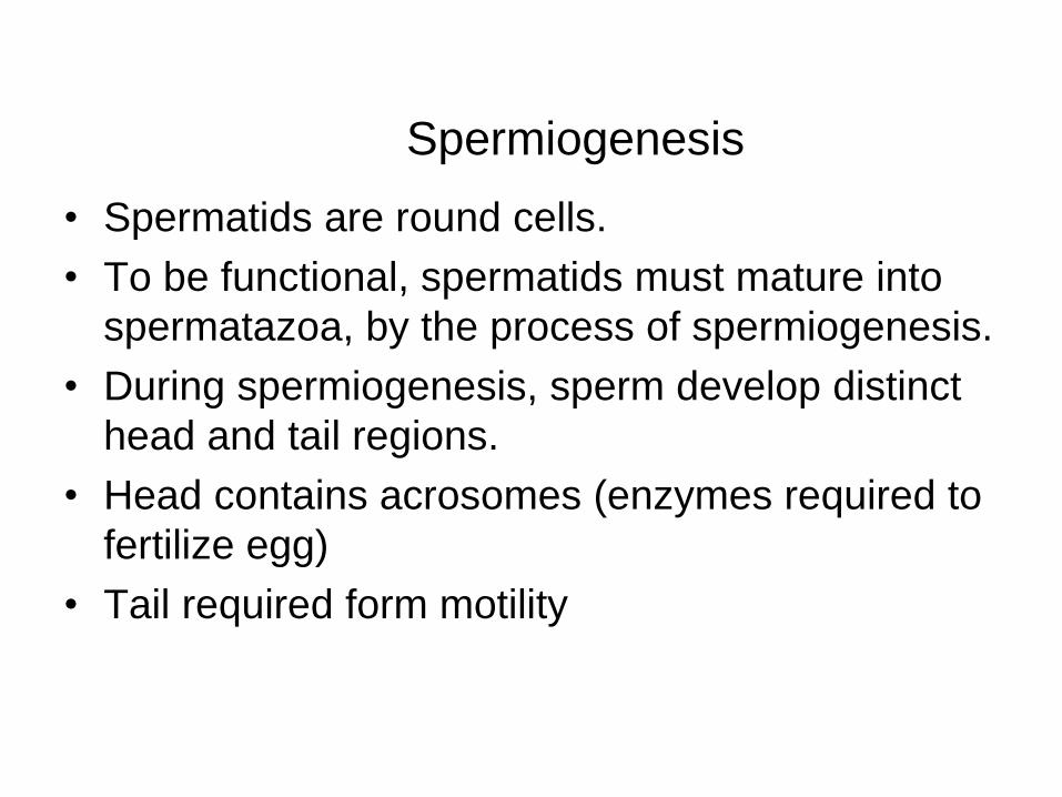
Spermiogenesis
• Spermatids are round cells.
• To be functional, spermatids must mature into
spermatazoa, by the process of spermiogenesis.
• During spermiogenesis, sperm develop distinct
head and tail regions.
• Head contains acrosomes (enzymes required to
fertilize egg)
• Tail required form motility

Spermatogenesis

Oogenesis • Oogenesis: development of oocytes
• Oocytes are found in ovary, enclosed within follicles.
• During development, all stem cells for oocytes enter first part of meiosis, and then stops (at prophase I, “primary oocyte”).
• Follicles remain “suspended” for years (or decades)
• During each ovulatory cycle, some follicles begin growth.
• Each month, one follicle reaches full maturation (preovulatory or Graafian follicle).
• The ova is released from the follicle (ovulated) due to the midcycle surge of LH.

Follicle Development & Ovulation

Oogenesis
• Just before ovulation, in response to the LH surge, the
oocyte completes meiosis I (now “secondary oocyte”).
• End product of meiosis I: secondary oocyte and 1st polar
body.
• If the ovulated secondary oocyte is fertilized, it finishes
meiosis II, becoming a mature ova (egg)
• End product of meiosis II: mature ova and second polar
body.

Summary of
Oogenesis

End Products of Spermatogenesis and
Oogenesis
• Spermatogenesis: One primary
spermatocytes results in four spermatids,
which result in four spermatazoa.
• Oogenesis: One primary oocyte gives rise
to one mature ova and three polar bodies.
Recommended