Chapter 4:
Microscopy, Staining, and
Classification

Microscopy: The technology of making very
small things visible to the naked eye.
Units of Measurement: The metric system is
used to measure microorganisms.
Metric system:
• Basic unit of length: Meter.
• All units are related to each other by factors
of 10.
• Prefixes are used to indicate the relationship
of a unit to the basic unit (e.g.: meter).

Metric Units of Length and U.S.
Equivalents:
Metric Relationship to U.S.
Unit basic unit (meter) Equivalentkilometer (km) 1 km = 1000 m 1 mile = 1.61 km
meter (m) 1 m = 39.37 in
decimeter (dm) 1 dm = 0.1 m = 10-1m 1 dm = 3.94 in
centimeter (cm) 1 cm = 0.01 m = 10-2m 2.54 cm = 1 in
millimeter (mm) 1 mm = 0.001 m = 10-3m
micrometer (um) 1 um = 0.000001 m = 10-6m
nanometer (nm) 1 nm = 0.000000001 m = 10-9m
picometer (pm) 1 pm = 0.000000000001 m = 10-12m

General Principles of Microscopy:
1. Wavelength of radiation: The smaller the
wavelength used, the higher the magnification
and resolution of the microscope.
• Visible light: 400-700 nanometers
• Ultraviolet light: 100-400 nm
• Electron beams: 0.01-0.001 nm

The Electromagnetic Spectrum
General Principles of Microscopy:
2. Magnification
• Apparent increase in size of an object
• Occurs when radiation bends (refracts) as it
passes through a lens
• Magnification can be very high, but image quality
can get very blurry
• Useful magnification depends on resolution and
contrast

Light refraction and Magnification

General Principles of Microscopy:
3. Resolution (Resolving Power): Ability to
distinguish between objects that are close
together. Depends on two factors:
• Wavelength of electromagnetic radiation
• Aperture of lens: Ability to gather light
Resolution = 0.61 x wavelength
Distance numerical aperture

Limits of Resolution: Eye versus Microscopes

General Principles of Microscopy:
4. Contrast: Differences in intensity between:
• Two objects
• An object and its background
• Staining will increase the contrast between a cell and
its background

Instruments of Microscopy:
1. Simple Microscopes:
Only have one lens, similar to a magnifying glass.
Leeuwenhoeck’s simple microscopes allowed him
to magnify images from 100 to 300 X.
• They were so difficult to focus, he built a new one for
each specimen, a total of 419.
• He did not share his techniques with other scientists.
Even today, his source of lighting is unknown.
• His daughter donated 100 of his microscopes to the
Royal Society shortly before his death in 1723.

Simple Microscopes Have a Single Lens
Instruments of Microscopy:
2. Compound Light (CL) Microscopy
History of CL Microscopes:
First developed by Zaccharias Janssen, Dutch
spectacle maker in 1600.
• Poor quality
• Could not see bacteria
Joseph Jackson Lister (Lister’s father) developed
improved compound light microscope in 1830s.
• Basis for modern microscopes
Use visible light as a source of illumination.

Compound Light Microscope

Instruments of Microscopy:
2. Compound Light Microscopy
Have several lenses:
1. Light originates from an illuminator and passes
through condenser lenses, which direct light onto the
specimen.
2. Light then enters the objective lenses, which magnify
the image. These are the closest lenses to the specimen:
• Scanning objective lens: 4 X
• Low power objective lens: 10 X
• High power objective lens: 40-45 X
• Oil immersion lens: 95-100 X
3. The image of the specimen is magnified once again by
the ocular lens or eyepiece (10 X).
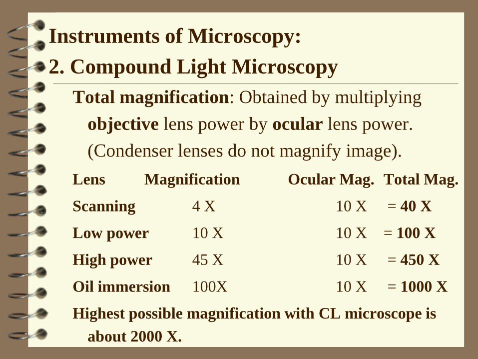
Instruments of Microscopy:
2. Compound Light Microscopy
Total magnification: Obtained by multiplying
objective lens power by ocular lens power.
(Condenser lenses do not magnify image).
Lens Magnification Ocular Mag. Total Mag.
Scanning 4 X 10 X = 40 X
Low power 10 X 10 X = 100 X
High power 45 X 10 X = 450 X
Oil immersion 100X 10 X = 1000 X
Highest possible magnification with CL microscope is
about 2000 X.

Instruments of Microscopy:
2. Compound Light Microscopy
Resolution (Resolving power): Ability of microscope to see two items as separate and discrete units.
• The smaller the distance between objects at which they can be distinguished as separate, the greater the resolving power.
• Light must pass between two objects in order for them to be seen as separate.
• Depends on light wavelength. If wavelength is too long to pass between objects, they will appear as one.
• White light has a relatively long wavelength (550 nm), and cannot resolve structures less than 220 nm (0.2 um) apart.
• Ultraviolet (UV) light has a shorter wavelength (100 to 400 nm), and can resolve distances as small as 110 nm.

Instruments of Microscopy:
2. Compound Light Microscopy
Refraction: Bending of light as it passes from one medium to another of different density.
• Index of refraction: A measure of the speed at which light passes through a material.
• Can be changed by staining, which increases contrastbetween specimen and surrounding medium.
• When two substances have a different index of refraction, the light will bend as it passes from one material to another.
• As light passes through a glass slide, air, and the objective lens, it bends each time, causing loss of light and a blurred image.
• Immersion oil has the same index of refraction as glass slide, preventing light loss from refraction.

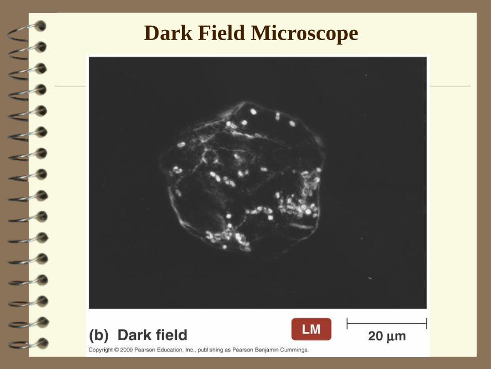
Instruments of Microscopy:
3. Darkfield Microscopy
• Useful to examine live or unstained specimens.
• Light sensitive organisms
• Specimens that lack contrast with their background.
• Spirochetes which cause syphilis.
• Darkfield condenser with opaque disc blocks
light that would enter objective lens directly:
• Light reflects off specimen at an angle.
• Only light reflected by specimen enters objective lens.
• No direct background light.
• Image: Light specimen against dark
background.
Dark Field Microscope

Instruments of Microscopy:
4. Phase Contrast Microscopy
• Useful to examine live specimens:
• Doesn’t require fixing or staining, which usually kill
and/or distort microorganisms.
• Permits detailed examination of internal
structures.
• Special objective lenses and condenser with ring
shaped diaphragm accentuate small differences in
refractive indexes of internal structures.
• Image: Direct rays and reflected light rays come
together, forming an image with many shades of
gray to black.

Phase Contrast Microscope

Instruments of Microscopy:
5. Fluorescence Microscopy
• Fluorescence: Ability of substances to absorb short wavelengths of light (ultraviolet light) and emit them at a longer wavelength.
• Natural Fluorescence: Some microorganisms fluoresce naturally under UV light (Pseudomonas).
• Fluorochrome: Fluorescent dye.
• Acridine orange: Binds to nucleic acids, colors cells orange, green, or yellow depending on light source.
• Immunofluorescence: Fluorescent antibodies can be used to detect specific antigens. Very useful for the rapid diagnosis of specific diseases (e.g.: syphilis).
• Image: Luminescent bright object against a dark background.

Fluorescent Microscopes & Immunofluorescence

Instruments of Microscopy:
Limitations of light microscopy:
• Magnification: Up to 2000 X.
• Resolving Power: Up to 0.2 um.
Because of the limits of magnification and
resolving power, viruses and most internal
structures of cells cannot be seen with a light
microscope.
Instruments of Microscopy:
6. Electron Microscopy
• Electron microscopes were first developed in 1932, and became widely available in 1940s.
• Use a beam of electrons instead of a beam of light.
• Wavelength of electron beam is about 100,000 times smaller than visible light.
• Used to examine structures too small to be resolved with a light microscope.
• Two types of electron microscope:
A. Transmission Electron Microscope (TEM)
B. Scanning Electron Microscope (SEM)

Instruments of Microscopy:
6. Electron Microscopy
A. Transmission Electron Microscope (TEM)
• Gives excellent view of internal structures.
• Magnification: 100,000 X or more.
• Resolving power: 2.5 nm or better.
• Two dimensional image.
• Drawbacks of TEM:
• Due to limited penetrating power of electrons, can only view very thin slices (70-90 nm) of specimen.
• Must slice, fix, dehydrate, and view specimen under a vacuum. Staining may be used to enhance image contrast.
• Treatments kill specimen and may cause shrinkage and distortion of cells (artifacts).

Transmission Electron Microscope

Instruments of Microscopy:
6. Electron Microscopy B. Scanning Electron Microscope (SEM)
• Gives excellent view of external surface.• Magnification: 10,000 X or more.
• Resolving power: 20 nm or better.
• Three dimensional image.
• More recent invention than TEM. Used mainly to observe the surfaces of cells and viruses.• Specimens are covered with a layer of heavy metal
(gold or palladium).
• A narrow beam of electrons (primary electron beam) is swept across specimen surface.
• Electrons on the specimen surface are knocked out, creating a secondary electron beam which is collected and amplified to produce an image.

Scanning Electron Microscope Images

7. Scanning Tunneling Microscopy and
Atomic Force Microscopy (AFM)
• Developed in the 1980s.
• Used to observe structure and surface of biological molecules and silicon computer chips.
A. Scanning Tunneling Microscope (STM)
Uses a thin metal probe that scans the surface of a specimen.
B. Atomic Force Microscopy (AFM)
Uses a diamond and metal probe that scans surface of specimen.
Advantages of both microscopes:• Higher resolving power than electron microscopes
• No special specimen preparation required

Preparation of Specimens for Light
Microscopy
1. Smear: Spread a thin film of material containing
microorganisms over slide surface. Allow to air
dry.
2. Fixing: Process that kills microorganisms and
attaches them to a microscope slide. Fixing
preserves and minimizes distortion of cells.
Two main methods of fixation:
• Heat fixation: Pass over Bunsen burner flame several
times.
• Chemical fixation: Cover with methanol for 1 minute.

Preparation & Heat Fixing of Smear
Preparation of Specimens for Light
Microscopy
3. Staining: Coloring microorganisms with a dye
that emphasizes certain structures. Before staining
a sample, it must be fixed.
Stains are salts composed of a positive ion
(cation) and a negative ion (anion).
The colored ion is called the chromophore.
Two types of dyes:
A. Basic dyes
B. Acidic dyes

Preparation of Specimens for Light
Microscopy
A. Basic dyes:
• Chromophor is in positive ions.
• Most commonly used dyes.
• Bacteria are slightly negatively charged at
pH 7, therefore they stain with basic dyes.
• Examples:
• Crystal violet
• Methylene blue
• Saffranin
Preparation of Specimens for Light
Microscopy
B. Acidic dyes:
• Color is in negative ions.
• Stain the background: negative staining.
• Bacteria do not stain with acidic dyes.
• Used to observe cell shape, size, and capsules.
• Minimal distortion because heat fixing is not necessary an dye is not taken up by cells.
• Examples:
• Eosin
• Nigrosin
• India ink.
u

Preparation of Specimens for Microscopy
1. Simple Stains
• Aqueous or alcohol solution of a single basic dye.
• Primary purpose is to stain entire microorganism to view cell shape and basic structures.
• Procedure:
• Stain is applied for a certain time, and then washed off.
• Slide is dried and examined.
• Mordant: May be used to increase stain intensity. Increases affinity of stain for specimen.
• Examples: Safranin, methylene blue, crystal violet, and carbolfuchsin.
Preparation of Specimens for Microscopy
2. Differential Stains
• React differently to different types of bacteria.
• Can be used to distinguish among different
groups of bacteria.
• There are two important differential stains used in
microbiology:
A. Gram stain
B. Acid-Fast stain

Preparation of Specimens for Microscopy
2. Differential Stains
A. Gram Stain
• Developed in 1884 by Hans Gram, a Danish
microbiologist.
• The most useful staining procedure in medical
microbiology.
• Distinguishes bacteria of two large and medically
important groups:
• Gram-positive bacteria
• Gram-negative bacteria
• Provides useful information for disease treatment.

Preparation of Specimens for Microscopy
2. Differential Stains
Steps of Gram Stain
1. Primary stain: Cover a heat fixed smear with a
basic dye (crystal violet).
• All cells, gram-positive and gram-negative, are stained
with crystal violet (appear purple).
2. Mordant: After smear is rinsed with water, an
iodine mordant solution is applied.
• Crystal violet-iodine [CV-I] complex forms

Preparation of Specimens for Microscopy
2. Differential Stains
Steps of Gram Stain
3. Decolorizing: Slide is washed with alcohol,
which will remove stain from Gram-negative cells
but not from Gram-positive cells.
• Gram-negative cells will be decolorized.
• Gram-positive cells will remain purple.
4. Counterstain: Alcohol is rinsed off. Safranin is
applied, which will stain cells that were
decolorized.
• Gram-negative cells are stained pink.
• Gram-positive cells remain purple.
Preparation of Specimens for Microscopy
2. Differential Stain
What accounts for the differential
staining between Gram-positive and
Gram-negative cells?
• Gram-positive cells have very thick peptidoglycan cell
walls, whereas gram-negative cells have very thin cell
walls. Crystal violet easily penetrates both cell types.
• Because of its larger size, the crystal violet-iodine complex
[CV-I] is not easily removed from gram-positive cells, due
to their thick cell wall. The CV-I complex is readily
washed out of gram-negative cells with alcohol.
• Counterstain only colors gram-negative cells.

Preparation of Specimens for Microscopy
2. Differential Stain
Applications and Limitations of the
Gram stain
Chemotherapy:
• Gram-positive cells with their very thick peptidoglycan cell walls,
are susceptible to penicillins and cephalosporins.
• Gram-negative cells with their thin cell walls and
lipopolysaccharide layer are resistant to these antibiotics.
Limitations:
• Not all bacterial cells stain well with the Gram-stain.
• Gram-stain only works well on young bacterial cultures, that are
actively growing. Therefore it is best to use cultures that are 18 to
24 hours old.
• Older cultures (over 24-48 hours), are often gram-variable.
Preparation of Specimens for Microscopy
2. Differential Stains
B. Acid-Fast Stain (Ziehl-Nielsen Stain)
• Modification of a method developed in 1882 by
Paul Ehrlich.
• Used to detect tuberculosis and leprosy causing
organisms of the genus Mycobacterium and
pathogens of the genus Nocardia.
• These bacteria have waxy cell walls, which
makes them difficult to stain.
Preparation of Specimens for Microscopy
2. Differential Stains
Steps of Acid-Fast Stain
1. Primary stain:
• Cover a heat fixed smear with carbolfuchsin, a
red basic dye.
• Allow to penetrate for several minutes to
increase penetration and retention of dye.
• Rinse with water.
Preparation of Specimens for Microscopy
2. Differential Stains
Steps of Acid-Fast Stain
2. Decolorizing: Slide is washed with acid-alcohol.
• Non acid-fast cells will be decolorized.
• Acid-fast cells will remain red.
3. Counterstain: Acid-alcohol is rinsed off.
Methylene blue is applied, which will stain cells
that were decolorized.
• Non acid-fast cells are stained blue.
• Acid-fast cells remain red.

Preparation of Specimens for Microscopy
3. Special Stains
Used to color and isolate specific parts of
microorganisms such as:
• Endospores
• Capsules
• Flagella
Preparation of Specimens for Microscopy
3. Special Stains
A. Endospore Stain
• Endospores are extremely resistant, dormant structures
that are formed by some gram-positive bacteria to protect
them from harsh environmental conditions: heat, drought,
chemicals, radiation, etc.
• Ordinary staining methods cannot penetrate the thick
endospore wall.
• Most commonly used method is Schaeffer-Fulton
endospore stain.
Endospore Stain

Preparation of Specimens for Microscopy
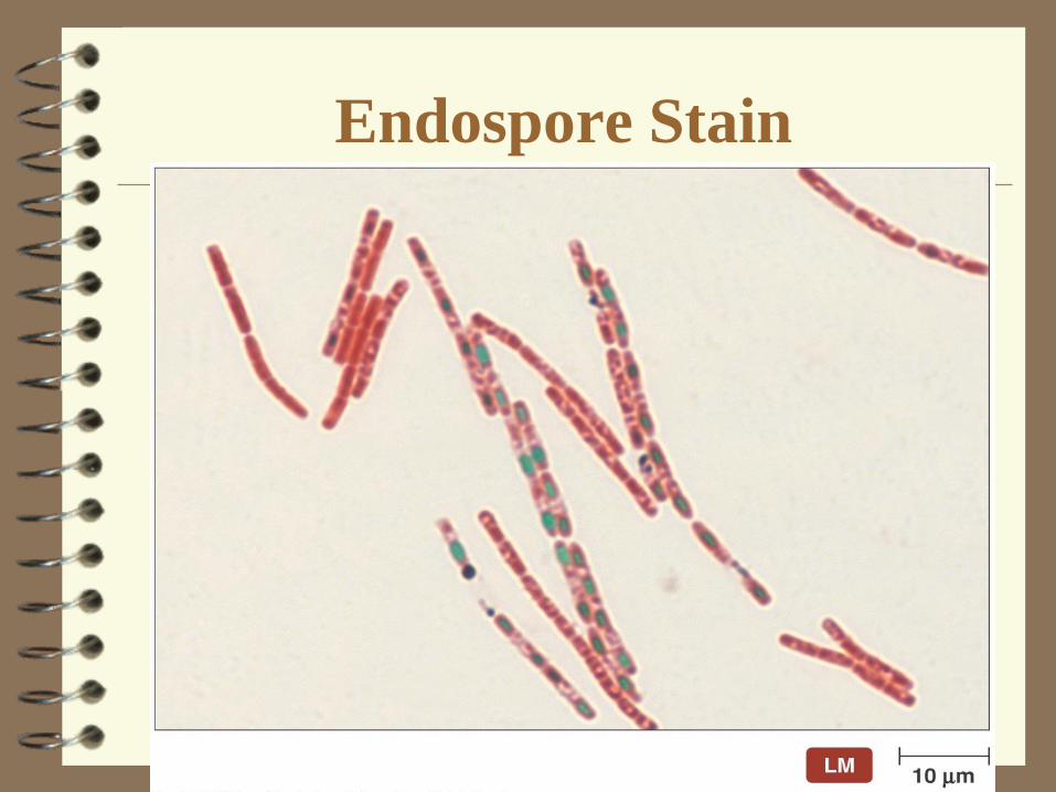
3. Special Stains
A. Endospore Stain
Steps for Schaeffer-Fulton Endospore Stain
1. Primary stain: Malachite green is applied to heat
fixed smear and steamed for about 5 minutes.
• Malachite green will penetrate endospore.
2. Wash: Rinse with water for 30 seconds.
• Removes green dye from rest of the cell, except for
endospore
3. Counterstain: Safranin will stain rest of the cell.
Appearance of cell with endospore:
Pink cell with green endospore.

Preparation of Specimens for Microscopy
3. Special Stains
B. Capsule Stain
• Capsules are gelatinous covers on top of the cell wall,
which are important virulence (disease) factors.
• Capsules are difficult to stain because they repel most
stains, are water soluble, and are easily disrupted with
harsh treatment.
• Negative stain is used to obtain a dark background (E.g.:
India ink or nigrosin).
• Cell is stained with a basic dye (E.g.: safranin).
Capsule appearance: Light halo around stained cell, dark
background.
Preparation of Specimens for Microscopy
3. Special Stains
C. Flagella Stain
• Flagella are appendages used for locomotion that are too
thin to be seen easily with a light microscope.
• Staining procedures are difficult. Usually involve using a
mordant and coating the flagellar surface with a dye or
metal (e.g.: silver).
• The number and arrangement of flagella can be used as
diagnostic aids.
Recommended