Zurich Open Repository andArchiveUniversity of ZurichMain LibraryStrickhofstrasse 39CH-8057 Zurichwww.zora.uzh.ch
Year: 2018
Microbiome and transcriptome interactions in epithelial tissues in thecontext of allergic diseases
Altunbulakli, Can
Posted at the Zurich Open Repository and Archive, University of ZurichZORA URL: https://doi.org/10.5167/uzh-152948DissertationPublished Version
Originally published at:Altunbulakli, Can. Microbiome and transcriptome interactions in epithelial tissues in the context ofallergic diseases. 2018, University of Zurich, Faculty of Science.

1.1. Skin barrier dysregulation upon exposure to p-phenylenediamine in hair dye
allergic individuals and non-responding hairdressers
Sanne S. Steengaard-Meisser1,2,3*, Can Altunbulakli1*, Josefine Bandier3, Morten S Opstrup3,
Francesc Castro-Giner1, Charlotte M. Bonefeld2, Jeanne D Johansen3, Cezmi A. Akdis1
1 Swiss Institute of Allergy and Asthma Research (SIAF), University of Zurich, Davos,
Switzerland. Christine Kühne- Center for Allergy Research and Education (CK-CARE),
Davos, Switzerland
2 Institute of immunology and microbiology, Copenhagen University, Denmark.
3 National Allergy Research Center, University Hospital Gentofte, Denmark.
* These authors contributed equally to this work.
Corresponding author
Cezmi A. Akdis, MD
Swiss Institute of Allergy and Asthma Research (SIAF)
Obere Strasse 22
CH-7270, Davos Platz, Switzerland
Telephone: +41 (0) 81 410 08 48
Fax: + 41 (0) 81 410 08 40
E-mail: [email protected]
1
Abstract
Background: p-Phenylenediamine (PPD) is a strong contact allergen used in hair dye products
and often the cause of allergic contact dermatitis among consumers and hairdressers. Previous
studies have established PPD as a hapten capable of sensitization through penetration of skin,
however, the full extent of changes PPD causes in skin of exposed individuals remains
unknown.
Methods: Skin biopsies were collected from 9 PPD allergic individuals and 7 hairdressers with
no allergy to PPD, on day 4 after patch test with 1% PPD in vaseline or vaseline alone. RNA-
sequencing and transcriptomics analysis were performed on skin biopsies and confirmed with
quantitative RT-PCR. Protein expression was analysed in skin sections with
immunofluorescence staining from 5 non-responder hairdressers and 1 allergic individual.
Reconstructed human epidermis cultures were used to test the effects of PPD in vitro where
both RNA analysis and immunofluorescent staining were performed.
Results: RNA-sequencing uncovered significant downregulation of epidermal tight junction
and stratum corneum barrier molecules in skin of severe allergic individuals stimulated with
PPD. Tight junction molecules CLDN1, CLDN8, CLDN11, CLMP, OCLN, MAGI1, MAGI2
were found to be downregulated in severe allergic individuals, whereas no changes were
observed in non-responding control patients and mild allergic individuals. FLG1, FLG2,
CRNN and LOR from the stratum corneum molecules were also found to be downregulated in
severe allergic individuals. Confocal microscopy imaging of CLMP and CLDN1 in PPD
exposed allergic skin also revealed downregulation of TJ proteins and disassociation of TJ
molecules from cell membranes. Exposure of PPD to 3D skin tissue culture of healthy
mammary skin revealed upregulation of FLG1 and FLG2 in response to allergen stimulation.

2
Conclusion: We demonstrate that the clinical outward signs of inflammation in PPD exposed
skin correlates with the extent of transcriptomic changes in the skin. Barrier dysregulation and
downregulation of tight junction and stratum corneum molecules were observed in severe
allergic individuals, while mild lesional skin and skin-of non-responders differ minimally in
response to PPD. Our results additionally demonstrate a healthy tissue response to PPD in the
form of upregulation of stratum corneum barrier molecules, such as FLG1 and FLG2.

3
Introduction
Allergic contact dermatitis (ACD); also referred to as allergic contact hypersensitivity reaction
is mediated by T-cells and the reaction typically appears as a lesion at 2-7 days after allergen
exposure. The presentation and severity of clinical reactions depends on exposure site and type
of allergen. Reactions are typically observed as eczema or swelling at the site of allergen
exposure(1). Irritant stimulus; in the form of cold and dry weather, abrasive activity in damp
conditions, exposure to chemical or mechanical stress might contribute to the severity of
lesions. ACD, together with irritant contact dermatitis, is one of the most abundant
occupational diseases in Europe and a considerable socioeconomic burden due to cost of
treatment and loss of productivity (2). Occupations particularly under risk of ACD development
include hairdressers, who have regular contact with irritants and contact allergens. Hair dye
component p-Phenylenediamine (PPD) used in dark permanent hair dyes is a well-known
allergen for ACD (1, 3). Allergic reactions to PPD are not only observed as an occupational
disease, but among the general population as well. Severe reactions to hair dye can result in
hospitalization due to swelling of the face, obstructing the airways as well as severe scalp
eczema, which can lead to complete or partial hair loss. Recent studies have shown that
approximately 4 % of patients patch tested at dermatology clinics in Europe are sensitized to
PPD (4). Hair dye is suspected to be the main route of exposure but also temporary black henna
tattoos pose a common route of sensitization, especially in children and young adults (5, 6).
Hairdressers are exposed to PPD on a regular basis through their work with hair dyes where
protective work habits are only able to limit the exposure, but not eliminate it (7). Exposure to
PPD can be detected both externally on the skin surface, but also internally in urine samples,
with little effect seen of glove use (8). PPD has been shown to sensitize majority of exposed

4
individuals after only two exposures (9) and hairdressers are therefore like to be sensitized to
PPD. However; the majority of the PPD exposed individuals but the majority do not develop
allergy. The exact mechanism of tolerance development in exposed but not responding
individuals in currently unknown.
PPD is a highly reactive small aromatic compound and contact allergy to PPD is thought to be
initiated by the products formed by reaction of PPD with proteins found in the skin (1). Among
the suggested allogenic mechanisms is the oxidation of the amino acid cysteine in human serum
albumin, (10) whereas the acetylation of PPD by N-acetyltransferase from keratinocytes is
reported to neutralize the allergenic potential of PPD (11). The skin barrier plays a crucial role,
not only in limiting the penetration of PPD but also in generating the epitopes causing allergy.
The epidermal barrier of the skin forms the first layer of defense against potential pathogens,
pollutants and allergens. It is composed of elements of innate immunity such as anti-microbial
peptides, pathogen recognition receptor systems, lipid and ions designed the prevent
colonization by pathogens, as well as protecting the commensal microbiota of skin. Tight
junctions (TJ) seal the epidermal layer against the outer environment, preventing access of
pathogens and antigen into deeper layers of skin. It is now known that disruption of epithelial
barrier by dysregulated TJs and stratum corneum (SC) proteins is one of the underlying causes
of many allergic skin diseases. The TJ family proteins Claudin-1 (CLDN-1) and CLDN-8, as
well as CLDN-23 are expressed at lower levels in the lesional skin of atopic dermatitis (AD)
patients (12, 13) and loss of adherens junctions and TJs, marked by decreased expression of
CLDN-18 and E-cadherin is typical for asthma (14, 15).
Filaggrin-1 (FLG) and FLG-2 are expressed at lower levels in lesional skin of AD patients (16).
FLG-null mutations in humans leads to a susceptibility to hand eczema, childhood-onset AD
and sensitization to contact allergens (17). Furthermore, AD and hand eczema patients with
FLG-null mutations showed exacerbation of skin symptoms and a lower quality of life (18, 19).

5
The skin barrier is therefore not only involved in the development of various skin diseases but
also the severity of said diseases.
However, the full extent of changes in epidermal barrier, as well as the effect of epidermal
barrier in contact hypersensitivity reactions against hair-dye allergens such as PPD are
currently unknown. In this report, we have for the first time performed RNA-sequencing on
skin biopsies treated with PPD collected from clinically non-responder hairdressers and
patients allergic to PPD. Furthermore, we show for the first-time expression changes in
epidermal barrier molecules and TJs in PPD exposed skin. We here show that multi-level
damage caused by PPD to the skin barrier is not only seen in allergic skin but also in non-
responder individuals.
Materials and methods
Study population
The study included two groups; a PPD allergic patient group from the allergy clinic at Gentofte
Hospital, Copenhagen and a group of PPD exposed but non-allergic hairdressers referred to as
non-responders. The allergic group was diagnosed PPD allergy within the past 5 years at our
clinic. Non-responders were all hairdressers with 5 or more years in the field and with no
history of contact allergy or eczema. The participants were adults between 18 and 60 years,
further characteristics are listed (Table 1). Patients with other inflammatory skin diseases or
receiving immunosuppressive medication were excluded. All participant gave informed written
consent and the study was conducted in accordance with the Helsinki declaration.

6
Table 1. Patient characteristics
Patients from the non-responder group did not have any type-1 allergies or non-allergic eczema, whereas some allergic
individuals were multi-allergic to both type-IV and type-I allergens. All non-allergic eczema was described as hand eczema
with dry skin in the winter but with no relevant allergen sensitization during patch-testing. Occupational exposure was based
on the participants own recollection and description of work, all allergic individuals had reacted to personal exposure to either
dark hair color, dark henna tattoos or other known sources of PPD exposure.
RNA sequencing
9 allergic individuals and 7 non-allergic hairdressers where patch tested for 48 hours using
8mm Finn chambers with 20mg 1% PPD in Vaseline and a vehicle (Vaseline) control. The
patch test scored at day 2 and day 4 according to the true-test criteria (IR, ?, +,2+ or 3+). 4 mm
punch biopsies where collected at day 4 from both the PPD and vehicle test-site. The biopsies
were placed in RNA later and frozen in liquid nitrogen before storage at -80°C. Total RNA
was prepared from skin biopsies using the RNeasy Universal Plus kit (QIAGEN, Hilden,
Germany). The quantity and quality of the isolated RNA was determined with a Qubit® (1.0)
Fluorometer (Life Technologies, California, USA) and a Bioanalyzer 2100 (Agilent,
Waldbronn, Germany) and samples with RNA integrity number >7.0 were chosen for
sequencing. Library preparation for RNA-seq was performed using the TruSeq Stranded
mRNA Sample Prep Kit (Illumina, Inc, California, USA).
Total RNA samples (400 ng) were ribosome depleted and then reverse-transcribed into double-
stranded cDNA with Actinomycin added during first-strand synthesis. The cDNA samples
were fragmented, end-repaired and polyadenylated before ligation of TruSeq adapters. The

7
adapters contain the index for multiplexing. Fragments containing TruSeq adapters on both
ends were selectively enriched with PCR. The quality and quantity of the enriched libraries
were validated using Qubit® (1.0) Fluorometer and the Bioanalyzer 2100 (Agilent, Waldbronn,
Germany). The product is a smear with an average fragment size of approximately 360 bp. The
libraries were normalized to 10nM in Tris-Cl 10 mM, pH8.5 with 0.1% Tween 20.
The TruSeq SR Cluster Kit v4-cBot-HS or TruSeq PE Cluster Kit v4-cBot-HS (Illumina, Inc,
California, USA) was used for cluster generation using 8 pM of pooled normalized libraries on
the cBOT. Sequencing were performed on the Illumina HiSeq 2500 paired end at 2x126 bp or
single end 126 bp using the TruSeq SBS Kit v4-HS (Illumina, Inc, California, USA).
Sequencing images were transformed with Illumina Basecaller software to bcl files, which
were demultiplexed to fastq files with CASAVA v1.8.2 software (Illumina). Quality check on
the reads was performed with fastqc (v.0.10.0, Babraham Institute, Cambridge, UK).
Raw sequencing reads were mapped to the Homo Sapiens genome (GRCh38 build) using
RSEM (v1.2.12) [PMID: 21816040] implementation of Bowtie software (v 1.0.0) [PMID:
19261174] alignment program with the Ensembl annotation (v 75). Gene and isoform level
abundances were quantified as RPKM values. Clustering analyses were performed using the
“ward.D2” clustering algorithm implemented in the “hclust” function of R statistics package.
Heatmap plots were performed with the function “heatmap.2” implemented in the gplots R
package.
Differential expression analysis between two groups was performed using edger R
bioconductor package [PMID: 19910308]. Genes present in less than 75% of samples in both
conditions were removed. Q-values were calculated using the Benjamini-Hochberg method
and genes with a q-value <0.05 and an absolute value of log2 (fold change)>1 were kept for
further analysis.

8
Gene ontology (GO) term enrichment analysis was performed using GOseq bioconductor
package [PMID: 20132535] using the Wallenius approximation.
Skin cultures
Skin cultures from mammary skin grown for 13 days (Episkin, France) were grown in air-
liquid interface cultures overnight before use. Cultures were stimulated with 0.5% PPD in PBS,
0.5% H2O2 in PBS or PBS alone. Skin cultures and supernatants were collected at 8 hours and
24 hours for confocal microscopy, qPCR analysis and cytokine analysis respectively.
Confocal microscopy
Biopsies were collected at day 4 from 4 non-responder hairdressers and 1 PPD allergic patient.
Upon collection the biopsies where embedded in tissue-tek, frozen in liquid nitrogen and stored
at -80°C. The biopsies were cut in 7µm slices, fixed with 4% PFA on glass slides and stained
with primary antibodies CLDN1, CLMP, FLG1 and FLG2 and secondary fluorochrome
labeled antibodies goat anti-rabbit AF546, goat anti-rabbit AF488, goat anti mouse AF546.
Statistics
Significant differential expressed genes was defined as genes with a false discovery rate (FDR)
below 0.015. Gene expression between paired samples, PPD exposed and vaseline exposed
skin from the same individual, was analyzed using Wilcoxon test and comparison between
groups was done using Mann Whitney test. Pathway analysis was done using the Enricher
platform and the Panther database. significant pathways were defined as pathways with an
adjusted P-value below 0.01. Venn diagrams of significantly regulated genes between
comparison groups were visualized by the online application “genevenn.sourceforge.net”
developed by Vijayaraj Nagarajan and Mehdi Pirooznia.

9
Results
Altered gene expression in skin after PPD exposure corresponds to clinical manifestations
Non-responder individuals patch tested with 1% PPD showed no clinical reaction and no
changes in general histology whereas allergic individuals showed reactions ranging from mild
swelling and redness to severe blister formation followed by necrosis (Figure 1A). RNA
sequencing, subsequent principal component analysis (PCA) and hierarchical clustering of skin
biopsies collected from patch tested individuals show a unique gene expression profile for PPD
exposed skin from PPD allergic patients who developed skin lesions with higher severity
(graded +2 and +3) (Figure 1B). Gene expression profile of PPD allergic individuals with mild
reaction to PPD (graded +1 and ?) were observed to cluster closely with vaseline exposed skin
from non-responder and PPD allergic patients, as well as PPD exposed skin from non-
responder individuals. Overall hierarchical clustering of skin biopsies according to the top 2000
genes with high standard deviation shows that the transcriptomic signature of PPD exposed
skin depends on severity of clinical reaction (Figure 1C). Furthermore; inflammatory signs
such as erythema, thickening of the skin and inflammatory papules are visible in severe and
mild allergic skin are visible after second day of PPD stimulation, and gets more severe after
fourth day. Skin characteristics and phenotype do not change with vaseline stimulation and in
non-responders at day 2 or day 4 (Fig 1D)

10
Figure 1. Reactions to PPD patch test in non-responder and allergic individuals. Clinical
reactions and corresponding HE staining of tissue at day 4 after patch with 1% PPD in vaseline
or vaseline control (A). 3D PCA plot of sample clustering when looking at the top 2000
differential expressed genes (B). Hierarchical clustering of samples in top 2000 differentially
expressed genes (C). Outward clinical reactions of PPD and vaseline stimulated skin at days 2
and 4 after patch is applied (D). Severe allergic reactions (red) n=5, mild allergic reaction
reactions (yellow) n=4, PPD exposed skin from non-responder individuals (green) n= 7 and
vaseline controls (purple) n= 16.
Patterns of gene expression differ between Allergic and non-responder groups
Next, we investigated the enriched pathways for the differentially expressed genes between
non-responder, mild allergic and severe allergic skin biopsies. Differentially expressed genes
from the 3 groups was divided to up-regulated genes (Figure 2A-B) and down-regulated genes
(Figure 2C-D) and compared between the 3 groups. 22 upregulated genes were found to be
11
unique for the non-responder group, 53 for the mild allergic and 2636 genes was unique to the
severe allergic group. 376 genes were up-regulated in all groups. The genes in each group,
shared or unique, was analyzed for significant pathway association (Figure 2A). Up-regulated
pathways in allergic individuals were in majority belonged to immune response pathways
involved in T-cell recruitment and activation, inflammation, cytokine and chemokine
signaling. In the severe reaction group, genes belonging to response against cellular damage,
like FAS and apoptosis signaling was significantly up-regulated. The non-responder group
showed significant up regulation of 3 immune relevant pathways and a gene involved in
serotonin degradation. Among the down-regulated genes was 17 genes unique for the non-
responder group, 69 for the mild allergic and 3382 for the severe allergic (Figure 2C). The most
significant down-regulated gene pathways were pathways involved in cell adhesion, tissue
regeneration and cell metabolism (Figure 2D). Association of downregulated genes with
cellular adhesion and cellular junction pathways, as well as identification of multiple genes
with known function on skin barrier in individual gene lists led us to investigate the state of
skin barrier and cell adhesion components in PPD allergic skin. (Figure E1).

12
Figure 2. Comparison and pathway analysis of significantly up-regulated (A) or down-
regulated (D) genes among non-responder individuals (blue), mild allergic individuals (yellow)
and severe allergic individuals (red) (significance defined as a fdr > 0.015). Pathway analysis
using the EnrichR/panther databases of up or down regulated genes in all groups in genes
overlapping between one or more groups. N = involved genes in the given pathway. Venn
diagrams indicating the number of unique and shared genes significantly upregulated (B) or
downregulated (C) between comparisons.

13
Figure E1. Significantly regulated gap junctions, desmosomes and keratin molecules in
PPD exposed severe allergic skin. Heatmap visualizing significantly changing molecules in
severe allergic PPD exposed skin (p-value<0.05).

14
Down regulation of TJ genes corresponds to clinical severity of the allergic reaction
To analyze the expression for the TJ components of skin, 47 genes with known association of
function with the TJ gene family were curated from Gene Ontology (GO), Kyoto Encyclopedia
of Genes and Genomes (KEGG) databases and previous publications. 32 genes out of 47 were
found to be expressed in skin, according to the mean expression threshold value of >10 unique
read counts. Analysis of the expression of TJ genes expressed in non-responder, mild allergic
and severe allergic skin groups revealed 15 out of 32 (% 46.8) TJ genes were significantly
down regulated genes in one or more groups (Figure 3A). All 15 genes were down regulated
in severe allergic patients whereas CLDN1 and CLMP was significantly down regulated in the
mild allergic. CLMP and CLDN1 downregulation did not reach statistical significance in the
PPD exposed non-responder group but showed a similar picture to the downregulation
observed in PPD exposed allergic patients (Figure 3B). Validation of RNA sequencing results
with qPCR, confirmed observation made for TJ genes CLDN1, CLDN8 and CLMP (Figure
3C).

15
Figure 3. Significantly regulated TJ molecules in PPD exposed severe allergic skin.
Heatmap visualizing significantly changing TJ molecules in severe allergic PPD exposed skin
(p-value<0.05) (A). Scatter plots for the top 8 significantly downregulated TJ molecules in
PPD exposed severe allergic skin, with statistical comparisons for all subject groups (p<0.05
*, p<0.01 **, p<0.001 ***) (B). qPCR confirmation of several TJ molecules significantly
downregulated in severe allergic PPD exposed human skin. Gene expression calculated as
arbitrary units (2(-ΔCt)*1000) according to EEF1A expression (C)

16
PPD causes dysregulation of Filaggrin family proteins in allergic skin
In line with the downregulated pathways identified in our differential expression analysis of
PPD exposed skin groups, we analyzed the expression of the 10 filaggrin family genes and
found 5 (% 50) differential expressed genes in one or more groups after PPD exposure (figure
4 a). Severe allergic reactions to PPD results in down regulation of 4 filaggrin family members;
FLG1, FLG2, loricrin (LOR) and cornulin (CRNN) (Figure 4A). When looking at expression
levels in each donor individually, only some donors in the mild allergic and non-responder
group decreased, but not as much as seen in the severe allergic group (Figure 4B). In contrast,
involucrin (IVL), showed significant upregulation in the severe allergic reactions, but no
change in neither mild allergic reactions nor non-responder individuals.

17
Figure 4. Significantly regulated stratum corneum molecules in PPD exposed severe
allergic skin. Heatmap visualizing significantly changing stratum corneum molecules in
severe allergic PPD exposed skin (p-value<0.05) (A). Scatter plots for the top 5 significantly
downregulated stratum corneum molecules in PPD exposed severe allergic skin, with statistical
comparisons for all subject groups (p<0.05 *, p<0.01 **, p<0.001 ***) (B). qPCR confirmation
of FLG1 and FLG2 in severe allergic PPD exposed human skin. Gene expression calculated as
arbitrary units (2(-ΔCt)*1000) according to EEF1A expression (C)

18
PPD changes protein expression and organization of TJ molecules in allergic and non-
responder individuals
Changes in barrier protein expression levels and the location in epidermis was visualized via
confocal microscopy of skin biopsies day 4 of patch test with 1% PPD in Vaseline from 5 non-
responder hairdressers and 1 severe allergic patient (Figure 5). CLDN1 and CLMP from the TJ
proteins was seen localized to the cell surface membrane forming a net-like pattern between
the keratinocytes throughout epidermis in vaseline controls of both the non-responder
hairdressers and the severe allergic individual (Figure 5A). We saw a decrease in CLDN1 after
PPD exposure in non-responder individuals but with an intact net-like structure whereas there
was a complete disruption of the epidermis in the allergic individual causing the net-like
structure to disappear when looking at both CLDN1 and CLMP.
PPD causes decreased protein expression of FLG1 and FLG2 in both non-responder and
allergic individuals. An even distribution of both FLG1 and FLG2 proteins was seen in the
outer layer of epidermis in the Vaseline controls of both the non-responder hairdressers and the
severe allergic individual (Figure 5B). PPD exposure caused a visible decline in of both protein
in the outer layer of epidermis. FLG2 was also seen expressed in the basal membrane, this
expression did not seem to be affected by PPD exposure in the non-responder individuals.

19
Figure 5. Confocal staining of barrier proteins in human skin biopsies. TJ proteins CLDN1
and CLMP (a) and Filaggrin family proteins FLG1 and FLG2 (b) in Non-responder and allergic
individuals at day 4 after patch with Vaseline or 1% PPD in Vaseline. Non-responder
individuals N=5, allergic N=1.
20
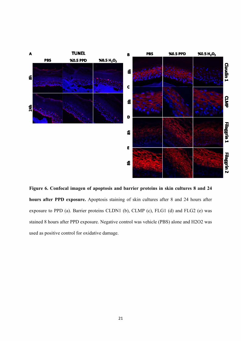
PPD did not induce apoptosis in healthy skin cultures but changes expression of CLDN1,
CLMP and FLG2
Since PPD is highly oxidative, we tested the direct toxicity of PPD on healthy skin cultures
after 8 and 24 hours of exposure but no indication of increased apoptosis was seen, though a
general cell death in the basal membrane was seen after 24h in all cultures. We there next
looked at the barrier protein expression CLND1 was expressed at the cell surface of
keratinocytes and showed limited decrease in expression but signs of disorganization 8 hours
after exposure to PPD whereas there was visibly decreased CLDN1 expression after hydrogen
peroxide exposure. CLMP was mainly seen expressed at the cell membrane and in lower levels
in the cytosol. CLMP shoved decreased expression 8 hours after PPD exposure, but still
localized to the cell surface whereas hydrogen peroxide exposure showed a disruption of
expression at the cell surface and a more homogenous expression in the cytosol. FLG1 showed
no change after neither PPD nor hydrogen peroxide exposure, but a decrease in expression was
seen for FLG2 in both PPD and hydrogen peroxide exposed cultures (Figure 6A-B).
21
Figure 6. Confocal imagen of apoptosis and barrier proteins in skin cultures 8 and 24
hours after PPD exposure. Apoptosis staining of skin cultures after 8 and 24 hours after
exposure to PPD (a). Barrier proteins CLDN1 (b), CLMP (c), FLG1 (d) and FLG2 (e) was
stained 8 hours after PPD exposure. Negative control was vehicle (PBS) alone and H2O2 was
used as positive control for oxidative damage.

22
Discussion
Hairdressers are known to be at risk of developing allergic skin diseases due to their high
exposure to strong allergens like PPD. PPD has long been known as a strong allergen and the
industry has tried over the past 15 years to modify or find alternatives PPD to make it less
allergenic generating an array of new components with various success. PPD is slowly being
replaced in many consumer products, but both hairdressers and private consumers still
experience a high level of exposure as reflected in the general sensitization rate. Several studies
have shown how glove use and work habits can decrease the exposure in hairdressers, but there
is still a low-dose exposure seen regardless of how well trained the hairdressers are (7, 20).
Therefore, it remains relevant to illuminate what effects PPD exposure has on the human body,
especially the skin barrier.
It is thought that development of sensitization to PPD might be a question of skin penetration.
Non-responders to PPD have undergone long exposure to the allergen, but a stable and strong
epidermal barrier might be the key to the tolerance towards the allergen. However, our study
did not observe any differences in barrier molecule expression in allergic and non-responder
individuals before PPD exposure. Overall the TJ molecules and stratum corneum molecules
show similar levels of expression in both mild and severe allergic individuals as well as non-
responders in the stable state. We propose that instead of inherent differences in skin barrier
stability, other tissue factors and severity of inflammation as a result of PPD exposure drives
development of contact allergy lesions.
Expression of TJ molecules CLDN1 and CLMP in particular seems to be downregulated upon
PPD exposure in both mild and severe allergic individuals, as well as non-responders. On the
other hand, the rest of the significantly regulated TJ and stratum corneum barrier molecules
23
are mainly downregulated in severe allergic individuals and to a lesser extent, in mild allergics
upon PPD stimulation. Confocal microscopy imaging of FLG1, FLG2 and CLDN1 confirms
loss of these barrier molecules in non-responding individuals. Skin inflammation in atopic
dermatitis, as well as contact dermatitis and psoriasis have been reported to downregulate
FLG1.
Oxidative stress has been proposed as the main mechanism of action for PPD damage on skin.
Other studies have suggested the induction of reactive oxygen species as an important
mechanism in induction of PPD allergy as that could provide the first damage signal activating
the innate immune system and allowing antigen presenting cells to activate and migrate to the
local lymph nodes. The difference between allergic and non-responder individuals could
thereby also be found in response of skin to oxidative damage and damage control. In our study,
we observed damage to skin barrier in both non-responder and allergic individuals at day 4
after exposure. A more extensive analysis of earlier time points would be needed to conclude
if non-responder individuals have a different immediate response to PPD than allergic
individuals.
It is known that the PPD is neutralized to a large degree in the uppermost layers of the skin.
Therefore, the specific epitopes and the nature of the signals that reach Langerhans cells and
resident memory T lymphocytes that lead to sensitization for PPD and the development of
contact allergy are currently unknown. It is thought that the cellular damage and exposure of
self-molecules as a result of PPD exposure leads to activation of skin resident dendritic cells
and T lymphocytes.
The response observed at fourth day in non-responder individuals after PPD exposure might
be mechanisms of damage control and wound healing in response to the oxidative stress. It
might be also a response mechanism against uncontrolled initiation of inflammation. The non-
responder skin might have a higher threshold of exposure before development of allergic
24
reactions. This could be either a higher tolerance to allergen exposure, or a higher tolerance to
tissue damage and oxidative stress.
References
1. McFadden JP, Yeo L, White JL. Clinical and experimental aspects of allergic contact
dermatitis to para-phenylenediamine. Clin Dermatol. 2011;29(3):316-24.
2. Saetterstrom B, Olsen J, Johansen JD. Cost-of-illness of patients with contact dermatitis
in Denmark. Contact Dermatitis. 2014;71(3):154-61.
3. Kasemsarn P, Bosco J, Nixon RL. The Role of the Skin Barrier in Occupational Skin
Diseases. Curr Probl Dermatol. 2016;49:135-43.
4. Schuttelaar ML, Vogel TA, Rui F, Krecisz B, Chomiczewska-Skora D, Kiec-
Swierczynska M, et al. ESSCA results with the baseline series, 2002-2012: p-
phenylenediamine. Contact Dermatitis. 2016;75(3):165-72.
5. Panfili E, Esposito S, Di Cara G. Temporary Black Henna Tattoos and Sensitization to
para-Phenylenediamine (PPD): Two Paediatric Case Reports and a Review of the Literature.
Int J Environ Res Public Health. 2017;14(4).
6. Vogel TA, Coenraads PJ, Bijkersma LM, Vermeulen KM, Schuttelaar ML, Group EFS.
p-Phenylenediamine exposure in real life - a case-control study on sensitization rate, mode and
elicitation reactions in the northern Netherlands. Contact Dermatitis. 2015;72(6):355-61.
7. Lind ML, Boman A, Sollenberg J, Johnsson S, Hagelthorn G, Meding B. Occupational
dermal exposure to permanent hair dyes among hairdressers. Ann Occup Hyg. 2005;49(6):473-
80.
8. Gube M, Heinrich K, Dewes P, Brand P, Kraus T, Schettgen T. Internal exposure of
hairdressers to permanent hair dyes: a biomonitoring study using urinary aromatic diamines as
biomarkers of exposure. Int Arch Occup Environ Health. 2011;84(3):287-92.
9. Kligman AM. The identification of contact allergens by human assay. II. Factors
influencing the induction and measurement of allergic contact dermatitis. The Journal of
investigative dermatology. 1966;47(5):375-92.
10. Jenkinson C, Jenkins RE, Aleksic M, Pirmohamed M, Naisbitt DJ, Park BK.
Characterization of p-phenylenediamine-albumin binding sites and T-cell responses to hapten-
modified protein. The Journal of investigative dermatology. 2010;130(3):732-42.
11. Kawakubo Y, Merk HF, Masaoudi TA, Sieben S, Blomeke B. N-Acetylation of
paraphenylenediamine in human skin and keratinocytes. J Pharmacol Exp Ther.
2000;292(1):150-5.
12. De Benedetto A, Rafaels NM, McGirt LY, Ivanov AI, Georas SN, Cheadle C, et al.
Tight junction defects in patients with atopic dermatitis. The Journal of allergy and clinical
immunology. 2011;127(3):773-86 e1-7.
13. Suarez-Farinas M, Ungar B, Correa da Rosa J, Ewald DA, Rozenblit M, Gonzalez J, et
al. RNA sequencing atopic dermatitis transcriptome profiling provides insights into novel
disease mechanisms with potential therapeutic implications. The Journal of allergy and clinical
immunology. 2015;135(5):1218-27.
14. Heijink IH, Kies PM, Kauffman HF, Postma DS, van Oosterhout AJ, Vellenga E.
Down-regulation of E-cadherin in human bronchial epithelial cells leads to epidermal growth
factor receptor-dependent Th2 cell-promoting activity. Journal of immunology.
2007;178(12):7678-85.
15. Sweerus K, Lachowicz-Scroggins M, Gordon E, LaFemina M, Huang X, Parikh M, et
al. Claudin-18 deficiency is associated with airway epithelial barrier dysfunction and asthma.
The Journal of allergy and clinical immunology. 2017;139(1):72-81 e1.
25
16. Pellerin L, Henry J, Hsu CY, Balica S, Jean-Decoster C, Mechin MC, et al. Defects of
filaggrin-like proteins in both lesional and nonlesional atopic skin. The Journal of allergy and
clinical immunology. 2013;131(4):1094-102.
17. Thyssen JP, Linneberg A, Ross-Hansen K, Carlsen BC, Meldgaard M, Szecsi PB, et al.
Filaggrin mutations are strongly associated with contact sensitization in individuals with
dermatitis. Contact Dermatitis. 2013;68(5):273-6.
18. Heede NG, Thyssen JP, Thuesen BH, Linneberg A, Szecsi PB, Stender S, et al. Health-
related quality of life in adult dermatitis patients stratified by filaggrin genotype. Contact
Dermatitis. 2017;76(3):167-77.
19. Andersen YMF, Egeberg A, Balslev E, Jorgensen CLT, Szecsi PB, Stender S, et al.
Filaggrin loss-of-function mutations, atopic dermatitis and risk of actinic keratosis: results from
two cross-sectional studies. Journal of the European Academy of Dermatology and
Venereology : JEADV. 2017;31(6):1038-43.
20. Oreskov KW, Sosted H, Johansen JD. Glove use among hairdressers: difficulties in the
correct use of gloves among hairdressers and the effect of education. Contact Dermatitis.
2015;72(6):362-6.
Recommended