International J. of Healthcare and Biomedical Research, Volume: 03, Issue: 04, July 2015, Pages 55-58
55
www.ijhbr.com
Case Report:
Medullary carcinoma of breast : Case Report
Sunil V Jagtap1, Neerav Saini1,P G Chougule2,Swati S Jagtap3, Saini, Sneha1
1Department of Pathology, Krishna Institute of Medical Sciences University, Karad
2Department of Surgery,Krishna Institute of Medical Sciences University, Karad
3Department of Physiology,Krishna Institute of Medical Sciences University, Karad
*Corresponding author : Dr.Sunil V.Jagtap , Professor, Department of Pathology, Krishna Institute of Medical
Sciences University, Karad , Maharashtra , India
Abstract:
Medullary carcinoma of breast is a rare variant of invasive ductal carcinoma of breast and its incidence is less than
5% of invasive breast carcinomas. We are presenting this case in a 49 years old female having single, large, well
circumscribed mass in right breast since 6 months. Fine needle aspiration cytology report was given as positive for
high grade duct carcinoma of right breast. Histopathology report was given as medullary carcinoma of breast.
Immunohistochemistry showed Estrogen Receptor (ER), Progesterone Receptor(PR) negativity and Her-2 neu
positivity. We are presenting this case of Medullary carcinoma of breast for being a specific histopathological
subtype.
Keywords: Breast cancer, Medullary carcinoma, Breast lump
Introduction
Medullary carcinoma of breast is uncommon
variant of invasive ductal carcinoma, which
constitutes about 5% of all breast cancers1.
Despite its aggressive histopathological
appearance, these type of breast cancers have a
favorable prognosis. Histopathological features
of this type of tumor has specific findings
which play important role in final diagnosis
and management of the patient.
Case report:
A 49 year old female presented with lump in
upper outer quadrant of right breast since 6
months. On local examination a lump which
was 5x4 cm in size, painless, soft to firm and
mobile. Skin nipple,areola were unrema-
rkable.No significant contributory history was
there. All routine investigations were within
normal limits. Radiological examination of
chest, pelvis and abdomen showed no evidence
of metastasis.
Cytopathological evaluation showed cellular
smears of malignant cells arranged in sheets
and clusters admixed with mature lymphocytes
and plasma cells. Cytopathological features
suggestive of high grade carcinoma of right
breast. Modified Radical Mastectomy (MRM)
of right breast was done and specimen was
sent for histopathological examination.
Gross
We received a specimen of right modified
radical mastectomy specimen with axillary
clearence. MRM specimen on serial cut
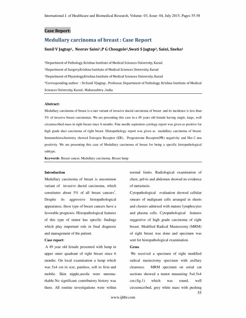
sections showed a tumor measuring 5x4.5x4
cm.(fig.1) which was round, well
circumscribed, grey white mass with pushing
International J. of Healthcare and Biomedical Research, Volume: 03, Issue: 04, July 2015, Pages 55-58
56
www.ijhbr.com
margins. Nipple, areola and skin were
unremarkable.
Microscopic examination
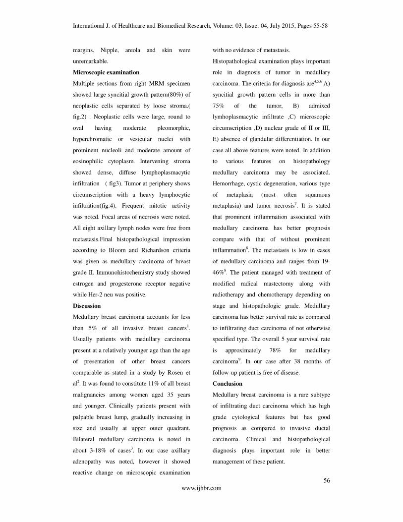
Multiple sections from right MRM specimen
showed large syncitial growth pattern(80%) of
neoplastic cells separated by loose stroma.(
fig.2) . Neoplastic cells were large, round to
oval having moderate pleomorphic,
hyperchromatic or vesicular nuclei with
prominent nucleoli and moderate amount of
eosinophilic cytoplasm. Intervening stroma
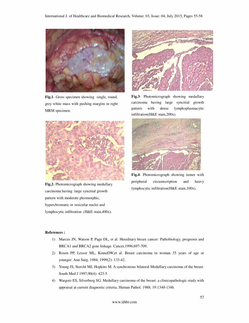
showed dense, diffuse lymphoplasmacytic
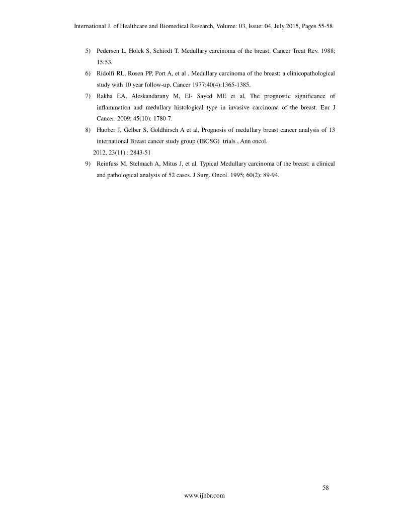
infiltration ( fig3). Tumor at periphery shows
circumscription with a heavy lymphocytic
infiltration(fig.4). Frequent mitotic activity
was noted. Focal areas of necrosis were noted.
All eight axillary lymph nodes were free from
metastasis.Final histopathological impression
according to Bloom and Richardson criteria
was given as medullary carcinoma of breast
grade II. Immunohistochemistry study showed
estrogen and progesterone receptor negative
while Her-2 neu was positive.
Discussion
Medullary breast carcinoma accounts for less
than 5% of all invasive breast cancers1.
Usually patients with medullary carcinoma
present at a relatively younger age than the age
of presentation of other breast cancers
comparable as stated in a study by Rosen et
al2. It was found to constitute 11% of all breast
malignancies among women aged 35 years
and younger. Clinically patients present with
palpable breast lump, gradually increasing in
size and usually at upper outer quadrant.
Bilateral medullary carcinoma is noted in
about 3-18% of cases3. In our case axillary
adenopathy was noted, however it showed
reactive change on microscopic examination
with no evidence of metastasis.
Histopathological examination plays important
role in diagnosis of tumor in medullary
carcinoma. The criteria for diagnosis are4,5,6
A)
syncitial growth pattern cells in more than
75% of the tumor, B) admixed
lymhoplasmacytic infiltrate ,C) microscopic
circumscription ,D) nuclear grade of II or III,
E) absence of glandular differentiation. In our
case all above features were noted. In addition
to various features on histopathology
medullary carcinoma may be associated.
Hemorrhage, cystic degeneration, various type
of metaplasia (most often squamous
metaplasia) and tumor necrosis7. It is stated
that prominent inflammation associated with
medullary carcinoma has better prognosis
compare with that of without prominent
inflammation8. The metastasis is low in cases
of medullary carcinoma and ranges from 19-
46%8. The patient managed with treatment of
modified radical mastectomy along with
radiotherapy and chemotherapy depending on
stage and histopathologic grade. Medullary
carcinoma has better survival rate as compared
to infiltrating duct carcinoma of not otherwise
specified type. The overall 5 year survival rate
is approximately 78% for medullary
carcinoma9. In our case after 38 months of
follow-up patient is free of disease.
Conclusion
Medullary breast carcinoma is a rare subtype
of infiltrating duct carcinoma which has high
grade cytological features but has good
prognosis as compared to invasive ductal
carcinoma. Clinical and histopathological
diagnosis plays important role in better
management of these patient.
International J. of Healthcare and Biomedical Research, Volume: 03, Issue: 04, July 2015, Pages 55-58
57
www.ijhbr.com
Fig.1- Gross specimen showing single, round,
grey white mass with pushing margins in right
MRM specimen.
Fig.2- Photomicrograph showing medullary
carcinoma having large syncitial growth
pattern with moderate pleomorphic,
hyperchromatic or vesicular nuclei and
lymphocytic infiltration .(H&E stain,400x).
Fig.3- Photomicrograph showing medullary
carcinoma having large syncitial growth
pattern with dense lymphoplasmacytic
infiltration(H&E stain,200x).
Fig.4- Photomicrograph showing tumor with
peripheral circumscription and heavy
lymphocytic infiltration(H&E stain,100x).
References :
1) Marcus JN, Watson P, Page DL, et al. Hereditary breast cancer: Pathobiology, prognosis and
BRCA1 and BRCA2 gene linkage. Cancer,1996;697-709.
2) Rosen PP, Lessor ML, KinneDW,et al. Breast carcinoma in woman 35 years of age or
younger: Ann Surg. 1984; 1999(2): 133-42.
3) Young JS, Sterchi MJ, Hopkins M. A synchronous bilateral Medullary carcinoma of the breast.
South Med J 1997;90(4): 423-5.
4) Wargotz ES, Silverberg SG. Medullary carcinoma of the breast: a clinicopathologic study with
appraisal at current diagnostic criteria. Human Pathol. 1988; 19:1340-1346.
International J. of Healthcare and Biomedical Research, Volume: 03, Issue: 04, July 2015, Pages 55-58
56
www.ijhbr.com
5) Pedersen L, Holck S, Schiodt T. Medullary carcinoma of the breast. Cancer Treat Rev. 1988;
15:53.
6) Ridolfi RL, Rosen PP, Port A, et al . Medullary carcinoma of the breast: a clinicopathological
study with 10 year follow-up. Cancer 1977;40(4):1365-1385.
7) Rakha EA, Aleskandarany M, El- Sayed ME et al, The prognostic significance of
inflammation and medullary histological type in invasive carcinoma of the breast. Eur J
Cancer. 2009; 45(10): 1780-7.
8) Huober J, Gelber S, Goldhirsch A et al, Prognosis of medullary breast cancer analysis of 13
international Breast cancer study group (IBCSG) trials , Ann oncol.
2012, 23(11) : 2843-51
9) Reinfuss M, Stelmach A, Mitus J, et al. Typical Medullary carcinoma of the breast: a clinical
and pathological analysis of 52 cases. J Surg. Oncol. 1995; 60(2): 89-94.
58
Recommended