Dr. Abdullah Ahmed Hama
PhD. Molecular and Medical Parasitology
1
Sulaimani University College of Pharmacy
Dr. Abdullah A. Hama Microbiology / parasitology & virology Lec. 5 part 1
Microbiology/ parasitology and virology
Protozoa/ Class: Mastigophora (Flagellates)
Blood Flagellates : Leishmaniasis part1

Outline
Part1 : Leishmaniasis
1.Introduction to Leishmania species
2. Classification
3.Morphology
4.Life cycle.
5. Visceral leishmaniasis
6.Cutaneous Leishmaniasis
7. Mucocutaneous Leishmaniasis
8. Transmission and vector
9. Treatment and Laboratory diagnosis. 2
Part2: Trypanosomiasis • Introduction • Morphology • African Trypanosomiasis • Life cycle • Disease • Laboratory Diagnosis of African
trypanosomiasis • American Trypanosomiasis • Morphology • Life cycle • Disease • Treatment and Laboratory Diagnosis
Dr. Abdullah A. Hama Microbiology / parasitology & virology Lec. 5 part 1

Leishmania species
Introduction:
Leishmania species are mainly parasites of man and other animals, especially
dogs and rodents.
They cause disease known as Leishmaniasis; there are three type of
Leishmaniasis (Visceral Leishmaniasis, Cutaneous Leishmaniasis, and
Mucocutaneous Leishmaniasis). The parasites are unusual in that they live
entirely within the cells of the reticulo-endothelial cells.
3
Dr. Abdullah A. Hama Microbiology / parasitology & virology Lec. 5 part 1

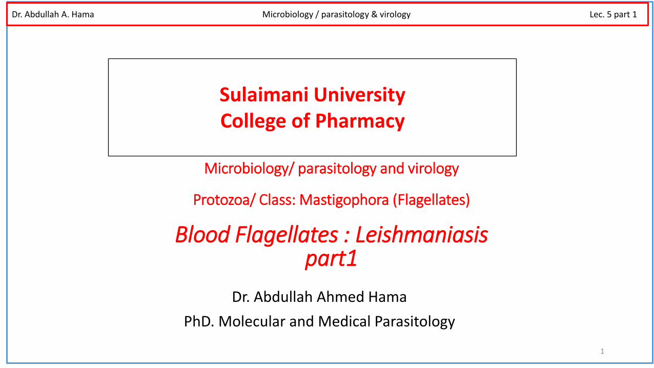
Leishmania stages: flagellated extracellular known as (promastigotes) in the host
Sand fly (Phlebotomus species ) and as obligate intracellular amastigotes (no
flagella) within mononuclear phagocytes of their vertebrate hosts including human.
The various species are not distinguishable morphologically from one another. When
stained with Romanowsky stains such as Giemsa, amastigotes appear as round or
oval bodies ranging from 2 – 3 μm in diameter with a well defined nucleus and
kinetoplast, a rod shaped specialized mitochondrial structure that contains extra-
nuclear DNA.
The flagellated promastigote form is spindle shaped, measuring 10 - 20μm in length, nucleus and kinetoplast are clearly visible.
4
Morphology
Dr. Abdullah A. Hama Medical Parasitology Lec.9 Text book: 978-1-4354-4816-2 Dr. Abdullah A. Hama Microbiology / parasitology & virology Lec. 5 part 1

5
Amastigote of Leishmania sp. in a macrophage of a vertebrate host.
Leishmania promastigote. This stage of the parasite are seen in the vector, sandfly.
Dr. Abdullah A. Hama Medical Parasitology Lec.9 Text book: 978-1-4354-4816-2
Dr. Abdullah A. Hama Microbiology / parasitology & virology Lec. 5 part 1
6
Dr. Abdullah A. Hama Medical Parasitology Lec.9 Text book: 978-1-4354-4816-2
Dr. Abdullah A. Hama Microbiology / parasitology & virology Lec. 5 part 1
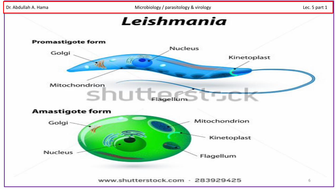
7 Life cycle of Leishmania sp.
Dr. Abdullah A. Hama Medical Parasitology Lec.9 Text book: 978-1-4354-4816-2
Dr. Abdullah A. Hama Microbiology / parasitology & virology Lec. 5 part 1
1. Visceral Leishmaniasis (Leishmania donovani )
8
Human visceral leishmaniasis (VL), sometimes known as Kala-azar, (black sickness) or
dumdum fever is caused by Leishmania donovani complex; L. donovani and L. donovani
infantum in the Old World (Africa, Europe, and Asia) and L. donovani chagasi in the New
World (America). The spleen and liver become markedly enlarged, and lymphadenopathy
also occurs. Increased production of globulin results in hyperglobulinemia, and reversal of
the albumin-to-globulin ratio.
Disease:
Dr. Abdullah A. Hama Medical Parasitology Lec.9 Text book: 978-1-4354-4816-2
Symptoms begin with intermittent fever, weakness, and diarrhea; chills and
sweating that may resemble malaria symptoms.
Dr. Abdullah A. Hama Microbiology / parasitology & virology Lec. 5 part 1

3- Serodiagnosis: VL produces large amounts of specific IgG which can be used for diagnosis. Currently the most
used sero-diagnostic tests are Indirect-immuno Fluorescent Antibody Test (IFAT), Enzyme Linked Immunosorbent
Assay (ELISA) and Direct Agglutination Test (DAT).
Laboratory diagnosis 1- Microscopy: Parasites may be found in a splenic aspirate, liver biopsy or bone marrow biopsy.
These techniques, especially splenic aspirate and liver biopsy, can be hazardous and require previous
expertise.
*Stain the air drying smear with Giemsa 1 in 10 in buffered distilled water pH 6.8 for 30 minutes (or
)stain Romanowsky( is a version of a stain rapid Field’s use the
2. Culture: The aspirates can be cultured in Novy-Nicolle-MacNeal (NNN) or Schneider's Drosophila
Medium (SDM). In culture the amastigote stage converts to the promastigote stage. However, this is
not a rapid technique, as the parasites may take from 10-21 days to grow.
9
Dr. Abdullah A. Hama Medical Parasitology Lec.9 Text book: 978-1-4354-4816-2 Dr. Abdullah A. Hama Microbiology / parasitology & virology Lec. 5 part 1
Cutaneous Leishmaniasis (Oriental sore) L. tropica
These are parasites of the skin found in endothelial cells of the
capillaries of the infected site, nearby lymph nodes, within large
mononuclear cells.
Cutaneous leishmaniasis is present in many parts of Asia, Africa, Mediterranean
Europe and the southern region of the former Soviet Union. The vector for the
world cutaneous leishmaniasis is the Phlebotomus sand fly.
Dr. Abdullah A. Hama Medical Parasitology Lec.9 Text book: 978-1-4354-4816-2
Dr. Abdullah A. Hama Microbiology / parasitology & virology Lec. 5 part 1
Disease
11
• L. tropica minor - dry or urban cutaneous leishmaniasis
• L. tropica major - wet or rural cutaneous leishmaniasis • L. ethiopica - cutaneous leishmaniasis
The red papule, appears at the site of the fly’s bite.This lesion becomes
irritated, with intense itching, and begins to enlarge and ulcerate.
Secondary bacterial infection may complicate the disease. In the case of
the Ethiopian cutaneous leishmaniasis, they may also give rise to diffuse
cutaneous leishmaniasis (DCL) in patients who produce little or no cell
mediated immunity against the parasite. This leads to the formation of
disfiguring nodules over the surface of the body.
Dr. Abdullah A. Hama Medical Parasitology Lec.9 Text book: 978-1-4354-4816-2 Dr. Abdullah A. Hama Microbiology / parasitology & virology Lec. 5 part 1

1- Skin smear:
The margin of the lesion contains amastigotes whereas the center
contains debris and dead skin material.
2. Polymerase chain reaction:
Gene amplification techniques are powerful and sensitive
methods and are useful in diagnosis of Cutaneous Leishmaniasis
particularly when organisms cannot be detected microscopically.
Laboratory Diagnosis of cutaneous Leishmaniasis
Dr. Abdullah A. Hama Medical Parasitology Lec.9 Text book: 978-1-4354-4816-2 Dr. Abdullah A. Hama Microbiology / parasitology & virology Lec. 5 part 1

Mucocutaneous Leishmaniasis:Is caused by the L. braziliensis complex and is
found in Brazil, Eastern Peru, Bolivia, Paraguay, Ecuador, Columbia and Venezuela.
Mucocutaneous Leishmaniasis or espundia initially develops like cutaneous
leishmaniasis but develops into lesions in the mucocutaneous junction of the
pharynx resulting in the break down of the palate of the mouth and nose or more
rarely the genitalia or anus. This occurs from a few weeks to several years,these
lesions result in disfiguring deformities of the nose and mouth.
Mucocutaneous Leishmaniasis
Dr. Abdullah A. Hama Medical Parasitology Lec.9 Text book: 978-1-4354-4816-2 Dr. Abdullah A. Hama Microbiology / parasitology & virology Lec. 5 part 1
Leishmaniasis
Several species of Leishmania are pathogenic for man:
L. donovani causes visceral leishmaniasis (Kala-azar, black disease, dumdum fever)
L. tropica (L. t. major, L. t. minor and L. ethiopica) cause cutaneous leishmaniasis (oriental sore, Delhi ulcer,
Aleppo, Delhi or Baghdad boil)
L. braziliensis and L. mexicana are etiologic agents of mucocutaneous leishmaniasis (espundia, Uta, chiclero
ulcer).
Vectors:
The vectors (sand fly ) have two scientific name Lutzomyia- in new world Phlebotomus- in old
world
Habitat: Macrophages and Reticuloendothelial cells of vertebrates.
Summary: Dr. Abdullah A. Hama Medical Parasitology Lec.9 Text book: 978-1-4354-4816-2 Dr. Abdullah A. Hama Microbiology / parasitology & virology Lec. 5 part 1

Dr. Abdullah Ahmed Hama
PhD. Molecular and Medical Parasitology
15
Sulaimani University College of Pharmacy
Dr. Abdullah A. Hama Microbiology / parasitology & virology Lec. 5 part 2
Microbiology/ parasitology and virology
Protozoa/ Class: Mastigophora (Flagellates)
Blood Flagellates : Trypanosomiasis Part2
Introduction Trypanosomes are haemo-flagellates and three species of the genus
Trypanosoma are responsible for humans infection ( sleeping sickness or
trypanosomiasis).
Trypanosomes live in the blood of the majority of vertebrate animals in the endemic area.
The life cycle involves intermediate host (Tsetse fly) which is known as biological vector
Many species of trypanosomes can live in harmony with their hosts producing no
pathogenic effect, but the best known species are those that are pathogenic to their
definitive hosts.
Dr. Abdullah A. Hama Medical Parasitology Lec. 10 Text book: 978-1-4354-4816-2 Dr. Abdullah A. Hama Microbiology / parasitology & virology Lec. 5 part 2

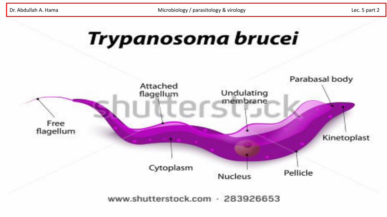
Morphology
The parasite is an elongated cell with single nucleus which usually lies near the
center of the cell. Each cell bears a single flagellum which appears to arise from a
small granule –called kinetoplast.
The kinetoplast: is a specialized part of the mitochondria and contains DNA. The
length and position of the trypanosome’s flagellum is variable. In trypanosomes
from the blood of a host the flagellum originates near the posterior end of the cell
and passes forward over the cell surface, and forms undulating membrane.
Dr. Abdullah A. Hama Microbiology / parasitology & virology Lec. 5 part 2
Dr. Abdullah A. Hama Microbiology / parasitology & virology Lec. 5 part 2

Life cycle of African Trypanosomiasis (salivaria phase) The vector of African Trypanosomiasis is the Tsetse fly (Glossina spp.) which cause Trypanosoma brucei
gambiense and Trypanosoma brucei rhodesiense. Metacyclic (infective) trypomastigotes are inoculated
through the skin when a tsetse fly takes a blood meal. The parasites develop into long slender
trypomastigotes which multiply at the site of inoculation where ulceration occurs. The trypanosomes
continue to develop and then may invade the lymphatic tissues, the heart, various organs and in later
stages, the central nervous system.
Trypomastigotes are taken up by the tsetse fly (male and female) during a blood meal. The
parasites develop in the midgut of the fly where they multiply. 2-3 weeks later the trypomastigotes
move to the salivary glands transforming from epimastigotes into metacyclic (infective)
trypomastigotes. The tsetse fly remains infective for life about three months.
Dr. Abdullah A. Hama Microbiology / parasitology & virology Lec. 5 part 2

Dr. Abdullah A. Hama Microbiology / parasitology & virology Lec. 5 part 2

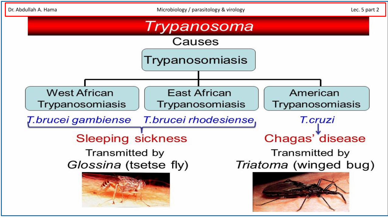
Disease Sleeping sickness, African trypanosomiasis, is a fatal parasitic disease caused by two type of Trypanosoma
brucei, both types are transmitted by tsetse fly. Trypanosoma brucei rhodesiense causes East African
trypanosomiasis. 1000 new cases of T. b. rhodesiense infections are reported by World Health Organization
annually. Trypanosoma brucei gambiense causes West African trypanosomiasis (also known as Gambian sleeping
sickness).
The early stages of African trypanosomiasis may be asymptomatic and there is a low grade parasitiaemia. This
period may remain for several weeks to several months. The disease may terminate untreated at this stage or
may be invade the lymph glands. Invasion of the lymph glands is usually accompanied by a high irregular fever
with shivering, sweating and an increased pulse rate. The lymph glands near the bite often become swollen
(chancre), in T. b. gambiense the glands at the back of the neck and T. b. rhodesiense and usually the glands
under the jaw are affected (Winter-bottom's sign).
Dr. Abdullah A. Hama Microbiology / parasitology & virology Lec. 5 part 2
Dr. Abdullah A. Hama Microbiology / parasitology & virology Lec. 5 part 2
Laboratory Diagnosis of African trypanosomiasis
1. Microscope Examination of blood :
a. Thick and Thin Blood Films Thick and thin blood films are made and stained with Fields stain and examined under microscope.
b. Triple Centrifugation Technique and Parasite concentration techniques
(detail in the next slide)
2. Microscope Examination of liver or spleen aspirate
The aspirate can be examined microscopically by making a wet preparation, or if there is not much material, it can be allowed to dry on a slide and then stained with either rapid Field’s stain or with Giemsa and examined microscopically.
3. Examination of the CSF for the parasite:
In the late stages of African trypanosomiasis, trypanosomes may be found in the CSF together with IgM
Dr. Abdullah A. Hama Microbiology / parasitology & virology Lec. 5 part 2

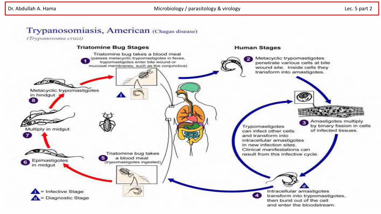
American Trypanosomiasis (stercoraria phase)
Trypanosoma cruzi occurs throughout South and Central America, especially in Brazil, Argentina and
Mexico causing the disease known as Chagas’ disease. It is estimated that over 24 million people are
infected with this species. It is a zoonotic parasite with over 150 species of wild animals known to
harbor the parasites, for example opossums, dogs, rates, pigs and cats.
The parasite can be transmed to human and other animals by brightly colored bugs
belonging to the Reduviidae family (Reduviid bug), subfamily Triatominae. All stages of
these bugs are known to become infected and also called assassin bug. The bugs live in
the crack of the walls and vegetal roofs of the poorly maintained houses, coming out at
night to feed on the exposed parts of the host’s body.
Dr. Abdullah A. Hama Microbiology / parasitology & virology Lec. 5 part 2

Multiplication of Trypanosoma cruzi in man only occurs in the amastigote phase, which
grows in a variety of tissue cells especially muscle. In vitro infected fibroblast showing a
large number of intracellular amastigotes. (Giemsa stain)
Intracellular amastigotes. (Giemsa stain) Mastigote phase
Dr. Abdullah A. Hama Microbiology / parasitology & virology Lec. 5 part 2

Dr. Abdullah A. Hama Microbiology / parasitology & virology Lec. 5 part 2

Disease
Multiplication of T. cruzi at the site of infection can produce inflamation and swelling
(chagoma) which persists for many weeks. Trypomastigotes or amastigotes may be seen in
the aspirate of the chagoma. Regional lymph nodes may become infected which frequently
involve one side of the face. Unilateral edema of the upper and lower eyelid may occur along
with conjunctivitis. This is known as Romanas' sign.
Many people infected with T. cruzi and they may be remain asymptomatic and free from
Chagas’ disease. The most severe form of the disease is most commonly seen in children
younger than five years of age.
Chronic manifestations include signs of cardiac muscle damage with a weak and irregular
heartbeat, edema, heart enlargement leading to heart failure. Dilation of the digestive tract
resulting in megaesophagus and megacolon may also occur. About 10% of persons infected
with T. cruzi the disease may be developed to chronic Chagas cardiomyopathy.
Dr. Abdullah A. Hama Microbiology / parasitology & virology Lec. 5 part 2

Life cycle
The amastigotes develop into trypomastigotes which are released into the
blood when the cell ruptures. No multiplication of the parasite takes place in
the blood in its trypomastigote stage. The trypomastigotes reach tissue cells
especially heart muscle, nerves, skeletal muscle and smooth muscle of the
gastrointestinal system by way of the blood and lymphatic system.
Metacyclic trypomastigotes are deposited in feces on the skin as the triatomine bug
(reduviid bug) feeds. The bug usually bites round the edges of the mouth and eyes.
The trypomastigotes are either rubbed into the skin by scratching the irritated area or
penetrate the conjunctiva or membranes of the nose and mouth. Trypomastigotes
become amastigotes in localized reticuloendothelial cells and multiply.
Dr. Abdullah A. Hama Microbiology / parasitology & virology Lec. 5 part 2
Dr. Abdullah A. Hama Microbiology / parasitology & virology Lec. 5 part 2

Laboratory Diagnosis of American Trypanosomiasis
• Microscopical Examination of blood.
• Xenodiagnosis
• Blood culture
• Serology Xenodiagnosis
Xenodiagnosis is useful in chronic and sub acute (low parasitaemia) disease.
Sterile bugs are fed on patients by attaching a black bag containing the bugs to
the arm of the patient and allowing them to feed for 30 minutes. Twenty five to
thirty days later the bugs are dissected and the contents of the hindgut and
rectum are examined microscopically for the presence of trypanosomes.
Dr. Abdullah A. Hama Microbiology / parasitology & virology Lec. 5 part 2

31
TREATMENT VISCERAL LEISHMANIASIS • Liposomal amphotericin-B (AmBisome): is the drug of choice 3mg/kg per day on days
1-5, day 14 and day 21 • Pentostam: is an alternative therapy 28 days of therapy is required
TREATMENT CUTANEOUS AND MUCOCUTANEOUS Antimony (Pentostam , Sodium stibogluconate): is the drug of choice will be given for 20 days of intravenous therapy . Fluconazole may decrease healing time in L. major infection Six weeks of therapy is needed.
Treatment of Sleeping Sickness and Chagas Disease In early stage of the disease: Pentamidine OR SuraminIn late stages of the disease:Tryparsamide For both early and late stages of the disease:Eflornithine (DFMO) OrnidylNifurtimoxinhibits intracellular development. Drug of choice in acute and early chronicORPrimaquinedestroys Trypanosoma in blood
Dr. Abdullah A. Hama Microbiology / parasitology & virology Lec. 5 part 2
Recommended