Loyola University ChicagoLoyola eCommons
Dissertations Theses and Dissertations
1975
Mechanisms and Regulation of MacrophageGlycogen MetabolismPaul GudewiczLoyola University Chicago
This Dissertation is brought to you for free and open access by the Theses and Dissertations at Loyola eCommons. It has been accepted for inclusion inDissertations by an authorized administrator of Loyola eCommons. For more information, please contact [email protected].
This work is licensed under a Creative Commons Attribution-Noncommercial-No Derivative Works 3.0 License.Copyright © 1975 Paul Gudewicz
Recommended CitationGudewicz, Paul, "Mechanisms and Regulation of Macrophage Glycogen Metabolism" (1975). Dissertations. Paper 1579.http://ecommons.luc.edu/luc_diss/1579

MECHANISMS AND REGULATION OF MACROPHAGE GLYCOGEN METABOLISM
by
Paul W. Gudewicz
A Dissertation Submitted to the Faculty of the Graduate School
of Loyola University in Partial Fulfillment
of the Requirements for the Degree of
Doctor of Philosophy
February
1975
DB~.:\RY LOYOT A Ul'TTVFP::,ny ~.lf':.lJiCAL CFirr;:;:.:

\
ACKNOWLEDGEMENTS
I am indebted and grateful to my' advisor, Dr. James P. Filkins,
who fostered and.guided my scientific development by sharing of his
time, wisdom and understanding throughout the course of my graduate
studies. Furthermore, I want to express my sincere thanks to Dr.
Walter C. Randall and the faculty of the Department of Physiology for
their untiring dedication and support during my graduate training.
I would also like to extend my appreciation to Mrs. Georgine
Hoppe and Chio Tan for their expert technical assistance during my
research project.
Furthermore, I want to extend a special thanks to Dr. Phillip
Hawley, a friend and physiologist, who first introduced me to physiology
and nurtured my scientific curiosity by his example and advice.
Finally, I am indebted to Mrs. Mary Berchos for her excellent
assistance in the preparation of this dissertation.
ii

Chapter
I.
II.
III.
TABLE OF CONTENTS
Page
INTRODUCTION ..................................... . 1
HISTClRICAL REVIEW .........•....................... 4
A. The Role of the Macrophage in Inflammation.... 4 1. Historical Development and Knowledge of
the Macrophage System. . . . . . . . . . . . . . . . . . . . . 4 2. Origin of the Inflammatory Macrophage..... 5 3. Morphology and Maturation of the
Inflammatory Macrophage................... 6 4. Migration Behavior of the Macrophage. . . . . . 8 5 .. Endocytosis and Digestion by the' Macrophage 10
a. Phagocytosis.......................... 10 b. Pinocytosis. . . . . . . . . . . . . . . . . . . . . . . . . . . 12
B. Macrophage Metabolism......................... 14 1. Mononuclear vs. Polymorphonuclear Leukocyte 14 2. Macrophage Carbohydrate Metabol~sm and
Energy Supply............................. 15 3. Macrophage Lipid Metabolism............... 17 4. Macrophage Protein Synthesis.............. 18 5. Phagocytosis Associated Metabolism in the
Polymorphonuclear Leukocyte and Macrophage 20 a. Leukocytes ........................... ~ 20 b. Macrophages. . . . . . . . . . . . . . . . . . . . . . . . . . . 21
6. Glycogen Metabolism in the Polymorpho-nuclear Leukocyte. . . • . . . . . . . . . . . . . . . . . . . . . 24
7. Glycogen Metabolism in Inflammatory Cells. 28 C. Statement of the Problem...................... 30
EXPERIMENTAL METHODS ............................. . A. Experimental Animals ......................... . B. Preparation of Inflammatory Cell Populations ..
1. Exudate Production ....................... . 2. Harvesting Procedures .................... . 3. Cell Counting, Differential and Protein
Determinations ........................... . C. Glycogen Determinations in Inflammatory
Leukocytes and Macrophages ...•................ 1. Chemical Determination of Glycogen .•...•.• 2. Incorporation of 14c-U-Glucose into
Macrophage Glycogen ..•....•.....•.•.......
iii
32 32 32 32 32
33
33 33
34

TABLE OF CONTENTS (continued)
Chapter
3. 14c-Glycogen Isolation from Rat Liver ..... 4. Uptake of 14C-Glycogen by Inflammatory
Macrophages .........................•..... D. Glycogen Regulatory Enzyme Determinations in
Inflammatory Macrophages .................••..• 1. Macrophage Phosphorylase Activity ........ . 2. Macrophage ~-Glucosidase Activity ...•..•.. 3. Macrophage Glycogen Synthetase Activity •.. 4. Macrophage Glucose-6-Phosphatase Activity.
E. Glucogenesis in Inflammatory Macrophages and Polymorphonuclear Leukocytes ......•........••.
F. Statistics and Data Analysis .....•....•.....•.
IV. RESULTS • ..•..•.•.....•..•.••.•.•......••.••...•••• A. Factors Regulating the Glycogen Content of
Inflammatory Macrophages .•........... , ....... . 1. Inflammatory Cellular Yields from the
Rat Peritoneum ...............•.......••... 2. Glycogen Content of Inflammatory PMNL
and Macrophages .................•......... 3. Effect of Anaerobiosis and Phagocytosis
on Glycogen Content in Inflammatory Macrophages ......•...•....................
4. ~ffect of Glucose and Exogenous Glycogen on Inflammatory Macrophage Glycogen Content
5. Effect of Glucose or Glycogen on the Glycogen Content in Glycogen-Depleted Inflammatory Macrophages ................. .
6. Incorporation of 14c-U-Glucose into Inflammatory Macrophage Glycogen ......... .
7. The Influence of ·a· Pinocytic Activator and Insulin in Stimulating 14C-U-Glucose Incorporation into Macrophage Glycogen ....•.
8. 14c-Glycogen Uptake by Inflammatory
9. ~t~~~i~~~=:~·u~t~k~.i~·Gi;~~~~~~n~~i~t~d·· Inflammatory Macrophages ...........••.•..•
B. Enzymes Regulating Macrophage Glycogen Metabolism ................................... . 1. Macrophage Phosphorylase Activity •.••••...
a. Effect of Glycogenolysis on Macrophage Total Phosphorylase Activity ...••.....
b. Effect of Exogenous Glycogen on Macrophage Total Phosphorylase Activity ... .......................... .
iv
Page
35
36
36 36 38 39 40
41 41
42 • 42
42
42
44
47
49
49
51
53
56
58 58
58
58

TABLE OF CONTENTS (continued)
Chapter
v.
VI.
VII.
\
Page
c. The Active Form of Macrophage Phosphorylase......................... 61
d. Effect of Mg++-ATP on Macrophage Phosphorylase Activity................ 61
2. Macrophage Glucosidase Activity........... 63 a. o<.-1,4 ando(-1,6 Giucosidase Activity in
Inflammatory Macrophage............... 63 b. Effect of pH on Inflammatory Macrophage
~-1,4 Glucosidase Activity............ 66 3. Macrophage Glycogen Synthetase Activity... 66
a. I and D Forms of Macrophage Glycogen Synthetase............................ 66
b. Effect of Glycogen Depletion on Macrophage Glycogen Synthetase Activity................. . . . . . . . . . . . . . 69
4. Macrophage Glucose-6-Phosphatase Activity. 69 C. Glucogenesis in Inflammatory Macrophages...... 71
1. Glucose Production in Normal and Inflammatory Exudate Cell Populations..... 71
2. Glucogenesis by Inflammatory Macrophage Cell and Sonicate Preparations............ 74
3. Effect of Acid pH Conditions on Macrophage Glucogenesis. . . . . . . . . . . . . . . . . . . . . . . . . . . . . . 11
4. Substrate and Glycoside Bond Specificity of Macrophage Glucogenesis................ 11
5. Effect of Cyclic AMP on Macrophage Glucogenesis... . . . . . . . . . . . . . . . . . • . . . . . . . . . 19
DISCUSSION ....................................... . 83
SUMMARY ..................•............. ; .•........ 99
SELECTED BIBLIOGRAPHY ............................ . 103
v

LIST OF TABLES
Table
GLYCOGEN CONTENT AND DEPLETION IN INFLAMMATORY PMNL AND MACROPHAGES ......•.......................
2. EFFECT OF ANAEROBIOSIS AND PHAGOCYTOSIS ON THE GLYCOGEN CONTENT IN INFLAMMATORY MACROPHAGES ..... .
3. EFFECT OF GLUCOSE AND EXOGENOUS GLYCOGEN ON INFLAMMATORY MACROPHAGE GLYCOGEN CONTENT ......... .
4. INCORPORATION OF l4c-U-GLUCOSE INTO MACROPHAGE GLYCOGEN:EFFECT OF INSULIN AND ALBUMIN PINOCYTIC
Page
43
46
48
ACTIVATION .....•........................ ·.. . . . . . . . . 54
5. l4c-GLYCOGEN UPTAKE BY INFLAMMATORY MACROPHAGES: EFFECT OF GLYCOGEN DEPLETION ON l4c-GLYCOGEN UPTAKE............................................ 57
6. EFFECT OF GLYCOGENOLYSIS ON MACROPHAGE TOTAL
7.
8.
9.
PHOSPHORYLASE ACTIVITY............................ 59
EFFECT OF EXOGENOUS GLYCOGEN ON TOTAL MACROPHAGE PHOSPHORYLASE ACTIVITY ........................... .
ACTIVE AND TOTAL PHOSPHORYLASE ACTIVITY IN INFLAMMATORY MACROPHAGES ...................•......
o< -GLUCOSIDASE ACTIVITY IN INFLAMMATORY MACROPHAGES ...................................... .
60
62
10. INFLAMMATORY MACROPHAGE GLYCOGEN SYNTHETASE ACTIVITY. . • . . . . . . . . . . . . . • . . . . . . . . . . . . . . . . . . . . . . . . . 68
ll. EFFECT OF GLYCOGEN DEPLETION ON MACROPHAGE GLYCOGEN SYNTHETASE ACTIVITY .............................. . 70
12. GLUCOSE-6-PHOSPHATASE ACTIVITY IN INFLAMMATORY MACROPHAGES ...................................... . 72
13. GLUCOSE PRODUCTION IN NORMAL AND INFLAMMATORY EXUDA'TE CELL POPULATIONS ...•..... , ............... . 73
vi

LIST OF TABLES (continued)
Table Page
14. GLUCOSE PRODUCTION BY INFLAMMATORY MACROPHAGE CELL AND SONICATE PREPARATIONS ................... . 76
15. THE EFFECT OF pH ON GLUCOSE PRODUCTION BY MACROPHAGE SONICATES ....... .'· .......•••••••••...... 78
16. SUBSTRATE AND GLYCOSIDE BOND SPECIFICITY OF MACROPHAGE GLUCOGENESIS .......................... . 80
17. EFFECT OF CYCLIC AMP ON MACROPHAGE AND LEUKOCYTE GLUCOGENESIS ........................... . 81
vii
LIST OF FIGURES
Figure Page
l. EFFECT OF IN VITRO INCUBATION IN GFH ON MACROPHAGE GLYCOGEN CONTENT ....................... 45
2. EFFECT OF GLUCOSE (10 mM) OR GLYCOGEN (10 mg/m1) ON GLYCOGEN CONTENT IN GLYCOGEN-DEPLETED MACROPHAGES ....................................... 50
3. INCORPORATION OF 14c-U-GLUCOSE INTO INFLAMMATORY MACROPHAGE GLYCOGEN.EFFECT OF GLYCOGEN DEPLETION ON 14c-GLUCOSE INCORPORATION ....•................. 52
4. 14c-GLYCOGEN UPTAKE BY INFLAMMATORY MACROPHAGES ... 55
5. EFFECT OF Mg-++-ATP ON INFLAJvlMATORY MACROPHAGE PHOSPHORYLASE ACTIVITY •........................... 64
6. EFFECT OF pH ON MACROPHAGE ~-1,4 GLUCOSIDASE ACTIVITY .......................................... 67
7. GLUCOGENESIS BY INFLAMMATORY MACROPHAGES ••........ 75
viii

CHAPTER I
INTRODUCTION
The inflammatory response is a complex interdependent seQuence
of local vascular, cellular and tissue events that launch reparative
processes at the host's damaged tissue. The acute inflammatory re
sponse is heralded by vascular dilatation and marked alterations in
vascular permeability leading to the exudation of protein and cellular
elements at the inflammatory site. The macrophage~ i.e. the mono
nuclear phagocyte, and polymorphonuclear leukocyte (PMNL) constitute
the two major phagocytic cell populations that accumulate at the
site of tissue injury .. The cellular development of the local in
flammatory reaction classically features an early infiltration by the
PMNL which is subseQuently replaced by the inflammatory macrophage as
the principal phagocytic mediator of the host's defense reactions
(105 ). Macrophages and PMNL at the inflammatory locus perform the
vital functions of clearance, degradation and detoxification of in
vading foreign organisms and cellular debris by employing endocytic
and intracellular digestive mechanisms. However, the homeostatic con
trol mechanisms and cellular interactions influencing the metabolic
demands among inflammatory cell populations are poorly understood.
The maintenance of PMNL and macrophage function is ultimately con
tingent upon t~e continual provision of substrate supply and cellular
energy. The relative importance of glycolytic and oxidative pathways
l

as energy sources varies markedly among the different macrophage and
PMNL populations ( 85 ). Early in vitro studies centering on macro
phage metabolism focused attention on metabolic alterations subsequent
to phagocytosis (66). Glycolysis and respiration were transiently
stimulated during the phagocytic event in macrophages, however certain
macrophage populations are capable of deriving their energy for pha
gocytosis exclusively from glycolysis under anaerobic conditions either
in vitro or at the inflammatory site where low oxygen tensions prevail.
Macrophage endocytic capacities include both phagocytosis and
pinocytosis - terms utilized to describe the cellular ingestion of
particulate matter and soluble components of their external environ
ment, respectively. In addition to its role in microbial host defense,
endocytosis affords a mechanism whereby the inflammatory macrophage
enriches its fuel supply by ingestion and degradation of macromolecules
found at the inflammatory site. Glycogen, an important carbohydrate
fuel reserve, is one such macromolecule which is found in abundance in
the PMNL (109). Histological studies have provided evidence that the
glycogen-laden PMNL shedscytoplasmic fragments during the early course
of the inflammatory response while the macrophage rapidly engulfs this
potential source of fuel molecules (141).
Although glycogen content and the mechanisms responsible for
glycogen synthesis and degradation have been extensively investigated
in various leukocyte populations, glycogen content and its regulation
within macrophages have not received similar quantitative attention.
Thus the intents of this study are to evaluate in vitro the physio
logical responses of macrophages to exogenously added glycogen and to
2

assess glycogen metabolism and those control mechanisms responsible for
glycogen synthesis and utilization within the macrophage derived from
the inflammatory locus.
3

CHAPTER II
HISTORICAL REVIEW
A. The Role of the Macrophage in Inflammation
\
1. His·torical development and knowledge of the macrophage
system
Although it was known that different forms of leukocytes oc
curred in blood was in evidence as early as 1865.(108), the early
functional history of the mononuclear phagocyte pivots around the ex
haustive studies of Metchnikoff (82). In a series of comparative
studies concerned with the cellular response following tissue injury,
Metchnikoff investigated the phagocytic activity of inflammatory cells
derived from both invertebrate and vertebrate species. He demonstrated
the ultimate dominance of the phagocyte in the defense reactions of
the host to inflammatory stimuli. Furthermore, he also was the first
to distinguish between the macrophage, i.e., big eater, and the micro
phage, the smaller polymorphonuclear leukocyte. In the following dec
ade, Goldman (49) and others employed vital staining techniques and
illustrated the ubiquitous occurrence and organ localization of mono
nuclear phagocytes. As an outcome of these early histological studies
Aschoff (3) coined the term reticuloendothelial system (RES) to des
cribe the concept of a widely distributed collection of mononuclear
phagocytes which are united by similar morphological and functional
properties and potentialities. The RES includes both the fixed
macrophage populations localized primarily in the blood
4

sinuses of the liver, lung, spleen, and bone marrow an~ the mobile mac
rophage pool found in the serous cavities of the organism. Thus fol
lowing Aschoff's formulation of the RES concept as a phagocytic
functional system and metabolic apparatus involved in host defense
mechanisms, all subsequent investigations have resulted in determining
the potential and multiple roles of the mononuclear phagocyte in phy
siological and pathophysiological settings.
2. Origin of the inflammatory macrophage
The origin and identity of the precursor cell which gives rise
to the macrophage of the inflammatory exudate have been studied and
debated for more than thirty years. Ebert and Florey (39) using rabbit
ear chambers observed that monocytes infiltrated from the peripheral
blood into the inflammatory site and differentiated into macrophages.
However, it was argued by Rebuck and his colleagues (96, 98) that
inflammatory macrophages were derived primarily from lymphocytes.
Rebuck utilized the "skin window" technique in which glass coverslips
were applied to areas of abraded human skin and then periodically
removed and examined microscopically. It was concluded from these
studies that small lymphocytes evolved through a series of intermediate
cell types into inflammatory macrophages.
Recent in vivo and in vitro labelling studies have supported
the conclusions that under physiological conditions and during the
acute inflammatory response, the inflammatory macrophage is principally
derived from peripheral blood monocytes. That mononuclear phagocytes
ultimately originated from bone marrow precursors was first demonstrated
by Balner (5) in radiation chimeras give~ allogenic bone marrow cells.
5

Both transfusion studies with 3H-thymidine labelled bone marrow cells
into syngenic rats (129) and labelling studies in X-irradiated animals
with shielding of the bone marrow (126) have provided direct evidence
not only for the existence of monocyte precursors in the bone marrow
but also that the peritoneal macrophage population is derived from
blood monocytes.
Induction of an inflammatory response in the peritoneal cavity
of mice resulted in a two- to threefold increase in the number of peri-
toneal macrophages by 72 hours following the introduction of an ir-
ritant (19). In vitro incubation of these periton~al macrophages in
the presence of 3H-thymidine indicated a labelling index comparable to
peritoneal macrophages from unstimulated animals, suggesting that the
increase in macrophage population as a result of the inflammatory
stimuli is not the result of increased mitotic activity (126). How-
ever, when peripheral blood monocytes were labelled in vivo with re-
peated injections of 3H-thymidine and a peritoneal inflammatory re-
sponse was induced, these experiments resulted in a 45 to 65% increase
in the labelling of inflammatory peritoneal macrophages. Thus these
experiments add overwhelming evidence in favor of the blood monocyte
origin of the inflammatory macrophage.
3. Morphology and maturation of the inflammatory macrophage
Maximow and Bloom (80) provided the first detailed account
of macrophage morphology and distinguished macrophages from the blood
lymphocyte by their "active amoeboid protoplasm" and inclusion bodies.
Macrophages are large (20-40~ cells having an eccentrically located
horseshoe-shaped nucleus with an azurophilic granular cytoplasm
6

7
containing numerous dense bodies and mitochondria (17). An extensive
Golgi apparatus occupies the perinuclear area with lysosomal-like
granules and mitochondria located at the periphery of the Golgi zone.
At the light microscope level, the macrophage cell membrane is charac
terized by a ruffled appearance. When macrophages are allowed to ad
here to a glas·s surface, the cell membrane becomes dramatically well
spread out and intensely ruffled (46).
Recently, the ultrastructure of the peritoneal macrophage has
been under intense investigation (13, 22, 37) and has revealed a com
plex cytoplasmic structure and many prominent cell membrane processes.
The macrophage membrane surface is studded with spherical indentations
O.~in diameter suggesting ongoing active pinocytic vacuole formation.
A granular endoplasmic reticulum and Golgi apparatus are well developed
and have been shown to be the site of formation of the primary lysosome.
In the mature macrophage many smooth surfaced vesicles (50-100 nm in
diameter), the primary lysosomes, are distributed throughout the Golgi
zone. The most prominent cytoplasmic inclusions are the large number
of electron-dense granules demonstrated to be lysosomal in character
by their positive acid phosphatase reaction (14). Such heterogeneous
granules are seen to contain varying amounts of lipid or dense staining
material and are clearly secondary lysosomes. Labelled marker par
ticles have been demonstrated to be incorporated within these lysosomal
structures by combination with phagocytic or pinocytic vacuoles formed
at the membrane surface (16).
Peritoneal macrophages in culture have been extensively used
to study the maturation process of monocyte to macrophage differentiation.

8
Lewis (77) first observed, by culturing mixed blood cells, that blood
monocytes altered their morphological appearance and matured into large
macrophages, epithelioid and giant cells. More recent studies have con
cluded that the induction of macrophage maturation by in vitro culturing
techniques or in vivo, by lipopolysaccharide stimulated animals, resulted
in similar morphological and biochemical alterations (19, 21). It was
demonstrated by these authors that mouse peritoneal cells underwent a
temporal sequence of morphological alterations including an increase in
the number of phase-dense, acid phosphatase-positive granules and mito
chondria. In addition, the size of the Golgi apparatus was markedly
increased as well as the number of large lipid inclusions that were
associated with the rough endoplasmic reticulum. Thus it is apparent
that the arrival of the blood monocyte to the inflammatory site alters
the macrophage into a large cell with more digestive capacity and
phagocytic potential resulting in a dramatic stimulation in the
functional properties of the inflammatory macrophage.
4. Migration behavior of the macrophage
Subsequent to all injury, alterations occur in the immediate vas
culature and local tissue milieu leading to the emigration of phagocytic
cells and the extravasation of plasma constituents. Although the PMNL
is the preponderant cell type mobilized during the initial stage of the
inflammatory reaction, within 24 hours, macrophages infiltrate the in
flamed area and develop into the dominant phagocytic cell (105). The
mechanisms underlying this temporal sequence and the eventual lo
calization of the macrophage are poorly understood. Difference in PMNL
and macrophage migration rates have been postulated as to why these t1vo

9
cell types do not appear simultaneously during the developing inflam-
matory reaction (138).
chemotaxis, the directional movement of cells toward a chemical
substance, has been the in vitro approach utilized to study the mechanism
of phagocytic cell migration (54, 81). The chemotactic effect of a
variety of substances is thought to be due to the formation of mediators
(cytotaxins) produced by the interaction of these substances with normal
plasma or serum (117). Several authors have suggested that there is no
striking difference in the chemotactic response in vitro of macrophages l
and PMNL (54, 69). However, Ward (133) has recently reported several
chemotactic factors specific for mononuclear cells. One of these fac-
tors was derived from PMNL lysates indicating a functional relationship
between these two cell's migratory behavior. Furthermore, the absence
of circulating neutrophils has been reported to markedly reduce the
appearance of mononuclear cells in experimentally induced inflammatory
reactions (87).
Other chemotactic factors to which mononuclear cells respond in-
elude a factor generated in serum by interaction with antigen-antibody
complex, fragmentation products of the third and fifth components of
complement, and soluble bacterial factors. The interaction of lympho-
cytes derived from animals exhibiting delayed hypersensitivity reactions
wit~ specific antigen produces a material, migration inhibiting factor,
inhibiting the random motion of normal macrophages in vitro (30).
From these studies, it is obvious that a variety of processes
at the inflammatory site generate soluble chemotactic factors whose

10
activities can be demonstrated in vitro, however the relative importance
of the factors under in vivo conditions remains to be identified. Al
though the cellular energetics involved in macrophage migration has not
been adequately explored, recent information relative to the biochemical
events of how chemotactic agents organize cell movements has assigned
a major role in the mobility of phagocytes to contractile proteins and
a microtubular system which possesses divalent cation-sensitive ATPase
activity (119) ..
5. Endocytosis and digestion by the macrophage
The physiological expression and functional classification of
the macrophage are ultimately dependent upon its ability to engulf ma
terial from the external environment. This endocytic process encom
passes both phagocytosis, the ingestion of particulate matter, and
pinocytosis, the ingestion of soluble components of the external medium.
The literature pertaining to phagocytosis is enormous and has been re
peatedly reviewed ( 9, 17, 83, 84, 119). Although pinocytosis was first
described over forty years ago ( 77), only recently has this endocytic
process been extensively examined and reviewed (17, 41).
a. Phagocytosis:
Phagocytosis is a complex cellular phenomenon requiring on the
part of the macrophage the expenditure of energy and the interaction of
the particulate matter with certain properties of its limiting membrane.
Investigations into the mechanism of phagocytosis have clas
sically divided the endocytic event into the following sequence
recognition and attachment, ingestion, and digestion of foreign material.
The macrophage displays selectivity in what will be phagocytized
11
by recognizing certain characteristics on the surface of the material
to be ingested. The importance of surface requirements for phago-
cytosis was first recognized by Wright and Douglas (140) who observed
that bacteria had to interact with unknown serum components, which
they named "opsonin", before phagocytosis would occur in vitro. Recent
investigations· have identified certain immunoglobulins that are able
to bind selectively to the macrophage surface membrane (10). These
antibodies cytophilic for macrophages have been identified as IgG
and represent the heat stable portion of hyperimmune serum opsonic
activity although other immunoglobulins can also bind to the macro-
phage surface (8). A heat labile component of serum recently demon-
strated to express opsonic activity has been identified as the C3 com-
ponent of the complement protein (65). Although the mechanism by which
macrophages respond to an opsonic surface is not known, there is evi-
dense that the Fe fragment of the IgG molecule attaches to the membrane
surface of the macrophage and somehow the cell recognizes a conforma-
tional change in the antibody molecule when it combines with the par-
ticular antigenic determinant (91). The attachment of large particu-
late matter, i.e. red blood cells, to the macrophage surface has been
studied (93, 127) with the result that attachment of aldehyde-treated
red blood cells to the macrophage cell membrane is temperature de-
pendent but independent of serum or cation, while the ingestion
process required the presence of both serum and cations in the medium.
Thus the coupling of macrophage receptor molecules with opsonized '
particles may not only serve a recognition role but also function to
trigger the ingestion phase in some phagocytic systems. Ingestion of

particulate matter by phagocytic cells is an energy dependent process
initiating a complex sequence of morphological events. Following
contact with the macrophage's cell surface, the particle is sur
rounded by the plasma membrane by pseudopodia which fuse at the dis
tal side of the particle to form the phagocytic vacuole or phagosome.
As the phagosome· moves centripetally into the cytoplasm, primary
lysosomes interact and fuse with the phagosome (60), resulting in
the formation of a phagolysosome. A network of microfilaments has
been observed in macrophage pseudopodia during ingestion and is
thought to be the intracellular contractile element involved in mac
rophage movement and membrane deformability (95). Cohn and Wiener
(25) have investigated the intracellular events subsequent to in
gestion and have demonstrated that acid hydrolyases are released from
the primary lysosomes and are redistributed about the newly-formed
phagocytic vacuole. Thus the degranulation of the vacuole contents
involves the transfer of digestive enzymes from the lysosome ·converting
the phagocytic vacuole into a digestive organelle.
b. Pinocytosis
Although both PMNL and macrophages actively engage in phago
cytosis, of the two, only the macrophage is capable of pinocytic ac
tivity (84). Pinocytosis serves as a mechanism whereby the macrophage
transports exogenous material into lysosomal granules. Small invagi
nations of the membrane surface form pinosomes which migrate inward and
fuse with Golgi residues containing newly synthetized acid hydrolases
forming secondary lysosomes. Pinocytosis in peritoneal macrophages in
~ has been demonstrated to be regulated by medium constituents (24)
12

and depressed by inhibitors of glycolytic or oxidative phosphorylation
(16). The rate of pinocytic activity in vitro is significantly in
creased by a variety of molecules: l) anionic molecules including al
bumin, acidic polysaccharides, RNA, and DNA and 2) nucleotides such
as ATP. The presence of optimal concentrations of serum in the in
cubation media not only stimulated pinocytic activity but also in
creased the number of lysosomes as well as the levels of acid hydro~
lases, suggesting a direct correlation between serum concentrations,
pinocytosis, and enzyme accumulation. Macrophage lysosomes contain
a variety of acid hydrolases that are capable of degrading macro
molecules to low molecular weight products which then are excreted
or utilized by the cell. Recent observations have been made on the
uptake and intracellular fate of soluble proteins by peritoneal
macrophages (40). Iodinated human serum albumin was pinocytosized
and transferred into lysosomes where the protein was degraded and a
TCA-soluble isotope excreted. These studies point out the probable
role of macrophage proteases and peptidases in digestion of foreign
protein with the concomitant return of TCA-soluble peptides or amino
acids to the extracellular environment. The mechanisms of uptake,
storage and hydrolysis of carbohydrates were recently studied in the
mouse peritoneal macrophage (20 ). Nonutilizable disaccharides were
pinocytized and formed large acid phosphatase-positive vacuoles in
the perinuclear region. Monosaccharides with molecular weights up
to 220 did not produce lysosomal vacuolization. Those oligosac
charides which did produce vacuolization were shown to be resistant
to the complement of macrophage acid hydrolases and are quantitatively
13
retained within the cell during the incubation period. Thus at the in
flammatory site the macrophage is capable of responding to a variety of
stimuli by displaying a state of heightened physiological activity.
This maturation process is accompanieq by profound alterations in
function and morphology which have been referred to as differentiation.
It is evident that the potential of macrophages to adapt and respond
to external stimuli at the inflammatory locus by functional hyper
trophy and subsequent modification of the immediate environment is of
vital importance in the.functioning of macrophages at sites of tissue
injury and repair. The increased pinocytic activ~ty and content of
hydrolytic enzymes of the activated macrophage suggest an increased
synthetic activity requiring an abundant and continuous supply of
metabolic energy. Therefore, investigations into the pathways of
energy supply and production would add further insight into the
energetics of activation and maturation of the inflammatory macro
phage.
B. Macrophage Metabolism
1. Mononuclear phagocyte vs. polymorphonuclear leukocyte
The biochemical characterization of leukocyte function has
yielded valuable information concerning the metabolic consequences
underlying many macrophage and PMNL functions in host defense physi
ology. However, a direct comparison of leukocyte metabolism is com
plicated by the fact that several morphologically different types of
white blood ce+ls exist and are distinctive in their complement of
enzymes and other features of their metabolism. Furthermore, a homo
geneous white cell population may display significant alterations in
14
its metabolic patterns depending upon the functional state of the cell
(e.g. resting vs. phagocytizing, stage of differentiation, etc.) or
whether the cells were derived from the normal or diseased state.
Therefore such factors must be considered in attempting to critically
review or evaluate leukocyte metabolism.
Until recently, the biochemical properties of the PMNL have
received almost exclusive attention in evaluating leukocyte metabolism
due to the relative ease of obtaining a homogeneous population from the
peripheral blood or inflammatory exudate. The general aspects of leu-
kocyte metabolism have consequently been thoroughl~ reviewed (15, 67,
124). Since the role of the PMNL in host defense reactions is inti-
mately related to its ability to engulf and destroy a wide variety of
microorganisms, the metabolic events accompanying phagocytosis by
leukocytes have received considerable attention and have been exten-
sively revie~ed (66, 67). In contrast, the biochemical literature per-
taining to macrophage metabolism is relatively sparse when compared
to the PMNL. This is due in part to the smaller yields of a homo-
geneous population of macrophages from blood, tissue, or exudate pre-
parations. Recently, however, relatively pure suspension of macro-
phages have been obtained from lung, peripheral blood, and peritoneal
cavity (7, 85 ) resulting in more extensive investigations into the
functional biochemistry of the macrophage (4, 68).
2. Macrophage carbohydrate metabolism and energy supply
The relative contribution of either glycolysis or oxidative I
respiration as the principal energy source is significantly different
among the various macrophage populations examined. The primary energy
15

source for the blood monocyte and peritoneal macrophage is glycolysis;
the alveolar macrophage however appears to be more dependent on oxi-
dative mechanisms (7, 85). Harris and Barclay (55 ) observed that
most of the glucose utilized by peritoneal macrophages was converted
to lactate, even in the presence of adequate oxygen, and that under an
aerobic conditions glycolysis supplied cellular energy reQuirements.
Thus, they concluded that peritoneal macrophages are facultative
anaerobes which utilize glycolysis as their principal energy pathway.
Recently, West et al. (135) confirmed the high glucose utilization
(31-37Junoles/hr/108 cells) displayed by peritoneal'macrophages and
16
also demonstrated a high aerobic lactate production. In addition, a
comparison of the rates of glucose utilization by PMN leukocytes, lympho
cytes and macrophages indicated that the macrophage is the most active
of the three cell types studied.
Glucose oxidation via the hexose monophosphate shunt is present
in the non-particulate fraction of PMN leukocytes and macrophages. The
alveolar macrophage of the guinea pig is far more active in converting
glucose-l-14c and glucose-6-14c to 14co2 than the peritoneal macrophage
(85). The ratio of 14co2 production from glucose-l-14c to that from
glucose-6-14c is about 6:1 for the resting alveolar macrophage and 20:1
for the peritoneal macrophage. The oxidation of glucose-6-14c to 14co2
is extremely low in rat exudate PMNL ( 99 ), demonstrating a glucose-l-
14co2/glucose-6-14co2 ratio of 92 in the resting state. Rat PMNL are
also character1zed by a low rate of oxygen uptake, a high aerobic glyco
lytic rate, and an active direct oxidative pathway, thus the resting
metabolism of peritoneal PMNL appears to be similar to that of the

17
peritoneal macrophage.
3. Macrophage lipid metabolism
Studies in macrophage lipid metabolism have addressed themselves
to two important unsolved problems in macrophage physiology: 1) the
tremendous lipid turnover of the cell membrane subsequent to endocy
tosis and 2) the function of macrophages at the site of fatty plaque
formation and their role in the clearance of blood cholesterol.
Day (32) and his colleagues have investigated the uptake and
metabolism of 14c-labelled cholesterol either in aqueous solution or
as a, component of chylomicra and determined that exogenous cholesterol
was incorporated by macrophages and rapidly hydrolyzed. The in vitro
synthesis of cholesterol from 14c-acetate was demonstrated by peritoneal
macrophages (31), along with cholesterol esterifying activity (33).
The principal fatty acids synthesized by peritoneal macrophages from
14c-acetate were palmitic, oleic and linoleic acids.
The uptake of 14c-labelled fatty acids has been investigated
in the alveolar macrophage. 14c-linoleic and 14c-palmitic acid were
readily taken up by the macrophage, esterified and incorporated into
the triglyceride and phospholipid pool (42). Inhibitors of glycolysis
effectively eliminated esterification, whereas inhibition of oxidative
metabolism was without effect. Furthermore, triglyceride incorporation
was investigated by the use of labelled tripalmitate, free or chylomicra
bound, and the cellular incorporation of labelled palmitate was ex
amined following 60 minutes of incubation. Approximately 60% of the
labelled palmitate was found in the triglyceride fraction, 35% in
phospholipids, and 5% as free fatty acids.

18
A prominent feature in atherosclerotic plaque formation is the
presence of a large number of lipid-laden macrophages infiltrating the
vascular endothelium. Duff et al. (36) concluded that these lipid
laden macrophages, called foam cells, were derived from the blood
monocyte population and were incorporated into the vessel wall while
the endothelium grew over the macrophages. Recently, Cookson (27) has
proposed a dual origin for foam cells in atherosclerosis. One cell
type contained numerous cytoplasmic fibrils suggestive of a smooth
muscle cell origin. The other cell type manifested itself as a mac
rophage, exhibiting many cytoplasmic processes, inclusions, and posi
tive acid phosphatase reactive granules. Cholesterol and its esters
have been shown to be fibrogenic and produce large granuloma when in
jected into connective tissue (1). These authors suggest that phos
pholipid injection somehow stimulated macrophage metabolism of cho
lesterol and thereby removes the cholesterol before it can exert a
fibrogenic effect on the vascular wall.
Thus the macrophage's role in the metabolism of a variety of
lipids establishes an important trophic function by the RES and clearly
implicates the macrophage as an important site of metabolic fuel up
take and processing in health and disease states.
4. Macrophage protein synthesis
The fact that macrophages are quite active in the synthesis and
subsequent release of active molecules necessary to host defense mecha
nisms suggests,an active protein forming machinery. On morphological
evidence alone, macrophages and monocytes are quite active in protein syn
thesis as shown by the presence of abundan~ endoplasmic reticulum and

19
ribosomes. Cohn et al. (21) have demonstrated in macrophages cultured
in vitro that, following a pulse of leucine- 3H, a flow of newly formed --protein occurs from the site of protein synthesis in the endoplasmic
reticulum through the Golgi apparatus, and finally to the primary ly-
sosome. These authors concluded that the majority of the newly syn-
thesized prote.in accumulated in Golgi vesicles prior to their fusion
with endocytic vacuoles.
Interferon represents a heterogeneous class of antiviral pro-
teins produced in many tissues in response to viral infection. Several
investigators have now clearly established that macrophages can produce
and secrete large amounts of interferon. Acton and Myrvik (2) demon-
strated that alveolar macrophages can produce interferon following the
in vitro inoculation of parainfluenza-3 virus. Incubation of normal
alveolar macrophages with this substance before challenging the macro-
phages with pox virus protected the cells against destruction. Perito-
neal macrophages are also capable of producing interferon which is de-
tectable in the culture medium two hours following the exposure of mac-
rophages to viral agents (116).
Stecher and Thorbecke (118) have studied t·he in vitro incorpora
tion of 14c-labelled amino acids into serum proteins by macrophages.
By utilizing autoradiographic and immunoelectrophoretic techniques,
they found that peritoneal macrophages synthesize in vitro Blc globulin
(C'3) and transferrin far more actively than either thoracic duct
lymphocytes or,blood leukocytes. Although the macrophage is important
for complement component synthesis and probably the major site of com-
plement formation in lymphoid tissue, it must be realized that it is

20
probably not the only cell capable of producing these proteins. In all
likelihood the parenchymal cells of the liver are also capable of syn-
thesizing serum complement proteins.
Blood monocytes and peritoneal macrophages do not normally syn-
thesize DNA under culture conditions (126), although following stimu-
lation by an adjuvant, macrophages are able to divide and incorporate
thymidine into DNA. Active RNA synthesis has been demonstrated in
macrophages (17). Utilizing 3H-uridine and radioautographic techniques,
macrophages incorporate uridine initially into the nucleus and nucleoli
followed by migration of the label into regions of the cytoplasm con-
taining ribosomes.
5. Phagocytosis associated metabolism in PMNL and macrophages
Phagocytosis is associated with profound alterations in the
metabolism of phagocytic cells. The nature and magnitude of these
metabolic perturbations vary and are ultimately dependent upon a host
of factors including: 1) the cell type (PMNL vs. macrophage), 2) the
origin of the phagocytic cell (blood, lung, or peritoneal cavity),
3) the type of particle ingested (bacteria vs. synthetic), and 4) the
incubation conditions (serum concentration, gas phase, cell suspension
or monolayer, etc.).
a. PMN Leukocytes
Until recently, the PMNL has been the phagocytic cell population
which has been most studied in an effort to understand the metabolic
events accompanying phagocytosis ( 6~. For example, it has been demon-
strated that the uptake of particulate matter by the PMNL is accompanied
by a stimulation of oxygen consumption, as well as by increases in aerobic

21
and anaerobic glycolysis, glycogenolysis, hexose monophosphate shunt
activity, lipid turnover, and formate oxidation (23, 63, 106, 107).
Glycolytic inhibitors (e.g. sodium fluoride and iodoacetate) have a
profound inhibitory effect on phagocytosis, whereas the respiratory in-
hibitors (e.g. sodium cyanide and antimycin A) have little effect. It
is clear that ~hagocytosis by PMNL is an active process requiring a
net expenditure of energy derived primarily from glycolysis.
The dramatic respiratory burst that is associated with phagocy-
tosis has been linked functionally to a bactericidal mechanism in the
PMNL. Klebanoff (70) has postulated an effective antimicrobial scheme
consisting of a halide, myeloperoxidase, and the production of hydrogen
peroxide. The increment in oxygen uptake subsequent to phagocytosis ap-
pears to be largely insensitive to inhibitors of cytochrome-linked respi-
ration and is accompanied by the direct oxidation of glucose via the
hexose monophosphate shunt. Two oxidases have been demonstrated in the
PMNL capable of producing hydrogen peroxide (12, 145). It ·has been sug-
gested that the activation of one of these two oxidases during phagocy-
tosis accounts for the increased oxygen utilization and direct oxidation
of glucose-l-14c due to the generation of NADP either by indirect coupling
with an NADPH lactic dehydrogenase or by direct NADPH oxidation.
b. Macrophages
The recent advent of improved techniques for separating mono-
nuclear cells from the peritoneal cavity, lung, or peripheral blood has
elicited quantitative information concerning the metabolic events of I
macrophages during phagocytosis. Oren et al. (85) compared the meta-
bolic characterizations of peritoneal macrophages and PMNL along with
22
alveolar macrophages harvested from the guinea pig. Following the in
gestion of starch particles, these authors observed that both respi
ration and glycolysis were stimulated in peritoneal macrophages as evi
denced by a three-fold increase in oxygen uptake concomitant with a
ten-fold increase in the recovery of 14coz from glucose-6-14c. Phago
cytosis by peritoneal macrophages was not impaired by anaerobiosis, in
hibitors of respiration (1o-3M cyanide) or by uncouplers of oxidative
phosphorylation (1o-4M 2,4-dinitrophenol). However, the glycolytic in
hibitors, sodium fluoride (1o-2M) and iodoacetate (1o-4M), exerted a
dramatic inhibitory effect on phagocytosis. Thus it was concluded that
the peritoneal macrophage derived its energy needs for phagocytosis
from glycolysis.
The alveolar macrophage, with a resting respiratory rate three
times that of the peritoneal macrophage, demonstrated only a slight in
crease in oxygen uptake or 14coz production from either glucose-l-14c
or glucose-6-14c following phagocytosis. Furthermore, phagocytosis by
alveolar macrophages was markedly depressed in the presence of inhibi
tors of both oxidative and glycolytic metabolism.
Cytochemical investigations involved with the metabolism of al
veolar and peritoneal macrophages basically concur with the biochemical
evidence that alveolar macrophages require oxidative metabolism to meet
energy demands while peritoneal macrophages derive their energy needs
predominantly from glycolytic metabolism. Portugalov et al. (92) demon
strated that peritoneal macrophages displayed high activity of such
glycolytic enzymes as phosphorylase and lactic dehydrogenase, whereas
the alveolar macrophage demonstrated higher levels of tricarboxylic

acid cycle enzymes than did the peritoneal macrophage. '
In contrast to the PMNL where phagocytosis was associated with
a three-fold increase in the ratio of l4co2 from glucose-l-l4c to
glucose-6-l4c, Oren et al. (85) could not demonstrate any significant
increase in hexose monophosphate shunt activity in either the alveolar
or peritoneal macrophage subsequent to phagocytosis. However, West
et al. (135 ) calculated hexose monophosphate shunt activity in guinea
pig peritoneal macrophages and were able to demonstrate a small but
significant increase during phagocytosis.
Recently Romeo et al. (101) have analyzed the link between the
23
stimulated respiration and HMP shunt activity in the macrophage. Their
results show a stimulation of both processes within seconds after the
addition of bacteria. Furthermore, NADPH oxidase activity has been
demonstrated in macrophage 20,000 x g subcellular fraction sug
gesting that the activation of this oxidase, producing hydrogen per
oxide, may be the event linking the stimulation of macrophage respira
tion to that of HMP shunt activity, a mechanism similar to that found
in the PMNL.
Stimulation of macrophage metabolic pathways is not only de
pendent upon particle ingestion but also on the state of maturation of
the mononuclear cell population. Nonstimulated macrophages washed from
unstimulatedmouseperitoneum displayed no respiratory burst during
phagocytosis (68). However,peritoneal macrophages harvested five days
following the intraperitoneal injection of caseinate, demonstrated the
typical respiratory response subsequent to the ingestion of particles.
The metabolic requirements for macrophage pinocytic activity
have been investigated in the mouse peritoneal macrophage (16) and
are clearly distinctive from those required for phagocytosis. Pino-
cytosis by the peritoneal macrophage was depressed by inhibitors of
respiration (1o-5M cyanide, 1o-7M antimycin A), by anaerobiosis, and
by inhibitors of oxidative phosphorylation (0.6 g/ml oligomycin,
10-6M 2,4-dinitrophenol). Pinocytosis can also be arrested by inhi-
bitors of protein synthesis suggesting that the synthesis of new plasma
membrane is probably required for continuing membrane interiorization
processes.
6. Glycogen metabolism in the PMNL
The preceding summary of selected aspects of the metabolism of
24
PMNL and macrophages have emphasized the major role performed by aerobic
and anaerobic glycolysis in meeting the energy demands of phagocytic
cells. The high rate of glucose utilization in the macrophage and the
accelerated rates of glycolysis demonstrated by PMNL and macrophages
during the endocytic event necessitate a ready supply of glucose in
order to maintain functional integrity. Furthermore, at the inflam-
matory site, where low oxygen and substrate supply may prevail, it would
be advantageous if exudate cells were able to store glucose in a reserve
form to meet continual energy demands. Glycogen is the principal macro-
molecular storage form of carbohydrate in animal cells and, in concert
with glycogen synthetic and degradative enzymes, functions as a dynamic
ready source of glucose.
Investigations approaching the study of glycogen metabolism and #
its regulation in white blood cells have primarily focused attention on
the leukocyte phagocytic cell population. Early studies using histological

25
techniques demonstrated that most of the glycogen in circulating white
blood cells is found in neutrophils and platelets, while both lymphocytes
and monocytes contained relatively small amounts of stainable glycogen.
wachstein (130) demonstrated that eosinophils and basophils contained
glycogen but in much smaller amounts than in the neutrophil. This his
tochemical data in conjunction with the relative ease of procuring a
homogeneous population of blood leukocytes stimulated the appearance of
quantitative· estimates of leukocyte glycogen.
Wagner (131) estimated leukocyte glycogen, utilizing the micro
method of Pfluger (90), to be 4.2 micrograms per 106 granulocytic leuko
cytes and calculated that the glycogen content of leukocytes was in the
same order of magnitude as striated muscle (i.e. 0.5-1.0%). Valentine
et al. (125) investigated the glycogen content of human leukocytes in
health and in various disease states by the anthrone technique (113).
The mean leukocyte glycogen per 1010 granulocytic leukocytes was 75.1 mg.
Leukocyte glycogen remained relatively unchanged during. fasting and the
postprandial rise in blood glucose. Leukocytes obtained from chronic
myelocytic leukemic patients displayed a glycogen content approximately
one half that of normal subjects. In contrast, leukocyte glycogen in
polycythemia vera was elevated to a mean value per 1010 leukocytes of
116.2 mg.
Recently, an elegant study on leukocyte glycogen turnover and
the macromolecular state of leukocyte glycogen was performed by Scott
et al. (112).' The glycogen content of human leukocytes averaged 7.36 +
2.05 mg glycogen per 109 neutrophils and, when placed in a glucose-free
medium, decreased 38% following a two hour incubation. Leukocyte glycogen

synthesis was investigated in vitro by determining the glucose level at
which intracellular glycogen was not consumed and the glucose level
which provided maximal glycogen resynthesis. The glucose load which
gave no net change in leukocyte glycogen for 60 minutes of incubation
was found to average 17.6 mg %, while the maximal change in glycogen
content was obtained at a glucose load of 200 mg %. After preincu
bation of leukocytes in glucose-free medium, glycogen resynthesis with
glucose-l4c was estimated and revealed that nearly 90% of the intra
cellular radioactivity was found in the isolated glycogen fraction. An
electron micrograph study of leukocyte glycogen revealed a rather
uniform particle size of about 20-30 ~ in diameter, unlike liver gly
cogen particles which appear in a spectrum of sizes ranging from 40 to
200 II¥-t in diameter (ll2).
26
In view of the observations that leukocyte glycogen levels ap
peared to be markedly labile in various physiological and disease states,
several investigators have attempted to elucidate those factors con
trolling glycogenolysis and glycogenesis in the leukocyte. Williams
and Field (137) measured leukocyte phosphorylase activity in normal
humans and in two patients with glycogen storage disease with low liver
phosphorylase activity. The mean level of leukocyte phosphorylase was
29.6 micrograms of inorganic phosphorus per 107 leukocytes in normal
patients and an abnormally low mean value of 6.3 micrograms of inorganic
phosphorus per 107 in those patients manifesting glycogen storage
disease. The authors concluded that the determination of leukocyte
phosphorylase activity derived from a blood sample may assist in diag
nosing certain glycogen storage diseases, ideally eliminating the need

27
for a liver biopsy. Glycogen phosphorylase was assayed in normal leu
kocytes and those derived from patients with chronic granulocytic or
lymphocytic leukemia (144). Their results indicated that normal leu
kocyte phosphorylase existed predominantly in .the active form (i.e.
active in the absence of 5'-AMP) suggesting that this leukocyte enzyme
is closely rel·ated to liver phosphorylase. Phosphorylase levels in
leukemic leukocytes did not differ significantly from the normals.
The properties of leukocyte glycogen phosphorylase were further
studied in rats with chloroma, a tumor composed entirely of immature
granulocytes (143). Two forms of the enzyme were demonstrated to
exist, an active form exhibiting 65-80% of its activity in the ab
sence of 5'-AMP and an inactive form that was not significantly active
even in the presence of the nucleotide. The authors noticed striking
similarities in the properties of leukocyte glycogen phosphorylase when
compared to liver phosphorylase.
Sbarra and Karnovsky (106) observed that, when leukocytes are in
gesting latex particles in a glucose-free medium, glycogen breakdown was
accelerated during the first fifteen minutes of incubation. However,
these same authors found that in the presence of glucose no change in
glycogen content occurred during phagocytosis. Recently, Stossel et al.
(120) incubated leukocytes with latex particles in the absence of glu
cose and demonstrated that during the first fifteen minutes the rate
of glycogenolysis was accelerated while glycogen phosphorylase or syn
thetase activity did not significantly differ from leukocytes incubated
without particles. Epinephrine and glucagon, hormones which increase
phosphorylase activity in liver preparations, had no effect in leukocyte

phosphorylase activity, although Williams and Field (13() did report
. that glucagon increased phosphorylase activity.
28
The biosynthesis of glycogen via glycogen synthetase or trans
ferase has been demonstrated in human PMNL (103) and in lymphocytes (57).
In normal human PMNL,synthetase activity existed only in the glucose-6-
phosphate dependent D-form, whereas lymphocytes possessed the system
for interconversion between the D and I-form (i.e. independent of glu
cose-6-phosphate). However, in leukocytes from diabetic patients, gly
cogen synthetase was found in both the I and D-form (45) and the D to I
transformation was greatly increased by insulin treatment. Leukocytes
from rat peritoneal exudates differed from human PMNL in that incubation
greatly stimulated the D to I transformation in both normal and diabetic
leukocytes (102). Recently Wang et al. (132) incubated human PMNL in a
glucose-free buffer for two hours and observed a large decrease in gly
cogen content and phosphorylase activity. The subsequent addition of
a 2 mg/ml glucose load resulted in a three-fold increase in ·the I ac
tivity, thus demonstrating that a D to I interconversion system, similar
to those found in liver, muscle and rat PMNL preparations, is present in
normal human PMNL. Insulin had no demonstrable effect on
either the D to I conversion of glycogen synthetase or on the glycogen
content of human PMNL.
1. Glycogen metabolism in inflammatory cells
Information regarding the role of glycogen in inflammatory exu
date cells is meager .and consists primarily of histological studies
demonstrating the presence of stainable glycogen deposits. Page and
Good (87}, utilizing the skin window technique of Rebuck followed the

29
progression of phagocytic cells infiltrating into the iQflammatory site
in neutropenic patients. They observed a marked depletion of mononuclear
cells at the inflammatory site in the absence of circulating neutrophils
and concluded that viable leukocytes somehow contributed to the sub-
sequent appearance of macrophages in the inflammatory response. Wulff
(141), utilizing the periodic acid-Schiff (PAS) technique to stain for
glycogen~ investigated the glycogen content of leukocytes and macrophages
infiltrating into the inflammatory locus. Initially, neutrophils at the
exudate site were observed to contain variable amounts of PAS positive
reaction product and as the inflammatory reaction proceeded, the neu
trophils stained more intensely. With the appearance of macrophages,
neutrophil cytoplasmic fragments containing PAS positive reaction pro
duct were eventually observed to be amassed in membraneous aggregations
aroundthe macrophage population. Rebuck et al. (97) also demonstrated
a direct transfer of neutrophil cytoplasmic glycogen to monocytes during
the course of the inflammatory response in humans. Furthermore, they
observed a failure to transfer glycogen to mononuclear cells in severely
acidotic patients and suggested that the absence of membraneous-glycogen
in the mononuclear cells accounted for the depressed phagocytic ability
in diab.etic phagocytes and offered a mechanism for the increased suscep
tibility of diabetics to infection.
Recently, Scott and Cooper ( 111) studied the leukocyte glycogen
responses in guinea pig inflammatory exudates and demonstrated a dra
matic rise in exudate leukocyte glycogen content as compared to blood
leukocyte glycogen levels. Glycogen synthetase activity was found to be
significantly elevated in inflammatory leukocytes when compared to

30
peripheral blood leukocytes whereas, no significant change in glycogen
phosphorylase activity was detected in blood or exudate leukocytes.
Fasting the animals for up to three days appeared· to have no effect on
the glycogen a~cumulating ability of inflammatory leukocytes. Thus the
authors concluded that the dramatic rise in leukocyte glycogen levels
at the inflammatory site is a significant event in the course of the
inflammatory response and can be accounted for by the relative increase
in the D-form of glycogen synthetase activity in the exudate leukocyte.
c. Statement of the problem
In contrast to the abundant data which exists relevant to gly
cogen metabolism and its regulatory mechanisms in blood and exudate
leukocytes, no quantitative information is available that has explored
the role of glycogen or its metabolism within the inflammatory macro
phage nor the mechanisms by which macrophages acquire glycogen at the
inflammatory site.
The overall purpose of this study is to evaluate in vitro the
physiological responses of macrophages derived from the inflammatory
site to exogenous glycogen. Studies were undertaken to demonstrate
the mechanisms by which inflammatory macrophages acquire a large intra
cellular glycogen level from the inflammatory environment. Additional
studies were also performed to examine those mechanisms employed by
inflammatory macrophages to maintain and utilize this carbohydrate re
serve for continual macrophage function.
Furthe~more, in the present study in vitro methods were developed
and employed to demonstrate macrophage glycogen synthesis and degrada
tion by determining the activity of glycogen phosphorylase and synthetase

and also by identifying an O<.-glucosidase of probable lysosomal origin.
In view of the observation that macrophages perform trophic
functions by ingestion of particulate material and the subsequent re
lease of digested material into the extracellular environment, studies
were employed to examine a new trophic function of glucose export
and the relevant contribution of extracellular glycogen ingestion to
this glucogenic mechanism.
31

CHAPTER III
EXPERIMENTAL METHODS
A. Experimental Animals
Male rats of the Sprague Dawley strain weighing between 250
and 350 grams were obtained from the Holtzman Co., Madison, Wisconsin.
The rats were housed at a room temperature of 74-75°F and maintained
on Purina Laboratory Chow and tap water ad libitum.
B. Preparation of Inflammatory Cell Populations
1. Exudate production
Inflammatory peritoneal exudates containing either PMNL or mac
rophages were induced by a modification of the procedure described by
Reed and Tepperman (99). Rats were anesthetized with ether and in
jected intraperitoneally (16 ml/100 gm body wt) with a 1% solution of
sodium caseinate (Eastman Kodak Co., Rochester, N.Y.) in 0.02M phos
phate-buffered saline (PBS) at pH 7.4. Non-inflammatory mononuclear
cells were obtained by lavage of normal rat peritonelw without prior
stimulation with caseinate. Peritoneal exudates containing PMNL were
collected three hours following the introduction of caseinate while
inflammatory macrophages were harvested 96 hours after the injection
of the irritant.
2. Harvesting procedures
Followitig decapitation, 20 ml of cold PBS containing 20 units/ml
of heparinwere injected into the peritoneal cavity, mixed briefly, and
32

33
the exudate contents withdrawn with a large syringe and needle and
delivered into 50 ml Nalgene centrifuge tubes at 40C. Cells were
sedimented by centrifugation at 150-200 x g for 10 minutes at 40C
and erythrocytes were effectively removed from the resulting cell
pellets by a 10 second exposure to 5.0 ml of distilled water followed
immediately by the addition of 15.0 ml of 1.2% saline to adjust iso-
tonicity. Following recentrifugation at 200 x g for 10 minutes at
4oc, cells were resuspended in cold glucose-free Hanks solution (GFH),*
pH 7.4, and washed twice in GFH prior to resuspension to their final
volume.
3. Cell counting, differential and protein determination
Duplicate cell counts were ·obtained from 0.1 ml sample of the
final cell suspension diluted into 10 ml of GFH by routine hemocytometry
and expressed as 106 cells per ml of cell suspension. Cell differentials
of PMNL, macrophages and lymphocytes were performed from Wright-stained
smears. Cell protein was determined from a 1:100 dilution 'of cell sus-
pension according to the method of Lowry et al. (78) utilizing the Oyama
and Eagle modification (86) with bovine albumin as a reference standard.
C. Glycogen Determination in Inflammatory Leukocytes and Macrophages
1. Chemical determination of glycogen
Glycogen was isolated from inflammatory macrophages and PMNL by
a modification of the procedure originally described by Good, Kramer,
and Somogyi (50). Glycogen was extracted by adding 2.0 ml of 30% KOH to
cell pellets (~0-120 x 106 cells) in 12 ml glass centrifuge tubes and
*Glucose-free Hank's balanced salt solution (pH=7.4), concentrations in grams/L: NaCL 8.0, KCl 0.40,-Naz HP04 0.09, KHZ P04 0.06, Mg S04 0.1, CaClz 0.14, Mg CLz 0.1, NaHC03 0.35.

heating in a boiling water bath for 15 minutes until the pellet dis
solved. Following hydrolysis, tubes were cooled to room temperature
and 2.0 ml of 95% ethanol (ETOH) was added, mixed, reheated to near
boil, and stored at 4°C overnight to precipitate glycogen. Glycogen
34
was sedimented by centrifugation at 1000 x g for 10 minutes at 4°C and
the resulting supernatant discarded. The glycogen pellet was washed
twice in 60% ETOH and the final pellet drained briefly to remove the
alcohol. 1.0 ml of distilled water and 1.0 ml of 2.0 N H2S04 were added
to the dried glycogen pellets and heated in ~ boiling water bath for 3
hours. Following acid hydrolysis of the glycogen, the tubes were cooled
to room temperature and neutralized to pH 7.0 by titrating with 1.0 N
NaOH and the final volume brought to 5.0 ml with distilled water. Dup
licate 1.0 ml aliquots were added to 4.0 ml :of glucose oxidase reagent
(Worthington Biochemical Co., Freehold, N.J.) and color development
read after 60 minutes of incubation at 37°C using a Klett-Summerson
colorimeter with blue filter (400-450 ~). Results are exp~essed as
either fg of glycogen per mg of protein or /<g glycogen per 106 cells.
2. Incorporation of 14c-U-glucose into macrophage glycogen
4.0 ml of 96 hour inflammatory macrophages (30 to 40 x 106/ml)
in GFH, pH 7.4, were incubated at either 370 or 4°C in 25 ml Erlenmeyer
flasks containing 14c-U-glucose at a final concentration of 10 mM.
14c-U-glucose (specific activity 285 mC/mmole) was diluted with non-
radioactive glucose in GFH to a specific activity of either 0.1 or
1.0 ;tCi/10 mM glucose. Following the incubation interval, the contents
of the flasks were transferred to 12 ml glass centrifuge tubes at 4°C
and the macrophages washed twice in cold GFH to remove any extracellular

35
14c.:.glucose. 2.0 ml of 30% KOH was added to the final'macrophage pellet
and glycogen precipitated by the method of Good, Kramer, and Somogyi (50)
as described above. Following two washings in 60% ETOH, the final gly
cogen pellet was dissolved in 1.5 ml of. distilled water and duplicate
0.5 ml aliquots of the glycogen solution were added to 10 ml of PCS and
counted in the Isocap 300 (Searle Analytic Inc., Des Plaines, Illinois)
for 10 minutes. Activity was expressed as the dpms incorporated into
macrophage glycogen per mg protein per incubation time.
3. 14c-glycogen isolation from rat liver
Rat liver 14c-glycogen was prepared by the.incorporation of 14c
U-glucose into liver glycogen as described by Brown (11). Male rats
weighing 300-350 gms were fasted overnight and anesthetized with sodium
pentobarbital (12 mg/300 gm). 2-deoxyglucose (40 mg/100 gm body weight)
was injected i.v. into the dorsal vein of the penis followed after 30
minutes with an intraperitoneal injection of D-glucose (150 mg/100 gm
body weight) containing 14c-U-glucose at a specific activit.y of 6.0 fCi/
mmole. Following three hours of light anesthesia maintained by subcu
taneous injection of pentobarbital, the rats were decapitated, the livers
quickly removed, weighed, cut into small pieces, and placed immediately
into 250 ml beakers containing hot 30/o KOH and heated to near boil with
stirring for 15 minutes. After tissue digestion, beakers were cooled to
room temperature and 50 ml of 95% ETOH added, mixed and the contents
poured into centrifuge tubes and the glycogen allowed to precipitate
overnight at 4~C. Glycogen pellets were centrifuged for 10 minutes at
200 x g and washed twice in three volumes of 60% ETOH. The final gly
cogen pellets were dissolved in distilled water and dialyzed (mol.

36
weight cut off l2,QOO-l4,000) against water overnight at room tempera
ture. Following dialysis, glycogen was reprecipitated by adding l
volume of 95% ETOH, washed in 60% ETOH and dried in a vacuum desiccator.
The resulting glycogen was weighed and di.luted in GFH and activity ex
pressed as cpm per mg of glycogen by adding 1.0 ml of a known concentra
tion of l4c-glycogen in GFH to 10.0 ml of Phase Combination System (PCS)
Solubilizer and counted for 10 minutes in Isocap 300 Liquid Scintillation
System, using efficiency calculations based on quench correction factors
determined by the sample channels ratio method.
4. Uptake of l4c-glycogen by inflammatory macrophages
4.0 ml of inflammatory macrophages (30-40 x 106 cells/ml) in GFH,
pH 7.4, were incubated in 25 ml Erlenmeyer flasks containing 10 mg/ml
of l4c-glycogen, specific activity (2-3 x 10³ cpm/mg glycogen), at either
37oc or 4°C with shaking (60/min). Following the incubation period,
contents of the flasks were transferred to 12 ml glass centrifuge tubes
at 4°C and the flasks rinsed with cold GFH. Cells were washed twice in
cold GFH to remove extracellular l4c-glycogen and 2. 0 ml of 30% KOH was
added to the cell pellets and macrophage glycogen isolated by the method
of Good, Kramer, and Somogyi (50) as described above. The final glyco
gen pellet was dissolved in 1.5 ml of distilled water and duplicate 0.5
ml aliquots were added to 10 ml of PCS and counted in the Isocap 300 for
10 minutes. Activity is expressed as the~g of l4c-glycogen incorporated
into macrophage glycogen per mg protein.
D. Glycogen Regulatory Enzyme Determinations in Inflammatory Macrophage
l. Macrophage phosphorylase activity
Macrophage phosphorylase activity was determined in the direction

37
of polysaccharide synthesis by measuring the rate of liberation of in
organic phosphorus from glucose-1-phosphate in the presence of glycogen
by a modification of the method of Sutherland (121). 3.0 ml of 96 hour
inflammatory macrophages (20-40 x 106 cells/ml) in GFH, pH 7.4, were
incubated in 25 ml Erlenmeyer flasks containing 1.0 ml of additions at
37oc in a Dubnoff metabolic shaking bath (60/min) with air as the gas
phase. Incubations were terminated by the rapid transfer of the flask
contents into 12 ml glass centrifuge tubes at 4°C, the flasks rinsed
with cold GFH, and the cell suspensions centrifuged at 200 x g for 10
minutes at 4°C. Following centrifugation, the su~ernatant was discarded
and the macrophage pellets were resuspended in 2.0 ml of 0.1 M NaF at
4oc. Macrophages were effectively disrupted by a 2 minute sonication
at 4oc using a Biosonik IV Sonifer (Brownwill Scientific Inc., Rochester,
N.Y.) with microprobe and centrifuged for 10 minutes at 3000 x g to re-
move cellular debris. 0.4 ml of the resulting sonicate supernatant was
added to duplicate 12 ml glass centrifuge tubes at 4°C cont·aining 1. 0 ml
of 0.05 M glucose-1-phosphate (pH 6.1) containing 5.7 mg/ml of glycogen
and 0.05 M NaF and either 0.1 ml of 0.02 M adenosine-5 -phosphate or
0.1 ml of distilled water. Reagent blank tubes without sonicate were
included in each experiment. Reaction mixtures were incubated for 15
minutes at 37°C and terminated by the addition of 2.0 ml of cold 5% TCA
and centrifuged for 10 minutes at 500 x g at 4°C. Following centrifu-
gation, duplicate 0.2 ml aliquots of deproteinized supernatants were
added to cuvettes containing 0.8 ml of distilled H20 and inorganic phos
phorus was determined by the method of Fiske and Subbarow (47). 3.0 ml
of molybdic acid reagent were added to each cuvette and, at 10 second

38
intervals, 1.0 ml of elon reagent was added to each tube and incubated
for 15 minutes at 37°C. Color development was read immediately in a
Klett-Summerson colorimeter using a red filter. Results are expressed
as micrograms of inorganic phosphorus liberated per mg protein per 15
minutes of incubation.
2. Macrophage ~glucosidase activity
Macrophage ~-glucosidase activity was measured by the formation
of glucose from maltose, isomaltose, and glycogen as described by Hers
(58). A 96 hour inflammatory macrophage cell suspension (1-10 x 106
cells/ml) in saline was sonicated for 2 minutes at 4°C. 0.5 ml dupli
cate aliquots of macrophage sonicate were added to 12 ml glass centri
fUge tubes containing either (1) 0.5 ml of 8 mg/ml of glycogen in 0.1 M
acetate buffer, pH 4.0; (2) 4 mg/ml of maltose in 0.1 M acetate buffer,
pH 4.0; or (3) 4 mg/ml of isomaltose in 0.1 M acetate buffer, pH 4.0 and
incubated for 60 minutes at either 40 or 37oc. Protein was determined
in the macrophage sonicate according to the method of Lowry. (78). A
substrate blank containing 0.5 ml of substrate in 0.1 M buffer and 0.5
ml of saline was incubated with each experimental run. 4 mg/ml of mal
tose substrate was also prepared in 0.1 M sodium cacodylate-HCl, pH 6.0;
0.1 M imidazole-HCL, pH 7.0-8.0; and 0.1 M glycine -NaOH, pH 9.0 to
obtain a pH profile of macrophagec{-1,4 glucosidase activity. Following
incubation, samples were deproteinized with 1 ml of 1.8% BaOH and 1 ml
of 2% ZnS04 and centrifuged for 10 minutes at 200 x g. Duplicate 1.0 ml
aliquots of deproteinized supernatant were added to Hycell cuvette tubes
and glucose concentration determined by the glucose oxidase method with
the glucose oxidase reagent dissolved in .025 M tris-HCl buffer to

39
inhibit the maltase present in corrnnercial preparations of glucose oxi
dase (28). The ofglucosidase activity was measured by the difference
between the amounts of glucose present in samples incubated at 40 or
37oc and expressed as ~ glucose per mg of protein per 60 minutes of
incubation.
3. Macrophage glycogen synthetase activity
Uridine diphosphate glucose: o(-1,4-glucana<-4-glucosyl trans
ferase (E.C. 2.4.1.11), the enzyme responsible for the elongation of
the outer chains of glycogen, was determined by measuring the amount of
14c-glucose activity incorporated into macrophage glycogen from UDP-u-14c
glucose according to a modification of the method described by Gold and
Segal (48). 1.0 to 2.0 ml of packed 96 hour inflammatory macrophages
were sonicated in two volumes of 0.1 M glycylglycine buffer (pH 7.4) at
40C for 2 minutes and the sonicate centrifuged for 10 minutes at 3000 x g
at 4oc. The assay mixture contained ~mol of UDP-glucose, O.~i of UDP
glucose -u-14c,26~ol of glycylglycine, pH 7.4, and 20 mg.of glycogen
in a total volume of 3.8 ml. When the glucose-6-phosphate dependent or
D form of the enzyme was as,sayed, 0. 01 M glucose-6-phosphate was also
present. The reaction was initiated by adding 0.2 ml of macrophage 3000
x g sonicate supernatant to the assay mixture in 25 ml Erlenmeyer flasks
and incubated at 37oc for 10 minutes with shaking (60/min). The reaction
was stopped by the addition of 6.0 ml of hot 30% KOH to each flask and
the contents of the flasks transferred to 40 ml centrifuge tubes and
heated for 15 minutes in a boiling water bath. Following heating, 0.5 ml
of saturated NaS04 was added to each centrifuge tube and glycogen preci
pitated with 14.0 ml of 95% ETOH. The mixture was again heated to boil,

40
cooled for 30 minutes at 4°C and centrifuged for 10 minutes at 200 x g.
The glycogen-Na2S04 pellets were dissolved in 5.0 ml of distilled water
and reprecipitated by adding 6.0 ml of 95% ETOH. The tubes were heated,
cooled and centrifuged as above. The glycogen pellet was dissolved in
distilled water to a final volume of 3.0 ml and duplicate 1.0 ml sam
ples of the glycogen solutions were added to 10-15 ml of Phase Combining
System (PCS) Solubilizer, (Amersham/Searle Corp., Arlington Heights,
Illinois,) and counted in the Isocap 300 for 10 minutes. Background
incorporation was determined in samples to which the enzyme was added
after the addition of KOH to the flask. Enzyme activities are ex
pressed as nanomoles of glucose transferred from 14c-uridine diphosphate
glucose (UDPG) to glycogen per 10 minutes per mg of protein.
4. Macrophage glucose-6-phosphatase activity
Macrophage glucose-6-phosphatase activity was measured by the
liberation of inorganic phosphorus from glucose-6-phosphate as described
by DeDuve (34). Inflammatory macrophages (40-50 x 106 ml) -.vere sus
pended in 0.25 M sucrose and sonicated for 2 minutes at 40C using a
Biosonik IV (Brownwill Scientific Inc., Rochester, N.Y.) with micro
probe. Protein was determined in the macrophage sonicate according to
the method of Lowry (78). Duplicate 0.5 ml aliquots of macrophage soni
cate were added to 12 ml centrifuge tubes containing 0.5 ml of .04 M
glucose-6-phosphate, .001 M EDTA, and .007 M histidine monohydrate, pH
6.5, and incubated for 10 minutes at 37oc. Enzyme blanks were determined
in tubes to which the macrophage sonicate was added after the addition
of 10% TCA and incubated for 10 minutes at 37°C. Reagent blanks were
prepared by incubating 0.5 ml of the glucose-6-phosphate substrate with

41
0.5 ml of 0.25 M sucrose. Following incubation, 5.0 ml of 10% TCA were
added to assay and reagent blank tubes and all samples were centrifuged
for 15 minutes at 200 x g at 4°C. 1.0 ml of deproteinized supernatant
was added to cuvettes and inorganic p~osphorus was determined by the
method .of Fiske and Subbarow (47). Activity was expressed as mg phos-
phorus liberated per 10 minutes per mg protein.
E. Glucogenesis in Inflammatory Macrophages and Polymorpho-
nuclear Leukocytes
Two ml of cells (20-50 x 106/ml) or derived sonicate were sus-
pended in GFH and added to 25 ml Erlenmeyer flask~ at 4oc. One ml of
additions was added and all incubations were conducted at 37°C with
air as the gas phase and the flasks were shaken at 60/minute in a
Dubnoff metabolic shaking bath. Incubations were terminated by rapidly
freezing all samples along with appropriate blanks at -2ooc. After
thawing, samples were deproteinized with 1 ml of 1.8% BaOH and 1 ml
of 2.0% ZnS04 and the glucostat method (Worthington Biochemical Co.,
Freehold, N.J.) was used to determine glucose concentrations from de-
proteinized supernatants. Glucose concentrations are expressed as~
glucose per mg of protein per hour of incubation.
F. Statistics and Data Analysis
Throughout the entire study, all results are expressed as the
mean + the standard error of the mean with the number of independent
in vitro observations described as "n". Non-paired analysis were em-
ployed, placing the confidence limit at 95% for all experiments. I

CAAP~RIV
RESULTS
A. Factors Regulating the Glycogen Content of Inflammatory Macrophages
1. Inflammatory cellular yields from the rat peritoneal cavity
Lavage of normal, unstimulated rat peritoneal cavities yielded
an average 18-20 x 106 mononuclear cells per rat with small macrophages
and lymphocytes constituting 90% of the cell population recovered.
Caseinate induction resulted in consistently high'yields of inflammatory
exudate cells from the rat peritoneum; from the 3 hour exudate, 6-7 x
107 cells were collected from each rat with greater than 90% of the
cell population examined being PMNL. The 96 hour inflammatory exudate
produced 2-3 x 10⁸ cells per rat with inflammatory macrophages accounting
for 85-90% of the cell population; PMNL and lymphocytes comprised the
remainder of the exudative population. Approximately 90% of the 96 hour
inflammatory cell population was observed to adhere to a glass surface
when incubated for 60 minutes in Leighton tubes; furthermore, the cells
avidly phagocytized latex beads, o.~ in diameter, and carbon particles
as determined by light microscopy.
2. Glycogen content of inflammatory PMNL and macrophages
The glycogen contents of caseinate-induced inflammatory PMNL and
macrophages harvested from the rat peritoneum are presented in Table 1.
The 96 hour in~lammatory macrophage was observed to contain a large in
tracellular glycogen reserve. When the glycogen level of inflammatory
42

TABLE 1 GLYCOGEN CONTENT AND DEPLETION IN INFLAMMATORY PMNL AND MACROPHAGES a.)
Number of Glycogen Content Incubation
Cell Type Samples JAg glycogen/106 cells
Inflammatory PMNL
Control .. 60 min at 4°C 4
60 min in GFH at 37°C 4
Inflammatory Macrophages
Control - 60 min at 4oc 7
60 min in GFH at 37°C 6
9-55.:!:. 0.44
5.53.:!:. 0.36
11.79 .:!:. l. 59
5.78.:!:. 0.58
% of control
100
58
100
49
a.) 4.0 ml (20-40 x 106/ml) of inflammatory macrophages were incubated in GFH for 60 minutes at either 4° or 37°C. Glycogen content is expressed asfog glycogen per 106 cells.
+:w

44
macrophages was compared with the 3 hour exudate PMNL,'.there was no sig
nificant difference in the glycogen levels of these two inflammatory
cell populations. Furthermore, when either inflammatory PMNL or macro
phages were incubated in glucose-free,Hanks (GFH) media for 60 minutes,
both cell populations exhibited a labile intracellular glycogen pool
as demonstrated by an approximately 50% reduction in either inflammatory
cell's carbohydrate reserve.
Figure 1 depicts the effect of incubation in GFH on the glycogen
content in inflammatory macrophages. Increasing the length of incuba
tion in a glucose-free media to 120 minutes did not further reduce the
glycogen levels in inflammatory macrophages. This inability to further
deplete the intracellular glycogen store suggests that only a certain
fraction of macrophage glycogen is available for rapid mobilization as
a fuel reserve.
3. Effect of anaerobiosis and phagocytosis on glycogen
content in inflammatory macrophages
Since the peritoneal macrophage has been demonstrated to ulti
mately rely on glycolytic mechanisms to meet energy demands under
anaerobic conditions (85), it was of interest to determine whether in
vitro incubations of inflammatory macrophages in the presence of a ni
trogen atmosphere would markedly affect macrophage glycogen stores
(Table 2). During 60 minutes of incubation under anaerobic conditions
in a glucose-free media, inflammatory macrophages did not demonstrate
a further reduction in their glycogen content when compared to macro
phages incubated similarly with air as the gas phase.
Furthermore, since leukocyte glycogen breakdown has been shown

.. IC • .. c: 0
CJ c: • Cll 0 u >-
c:J
Gl
.: I'll >
0 .. ... c: 0 u -0
eft
100
80
60
40
20
15
FIGURE 1
EFFECT OF IN VITRO INCUBATION IN GFH
30
ON MACROPHAGE GLYCOGEN CONTENT
60 minutes
120
4.0 ml of macrophages (20-40 x 106/ml) were incubated at 37°C in GFH shaking (60/min) for the various incubation intervals. Glycogen content is compared with control value at 4° and expressed as per cent of control. Each point represents the mean + standard error of at least four experiments.

TABLE 2 EFFECT OF ANAEROBIOSIS AND PHAGOCYTOSIS ON THE GLYCOGEN CONTENT
IN INFLAMMATORY MACROPHAGES
) Number of Glycogen Content In~ubation a. Incubation Conditions Samples ~g glycogen/mg Protein
A. Control - 60 min at 4°C 8 110.3.:!:.. 12.8
B. 60 min in GFH 8 58.5 .:!:.. 6.12
c. 60 min + N2 Atmosphere 4 56.0 .:!:.. 11.8
D. 60 min+ Latex Particles b.) 4 40.0 + 3. 40 c.)
a.) 4.0 ml of inflammatory macrophages (20-40 x 106 cells/ml) were incubated at 37°C with shaking (60/min) for 60 min. Glycogen content is expressed as ~g glycogen/mg protein.
b.) Latex particles (0.8~dia) were added at a particle to cell ratio of 50:1 in a total volume of 0.1 ml in GFH.
c. ) p {. 05 as compared to group B
+:"" 0\

' t
L
47
to be accelerated during phagocytosis in the absence of\adequate glucose
in the media (23, 107), experiments were undertaken to determine if
macrophage glycogen provided additional fuel molecules during the course
of the phagocytic event. When inflammatory macrophages were incubated
in the presence of latex particles (0.8~ in dia) in a glucose-free media,
the glycogen content of phagocytizing macrophages was significantly di-
minished when compared to non-phagocytizing macrophages (Table 2).
4. Effect of glucose and exogenous glycogen on inflammatory
macrophage glycogen content
The maintenance of intracellular macrophage glycogen content by
the addition of either glucose or exogenous glycogen is presented in
Table 3. Inflammatory macrophages were incubated in the presence of
lOmM glucose or 10 mg/ml of glycogen (Rabbit Liver Glycogen, Type III,
Sigma Co., St. Louis, Mo.) for 60 minutes and macrophage glycogen con-
tent determined. The presence of lOmM glucose in the incubation media
adequately maintained macrophage glycogen content during the 60 minute
incubation interval. Moreover, the addition of 10 mg/ml of exogenous
glycogen to the glucose-free medium also enabled macrophages to maintain
their intracellular glycogen pool. In the presence of exogenous gly-
cogen, the glycogen content of inflammatory macrophages following 60
minutes of incubation did not differ significantly from those samples
incubated in the presence of a high glucose load. In addition, incuba-
tion of exogenous glycogen with inflammatory macrophages in an anaerobic
atmosphere did not significantly diminish the macrophage~'s ability to
maintain glycogen levels when compared to samples incubated under air
atmosphere.

A.
B.
c.
D.
E.
TABLE 3 EFFECT OF GLUCOSE AND EXOGENOUS GLYCOGEN ON INFLAMMATORY MACROPHAGE GLYCOGEN CONTENT
Incubation Incubation a· ) Number of Glycogen Content
Candi tions Samples jA.g Gl;ycogen/mg Protein % of Control
Control - 60 min at 4oc 8 110.3 ~ 12.8 c.) 100
60 min in GFH 8 58.5 ~ 6.12 53
60 min + lOmM Glucose 6 95.8 ~ 4.20 87
60 min + lOmg/ml Glycogen b.) 6 85.4 ~ 9.08 75
60 min + lOmg/ml Glycogen + N2 Atmosphere 3 76.6 + 3.65 70
a.) 4.0 ml of macrophages (20-40 x 106/ml) were incubated·for 60 min at 37°C with shaking (60/min)
b.) Rabbit liver glycogen (Sigma Co., St. Louis, Mo.) was dissolved in GFH to a final incubation concentration of 10 mg/ml.
c.) P values in determining significant differences between groups gp A vs B gp A vs C,D gp B vs E
p <.01 not significantly different P<·os
+:OJ

5~ Effect of glucose or glycogen on the glycogen content
in glucose-depleted inflammatory macrophages
One approach to determine by what mechanism exogenous glycogen
is able to maintain macrophage glycogen content is shown in figure 2.
Incorporation of exogenous glycogen into the inflammatory macrophage
glycogen pool could occur by two possible mechanisms, i.e., phagocy
tosis of macromolecular glycogen particles or by the extracellular
hYdrolysis of glycogen to glucose with the subsequent transport of
glucose into the macrophage directed toward glycogen resynthesis.
Since an adequate glucose supply to maintain glycolytic mechanisms is
49
one necessary requirement for particle ingestion by phagocytic cells
(23), incubation of inflammatory macrophages in a glucose deficient en-
vironment would therefore suppress phagocytosis of glycogen particles.
Macrophage glycogen resynthesis from glucose can be adequately stimu-
lated by preincubating macrophage cell suspensions for two hours in a
glucose-free medium. The addition of lOmM glucose to glycogen-depleted
inflammatory macrophages initiated a dramatic increase in glycogen re
synthesis by inflammatory macrophages, reaching control levels by 60
minutes of incubation. In contrast, the addition of 10 mg/ml of gly-
cogen to macrophages with a low energy reserve in a glucose-free medium
did not result in any significant changes in macrophage intracellular
glycogen content.
6. Incorporation of l4c-U-glucose into inflammatory
macrophage glycogen
14c-u-g{ucose was utilized to compare the rates of glycogen
synthesis in glycogen-laden inflammatory macrophages and glycogen

.... c: • .. c: a
(,)
I: ., • 0 u ~
"
50
FIGURE 2
EFFECT OF GLUCOSE (lOmM) OR GLYCOGEN (lOmg/ml) ON GLYCOGEN
CONTENT IN GLYCOGEN-DEPLETED MACROPHAGES
120
10 mM Glucos•
100
c: ., -0 0. 80 Cll
~ c ., 60 Ci 0 ()
>-a 10+1 Glycogen Qt 40 =t
20
0 120 60 90 120 minutes
4.0 ml of macrophages (20-40 x 106/ml) were preincubated for 2 hours at 37°C with shaking (60/min) in GFH. lOmM glucose or 10 mg/ml of glycogen were then added and glycogen content determined at 30 minute intervals. Ea~h point represents the mean+ standard error of at least three experiments.

51
resynthesis in macrophage depleted of their glycogen pools (Figure 3).
Inflammatory macrophage cell suspension were preincubated in a glucose-
free media at either 4° or 37°C. Following the preincubation period,
lOmM glucose containing 0.1~ of 14c-U-glucose was added to each flask
and, following various incubation intervals, macrophage glycogen was
isolated and r·adioactivity counted from aliquots of dissolved glycogen.
Glycogen-laden inflammatory macrophages incorporated 14c-U-glucose into
intracellular glycogen at a rate of 27.2 + 3.8 x 10³ dpm of 14c-glucose
per 30 minutes per gram of cell protein. Glycogen resynthesis in gly-
cogen-depleted.macrophages was determined to be 71.5 + 4.4 x 10³ dpm of
14c-U-glucose incorporated into macrophage glycogen per 30 minutes per
gram of cell protein. The rate of 14c-glucose incorporation into macro-
phage glycogen was directly related to the length of incubation in both
macrophage populations (Figure 3). Thus glycogen depleted macrophages
demonstrated an accelerated rate of glycogen resynthesis and suggests
a probable inverse relationship between macrophage glycogen concentration
and the percent of glycogen synthetase in the active form as has been
observed in such tissues as skeletal muscle (29), cardiac muscle, (62)
and liver (35).
7. The influence of a pinocytic activator and insulin in
stimulating 14c-U-glucose incorporation into macrophage
glycogen
Pinocytosis affords the macrophage another mechanism of trans-
porting extracellular solutes by membrane vesicle formation and in-,
vagination into the macrophage interior. Numerous factors stimulate
macrophage pinocytosis including anionic proteins, serum factors, ATP,
I

0 .. ;
• • 0 C,l :2
CJ ' ~ .
,.u ... "'o ...
)C
E a. ,
FIGURE 3
INCORPORATION OF 14c-U-GLUCOSE INTO INFLAMMATORY MACROPHAGE GLYCOGEN
EFFECT OF GLYCOGEN DEPLETION ON 14c-GLUCOSE INCORPORATION
r:: Gl ... 0 Q. E
~ r:: • Cll 0 t.l ,., Cll
• Cll CQ
~ a. 0 ... t.l • E
100
80
60
40
20
15 minute Incubation
30 minute Incubation
glycogen depleted
0 glycogen laden
4.0 ml of macrophages (30-40 X 106/ml) were incubated in the presence of lOmM glucose containing O.l~Ci of 14c-U-glucose per flask and glycogen isolated from cell pellet.
52
Glycogen depleted macrophages were preincubated for 120 minutes at 37°C in GFH prior to the introduction of 14C-glucose.
Radioactivity was counted from duplicate 0.5 ml samples of isolated glycogen dissolved in 1.5 ml of distilled H20.
Each point represents the mean + standard error of at least four experiments.

53
and mucopolysaccharides (24). Since albumin is one such activator of
macrophage pinocytic activity, experiments were undertaken to determine
whether activation of pinocytosis increased the incorporation of extra-
cellular 14c-U-glucose into macrophage glycogen (Table 4). Inflammatory
roacrophages incubated in the presence of 1% albumin doubled their incor-
poration of 14~-U-glucose into macrophage glycogen as compared to cells
incubated in a glucose-free media alone.
Insulin has been shown to be without effect in PMN leukocytes in-
cubated in vitro in either augmenting glycogen synthetase activity or
increasing leukocyte glycogen levels (44). Inflammatory macrophages in-
cubated in the presence of insulin (200~U/ml) did not demonstrate any
increased incorporation of 14c-U-glucose into macrophage glycogen. How-
ever, the addition of insulin to the incubation media containing 1% al
bumin significantly decreased 14c-U-glucose incorporation into macro-
phage glycogen when compared to macrophages incubated in the presence
of 1% albumin alone (Table 4).
8. 14c-glycogen uptake by inflammatory macrophages
In view of the apparent significance that exogenous glycogen
serves as a source of energy reserve to the inflammatory macrophage and
the lack of any quantitative information concerning the ingestion of
this important fuel macromolecule, preliminary experiments were under-
taken to determine whether l4c-labelled glycogen could be ingested in
vitro by inflammatory macrophages. Figure 4 presents a plot of the in
vitro uptake of 14c-labelled glycogen by inflammatory macrophages in a ,
glucose-free media. 14c-glycogen was prepared from rat liver as des-
cribed in Methods and dissolved in GFH containing unlabelled glycogen
to a final incubation concentration of 10 mg/ml. Using a medium glycogen

TABLE 4 INCORPORATION OF l4c-U-GLUCOSE INTO MACROPHAGE GLYCOGEN
EFFECT OF INSULIN AND ALBUMIN PINOCYTIC ACTIVATION
Addition to a.) Number of dpm of 14c-U-glucose b.) Ineubation Incubation incorporation into Medium . Samples glycogen/mg protein/60 min
lOmM lhc-U-Glucose
10w1 l4c-U-Glucose + 1% Albumin
lOmM l4c-U-Glucose + 1% Albumin + 200~U/ml Insulin
8
12
13
71.9 .:!:. 2.23
146.9 .:!:. 3.42
122.8 .:!:. 3.15
a.) 4.0 ml of macrophages (30-40 x 106 cells/ml) were incubated with 1.0 ml of additions at 37°C with shaking (60/min) for 60 minutes.
b.) Radioactivity was counted from duplicate 0.5 ml samples of isolated macrophage glycogen dissolved in 1.5 ml of distilled H2o.
Vl +:-

FIGURE 4
14c-GLYCOGEN UPTAKE BY INFLAMMATORY MACROPHAGES
26-
-22-
-c 18-"i 0 ... a. .. CJI E
14 .. ... Ill Q.
c -Ill at
10. 0 u >o
C) . . 0
:t 6. at =l .
2 ...
15min 30mln
4.0 ml of macrophages (30-40 x 106 cells/ml) were incubated at 37oc and 4°C with 0.5 ml of 14c-glycogen in a total volume of 5.0 ml.
Radioactivity was counted from duplicate 0.5 ml samples of isolated macrophage glycogen dissolved in 1.5 ml of distilled H20.
55
0.5 ml of a 100 mg/ml glycogen in GFH contained 90 mg/ml of unlabeled glycogen + 10 mg/ml of 14c-glycogen. Specific activity of final 14c-glycogen solution was approximately 1600 cpm/mg of glycogen.

concentration of 10 mg/ml containing 16 x 103 cpm/ml of, 14c-glycogen,
the amount of isotope incorporated into inflammatory macrophage glycogen
by 15 minutes of incubation corresponded to lOfg of glycogen per mg of
cell protein. Samples containing 14c-glycogen were incubated at 4°C
with each experimental run and any activity in the isolated glycogen was
subtracted from those samples incubated at 37°C. The incorporation of
14c-glycogen was linear over a 30 minute incubation period utilized in
these experiments. Assuming no change in the total macrophage glycogen
level of llOpg of glycogen/mg protein over the 15 minute incubation
period, the incorporation of exogenous glycogen amounted to approximately
10% of the total macrophage intracellular pool.
9. 14c-glycogen uptake in glycogen-depleted inflammatory
macrophages
Since glycogen-depleted macrophages manifested a marked inability
to increase their glycogen content in the presence of exogenous glycogen
in a glucose-free media, it was of interest to compare the rate of in
corporation of 14c-glycogen by glycogen-laden and glycogen-depleted in
flammatory macrophages. In Table 5 is presented the in vitro incorpora
tion of l4c-labelled glycogen by glycogen laden macrophages and by mac
rophages depleted of their glycogen reserve by a 2 hour preincubation in
a glucose-free media. Incorporation of l4c-glycogen by glycogen-depleted
macrophages w~s markedly depressed in the presence of 10 mg/ml of 14c_
labelled glycogen at either 15 or 30 minutes of incubation at 37°C.
When compared to inflammatory macrophages containing their full comple
ment of glycogen reserve, the ingestion of exogenous glycogen was de
pressed nearly 50% in glycogen-depleted inflammatory macrophages,

TABLE 5 14C-GLYCOGEN UPTAKE BY INFLAMMATORY MACROPHAGES
EFFECT OF GLYCOGEN DEPLETION ON 14C-GLYCOGEN UPTAKE
Macrophage a.) Incubation Population Interval
Number of Incubation Samples
l4C-Glycogen Content c.) ~g 14-c Glyco~e~ mg Protein b.) %of Control
Glycogen-Laden 15 min
Gly~ogen-Depleted 15 min
Glycogen-Laden 30 min
Gl~ogen-Depleted 30 min
4
4
4
4
29.69 ~ 0.75
12.56 ~ 0.63
36.26 ~ 1.60
21.38 ~ 1.92
100
56
100
56
a.) 4.0 ml of macrophages (30-40 x 106/ml) were incubated at 37°C with 0.5 ml of 10 mg/ml of 14C-glycogen in a total volume of 5.0 ml.
b.)
c.)
Radioactivity was counted from duplicate 0.5 ml samples of isolated macrophage dissolved in 1.5 ml of distilled H20.
14c-glycogen specific activity was 2100 cpm/mg of glycogen.
glycogen
/
VT ~

58
suggesting a reduced functional endocytic capacity in inflammatory macro-
phages whose carbohydrate reserve has been severely diminished.
B. Enzymes Regulating Macrophage Glycogen Metabolism
l. Macrophage phosphorylase activity
a. Effect of glycogenolysis on macrophage total
phosphorylase activity
Table 6 demonstrates that the initial total phosphorylase ac-
tivity, derived from whole sonicates of 96 hr inflammatory macrpphages,
was 37.9 ~ l.56~g inorganic phosphorus (Pi) liberated per 15 minutes
per mg of protein. Total macrophage phosphorylase activity was de-
termined in the presence of 51
-AMP in the reaction mixture. Since it
was of interest to determine the relative states of macrophage phos-
phorylase activation prior to and during active glycogenolysis, ex-
periments were performed to determine if any change occurred in macro-
phage total phosphorylase activity during 60 minutes of active glyco-
genolysis in a glucose-free media. In Table 6 it is observed that in-
flammatory macrophages incubated in GFH demonstrated no sig-
nificant change in total phosphorylase during the 60 minutes of active
glycogenolysis.
b. Effect of exogenous glycogen on macrophage total
phosphorylase activity
I The activation of macrophage 5 -AMP dependent phosphorylase
activity by exogenous glycogen is presented in Table 7. Inflammatory
macrophages were incubated in GFH for 15 minutes in the presence or
absence of 10 mg/ml of exogenous glycogen in the incubation media.
Following the 15 minute incubation interval, total phosphorylase
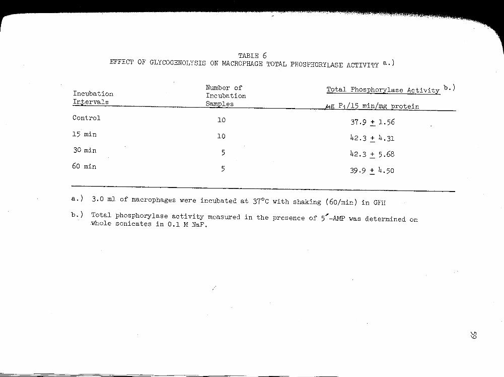
TABLE 6 EFFECT OF GLYCOGENOLYSIS ON MACROPHAGE TOTAL PHOSPHORYLASE ACTIVITY a.)
Number of Total Phosphorylase Activity b.) Incubation Incubation Intervals Samples 4g P;/15 min/mg protein
Control 10 37.9~1.56
15 min 10 42.3 ~ 4.31
30 min 5 42.3 ~ 5.68
60 min 5 39.9 ~ 4.50
a.) 3.0 ml of macrophages were incubated at 37°C with shaking (60/min) in GFH
b.) Total phosphorylase activity measured in the presence of 5'-AMP was determined on whole sonicates in 0.1 M NaF.
VI \0
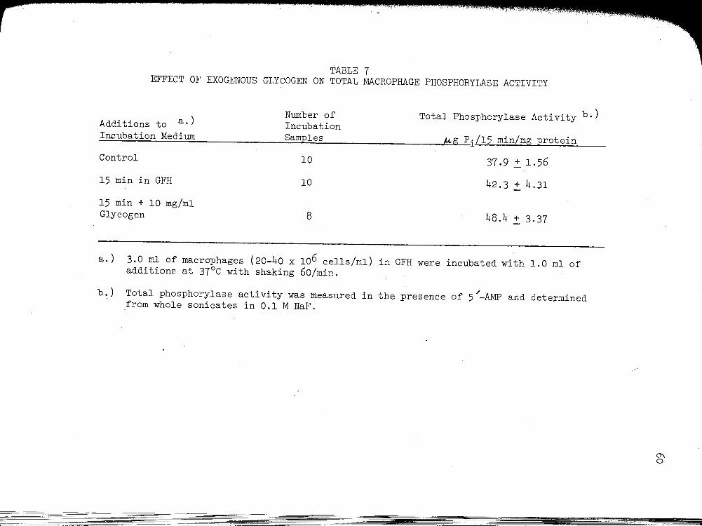
TABLE 7 EFFECT OF EXOGENOUS GLYCOGEN ON TOTAL MACROPHAGE PHOSPHORYLASE ACTIVITY
Number of Additions to a.) Incubation
Total Phosphorylase Activity b.)
Incubation Medium Samples M,g Pi/15 min/mg protein
Control
15 min in GFH
15 min + 10 mg/ml Glycogen
10
10
8
37.9.:!:. 1.56
42.3.:!:. 4.31
48.4 + 3.37
a.) 3.0 ml of macrophages (20-40 x 106 cells/ml) in GFH were incubated with 1.0 ml of additions at 37°C with shaking 60/min.
b.) Total phosphorylase activity was measured in the presence of 5/-AMP and determined from whole sonicates in 0.1 M NaF.
-------=-=-- -----=-------:-- - --- -~-----=====-=---=----=----===
-----~
0'\ 0

61
activity was immediately determined on macrophage sonic~tes. Inflam-
matory macrophages incubated in the presence of 10 mg/ml of glycogen
significantly increased phosphorylase activity by nearly 30%, whereas
macrophages incubated in the absence of glycogen did not demonstrate
/ any further activation of 5 -AMP dependent phosphorylase activity.
c.· The active form of macrophage phosphorylase
An active or a form of the phosphorylase enzyme, whose activity
/ is measured in the absence of 5 -AMP, has been demonstrated in such
tissues as skeletal muscle (26), liver (139) and leukocytes (143, 144). ,
Since the form independent of 5 -AMP has been shown to be the fraction
of phosphorylase activity responsible for initiating glycogenolysis,
studies were attempted to determine what fraction of macrophage phos-
phorylase activity is in the active form. The active and total phos-
phorylase activities derived from 3000 x g sonicate supernatant is pre-
sented in Table 8. Total macrophage phosphorylase activity measured in
the presence of 5,-AMP was 41.2 ~ 1.82 g Pi/15 min/mg protein. Macro-
/ phage active phosphorylase activity measured in the absence of 5 -AMP
was 30.4 ~ 1.86~g Pi/15 min/mg protein. Thus approximately 73% of mac-
rophage phosphorylase activity was in the active form. The macrophage
phosphorylase activity ratio (active phosphorylase activity/total phos-
phorylase activity) of 0.73 was similar to the 0.75 activity ratio of
liver phosphorylase (139) and the 0.80 activity ratio of leukocyte
phosphorylase (143).
d. Effect of Mg++_ATP on macrophage phosphorylase activity
The conversion of inactive to active phosphorylase has been shown
to occur in crude liver extracts (94) and, more recently, in leukocyte

TABLE 8 ACTIVE AND TOTAL PHOSPHORYLASE ACTIVITY IN INFLAMMATORY MACROPHAGES
Number of Macrophage Phosphorylase Activity a.) Incubation
. Samples pg Pi[l_5 min/Plg protein
Total Phosphorylase Activity (+ 0.02 M 5/-AMP)
Phosphorylase a Activity (- 0.02 M 5/-AMP)
Per Cent Activity in the Active Form
9 41.2 + 1.82
9 30.4 ~ 1.86 b.)
9 73.1
a.) Macrophage phosphorylase activity was determined on 3000 x g sonicate supernatants in 0.1 M NaF.
b.) Significantly different from total phosphorylase activity (p<.OOl).
/
0\ 1\)

homogenates (143) by a mechanism involving a specific p:rotein kinase
requiring Mg++ and ATP. In order to determine whether macrophage phos
pho~ylase was also capable of interconversion of the inactive to active
form, macrophage 3000 x g sonicate supernatants were incubated with
o.lM Mg-ATP and the inactive and active phosphorylase activities de
termin~d (Figure 5). The preincubation of macrophage 3000 x g sonicate
in O.lM NaF for 30 minutes decreased the percent of macrophage phos
phorylase in the active form from 73% to 55%. Following this preincu
bation interval, the introduction of O.lM Mg++_ATP to the samples re
sulted in a nearly complete conversion of phosphorylase activity to the
active form by 15 minutes (90% in the active form). Total phosphorylase
activity measured in the presence of 5"-AMP did not significantly change
throughout the incubation interval.
2. Macrophage glucosidase activity
a. o\-1,4 and ~-1,6 glucosidase activity in inflammatory
macrophage
Since acid ~1,4 glucosidase is capable of hydrolyzing maltose,
linear oligosaccharides, and the outer branches of glycogen to glucose
and of catalysing the transglucosylation from maltose to glycogen in
liver, skeletal muscle (58) and in human leukocytes (136), it was of
interest to determine whether this mechanism of glycogen hydrolysis was
present in inflammatory macrophages. Table 9 contains the results of
determinations of~ -glucosidase activities measured at pH 4.0 in whole
sonicates of 96.hour inflammatory macrophages. Macrophages exhibited
a large ~-1,4-glucosidase activity of 201.1 ~ 1.07~g of glucose hy
drolyzed per hour per mg of protein utilizing maltose as the enzyme

64
FIGURE 5
EFFECT OF Mg-ATP ON
INFLAMMATORY MACROPHAGE PHOSPHORYLASE ACTIVITY
100 0.1 M Mg-ATP
80 ' Gl E > ... - 0 u -IV Gl 60
.!: • .!! >- >-... > 0
.c - r::a. 40 u • IV 0 ;I! .c
r::a.
20
15 30 45 60
minutes
a) Macrophage 3000 xg sonicate supernatants were preincubated at 37°C in O.lM NaF for 30 minutes prior to the addition of O.lM Mg+2-ATP.
b) Per cent in active form was determined by the phosphorylase activity ratio '(activity- AMP/activity+ AMP).
c) Mean+ standard error of four experiments.
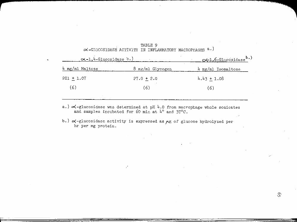
TABLE 9 o<-GLUCOSIDASE ACTIVITY IN INFLAMMATORY MACROPHAGES a.)
o{-1,4-Glucosidase b.) o<.-1, 6-Gl.ucosidase b ·)
4 mg/ml Maltose 8. mg/ml Glycogen 4 mg/ml Isomaltose
201 ~ 1.07 27.0 ~ 2.0 4.43 ~ 1.08
(6) (6) (6)
a.) o<-glucosidase was determined at pH 4.0 from macrophage whole sonicates and samples incubated for 60 min at 4° and 37°C.
b.) o(-glucosidase activity is expressed as~ of glucose hydrolyzed per hr per mg protein.
0\ \Jl

66
substrate and 27.0 ~ 2.0~ of glucose hydrolyzed per hour per mg of pro
tein using glycogen as substrate. ~-1,6-glucosidase activity resulted
in a very low activity of 4.43 ~ l.08~g of glucose hydrolyzed per hour
per mg of protein when isomaltose, a dissacharide comprised entirely of
~-1,6 glucosidic bonds was added as substrate for glucosidase activity.
b. · Effect of pH on inflammatory macrophage 0<.. -1,4-
glucosidase activity
Lejeune et al. (76) reported that liver 0(-glucosidase had its
maximum activity at pH 4.0 and was present in the lysosomal fraction of
rat-liver homogenates. Figure 6 presents the results of incubating mac
rophage sonicates with maltose substrate at pH values ranging from 3.0
to 9.0. Macrophage ~-1,4 glucosidase activity manifested its greatest
activity at pH 4.0, suggesting that acid pH optimum of macrophage glu
cosidase activity favors a lysosomal environment.
3. Macrophage glycogen synthetase activity
a. I and D forms of macrophage glycogen synthetase
Table 10 presents the glycogen synthetase or transferase ac
tivity of inflammatory macrophage 3000 x g sonicate supernatants.
Macrophage glycogen synthetase was found to exist in two forms, a de
pendent or D form, requiring the presence of glucose-6-phosphate and an
independent or I form, manifesting activity in the absence of glucose-
6-phosphate. Macrophage dependent or total glycogen synthetase ac
tivity measured in the presence of lOmM glucose-6-phosphate was 14.3 +
2.2mrm of UDP glucose incorporated into macrophage glycogen per 10
minutes per mg of protein, while the independent or I for.m of macrophage
glycogen synthetase was 0.84~ of UDP glucose incorporated into glycogen

FIGURE 6
EFFECT OF pH ON MACROPHAGE ~-1,4 GLUCOSIDASE ACTIVITY
c • -f.
0 ~
~
m
+ E
" ~ ~ " t • • 0 u ~
m 0 100
' + + +
t 3 4 5 e 7 8 9
pH
a) 0.5 ml of macrophage sonicate was incubated with 4 mg/ml of maltose in a total volume of 1.0 ml for 60 minutes at 4° and 37°C.
I
b) Mean+ standard error of nine experiments.

TABLE 10 INFLAMMATORY MACROPHAGE GLYCOGEN SYNTHETASE ACTIVITY a.)
Number of ~oles of UDP glucose Incubated incorporated into glycogen/
~ Samples 10 min/mg protein
Total Synthetase Activity + lOmM Glucose-6-Phosphate
Independent Synthetase Activity - lOmM Glucose-6-Phosphate
Per Cent Activity in the Independent Form
8
8
8
14.3 .:': 2.23
0.84 + 0.08
5.9%
a.) Glycogen synthetase activity was determined on macrophage 3000 x g sonicate supernatants in O.lM glycylglycine buffer.
0\ co
69
per 10 minutes per mg protein and represented approximately 6% of the
total activity.
b. Effect of glycogen depletion on macrophage glycogen
synthetase activity
In view of the observation that preincubation of macrophages in
a glucose-free media accelerated the rate of l4c-glucose incorporation
into glycogen, it was of interest to investigate whether an increase in
the activity of glycogen synthetase was the underlying mechanism for the
acceleration of glycogen resynthesis in glycogen-depleted macrophages.
In Table 11 is shown the results of a comparison of macrophage glycogen
synthetase activity, both in the glucose-6-phosphate independent and
dependent forms, from glycogen-laden macrophages and macrophages de-
pleted of their glycogen reserve by a 2 hour preincubation in a glucose-
free media. Subsequent to glycogen depletion macrophages exhibited a
dramatic 267% increase in the glucose-6-phosphate dependent form of the
enzyme to a value of 38.2 ~ 2.~~ of UDP glucose incorporated into
glycogen per 10 minutes per mg of protein. The glucose-6-phosphate in-
dependent form of glycogen synthetase was observed to increase over five
fold to a value of 4.69 ~ 0.7~\m of UDP glucose incorporated into gly-
cogen per 10 minutes per mg of protein. As a consequence of this dra-
matic rise in the independent form of macrophage glycogen synthetase in
glycogen depleted macrophages, glycogen synthetase activity in the in-
dependent form increased 83% as compared to glycogen laden macrophages.
4. Macrophage glucose-6-phosphatase activity
Glucose-6-phosphatase catalyzes the hydrolysis of glucose-6-
phosphate to inorganic phosphate and glucose and its activity has been
'I :' I

TABLE 11 EFFECT OF GLYCOGEN DEPLETION ON MACROPHAGE GLYCOGEN SYNTHETASE ACTIVITY a.)
Number of Glycogen ~ UDPG Glucose Incubation Incubation Synthetase Incorporatedinto glycogen/ Conditions Samples Activity 10 min/mg protein
% of Control Glycogen-Laden
Glycogen-Depleted b · )
Glycogen-Laden
Glycogen-Depleted
8
4
8
4
D-form
D-form
I-form c ·)
I-form
14.3.:!:. 2.23
38.2 .:!:. 2.15
0.84 + 0.08
4.69.:!:. 0.73
100
267
100
558
a.) Glycogen synthetase activity was determined on macrophage 3000 x g sonicate supernatant in O.lM glycylglycine . .
b.) Glycogen-depleted macrophages were preincubated for 2 hours at 37°C in GFH.
c.) Percent activity in I-form in glycogen-laden macrophages = 5.9%. Percent activity in I-form in glycogen-depleted macrophages = 10.9%.
~ 0

71
reported in liver, kidney and in small intestine (59). \Since glucose-6-
phosphatase represents the final key regulatory step in glycogen me
tabolism in terms of glucose output, it was of interest to determine
whether inflammatory macrophages could demonstrate hydrolytic activity
towards glucose-6-phosphate. Table 12 presents the glucose-6-phosphatase
activity of inf-lammatory macrophage sonicates in 0.25M sucrose. Macro
phages displayed a significant glucose-6-phosphatase activity of 1.52 +
O.llmg Pi/mg protein/10 min, suggesting the enzymatic capability to
liberate free glucose from intracellular glycogenolytic mechanisms.
C. Glucogenesis in Inflammatory Macrophages
Since the macrophage is known to perform trophic functions by
the ingestion of soluble macromolecules and the subsequent transport
of digested products back into the immediate extracellular environment,
it was of interest to determine whether the inflammatory macrophage
responded trophically to the uptake and intracellular digestion of exo
genous glycogen by the production and subsequent transport of glucose,
i.e. glucogenesis, back into the extracellular milieu.
1. Glucose production in normal and inflammatory exudate cell
populations
In Table 13, the glucogenic capability of unstimulated perito
neal mononuclear cells, inflammatory PMNL and inflammatory macrophages
is evaluated by measuring the glucose liberated by these cell popula
tions into a glucose-free media. In the absence of exogenous glycogen,
there was no significant amount of glucose liberated during a 3 hour
incubation period by the three peritoneal cell populations. The ad
dition of 50 mg of glycogen to the incubation flasks significantly

TABLE 12 GLUCOSE-6-PHOSPHATASE ACTIVITY IN INFLAMMATORY MACROPHAGES
Number of Incubation Glucose-6-Phosphatase b.)
Cell Population a.) Samples Acti~ity
Inflammatory Macrophage Sonicate 12 1.52 + 0.11
a.) Inflammatory macrophages were sonicated in 0.25M sucrose at 4°C. Duplicate samples were incubated at 4° or 37°C for 30 min.
b.) Glucose-6-phosphatase activity is expressed as mg Pi/mg protein/10 min.
-.:] 1\)

TABLE 13 GLUCOSE PRODUCTION IN NORMAL AND INFLAMMATORY EXUDATE CELL POPULATIONS a.)
Glucogenesis -,HS of glucose/mg protein/hour
Additions to incubation media
Cell Population No Additions Glycogen (10 mg/ml) Glycogen (10 mg/ml) + NaF ( 3 mM)
Unstimulated Peritoneal Cells 0 (3)b·) 2.26.!.. 0.78 ( 4) 1.63 .!.. 0.98 (4)
Inflammatory Leukocytes 0.63 .!.. 0.31 (3) 1.42 .!.. 0.13 (3) 1.80 .!.. 1. 06 (4)
Inflammatory Macrophages 0.23 ~ 0.13 (3) 2.68 .!.. 0.35 ( 4) 12.30 ~ 1.75c.)(5)
a.) Two ml of cells (20-50 x 106/ml) were suspended in GFH in~ total incubation volume of 3 ml. Incubations were for 180 min. at 37°C. Glucose production is expressed as pg of glucose per mg of protein per hour. All data represent the differences as compared to control samples incubated at 4°C.
b.) Number of experiments
c.) Significantly different from the unstimulated and leukocyte populations (p~.OOl)
--::J w
74
augmented the glucogenic response of all three cell types examined.
While the addition of 3 mM NaF, a potent glycolytic inhibitor, to the
incubation media containing exogenous glycogen did not significantly
alter glucose liberation in either the unstimulated peritoneal mono
nuclear cells or in the inflammatory PMNL, glucose production by the
inflammatory macrophage was augmented from 2.68 ~ 0.35~ glucose per
mg protein per hour to 12.30 ~ 1.75~ glucose per mg protein per hour,
nearly a six fold increase in the macrophage glucogenic capacity. In
contrast, macrophages did not manifest a glucogenic response to 3 mM NaF
when exogenous glycogen was deleted from the incubation media. Further
more, incubation of 50 mg of glycogen in GFH for 3 hours at 37°C did not
result in any measurable glucose liberation into the media.
Glucogenesis by inflammatory macrophages as related to length of
incubation is represented in Figure 7. The rate of macrophage gluco
genesis was directly related to the 3 hour incubation interval utilized
in these experiments. Additional studies have also demonstrated that
the glucogenic rate was independent of exogenous glycogen concentration
above 8 mg/m1. These results indicate not only that the rates of re
actions involved in glycogenolysis and glucogenesis were dependent on
length of incubation and substrate concentration but also that these me
chanisms were maintained over the course of incubation time chosen in
these experiments.
2. Glucogenesis by inflammatory macrophage cell and sonicate
preparations
Table 14 presents the glucogenic response of macrophage whole
cells and sonicate preparations. Macrophage sonicate preparations when
compared to intact cells demonstrated a significant increase in the rate

75
FIGURE 7
GLUCOGENESIS BY INFLAMMATORY MACROPHAGES
z 30 ... ... on 0 - • Ill L ...
" z ~ ... ........ 20
" ... 0 Ill
u 0 u
:::) :::) ... ... " "
" 10
:::t.
30 60 120 180
MINUTES
Macrophages were incubated in the presence of 50 mg of glycogen and 3mM NaF. Each point represents the mean+ S.E. of at least four experiments.

TABLE 14 GLUCOSE PRODUCTION BY INFLAMMATORY MACROPHAGE CELL AND SONICATE PREPARATIONS
Glucogenesis - ,pg glucose/mg protein/hr Glycogen 11C mg/ml) +
Glycogen(lO mg/ml) NaF (3 mM) No Additions
Macrophage Whole Cells 0.23 .:!:. 0.13 (3) 2.68.:!:. 0.35 (4) 12.30.:!:_1.75 (5)
Macrophage a.) Sonicate 3.13.:!:. 0.97 (3) 15.56 .:!:. 2.49 ( 3) 16.71 .:!:. 2.44 ( 3)
a.) Macrophages (20-50 x 106/ml) were sonicated for two minutes at 4°C in GFH, pH 7.4.
-.;J 0'\

77
of glucose formation either in the absence or presence pf exogenous gly
cogen. However, the addition of 50 mg of glycogen and 3mM NaF to mac
rophage sonicate preparatiomresulted in a rate of glucose liberated of
16.71 ~ 2.4~ of glucose per mg protein per hour comparable to that
found in the intact cell preparation, i.e. 12.3 + 1. 75flg glucose per mg
protein per hour. Furthermore, the glucogenic response of 15.56 ~ 2.49~g
glucose per mg protein per hour demonstrated by the addition of exogenous
glycogen alone to macrophage sonicates illustrates that the rate of glu
cose formation from sonicate glycogenolysis in the absence of any meta
bolic inhibitor is similar to the rate of glucogenesis by intact macro
phages when substrate utilization is effectively inhibited by NaF.
3. Effect of acid pH conditions on macrophage glucogenesis
In order to evaluate the possible role of macrophage acid-hydro
lytic mechanisms on glycogenolysis and subsequent glucose formation,
exogenous glycogen was added to macrophage sonicate preparations under
neutral and acid pH conditions (Table 15). Macrophage sonicates were
incubated for 180 minutes in a citrate phosphate buffer at either pH 7.0
or pH 5.0. Glucose production by macrophage sonicates without the ad
dition of exogenous glycogen demonstrated a probable endogenous glyco
genolysis at both neutral and acid pH conditions. The addition of exo
genous glycogen to macrophage sonicates resulted in an augmentation of
macrophage glycogenolysis. Moreover, macrophage sonicate glycogenolysis
and glucose formation in the presence of exogenous glycogen was further
augmented at pH 5.0 when compared to neutral pH conditions.
4. Substrate and glycoside bond specificity of macrophage
glucogenesis

TABLE 15 THE EFFECT OF pH ON GLUCOSE PRODUCTION BY MACROPHAGE SONICATES
Macrophage Sonicate a.) Glucogenesis- #g glucose/mg protein/hour
+ 10 mg/ml Glycogen
- 10 mg/ml Glycogen
pH 7.0
21.63 2:. 3. 70 b. )
8 . 50 2:. 1. 29
pH 5.0
32.72 2:. 5.02
9.02 2:. 1.97
a.) Three ml of macrophage sonicates were incubated in citrate phosphate buffer for 180 min.
b.) Each value represents the mean+ standard error of the mean of 3 experiments.
-.J ():>

79
In view of the fact that o<-1,4 glucosidase prepominates as the
principal acid glucosidase present within the inflammatory macrophage,
substrate and glycoside bond specificity of macrophage glucogenesis was
investigated in intact macrophage preparations (Table 16). Glycogen and
soluble starch containo<-1,4 and o(-1,6 glycoside bonds linking glucose
residues and both substrates can vary enormously in their particle size.
Dextrans are polysaccharides produced by the action of certain bacteria
(e.g. Leuconostoc mesenteroides) on sucrose and are composed almost en
tirely of glucose units joined by c<-1,6 glycosidic linkages. Incubation
of intact inflammatory macrophages with either glycogen or starch re
sulted in comparable rate of glucogenesis, 12.3 ~ l.75~g of glucose per
mg protein per hour and 11.00 ~ 0.31~ of glucose per mg protein per hour,
respectively, while the addition of two different molecular weight dex
trans failed to liberate any significant amount of glucose.
5. Effect of cyclic AMP on macrophage glucogenesis
In order to evaluate the potential role of a phosphorylytic me
chanism mediated by glycogen phosphorylase on the glucogenic mechanism,
experiments were performed to determine whether dibutyryl cyclic AMP
could influence macrophage glucose formation from exogenous glycogen.
Cyclic AMP and, in the presence of intact cells, the dibutyryl deriva
tive are nucleotides known to activate phosphorylase kinase and gly-
cogen phosphorylase (56). Macrophages and PMNL were incubated under
identical conditions in the presence of 50 mg of glycogen, 3mM NaF and
lmM dibutyryl cyclic AMP for 180 minutes (Table 17). The results in
dicate that in the inflammatory macrophage, the addition of dbc AMP
produced a 63% increase in the rate of glucose formation in the presence

TABLE 16 SUBSTRATE AND GLYCOSIDE BOND SPECIFICITY OF MACROPHAGE GLUCOGENESIS a.)
Addition to Number of Glucogenesis Incubation Incubation Medium a.) Samples ~g glucose/mg protein/hour
Glycogen 5 12.30.:!:_1.75
Starch 3 11.00.:!:. 0.31
Dextran 6 mw= 2 x 10 3 0.28 .:!:. 0.23
Dextran 3 0.42.:!:. 0.31 mw= 2 x 10⁵
a. ) Two ml of macrophages were in cuba ted iri the presence of NaF ( 3 ml1) and 50 mg of carbohydrate in GFH for 180 min at 37°C.
co 0

TABLE 17 EFFECT OF CYCLIC AMP ON MACROPHAGE AND LEUKOCYTE GLUCOGENESIS a.)
Glucogenesis -~g glucose/mg protein/hour . Cell Population -dbc AMP +dbc AMP (1 rnM)
Inflammatory Leukocyte 1.80 + 1.06 (4) 1. 20 .:!:. 0 . 31 ( 3)
Inflammatory Macrophage 12.20.:!:_1.75 (5) 19.92.:!:. 0.90 (3)b.)
a.) Two ml of cells were incubated with 10 mg/ml of glycogen and 3 rnM of NaF in a total volume of 3.0 ml for 180 min. at 37°C with or withour 1 mM dbc AMP.
b.) Significantly different from macrophages incubated without dbc AMP (p<. 01).
())
1-'

of exogenous glycogen. In contrast, the inflammatory P~ did not
exhibit any demonstrable change in their ability to liberate glucose
from a glycogen source in the whole cell system.
82

C~T~ V
DISCUSSION
The results of this study demonstrate a prominent glycogen
content in inflammatory macrophages originating from a 96 hour perito-
neal exudate. The inflammatory macrophage glycogen content of 9.55
~ 0.44.ug glycogen per 106 cells is not significantly different from
the glycogen content of 3 hour inflammatory leukocytes utilized in
this study and is also comparable to glycogen values reported for nor-
mal PMN leukocytes (43, 112). Although a quantitative analysis of mac-
rophage glycogen content has not been previously reported, histological
studies have suggested glycogen to be a significant cytoplasmic con-
stituent of mononuclear phagocytes. Weiss and Fawcett (134) cultured
chicken buffy coat cells in autologous serum, allowing granulocytes and
platelets to disintegrate with continuing culture and leaving a uniform
population of adherent macrophages. During the in vitro culture in-
cubations, macrophages were observed to markedly increase cytoplasmic
inclusions exhibiting a positive periodic acid-Schiff (PAS) reaction by
the ingestion of effete cells. Wulff (142) investigated the glycogen
metabolism of developing macrophages and observed that, parallel to
maturation, an increase in PAS-positivity occurred. Recently, Dvorak
et al. (38) demonstrated dense aggregations of glycogen within the
peripheral pseudopodia of migrating macrophages in capillary tube cul-
tures. Furthermore, in cultures where macrophage migration was
83
\ L

84
inhibited by the presence of sensitized lymphocytes, glycogen-packed
pseudopodia were less prominent. Interestingly, in the invertebrate
chiton Liolophura where the phagocytic cell represents the only.known
host defense mechanism to fore{gn body invasion or tissue injury, wan-
dering macrophages have been collected from the hemolymph and have been
observed to coritain large aggregations of glycogen particles, at times
filling more than half of the cell's cytoplasmic space (71). Therefore,
the present quantitative data discounts the previous notion that the
PMN leukocyte is the only white blood cell population to contain sig-
nificant stores of glycogen (109, 125) and support~ the histological
evidence that macrophages can accumulate a large glycogen reserve.
The acquisition of glycogen by the macrophage at the inflam-
matory locus appears to occur by the active ingestion of cellular de-
bris containing large deposits of glycogen. Quantitative studies on
inflammatory leukocyte glycogen content have recently been conducted
on guinea pig blood and exudate leukocytes (111). The glycogen content
of leukocytes entering the inflammatory site increased dramatically when
compared to control peripheral blood leukocytes. The mean glycogen con-
tent of peripheral blood leukocytes of 6.5 ~ 1.3 mg of glycogen per 109
cells was elevated to 28.7 ~ 3.1 mg of glycogen per 109 cells in 24 hour
exudate leukocytes. Indeed, the glycogen content of inflammatory leu-
kocytes was already significantly elevated after 4 hours following the
induction of the inflammatory response. Examination of histological
evidence has repeatedly suggested that exudate macrophages acquire gly-,
cogen by the ingestion of leukocyte debris. Wulff (141) demonstrated
that inflammatory macrophages progressively increased their glycogen

85
content by the ingestion of neutrophil cytoplasmic fragments. Rebuck
et al. (97)havefurther suggested that the acquisition of PAS- positive
neutrophilic debris by inflammatory macrophages was apparently depen-
dent upon a functional macrophage carbohydrate metabolism. The authors
proposed the concept that the depressed phagocytic capability of mac-
rophages originating from insulin-deficient diabetics was due, in part,
to a failure of macrophages to increase their glycogen reserve.
The results of in vitro experiments in this study demonstrating
the uptake by inflammatory macrophages of exogenous glycogen clearly
arein agreement with the histological evidence tha~ macrophages areca-
pable of ingesting glycogen at the inflammatory site. Macrophages were
able to incorporate 14c-labelled glycogen into intracellular glycogen
stores, amounting to nearly 10% of the total glycogen content in a 15
minute incubation period. Furthermore, inflammatory macrophages demon
strated a profound increase in 14c-glycogen uptake at 37°C as compared
to incubation at 4°C, suggesting that the labelled glycogen was not
merely being adsorbed to the macrophage membrane during the incubation
interval. Moreover, macrophages were able to maintain their glycogen
stores not only by the provision of glucose to the incubation media
but also by the ingestion of exogenous glycogen in a glucose-free en-
vironment. These results suggest that the macrophage at the inflam-
matory site is able to maintain functional integrity by the ingestion
of glycogen-containing cellular debris even though adequate substrate
supply may be interrupted. '
A functional carbohydrate metabolism is mandatory for the provision
of cellular energy for the active endocytic process in phagocytic cells.

The findings that, in peritoneal PMN leukocytes and macrophages, gly
colytic inhibitors prevent phagocytosis, whereas inhibitors of respi
ration do not indicate that the energy requirement for particle up
take is ultimately derived from glycolysis (85, 106). Pinocytosis
86
by peritoneal macrophages, in contrast, has been shown to be severely
depressed by inhibitors of oxidative phosphorylation and respiration
(16). In vitro incubation under N2 atmospheric conditions did not in
terfere with glycogen uptake by macrophages in a glucose-free media
when compared to macrophages incubated under air as the gas phase, sug
gesting that anaerobic glycolysis provided the necessary energy for
macrophage glycogen uptake. Whether in vitro glycogen uptake was per
formed by pinocytic or phagocytic mechanisms was not determined in
this study, although under in vivo conditions at the inflammatory site,
both endocytic mechanisms would be operative in the ingestion of soluble
glycogen or aggregates of the glycogen macromolecule.
The investigation into the alteration of macrophag~ glycogen
content during phagocytosis was an attempt to gain further insight into
the functional significance of the macrophage glycogen reserve. Studies
examining the metabolic perturbations during phagocytosis by either leu
kocytes or macrophages have demonstrated a significant stimulation in
glucose utilization during the phagocytic event (23, 135). The role of
leukocyte glycogen as an energy store to be utilized during phagocytosis
has been proposed by Scott (109). Indeed, several studies have reported
leukocyte glyco£en levels to be depleted in vitro during phagocytosis
(6, 23, 120). Inflammatory macrophages incubated in the presence of
latex particles were observed to significantly accelerate glycogenolysis

in the absence of glucose when compared to non-phagocytizing controls.
Thus the evidence suggests that the inflammatory macrophage glycogen
reserve can also be called upon to provide for the augmented glucose
demands during phagocytosis.
Incubation for 60 minutes in a glucose-free media mobilized
half of the ma~rophage glycogen supply, however, incubation for longer
intervals did not further deplete endogenous glycogen stores. The in
ability to mobilize the entire glycogen reserve in macrophages has not
been determined, although studies involved in the molecular structure
of glycogen have revealed that apparently only 60% of the total glucose
residues forming the outer linear chains of the .glycogen molecule are
readily accessible to the action of phosphorylase (115). Additional
support for the role of macrophage glycogen in maintaining active
phagocytic mechanisms was provided utilizing glycogen-depleted macro
phages. The results in this in vitro study demonstrate that the addition
of exogenous glycogen in a glucose-free media to macrophages,whose energy
stores have been depleted did not increase macrophage glycogen levels to
initial control values, although glycogen-depleted macrophages readily
synthesized glycogen when extracellular glucose was available. The de
pression of macrophage ·phagocytic ability in glycogen-depleted cells
was further supported by the fact that the uptake of 14c-labelled glyco
gen by glycogen-depleted macrophages in a glucose-free media was de
pressed nearly 50% when compared to glycogen-laden macrophages. These
results suggest that the depletion of macrophage carbohydrate reserve
may significantly reduce the capacity of inflammatory macrophages to par
ticipate in the inflammatory reaction by depressing macrophage function

88
when glucose is not readily available.
The in vitro incorporation of 14c-glucose into macrophage gly
cogen offers some insight into the changing balance of synthesis at
different glycogen loads. The rate of glycogen synthesis in inflam
matory macrophages partially depleted of their glycogen reserve was ac
celerated more than three-fold when compared to glycogen-laden cells
when incubated in the presence of 14c-U-glucose. These results were
consistent with similar observations made in the PMN leukocyte (109,
111) and in glycogen-depleted human cytotrophoblastic cells -- a cell
line that relies heavily on glycogen as an energy source (89). The
in vitro results in the present study raise the possibility that a re
lationship exists between the initial glycogen content of macrophages
and the rate of incorporation of glucose into glycogen. In skeletal
muscle (75) and liver (35), glycogen decreases its own synthesis non
hormonally by inhibition of glY,cogen synthetase D phosphatase, the en
zyme that converts synthetase D to synthetase I. It was suggested by
Wang et al. (132) that glycogen reple~ion of PMN leukocytes is as
sociated with a rapid change from the D-form of the enzyme to the I-form
with a small change in total enzyme activity. More recently, Scott and
Cooper (111) observed a significant elevation in glycogen synthetase ac
tivity in exudate leukocytes when compared to blood leukocytes and at
tributed the change in enzyme activity to an increase in the glucose-6-
phosphate dependent (D) form of the enzyme.
The present results clearly demonstrate the activity of UDP
glucose-glucan glucosyl transferase (i.e. glycogen synthetase) in macro
phage sonicates. Macrophage glycogen synthetase activity independent of
glucose-6-phosphate (I-form) was equal to 5.9% of the total (I and D)
activity. Furthermore, when macrophages were incubated for 2 hours in
a glucose-free media, the I activity increased to nearly 11%. This two
fold increase in I activity suggests a decreased inhibitory effect of
glycogen on the synthetase phosphatase. A similar glycogen synthetase
D to I conversi~n has recently been reported in the human PMN leukocyte
(132) and also in human lymphocytes {57). Depletion of macrophage gly
cogen content for 2 hours significantly increased glycogen synthetase
activity not only in the glucose-6-phosphate independent or I form but
also in the dependent or D form. In liver, the activation of glycogen
synthetase (D to I conversion) involves a dephosphorylation catalyzed by
a specific phosphatase, thus the activity of glycogen synthetase, like
that of phosphorylase, appears to depend on a finely tuned regulatory
mechanism between the activities of kinases and phosphatases. The pre
sent evidence suggests such a regulatory mechanism for glycogen synthesis
is operational within the inflammatory macrophage and furthermore, that
alterations in glycogen levels within the cell will exert a profound in
fluence on the macrophage glycogen synthetic machinery.
Insulin, a potent stimulator of glycogen synthetic mechanisms
in adipose tissue and skeletal muscle, failed to increase l4c-glucose in
corporation into macrophage glycogen at an insulin concentration of 200fU/ml.
These results support the conclusions of Esmann. ( 44) who also failed to
find any in vitro effect of insulin at physiological concentrations on
glucose transport or glycogen synthesis in the PMNL, although prolonged
incubation and high insulin concentration (lU/ml) did result in a non
specific increase in glucose transport. However, the addition of 1%

90
albumin to the incubation media significantly doubled the in vitro in
corporation of 14c-glucose into macrophage glycogen in a 60 minute in
cubation period. Cohn and Parks (24) have investigated the pinocytosis
inducing effect of a number of molecular species and have found certain
proteins, in particular plasma albumin, to be potent stimulators of ve
sicle formation in macrophages. The addition of insulin to the media
containing albumin did not further enhance 14C-glucose incorporation into
macrophage glycogen. Thus the present results suggest that stimulation
of macrophage pinocytosis results in an increased transport of extracell
ular glucose, offering endocytosis as a mechanism facilitating glycogen
synthesis by increasing the incorporation of available extracellular
glucose.
The demonstration of macrophage phosphorylase activity indicates
that, in view of the broad variation in macrophage energy demands at the
inflammatory site, glycogen mobilization is controlled by a finely balanced
regulatory mechanism. Rapid glycogenolysis is accomplished'bY the acti
vation of glycogen phosphorylase and is controlled by a complex cascade
of phosphorylation-dephosphorylation reactions. Green and Cori (Sl) were
the first to isolate rabbit skeletal muscle phosphorylase and found
readily interconvertible active and inactive forms. Sutherland et al.
(123) characterized the two forms of phosphorylase in liver homogenates
and demonstrated that the conversion of the inactive to active form re
quired the presence of ATP, Mg++ and the enzyme phosphorylase b kinase.
Distinct differences exist in the phosphorylase activity from these two
tissues. The inactive skeletal muscle enzyme becomes active in the pre
sence of AMP, while the active form has an activity independent of AMP.

In contrast, the inactive liver enzyme demonstrates little activity
even in the presence of the nucleotide, whereas the active form is
stimulated only 40% by AMP. Detailed studies on the protein kinases
responsible for the phosphorylation of muscle phosphorylase b has re
vealed that they also occur as active and inactive forms. Studies by
Krebs et al. (7·4) have shown that the conversion of the inactive to
active form of phosphorylase b kinase takes place on incubation with
ATP and Mg++, resulting in the phosphorylation of the inactive form.
Maddaiah and Madsen (79) have demonstrated that 60% of resting liver
phosphorylase activity is in the active form while resting skeletal
muscle phosphorylase activity is predominantly in the b or inactive
91
form (72). The levels of cofactors and substrates in the cell have been
shown to influence the interconversion reactions of phosphorylase and
synthetase. Krebs et al. (74) have demonstrated that elevated glycogen
levels stimulate the phosphorylase b to a conversion and also the acti
vation of phosphorylase b kinase whereas, increased glycogen .. levels in
hibited the synthetase D to I conversion (128).
Determination of leukocyte phosphorylase activity demonstrated
a behavior similar to that found in liver in that it was possible to
activate leukocyte phosphorylase activity in vitro by glucagon (137).
Recently, Yunis and Arimura (143) examined the properties of glycogen
phosphorylase in rat chloroma cells, a tumor composed entirely of im
mature granulocytes, and noticed that leukocyte phosphorylase exhibited
considerable activity in the absence of AMP and thus resembled liver
phosphorylase. The present results of phosphorylase assays in inflam
matory macrophage extracts indicate that a significantly large fraction
92
of the total phosphorylase activity exists in the active form (73%),
thus, in this one respect, macrophage phosphorylase activity resembled
leukocyte and liver phosphorylase activity. Total phosphorylase ac
tivity did not significantly change during a l hour period of glyco
genolysis in a glucose-free media, however, phosphorylase activity in
creased significantly 15 minutes following the addition of exogenous
~lycogen when compared to initial control values, again comparable to
liver phosphorylase. Conversion of inactive to active liver phosphory
lase has been shown to occur readily in the presence of Mg++ and ATP
(94). Macrophage active phosphorylase activity was markedly enhanced
in macrophage sonicates incubated in the presence of O.lM Mg++_ATP fol
lowing the partial inactivation of the active form by a 30 minute pre
incubation. The results suggest that the Mg++_ATP stimulation of macro
phage active phosphorylase activity is similar to those mechanisms des
cribed in liver and muscle. The activation of this protein kinase in
these tissues by Mg++_ATP is strongly stimulated by cyclic AMP. The
now classic studies by Sutherland and Rall (122) on the physiology of
cyclic AMP have demonstrated that phosphorylase activation via the
adenyl cyclase system provides rapid glycogenolysis and increased glu
cose output by liver preparations. In support of this concept, Reik
et al. (10~ have localized a hormone-sensitive adenyl cyclase activity
in reticuloendothelial cells of the liver utilizing both cytochemical
and biochemical techniques. In addition, alveolar macrophages have been
found to exhibit a significant increase in the intracellular level of
cyclic AMP and thus suggesting a link between particle uptake and the
initiation of metabolic events requiring the accelerated expenditure

93
of energy in the form of glycogenolysis (114). Thus the glycogen mo
bilization via phosphorylase by the, inflammatory macrophage may also
respond in a similar fashion via a hormone-sensitive adenyl cyclase me
chanism that would function in rapidly providing glucose to meet the
substrate demand of the macrophage at the inflammatory locus.
The pre'sence of an acid {)(,-glucosidase in inflammatory macro
phages provides an alternate mechanism for the degradation of the gly
cogen molecule. This is the only known lysosomal enzyme capable of de
grading glycogen. Jeffrey et al. (64) have recently purified an ~
glucosidase from the lysosomal fraction of rat liver homogenates and
have demonstrated that the enzyme had both o(-1,4 and o(.-1,6 glucosidase
activity. Hers (58) initially demonstrated the deficiency of a lyso
somal acid maltose, c<-1,4 glucosidase, in the livers of patients with
Pompe's disease (Type II glycogenesis), a fatal disorder characterized
by the accumulation of glycogen in the heart, skeletal muscle, brain,
liver and kidney. Williams (136) measured <::>(-glucosidase a'ctivity in
human PMN leukocytes from normal subjects and from patients with Type
II glycogenesis and demonstrated that leukocytes from patients with
Pompe's disease display approximately one half the activity of the
normal controls.
Hsia et al. (61) have characterized an o<-glucosidase in human
PMN leukocytes and were able to show activity with maltose as a substrate;
however, no ~-1,6 glucosidase activity was observed utilizing isomaltose
as substrate. The results of the present study utilizing the macrophage
indicate a prominent o(,-1,4 glucosidase activity measured at pH 4.0 with
maltose as substrate. Furthermore, that only ~-1,4 glucosidase activity
was manifest in macrophage sonicates and negligible ~-+,6 glucosidase
activity was observed is comparable to the results found in the PMN
leukocyte. The determination of a 4.0 pH optimum for macrophage o<-
1,4 glucosidase activity is in agreement with results described for
liver o( -1,4 glucosidase (58) and in PMN leukocytes (136) and strongly
suggests a probable lysosomal origin within the macrophage for the acid
glucosidase activity. Therefore, the identification within macrophages
of a hydrolytic degradative mechanism for glycogen indicates that sub-
sequent to the discharge of lysosomal hydrolases into a glycogen-con-
taining phagocytic vacuole, lysosomal glucosidase could readily attack
the outer linear branches of the glycogen molecule providing available
free glucose for macrophage functional energy demands.
In view of the fact that glucose-6-phosphatase provides the
final enzymatic link in the provision of free extracellular glucose
from the glycogenolytic sequence, particular interest is raised in this
study when glucose-6-phosphatase activity was observed in maGrophage
sonicates. It is generally accepted that glucose-6-phosphatase acti-
vity is manifested predominantly in those organs which function in the
regulation of blood glucose. Thus, Hers and DeDuve (59) found glucose-
6-phosphatase activity greatest in liver, kidney and in small intestine.
Although no glucose-6-phosphatase activity has been reported biochemi-
cally in other white blood cells (61, 109), the blood platelet however,
does contain measurable amounts of glucose-6-phosphatase (52). Recent
histochemical evidence revealed that glucose-6-phosphatase activity was
localized in rat and mouse RES cells, in particular, alveolar macro-
phages and Kupffer cells of the liver (104). The present evidence

supplies the quantitative data in support of the histochemical demon
stration of glucose-6-phosphatase activity in macrophages.
95
The functional significance of glucose-6-phosphatase in RES
cells has not been previously investigated. The active phosphorylytic
attack of macrophage glycogen by phosphorylase would result in the for
mation of glucase-6-phosphate which predominantly would be further me
tabolized via glycolytic or hexose monophosphate shunt pathways to
meet macrophage energy demands. However, the presence of glucose-6-
phosphatase in inflammatory macrophages provides a third alternative to
the metabolism of glucose-6-phosph~te, whereby the hydrolytic attack on
the molecule would result in the formation of free glucose. Therefore,
the macrophage at the inflammatory site is equipped with the full com
plement of glucogenic enzymes and is capable enzymatically of supplying
glucose to its extracellular environment. Thus the present characteri
zation of glycogenolytic and glucogenic enzymes within the inflammatory
macrophage reveals an additional trophic functional role for,the large
carbohydrate supply ingested and stored by the inflammatory macrophages.
The process of glucogenesis, i.e. the liberation of glucose,
was demonstrated in vitro in the peritoneal exudate cells and significant
increases were observed in macrophages in the presence of glycogen and
NaF, a glycolytic inhibitor. In contrast, neither inflammatory PMN leu
kocytes nor unstimulated peritoneal mononuclear cells responded by aug
mented glucose liberation when exogenous glycogen and the glycolytic in
hibitor were added. The use of a glycolytic inhibitor in vitro to ex
aggerate the glucogenic response by inflammatory macrophages may mimic
the in vivo situation whereby the glycolytic pathway is depressed by a

decrease in intracellular pH initiated by the release of acid hydrolases
into the cytosol. In support of this concept, Halpern et al. (53) re
cently compared the rate of glycolysis in leukocytes incubated in media
of low pH values and found that the inhibition of glycolysis was due to
an inhibition of the activity of phosphofructokinase, a key regulatory
enzyme of the glycolytic pathway.
Recent studies by Cohn and Ehrenreich (20) relevant to the me
chanisms involved in the uptake of digestible and nondigestible carbo
hydrates by cultured macrophages have demonstrated that large carbohy
drate molecules are pinocytized and stored within lysosomal associated
vacuoles. Intralysosomal hydrolysis of carbohydrates then takes place
with the formation of utilizable monosaccharides which escape from
the vacuole. It appears reasonable to speculate that such a mechanism
is employed following the ingestion of glycogen-contai~ing debris from
the inflammatory environment. The resulting release of glucose from the
phagocytic vacuole would then be either further metabolized ·or liberated
from the macrophage and utilized by ongoing reparative processes at the
inflammatory site.
In an attempt to extend these observation~macrophage sonicate
preparations were employed to investigate the hydrolytic degradation of
glycogen by inflammatory macrophages. The rate of glucogenesis in macro
phage whole cells was found to be comparable to the rate of glycogenolysis
in macrophage sonicates indicating that macrophage lysosomal glucosidase
released upon sonication could account for the observed rate of glucose
liberation in the intact macrophage when glucose utilization was de
pressed. Glycogen and starch are composed of D-glucose units linked by

97
o{-1,4 and o(-1,6 glycoside bonds whereas dextrans are\polysaccharides
composed entirely of glucose units joined by ~-1,6 glycoside bonds.
A comparison of the hydrolytic action by macrophage sonicates on large
molecular weight polysaccharides revealed that glucogenic activity was
manifest only in the presence of carbohydrates where ~-1,4 glycoside
bonds are prominent. Thus these present studies concur with the enzyma-
tic demonstration that c<-1,6 glucosidase is not active within the mac-
rophage lysosome. Although glucosi~ase activity in macrophage subcellu-
lar compartments by fractionation techniques was not determined in this
study, the probable lysosomal origin of the hydr~lytic activity on the
glycogen molecule was inferred when exogenously added glycogen was more
rapidly hydrolyzed to glucose by macrophage sonicates under acid pH
conditions favored by lysosomal hydrolases.
An important functional role for phosphorylase as a degradative
process involved in macrophage glycogen breakdown was illustrated by the
in vitro addition of dibutryl cyclic AMP to macrophages ingesting exo-
genous glycogen. Macrophage glycogenolytic and glucogenic mechanisms
in intact cells responded to the addition of dibutyryl cyclic AMP by a
notable increase in the glucose liberated from intact inflammatory mac-
rophages. It is reasonable to assume that dibutyryl cyclic AMP accele-
rated intracellular glycogenolysis by stimulating the activation of mac-
rophage phosphorylase and, coupled with the hydrolytic action of glucose-
6-phosphatase, augmented the liberation of glucose supplied by the acid
hydrolytic mechanism. Park et al. (88) postulated that adenyl cyclase I
activation during phagocytosis may be the initiating event triggering
subsequent metabolic alterations during phagocytosis, thus emphasizing a

98
role for phosphorylase activation and the ensuing glycogenolysis in the
energetics of particle engulfment within phagocytic cells. The present
study suggests that the degradative pathways involved in glycogenolysis
within the macrophage include both hydrolytic and phosphorylytic mecha
nisms and furthermore, that both mechanisms may be operative in sup
porting macrophage glucogenesis.
The present studies have demonstrated that the inflammatory mac
rophage is equipped to harvest a prominent carbohydrate reservoir by the
ingestion of exogenous glycogen as well as by glycogen synthetic enzymes.
Furthermore, the dynamic state of macrophage glycogen metabolism has
been evaluated by the demonstration of a complex glycogen degradative
apparatus, capable of responding to regulatory control mechanisms, to
provide additional glucose to meet a wide spectrum of macrophage energy
demands. The studies illustrating the effect of glycogen depletion on
the phagocytic ability of the macrophage emphasizes that further in
vestigation is needed to adequately clarify the physiological role of
glycogen metabolism within the phagocytic cell. The demonstration of a
glucogenic capacity within the inflammatory macrophage adds a new di
mension to macrophage function suggesting that an important nutrient role
may be served by the macrophage at the inflammatory site in the form of
glucose provision to assist the.reparative processes of tissue healing
mechanisms.

CHAPTER VI
SUMMARY
l. , Caseinate-induced 3 hour exudates yielded 6 to 7 x 10⁶
cells with greater than 90% of the cell population examined being PMNL.
The 96 hour inflammatory exudate produced 2 to 3 x 10⁸ cells per rat
with macrophages comprising 85 to 90% of the cell population from the
peritoneum.
2. The 96 hour inflammatory macrophage demonstrated a prominent
glycogen content of 11.79 ~ l.5~;g glycogen per 106 cells or 110.3 ~
l2.&~g glycogen per mg of cell protein that was comparable to the glyco
gen content of the inflammatory PMNL.
3. Both PMNL and macrophages exhibited a labile intracellular
glycogen pool that was decreased to approximately 50% of control values
by incubation in glucose-free Hank's for 60 minutes. Furthermore, when
macrophages were incubated in the presence of latex particles in a glu
cose-free medium, the glycogen content of phagocytizing macrophages was
further diminished when compared to non-phagocytizing macrophages.
4. Inflammatory macrophages incubated in the presence of
either 10 mM glucose or 10 mg/ml of exogenous glycogen maintained their
large glycogen reserve during a 60 minute incubation interval.
99

100
5. Macrophage glycogen resynthesis from glucose was stimulated
by preincubation for 2 hours in a glucose-free medium. However, the
addition of exogenous glycogen to glycogen depleted rnacrophages did not
result in an elevation of macrophage glycogen content in glycogen de
pleted cells.
6. Glycogen synthesis in glycogen depleted macrophages from
14c-U-glucose was accelerated three-fold when compared to 14c-U-glucose
incorporation into glycogen from glycogen laden macrophages.
7. Inflammatory macrophages incubated in the presence of 1%
albumin doubled their incorporation of 14c-U-glucose into macrophage
glycogen when compared to cells incubated without the pinocytic acti
vator. 20~U/rnl of insulin did not enhance the incorporation of 14-c
U-glucose into macrophage glycogen.
8. Exogenous 14C-labelled glycogen was readily incorporated
into the .macrophage glycogen pool. In contrast, incorporation of 14c
glycogen by glycogen depleted macrophages was markedly depressed.
9. Inflammatory macrophages exhibited glycogen phosphorylase
activity that was significantly increased in the presence of exogenous
glycogen. Macrophage active phosphorylase activity amounted to 73% of
the total phosphorylase activity and the introduction of 0.1 M Mg++_
ATP to macrophage sonicates resulted in a nearly complete conversion
of macrophage phosphorylase activity to the active form.
10. Inflammatory macrophages demonstrated considerable~-1,4

glucosidase activity that manifested its greatest acti~ity at pH 4.0.
In contrast, macrophages displayed no .o\-1,6 glucosidase activity.
11. Macrophage glycogen synthetase was found to exist in
101
two forms, a glucose-6-phosphate dependent or D-form and a glucose-6-
phosphate independent or I-form. The I-form amounted to approximately
6% of the total glycogen synthetase activity.
12. Glycogen depletion in inflammatory macrophages resulted
in a dramatic increase in both the I and D-forms of glycogen synthetase.
Glycogen depletion produced an 83% increase in the I-form as compared
to glycogen laden macrophages.
13. Inflammatory macrophages displayed a significant glucose-
6-phosphatase activity suggesting the enzymatic ability to liberate
free glucose from intracellular glycogenolytic mechanisms.
14. Glucogenesis, i.e., glucose liberation into the incu
bation medium was demonstrated by 96 hour inflammatory macrophages
in the presence of NaF and exogenous glycogen. Macrophage glucogenesis
was greater at pH 5.0 than at pH 1.0.
15. Macrophage glucogenesis was marked in the presence of
either exogenous glycogen or soluble starch while the addition of
various molecular weight dextrans was ineffective in liberating
glucose by macrophages.
16. Glucogenesis in both intact macrophages and sonicate
preparations was augmented by dbc AMP while PMN leukocyte glucogenesis

102
was not affected by dbc AMP.

BIBLIOGRAPHY
l. Adams, C.W.M., O.B. Bayliss, M.Z.M. Ibrahim, and M.W. Webster. Phospholipids in atherosclerosis: The modification of the cholesterol granuloma by phospholipid. J. Path. Bact. 86: 431-436·, 1963. - -- -- -
2. Acton, J.D. and Q.N. Myrvik. Production of interferon by alveolar macrophages. J. Bacterial. 91: 2300, 1966.
3. Aschoff, L. Das Reticulo-endotheliale System. Ergebn. inn. Med. Kinderheillc 26: l-118, 1924.
4. Axline, s:c. Functional biochemistry of the macrophage. Seminars in Hematology 1= 142-160, 1970.
5. Balner, H. Identification of peritoneal macrophages in mouse radiation chimeras. Transplantation _l: 217-223, 1963.
6. Bazin, S. and C. Avice. Le metabolisme glycogenique des polynucleares au cours de la phagocytose in vitro. Compt .. rend. Soc. de Biol. 147: 1027-1037, 1953.
7. Bennet, W.E. and Z.A. Cohn. The isolation and selected properties of blood monocytes. J. Exptl. Med. 123: 145-160, 1966.
8. Berken, A. and B. Benacerraf. Properties of antibodies cytophilic for macrophages. ~· Exptl. Med. 123: 119-147, 1966.
9. Berry, L.J. and T.D. Spies. Phagocytosis. Medicine 28: 239-300, 1949.
10. Boyden, S.V. Cytophilic antibody in guinea pigs with delayed type hypersensitivity. Immunology 1: 474-483, 1964.
11. Brown, J. Effects of 2-deoxyglucose on carbohydrate metabolism: Review of the literature and studies in the rat. Metabolism 11: 1098-1112, 1962.
12. Cagan, R.H. and M.L. Karnovsky. Enzymatic basis of the respiratory stimulation during phagocytosis. Nature 204: 255-257, 1964.
13. Carr, I. THe fine structure of the cells of the mouse peritoneum. Zeitschrift fUr Zellforschung 80: 534-555, 1967.
103

14. Carr, I. Lysosome formation and surface changes in,stimulated peritoneal cells. Z. Zellforsch. Mikrosk. Anat. 89: 328-354, 1968. -- -
15. Cline, M.J. Rev . .!±.2_:
Metabolism of the circulating leukocyte. 674-720, 1965.
Physiol.
16. Cohn, Z.A. The regulation of pinocytosis in mouse macrophages. J. Exptl. Med. 124: 557-571, 1966.
104
17. Cohn, Z.A. ·The structure and function of monocytes and macrophages. , Adv. in Immunology .2_: 163-214, 1968.
18. Cohn, Z.A. and B. Benson. The in vitro differentiation of mononuclear phagocytes. I. The influence of inhibitors and the results of autoradiography. J. Exptl. Med. 121: 279-288, 1965.
19. Cohn, Z.A. and B. Benson. The differentiation of mononuclear phagocytes. Morphology, Cytochemistry, and Biochemistry. J. Exptl. Med. 121: 153-170, 1965.
20. Cohn, Z.A. and B.A. Ehrenreich. The uptake, storage and intracellular hydrolysis of carbohydrates by macrophages. J. Exptl. Med. 129: 201-225, 1969.
21. Cohn, Z.A., M.E. Fedorko, and J.G. Hirsch. The in vitro differentiation of mononuclear phagocytes. J. Exptl. Med. 123: 757-766, 1966.
22. Cohn, Z.A., J.G. Hirsch, and M.E. Fedorko. The in vitro- differentiation of mononuclear phagocytes. IV. The ultrastructure of macrophage differentiation in the peritoneal cavity and in culture. ~· Exptl. Med. 123: 747-755, 1966.
23. Cohn, Z.A. and S.I. Morse. Functional polymorphonuclear leukocytes. I. ments and consequences of particle 111: 667-686, 1960.
and metabolic properties of Observations on the requireingestion. ~· Exptl. Med.
24. Cohn, Z.A. and E. Parks. The regulation of pinocytosis in mouse macrophages. II. Factors inducing vesicle formation. J. Exptl. Med. 125: 213-232, 1967.
25. Cohn, Z.A. and E. Wiener. The particulate hydrolases of macrophages. II. Biochemical and morphological response to particle ingestion. J. Exptl. Med. 118: 1009-1019, 1963.
26. Cori, C.F., G.T. Cori, and A.A. Green. Crystalline muscle phosphorylase. III. Kinetics. ~· Biol. Chem 151: 39-55, 1943.

105
27. Cookson, F.B. The or1g1n of foam cells in atherosclerosis. Brit. ~· Exptl. Path. 52: 62-69, 1971.
28. Dahlqvist, A. Determination of maltase and isomaltase activities with a glucose-oxidase reagent. Biochem. ~· 80: 547-551, 1961.
29. Danforth, W.H. Glycogen synthetase activity in skeletal muscle. Interconversion of two forms and control of glycogen synthesis. J. Biol. Chem. 240: 588-593, 1965.
30. David, J.R., S. Al-Askari, H.S. Lawrence, and L. Thomas. Delayed hypersensitivity in vitro. I. The specificity of inhibition of cell migration by antigens. J. Immunol. 93: 264-273, 1964. ,- -
31. Day, A.J. and N.H. Fidge. Incorporation of 14c-labelled acetate into lipids by macrophages in vitro. J. Lipid. Res. _L: 163-168, 1964.
32. Day, A.J., F.R.S. Gould-Hurst, and M.L. Wahlquist. The uptake and metabolism of cholesterol-3H labelled lipoprotein by macrophages in vitro. J. Reticuloendothelial Soc. 1: 40-49, 1964.
33. Day, A.J. and R.K. Tume. Cholesterol-esterifying activity of cellfree preparations of rabbit peritoneal macrophages. Biochim. Biophys. Acta 176: 367-376, 1969.
34. DeDuve, C., B.C. Pressmann, R. Gianetto, R. Wattiaux and F. Appelmans. Intracellular distribution patterns of enzymes in rat liver tissue. Biochem. J. 60: 604-617, 1955.
35. DeWulf, H. and H.G. Hers. The interconversion of liver .glycogen synthetase a and b in vitro. European J. Biochem. 6: 552-557, 1968.
36. Duff, G.L., G.C. McMillan, and A.C. Ritchie. The morphology of early atherosclerotic lesions of the aorta demonstrated by the surface technique in rabbits fed cholesterol. Am. ~·
Path. 33: 845-874, 1957.
37. Dumont, A. Ultrastructural study of the maturation of peritoneal macrophages in the hamster. J. Ultrastruct. Res. 29: 191 209, 1969.
38. Dvorak, A.M., M.E. Hammond, H.F. Dvorak, and M.J. Karnovsky. Loss of cell surface material from peritoneal exudate cells as
.sociated with lymphocyte-mediated inhibition of macrophage migration from capillary tubes. Lab. Invest. 27: 561-574, 1972. -
I
39. Ebert, R.H. and H.W. Florey. cytes observed in vivo. 1939.
Extravascular development of monoBrit. l· Exptl. Path. 20: 342-356,
40. Ehrenreich, B.A. and Z.A. Cohn. The uptake and digestion of iodinated human serum albumin by macrophages in vitro. l· Exptl. Med. 126: 941-958, 1967.
41. Ehrenreich, B.A. and Z.A. Cohn. Pinocytosis by macrophages. J.
42.
43.
44.
45.
Reticuloendo. Soc. ~: 230-242, 1968.
Elsbach, P. the role Biochim.
Uptake of fat by phagocytic cells. An examination of of phagocytosis. II. Rabbit alveolar macrophages. Biophys. Acta 98: 420-431, 1965.
Esmann, V. The glycogen content of leukocytes from diabetic and non-diabetic subjects. Scand. l· Clin. Lab. Invest. 13: 134-139, 1961.
Esmann, V. Effect of insulin on human leukocytes. _N_.Y_. __ 12: 545-549, 1963.
Diabetes,
Esmann, V., C.J. Hedeskov, and M. Perez Rosell. UDP-glucoseglucan glucosyl-transferase activity of polymorphonuclear leukocytes from diabetic subjects. Diabetologia 4: 181-187, 1968.
46. Fedorko, M.E. and J.G. Hirsch. Structure of monocytes and macrophages. Seminars in Hematology 2: 109-124, 1970.
47. Fiske, C.H. andY. Subbarow. The colorimetric determination of phosphorus. J. Biol. Chern. 66: 375-400, 1925.
48. Gold, A.H. and H.L. Segal. Time-dependent glycogen synthetase activity in vitro. Biophys. 120: 359-364, 1967.
increase in rat liver Arch. Biochem.
49. Goldman, E.E. Die aussere und innere sekretion des gesunden organismus im lichte der vitalen furbung. Beitr. Klin. Chir. 64: 192-265, 1909.
50. Good, C.A., H. Kramer, and M. Somogyi. The determination of glycogen. J. Biol. Chern. 100: 485-491, 1933.
51. Green, A.A. and G.T. Cori. Crystalline muscle phosphorylase. I. Preparation, properties, and molecular weight. l· Biol. Chern. 151: 21-29, 1943.
106

107
52. Gross, R., G.W. Luhr, and H.D. Waller. Genetic defects in platelet metabolism as cause of thrombocytopathies. In Biochemistry of Blood Platelets. ed. E. Kowalski and S. Niewiarowski. Academic Press, New York, 1967. p. 161-172.
53. Halperin, M.L., H.P. Connors, A.S. Relman, and M.L. Karnovsky. Factors that control the effect of pH on glycolysis in leukocytes. J. Biol. Chern. 244: 384-390, 1969.
54. Harris, H. Role of chemotaxis in inflammation. Physiol. Rev. 34: 529-564, 1954.
55. Harris, H. and W.R. Barclay. A method for measuring the respiration of animal cells in vitro with some observations on the macrophages of the rabbit. Brit. I· Exptl. Path. 36: 592-598,
56.
57.
58.
59.
1955.
Haugaard, N. and M.E. Hess. Actions of autonomic drugs on phosphorylase activity. Pharmacal. Rev. 17: 27-69, 1965.
Hedeskov, G.J., v. Esmann, and M. Perez Rosell. Glycogen content and UDP-glucose-glucan glucosyl-transferase activity of normal human lymphocytes. Biochim. et Biophysica Acta 130: 393-400, 1966.
Hers, H.G. <=><(-glucosidase deficiency in generalized glycogenstorage dis~ase (Pompe's Disease). Biochem. I· 86: 11-16, 1963.
' . Hers, H.G. and C. DeDuve. Le Systeme Hexose-phosphatasLque. II. Repartition de L'activite glucose-6-phosphatasique dans les tissus. Bull. Soc. Chim. Biol. 32: 20-29, 1950.
60. Hirsch, J.G., M.E. Fedorko, and Z.A. Cohn. Vesicle fusion and formation at the surface of pinocytic vacuoles in macrophages. I· Cell Biol. 38: 629-632, 1968.
61. Hsia, D.Y. Yung, J. Monteleone, and J. Farquhar. The nature of disaccharidases in human leukocytes. J. Lab.~ Clin. ~~d.
73: 111-120, 1969.
62. Huijing, F. Control of glycogen synthesis in rat heart by glycogen. Fed. Proc. 25: 449, 1966.
63. Iyer, G.Y.N., M.F. Islam, and J.H. Quastel. Biochemical aspects of phagocytosis. Nature 192: 535-541, 1961.
64. Jeffrey, P.L., D.H. Brown, and B.I. Brown. Studies of lysosomal "'(-glucosidase. I. Purification and properties of the rat liver enzyme. Biochemistry~: 1403-1415, 1970.
•

65. Johnston, R.B., M.R. Klemperer, and C.A. Alper. The\enhancement of bacterial phagocytosis by serum: The role of complement components and two cofactors. ~· Exptl. Med. 129: 1225-1290, 1969.
66. Karnovsky, M.L. Metabolic basis of phagocytic activity. Physiol. Rev. 42: 143-168, 1962.
67. Karnovsky, M.L. The metabolism of leukocytes. Seminars in Hematology ~: 156-165, 1968.
68. Karnovsky, M.L., S. Simmons, E.A. Glass, A.W. Shafer, and P. D'Aray Hart. Metabolism of macrophages. From Mononuclear Phagocytes. ed. Ralph van Furth. F.A. Davis Co., Philadelphia, 1970.
69. Keller, H.U. and E. Sorkin. Studies on chemotaxis. VI. Specific chemotaxis in rabbit polymorphonuclear leukocytes and mononuclear cells. Int. Arch Allergy ll: 575-586, 1967.
70, Klebanoff, S.J. Iodination of bacteria; a bactericidal mechanism. I· Exptl. Med. 126: 1063-1079, 1967.
71. Killby, V.A.A., R. Crichtori, and K.J. Lafferty. Fine structure of phagocytic cells in the chiton, Liolophura Gaimardi. ~·I·~·!·~·!· 2l: 373-391, 1973.
72. Krebs, E.G. and E.H. Fischer. Phosphorylase activity of skeletal muscle extracts. I· Biol. Chern. 216: 113-120, 1955.
73. Krebs, E.G., C. Gonzales, J.B. Posner, D.S. Love, G.E. Bratvold, and E.H. Fischer. In Control of Glycogen Metabolism. ed. W.J. Whelan and M.P. Cameron. J. & A. Churchill, London, p. 200-210, 1964.
74. Krebs, E.G., D.S. Love, G.E. Bratvold, K.A. Trayser, W.L. Meyer, and E.H. Fischer. Purification and properties of rabbit skeletal muscle phosphorylase b kinase. Biochemistry}: 1022-1033, 1964.
; a
108
75. Larner, J. and C. Palasi-Villar. Glycogen synthase and its control. Current Topics in Cellular Reg. }: 195-235, 1971.
76. Lejeune, N., D. Thines-Sempoux, and H.G. Hers. Tissue fractionation studies. Intracellular distribution and properties of c<-glucosidases in rat liver. Biochem. ~· 86: 16-21, 1963.
77. Lewis, M.R. The formation of macrophages, epithelioid cells and giant cells from leukocytes of incubated blood. Am. I· Path. 1: 91-100, 1925.

r ' .
78. Lowry, O.H., H.J. Rosebrough, A.L. Farr, and R.J. Randall. Protein measurement with the folin reagent. ~· Biol. Chern. 193: 265-275, 1951.
109
79. Maddaiah, V.T. and N.R. Madsen. Studies of glycogen metabolism in liver. I. of glycogen phosphorylase. Biochim. 268, 1966.
on the biological control State and activity pattern
Biophys. Acta 121: 261-
80. Maximow, A.A. and W. Bloom. A Textbook of Histology. Saunders, Philadelphia & London, 1931.
81. McCutcheon, M. Chemotaxis in leukocytes. Physiol. Rev. 26: 319-336, 1946.
82. Metchnikoff, E. Lectures on the comparative pathology of inflammation. Dover Publications Inc., New York, 1968.
83. Mudd, S., M. McCutcheon, and B. Lucke. Phagocytosis. Physiol. Rev. 14: 210-275, 1934.
84. North, R.J. Endocytosis. Seminars in Hematology 1: 161-171, 1970.
85. Oren, R., E. Farnham, K. Saito, E. Milofsky, and M.L. Karnovsky. Metabolic patterns in three types of phagocytizing cells. J. Cell. Biol. 17: 487-501, 1963.
86. Oyama, V.I. and H. Eagle. Measurement of cell growth in tissue culture with a phenol reagent (Folin-Ciocalteau). Proc. Soc. Exptl. Biol. ~ Med. 2!: 305-307, 1956.
87. Page, A.R. and R.A. Good. A clinical and experimental study of the function of neutrophils in the inflammatory response. Am. ~· Path. 34: 645-669, 1958.
88. Park, B.H., R.A. Good, N.P. Beck, and B.B. Davis. Concentration of cyclic adenosine 3',5'-monophosphate in human leukocytes during phagocytosis. Nature (New Biology) 229: 27-28, 1971.
89. Pattillo, R.A., R.O. Hussa, and J.C. Garancis. Glycogen metabolism in human hormone-producing trophoblastic cells in continuous culture. In Vitro 1: 59-67, 1971-72.
90. Pfluger, E.F.W. Glycogen. Arch. fur die gesammte Physiologie des menschen und der Thiere 96: 1-398, 1903.
91. Phillips-Quagliata, J.M., B.B. Levine, and J.W. Uhr. Studies on the mechanism of binding of immune complexes to phagocytes. Nature 222: 1290-1291, 1969.

r 92.
93.
94.
95.
96.
97.
98.
99.
100.
101.
102.
103.
104.
L
110
Portugalov, V., G.N. Durnova, A.S. Kaplansky, and N.S. Kolganova. Cytochemical investigation of the metabolism of alveolar and peritoneal macrophages. Acta Histochem. Bd. 46: 216-226, 1973.
Rabinovitch, M. The role of antibodies in the ingestion of aldehydetreated erythrocytes attached to macrophages. J. Immunol. 99: 232-237' 1967.
Rall, T.W., E.W. Sutherland, and W.D. Wosilait. The relationship of epinephrine and glucagon to liver phosphorylase. III. Reactivation of liver phosphorylase in slices and in extracts. J. Biol. Chern. 218: 483-495, 1956.
Reaven, E.P. and S.G. Axline. Subplasmalemmal microfilaments and microtubules in resting and phagocytizing cultivated macrophages. ::! . Cell. Biol. 59: 12-27, 1973.
Rebuck, J.W. and J.H. Crowley. A method of studying leukocytic function in vivo. Ann. ~·1· Acad. Sci. 59: 757-805, 1955.
Rebuck, J.W., F.W. Whitehouse, and S.M. Noonan. A major fault in diabetic inflammation: Failure of leukocytic glycogen transfer to histocytes. Adv. in Exptl. Med. ~ Biol. 1: 369-381, 1967.
Rebuck, J.W., F.W. Whitehouse, and S.M. Nqonan. Energization of lymphocytes transforming to macrophages in human inflammation. Biochemical Pharm. Suppl. 17: 159-170, 1968.
Reed, P.W. and J. Tepperman. Phagocytosis-associated metabolism and enzyme in the rat polymorphonuclear leukocyte. Am. J. Physiol. 216: 223-230, 1969.
Reik, L., G.L. Petzold, J.A. Higgins, P. Greengard, and R.J. Barrnett. Hormone sensitive adenyl cyclase: Cytochemical localization in rat liver. Science 168: 382-384, 1970.
Romeo, D., G. Zabucchi, and F. Rossi. Reversible metabolic stimulation of polymorphonuclear leukocytes and macrophages by Concanavalin A. Nature, New Biology 243: 111-112, 1973.
Rosell Perez, M., C.J. Hedeskov, and V. Esmann. Glycogen biosynthesis in leukocytes of normal and alloxan diabetic rats. Biochim. Biophys. Acta 156: 414-417, 1968.
Rosell Perez, M. and V. Esmann. UDPG-glucan-glucosyl-transferase in human polymorphonuclear leukocytes. Acta Chemica Scand. 19: 679-684, 1965.
Rosen, S.I. A histochemical survey of the glucose-6-phosphate hydrolysing enzyme activity in the RES: A light microscopic study. Acta Histochem. 38: 199-217, 1970.

r 105. Ryan, G.B. The origin and sequence of the cells found in the
acute inflammatory response. Aus. I· Biol. Med. 45: 149-162, 1967.
106. Sbarra, A.J. and M.L. Karnovsky. The biochemical basis of phagocytosis. I. Metabolic changes during the ingestion of particles by polymorphonuclear leukocytes. I· Biol. Chern. 234: 1355-1362, 1959.
107. Sbarra, A.J.· and M.L. Karnovsky. The biochemical basis of phagocytosis. II. Incorporation of 14c-labelled building blocks into lipid, protein, and glycogen of leukocytes during phagocytosis. I· Biol. Chern. 235: 2224-2229, 1960.
111
108. Schultze, M. Ein Heizbarer objekttish und seine ver wendunge bei Untersuchung des Beutes. Archiv i· mikroskop. Anatomie 1: 1865.
109-. Scott, R.B. The role of glycogen in blood cells. __ Me __ d. __ 27 __ 8: 1436-1439, 1968.
New England J.
110. Scott, R.B. Glycogen in human peripheral blood leukocytes. I. Characteristics of the synthesis and turnover of glycogen in vitro. I· Clin. Invest. 47: 344-352, 1968.
111. Scott, R.B. and L.W. Cooper. Leukocyte glycogen response in inflammatory exudates. Brit. I· Haematology 26: 485-496, 1974.
112. Scott, R.B. and W.J.S. Still. Glycogen in human peripheral blood leukocytes. II. The macromolecular-state of leukocy~e glycogen. I· Clin. Invest. 47: 353-359, 1968.
113. Seifter, S., S. Dayton, B. Novie, and E. Muntwyler. The estimation of glycogen with the anthrone method. Arch. Biochem. 25: 191-200, 1950.
114. Seyberth, H.W., H. Schmidt-Gayk, K.H. Jakobs, and E. Hackenthal. Cyclic adenosine monophosphate in phagocytizing granulocytes and alveolar macrophages. I· Cell. Biol. 57: 567-571, 1973.
115. Smith, E.E., P.M. Taylor, and W.J. Whelan. Enzymic processes in glycogen metabolism. In Carbohydrate Metabolism and its Disorders. ed. F. Dickens, P.J. Randle, and W.J. Whelan. Academic Press, London, 1968.
116. Smith, T.J. and R.R. Wagner. Rabbit macrophage interferons. I. Conditiohs for biosynthesis by virus infected and uninfected cells. J. Exptl. Med. 125: 559-577, 1967.

117. Sorkin, E., J.F. Borel, and V.J. Stecher. Chemotaxis of mononuclear and polymorphonuclear phagocytes in mononuclear phagocytes. Vol. 1. ed. R. van Furth. F.A. Davis Co., Philadelphia, 1970.
118. Stecher, V.J. and G.J. Thorbecke. Sites of synthesis of serum proteins. I. Serum proteins produced by macrophages in vitro. J. Immunol. 99: 643-652, 1967. ·
119. Stossel, T.P. Phagocytosis (Part I). New England~- Med. 290: 717-723,. 1974.
120. Stossel, T.P., F. Murad, R.J. Mason, and M. Vaughan. Regulation of glycogen metabolism in polymorphonuclear leukocytes. ~· Biol. Chern. 245: 6228-6234, 1970.
121. Sutherland, E.W. Polysaccharide phosphorylase, liver. In Methods in Enzymology, Vol. I. ed. S.P. Colowick and N.O. Kaplan. Academic Press, N.Y., 1955.
122. Sutherland, E.W. and T.W. Rall. The relation of adenosine 3',5'phosphate and phosphorylase to the actions of catecholamines and other hormones. Pharm. Rev. 12: 265-299, 1960.
123. Sutherland, E.W. and W.D. Wosilait. The relationship of epinephrine and glucagon to liver phosphorylase. I. Liver phosphorylase; preparation and properties. J. Biol. Chern. 218: 459-481, 1956.
124. Valentine, W.N. Quantitative biochemical studies on leukocytes in man. A review. Blood 6: 84,5-854, 1951.
125. Valentine, W.N., J.H. Follette, and J.S. Lawrence. The glycogen content of human leukocytes in health and in various disease states. ~- Clin. Invest. 32: 251-257, 1953.
126. vanFurth, R. and Z.A. Cohn. The origin and kinetics of mononuclear phagocytes. l· Exptl. Med. 128: 415-435, 1968.
127. Vaughn, R.B. and S.U. Boyden. Interactions of macrophages and erythrocytes. Immunology Z: 118-126, 1964.
128. Villar-Palasi, C. and J. Larner. Feedback control of glycogen metabolism in muscle. Fed. Proc. 25: 583, 1966.
129. Volkman, A. and J.L. Gowans. The origin of macrophages from bone marrow in the rat. Brit. ~· Exptl. Path. 46: 62-70, 1965.
I
112
130. Wachstein, M. The distribution of histochemically demonstrable glycogen in human blood and bone niarrow cells. Blood ~: 54-59, 1949.

131. Watner, R. Studies on the physiology of the white blood cell. The glycogen content of leukocytes in leukemia and polycythemia. Blood 1: 235-243, 1947.
113
132. Wang, P., L. Plesner, and v. Esmann. The effect of glucose and glucose analogues on the conversion of glycogen synthetase from phosphorylated to dephosphorylated form in human polymorphonuclear leukocytes. Eur. l· Biochem. 27: 297-300, 1972.
133. Ward, P.A. Chemotaxis of mononuclear cells. l· Exptl. Med. 128: 1201-1221, 1968.
134. Weiss, L.P., D.W. Fawcett. Cytochemical observations on chicken monocytes, macrophages and giant cells in tissue culture. l· Histochem. Cytochem. 1: 47-65, 1953.
135. West, J., D.J. Morton, v: Esmann, and R.L. Stjernholm. Carbohydrate metabolism in leukocytes. VIII. Metabolic activities of the macrophage. Arch. Biochem. ~ Biophysics 124: 85-90, 1968.
136. Williams, H.E. ~-glucosidase activity in human leukocytes. Biochim. Biophys. Acta 124: 34-38, 1966.
137. Williams, H.E. and J.R. Field. Low leukocyte phosphorylase in hepatic phosphorylase-deficient glycogen storage disease. J. Clin. Invest. 40: 1841-1845, 1961.
138. Wood, S., Jr. The motion of leukocytes in vivo. Am. J. Path. 52: 62a-63a, 1968.
139. Wosilait, W.D. and E.W. Sutherland. The relationship of epinephrine and glucagon to liver phosphorylase. II. Enzymatic inactivation of liver phosphorylase. l· Biol. Chern. 218: 469-481, 1956.
140. Wright, A.E. and S.R. Douglas. An experimental investigation of the role of blood fluids in connection with phagocytosis. Proc. Roy. Soc. (London) 72: 357-370, 1904.
141. Wulff, H.R. exudate. 1962.
Histochemical studies of leukocytes from an inflammatory Glycogen and phosphorylase. Acta Haemat. 28: 89-94,
142. Wulff, H.R. Leukocyte migration in "skin windows" in man. Morphological and histochemical studies. Danish Med. Bull. 14: 254-258, 1967. - -- -
143. Yunis, A.A. ~nd G.K. Arimura. Enzymes of glycogen metabolism in white blood cells. Activation and inactivation of glycogen phosphorylase of rat chloroma. Biochimica et Biophysica Acta 118: 325-334, 1966.
144. Yunis, A.A. and G.K. Arimura. Enzymes of glycogen metabolism in white blood cells. I. Glycogen phosphorylase in normal and leukemic human leukocytes. Cancer Res. 24: 489-492, 1964.
145. Zatti, M. and F. Rossi. Mechanism of the respiratory stimulation in phagocytosing leukocytes. The KCN-insensitive oxidation of NADPH2. Experientia 22: 758-759, 1966.
114

--
APPROVAL SHEET
The dissertation submitted by Paul W. Gudewicz has been read and approved by the following Committee:
Dr. James P. Filkins, Chairman of Committee Associate Professor, Physiology, Loyola
Dr. Berton Braverman Assistant Professor, Physiology, Loyola
Dr. Lawrence R. DeChatelet Associate Professor, Biochemistry, Bowman Gray School of Medicine, Wake Forest University, North Carolina
Dr. Philip L. Hawley -~ssociate Professor, Physiology, University of Illinois,
College of Medicine, Chicago
Dr. Maurice V. L'Heureux Professor, Biochemistry and Biophysics, Loyola
Dr. Clarence N. Peiss Professor, Physiology, and Associate Dean, Graduate School, Loyola
The final copies have been examined by the director of the dissertation and the signature which appears below verifies the fact that any necessary changes have been incorporated and that the dissertation is now given final approval by the Committee with reference to content and form.
The dissertation is therefore accepted in partial fulfillment of the requirements for the degree of Doctor of Philosophy.
Recommended