LV FUNCTION LV FUNCTION ASSESSMENTASSESSMENT
CLINICAL APPLICATIONS OF CLINICAL APPLICATIONS OF IMAGINGIMAGING
Nadine Gauthier, MD, FRCPCDivision of Cardiology
October 27, 201313th Annual Cardiac Imaging Symposium
LEARNING OBJECTIVESLEARNING OBJECTIVES
Role and evolution of diagnostic testing for assessment of LV function
Properties of typical imaging tests Current clinical applications of such
imaging techniques
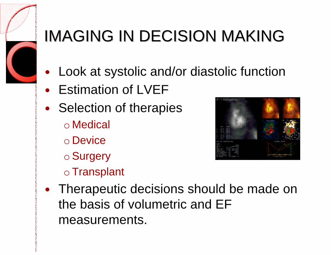
IMAGING IN DECISION MAKINGIMAGING IN DECISION MAKING
Look at systolic and/or diastolic function Estimation of LVEF Selection of therapies
o Medicalo Deviceo Surgeryo Transplant
Therapeutic decisions should be made on the basis of volumetric and EF measurements.
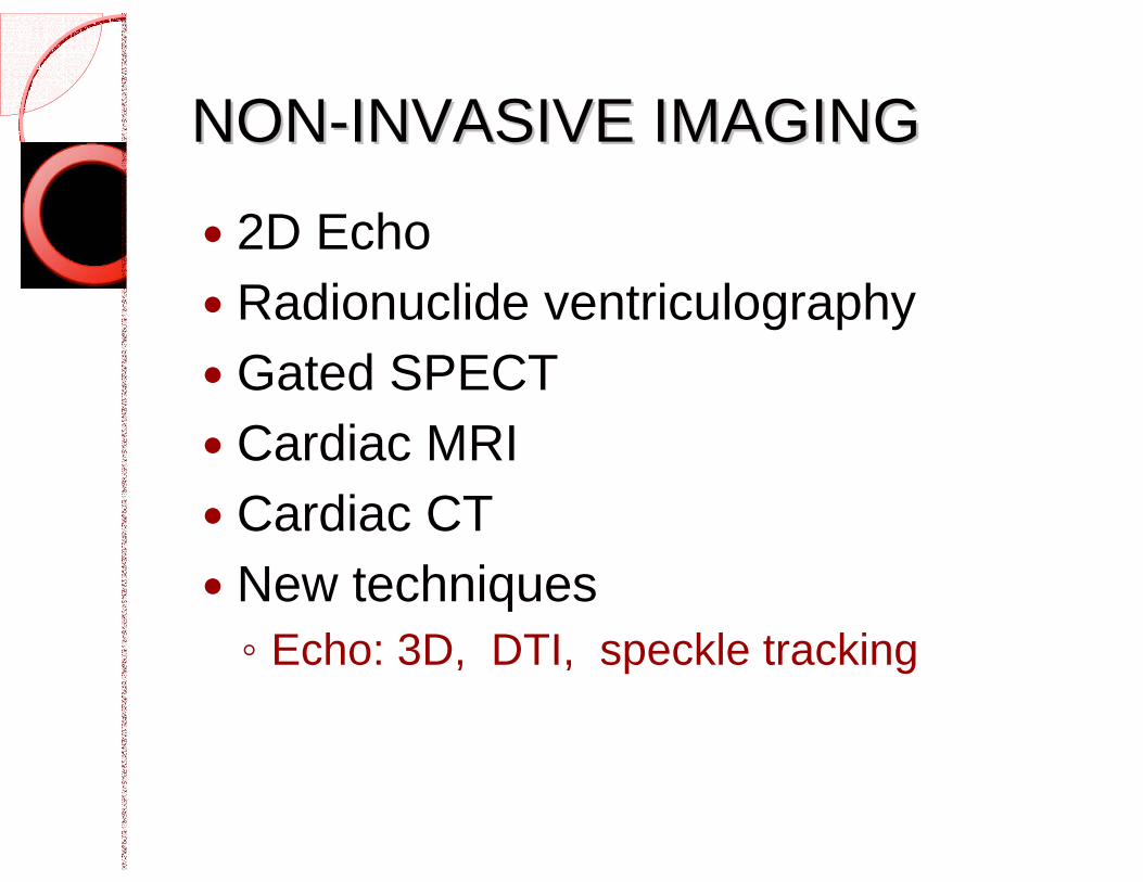
NONNON--INVASIVE IMAGINGINVASIVE IMAGING
2D Echo Radionuclide ventriculography Gated SPECT Cardiac MRI Cardiac CT New techniques◦ Echo: 3D, DTI, speckle tracking
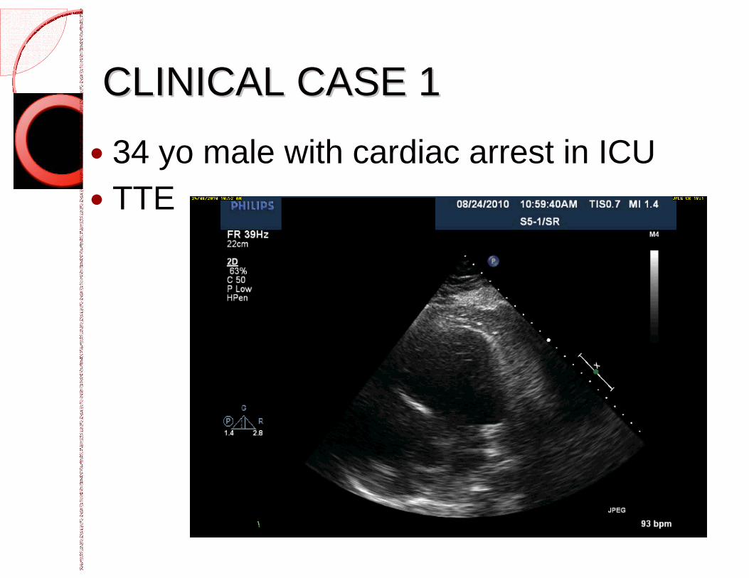
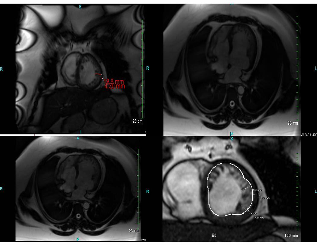
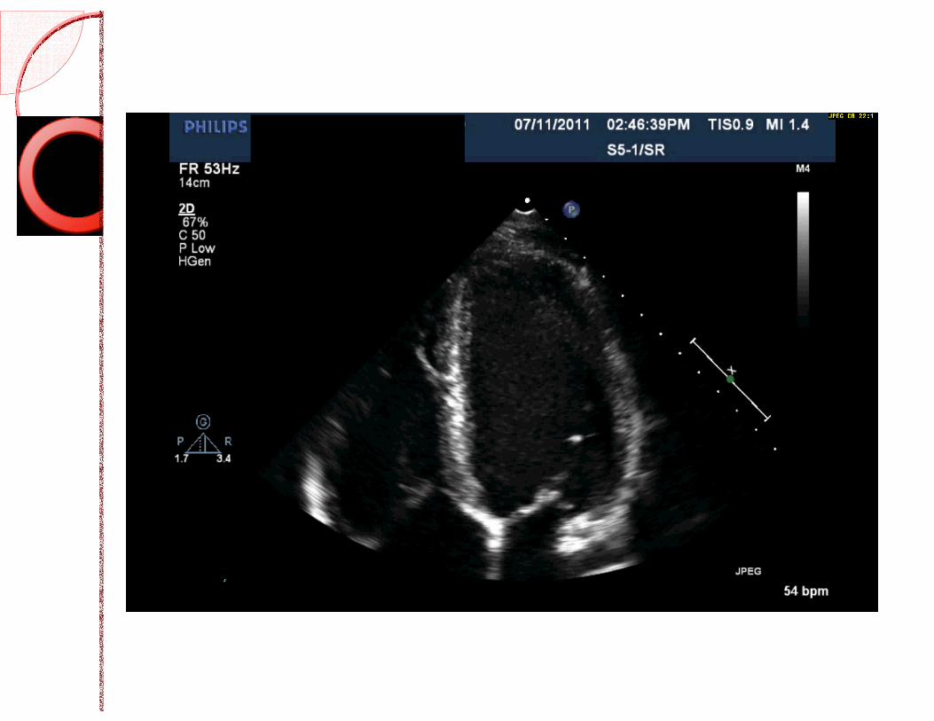
CLINICAL CASE 1CLINICAL CASE 1
34 yo male with cardiac arrest in ICU TTE
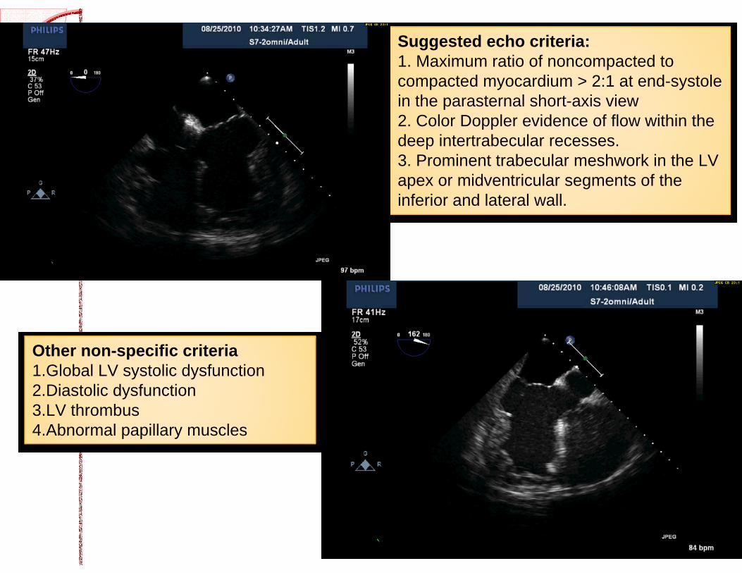
Suggested echo criteria:1. Maximum ratio of noncompacted to compacted myocardium > 2:1 at end-systole in the parasternal short-axis view2. Color Doppler evidence of flow within the deep intertrabecular recesses.3. Prominent trabecular meshwork in the LV apex or midventricular segments of the inferior and lateral wall.
Other non-specific criteria1.Global LV systolic dysfunction2.Diastolic dysfunction3.LV thrombus4.Abnormal papillary muscles
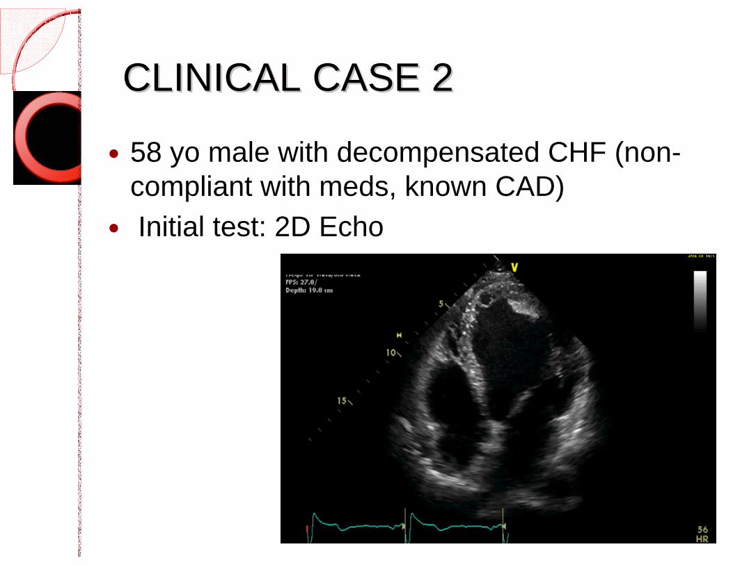
CLINICAL CASE 2CLINICAL CASE 2
58 yo male with decompensated CHF (non-compliant with meds, known CAD)
Initial test: 2D Echo
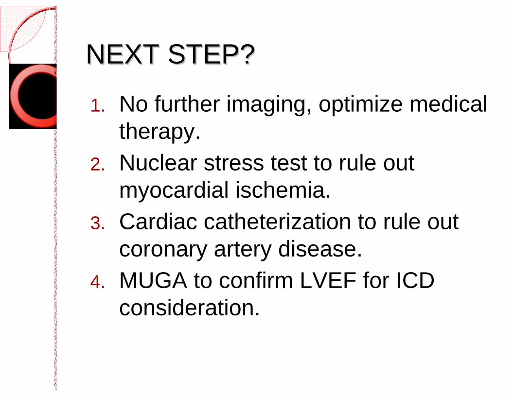
NEXT STEP?NEXT STEP?
1. No further imaging, optimize medical therapy.
2. Nuclear stress test to rule out myocardial ischemia.
3. Cardiac catheterization to rule out coronary artery disease.
4. MUGA to confirm LVEF for ICD consideration.
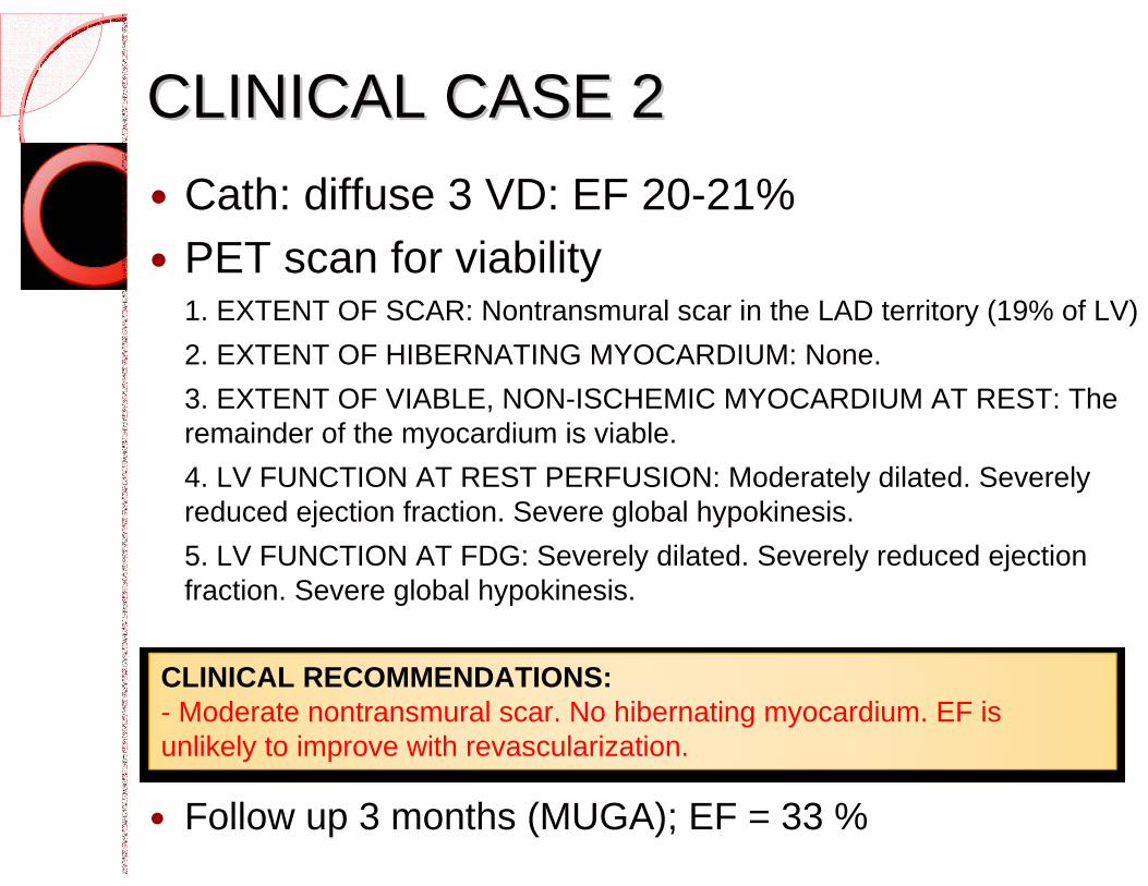
CLINICAL CASE 2CLINICAL CASE 2 Cath: diffuse 3 VD: EF 20-21% PET scan for viability
1. EXTENT OF SCAR: Nontransmural scar in the LAD territory (19% of LV)2. EXTENT OF HIBERNATING MYOCARDIUM: None.3. EXTENT OF VIABLE, NON-ISCHEMIC MYOCARDIUM AT REST: The remainder of the myocardium is viable.4. LV FUNCTION AT REST PERFUSION: Moderately dilated. Severely reduced ejection fraction. Severe global hypokinesis.5. LV FUNCTION AT FDG: Severely dilated. Severely reduced ejection fraction. Severe global hypokinesis.
Follow up 3 months (MUGA); EF = 33 %
CLINICAL RECOMMENDATIONS:- Moderate nontransmural scar. No hibernating myocardium. EF is unlikely to improve with revascularization.
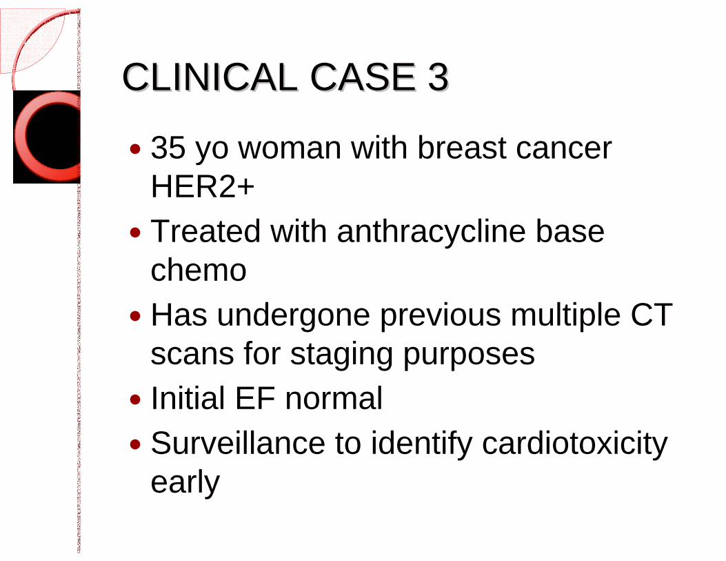
35 yo woman with breast cancer HER2+
Treated with anthracycline base chemo
Has undergone previous multiple CT scans for staging purposes
Initial EF normal Surveillance to identify cardiotoxicity
early
CLINICAL CASECLINICAL CASE 33
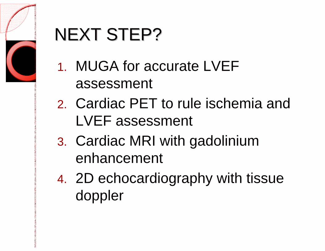
NEXT STEP?NEXT STEP?
1. MUGA for accurate LVEF assessment
2. Cardiac PET to rule ischemia and LVEF assessment
3. Cardiac MRI with gadolinium enhancement
4. 2D echocardiography with tissue doppler
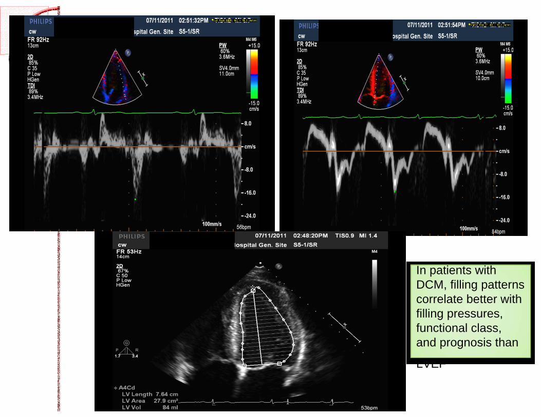
In patients withDCM, filling patterns correlate better with filling pressures, functional class, and prognosis than
LVEF
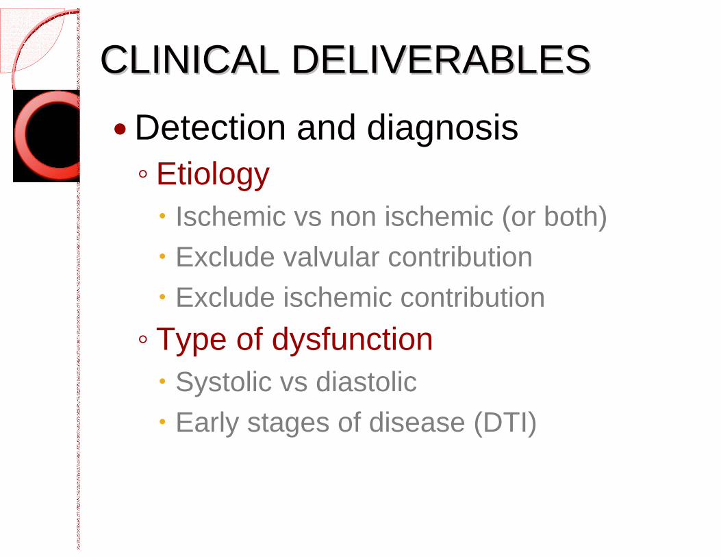
CLINICAL DELIVERABLESCLINICAL DELIVERABLESDetection and diagnosis◦ Etiology Ischemic vs non ischemic (or both) Exclude valvular contribution Exclude ischemic contribution◦ Type of dysfunction Systolic vs diastolic Early stages of disease (DTI)
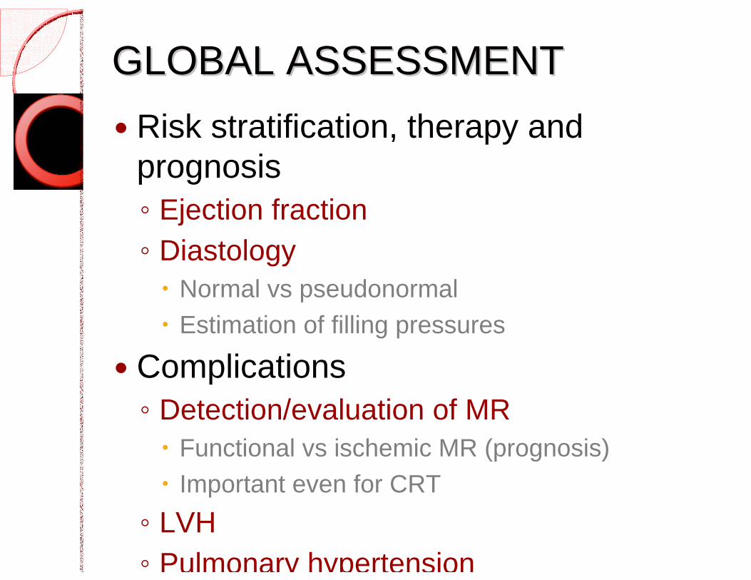
GLOBAL ASSESSMENTGLOBAL ASSESSMENT Risk stratification, therapy and
prognosis◦ Ejection fraction◦ Diastology Normal vs pseudonormal Estimation of filling pressures
Complications◦ Detection/evaluation of MR Functional vs ischemic MR (prognosis) Important even for CRT◦ LVH◦ Pulmonary hypertension
THE EVOLUTION OF ECHOTHE EVOLUTION OF ECHO Contrast echo to better delineate the
endocardial border◦ Scattering beam and oscillation of microbubbles
Doppler tissue imaging (DTI)◦ Low-velocity frequency shifts of ultrasound
waves to calculate myocardial velocity. ◦ Measurement of both regional and global LV
function through the assessment of myocardial velocity data using the basal segments.◦ Even if technically difficult study
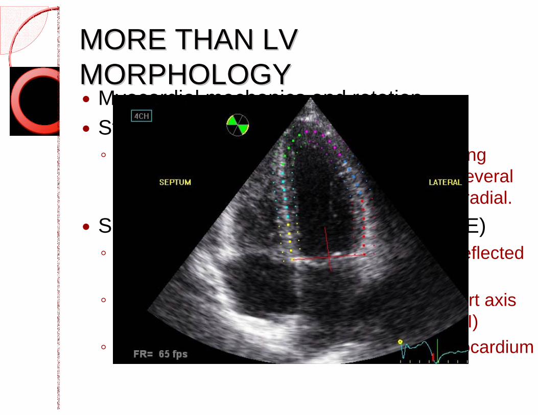
MORE THAN LV MORE THAN LV MORPHOLOGYMORPHOLOGY Myocardial mechanics and rotation Strain (function of TDI or stress echo)◦ Reflects deformation of the myocardium during
contraction/relaxation pattern calculated in several dimensions; longitudinal, circumferential, or radial.
Speckle-tracking echocardiography (STE)◦ Quantify myocardial motion in 2D by using reflected
particles as speckle tracked fram by frame◦ LV maximal internal dimension long axis/short axis
at end-diastole (not angle dependent like TDI)◦ Assessment of torsion or twist (layers of myocardium
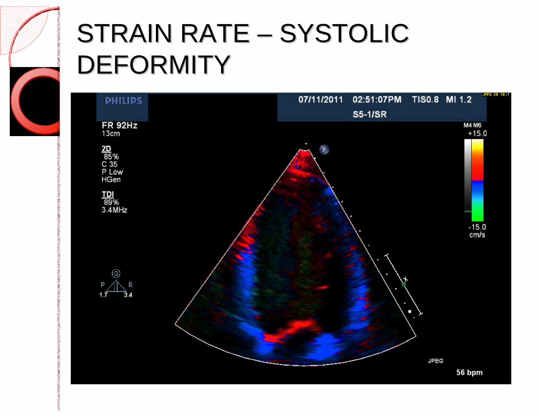
STRAIN RATE STRAIN RATE –– SYSTOLIC SYSTOLIC DEFORMITYDEFORMITY
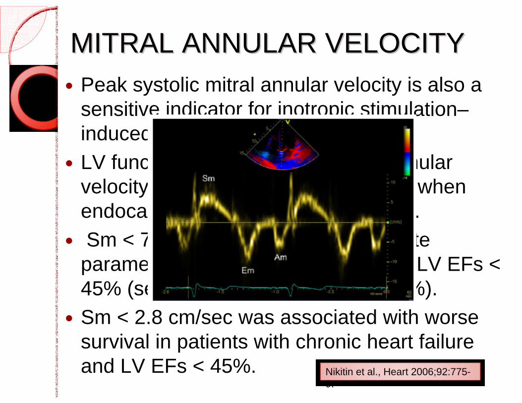
MITRAL ANNULAR VELOCITYMITRAL ANNULAR VELOCITY Peak systolic mitral annular velocity is also a
sensitive indicator for inotropic stimulation–induced alterations in LV contractility.
LV function assessment by mitral annular velocity on DTI is valuable especially when endocardial delineation is suboptimal.
Sm < 7 cm/sec was the most accurate parameter in identifying patients with LV EFs < 45% (sensitivity, 93%; specificity, 87%).
Sm < 2.8 cm/sec was associated with worse survival in patients with chronic heart failure and LV EFs < 45%. Nikitin et al., Heart 2006;92:775-
9.
Accuracy and reliability Structural◦ LV mass and geometry
Functional◦ LVEF (ESV and EDV)◦ LV size◦ Filling pressures E wave, A wave, E/A ratio, A width, DT◦ Filling characteristics◦ RV function◦ ?MR
COMPLETE ASSESSMENT COMPLETE ASSESSMENT --ECHOECHO
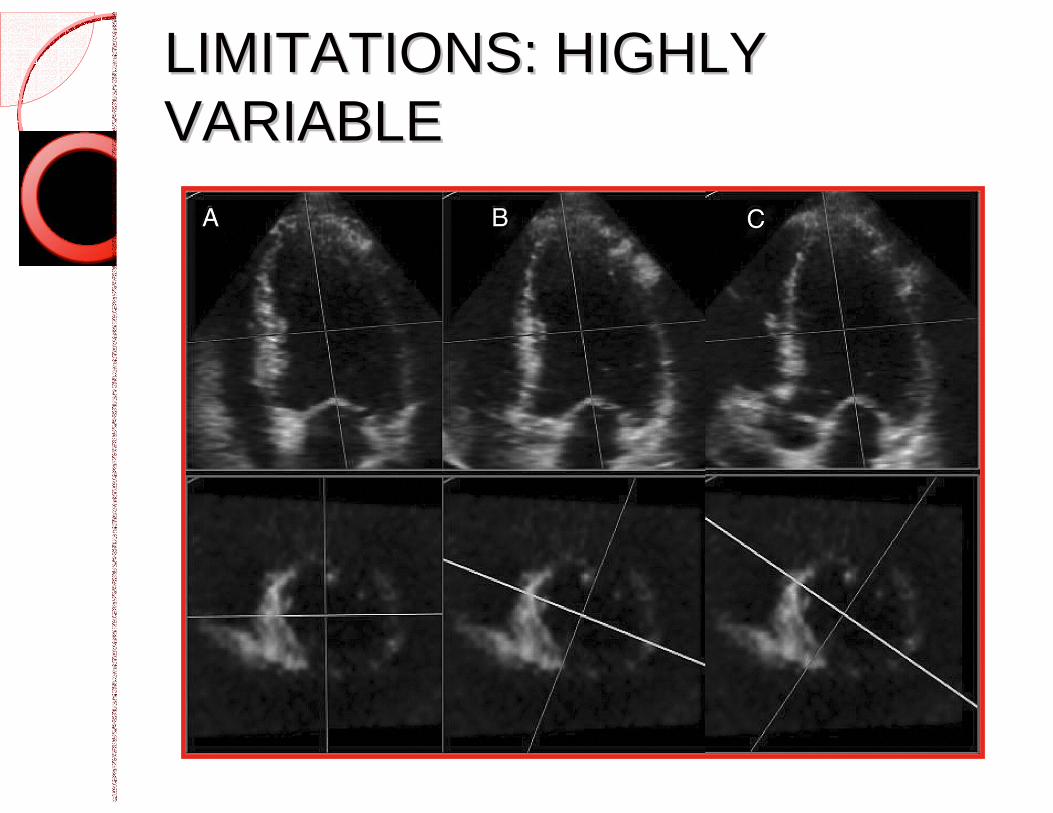
LIMITATIONS: HIGHLY LIMITATIONS: HIGHLY VARIABLEVARIABLE
LIMITATIONS: 2D IMAGINGLIMITATIONS: 2D IMAGING Standard 2D imaging planes are limiting Echo: modified Simpson’s (semi-
automated) Solutions
o Scintigraphic counts: attenuation and “overlying chambers”
o HR variability (arrhythmias)o 3D imaging
o Echo, MRI, single photon SPECT, cardiac CTo MRI: better spatial resolution, entire LV measuredo CT: single breath hold (radiation)o 3D echo very comparable with MRI for certain
measurements
ROLE OF LV FUNCTION IN HF ROLE OF LV FUNCTION IN HF
Diagnosis of early stage disease Imaging in new-onset HF Advanced HF Sequential imaging as surveillance
EARLY STAGE DISEASEEARLY STAGE DISEASE
Early detection of HF◦ Echo is the mainstay Consider strain/speckle tracking
Diastolic dysfunction◦ LV filling abnormalities◦ LA volumes
LVH◦ 3D techniques more accurate and
reproducible◦ Pathologic vs athlete’s heart ,HCM,
hypertensive heart disease
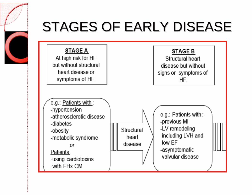
STAGES OF EARLY DISEASESTAGES OF EARLY DISEASE
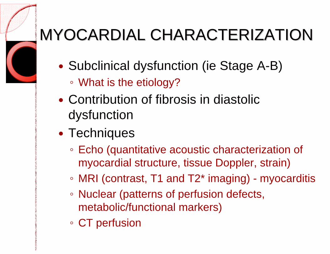
MYOCARDIAL CHARACTERIZATIONMYOCARDIAL CHARACTERIZATION
Subclinical dysfunction (ie Stage A-B)◦ What is the etiology?
Contribution of fibrosis in diastolic dysfunction
Techniques◦ Echo (quantitative acoustic characterization of
myocardial structure, tissue Doppler, strain)◦ MRI (contrast, T1 and T2* imaging) - myocarditis◦ Nuclear (patterns of perfusion defects,
metabolic/functional markers)◦ CT perfusion
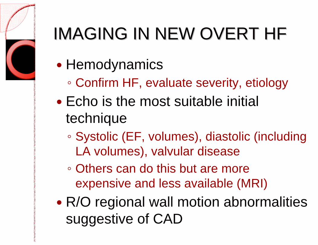
IMAGING IN NEW OVERT HFIMAGING IN NEW OVERT HF
Hemodynamics◦ Confirm HF, evaluate severity, etiology
Echo is the most suitable initial technique◦ Systolic (EF, volumes), diastolic (including
LA volumes), valvular disease◦ Others can do this but are more
expensive and less available (MRI) R/O regional wall motion abnormalities
suggestive of CAD
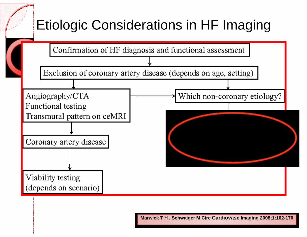
Etiologic Considerations in HF Imaging
Marwick T H , Schwaiger M Circ Cardiovasc Imaging 2008;1:162-170
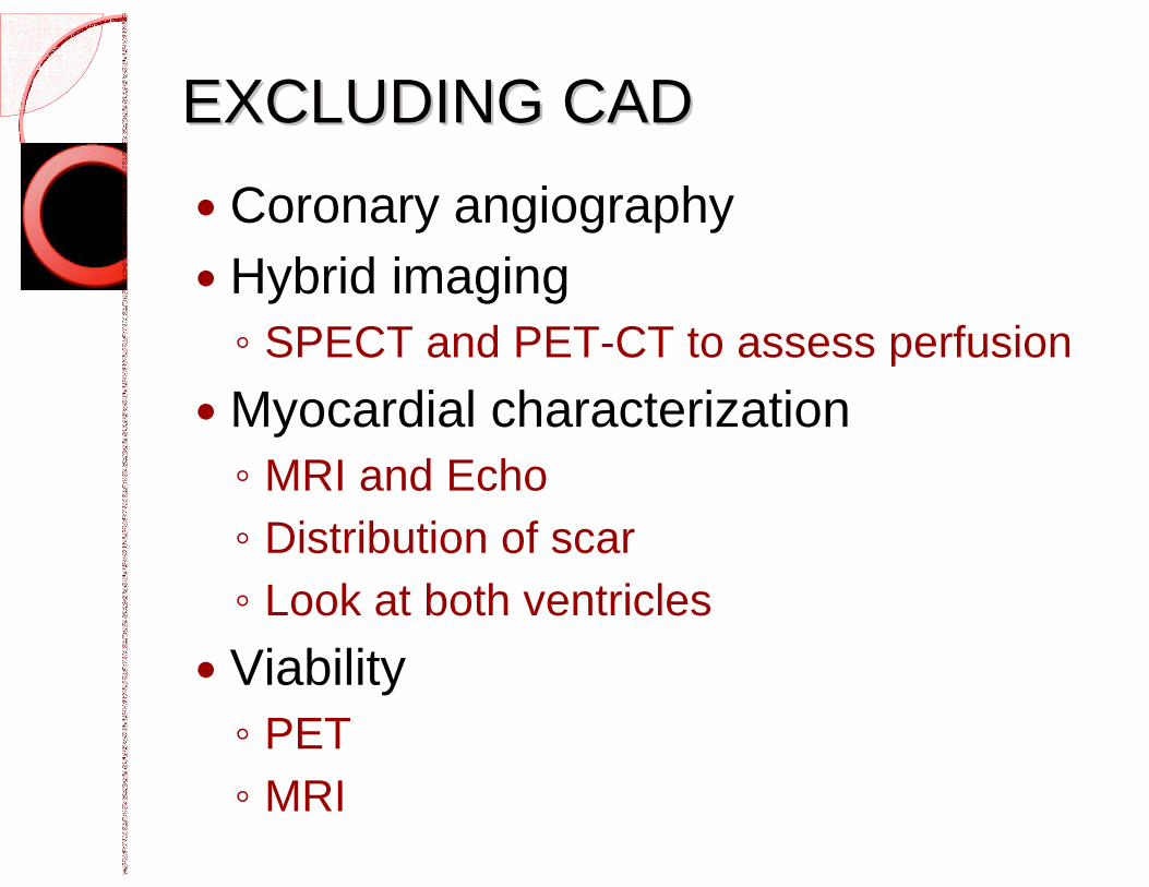
EXCLUDING CADEXCLUDING CAD Coronary angiography Hybrid imaging◦ SPECT and PET-CT to assess perfusion
Myocardial characterization◦ MRI and Echo◦ Distribution of scar◦ Look at both ventricles
Viability◦ PET◦ MRI
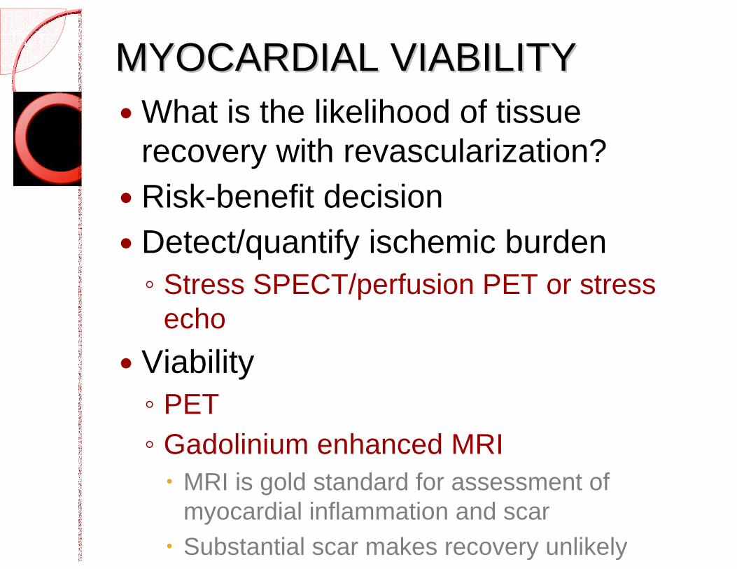
MYOCARDIAL VIABILITYMYOCARDIAL VIABILITY What is the likelihood of tissue
recovery with revascularization? Risk-benefit decision Detect/quantify ischemic burden◦ Stress SPECT/perfusion PET or stress
echo Viability◦ PET◦ Gadolinium enhanced MRI MRI is gold standard for assessment of
myocardial inflammation and scar Substantial scar makes recovery unlikely
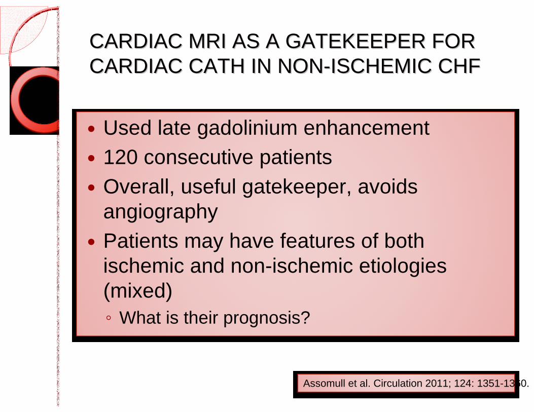
CARDIAC MRI AS A GATEKEEPER FOR CARDIAC MRI AS A GATEKEEPER FOR CARDIAC CATH IN NONCARDIAC CATH IN NON--ISCHEMIC CHFISCHEMIC CHF
Used late gadolinium enhancement 120 consecutive patients Overall, useful gatekeeper, avoids
angiography Patients may have features of both
ischemic and non-ischemic etiologies (mixed)◦ What is their prognosis?
Assomull et al. Circulation 2011; 124: 1351-1360.
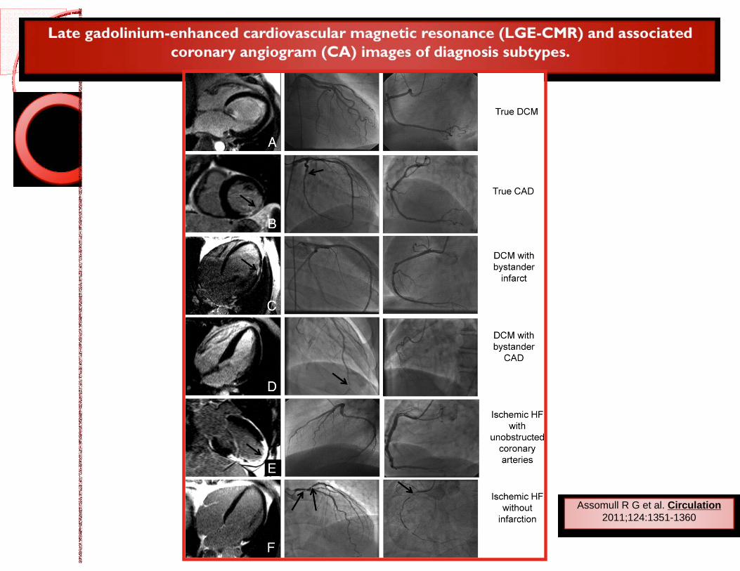
Assomull R G et al. Circulation 2011;124:1351-1360
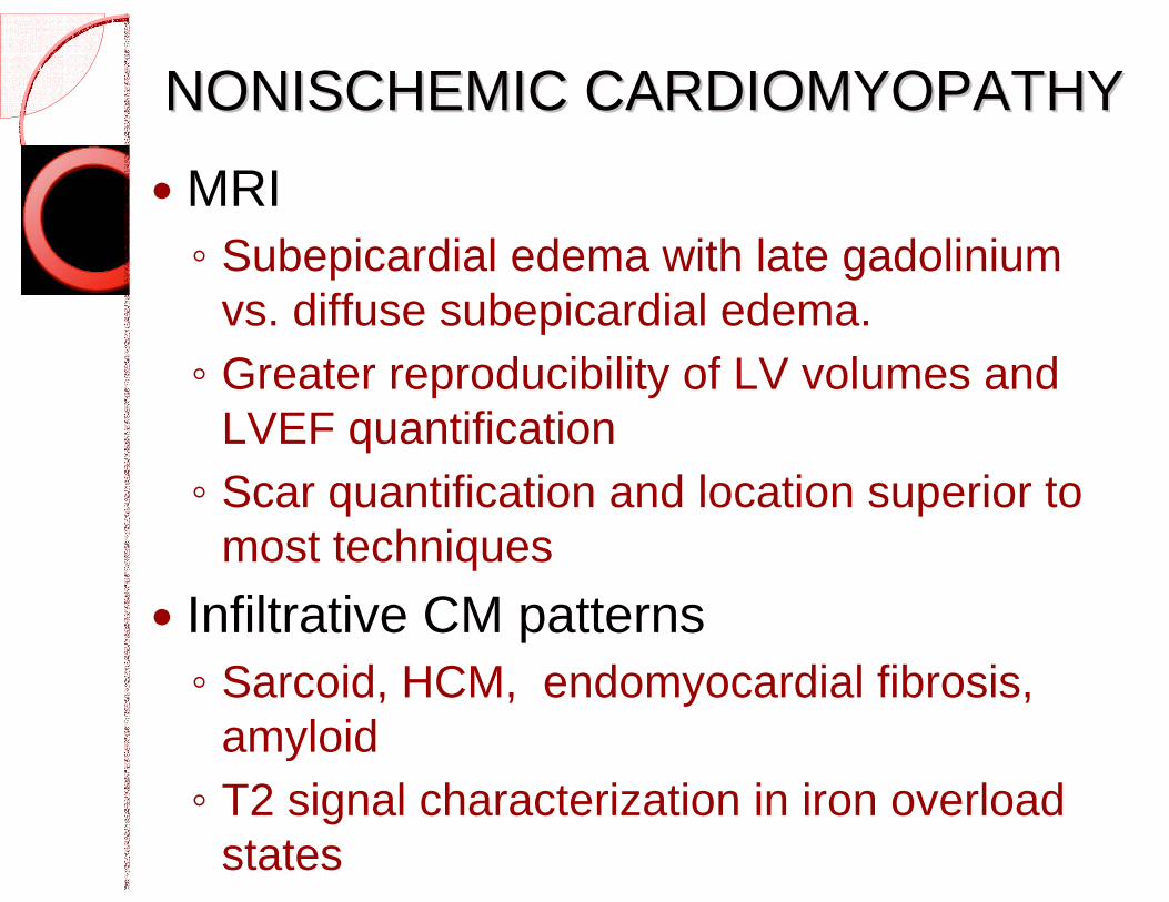
NONISCHEMIC CARDIOMYOPATHYNONISCHEMIC CARDIOMYOPATHY
MRI◦ Subepicardial edema with late gadolinium
vs. diffuse subepicardial edema. ◦ Greater reproducibility of LV volumes and
LVEF quantification◦ Scar quantification and location superior to
most techniques Infiltrative CM patterns◦ Sarcoid, HCM, endomyocardial fibrosis,
amyloid◦ T2 signal characterization in iron overload
states
MYOCARDITISMYOCARDITIS
Clinical assessment◦ Viral illness, inflammatory biomarkers◦ “Troponitis”
MRI◦ Spatial resolution plus late enhancement
can more precisely identify irreversible injury◦ Can be used to monitor therapy in such
diseases as sarcoid and CTD
ADVANCED HFADVANCED HF
Selection for ICD Therapy◦ LVEF 30-35%◦ Which technique? (Echo vs. MUGA)◦ 3D techniques less variable
Nuclear gated scan vs MRI◦ Accurate and reliable
Other prognostic markers◦ Cardiac sympathetic imaging with I131-
MIBG may be additive
ADVANCED HFADVANCED HF
Clinical evaluation before CRT (on optimal medical therapy)◦ LV dysfunction◦ Functional limitation (NYHA)◦ ECG evidence of dyssynchrony
Is the LVEF really less than 30% ?◦ Especially in close to cutoff values
MUGAMUGA Advantages◦ High accuracy and
reproducibility.◦ Measurements do not rely
on geometric assumptions.◦ Global and regional LV
systolic function.◦ Phase analysis of
segmental ventricular contraction conveys information for regional dysnergy.◦ Patient's body habitus is not
limiting.◦ Not time consuming
Disadvantages◦ Radiation◦ Overlapping of
structures◦ Improper visualization
of septum◦ ECG gating◦ Limited analysis of
other structures.
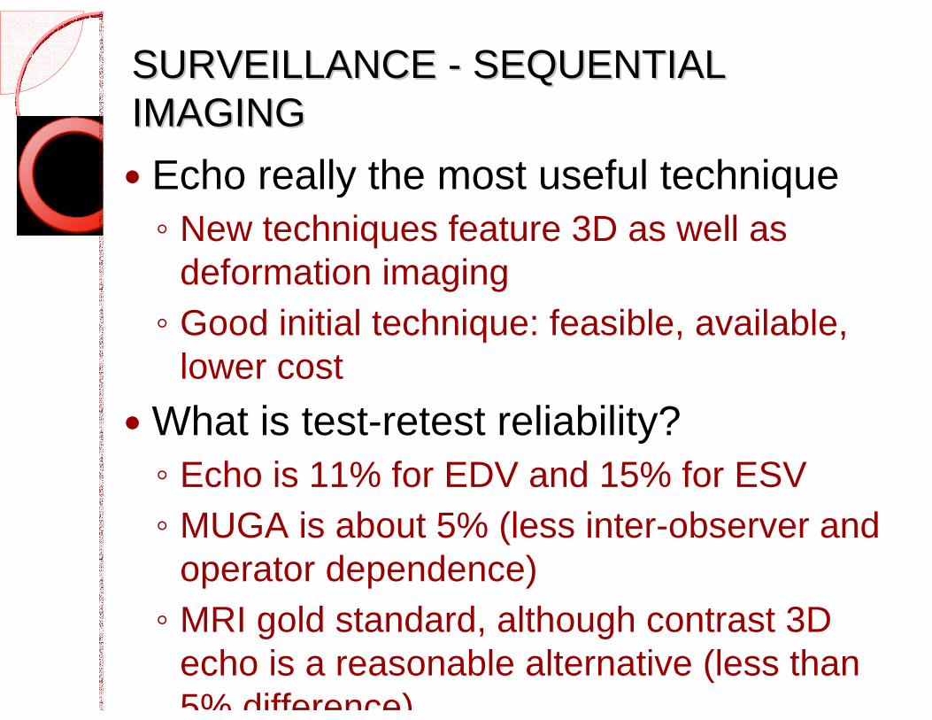
SURVEILLANCE SURVEILLANCE -- SEQUENTIAL SEQUENTIAL IMAGINGIMAGING Echo really the most useful technique◦ New techniques feature 3D as well as
deformation imaging ◦ Good initial technique: feasible, available,
lower cost What is test-retest reliability?◦ Echo is 11% for EDV and 15% for ESV ◦ MUGA is about 5% (less inter-observer and
operator dependence)◦ MRI gold standard, although contrast 3D
echo is a reasonable alternative (less than 5% difference)
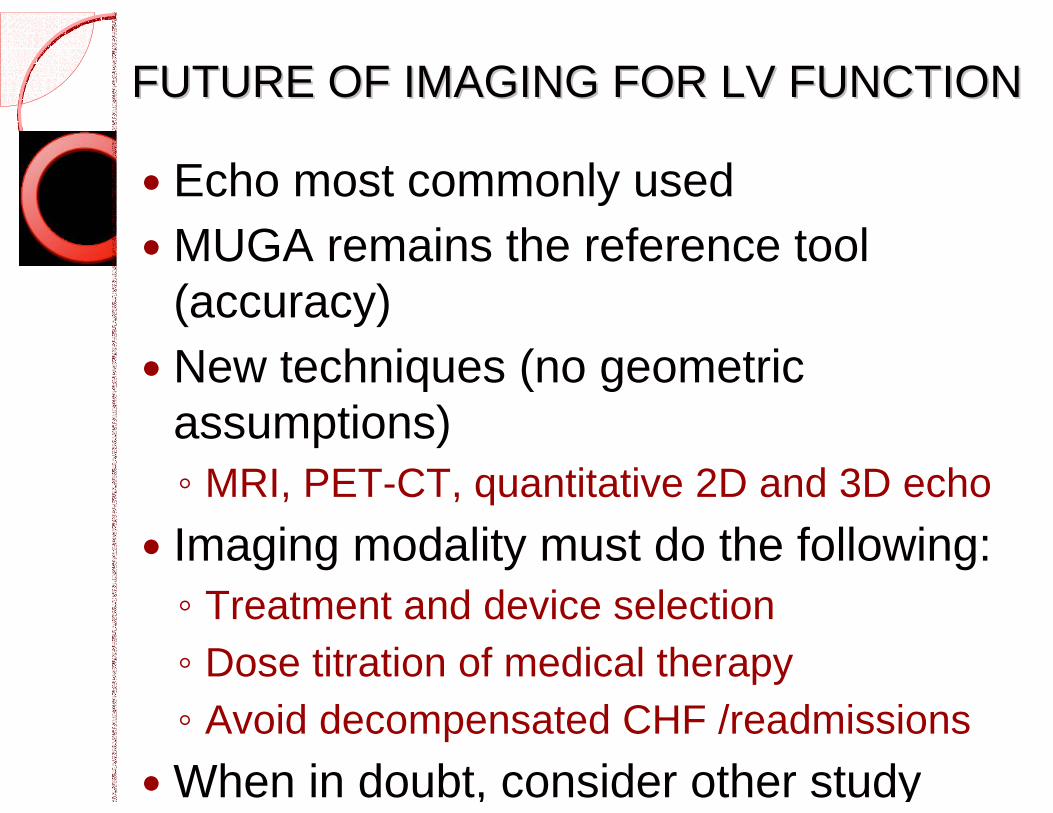
FUTURE OF IMAGING FOR LV FUNCTIONFUTURE OF IMAGING FOR LV FUNCTION
Echo most commonly used MUGA remains the reference tool
(accuracy) New techniques (no geometric
assumptions)◦ MRI, PET-CT, quantitative 2D and 3D echo
Imaging modality must do the following:◦ Treatment and device selection◦ Dose titration of medical therapy◦ Avoid decompensated CHF /readmissions
When in doubt, consider other study
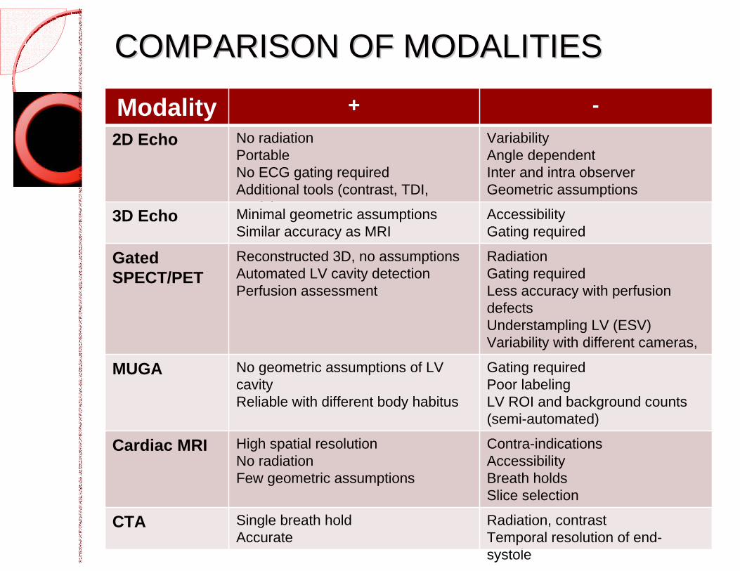
COMPARISON OF MODALITIESCOMPARISON OF MODALITIES
Modality + -2D Echo No radiation
PortableNo ECG gating requiredAdditional tools (contrast, TDI, strain)
VariabilityAngle dependentInter and intra observerGeometric assumptions
3D Echo Minimal geometric assumptionsSimilar accuracy as MRI
AccessibilityGating required
Gated SPECT/PET
Reconstructed 3D, no assumptionsAutomated LV cavity detectionPerfusion assessment
RadiationGating requiredLess accuracy with perfusion defectsUnderstampling LV (ESV)Variability with different cameras, softwareMUGA No geometric assumptions of LV
cavity Reliable with different body habitus
Gating requiredPoor labelingLV ROI and background counts (semi-automated)
Cardiac MRI High spatial resolutionNo radiationFew geometric assumptions
Contra-indicationsAccessibilityBreath holdsSlice selection
CTA Single breath holdAccurate
Radiation, contrastTemporal resolution of end-systole
Recommended