TRANSDUCTION IN ESCHERICHIA COLI K-12
M. L. MORSEa, ESTHER M. LEDERBERG, AND JOSHUA LEDERBERG
Department of Genetics, University of Wisconsin, Madison, Wisconsin Received August 26, 1955
SYSTEM of genetic transduction has been discovered in the sexually fertile A K-12 strain of Escherichia coli. This transduction is mediated by lambda, a temperate phage for which K-12 is normally lysogenic.
The distinctive features of the lambda-K-12 system include the following: (1) The transductions are limited to a cluster of genes for galactose fermentation. The GaZ loci are closely linked to each other and to Lp, the locus for lambda-mainte- nance. (2) The transducing competence of lambda depends on how it is prepared. Competent lambda is produced by induction of lysogenic bacteria; lambda har- vested from infected, sensitive hosts is incompetent. (3) The transduction clones are often heterogenotic, that is, heterozygous for the Gal genes which they con- tinue to segregate. Technical advantages of the lambda system include recombina- tional analysis by the sexual cycle and the availability of lysates in which nearly every lambda particle is competent.
MATERIALS AND METHODS
Cultures The origin and history of the Escherichia coli K-12 cultures studied have already
been described (E. LEDERBERG 1950, 1952; LEDERBERG and LEDERBERG 1953). The emphasis will be placed here on the Gal loci (+ = fermenting galactose; - = nonfermenting) and on the locus which controls the maintenance of lambda (Lpl).
The phenotypes of cultures with different alleles of Lpl are as follows:
Lpl* culture (sensitive) Lpl+ culture (lysogenic) Lplr culture (immune)
Lysed by lambda Lyses L)I' culture
Yes no no Yes no no
Regardless of their Lpl genotype, cultures have been found to adsorb lambda. Thus Lpl+ and LplT are resistant to lysis by lambda in spite of their ability to ad- sorb the phage. In contrast with this, mutants resistant to lambda-2, a virulent mutant of lambda, are resistant because they do not adsorb either lambda or lambda-2 under the experimental conditions used here.
Media The media used include: broth, Difco penassay; agar for phage assay, Difco
nutrient agar with 0.5 percent NaC1; indicator medium, EMB agar plus one percent
Paper No. 589 of the Department of Genetics. This work has been supported a t various times by the Atomic Energy Commission, Contract AT(ll-l)-64, Proj. 10; a researchgrant (C2157) from the National Cancer Institute, Public Health Service and grants from the Research Committee, University of Wisconsin, with funds allotted by the Wisconsin Alumni Research Foundation.
2 Predoctoral research fellow of the National Science Foundation. 1953-54.
M. L. MORSE, E. M. LEDERBERG AND J. LEDERBERG 143
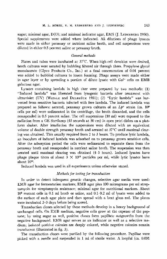
sugar; minimal agar, D(0); and minimal indicator agar, EMS (J. LEDERBERG 1950). Special supplements were added where indicated. ,411 dilutions of phage lysates were made in either penassay or nutrient saline broth, and cell suspensions were diluted in either 0.5 percent saline or penassay broth.
General methods
Plates and tubes were incubated a t 37°C. When high cell densities were desired, broth cultures were aerated by bubbling filtered air through them. Propylene glycol monolaurate (Glyco Products Co., Inc.) a t a final concentration of 0.01 percent was added to bubbled cultures to lessen foaming. Phage assays were made either in agar layer or by spreading a portion of dilute lysate with Gal- cells on EMB galactose agar.
Lysates containing lambda in high titer were prepared by two methods: (1) “Induced lambda” was liberated from lysogenic bacteria after treatment with ultraviolet (UV) (WEIGLE and DELBRUCK 1951); (2) “Lytic lambda” was har- vested from sensitive bacteria infected with free lambda. The induced lambda was prepared as follows: aerated, penassay grown cultures of an Lp+ strain (ca. lo9 cells per ml) were sedimented in the centrifuge, the broth discarded, and the cells resuspended in 0.5 percent saline. The cell suspensions (10 ml) were exposed to the radiation from a GE Sterilamp (45 seconds a t 50 cm) in open petri dishes on a plat- form shaker. After irradiation the suspensions were diluted with an equivalent volume of double strength penassay broth and aerated a t 37°C until maximal clear- ing was obtained. This usually required from 2 to 3 hours. To produce lytic lambda, an inoculum of induced lambda was adsorbed on to penassay grown sensitive cells. After the adsorption period the cells were sedimented to separate them from the penassay broth and resuspended in nutrient saline broth. The suspension was then aerated until maximal clearing was obtained (4-5 hours). Induced lysates have phage plaque titers of about 3 X 1Olo particles per ml, while lytic lysates have about lolo.
Induced lambda was used in all experiments unless otherwise stated.
Methods for testing for transduction
In order to detect infrequent genetic changes, selective agar media were used: EMB agar for fermentation markers; EMB agar plus 100 micrograms per ml strep- tomycin for streptomycin resistance; minimal agar for nutritional markers. About 108 mutant cells in 0.1 ml broth or saline, and 0.14.2 ml of lysate were added to the surface of each agar plate and then spread with a bent glass rod. The plates were incubated 2-3 days before being scored.
Transduction clones selected by these methods develop in a heavy background of unchanged cells. On EMB medium, negative cells grow at the expense of the pep- tone; by using sugar as well, positive clones form papillate outgrowths from the negative background. EMB agar serves as an indicator as well as a selective me- dium; isolated positive colonies are deeply colored, while negative colonies remain translucent (illustrated in fig. 3).
The transduction clones were purified by the following procedure. Papillae were picked with a needle and suspended in 1 ml of sterile water. A loopful (ca. 0.001

144 TRANSDUCTION IN E. COLI
ml) of this suspension was then streaked upon a portion of another plate of the EMB agar. These primary dispersals of the transduction clones were nearly always mixed. Direct picking and streaking, or spotting without any purification cannot be trusted. From the primary streaks a single colony that looked pure was picked to water and streaked as before. This operation was repeated once again, and a single colony from the last streaking was taken to represent the transduction clone. In addition to freeing the transduction clone from unchanged background cells, this method of purification may also act selectively within an unstable clone. Pick- ing apparently pure colonies leads to an overestimate of the fraction of non-segregat- ing clones.
RESULTS
The transduclions Although a number of different loci affecting diverse portions of the genotype
were tested, only genes of a cluster of loci for galactose fermentation were trans- duced by lambda lysates (MORSE 1954). The Gal loci, of which about seven have been investigated thus far, are closely linked to one another (less than one percent recombination) and to LpI, the locus for lambda maintenance (LEDERBERC and LEDERBERC 1953, and unpublished).
The transformation of Gal- cells to Gal+ by induced lambda is illustrated in figure 1. Each papilla is a clone of galactose fermenting cells; on the area of the plate to which lysate was added, most of the Gal+ papillae are transduction clones. The
0 5
*. .
FIGVRE 1.-The procluction of Col+ papillre from a Gal- 1)ackgrountl of cells bp r lamIda lysate. Left, the control, no lysate rtlcletl. Right, 0.1 ml of lysate from a Gal+ culture. Some of the papillae have ken picked with a needle.

M. L. MORSE, E. M. LEDERBERG A N D J. LEDERBERG
Recipient culture Lysate of:
Gal+ Gall- Gal+ Galz- Gal+ Gak- Gal+ Gala- Gal+ Gal4- Gal+ Gal+ Gal+ Gal+
TABLE 1 $ Gal- cdlures
Lambda titer in 1010 per ml
1.4 2.4 1.4 4 .9 1.4 1 . 7 1 .4 1.7 2.3 1.7 2.3 2.3 1.4 1.6
lysates of Gal+
Number of Gal+ papillae
Control (no lysate)*
2 2
20 20 47 47 4
163 10 10 5 40 29 28
1.1 ml lysate
405 2
356 10
394 50
2112 86
3020 18
1296 161 129 92
145
Sal+ papillae per 107 lambda
2 .9
2.4
2.5
15.1
13.1
5.6 0.5 0.7 0.4
-
- - -
* The Gal+ papillae on the control are spontaneous reversals of phenotype. t Different stocks. 2 Different experiments.
quantitative relationships are illustrated in figure 2. The data can be summarized: (1) Regardless of the Lpl genotype of the recipient, transductions were obtained; (2) with each genotype the number of transductions was proportional to the amount of lysate plated; (3) Lps recipient cultures gave 5 to 10-fold more papillae per unit of lysate than either Lpl+ or Lpl'. Further, the transducing activity of lysates (which contain 1O1O lambda per ml) varies according to the number of cells plated: (1) with Lpl+ Gal- cultures there is a two-fold increase between 106-109 cells per plate, with a plateau of maximal yield around 108 cells per plate; ( 2 ) Lp8 Gal- re- cipient cultures show about a six-fold increase over a similar range of cell platings, with the highest yield at the highest cell density.
The transducing activity of lysates is specific; that is, a lysate of a Galz- culture will not transform Gal,- cultures (table 1) but Gal+ papillae were found with a Gal,- culture. The specificity is extended further in that some galactose positive phenotypic reversals of a Gal- culture give lysates with transducing activity on the original Galz- indicator (table 2). The different types of phenotypic reversals may be understood under the following hypothesis: (1) reverse mutations (Gal, to Gal,+) yield cultures that give active lysates, and (2) suppressor mutations (Gal, Sup- pressor- to Gal,- Suppressor+) will yield incompetent cultures when the suppressor lies outside the region transduced.
From the data in table 1 and figure 2 the ratio of the transducing particles to the lambda particles in a lysate may be obtained. Lp8 recipient cultures give about one transduction per lo6 lambda; Lpf recipients, one per lo7. One per 106-107 lambda will be referred to as LFT (low frequency of transduction).

146 TRANSDUCTION IN E. COLI
Lp+ recipient culture Numbers of Gal' papillae observed
Lysate of reversion Control (no lysate) I 0.1 ml lysate
Gall- Gd2-
Gd4-
0 10 6
39 25
648 96
552 204 291
,,, 3000 i 2000
4 0,
100
. Gal4- ~ p '
/ +
Gal4- Lp
Lambda PARTICLES FIGURE 2.-Proportionality between amount of lambda lysate (LFT) plated and number of
papillae formed from L p , Lp+ and Lpr Gal- cultures. The ratio of papillae to lambda particles is 10-6 for an Lp" culture, lo-' for Lp+ and Lpr cultures.
Examinalion of the Gal+ clones formed by transduclion After purification the transduction clones were examined for changes a t loci other
than the Gal series. A number of markers were examined, including fermentative, nutritional, and phage and drug resistance mutations. The only changes a t other loci were Lps to Lp+ in lambda sensitive recipients, and occasionally Lp' to Lp+ in lambda immune cultures. Transduction clones from Lp+ recipients were invari- ably Lp+.
To determine whether lysogeny was causally related to transduction, a recon- struction experiment was done. To a mixture of lysate and Gal- Lps cells, Gal+ Lpa cells labelled with a mutant character were added to estimate the frequency of chance lysogenization in the untransformed cells in a transduction mixture. After papillae had formed, they were picked, purified, and on the basis of the differential label, divided into: (1) the inserted Gal+, and (2) the transductions. The frequency

11. I.. MORSE, E. If. LEDERBEKG AND J. LEDERBERC 145
TABLE 3 Comparison o/ the lysogenkalion o/ lransjortned and twn-lrans/ormed seirsitke .drains:
reconslrtcclion e.rperiiirert -~ Type rcrowrnl from mixture' of Lp' bacteria ancl ' Sumtwr of cloncs t Pcrccnt of clones
LFT lysate cxnmincd lpsogcnizecl
I I 68.5
i 72.5 Inserted Cal+Iac"S* 46
40 Recipient Gal-lnc-s' (non-transformed) Transduction Ga1'I.c-s' I 103 I 100.
10" Cal-IAc-S', 100 Cal+I~c+S*, 109 Iaml)cla particles.
of lysogeny was determined in the two classes, antl in the Gal- background. Whereas unchanged Gal- cells antl the inserted Gal+ were each only 70 percent lysogenized, the transduction clones were 100 percent lysogeni7Rd (table 3).
When I-#' cultures were used as recipients, 14/112 (12 percent) of transduction clones formed were I$+. Although the fraction is small, all previous attempts to lysogenize these cultures have heen unsuccessful. The isolation of transduction clones evidently selects for these cells that have Imn infected with lambda par- ticles from the input lysate.
The original Gal+ strain and spontaneous reversions of the Gal- mutants have all been stable in ordinary culture. However, the Gal+ clones formecl by transduc- tion are unstable for galactose fermentation as shown by the recurrence of negative and mosaic colonies (fig. 3). Despite many serial single colony isolations the galac- tose-positives continue to segregate galactose negative progeny. They behave as if
*. *. e * t .
FIGURE 3.--1C11 II g:~I:~t i )w axar 1h1c spread with cell< f i r m n riilturr of :I Iictcrogcnote, showing Gal+, Gal- ancl sectoring colonics.

148 TRANSDUCTION IN E. COLI
TABLE 4 Frequency of instability for galactose fermentation among the transduction clones
Percent unstable I Recipient cells I Unstable clones/total examined
9/22 4 / 4 8 22/24 13/24 20124 29/48
28/48 16/24
618
41 83 92 54 83 60 75 58 67
heterozygous for a single gene (or short chromosome segment) and may be desig- nated as heterogenotes. Instability among the transduction clones is quite frequent; 484 of 609 clones (70 percent) were found unstable (representative data are given in table 4). This estimate is probably low because the purification procedure acts selectively against unstable clones.
The frequency of segregation has been estimated from the incidence of Gal- in small clones of heterogenotes. The probability of segregation per bacterial division is about 2 X (table 5). By repeated reisolation, however, heterogenotic lines can be maintained indefinitely.
The segregants from the heterogenotic clones were examined with regard to their Gal and Lp character. Lysates of the segregants have no transducing activity on the Gal- culture that was used as the recipient in forming the transduction clone and are therefore allelic to it. The same lysates continue to give one transduction per 106-107 lambda (LFT ratio) on non-allelic Gal- cultures. With different recipient cultures the Lp alleles of the segregants were (1) Lp+ recipient, all segregants Lp+; ( 2 ) Lps recipients, all segregants Lp+; (3) Lp' cells, the segregants were usually Lp'. In one instance, a heterogenote segregated both Lp+/Lpr and Gal+/Gal-.
Lysates prepared from the heterogenotes have two outstanding features: (1) instead of containing 1O1O lambda particles per ml, they seldom have titers higher than 5 X lo8, particularly if they originate from cultures containing few Gal- segre- gants; ( 2 ) the number of transducing particles in these lysates is often nearly equal to the number of lambda particles in the lysate (table 6). These lysates will be re- ferred to as HFT (giving a high frequency of transduction).
Transdwtions with lysates of heterogenotes Platings of highly diluted HFT lysate with Lps and Lp+ bacteria give a number
of papillae. The number of papillae obtained with Lp" cells is, however, less than that obtained with L$+. The lower yield with Lp* recipients may result a t least in part from the loss of potential transductions through lysis of the recipient cell or of some of its early progeny.
With HFT lysates it is possible to transform a large fraction of a cell population, and to observe transduction without strong selection. By adsorbing HFT lambda onto cells, diluting and plating on EMB galactose to obtain well isolated colonies

M. L. MORSE, E. M. LEDERBERG AND J. LEDERBERG 149
Heterogenote
G U J , - J / G ~ +
TABLE 5 Frequency of segregation from the heterogenoles
Number of cells in inoculum
2.11
1.50
Test clones*
Number of G a r cells
6 3 4
23 9
19 103 319
22 0
11 2 8 3
52 0
36 3 6
55
Total cells
1169 595 25 1
1252 1113 897
2750 1622 1966 237
323 176
1669 317
1236 10
1055 299 386
1965
ProbabilityTof segregation per 108 bacterial divisionst
1
1 4 3 . 6 2 4.3 6 . 6
36.8 2.0 0
8 .1 3 0 . 9 2 8.2 0 6.7 2 4 5.1
* A fully grown culture in penassay broth was diluted to give about 10 cells per ml. Twenty samples of 0.1 ml were taken up in 0.2 ml serological pipettes which were supported in a horizontal position on a tray. The pipettes were incubated at 37°C for 4.5 hours. Each pipette was then blown out on to an EMB galactose agar plate, and the inside of the pipette washed with 0.1 ml of broth. The washing was added to the plate, and the inoculum spread with a glass rod. After 18 hours
BY’*
t Using the equation a = 0.602r/N log N, (modified for the indicated units from LURIA and DELBRUCK 1943) where r = the number of Gal- segregants and N = the clone size. The probability of segregation is also estimated by the fraction of cultures containing no segregants.
incubation a t 37°C the number of Gal+ and Gal- colonies on the plates was determined.
2.3 1 N Po
a = - log- (Po = fraction of cultures with no segregants.)
In the first experiment, using N = 21° 2 .3 1024
a = __ log 1/1/19 = 2 .8 X loV3
In the second experiment, using N = 21° 2.3 1024
a = ~ log 1/1/11 = 2.6 x 10⁻³
$ The assay plates showed this culture to have Gal+: Gal- in the ratio 106:4. Of the twenty sam- ples in this experiment, one contained only Gal+, one contained only G a r , and 18, both Galf and Gal-. Only the plates that were counted are given. Nine plates were too crowded to be counted accurately.
5 The ratio of Gal+:Gal- in the parent culture was 128: 19. The twenty cultures were distributed as follows: failed to grow, 9; contained only Gal+, 1; contained both Gal+ and Gal-, 10. One plate had approximately equal numbers of Gal+ and Gal- and was assumed to have come from a mixed inoculum.

150 TRANSDUCTION IN E. COLI
W I- 4 n
.I
0.05 ML. o f HFT LYSATE
=FIGURE 4.-Proportionality between amount of HFT lysate and number of papillae formed from Lp* and Lp+ Gal- cultures. For the assays, a lysate containing 1.6 X 10⁸ phage/ml was di- luted a thousandfold.
it is possible to study individual transduction clones derived from single particle infections of isolated bacteria. A t the optimal ratio of about 10 lambda particles per cell, the fraction of cells transformed ranged from 5 to 15 percent.
Evidence that lambda i s the vector of transduction That lambda is the vector of Gal transduction is suggested by previous experi-
ments: (1) the 100 percent lysogenization of Lp8 recipients by LFT lysate trans- ductions; ( 2 ) the incidence of lysogenicity in transduction to Lpr recipients. In
TABLE 6 The high frequency of transduction ( H F T ) given by lysates of heterogenotes
Heterogenote Lysate of the heterogenote
Lambda particles per ml
1.2 x 10⁸ 5.8 x 10⁸ 5.4 x 10⁷ 7.6 x 10⁷ 1.5 x 10⁸ 7.3 x 10⁸
Transductions per ml
2.1 x 10⁷ 1.8 x 10⁷ 3 .6 x 107 4.2 x 10⁷ 7.4 x 10⁷ 2.5 x 10⁷
Transductions per lambda particle
1/5.7
1/1 .5
1/2.0
1/32*
1/1.8
1/29*
* With the exception of these cases, the cultures used for making the lysates were started from a single apparently pure Gal+ colony on EMB galactose. The lower ratio in the exceptional cases, and the higher lambda titer is probably the result of the presence in the source cultures of a larger number of Gal- segregants. Assay of the transductions was made with Lp+ cells.

M. L. MORSE, E. M. LEDERBERG AND J. LEDERBERG 151
Gall-
G a l z
Gd4-
Gall-
TABLE 7 Failure of transduction to lambda-2 resistant mutants
Recipient cells (Lp+) I Lambda-2 reaction' I Number of fermenting clones
No lysate added
sensitive 1 resistant 1 sensitive 20 resistant 14 sensitive 89 resistant 50 sensitive 2 resistant 3
0.1 ml of Gal+-lysate
426 (LFT) 2
356 14
296 57
4 107 (HFT)
addition, lambda and the transducing agent are adsorbed to about the same degree by Lp" cells, and both are inactivated by crude anti-lambda serum. More definite evidence was the failure of lambda-2 resistant cells to adsorb either lambda or trans- ducing activity or to be transformed even by HFT lysates (table 7).
Conclusive evidence that lambda is the vector of transduction is found from the behavior of single transduction clones: (1) Heterogenotes formed from HFT lysate and Lps cells a t low lambda multiplicity are always either overtly lysogenic (Lpf) or carry a defective prophage (Lp') (table 8). (2) Proportionality between number of transductions and amount of lysate a t high dilution (fig. 4). For a two-factor- system to be invoked at these dilutions, the accessory factor would have to exceed the lambda by at least lolo, which would imply a concentration of this fancied ele- ment in undiluted HFT lysate of 10l8 per ml, which should be compared with Avo- gadro's number.
Early segregation of Lp and Gal in transduction clones
HFT Gal+ lambda was mixed with a culture of Gal,- bacteria to give 2.6 X lo7 lambda and 7 X 10⁸ cells per ml, a multiplicity ratio of 0.04. The suspension was then diluted and plated on EMB Gal to give about 100 cells per plate. After 24 hours incubation, on 7 plates, a total of 8 colonies with Gal+ sectors was noted. Each of these colonies was sectored, with a large Gal- component. Each colony was restreaked, and 20 to 30 Gal- reisolated from each line. Of the 8 lines, the Gal- from 3 gave only Lp+, from 5 gave mainly Lps with a few Lp+. Ten Gal+ (heter- ogenote) colonies were also picked from each line. All of them were Lp+ and of a total of 297 Gal- segregants subsequently reisolated from these 60 heterogenotic colonies, all were Lp+ also. The frequent segregation of Lp+/Lp8 subclones from lambda-infected Lps cells has been noted previously (LEDERBERG and LEDERBERG 1953; LIEB 1953). The correlation of Lpf and Gal+ evidently extended, in the 5/8 clones that segregated both markers, to the early intraclonal progeny. Since the heterogenotes do not continue to segregate Lps, these results are economically in- terpreted on the basis of the multinucleate character of the bacterial cells. The early segregation would represent the separation of unaltered Lps Gal- nuclei from the

152 TRANSDUCTION I N E. COLI
TABLE 8 Incidence of lysogenicity in isolated lzeterogenotes
Colony type
Unaltered Gal- With Gal’
1. The transductions
Number of colonies Number of colonies examined
LP8 LP+ LP*
31 31 0 0 26 0 23 3
Number of colonies observed
Unaltered Gal- 1 With Gal+ I Gal- partially lysed Gal- cells exposed to:
Broth I 3280 I 0 I 0 2801 I 31 1 54 ~ ~ ~ i y s a t e * I
2. Examination of the colonies after exposure to H F T lysate
* One ml of cell suspension (4.1 X 1 0 9 cells) was added to one ml of HFT lysate (1.2 X log plaques per ml, 3.0 X 10⁸ transducing particles per ml) and the mixture incubated a t 37OC for 10 minutes. The cells were then centrifuged down, the supernatant discarded, and the cells resus- pended in one ml of broth. The suspension was then diluted and plated on EMB galactose agar. The tube contained 3.5 X lo9 cells after HFT lysate exposure. 1.1 percent of exposed cells were transformed, and 1.8 X 10⁷ transductions per ml were accomplished.
nucleus with which the prophage-Gal+ complex has associated. The segregation of Gal and stability of Lp in the heterogenotic subclones will be taken in later com- munications.
The failure to observe transduction with lytic lambda
The experiments described above employed UV induced lysates. That lytic lambda, prepared by the growth of lambda on sensitive cells is incompetent in transduction is evident from the following: (1) lytic lambda failed to augment the number of papillae when added to Gal- cells on EMB galactose agar; (2) the oc- casional Gal+ clones that were found on plates to which lytic lambda was added were all stable and were presumably spontaneous reversions. The lysates used in these experiments were made by growing induced lambda from a Ga&- culture on a Gal+ culture, and the initial tests of competence of the lysates were made on Gal4- cultures. In this way, confusion by “carry-over” of the inoculum phage was avoided. The experiments were executed on a scale that should have detected as little as 3 % of the activity per phage of LFT induced lysates.
Failure to observe transduction at loci other than Gal Attempts with LFT, HFT, or lytic lysates to transduce genes at other loci were
unsuccessful. The unsuccessful tests for transductions of prototrophy to auxotrophic cultures
involved: histidine; leucine (two loci); methionine; proline; glycine or serine; tryp- tophane.

M. L. MORSE, E. M. LEDERBERG AND J . LEDERBERG 153
The fermentation markers that were not transduced included: lactose (Lacl); maltose (two loci); arabinose (two loci); xylose; glucose.
The attempt to transduce streptomycin resistance to sensitive cells was unsuc- cessful.
In the E. coli compatibility system, failure to transduce the following was noted: (1) by lysates of Hfr cultures, F+ and F- recipients to Hfr; and F- recipients to F+; (2) by lysates of F+, F- recipients to F+.
The most extensive tests were made on genes a t loci known to be linked to the Gal series (Hfr, histidine; CAVALLI-SPORZA personal communication, and proline), or mutations other than Gal (W435, Lacs-, LEDERBERG and LEDERBERG 1953 and some auxotrophs) which had occurred coincidently with changes of Lp+ cultures
In considering the transduction of specific loci, interactive effects should be kept in mind. For example, papillae were observed on EMB lactose, arabinose, and xylose, respectively, in tests with multiple marker stocks. When purified, however, these papillae were negative for the indicated sugar, but gave galactose-positive colonies. Historically, transduction papillae were first observed in platings of a treated Gal- L a c culture on EMB lactose. The papillae proved to be Gal+ Lac- rather than Gal- Lac+. Evidently, all these sugars have slight selective potentials for Gal+ clones.
to Lpa.
Other observations Most lambda lysates are viscous when first obtained. The viscosity is destroyed:
(1) by DNAase, an indication that DNA is the cause of viscosity; (2) spontaneously a t a slow rate. Exposure of lambda lysates to DNAase has not affected either trans- duction or plaque titers.
Transduction of the Gal gene is not restricted when either the donor or the re- cipient culture is (1) a prototroph or any of a variety of auxotrophs; (2) Hfr, Ft or P, in any combination. Transduction is controlled (1) by the method of lysate production, and (2) the ability of the recipient cells to adsorb lambda. The only genes transduced are the Gal loci.
Gal- mutants in E. coli strains other than K-12 that adsorb lambda can be trans- formed. As in strain K-12 the transformation does not require that the recipient be sensitive; among the susceptible strains are lambda sensitives, lambda immunes, and host modifiers of K-12 lambda (E. LEDERBERG 1954). However, lambda was incompetent when tested on galactose negative mutants of Salmonella, and trans- ducing Salmonella phage (ZINDER and LEDERBERG 1952) failed to transform E. coli.
DISCUSSION
Galactose-negative cultures of E. coli are transformed to galactose-positive by certain lysates containing the phage lambda. That this process is genetic transduc- tion by lambda particles is established by the following: (1) Gal,- cells are trans- formed to Gal+ by lysates of Gal,+ cultures but not by Gal,-. (2) However, Gal,+ obtained by reversion regains its ability to transform the Gal,, which emphasizes

154 TRANSDUCTION IN E. COLI
the role of the donor genotype in effective transformation. ( 3 ) The transformed positives are unstable, and segregate Gal,- and not other galactose types. The various “Gal,” used for these experiments include Gall, Galz, Gals, Gal4, Gal6, Gal7 and Gals. (4) All transduction clones obtained from Lp” recipients become lysogenic for lambda (either Lp+ or Lp‘). ( 5 ) Transduction is not obtained with cells unable to adsorb lambda.
The contrasting features of the E. coli-lambda and the Salmonella systems of transduction are summarized as follows:
Range of genes transduced E. coli K-12 phage lambda only Gal
Salmonella phage PLT22 any selectable marker
Localization of prophage Competence of lytic phage Transduction clones Efficiency of transduction, per
Sexual fertility of the host phage
Lp locus linked to Gal No unstable heterogenotes LFT lo6 HFT 10-l Fertile, subject to F compatibility
system
Unknown Yes stable 10-5-10-6
Unknown
The two systems are alike in the following respects: (1) Genetic factors are carried by phage particles; (2) The specificity of the transducing particles is determined by the genetic content of the donor bacteria, in contrast to lysogenic conversions (UETAKE, ET AL. 1955); (3) The genetic material is inaccessible to DNAase and other enzymes; (4) Transduction occurs without regard (except for quantitative changes in yield) to the lysogenic or sensitive status of the recipient cells. In both systems UV induced phage is competent, but lytic phage is competent only in Salmonella.
However, the two systems evidently do not cross-react; lambda does not trans- form Salmonella and conversely, probably because of the specificity of phage ad- sorption.
Several of these features may be related in origin. For example, the limitation both on the mode of inclusion in the phage (i.e., only after induction of a lysogenic bacterium), and on the genetic material that can be transduced suggest that the physical proximity of the Gal loci to the prophage site determines transduction competence of lambda. This is supported by the linkage observed in crosses of Lp to Gal. Presumably the linked Gal genes may sometimes accompany the prophage into the maturing lambda particle when lysogenic bacteria are irradiated. The failure to obtain lambda particles with transducing activity when the phage is grown lytically on sensitive cells would be explained on this hypothesis, since the lambda may have no specific association with the Lp-Gal chromosomal segment during lytic growth.
The heterogenotic clones which result from transduction are isolated through the effectiveness of the Gal genes that accompany the prophage. In LFT transductions, this is a rare event; the HFT quality of lysates from heterogenotes may result in part from the prior selection of an effective fragment and its reproduction as such in the growth of the clone.

M. L. MORSE, E. M. LEDERBERG AND J. LEDERBERG 155
The persistence of the fragment in transduction clones requires an ad hoc explana- tion, possibly related to the presence of an Lp region in the fragment. For example, Lp might be closely linked to a centromere; it may function as a centromere itself; it may be adapted to synapse with the homologous site of an intact chromosome.
At any rate, the Lp region is singular in a t least two respects: it is close to a regu- lar point of breakage in crosses determined by F polarity (LEDERBERG and LEDER- BERG 1953; NELSON and LEDERBERG 1954; CAVALLI-SFORZA and JINKS 1956) and the Lp segment (considered as prophage) is capable of independent replication as a phage. If comparable singular regions exist in Salmonella, they have not yet been revealed in the occurrence of heterogenotes.
The occurrence of sexual recombination and transduction in the same organism raises the technical question of their experimental confusion. Since sexual recom- bination requires intact cells, and transduction is accomplished with a cell-free lysate, sexual recombination can have no direct bearing on transduction experi- ments. Furthermore, although crossing is completely blocked between F- cultures, the compatibility status has no effect on transduction. On the other hand, the rarity of LFT transduction makes it a priori unlikely that transduction will signifi- cantly interfere with segregation ratios in crosses.
Crosses of the various combinations of cultures carrying different Lp alleles will be presented in detail in further reports. However, they have indicated that com- binations involving Le+ (where transduction could occur) do not give appreciably different frequencies of Gal+ than Lps X Lp8 crosses (where lambda transduction is not possible). In addition, the Gal+ prototrophic recombinants obtained are stable for galactose fermentation. Even crosses of known heterogenotes (capable of HFT lambda) have not given increased frequencies of Gal+. These observations suggest that lambda transduction has not significantly affected results obtained by crossing.
The mosaic colonies of heterogenotic cultures (fig. 3) are reminiscent of those formed by segregating diploids of E. coli K-12. The latter, of course, are segregating blocks of many linked markers, not merely the Gal genes. Diploids are, however, more difficult to maintain without the benefit of balanced selective markers. They segregate twenty times as frequently as heterogenotes, as can be judged from the appearance of the colonies and from rates calculated from cell pedigrees (ZELLE and LEDERBERG 1952 and unpublished).
Further studies involving the use of two or more Gal markers, and relating trans- duction to sexual recombination analysis will be presented shortly, together with further consideration of the genetics of the prophage.
SUMMARY
Transduction of several Gal+ genes from galactose positive (GaZ+) to galactose negative cells (Gal-) by the bacteriophage lambda has been demonstrated. The resultant galactose positive clones have been found to be heterozygous for the Gal region and have been designated as heterogenotes (Gal--//Gal+). Segregation and the reappearance of Gal- from the heterogenotes occurs about once per lo3 bacterial divisions. The low frequency of lambda particles with Gal genes (1/106) from haploid cultures resembles other transduction systems. However, heterogenotic cultures

156 TRANSDUCTION IN E. COLI
produce lysates in which nearly every lambda particle carries Gal genes. No other markers have been transduced by lambda, and the competence of lambda in trans- duction depends upon its production from lysogenic cells, rather than by lytic growth on sensitive bacteria.
LITERATURE CITED
CAVALLI-SFORZA, L. L., and J. L. JINKS, 1956 Studies on the genetic system of E . coli K-12. J. Genet. In press.
LEDERBERG, E., 1950 Genetic control of mutability in the bacterium Escherichia coli. Doctoral Dis- sertation, University of Wisconsin.
1952 Allelic relationships and reverse mutation in Escherichia coli. Genetics 37: 469483. 1954 The inheritance of lysogenicity in interstrain crosses of Escherichia coli. Genetics 39:
978. LEDERBERG, E., and J. LEDERBERG, 1953 Genetic studies of lysogenicity in Escherichia coli. Genet-
ics 38: 51-64. LEDERBERG, J., 1950 Isolation and characterization of biochemical mutants of bacteria. Methods
in Medical Research 3: 5-22. LIEB, M., 1953 The establishment of lysogenicity in Escherichia coli. J. Bacteriol. 66: 642-651. LURIA, S. E., and M. DELBRUCK, 1943 Mutations of bacteria from virus sensitivity to virus re-
sistance. Genetics 28: 491-511. MORSE, M. L., 1954 Transduction of certain loci in Escherichia coli K-12. Genetics 39: 984. NELSON, T. C., and J. LEDERBERG, 1954 Postzygotic elimination of genetic factors in Escherichia
coli. Proc. Nat. Acad. Sci. U. S. 40: 415419. UETAKE, H., T. NAKAGAWA, and T. AKIBA, 1955 The relationship of bacteriophage to antigenic
changes in salmonellas of group E. J. Bacteriol. 69 : 571-579. WEIGLE, J. J., and M. DELBRUCK, 1951 Mutual exclusion between an infectingphageand a carried
phage. J. Bacteriol. 62: 301-318. ZELLE, M. R., and J. LEDERBERG, 1951 Single-cell isolations of diploid heterozygous Escherichia
coli. J. Bacteriol. 61: 351-355. ZINDER, N., and J. LEDERBERG, 1952 Genetic exchange in Salmonella. J. Bacteriol. 64: 679-699
Recommended