Label-free optical biosensing using a
horizontal air-slot SiNx microdisk
resonator
Shinyoung Lee,1 Seok Chan Eom,
1 Jee Soo Chang,
1 Chul Huh,
3 Gun Yong Sung,
3 and
Jung H. Shin1,2
1Department of Physics, KAIST 373-1 Guseong-dong, Yuseong-Gu, Daejeon, Korea
2Graduate School of Nanoscience and Technology (WCU), KAIST 373-1 Guseong-dong, Yuseong-Gu, Daejeon,
Korea 3Biosensor Research Team, ETRI, Daejeon 305-700, Korea
Abstract: We demonstrate label-free optical biosensing using a horizontal
air-slotted silicon-rich silicon nitride (SiNx) microdisk resonator. Due to the
strong confinement of light in the air-slot, a large resonance shift of 6.2 nm
is observed upon reaction of the biotin-functionalized disk with a
streptavidin solution with concentration of 2.5 µg/ml. Assuming a linear
relationship between resonance shift and streptavidin concentration, we
estimate the sensitivity to be 2.5 ± 0.2 nm/(µg/ml). Comparing this value
with surface sensitivity of 5.7 nm/nm calculated using FDTD simulations, a
detection limit of 30 ± 2 ng/ml, is extrapolated.
©2010 Optical Society of America
OCIS codes: (230.5750) Resonators; (280.1415) Biological sensing and sensors.
References and links
1. X. Fan, I. M. White, S. I. Shopova, H. Zhu, J. D. Suter, and Y. Sun, “Sensitive optical biosensors for unlabeled
targets: a review,” Anal. Chim. Acta 620(1-2), 8–26 (2008).
2. A. Densmore, M. Vachon, D. X. Xu, S. Janz, R. Ma, Y. H. Li, G. Lopinski, A. Delâge, J. Lapointe, C. C.
Luebbert, Q. Y. Liu, P. Cheben, and J. H. Schmid, “Silicon photonic wire biosensor array for multiplexed real-
time and label-free molecular detection,” Opt. Lett. 34(23), 3598–3600 (2009).
3. K. De Vos, I. Bartolozzi, E. Schacht, P. Bienstman, and R. Baets, “Silicon-on-Insulator microring resonator for
sensitive and label-free biosensing,” Opt. Express 15(12), 7610–7615 (2007).
4. V. R. Almeida, Q. Xu, C. A. Barrios, and M. Lipson, “Guiding and confining light in void nanostructure,” Opt.
Lett. 29(11), 1209–1211 (2004).
5. G. S. Wiederhecker, C. M. B. Cordeiro, F. Couny, F. Benabid, S. A. Maier, J. C. Knight, C. H. B. Cruz, and H. L.
Fragnito, “Field enhancement within an optical fibre with a subwavelength air core,” Nat. Photonics 1(2), 115–
118 (2007).
6. J. T. Robinson, L. Chen, and M. Lipson, “On-chip gas detection in silicon optical microcavities,” Opt. Express
16(6), 4296–4301 (2008).
7. C. A. Barrios, K. B. Gylfason, B. Sánchez, A. Griol, H. Sohlström, M. Holgado, and R. Casquel, “Slot-
waveguide biochemical sensor,” Opt. Lett. 32(21), 3080–3082 (2007).
8. C. A. Barrios, M. J. Bañuls, V. González-Pedro, K. B. Gylfason, B. Sánchez, A. Griol, A. Maquieira, H.
Sohlström, M. Holgado, and R. Casquel, “Label-free optical biosensing with slot-waveguides,” Opt. Lett. 33(7),
708–710 (2008).
9. T. Claes, J. G. Molera, K. De Vos, E. Schacht, R. Baets, and P. Bienstman, “Label-Free Biosensing With a Slot-
Waveguide-Based Ring Resonator in Silicon on Insulator,” IEEE Photo. J. 1(3), 197–204 (2009).
10. S. Lee, S. C. Eom, J. S. Chang, C. Huh, G. Y. Sung, and J. H. Shin, “A silicon nitride microdisk resonator with
a40-nm-thin horizontal air slot,” Opt. Express 18(11), 11209–11215 (2010).
11. L. Xianghuai, Y. Yuehui, Z. Zhihong, H. Wei, Z. Shichang, J. Zuqing, C. Ming, X. Shoulian, S. Taniguchi, T.
Shibata, and K. Nakamura, “Properties and structure of silicon nitride films synthesized by ion-beam-enhanced
deposition,” Surf. Coat. Tech. 46(2), 227–232 (1991).
12. J. T. Robinson, K. Preston, O. Painter, and M. Lipson, “First-principle derivation of gain in high-index-contrast
waveguides,” Opt. Express 16(21), 16659–16669 (2008).
13. I. M. White, and X. Fan, “On the performance quantification of resonant refractive index sensors,” Opt. Express
16(2), 1020–1028 (2008).
#132835 - $15.00 USD Received 3 Aug 2010; revised 7 Sep 2010; accepted 7 Sep 2010; published 14 Sep 2010(C) 2010 OSA 27 September 2010 / Vol. 18, No. 20 / OPTICS EXPRESS 20638
14. K. M. De Vos, I. Bartolozzi, P. Bienstman, R. Baets, and E. Schacht, “Optical biosensor based on silicon-on-
insulator microring cavities for specific protein binding detection,” in Nanoscale Imaging, Spectroscopy,
Sensing, and Actuation for Biomedical Applications IV(SPIE, San Jose, CA, USA, 2007), pp. 64470K–64478.
15. N. Daldosso, M. Melchiorri, F. Riboli, M. Girardini, G. Pucker, M. Crivellari, P. Bellutti, A. Lui, and L. Pavesi,
“Comparison Among Various Si3N4 Waveguide Geometries Grown Within a CMOS Fabrication Pilot Line,” J.
Lightwave Technol. 22(7), 1734–1740 (2004).
16. J. S. Chang, S. C. Eom, G. Y. Sung, and J. H. Shin, “On-chip, planar integration of Er doped silicon-rich silicon
nitride microdisk with SU-8 waveguide with sub-micron gap control,” Opt. Express 17(25), 22918–22924
(2009).
17. I. Fränz, and W. Langheinrich, “Conversion of silicon nitride into silicon dioxide through the influence of
oxygen,” Solid-State Electron. 14(6), 499–505 (1971).
18. A. V. Pesse, G. R. Warrier, and V. K. Dhir, “An experimental study of the gas entrapment process in closed-end
microchannels,” Int. J. Heat Mass Transfer 48(25-26), 5150–5165 (2005).
19. D. Hohlfeld, and H. Zappe, “An all-dielectric tunable optical filter based on the thermo-optic effect,” J. Opt. A,
Pure Appl. Opt. 6(6), 504–511 (2004).
1. Introduction
With the development of microphotonic technology, great strides have been made in
developing compact and highly sensitive optical biosensors on a chip [1]. Typically, label-free
optical biosensors sense biomaterials by detecting the change in the effective refractive index
of itself or its surroundings. In the widely used configuration, the light used for sensing is
guided within the sensor by total internal reflection due to refractive index contrast.
Unfortunately, this can result in small overlap between the optical mode and the sensing
material, since much of the optical mode is confined within the sensor itself, and changes in
the surrounding analyte are sensed via evanescent field. As a consequence, the devices tend to
require either long interaction distance [2] or a high Q factor to obtain sufficient sensing
efficiency [3].
Recently, a slot-waveguide structure that consists of a thin, low-index “slot” fabricated in
a high-index waveguide has attracted a great attention [4]. Similar to a holey optical fiber [5],
this opens up the interior region of the waveguide, where the electric field intensity is highest,
available for sensing. Furthermore, the requirement that dielectric displacement be continuous
results in strong concentration of electric field right on the inside surface of the slot, where a
biomaterial to be sensed would be adsorbed. This has led research into to application of slot-
waveguide structure for optical biosensor that can overcome the possible limitations of
evanescent field sensing. By now, sensors based on resonators that employ the slot-structure
to enhance the interaction between photons and materials in the slot region have been reported
[6–9].
So far, most research into slot-structure based optical biosensors used a vertical slot
structure which consists of a thin, vertical slot etched in the center of a high-index waveguide
that forms a resonator. Etching such thin, high aspect ratio slot, however, is difficult, and
usually requires expensive lithography techniques such as e-beam or deep-UV. Furthermore,
etching such a vertical slot can result in roughness on the slot surface, right where the E-field
intensity is the highest, and can lead to subsequent degradation in the Q-factor and the limit of
detection.
We have previously reported that such problems can be overcome by using a horizontal
air-slotted SiNx microdisk resonator fabricated by multilayer deposition and selective etching.
In this case, as the slot is defined through deposition, ultra-thin slots with extremely smooth
slot walls can easily be fabricated using conventional, micron-scale photolithography only
[10]. In this paper, we report on using such SiNx disk resonators with a 40-nm thin, horizontal
air-slot for bio-sensing applications. We find that the resonators are quite robust such that the
40-nm thin air slot is preserved throughout the fabrication and measurement processes that
involve repeated washing/drying steps and wide variations in temperature and pH. We
observe a resonance peak shift of 6.2 nm upon reaction of the biotin-functionalized disk with
a streptavidin solution with concentration of 2.5 µg/ml, with a saturation value of more than 9
nm. Assuming a linear relationship between resonance shift and streptavidin concentration,
#132835 - $15.00 USD Received 3 Aug 2010; revised 7 Sep 2010; accepted 7 Sep 2010; published 14 Sep 2010(C) 2010 OSA 27 September 2010 / Vol. 18, No. 20 / OPTICS EXPRESS 20639
we estimate the sensitivity to be 2.5 ± 0.2 nm/(µg/ml). Comparing this value with results of
FDTD simulations, we calculate the surface sensitivity and the limit of detection to be 5.7
nm/nm and 30 ± 2 ng/ml, respectively.
2. Experimental
A 240nm-thin SiNx/ 40nm-thin SiO2/ 240nm-thin SiNx multilayer thin film was deposited on a
silicon substrate using reactive ion beam sputter deposition method. After deposition, the film
was annealed at 800°C for 30 min in Ar environment to densify the film. The refractive index
of SiNx layer after annealing was obtained to be 2.308 by ellipsometry (data not shown) in the
1500nm region. We note that this is much higher than the value of ~2.0 reported for refractive
index of Si3N4 [11] due to the excess Si content of the deposited layers, and will result in
stronger slot effect. Furthermore, using such SiNx thin films for resonator fabrication also give
us the advantages of lower cost, higher stiffness, and transparency in the visible region for
possible operation in aqueous environment over SOI [13]. 15µm diameter microdisks were
then patterned with photolithography and dry etching. 30% KOH solution was then used to
selectively undercut the multilayer film to fabricate pedestal-type microdisk resonators.
Finally, a buffered hydrofluoric acid etch followed by a simple air drying was used to
selectively etch the SiO2 layer and define a ~2 µm deep horizontal air slot. In the previous
report, we have shown that such a depth is sufficient to eliminate the effect of the central SiO2
spacer on the fundamental mode [10].
The surfaces of the fabricated slot disks were functionalized by following process. First,
the sample was cleaned and oxidized by immersing in solutions of 5:1:1 H2O:H2O2:HN3-H2O
and then 4:1:1 H2O:H2O2:HCl at 80°C for 10 min each in order to expose hydroxyl group (-
OH) on the surface of SiNx. Afterwards, amine group (-NH2) was formed by immersing the
samples in 5% solution of 3-aminoprophltriethoxysilane (APTES) in dry ethanol for 2 hours
at room temperature. After rinsing with ethanol and distilled water, the surface was still
hydrophilic which indicates hydrophilic amino end-groups [14]. The sample was dried with
N2 gas and annealed on the hotplate of ~100 °C for 20 min to stabilize the amino group. After
silanization, the sample was dipped in NHS-biotin (Pierce) in dimethyl sulfoxide (DMSO)
during 15 hours at room temperature and cleaned with distilled water. Afterward, the samples
were allowed to react with 500µl of fluorescein (FITC) conjugated streptavidin (Peirce) in
PBS (pH 7.3) with concentration of 1.25, 2.5, 5, 10, 20 µg/ml for 1 hour and then rinsed with
water.
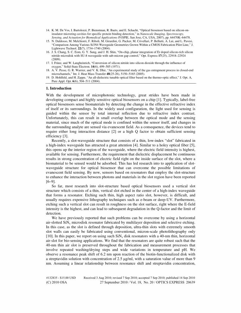
The images of the slot-sensor during the various stages of its fabrication and usage are
shown in Fig. 1. We observe successful fabrication of the air-slot disk resonator. More
importantly, Fig. 1 demonstrates that the 40-nm thin air-slot structure is preserved throughout
the fabrication process despite repeated washing/drying steps and wide variations in
temperature and pH, and repeated measurements over long period of time in the ambient air.
#132835 - $15.00 USD Received 3 Aug 2010; revised 7 Sep 2010; accepted 7 Sep 2010; published 14 Sep 2010(C) 2010 OSA 27 September 2010 / Vol. 18, No. 20 / OPTICS EXPRESS 20640
(b)
15µµµµm
~ 2µµµµm
(a)
SiNx
SiNx
SiO2
500nm
(c)
5 µµµµm
(d)
2 µµµµm
Fig. 1. (a) SEM image of the deposited multilayer thin film prior to disk formation, (b) DIC
optical microscope image of the fabricated microdisk after air slot fabrication. The green inner
boundary indicates the silicon post and the whiter outer region indicates air slot with depth of
more than 2µm. (c)(d) SEM images of fabricated slot disk after whole sensing process. Note
that the air-slot is maintained despite repeated washing/drying measurement steps.
3. Results and Discussion
Based on the fabricated structure shown in Fig. 1, the sensing efficiency of the air-slot
microdisk resonator was calculated using finite difference time domain (FDTD) method. The
edge of the disk was set to be 10° sloped based on the SEM image. The grid size of vertical
direction was 2 nm and a single fundamental TM-like mode was excited using a single
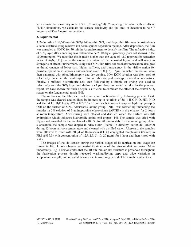
Gaussian point source. Figure 2(a) shows the calculated mode profile, demonstrating the
highly concentrated |E|2 in the air slot region. The confinement factor is defined as [12]
{ } �
2 2
0
2
E E, where .
Re E H E
A gA A
A A
Az
n c dxdy dxdyn
ne dxdy dxdy
ε εγ γ
ε∗
∞ ∞
Γ = ≡ =× ⋅
∫∫ ∫∫∫∫ ∫∫
(1)
Here c is the speed of light in vacuum, ng is the group index and nA is the refractive index
in the region A. The γA term represents the spatial confinement factor. Based on the simulated
mode profile, the shift in the position of the resonance peak of the resonator due to adsorption
of a thin layer with a refractive index of 1.45 typical for a dried biofilm is calculated. For a
configuration described schematically in Fig. 2 (b), the resonance position shifts linearly with
layer thickness, as is shown in Fig. 2 (c). The surface sensitivity of the resonator, Ssurface
defined as
[ / ],surface res bio
S t nm nmλ= ∂ ∂ (2)
is found to be 5.73 nm/nm for the mode number of m = 45. It should be noted here that the
high Ssurface value is mostly due to the adsorption of biofilm on the inside surface of the slot.
For example, give a 5 nm thick adsorbed film, the spatial confinement factor (γΑ) with the film
is calculated to be 6.3%. Of this, the spatial confinement factor with the film adsorbed on the
inside surface is calculated to be 5.4%, which corresponds to 85% of the total sensitivity. In
contrast, the surface sensitivity with the film adsorbed on the top of the upper disk and the
bottom of the lower disk is calculated to be 0.80 nm/nm only, confirming the importance of
the slot-structure for achieving the high sensitivity.
#132835 - $15.00 USD Received 3 Aug 2010; revised 7 Sep 2010; accepted 7 Sep 2010; published 14 Sep 2010(C) 2010 OSA 27 September 2010 / Vol. 18, No. 20 / OPTICS EXPRESS 20641
min
max
tbio
nbio = 1.45
(a)
(b)
(c)
0 2 4 6 8 10
1480
1490
1500
1510
1520
1530
1540
Re
so
na
nc
e w
av
ele
ng
th (
nm
)
Adsorbed layer thickness, tbio
(nm)
Simulated resonance wavelength
linear fit
Surface sensitivity
: 5.73 nm/nm
500nm
Fig. 2. (a) Simulated mode profile of |E|2 with bare fabricated structure (b) schematic of surface
sensing (c) Calculated shift in the resonance wavelength versus thickness of adsorbed layer
with a refractive index of 1.45. The surface sensitivity is obtained as 5.73nm/nm with linear fit.
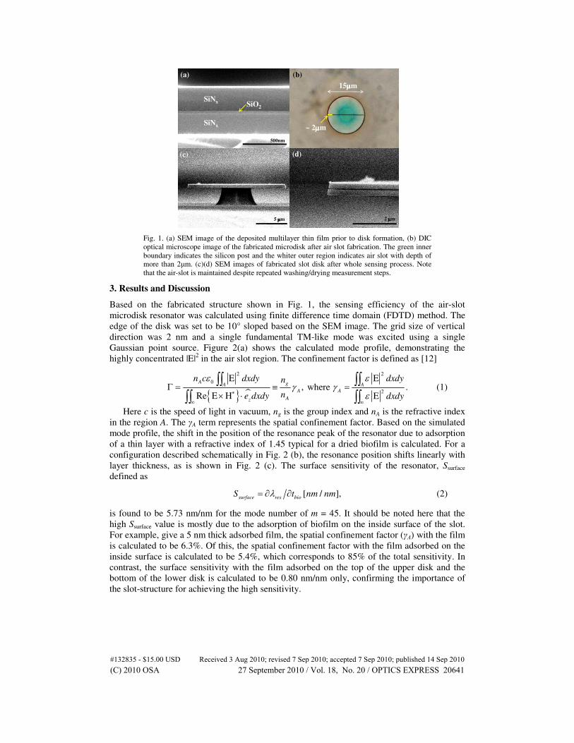
The actual surface sensing capability of fabricated air slot resonators was measured using
tapered fiber coupling. Figure 3 shows the schematic diagram of measurement setup. A
tunable laser which spans from 1470nm to 1545 nm was used as the light source, and a
tapered fiber with a diameter of ~1µm was used to evanescently couple light into and out of
the resonator. The input polarization was controlled with a fiber polarization controller to TM-
like mode in order to excite the slot-modes of the resonator. The fiber position was controlled
not to touch the disk in order to reduce the scattering loss and unwanted peak shift due to
contact between the resonator and the fiber.
TLD
1470nm~
1550nm Detector
Polarization
controller
U-bent tapered fiber
CCD
x50
Picomotor
xyz stage
(a) (b)
Fig. 3. (a) Schematic of the measurement system. (b) CCD image of a slot disk coupled with a
tapered fiber. The fiber diameter is ~1µm.
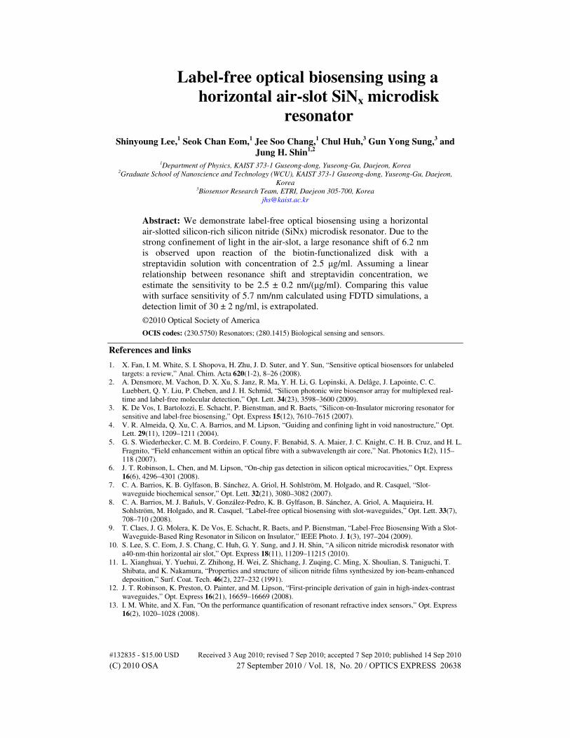
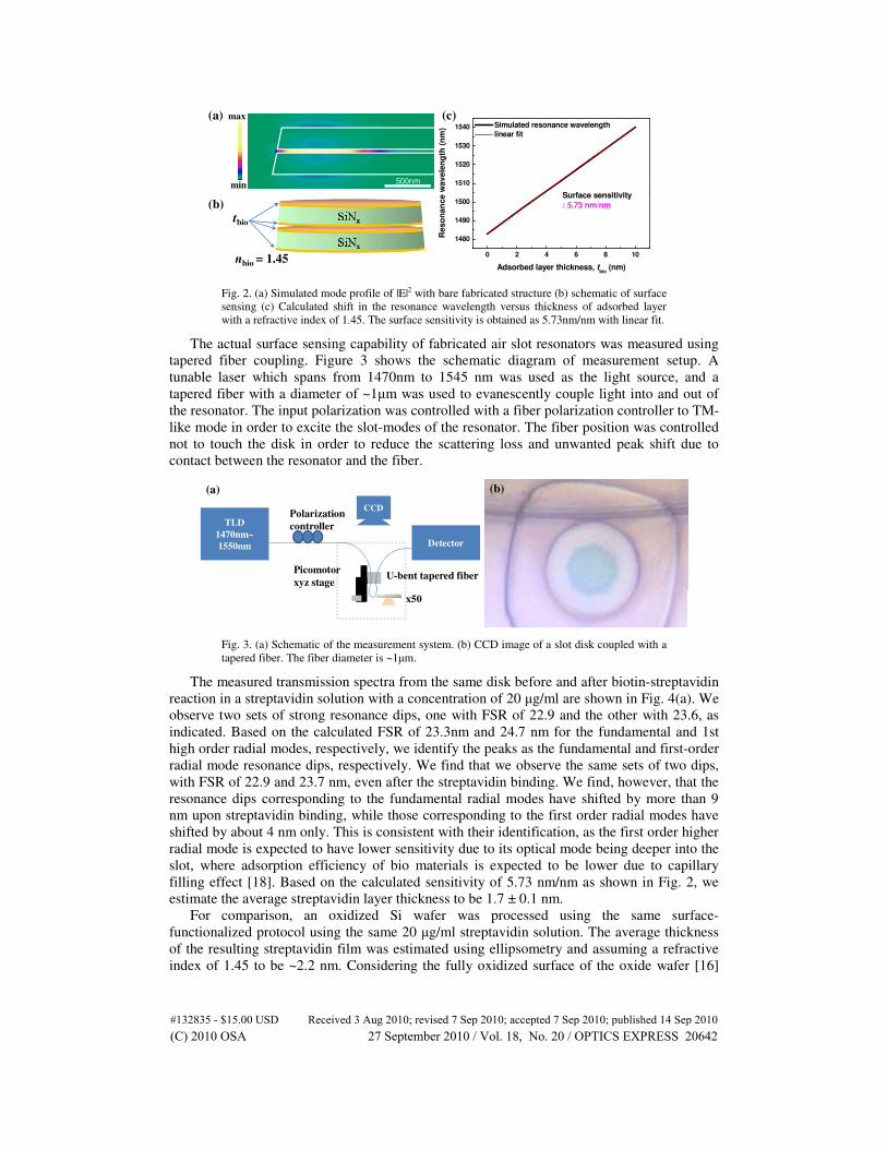
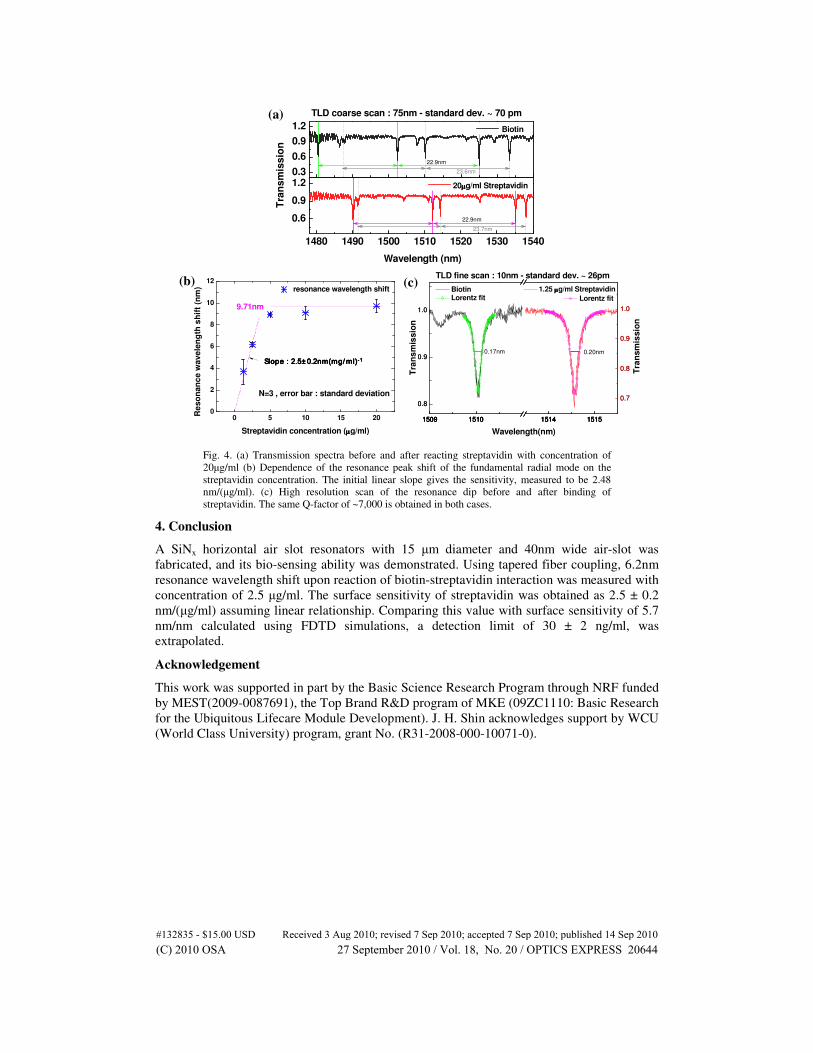
The measured transmission spectra from the same disk before and after biotin-streptavidin
reaction in a streptavidin solution with a concentration of 20 µg/ml are shown in Fig. 4(a). We
observe two sets of strong resonance dips, one with FSR of 22.9 and the other with 23.6, as
indicated. Based on the calculated FSR of 23.3nm and 24.7 nm for the fundamental and 1st
high order radial modes, respectively, we identify the peaks as the fundamental and first-order
radial mode resonance dips, respectively. We find that we observe the same sets of two dips,
with FSR of 22.9 and 23.7 nm, even after the streptavidin binding. We find, however, that the
resonance dips corresponding to the fundamental radial modes have shifted by more than 9
nm upon streptavidin binding, while those corresponding to the first order radial modes have
shifted by about 4 nm only. This is consistent with their identification, as the first order higher
radial mode is expected to have lower sensitivity due to its optical mode being deeper into the
slot, where adsorption efficiency of bio materials is expected to be lower due to capillary
filling effect [18]. Based on the calculated sensitivity of 5.73 nm/nm as shown in Fig. 2, we
estimate the average streptavidin layer thickness to be 1.7 ± 0.1 nm.
For comparison, an oxidized Si wafer was processed using the same surface-
functionalized protocol using the same 20 µg/ml streptavidin solution. The average thickness
of the resulting streptavidin film was estimated using ellipsometry and assuming a refractive
index of 1.45 to be ~2.2 nm. Considering the fully oxidized surface of the oxide wafer [16]
#132835 - $15.00 USD Received 3 Aug 2010; revised 7 Sep 2010; accepted 7 Sep 2010; published 14 Sep 2010(C) 2010 OSA 27 September 2010 / Vol. 18, No. 20 / OPTICS EXPRESS 20642
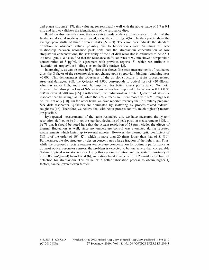
and planar structure [17], this value agrees reasonably well with the above value of 1.7 ± 0.1
nm, and further validates the identification of the resonance dips.
Based on this identification, the concentration-dependence of resonance dip shift of the
fundamental radial mode is investigated, as is shown in Fig. 4(b). The data points show the
average peak shifts of three different disks (N = 3). The error bars indicate the standard
deviation of observed values, possibly due to fabrication errors. Assuming a linear
relationship between resonance peak shift and the streptavidin concentration at low
streptavidin concentrations, the sensitivity of the slot disk resonator is estimated to be 2.5 ±
0.2 nm/(µg/ml). We also find that the resonance shifts saturates at 9.7 nm above a streptavidin
concentration of 5 µg/ml, in agreement with previous reports [3], which we attribute to
saturation of streptavidin binding sites on the disk surfaces [3].
Interestingly, as can be seen in Fig. 4(c) that shows fine scan measurements of resonance
dips, the Q-factor of the resonator does not change upon streptavidin binding, remaining near
~7,000. This demonstrates the robustness of the air-slot structure to resist process-related
structural damages. Still, the Q-factor of 7,000 corresponds to optical loss of ~20 dB/cm,
which is rather high, and should be improved for better sensor performance. We note,
however, that absorption loss of SiN waveguides has been reported to be as low as 0.1 ± 0.05
dB/cm even at 780 nm [15]. Furthermore, the radiation-loss limited Q-factor of slot-disk
resonator can be as high as 107, while the slot-surfaces are ultra-smooth with RMS roughness
of 0.51 nm only [10]. On the other hand, we have reported recently that in similarly prepared
SiN disk resonators, Q-factors are dominated by scattering by process-related sidewall
roughness [16]. Therefore, we believe that with better process control, much higher Q-factors
are possible.
By repeated measurements of the same resonance dip, we have measured the system
resolution, defined to be 3 times the standard deviation of peak position measurements [13], to
be 78 pm. It should be noted here that the system resolution of 78 pm includes the effects of
thermal fluctuation as well, since no temperature control was attempted during repeated
measurements which lasted up to several minutes. However, the thermo-optic coefficient of
SiN is of the order of 10−5
K−1
, which is more than 20 times lower than that of Si [19].
Furthermore, the slot structure by design concentrates a large fraction of the light in air. Thus,
while the proposed structure requires temperature compensation for optimum performance as
do most optical resonator sensors, the problem is expected to be less severe than comparable
Si-based optical resonator sensors. Using this system resolution and the system sensitivity of
2.5 ± 0.2 nm/(µg/ml) from Fig. 4 (b), we extrapolated a value of 30 ± 2 ng/ml as the limit of
detection for streptavidin. This value, with better fabrication process to obtain higher Q
factors, can be lowered even further.
#132835 - $15.00 USD Received 3 Aug 2010; revised 7 Sep 2010; accepted 7 Sep 2010; published 14 Sep 2010(C) 2010 OSA 27 September 2010 / Vol. 18, No. 20 / OPTICS EXPRESS 20643
1509 1510 1514 1515
0.8
0.9
1.0
1509 1510 1514 1515
0.8
0.9
1.0
1509 1510 1514 1515
0.7
0.8
0.9
1.0
1509 1510 1514 1515
0.7
0.8
0.9
1.0
Tra
ns
mis
sio
n
Biotin
Wavelength(nm)
Lorentz fit
1.25 µµµµg/ml Streptavidin
TLD fine scan : 10nm - standard dev. ~ 26pm
0.20nm
Tra
nsm
issio
n
Lorentz fit
0.17nm
0 5 10 15 200
2
4
6
8
10
12
resonance wavelength shift
Streptavidin concentration (µµµµg/ml)
Re
so
nan
ce w
ave
len
gth
sh
ift
(nm
)
slope : 2.48nm(µµµµg/ml)-1
N=3 , error bar : standard deviation
9.71nm
0.3
0.6
0.9
1.2
1480 1490 1500 1510 1520 1530 1540
0.6
0.9
1.2
Tra
nsm
issio
n
23.7nm
22.9nm
23.6nm
Biotin
TLD coarse scan : 75nm - standard dev. ~ 70 pm
22.9nm
Wavelength (nm)
20µµµµg/ml Streptavidin
(a)
(b) (c)
Slope : 2.5Slope : 2.5Slope : 2.5Slope : 2.5±±±± 0.2nm(mg/ ml)0.2nm(mg/ ml)0.2nm(mg/ ml)0.2nm(mg/ ml) ---- 1111
Fig. 4. (a) Transmission spectra before and after reacting streptavidin with concentration of
20µg/ml (b) Dependence of the resonance peak shift of the fundamental radial mode on the
streptavidin concentration. The initial linear slope gives the sensitivity, measured to be 2.48
nm/(µg/ml). (c) High resolution scan of the resonance dip before and after binding of
streptavidin. The same Q-factor of ~7,000 is obtained in both cases.
4. Conclusion
A SiNx horizontal air slot resonators with 15 µm diameter and 40nm wide air-slot was
fabricated, and its bio-sensing ability was demonstrated. Using tapered fiber coupling, 6.2nm
resonance wavelength shift upon reaction of biotin-streptavidin interaction was measured with
concentration of 2.5 µg/ml. The surface sensitivity of streptavidin was obtained as 2.5 ± 0.2
nm/(µg/ml) assuming linear relationship. Comparing this value with surface sensitivity of 5.7
nm/nm calculated using FDTD simulations, a detection limit of 30 ± 2 ng/ml, was
extrapolated.
Acknowledgement
This work was supported in part by the Basic Science Research Program through NRF funded
by MEST(2009-0087691), the Top Brand R&D program of MKE (09ZC1110: Basic Research
for the Ubiquitous Lifecare Module Development). J. H. Shin acknowledges support by WCU
(World Class University) program, grant No. (R31-2008-000-10071-0).
#132835 - $15.00 USD Received 3 Aug 2010; revised 7 Sep 2010; accepted 7 Sep 2010; published 14 Sep 2010(C) 2010 OSA 27 September 2010 / Vol. 18, No. 20 / OPTICS EXPRESS 20644
Recommended