
Kanika Rai BSc (N), Gold Medalist in MSc (N), PhD (N)
Professor and Head of Department (Medical Surgical Nursing)Maharishi Markandeshwar College of Nursing
Maharishi Markandeshwar (Deemed to be University)Mullana, Ambala, Haryana
Founder
Kanika’s Nursing Academy
Chandigarh
ForewordSukhpal Kaur
CBS Publishers & Distributors Pvt Ltd• New Delhi • Bengaluru • Chennai • Kochi • Kolkata • Lucknow
• Mumbai • Hyderabad • Nagpur • Patna • Pune • Vijayawada
PrefacePathophysiology is an essential topic for the nurses to understand. The essence of critical thinking is the ability of nurses to act according to their knowledge and understanding. Because of this reason, the nursing students as well as nurses should understand the pathophysiological changes that take place in any disease. This helps them to think critically and apply that knowledge while taking care of the patients. Considering the importance of learning and understanding the concepts related to pathophysiology, it is an honest effort to bring out a compact but an enriched content for making the nursing students well-versed with the changes that occur in various disease conditions. Pathophysiology helps build a strong foundation of nursing practice enabling nurses to provide quality care. The book has been organized into 17 chapters divided in accordance with the curriculum given by Indian Nursing Council, parallel to the chapters of Medical Surgical Nursing. The content has been designed in an interesting manner with the help of illustrations, flowcharts and figures for a better understanding of pathophysiological concepts. The motivation behind writing this book is my own interest in the pathophysiological concepts as I am specialized in Medical Surgical Nursing. Moreover, I have observed the students facing issues regarding an organized content related to pathophysiology and searching from multiple sources. I am pretty confident that not only the nursing students and nurses, but the undergraduate students of other medical disciplines will also be benefitted from the content presented in this book. I sincerely thank the Almighty for His blessings and all near and dear ones for their constant support and motivation at the time of writing this book.
Kanika Rai
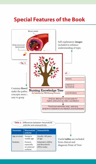
Special Features of the Book
Self-explanatory images included to enhance understanding of topic
Useful tables are included from clinical and diagnosis Point of View
Flowchart
Table 1: Differences between rheumatoid arthritis and osteoarthritis
Parameter Rheumatoid arthritis
Osteoarthritis
Age at onset Young to middle age
Usually >40 years of age
Nodules Present, especially on extensor surfaces
Heberden’s (DIPs) and Bouchard’s (PIPs) nodes
Fig. 1: Components of blood
Contains flowcharts to make the pathophysiological concepts more clear and easy to grasp
Understanding Pathophysiology of Diseases
xii
Also KnowIf the fibrous cap is thick, it can resist the stress from blood flow and vessel movement. If the fibrous cap is thin, lipid core may grow causing it to rupture and hemorrhage into plaque allowing a thrombus to form. This is called atherothrombosis.
SARS CORONAVIRUS-2
The Severe Acute Respiratory Syndrome (SARS) coronavirus-2 is a novel coronavirus belonging to the family Coronaviridae. It is known to be responsible for the outbreak of a series of recent acute atypical respiratory infections originating in Wuhan, China. The disease caused by this virus, termed coronavirus disease 19 or simply COVID-19, rapidly spread throughout the world at an alarming pace and was declared a pandemic by the World Health Organization (WHO) on March 11, 2020. Let us discuss the pathophysiology of Covid-19 and its different stages.
A highly relevant topic on SARS CORONAVIRUS-2 Pathophysiology has been included to keep the readers abreast of the various aspects of this deadly disease
Also Know boxes have been included in between the text to enhance the knowledge of the readers
Contents
Foreword........ ............................................................................................................iii
Contributor and Reviewers ....................................................................................v
Preface......... ..............................................................................................................vii
Acknowledgments..... ...........................................................................................viii
Special Features of the Book ..... ...........................................................................xi
SARS Coronavirus-2.... ..........................................................................................xxi
Glossary .... .............................................................................................................xxv
Chapter 1 Basics of Pathophysiology .....................................1–10
Cell 1
Cellular Adaptation 4
Repair of Cells 6
Homeostasis 7
Disease, Sickness and Illness 8
Pathophysiology and Disease 9
Importance of Pathophysiology in Nursing 10
Chapter 10 Pathophysiology of Cardiovascular Disorders ...11–40
Introduction 11
Coronary Artery Disease 12
Angina Pectoris 13
Myocardial Infarction 14
Heart Failure 15
Mitral Valve Prolapse 19
Mitral Regurgitation 20
Mitral Stenosis 20
Aortic Stenosis 21
Aortic Regurgitation 21
Rheumatic Heart Disease 21
Understanding Pathophysiology of Diseases
xiv
Pericarditis 23
Pericardial Effusion 24
Myocarditis 25
Endocarditis 25
Cor Pulmonale 27
Pulmonary Edema 27
Cardiogenic Shock 28
Cardiac Tamponade 30
Vascular System Disorders 30
Peripheral Vascular Disorders 32
Raynaud’s Disease 33
Aneurysm 35
Varicose Veins 35
Deep Venous Thrombosis 37
Chronic Venous Insufficiency 39
Chapter 3 Pathophysiology of Blood Disorders ....................41–50
Introduction 41
Anemia 42
Thalassemia 42
Thrombocytopenia 43
Disseminated Intravascular Coagulation 43
Leukemias 47
Myelomas 49
Lymphomas 49
Chapter 4 Pathophysiology of Respiratory Disorders ...........51–77
Introduction 51
Chronic Obstructive Pulmonary Disease 52
Bronchitis 54
Emphysema 55
Asthma 56
Contents
xv
Pleural Effusion 59
Pneumonia 60
Lung Abscess 61
Acute Respiratory Distress Syndrome 62
Respiratory Failure 64
Pulmonary Embolism 67
Chest Injuries 69
Tuberculosis 70
Atelectasis 73
Bronchiectasis 74
Empyema 76
Chapter 5 Pathophysiology of Gastrointestinal Disorders ............................................................ 79–109
Introduction 79
Gastritis 81
Typhoid Fever 81
Inflammatory Bowel Disease 82
Intestinal Obstruction 84
Ulcers 86
Malabsorption 89
Appendicitis 89
Hernias 90
Acute Pancreatitis 95
Chronic Pancreatitis 97
Liver Cirrhosis 97
Cholelithiasis 98
Cholecystitis 99
Portal Hypertension 100
Liver Abscess 103
Hepatic Failure 104
Hepatitis 105
Understanding Pathophysiology of Diseases
xvi
Hemorrhoids 108
Anal Fissures 109
Anal Fistula 109
Chapter 6 Pathophysiology of Renal System .................... 111–123
Introduction 111
Urinary Tract Infections 111
Cystitis 112
Glomerulonephritis 113
Nephrotic Syndrome 114
Renal Calculi 115
Acute Renal Failure 117
Chronic Renal Failure 119
Urethral Stricture 120
Benign Prostate Hyperplasia/Hypertrophy 121
Prostatitis 122
Chapter 7 Pathophysiology of Endocrine Disorders ........ 125–142
Introduction 125
Pituitary Disorders 127
Cushing’s Syndrome 128
Addison’s Syndrome 128
Syndrome of Inappropriate Antidiuretic Hormone 130
Hyperthyroidism 131
Hypothyroidism 134
Hyperparathyroidism 135
Hypoparathyroidism 136
Diabetes Mellitus 137
Chapter 8 Pathophysiology of Nervous System Disorders .. 143–169
Introduction 143
Headache 144
Head Injuries 145
Contents
xvii
Spinal Injuries—Paraplegia, Hemiplegia 146
Herniation of Intervertebral Disc 147
Cerebral Aneurysm 149
Meningitis 150
Brain Abscess 151
Encephalitis 152
Neurocysticercosis 153
Movement Disorders—Chorea 153
Seizures and Epilepsies 154
Cerebrovascular Disease 156
Hemorrhagic Stroke 158
Bell’s Palsy 159
Trigeminal Neuralgia 159
Peripheral Neuropathies 162
Carpal Tunnel Syndrome 162
Alzheimer’s Disease 163
Parkinson’s Disease 165
Guillain-Barré Syndrome 165
Myasthenia Gravis 166
Multiple Sclerosis 167
Chapter 9 Pathophysiology of Musculoskeletal Disorders .......................................................... 171–188
Introduction 171
Soft-tissue Injuries 172
Fracture 173
Osteomyelitis 175
Osteoarthritis 176
Rheumatoid Arthritis 178
Osteoporosis 182
Osteomalacia 183
Paget’s Disease 185
Understanding Pathophysiology of Diseases
xviii
Prolapse of Intervertebral Disc 186
Pott’s Spine/Disease 187
Chapter 10 Pathophysiology of Eye Disorders ................. 189–199
Introduction 189
Vision 189
Refractive Errors 190
Blepharitis 191
Conjunctivitis 191
Corneal Ulcer/Keratitis 193
Cataract 194
Glaucoma 196
Retinal Detachment 198
Chapter 11 Pathophysiology of Ear, Nose and Throat Disorders .............................................. 201–213
Ear 201
Nose 201
Throat 202
Disorders of External Ear 203
Disorders of Middle Ear 203
Disorders of Internal Ear 206
Upper Respiratory Tract Infections 208
Tonsillitis 210
Laryngitis 211
Epistaxis 212
Nasal Obstruction 212
Laryngeal Obstruction 213
Chapter 12 Pathophysiology of Integumentary System .............................................................. 215–224
Introduction 215
Systemic Lupus Erythematosus 216
Acne Vulgaris 217
Psoriasis 218
Contents
xix
Malignant Melanoma 219
Burns 220
Burn Edema 222
Chapter 13 Disorders of Female Reproductive System .............................................................. 225–232
Introduction 225
Menstrual Disorders 226
Pelvic Inflammatory Disease 228
Polycystic Ovarian Syndrome 228
Uterine and Cervical Disorders 229
Vaginal Disorders 231
Mastitis 232
Toxic Shock Syndrome 232
Chapter 14 Disorders of Male Reproductive System ........ 233–239
Introduction 233
Congenital Malformation 234
Cryptorchidism 235
Orchitis 235
Epididymitis 236
Phimosis 236
Sexual Dysfunction 237
Gynecomastia 238
Male Menopause 239
Chapter 15 Pathophysiology of Immune System Disorders ............................................. 241–245
Introduction—Immune System 241
Lymphatic System 241
Types of Immunity 242
Human Immunodeficiency Virus and Acquired Immune Deficiency Syndrome 243
Anaphylaxis 245
Understanding Pathophysiology of Diseases
xx
Chapter 16 Pathophysiology of Cancer ............................. 247–256
Introduction 247
Cancer of the Eye 249
Oral Cancer 249
Laryngeal Cancer 249
Breast Cancer 250
Lung Cancer 250
Gastric Cancer 251
Colorectal Cancer 251
Bladder Cancer 251
Liver Cancer 252
Prostate Cancer 253
Cervical Cancer 254
Ovarian Cancer 254
Brain Tumor 255
Chapter 17 Pathophysiology of Genetic Disorders .......... 257–265
Introduction 257
Single-gene Disorders 257
Chromosomal Disorders 263
Complex Disorders 265
Index ................................................................................................................267–273
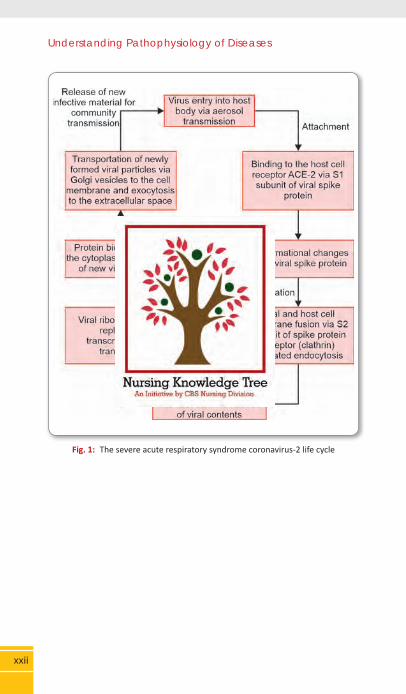
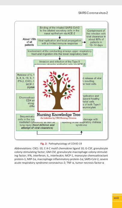
The Severe Acute Respiratory Syndrome (SARS) Coronavirus-2 is a novel coronavirus belonging to the family Coronaviridae. It is known to be responsible for the outbreak of a series of recent acute atypical respiratory infections originating in Wuhan, China. The disease caused by this virus, termed coronavirus disease 19 or simply COVID-19, rapidly spread throughout the world at an alarming pace and was declared a pandemic by the World Health Organization (WHO) on March 11, 2020. Let us discuss the pathophysiology of Covid-19 and its different stages. In the asymptomatic phase, SARS-CoV-2, received via respiratory aerosols, binds to the nasal epithelial cells in the upper respiratory tract. Local replication and propagation of virus occur in the conducting airways along with an infection of the ciliated cells. The duration of this stage is around two days with a limited immune response. The individuals tend to be highly infectious at this stage despite having a low viral load. In the next stage, the virus migrates to the upper respiratory tract from the nasal epithelium through the conducting airways. With the involvement of upper airways, manifestations, like fever, dry cough and malaise appear. The immune response in this stage is very high involving the release of C-X-C motif chemokine ligand 10 (CXCL-10) and interferons (IFN-β and IFN-λ) from the cells infected by the virus. This mounted immune response is enough to contain the spread of infection. About 1/5th of infected patients progress to the next stage in which there occurs an involvement of lower respiratory tract with an onset of acute respiratory distress syndrome (ARDS). The virus is able to invade the type-2 alveolar epithelial cells and starts replicating. The virus-laden pneumocytes start releasing different cytokines and inflammatory markers such as interleukins (IL-1, IL-6, IL-8, IL-120 and IL-12), tumor necrosis factor-α (TNF-α), IFN-λ and IFN-β, CXCL-10, monocyte chemoattractant protein-1 (MCP-1) and macrophage inflammatory protein-1α (MIP-1α). Owing to this cytokine storm, neutrophils, CD4 helper T cells and CD8 cytotoxic T cells begin to get sequestered in the lung tissues, leading to inflammation and injury to lung tissues. The persistent injury caused by the sequestered inflammatory cells and viral replication lead to loss of both types 1 and 2 pneumocytis. This diffuses alveolar damage ultimately culminated into ARDS.
SARS Coronavirus-2
Understanding Pathophysiology of Diseases
xxii
Fig. 1: The severe acute respiratory syndrome coronavirus-2 life cycle
xxiii
SARS Coronavirus-2
Fig. 2: Pathophysiology of COVID-19
Abbreviations: CXCL-10, C-X-C motif chemokine ligand 10; G-CSF, granulocyte colony-stimulating factor; GM-CSF, granulocyte-macrophage colony-stimulat-ing factor; IFN, interferon; IL, interleukin; MCP-1, monocyte chemoattractant protein-1; MIP-1α, macrophage inflammatory protein-1α; SARS-CoV-2, severe acute respiratory syndrome coronavirus-2; TNF-α, tumor necrosis factor-α
INTRODUCTION
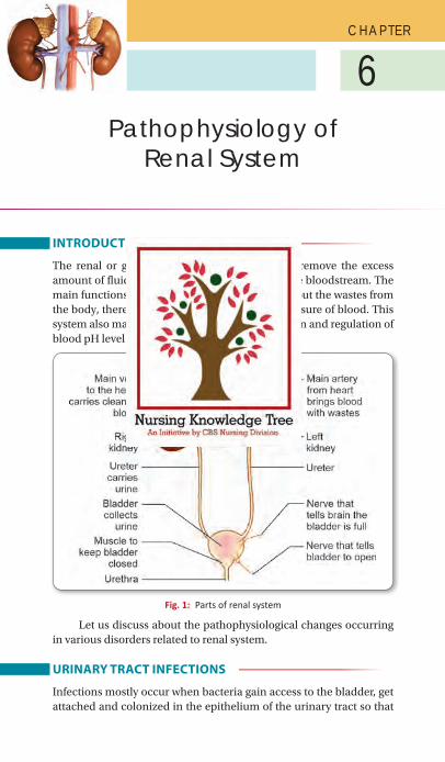
The renal or genitourinary system functions to remove the excess amount of fluid and toxic waste products from the bloodstream. The main functions of the renal system are to excrete out the wastes from the body, thereby, regulating the volume and pressure of blood. This system also maintains the electrolyte concentration and regulation of blood pH level (Fig. 1).
Fig. 1: Parts of renal system
Let us discuss about the pathophysiological changes occurring in various disorders related to renal system.
URINARY TRACT INFECTIONS
Infections mostly occur when bacteria gain access to the bladder, get attached and colonized in the epithelium of the urinary tract so that
6C H A P T E R
Pathophysiology of Renal System
Understanding Pathophysiology of Diseases
112
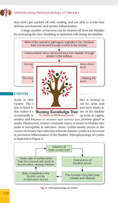
they don’t get washed off with voiding, and are able to evade host defense mechanisms, and initiate inflammation. A large number of bacteria can be cleared off from the bladder by increasing the slow shedding of epithelial cells lining the bladder.
CYSTITIS
Acute or chronic inflammation of urinary bladder is termed as Cystitis. The bladder serves as a storage/reservoir for urine and also is lined with mucus membrane having a protein layer above it, that makes it resistant to infection. But the infection of the bladder occasionally is the result of infection of neighboring areas as vagina, urethra and kidneys in females and urethra and prostate gland in males. Obstruction, tumors, traumatic injury or stones in bladder also make it susceptible to infection. Acute cystitis mostly occurs in the course of urinary tract infection whereas chronic cystitis is a recurrent or persistent inflammation of the bladder. Pathophysiology of cystitis is depicted in Figure 2.
Fig. 2: Pathophysiology of cystitis
Chapter 6 Pathophysiology of Renal System
113
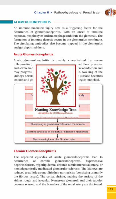
GLOMERULONEPHRITIS
An immune-mediated injury acts as a triggering factor for the occurrence of glomerulonephritis. With an onset of immune response, lymphocytes and macrophages infiltrate the glomeruli. The formation of immune deposit occurs in the glomerulus membrane. The circulating antibodies also become trapped in the glomerulus and get deposited there.
Acute Glomerulonephritis
Acute glomerulonephritis is mainly characterized by severe inflammation, renal insufficiency, swelling, increased blood pressure, and severe back pain. Kidneys get damaged because of infection and may progress on to subacute and chronic stages. Swelling of the kidneys occurs in the acute form of disease, the surface becomes smooth and grey and the capsule covering the kidneys is stretched.
Chronic Glomerulonephritis
The repeated episodes of acute glomerulonephritis lead to occurrence of chronic glomerulonephritis, hypertensive nephrosclerosis, hyperlipidemia, chronic tubulointerstitial injury, or hemodynamically medicated glomerular sclerosis. The kidneys are reduced to as little as one-fifth their normal size (consisting primarily the fibrous tissue). The cortex shrinks, making the surface of the kidney rough and irregular. Numerous glomeruli and their tubules become scarred, and the branches of the renal artery are thickened.
Understanding Pathophysiology of Diseases
114
The result is severe glomerular damage that results in end-stage renal disease (ESRD). Failure of kidneys to filter the waste products from the blood and accumulation of abnormal quantities of nitrogenous waste products is known as uremia.
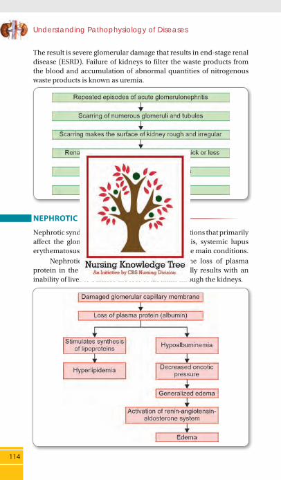
NEPHROTIC SYNDROME
Nephrotic syndrome results from the disease conditions that primarily affect the glomerulus, chronic glomerulonephritis, systemic lupus erythematosus and renal vein thrombosis being the main conditions. Nephrotic syndrome is characterized by the loss of plasma protein in the urine. Hypoalbuminemia eventually results with an inability of liver to balance the loss of albumin through the kidneys.
Chapter 6 Pathophysiology of Renal System
115
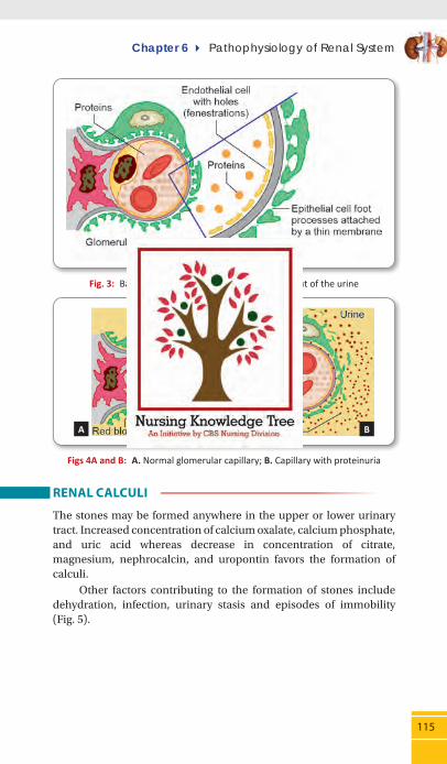
Fig. 3: Barriers that keep protein and blood cells out of the urine
A B
Figs 4A and B: A. Normal glomerular capillary; B. Capillary with proteinuria
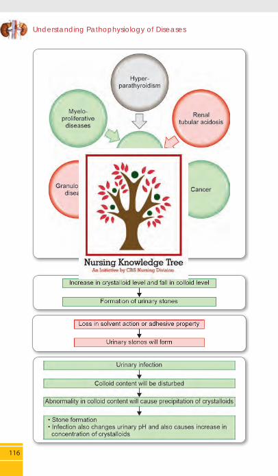
RENAL CALCULI
The stones may be formed anywhere in the upper or lower urinary tract. Increased concentration of calcium oxalate, calcium phosphate, and uric acid whereas decrease in concentration of citrate, magnesium, nephrocalcin, and uropontin favors the formation of calculi. Other factors contributing to the formation of stones include dehydration, infection, urinary stasis and episodes of immobility (Fig. 5).
Understanding Pathophysiology of Diseases
116
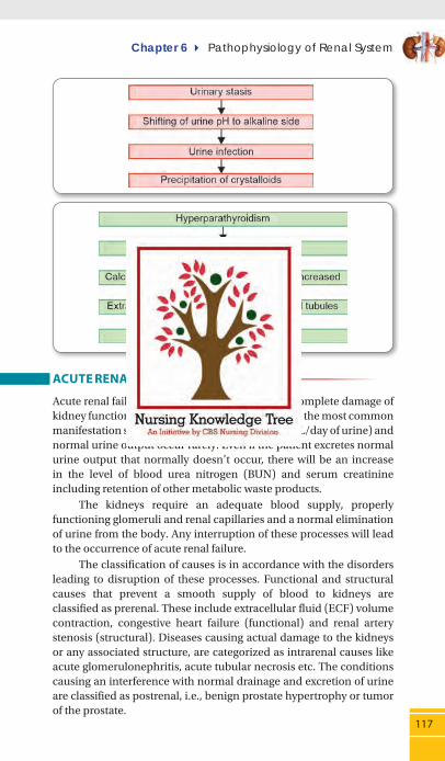
Fig. 5: Causes of hypercalcemia
Chapter 6 Pathophysiology of Renal System
117
ACUTE RENAL FAILURE
Acute renal failure (ARF) is a sudden and nearly complete damage of kidney function. Oliguria (<400 mL/day of urine) is the most common manifestation seen in ARF; whereas anuria (<50 mL/day of urine) and normal urine output occur rarely. Even if the patient excretes normal urine output that normally doesn’t occur, there will be an increase in the level of blood urea nitrogen (BUN) and serum creatinine including retention of other metabolic waste products. The kidneys require an adequate blood supply, properly functioning glomeruli and renal capillaries and a normal elimination of urine from the body. Any interruption of these processes will lead to the occurrence of acute renal failure. The classification of causes is in accordance with the disorders leading to disruption of these processes. Functional and structural causes that prevent a smooth supply of blood to kidneys are classified as prerenal. These include extracellular fluid (ECF) volume contraction, congestive heart failure (functional) and renal artery stenosis (structural). Diseases causing actual damage to the kidneys or any associated structure, are categorized as intrarenal causes like acute glomerulonephritis, acute tubular necrosis etc. The conditions causing an interference with normal drainage and excretion of urine are classified as postrenal, i.e., benign prostate hypertrophy or tumor of the prostate.
Understanding Pathophysiology of Diseases
118
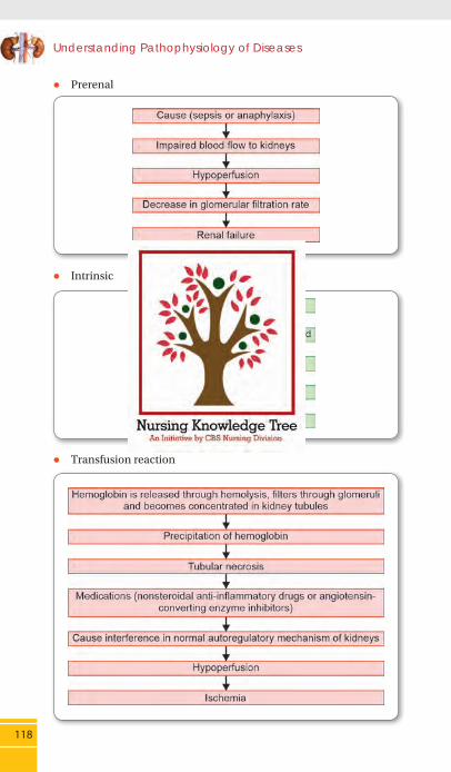
z Prerenal
z Intrinsic
z Transfusion reaction
Chapter 6 Pathophysiology of Renal System
119
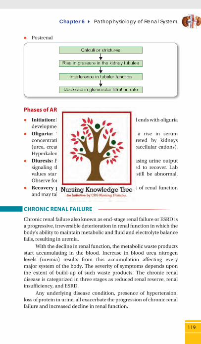
z Postrenal
Phases of ARF
z Initiation: Begins with the injury of kidneys and ends with oliguria development.
z Oliguria: This period is accompanied by a rise in serum concentration of substances normally excreted by kidneys (urea, creatinine, uric acid, organic acids, intracellular cations). Hyperkalemia may also develop.
z Diuresis: Patient experiences gradually increasing urine output signaling that glomerular filtration has started to recover. Lab values start decreasing. Renal function may still be abnormal. Observe for dehydration during this period.
z Recovery period: It signals the improvement of renal function and may take 3–12 months.
CHRONIC RENAL FAILURE
Chronic renal failure also known as end-stage renal failure or ESRD is a progressive, irreversible deterioration in renal function in which the body’s ability to maintain metabolic and fluid and electrolyte balance fails, resulting in uremia. With the decline in renal function, the metabolic waste products start accumulating in the blood. Increase in blood urea nitrogen levels (uremia) results from this accumulation affecting every major system of the body. The severity of symptoms depends upon the extent of build-up of such waste products. The chronic renal disease is categorized in three stages as reduced renal reserve, renal insufficiency, and ESRD. Any underlying disease condition, presence of hypertension, loss of protein in urine, all exacerbate the progression of chronic renal failure and increased decline in renal function.
Understanding Pathophysiology of Diseases
120
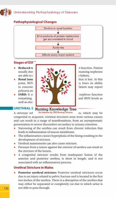
Pathophysiological Changes
Stages of ESRD
z Reduced renal reserve: 40–75% loss of nephron function. Patient usually does not have any symptom because remaining nephrons are able to carry out the normal functions of the kidney.
z Renal insufficiency: 75–90% of nephron function is lost. At this point, BUN and serum creatinine rise, kidney loses its ability to concentrate urine and anemia develops. Patient may report polyuria and nocturia.
z ESRD: It occurs when there is less than 10% nephron function remaining. Evidenced by elevated creatinine and BUN levels as well as electrolyte imbalances.
URETHRAL STRICTURE A stricture refers to the narrowing of the lumen, which may be congenital or acquired. Urethral strictures arise from various causes and can result in a range of manifestations, from an asymptomatic presentation to severe discomfort secondary to urinary retention.
z Narrowing of the urethra can result from chronic infection that leads to inflammation of mucus membrane.
z The inflammation causes hyperplasia of the lining resulting in the development of stricture.
z Urethral anastomosis can also cause stricture. z Pressure from a tumor against the exterior of urethra can result in
the stricture of the lumen. z A congenital stricture results from inadequate fusion of the
anterior and posterior urethra, is short in length, and is not associated with an inflammatory process.
Urethral Stricture in Males z Posterior urethral stricture: Posterior urethral strictures occur
due to an injury related to pelvic fracture and is located in the first two inches of the urethra. There is a disruption of the urethra that may either be separated or completely cut due to which urine is not able to pass through.
Chapter 6 Pathophysiology of Renal System
121
z Anterior urethral stricture: Main causes of anterior urethral stricture that is present in the last two inches of the urethra include direct traumatic injury to penis or catheterization.
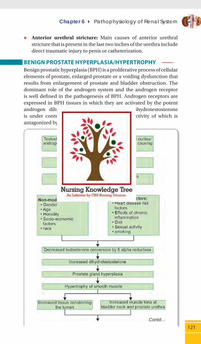
BENIGN PROSTATE HYPERPLASIA/HYPERTROPHY Benign prostatic hyperplasia (BPH) is a proliferative process of cellular elements of prostate, enlarged prostate or a voiding dysfunction that results from enlargement of prostate and bladder obstruction. The dominant role of the androgen system and the androgen receptor is well defined in the pathogenesis of BPH. Androgen receptors are expressed in BPH tissues in which they are activated by the potent androgen dihydrotestosterone. Synthesis of dihydrotestosterone is under control of the 5α-reductase enzyme, activity of which is antagonized by finasteride and dutasteride.
Contd…
Understanding Pathophysiology of Diseases
122
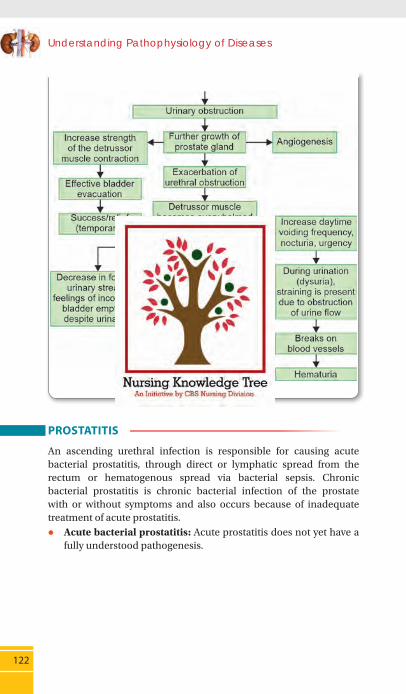
PROSTATITIS
An ascending urethral infection is responsible for causing acute bacterial prostatitis, through direct or lymphatic spread from the rectum or hematogenous spread via bacterial sepsis. Chronic bacterial prostatitis is chronic bacterial infection of the prostate with or without symptoms and also occurs because of inadequate treatment of acute prostatitis.
z Acute bacterial prostatitis: Acute prostatitis does not yet have a fully understood pathogenesis.
Chapter 6 Pathophysiology of Renal System
123
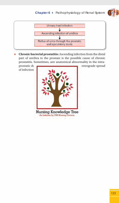
z Chronic bacterial prostatitis: Ascending infection from the distal part of urethra to the prostate is the possible cause of chronic prostatitis. Sometimes, any anatomical abnormality in the intra-prostatic ducts may also be responsible for the retrograde spread of infection.

Recommended