Journal of Pharmacy and Pharmacology 5 (2017) 668-680 doi: 10.17265/2328-2150/2017.09.009
Justicia insularis Improves the in vitro Survival and
Development of Ovine Preantral Follicles Enclosed in
Ovarian Tissue
Gildas Tetaping Mbemya1, Denise Damasceno Guerreiro1, Nathalie Jiatsa Donfack1, Luciana Mascena Silva1,
Luis Alberto Vieira1, Geovania Francisca Canafistula de Sousa1, Benner Geraldo Alves1, Aryele Pinto Izaguirry2,
Francielli Weber Santos2, Phelix Bruno Telefo3, Otília Deusdênia Loiola Pessoa4, Johan Smitz5, José Ricardo de
Figueiredo1 and Ana Paula Ribeiro Rodrigues1
1. Faculty of Veterinary (FAVET), State University of Ceará, Fortaleza 60714903, Brazil
2. Laboratory of Reproduction Biotechnology (Biotech), State of University of Pampa, Uruguaiana 97500970, Brazil
3. Laboratory of Biochemistry of Medicinal Plants, Food and Nutritional Sciences, Faculty of Science, University of Dschang,
Dschang 3451381, Cameroon
4. Laboratory of Phytochemical Analysis of Medicinal Plants (LAFIPLAN), Federal University of Ceará, Fortaleza 60020181, Brazil
5. Follicle Biology Laboratory, Center for Reproductive Medicine, Brussels 1090, Belgium
Abstract: Objectives: Evaluating the addition effect of J. insularis extract and FSH on the survival, activation and ROS production after in vitro culture of ovine preantral follicles enclosed in ovarian tissue. Methods: In the first experiment, ovarian fragments were fixed (non-cultured control) or in vitro cultured in α-MEM+ (cultured control), α-MEM+ supplemented with FSH 50 ng/mL, or in α-MEM+ supplemented with J. insularis (JUS0.3; 1.25 or 5 mg/mL) for 1 or 7 days, at 39˚C, 5% CO2. In the second experiment, fragments were fixed or cultured in α-MEM+ supplemented with anethole 300 µg/mL + FSH 50 ng/mL or in α-MEM+ supplemented with anethole 300 µg/mL + 0.3 mg/mL JUS. Key findings: JUS0.3 was the only treatment that maintained the percentage of morphologically normal follicles similar to non-cultured control even after 7 days of culture. After 7 days of culture, a higher (p < 0.05) percentage of developing follicles was observed in JUS5 treatment compared with the other treatments except JUS1.25. In the second experiment, FSH maintained the percentage of normal follicles and promoted activation of primordial follicles. A reduction (p < 0.05) of stromal cell density was observed in MEM++ANE supplemented with JUS or FSH. Conclusions: J. insularis in a concentration-dependent manner maintained the levels of ROS and improved in vitro follicular survival and activation of ovine primordial follicles.
Key words: Medicinal plant, antioxidant, in vitro folliculogenesis, preantral follicles.
1. Introduction
The in vitro follicle culture studies have been
performed either using primordial follicles enclosed in
ovarian slices (in situ culture) or in the isolated form
[1]. Such follicle culture systems have been used
respectively to investigate in vitro early and late
folliculogenesis at preantral follicle stage [2]. Culture
Corresponding author: Ana Paula Ribeiro Rodrigues, PhD., professor, reaearch fields: manipulation of oocyte and ovarian preantral follicles.
systems for primordial follicles are important tools for
studying the mechanism of oocyte development, and
are a potential source of oocytes that can be used for
in vitro embryo production. The factors that control
primordial follicle activation and further growth of
primary follicles are not well understood [2]. However,
endocrine hormones, like FSH (follicle stimulating
hormone), are known to regulate the production of
several growth factors that play a critical role in
primordial follicle activation and growth [3].
D DAVID PUBLISHING
Justicia insularis Improves the in vitro Survival and Development of Ovine Preantral Follicles Enclosed in Ovarian Tissue
669
Some in vitro studies have demonstrated that the
addition of FSH to the culture medium is important to
maintain viability and to promote ovine follicular
activation and further growth in vitro [3-5]. In
addition, Magalhães et al. [6] showed that the addition
of 50 ng/mL recombinant bovine FSH (rFSH) during
in vitro culture of goat preantral follicles maintained
viability, activation and follicular growth.
Despite the advances made in this field, the success
of in vitro culture of preantral follicles is still very
limited and the majority of reports are restricted to
investigative studies to elucidate how early or
preantral folliculogenesis works. As ARTs (assisted
reproductive technique) to date, this tool has been
limited to advance in mice, the only species to have
been reported the birth of animals from embryos
originated from completely developed preantral
follicles in vitro [7, 8]. In large animals such as sheep
[9] or even in human [10] the complete development
in vitro of preantral follicles from a primordial to an
ovulatory follicle has been hampered by growth
arrested at the primary follicle stage [9, 10]. One
reason for this shortcoming is that the requirements
for in vitro growth are not well characterized due to
lack of knowledge regarding activation of primordial
and development of primary follicles compared to
development of follicles in later stages [11]. In
addition, it is known that during in vitro culture of
preantral follicles, there is an increase production of
reactive oxygen species: ROS [12], which can affect
growth, survival and consequently can lead to cell
death [13]. In this context, at the present time, there is
an increase interest in natural products (medicinal
plants) that prevent oxidative damages caused by the
ROS and as a result may contribute to promote the
activation, survival, growth of preantral follicles in
vitro enclosed in ovarian tissue. Among these
potential natural products, it is important to highlight J.
insularis and anethole.
J. insularis T. Anders (family Acanthaceae), is an
herbaceous and perennial plant, widely distributed in
tropical area of Africa [14]. In ghomala’a (traditional
language spoken in Western Cameroon), J. insularis is
called “kwe mchie” [15]. Traditionally, in Senegal, the
leaf decoction of J. insularis is given to women during
the last month of pregnancy to reduce labour pains. In
Cameroon and specifically in the Western region, their
leaves are used in association with the leaves of three
others medicinal plants (Aloe buettneri, Hibiscus
macranthus and Dicliptera verticillata), to treat
dysmenorrhoea and some cases of women infertility
[15, 16]. The in vivo foliculogenic effect of their
leaves has been related to their composition. Besides
alkaloids, glycosides, polyphenols and triterpenoids,
studies undertaken by by Telefo et al. [14] and Goka
et al. [17] revealed the presence of flavonoids in their
leaves which can act as a natural antioxidant. A mix of
aqueous extract of J. insularis and others medicinal
plants (Aloe buettneri, Hibiscus macranthus and
Dicliptera verticillata) has also been proven, in a
series of studies to induce ovarian steroidogenesis and
folliculogenesis in female rats [17-19].
Anethole, other natural compound originated from
Croton zehntneri Pax & K. Hoffm (family
Euphorbiaceae), a plant locally known as “canela de
cunhã” or “canelinha” in the Northeast of Brazil [20]
has also showed antioxidant activity due its capacity
to decrease the concentrations of ROS both in vivo [21]
and in vitro [22]. Recently, Sá et al. [23] demonstrated
that anethole reduced the levels of ROS, proving its
antioxidant activity on goat isolated preantreal
follicles (secondary stage) cultured in vitro.
Despite the importance of the aforementioned
natural products for ARTs, to the best of our
knowledge, there is no study investigating the effect
of the aqueous extract of J. insularis on in vitro
folliculogenesis. In addition the effect of anethole on
the in vitro culture of primordial follicle enclosed in
ovarian tissue is not known. Therefore this study was
conducted to (1) investigate the addition effect of FSH
and J. insularis extract on the survival, activation,
growth and ROS generation after in vitro culture of
Justicia insularis Improves the in vitro Survival and Development of Ovine Preantral Follicles Enclosed in Ovarian Tissue
670
ovine preantral follicles enclosed in ovarian tissue; (2)
compare the efficiency of FSH and J. insularis on
stromal cell density and all parameters above
mentioned in culture medium containing anethole.
2. Materials and Methods
This study was approved and performed under the
guidelines of the Ethics Committee for Animal Use of
the State University of Ceará (N˚ 6004720/2015).
Unless mentioned otherwise, the culture media,
anethole and other chemicals used in the present study
were purchased from Sigma Chemical Co. (St. Louis,
USA).
2.1 Source of Ovaries
Ovaries (n = 22) were collected at a local
slaughterhouse from 11 adults (1-3 years old)
mixed-breed sheep (Ovis aries). Immediately
postmortem, ovaries were washed in 70% alcohol
followed by two rinses in minimum essential medium
(MEM) supplemented with 100 µg/mL penicillin and
100 µg/mL streptomycin plus 25 mM HEPES.
Ovaries were transported within 1 h to the laboratory
into tubes containing 15 mL of MEM-HEPES at 4 °C
[5].
2.2 Plants Materials to Extracts Preparation and
Culture Medium
The fresh leaves of J. insularis previously identified
in the National Herbarium of Cameroon under
voucher specimen code 34997 [17] were collected in
Western Cameroon (Batoufam subdivision,
Upper-Plateau division, 5°21′North 10°24′East,
Altitude 1,515 m). The fresh leaves were then dried at
room temperature in the shade. Subsequently, the
plant extract decoction was prepared according to the
protocol [16]. Finally, the plant decoction was
lyophilized and kept in the freezer at -20 ºC. The
lyophilized extract was then diluted in the distilled
water to obtain the desired concentrations (0.3 mg/mL,
1.25 mg/mL and 5 mg/mL).
The basic culture medium consisted of α-MEM (pH
7.2-7.4) supplemented with 1.25 mg/mL bovine serum
albumin, 10 µg/mL insulin, 5.5 mg/mL transferrin, 5
ng/mL selenium, 2 mM glutamine, 2 mM
hypoxanthine and antibiotics (100 µg/mL
penicillin-streptomycin) which was referred to as
α-MEM+ [24]. In a first experiment, to test the effect
of J. insularis on the culture of ovine preantral
follicles, the α-MEM+ was supplemented with FSH or
different concentrations of J. insularis, mentioned
above. In a second experiment, the α-MEM+ was
supplemented with anethole + FSH or with anethole +
J. insularis.
The concentrations of J. insularis were defined
based on a concentration curve done according to in
vivo [16] and in vitro [25]. Briefly, the best
concentration in vivo (5 mg/mL) was divided per 4 to
obtain 1.25 mg/mL which subsequently was divided
by the same factor (4) to obtain 0.3 mg/mL. This latest
concentration was close to the best concentration of
Amburana Cearensis, and other medicinal plant was
tested in in vitro culture of sheep preantral follicles
[25]. The concentration of anethole used was chosen
based on previous studies performed in our laboratory
on vitro culture of goat preantral follilces [23].
2.3 Experimental Design
As briefly mentioned, this work was divided into
two non-simultaneous experiments.
In the first one, sheep ovarian cortex from each
ovarian pair (n = 5) was cut using a tissue slicer
(Thomas Scientific, USA) into 22 fragments
(approximately 3 3 0.5 mm). One fragment was
taken randomly and immediately fixed for histological
analysis and identified as non-cultured control, the
remaining fragments were in vitro cultured in 1 mL of
α-MEM+; α-MEM+ supplemented with FSH 50 ng/mL,
or with different concentrations (0.3, 1.25 or 5 mg/mL)
of lyophilized plant extract J. insularis for 1 or 7 days
at 39 °C in 5% CO2 in air. These treatments were
referred to as: MEM+ (cultured control), FSH, JUS0.3,
Justicia insularis Improves the in vitro Survival and Development of Ovine Preantral Follicles Enclosed in Ovarian Tissue
671
JUS1.25 and JUS5, respectively. The culture medium
was equilibrated at least 3 h prior to use. Every two
days, whole culture medium was replaced. Based on
the histological analysis, the best treatments (higher
percentage of morphological normal follicles) were
selected to next experiment.
In the second experiment, ovarian fragments were
obtained as previously described in experiment 1. The
fragments from each ovarian pair (n = 6) were either
fixed for histological analysis (non-cultured control)
or in vitro cultured in 1 mL of α-MEM+ + anethole
300 µg/mL + FSH 50 ng/mL or α-MEM+ + anethole
300 µg/mL + J. insularis 0.3 mg/mL, corresponding
to the following treatments: MEM++ANE+FSH and
MEM++JUS+ANE, respectively.
2.4 Morphological Analysis and Evaluation of
Follicular Growth in vitro
Before (non-cultured control) and after 1 or 7 days
of culture, the ovarian fragments were fixed
individually in 4 % buffered paraformaldehyde for 2 h.
Subsequently, fragments were dehydrated in a graded
concentrations of ethanol. After paraffin embedding
(Synth, São Paulo, Brazil), the ovarian fragments were
cut into 7 µm sections and mounted on glass slides
and stained by periodic acid schiff-hematoxylin.
Follicle stage and survival were assessed
microscopically on serial sections.
The developmental stages of follicles are
primordial (one layer of flattened pregranulosa cells
around the oocyte) or growing follicles (intermediate:
one layer of flattened to cuboidal granulosa cells;
primary: one layer of cuboidal granulosa cells; and
secondary: two or more layers of cuboidal granulosa
cells around the oocyte) [26]. These follicles were still
classified individually as histologically normal when
an intact oocyte was present, surrounded by granulosa
cells which are well organized in one or more layers
and that have no pyknotic nucleus. Atretic follicles
were defined as those with a retracted oocyte,
pyknotic nucleus, and/or disorganized granulosa cells
detached from the basement membrane [26]. 150
follicles were evaluated for each treatment (30
follicles per each five repetitions) in both experiments.
To evaluate follicular activation, the percentages of
healthy primordial and growing follicles were
calculated before (non-cultured control) and after
culture in each treatment. Each follicle was examined
in every section in which it appeared and matched
with the same follicle on adjacent sections to avoid
double counting, thus ensuring that each follicle was
only counted once, regardless of its size [26].
2.5 Ovarian Stromal Cell Density
Ovarian stroma density was evaluated by
calculating the stromal cell per 100 µm2. For each
treatment, ten fields per slide were assessed and the
mean number of stromal cell per field was calculated
in experiment 2 [27]. All evaluations and
measurements were performed by a single operator.
2.6 Reactive Oxygen Species Levels
The ROS levels were determined by a
spectrofluorimetric method [28], using 2’,
7’-dihydrodichlorofluorescein diacetate (DCHF-DA)
assay. Sample aliquot (50 µL) was incubated with 5
µL of DCHF-DA (1 mM). The oxidation of
DCHF-DA to fluorescent dichlorofluorescein was
measured for the detection of ROS. The DCF
fluorescence intensity emission was recorded at 520
nm (with 480 nm excitation) 2 h after the addition of
DCHF-DA to the medium. The correlation between
the follicular viability and ROS levels was done to
better understand the effect of ROS by the cells during
in vitro culture of preantral follicles.
2.7 Statistical Analysis
Statistical analyses were carried out using the
Sigma Plot 11.0 software (Systat Software Inc, San
Jose, California, USA). Data that were not normally
distributed (Shapiro-Wilk test) were submitted to
logarithmic transformation. The percentage of
Justicia insularis Improves the in vitro Survival and Development of Ovine Preantral Follicles Enclosed in Ovarian Tissue
672
morphologically normal and growing preantral
follicles among treatments and days of culture were
compared by Fisher’s exact or chi-square tests. The
Mann-Whitney test was performed to analyze the
levels of reactive oxygen species and stromal cell
density among treatments and days of culture.
Spearman correlation test was used to assess the
association between normal preantral follicles and
reactive oxygen species. In addition, the association
between stromal cell density and percentage of normal
preantral follicles was evaluated by linear regression
analysis. Data were presented as mean (± standard
error of the mean) and percentage, unless otherwise
indicated. Statistical significance was defined as p <
0.05 and probability values > 0.05 and ≤ 0.1 indicated
that a difference approached significance.
3. Results
3.1 Sheep Preantral Follicles Morphology and
Development
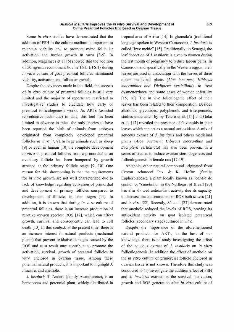
In the first experiment, a total of 1,374 preantral
follicles were analyzed. Morphologically normal or
degenerated follicles were observed in the
non-cultured control as well as in the in vitro cultured
ovarian tissues (Fig. 1).
The percentage of morphologically normal and
follicular growth before and after in vitro culture is
shown in Table 1 (experiment 1). JUS0.3 was the only
treatment that maintained the percentage of
morphologically normal follicles similar to
non-cultured control even after 7 days of culture. In
addition, this treatment showed a higher percentage
(p < 0.05) of normal follicles than the other treatments
regardless to the culture time. However, at day 7, J.
insularis significantly reduced (p < 0.05) the
percentage of morphologically normal follicles in a
concentration-dependent manner. On the other hand,
after 7 days of culture, a significantly higher
percentage of developing follicles was observed in the
JUS5 treatment compared to the other treatments
except JUS1.25.
In the second experiment, a total of 1,286 preantral
Fig. 1 Representative images of the morphology of ovine preantral follicles before and after in vitro culture. On the top panels we can see follicles from experiment 1 and on the bottom panels, follicles from experiment 2, after staining with periodic acid schiff-hematoxylin. Normal follicles are shown in non-cultured control (A), JUS0.3 (B), FSH (D) and FSH+ANE (E), while degenerated follicles are represented in JUS5 (C) and JUS+ANE (F) after 7 days of culture. Note the retracted oocyte with a pyknotic nucleus, disorganized granulosa cells (C and F). O: oocyte; Nc: oocyte nucleus; Gc: granulosa cells (400 x), bar 50 µm.
Justicia insularis Improves the in vitro Survival and Development of Ovine Preantral Follicles Enclosed in Ovarian Tissue
673
Table 1 Percentage of morphologically normal and growing preantral follicles before (non-cultured control) and after in vitro culture for 1 or 7 days in different treatments, in experiment 1.
Follicular morphology (%) Follicular development (%)
Primordial Developing
Non-cultured control 84.6 (127/150) 86.6 (110/127) 13.4 (17/127)
Day 1 Day 7 Day 1 Day 7 Day 1 Day 7
MEM+ (cultured control) 82.5 (109/132)aA 62.5 (70/112)*bA 88.9 (97/109)aA 85.7 (60/70)aA 11.1 (12/109) aA 14.3 (10/70)aA
FSH 79.8 (99/124)aA 65.2 (62/95)*bA 75.7 (75/99)*aB 87.1 (54/62)aA 24.3 (24/99) *aB 12.9 (8/62)aA
JUS0.3 91.3 (137/150)aB 89.5 (111/124)aB 77.4 (106/137)aB 84.7 (94/111)aA 22.6 (31/137) aB 15.3 (17/111)aA
JUS1.25 58.1 (57/98)*aC 47.3 (71/150)*aC 85.9 (49/57)aAB 74.6 (53/71)*aAB 14.1 (8/57) aAB 25.4 (18/71)*aAB
JUS5 66.1 (52/111)*aC 21.9 (29/132)*bD 82.5 (66/80)aAB 65.5 (19/29)*aB 17.5 (14/80) aAB 34.5 (10/29)*aB
* Differs from non-cultured control (p < 0.05). a,b Within a row and the same parameter evaluated, values without a commom superscript differed (p < 0.05). A,B,C,D Within a column, values without a commom superscript differed (p < 0.05).
follicles were analyzed. The percentages of
morphologically normal preantral and growing
follilces in non-cultured control and after 1 or 7 days
of culture in medium containing ANE supplemented
with FSH or JUS are shown in Table 2. After in vitro
culture, compared to non-cultured control, the
percentage of normal follicles was reduced (p < 0.05),
except in MEM++ANE+ FSH treatment on day 1.
Both cultured treated groups significantly reduced
(p < 0.05) the percentage of normal follicles from day
1 to day 7 (p < 0.05).
After 1 and 7 days of culture, there was a
significant reduction (p < 0.05) in the percentage of
primordial follicles with concomitant increase (p <
0.05) in the percentage of developing follicles in both
treatments compared to non-cultured control,
indicating the follicular activation process.
Furthermore, with the progression of the culture time,
only MEM++ANE+FSH significantly increased (p <
0.05) the percentage of developing follicles. In
addition, at day 7 of culture, MEM++ANE+FSH
treatment showed a higher percentage of developing
follicles than MEM++ANE+JUS treatment.
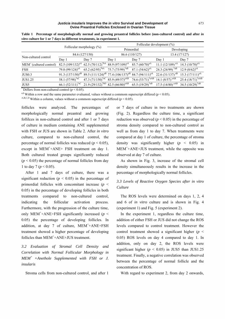
3.2 Evaluation of Stromal Cell Density and
Correlation with Normal Follicular Morphology in
MEM+ +Anethole Supplemented with FSH or J.
insularis
Stroma cells from non-cultured control, and after 1
or 7 days of culture in two treatments are shown
(Fig. 2). Regardless the culture time, a significant
reduction was observed (p < 0.05) in the percentage of
stroma density compared to non-cultured control as
well as from day 1 to day 7. When treatments were
compared at day 1 of culture, the percentage of stroma
density was significantly higher (p < 0.05) in
MEM++ANE+JUS treatment, while the opposite was
observed at day 7 of culture.
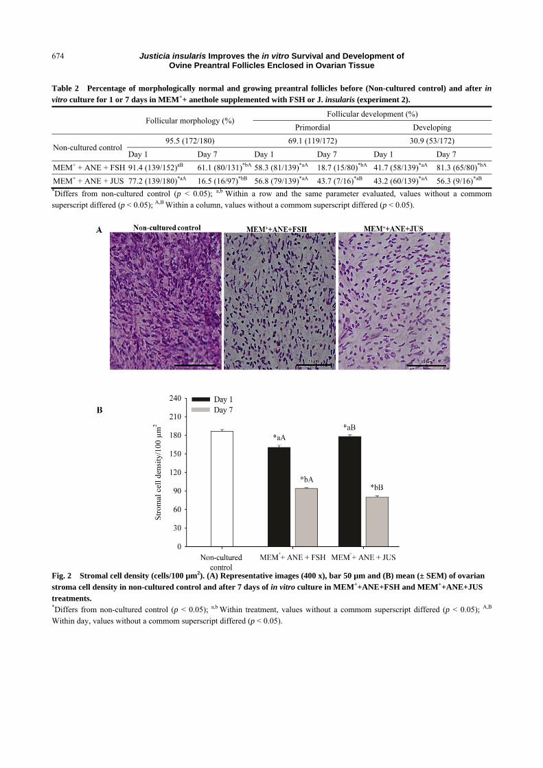
As shown in Fig. 3, increase of the stromal cell
density simultaneously results in the increase in the
percentage of morphologically normal follicles.
3.3 Levels of Reactive Oxygen Species after in vitro
Culture
The ROS levels were determined on days 1, 2, 4
and 6 of in vitro culture and is shown in Fig. 4
(experiment 1) and Fig. 5 (experiment 2).
In the experiment 1, regardless the culture time,
addition of either FSH or JUS did not change the ROS
levels compared to control treatment. However the
control treatment showed a significant higher (p <
0.05) ROS levels on day 4 compared to day 1. In
addition, only on day 2, the ROS levels were
significant higher (p < 0.05) in JUS5 than JUS1.25
treatment. Finally, a negative correlation was observed
between the percentage of normal follicle and the
concentration of ROS.
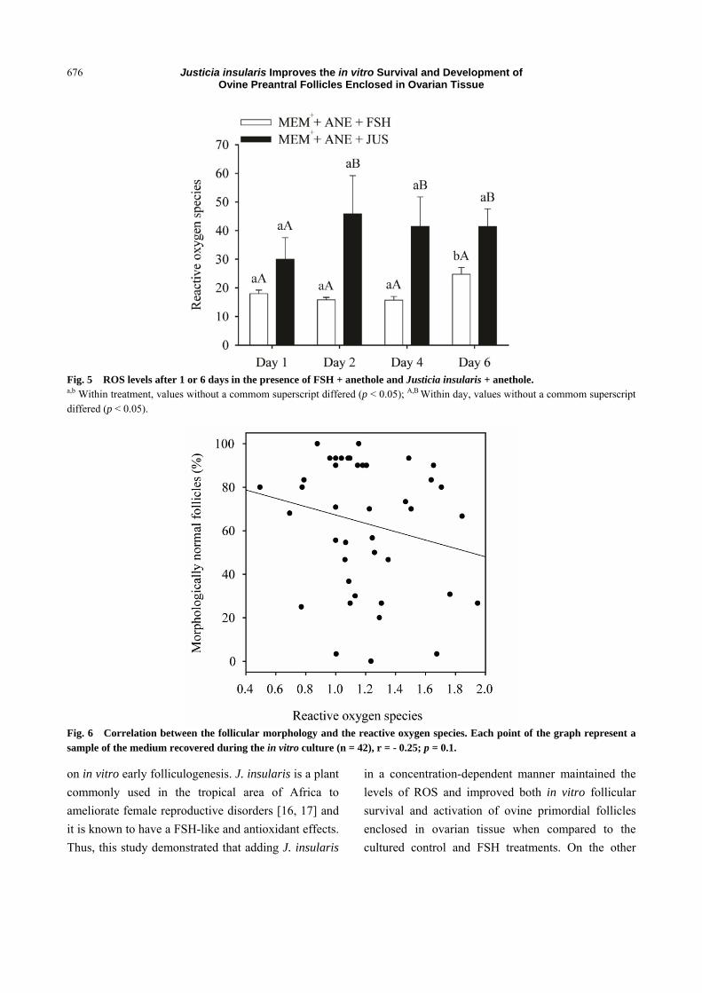
With regard to experiment 2, from day 2 onwards,
Justicia insularis Improves the in vitro Survival and Development of Ovine Preantral Follicles Enclosed in Ovarian Tissue
674
Table 2 Percentage of morphologically normal and growing preantral follicles before (Non-cultured control) and after in vitro culture for 1 or 7 days in MEM++ anethole supplemented with FSH or J. insularis (experiment 2).
Follicular morphology (%) Follicular development (%)
Primordial Developing
Non-cultured control 95.5 (172/180) 69.1 (119/172) 30.9 (53/172)
Day 1 Day 7 Day 1 Day 7 Day 1 Day 7
MEM+ + ANE + FSH 91.4 (139/152)aB 61.1 (80/131)*bA 58.3 (81/139)*aA 18.7 (15/80)*bA 41.7 (58/139)*aA 81.3 (65/80)*bA
MEM+ + ANE + JUS 77.2 (139/180)*aA 16.5 (16/97)*bB 56.8 (79/139)*aA 43.7 (7/16)*aB 43.2 (60/139)*aA 56.3 (9/16)*aB *Differs from non-cultured control (p < 0.05); a,b Within a row and the same parameter evaluated, values without a commom superscript differed (p < 0.05); A,B Within a column, values without a commom superscript differed (p < 0.05).
Fig. 2 Stromal cell density (cells/100 µm2). (A) Representative images (400 x), bar 50 µm and (B) mean (± SEM) of ovarian stroma cell density in non-cultured control and after 7 days of in vitro culture in MEM++ANE+FSH and MEM++ANE+JUS treatments. *Differs from non-cultured control (p < 0.05); a,b Within treatment, values without a commom superscript differed (p < 0.05); A,B
Within day, values without a commom superscript differed (p < 0.05).
Justicia insularis Improves the in vitro Survival and Development of Ovine Preantral Follicles Enclosed in Ovarian Tissue
675
Fig. 3 Relationship of stromal cell density with percentage of normal preantral follicles. The association between variables (black line) was analyzed by linear regression [Normal preantral follicles = 2.887 + (0.471 × stromal cell density); r = 0.66; R2 = 0.44; p < 0.001]. Each point on the chart represents one ovarian fragment evaluated.
Fig. 4 ROS levels after 1 or 6 days in the absence or presence of FSH or different concentrations of Justicia insularis. a,b Within treatment, values without a commom superscript differed (p < 0.05); A,B Within day, values without a commom superscript differed p < 0.05).
MEM++ANE+JUS treatment showed a significant
higher (p < 0.05) ROS levels than MEM++ANE+FSH
treatment. Contrary to MEM++ANE+JUS treatment,
MEM++ANE+FSH significantly increased (p < 0.05)
the ROS levels from day 1 to day 6.
As shown in Fig. 6, increase of the ROS levels
simultaneously results in the decrease in the
percentage of morphologically normal follicles.
4. Discussion
Although the action of FSH on the development of
preantral follicles cultured in vitro within the ovarian
cortex has already been investigated in different
species (human [29], sheep [3], goat [6], bovine [30],
buffalo [31], baboon [32]), this is the first study to
describe the effects of the aqueous extract of J. insularis
Justicia insularis Improves the in vitro Survival and Development of Ovine Preantral Follicles Enclosed in Ovarian Tissue
676
Fig. 5 ROS levels after 1 or 6 days in the presence of FSH + anethole and Justicia insularis + anethole. a,b Within treatment, values without a commom superscript differed (p < 0.05); A,B Within day, values without a commom superscript differed (p < 0.05).
Fig. 6 Correlation between the follicular morphology and the reactive oxygen species. Each point of the graph represent a sample of the medium recovered during the in vitro culture (n = 42), r = - 0.25; p = 0.1.
on in vitro early folliculogenesis. J. insularis is a plant
commonly used in the tropical area of Africa to
ameliorate female reproductive disorders [16, 17] and
it is known to have a FSH-like and antioxidant effects.
Thus, this study demonstrated that adding J. insularis
in a concentration-dependent manner maintained the
levels of ROS and improved both in vitro follicular
survival and activation of ovine primordial follicles
enclosed in ovarian tissue when compared to the
cultured control and FSH treatments. On the other
Justicia insularis Improves the in vitro Survival and Development of Ovine Preantral Follicles Enclosed in Ovarian Tissue
677
hand, addition of J. insularis or FSH in a culture
medium containing anethole promotes beneficial
effects on activation of ovine preantral follicles.
In the present study, JUS0.3 treatment maintained
the percentage of normal follicles similar to the
non-cultured control even after 7 days of culture and
showed a significantly higher value than the other
treatments. Such effect could be due to the action of
FSH-like compounds of the extract of J. insularis.
Phytochemical analysis of J. insularis has revealed the
presence of alkaloids, glycosides, polyphenols,
triterpenoids and flavonoids [14]. These metabolites
are responsible for the induction of ovarian follicle
growth and increase in the number of corpora lutea
recorded during the fertility assay performed on
immature female rats [16]. According to Andrade et al.
[4], addition of FSH to the culture medium is
important to the maintenance of morphology and
activation of sheep primordial follicles, as well as for
further follicular growth in vitro [33]. In addition,
Thomas et al. [34] reported that FSH stimulates the
expression of some growth factors, such as Kit Ligand,
Bone Morphogenetic Protein-15 and Growth
Differentiation Factor-9, which are important for the
regulation of early folliculogenesis. The presence of
FSH in the culture medium containing anethole also
maintained follicular morphology.
J. insularis in high concentration (expeiriment 1) or
in low concentration in the presence of anethole
(experiment 2) have detrimental effects on the ovarian
tissue. This could be due to an excess of antioxidant in
the culture medium which can be harmful to preantral
follicles. It is well known that oxidative metabolism is
indispensable for energy production of follicles during
in vitro culture [35], which in turn results in
generation of ROS. Although a critical amount of
ROS is essential for their physiological activities,
excessive amount of them generates a contrary effect
[12, 35]. Furthermore, some antioxidant substances
act as pro-oxidants when used at high concentrations
[36]. In this way, some studies have shown that high
concentrations of ascorbic acid, an important
antioxidant added to the culture medium, can inhibit
important physiological processes in the ovary,
resulting in follicular degeneration [37] besides
promoting oxidative damage to cellular DNA [38].
This study also focused on the features of stroma
tissue surrounding the follicles because any damage to
this compartment may affect follicular morphology.
After 1 and 7 days of culture, a reduction in the
percentage of stromal cell density was observed in
MEM++ANE supplemented either with J. insularis or
with FSH compared to non-cultured control. The in
vitro culture may have affected the bi-directional
contact between germinal and somatic cells which are
important to maintain the integrity of the cortex. In
addition, in our opinion, cells can die during in vitro
cultured by various factors such as nutrient deficiency
in the medium, ischemia, generation of ROS, toxicity
of the supplements. Our results are an in agreement
with findings using sheep [39] and goat [27] ovarian
tissues. Interestingly, a positive relationship between
morphology and stromal cell density was observed.
Previous reports have shown that ovarian stromal cells
are responsible for the production of growth factors
and peptides which are essential substances for
follicular development [2].
Finally, we have evaluated the antioxidant effect of
J. insularis and FSH in basic culture media in the
absence (experiment 1) or presence (experiment 2) of
anethole. In the first experiment (Fig. 4), addition of
either FSH or JUS did not change the ROS levels
compared to control treatment. The maintenance of
ROS levels could be due to the capacity of J. insularis
and FSH to scavenge ROS and their metal chelating
properties. Similar results have been found during in
vitro culture of ovine preantral follicles with
Amburana cearensis [25]. According to Gouveia et al.
[40], the extract of A. cearensis contains gallic acid,
PCA (protocatechuic acid), epicatechin, p-coumaric
acid and kaempferol. Gallic acid and PCA are
endogenous plant phenols which stimulate the
Justicia insularis Improves the in vitro Survival and Development of Ovine Preantral Follicles Enclosed in Ovarian Tissue
678
production antioxidant enzymes such as catalase
(CAT) and GPx (glutathione peroxidase) [41]. Reports
from Telefo et al. [17] and Goka et al. [14] showed
that J. insularis are constituted of several secondary
metabolites among which the phenols and flavonoids,
we thus belevied that those phenolic compounds act in
a similar way as metabolic of A. Cearensis to maintain
the ROS produced during in vitro culture.
In the present study, from day 1 to day 6, the
presence of FSH in culture medium containing
anethole increases the ROS levels. To date, it has been
shown that FSH stimulates catalase activity in goat
granulosa cells modulating intracellular ROS levels
[42]. ROS inhibitors, in a concentration-dependent
manner decreased oocyte maturation induced by FSH
[43]. In our study, we suggest that an adequate FSH
concentration (50 ng/mL) in a culture medium which
contained anethole contributed to the maintenance of
suitable levels of ROS after day 6 of culture, resulting
in higher rates of follicle survival and activation. As
above mentioned, we also observed that the
combination between J. insularis and anethole
increased the ROS level. This may be due to the high
concentration of antioxidant in the culture medium.
Indeed, antioxidant compounds in high concentration
can be converted in pro-oxidant compounds. Some
authors showed that some antioxidants could have
oxidative action depending on the concentration [44].
Furthermore, during in situ culture, many cells such as
granulosa, theca cells produce ROS. Those ROS with
the time progression during in vitro culture caused
injury to the follicular cells and thus affected the
follicular morphology [12].
5. Conclusions
J. insularis in a concentration-dependent manner
maintained the levels of ROS and improved both in
vitro follicular survival and activation of ovine
primordial follicles enclosed in ovarian tissue. Finally,
as well as FSH 50 ng/mL, the addition of J. insularis
0.3 mg/mL in a culture medium containing anethole
promotes beneficial effects on activation of ovine
preantral follicles kept in vitro at least for a short-term.
Although the mechanisms of interaction are unclear,
these results open a vast field of research regarding
interactions between plant extract and FSH in the
development of primordial follicles until the antral
stage. This study represents a hope for the use of
medicinal plants in more complex experiments which
aimed at the complete development of preantral
follicles in vitro. J. insularis could be used for futher
experiments to evaluate its beneficial effects on
isolated preantral follicles development and oocyte
maturation.
Conflict of Interest
The authors declare that there are no conflicts of
interest.
Acknowledgments
The authors would like to thank the National
Council for Scientific and Technological
Development (CNPq Brazil) for financial support.
Gildas Tetaping Mbemya is the recipient of a doctoral
scholarship from Coordenação de Aperfeiçoamento de
Pessoal de Nível Superior (PEC-PG/CAPES, Brazil).
Ana Paula Ribeiro Rodrigues is recipient of a grant
from CNPq Brazil through the projects
473968/2013-4 and 457226/2013-7. Johan Smitz is
Especial Visitor Researcher from CAPES
(88881.030.433/2013-01).
References
[1] Araújo, V. R., Gastal, M. O., Figueiredo, J. R., and Gastal, E. L. 2014. “In vitro Culture of Bovine Preantral Follicles: A Review.” Reprod Biol Endocrinol. 12: 78.
[2] Picton, H. M., Harris, S. E., Muruvi, W., and Chambers, E. L. 2008. “The in vitro Growth and Maturation of Follicles.” Reproduction 136: 703-15.
[3] Esmaielzadeh, F., Hosseini, S. M., Hajian, M., Nasiri, Z., Chamani, M., Afshar, A. M., et al. 2013. “Role of Follicle Stimulating Hormone in the Survival, Activation and Further Growth of in vitro Cultured Sheep Primordial Follicles.” Iran. J. Appl. Anim. Sci. 3: 785-90.
[4] Andrade, E. R., Seneda, M. M., Alfieri, A. A., Oliveira, J.
Justicia insularis Improves the in vitro Survival and Development of Ovine Preantral Follicles Enclosed in Ovarian Tissue
679
A., Bracarense, A. P. F., Figueiredo, J. R., et al. 2005. “Interactions of Indole Acetic Acid with EGF and FSH in the Culture of Ovine Preantral Follicles.” Theriogenelogy 64: 1104-13.
[5] Lima, L. F., Rocha, R. M. P., Alves, A. M. C. V., Saraiva, M. V. A., Araújo, V. R., Lima, I. M. T., et al. 2013. “Dynamized Follicle-Stimulating Hormone Affects the Development of Ovine Preantral Follicles Cultured in vitro.” Homeopathy 102: 41-8.
[6] Magalhães, D. M., Araújo, V. R., Lima-Verde, I. B., Matos, M. H. T., Silva, R. C., Lucci, C. M., et al. 2009. “Impact of Pituitary FSH Purification on in vitro Early Folliculogenesis in Goats.” Biocell. 33: 91-7.
[7] Eppig, J. J., and O’Brien, M. J. 1996. “Development in vitro of Mouse Oocytes from Primordial Follicles.” Biol. Reprod. 54: 197-207.
[8] O’Brien, M. J., Pendola, J. K., and Eppig, J. J. 2003. “A Revised Protocol for in vitro Development of Mouse Oocytes from Primordial Follicles Dramatically Improves Their Developmental Competence.” Biol. Reprod. 68: 1682-6.
[9] Cecconi, S., Barboni, B., Coccia, M., and Mattioli, M. 1999. “In Vitro Development of Sheep Preantral Follicles.” Biol. Reprod. 60: 594-601.
[10] Mery, L., Lefevre, A., Benchaib, M., Demirci, B., Salle, B., Guerin, J. F., et al. 2007. “Follicular Growth in vitro: Detection of Growth Differentiation Factor 9 (GDF9) and Bone Morphogenetic Protein 15 (BMP15) during in vitro Culture of Ovine Cortical Slices.” Mol. Reprod. Dev. 74: 767-74.
[11] Muruvi, W., Picton, H. M., Rodway, R. G., and Joyce, I. M. 2009. “In vitro Growth and Differentiation of Primary Follicles Isolated from Cryopreserved Sheep Ovarian Tissue. Anim.” Reprod. Sci. 112: 36-50.
[12] Costa, R. A. P., Romagna, C. D., Pereira, J. L., and Souza-pinto, N. C., 2011. “The Role of Mitochondrial DNA Damage in the Citotoxicity of Reactive Oxygen Species.” J. Bioenerg. Biomembr. 43: 25-29.
[13] Lima-Verde, I. B., Rossetto, R., Matos, M. H. T., Celestino, J. J. H., Bruno, J. B., Silva, C. M. G., et al. 2009. “Androstenedione and Follicle Stimulating Hormone Involvement in the Viability and Development of Goat Preantral Follicles in vitro.” Anim. Reprod. 7: 80-9.
[14] Goka, M. S. C., Telefo, P. B., Mbemya, G. T., Awouafack, M. D., Lienou, L. L., Yemele D. M., et al. 2016. “Potentialisation of Pregnant Mare Serum Gonadotropin Inducing Effect on Ovarian Follicles Growth by the Aqueous Extract of Aloe buettneri, Dicliptera verticillata, Hibiscus macranthus and Justicia insularis Leaves in Immature Rats.” Pharmacolology 7: 328-36.
[15] Telefo, P. B., Lienou, L. L., Yemele, M. D., Lemfack, M. C., Mouokeu, C., Goka, C. S., et al. 2011. “Ethnopharmacological Survey of Plants Used for the Treatment of Female Infertility in Baham, Cameroon.” J. Ethnopharmacol. 136: 178-87.
[16] Telefo, P. B., Tagne, S. R., Koona, O. E. S., Yemele, D. M., and Tchouanguep, F. M., 2012. “Effect of the Aqueous Extract of Justicia Insularis T. Anders (Acanthaceae) on Ovarian Folliculogenesis and Fertility of Female Rats.” Afr. J. Tradit. Complement. Altern. Med. 9: 197-203.
[17] Telefo, P. B., Moundipa, P. F., Tchana, A. N., Tchouanguep, D., and Mbiapo, F. T. 1998. “Effects of an Aqueous Extract of Aloe buettneri, Justicia insularis, Hibiscus macranthus, Dicliptera verticillata on Some Physiological and Biochemical Parameters of Reproduction in Immature Female Rats.” J. Ethnopharmacol. 63: 193-200.
[18] Telefo, P. B., Moundipa, F. P., and Tchouanguep, M. F., 2001. “Influence of the Aqueous Extract of Aloe buettneri, Justicia insularis, Hibiscus macranthus and Dicliptera verticillata on Fertility and Some Biochemical Parameters of Reproduction in Rats.” J. Cameroon Acad. Sci. 3: 144-50.
[19] Telefo, P. B., Moundipa, P. F., and Tchouanguep, F. M. 2004. “Inductive Effects of the Leaf Mixture Extract of Aloe buettneri, Justicia insularis, Dicliptera verticillata and Hibicus marcranthus on in vitro Production of Oestradiol.” J. Ethnopharmacol. 91: 225-30.
[20] Leal-Cardoso, J. H., and Fonteles, M. C. 1999. “Pharmacological Effects of Essential Oils Plants of the Northeast of Brazil.” An. Acad. Bra. Ciênc. 71: 207-13.
[21] Freire, R. S., Morais, S. M., Catunda-Junior, F. E., and Pinheiro, D. C. 2005. “Synthesis and Antioxidant, Anti-Inflammatory and Gastroprotector Activities of Anethole and Related Compounds.” Bioorg. Med. Chem. 13: 4353-8.
[22] Chainy, G. B., Manna, S. K., Chaturvedi, M. M., and Aggarwal, B. B. 2000. “Anethole Blocks both Early and Late Cellular Responses Transduced by Tumor Necrosis Factor: Effect on NF kappa B, AP-1, JNK, MAPKK and Apoptosis.” Oncogene 19: 2943-50.
[23] Sá, N. A. R., Araújo, V. R., Correia, H. H. V., Ferreira, A. C. A., Guerreiro, D. D., Sampaio, A. M., et al. 2016. “Anethole Improves the in vitro Development of Isolated Caprine Secondary Follicles.” Theriogenology 89: 226-34.
[24] Bandeira, F. T., Carvalho, A. A., Castro, S. V., Lima, L. F., Viana, D. A., Evangelista J. S. A. M., et al. 2015. “Two Methods of Vitrification Followed by in vitro Culture of the Ovine Ovary: Evaluation of the Follicular Development and Ovarian Extracellular Matrix.” Reprod.
Justicia insularis Improves the in vitro Survival and Development of Ovine Preantral Follicles Enclosed in Ovarian Tissue
680
Domest. Anim. 50: 177-85. [25] Barberino, R. S., Barros, V. R. P., Menezes, V. G., Santos,
L. P., Araújo, V. R., Queiroz, M. A. A., et al. 2015. “Amburana cearensis Leaf Extract Maintains Survival and Promotes in vitro Development of Ovine Secondary Follicles.” Zygote 1-9.
[26] Matos, M. H. T., Lima-verde, I. B., and Luque, M. C. A., 2007. “Essential Role of Follicle Stimulating Hormone in the Maintenance of Caprine Preantral Follicle Viability in vitro.” Zygote 15: 173-82.
[27] Carvalho, A. A., Faustino, L. R., Castro, S. V., Silva, C. M. G., Campello, C. C., Figueiredo, J. R., et al. 2013. “Tissue Thickness May Influence the Outcome of Vitrification of Goat Ovarian Cortex.” Acta Sci. Vet. 41: 1150.
[28] Loetchutinat, S., Kothan, S., Dechsupa, J., Meesungnoen, J., and Jay-Gerin, S. M. 2005. “Spectrofluorometric Determination of Intracellular Levels of Reactive Oxygen Species in Drug-Sensitive and Drug-Resistant Cancer Cells Using the 2’,7’-Dichlorofluorescein Diacetate Assay.” Radiat. Phys. Chem. 72: 323-31.
[29] Wright, C. S., Hovatta, O., Margara, R., Trew, G., Winston, R. M., Franks, S., et al. 1999. “Effects of Follicle-Stimulating Hormone and Serum Substitution on the in vitro Growth of Human Ovarian Follicles.” Hum. Reprod. 14: 1555-62.
[30] McLaughlin, M., Bromfield, J. J., Albertini, D. F., and Telfer, E. E., 2010. “Activin Promotes Follicular Integrity and Oogenesis in Cultured Pre-antral Bovine Follicles.” Mol. Hum. Reprod. 16: 644-53.
[31] Tamilmani, G., Varshney, P. K., Dubey, V. P., Pathak, M. C., and Sharma, G. T. 2013. “Influence of FSH on in vitro Growth, Steroidogenesis and DNA Synthesis of Buffalo (Bubalus bubalis) Ovarian Preantral Follicles.” Anim. Reprod. 10: 32-40.
[32] Xu, M., Fazleabas, A. T., Shikanov, A., Jackson, E., Barrett, S. L., Hirshfeld-Cytron, J., et al. 2011. “In vitro Oocyte Maturation and Preantral Follicle Culture from the Luteal-Phase Baboon Ovary Produce Mature Oocytes.” Biol. Reprod. 84: 689-97.
[33] Silva, J. R., Hurk, V. D., Matos, M. H., Santos, R. R., Pessoa, C., Moraes, M. O., et al. 2004. “Influences of FSH and EGF on Primordial Follicles during in vitro Culture of Caprine Ovarian Cortical Tissue.” Theriogenology 61: 1691-704.
[34] Thomas, F. H., Ethier, J. F., Shimasaki, S., and Vanderhyden, B. C. 2005. “Follicle-Stimulating Hormone
Regulates Oocyte Growth by Modulation of Expression of Oocyte and Granulosa Cell Factors.” Endocrinology 146: 941-9.
[35] Engel, R. H., and Evens, A. M. 2006. “Oxidative Stress and Apoptosis: A New Treatment Paradigm in Cancer.” Fro. in Biosc. 11: 300-12.
[36] Andrade, E. R., Maddox-Hyttel, P., Landim-Alvarenga, D. C., Silva, J. R. V., Alfieri, A. A., Seneda, M. M., et al. “Ultrastructure of Sheep Primordial Follicles Cultured in the Presence of Indol Acetic Acid, EGF, and FSH.” Vet. Med. Int. 1-7.
[37] Murray, A. A., Molinek, M. D., Baker, S. J., Kojima, F. N., Smith, M. F., Hillier, S. G., et al. 2001. “Role of Ascorbic Acid in Promoting Follicle Integrity and Survival in Intact Mouse Ovarian Follicles in vitro.” Reproduction 121: 89-96.
[38] Li, Y., and Schellhorn, H. E. 2007. “New Developments and Novel Therapeutic Perspectives for Vitamin C.” J. Nutr. 137: 2171-84.
[39] Faustino, L. R., Santos, R. R., Silva, C. M. G., Pinto, L. C., Celestino, J. J. H., Campello, C. C., et al. 2010. “Goat and Sheep Ovarian Tissue Cryopreservation: Effects on the Morphology and Development of Primordial Follicles and Density of Stromal Cell.” Anim. Reprod. Sci. 122: 90-7.
[40] Gouveia, B. B., Barros, V. R. P., Gonçalves, R. J. S., Barberino, R. S., Menezes, V. G., Lins, T. L. B., et al. 2015. “Effect of Ovarian Tissue Transportation in Amburana cearensis Extract on the Morphology and Apoptosis of Goat Preantral Follicles.” Anim. Reprod. 12: 316-23.
[41] Mansouri, M. T., Farbood, Y., Sameri, M. J., Sarkaki, A., Naghizadeh, B., and Rafeirad, M. 2013. “Neuroprotective Effects of Oral Gallic Acid against Oxidative Stress Induced by 6-hydroxydopamine in Rats.” Food Chem. 138: 1028-33.
[42] Behl, R., and Pandey, R. S. 2002. “FSH Induced Stimulation of Catalase Activity in Goat Granulosa Cells in vitro.” Anim. Reprod. Sci.70: 215-21.
[43] Chen, Q., Zhang, W., Ran, H., Feng, L., Yan, H., and Um, X., 2014. “PKCδ and θ Possibly Mediate FSH-induced Mouse Oocyte Maturation via NOX-ROS-TACE Cascade Signaling Pathway.” PLoS One 9: 1-8.
[44] Sadeghipour, M., Terreux, R., and Phipps, J. 2005. “Flavonoids and Tyrosine Nitration: Structure Activity Relationship Correlation with Enthalpy of Formation.” Toxicol. 19: 155-65.
Recommended