www.krspine.org
Which Criterion Is More Reliable for Selectingthe Distal Fusion Level in Cases of Adolescent
Idiopathic Scoliosis with Structural Thoracolumbar/Lumbar Curves: Static or Dynamic?
Jae Hwan Cho, M.D., Ph.D., Chang Ju Hwang, M.D., Ph.D., Jae Woo Park, M.D.,
Dong-Ho Lee, M.D., Ph.D., Choon Sung Lee, M.D., Ph.D.
J Korean Soc Spine Surg 2019 Dec;26(4):132-140.
Originally published online December 31, 2019;
https://doi.org/10.4184/jkss.2019.26.4.132
Korean Society of Spine SurgerySMG-SNU Boramae Medical Center, 20, Boramae-ro 5-gil, Dongjak-gu, Seoul 07061, Korea
Tel: +82-2-831-3413 Fax: +82-2-831-3414
©Copyright 2017 Korean Society of Spine Surgery
pISSN 2093-4378 eISSN 2093-4386
The online version of this article, along with updated information and services, islocated on the World Wide Web at:
http://www.krspine.org/DOIx.php?id=10.4184/jkss.2019.26.4.132
This is an Open Access article distributed under the terms of the Creative Commons Attribution Non-Commercial License (http://creativecommons.org/licenses/by-nc/4.0) which permits unrestricted non-commercial use, distribution, and reproduction in any medium, provided the original work is properly cited.
Journal of Korean Society of
Spine Surgery
132
J Korean Soc Spine Surg. 2019 Dec;26(4):132-140. https://doi.org/10.4184/jkss.2019.26.4.132Original Article
© Copyright 2019 Korean Society of Spine Surgery Journal of Korean Society of Spine Surgery. www.krspine.org. pISSN 2093-4378 eISSN 2093-4386 This is an Open Access article distributed under the terms of the Creative Commons Attribution Non-Commercial License (http://creativecommons.org/licenses/by-nc/4.0/) which permits unrestricted non-commercial use, distribution, and reproduction in any medium, provided the original work is properly cited.
Which Criterion Is More Reliable for Selecting the Distal Fusion Level in Cases of Adolescent Idiopathic Scoliosis with Structural Thoracolumbar/Lumbar Curves: Static or Dynamic?Jae Hwan Cho, M.D., Ph.D., Chang Ju Hwang, M.D., Ph.D., Jae Woo Park, M.D., Dong-Ho Lee, M.D., Ph.D., Choon Sung Lee, M.D., Ph.D.Department of Orthopedic Surgery, Asan Medical Center, University of Ulsan College of Medicine, Seoul, Korea
Study Design: Retrospective comparative study.Objectives: To compare the reliability of 2 criteria to predict the radiological outcomes of corrective surgery in cases of adolescent idiopathic scoliosis (AIS) with structural thoracolumbar/lumbar (TL/L) curves.Summary of Literature Review: Distal fusion level selection in AIS with structural TL/L curves is debatable.Materials and Methods: This study included 131 AIS patients with structural TL/L curves who underwent corrective surgery in which distal fusion was stopped at L3. Whole-spine standing radiographs and bending radiographs were obtained preoperatively. The patients were divided into 2 groups according to their findings on bending radiographs (dynamic criterion) and by the last touching vertebra and the lower end vertebra (static criterion). Radiological outcomes were assessed by reviewing postoperative radiographs. Reliability tests were conducted to compare the predictability of radiological outcomes using these 2 methods. In addition, radiological parameters were compared between both criteria.Results: Among 131 patients, 25 showed radiologically poor outcomes (19.1%). The sensitivity of the dynamic and static criteria was 0.69 and 0.50, respectively. The specificity of each criterion was 0.49 and 0.64, respectively. Overall, the dynamic criterion showed superior reliability (p=0.03). However, no significant difference in radiological parameters could be found in a comparison of both criteria.Conclusions: Although the dynamic criterion was more sensitive for predicting poor radiological outcomes when stopping fusion at L3 in patients with structural TL/L curves, its specificity was lower than that of the static criterion. Thus, both dynamic and static criteria should be considered when selecting the distal fusion level in cases of AIS with structural TL/L curves.
Key words: Adolescent idiopathic scoliosis, Double major curve, Distal fusion level, Lower instrumented vertebra
Received: September 24, 2019 Revised: September 28, 2019 Accepted: December 12, 2019 Published Online: December 31, 2019 Corresponding author: Jae Hwan Cho, M.D., Ph.D.ORCID ID: Jae Hwan Cho: https//orcid.org/0000-0002-1178-9778 Chang Ju Hwang: https://orcid.org/0000-0001-5666-3135 Jae Woo Park: https://orcid.org/0000-0002-6891-9246 Dong-Ho Lee: https://orcid.org/0000-0003-3704-6355 Choon Sung Lee: https://orcid.org/0000-0002-3263-9410Department of Orthopedic Surgery, Asan Medical Center, University of Ulsan College of Medicine 88, Oiympic-ro 43 Gil, PungNap-2-dong, SongPa-gu, Seoul, 05505 KoreaTEL: +82-2-3010-3549, FAX: +82-2-3010-8555E-mail: [email protected]
Introduction
There are several controversial issues in the treatment of
adolescent idiopathic scoliosis (AIS) although many problems
have been solved.1 Distal fusion level selection in patients
with thoracolumbar/lumbar (TL/L) curves (Lenke type 3C,
4C, 5C, and 6C) is one of debatable issues in AIS surgeries.
Traditionally, L4 is considered as lowest instrumented vertebra
(LIV) in those cases.2) However, stopping distal fusion at L3
has been successfully tried with the advent of powerful pedicle
screw system, which can save one more mobile segment (L3-4).
Although the clinical meaning of leaving unfused at L3-4 level

Distal Fusion Level Selection for AIS with Structural TL/L CurvesJournal of Korean Society of Spine Surgery
www.krspine.org 133
has not been validated, potential benefit could be anticipated in
that patients were generally so young.
Several studies have researched the distal fusion level selection
in AIS patients with TL/L curves.3-5) However, no concrete
criteria to select distal fusion level has been suggested. Distal
junctional wedging can be a problem when stopping fusion at
L3 although its clinical effect is not drawn.6) Less correction is
another potential problem when distal fusion stops at L3. So,
the criteria to guide distal fusion level is important.
To help the surgeons to choose appropriate LIV, several
criteria have been proposed (ref).3,4,7,8) Those criteria can
be generally divided into two, which is static or dynamic
according to the usage of preoperative dynamic radiographs.
Recently, 2 criteria were proposed to reveal the radiological
outcomes following corrective surgery in AIS patients with
structural TL/L curves. The one was a static criterion using
lower end vertebra (LEV) and last touching vertebra (LTV).4)
The other was a dynamic criterion using the findings on
preoperative bending radiographs.9) However, superiority of
those methods has not been proved.
Thus, the purpose of this study was (1) to compare the
predictability of radiological outcomes by 2 methods, and (2)
to compare radiological parameters between both criteria.
Materials and Methods
1. Study design and operative method
One-hundred thirty-one consecutive patients who
underwent corrective surgery for AIS with structural TL/L
curves (Lenke 3C, 4C, 5C, and 6C) between 2009 and 2013
at our institution were included. All surgeries were conducted
by single surgeon, and all patients were followed up for more
than 2 years. Demographic data were obtained from electronic
medical records. Radiological data were obtained from whole
spine standing radiographs (anteroposterior and lateral), and
side-bending radiographs preoperatively. Plain radiographs
were obtained with the patients in an upright standing
position in both views and with arms folded forward in lateral
views. Side-bending radiographs were obtained passively by
positioning a hard pillow under the apex vertebra. Patients were
regularly followed up at 1, 6, and 12 months postoperatively
and yearly thereafter with whole spine radiographs.
All surgeries were conducted by posterior only approach
with the rod derotation method with pedicle screw fixation
in every level. Direct vertebral rotation or additional
anterior surgery was not performed. The selection of upper
instrumented vertebra (UIV) and lower instrumented vertebra
(LIV) depended on the method by Lenke et al.10) However, we
tried to stop L3 distally whenever it was judged to be available.
Only cases which distal fusion level was L3 were included in
this study. This study was approved by the Institutional Review
Board of our institution (IRB No. 2018-0668), which waived
the requirement for informed consent due to the retrospective
nature of the data analysis.
2. Radiological Assessment
Radiological parameters included the degree of curves, trunk
shift, LIV+1 tilt, LIV disc angle, apical vertebral translation
(AVT), LEV, LTV, pelvic obliquity, lumbar lordosis, and
thoracic kyphosis. The degree of curves was measured by
Cobb angle method. The flexibility of the curve was assessed
by the equation: (Cobb angle in standing position-Cobb angle
in bending view)/Cobb angle in standing position * 100(%).
The correction rate was defined as follows: (Preoperative
Cobb angle-postoperative Cobb angle)/preoperative Cobb
angle * 100(%). Trunk shift was measured by the distance
between C7 plumb line and CSVL. LIV+1 tilt was assessed by
the angle between upper endplate of vertebra just below LIV
and horizontal line. LIV disc angle was defined by the angle
between lower endplate of LIV and upper endplate of vertebra
just below LIV. AVT was defined by the distance between
center of apical vertebra and CSVL. LEV was defined as the
most tilted vertebra at the distal end of the curve. LTV was
the most proximal vertebra that is most nearly touched by
CSVL. Pelvic obliquity was measured by the angle between the
line passing superior margin of both iliac crest and horizontal
line. Lumbar lordosis was measured by Cobb’s angle between
both upper endplates of L1 and S1. Thoracic kyphosis was
measured by Cobb’s angle between upper endplate of T1 and
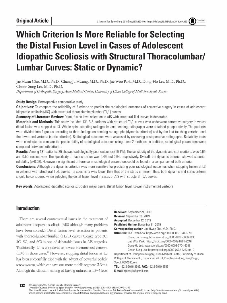
lower endplate of T12. All radiological measurements are
summarized in Fig. 1.
Radiological outcomes were divided by good or poor
outcomes checked in 2 years postoperatively. Radiologically
poor results were defined if more than one criterion was met
with following: (1) the distance between C7 plumb line and
CSVL > 20 mm, (2) LIV+1 tilt > 20°, (3) LIV disc angle > 7°,

Jae Hwan Cho et al Volume 26 • Number 4 • December 31 2019
www.krspine.org134
and (4) progression of distal lumbar curve > 5°.9)
3. Criteria to select distal fusion level
We retrospectively applied 2 criteria to select distal fusion
level. The one is a static criterion, which was suggested by
our previous study.4) If lower end vertebra (LEV) ≥L3 and
last touching vertebra (LTV) ≥L4, then L3 is selected as
appropriate LIV. If LEV ≤L4 or LTV=L5, then L4 is selected
as appropriate LIV. The other is a dynamic criterion, which
was also recently published.7) If L3 crosses CSVL with a
rotation less than grade II in side-bending radiographs, then
L3 is selected as appropriate LIV. If the curve is rigid, then LIV
should be L4.
4. Statistical Analysis
Radiological parameters were compared between pre- and
postoperatively by paired t-test. Reliability of prediction by
each criterion was assessed by sensitivity and specificity, and
ROC (receiver operating characteristic) curve. Cases showing
different prediction by 2 criteria (i.e. good by dynamic, and
poor by static criteria) were entered into subgroup analysis.
Demographic and radiological parameters between
subgroups were compared by chi-square test or independent
t-test. A multivariate regression analysis was performed
following a univariate analysis.
Statistical analyses were performed using the Statistical
Package for Social Sciences software package (version 21.0,
SPSS Inc., Chicago, IL) with p-values less than .05 considered
statistically significant.
Results
1. Demographic Data and Radiological Parameters
One hundred thirty-one patients (20 male and 111 female)
were included in this study, with a mean age of 15.3±3.4 years.
The mean height and weight of the patients were 160.0±7.6
cm and 51.2±9.8 kg, respectively. Body mass index was 19.9
±2.9. The mean follow-up period was 35.9±18.4 months,
and the average Risser grade was 3.5±1.5. Average fusion level
of operation was 9.8±2.1. Pre- and postoperative radiological
parameters were described in Table 1. The degree of the curve,
LIV+1 tilt, AVT, and TK were improved by corrective surgery
although L3-L4 disc wedging did not change.
2. Radiological outcomes
Postoperative poor radiological outcomes were shown in
25 patients (25/131, 19.1%). Fifty-one cases (38.9%) were
predicted to be poor radiological outcomes by static criteria,
Table 1. Comparisons of pre- and postoperative radiological parameters
Preop Postop p-value
TL/L curve (°) 56.8±11.1 15.4±7.4 <0.001
MT curve (°) 44.0±16.2 11.0±5.2 <0.001
C7-CSVL distance (mm) -10.3±13.4 -5.0±9.4 <0.001
L3-L4 disc wedging (°) 3.3±2.8 3.7±2.7 0.276
LIV tilt (°) 18.8±6.0 8.5±4.3 <0.001
AVT (mm) 42.2±11.8 19.3±8.3 <0.001
TK (°) 26.4±9.7 29.2±9.1 <0.001
LL (°) 46.5±11.2 46.3±9.5 0.773
*Mean and standard variation in continuous variables and number of cases in categorical variablesM:male, F:female, PTC: proximal thoracic curve, MTC: middle thoracic curve, AVT: apical vertebral translation, AVR: apical vertebral rotation, PTK: proximal thoracic kyphosis, RSH: radiographic shoulder height, CCAD: clavicle chest cage angle difference,F/U: follow up.
Fig. 1. Radiological measurements. (A) A preoperative radiograph show-ing the Cobb angle, LEV, LTV, AVT, and trunk shift (C7-CSVL distance) (B) A postoperative radiograph showing the L3-L4 disc wedge angle and the LIV+1 tilt. LEV, lower end vertebra; LTV, last touching vertebra; AVT, apical vertebral translation; CSVL, central sacral vertical line; LIV, lowest instru-mented vertebra.
A B

Distal Fusion Level Selection for AIS with Structural TL/L CurvesJournal of Korean Society of Spine Surgery
www.krspine.org 135
and 72 cases (55.0%) by dynamic criteria. The relationship
between 2 criteria and poor radiological outcome was shown
in Table 2. The sensitivity to predict poor radiological outcomes
following stopping distal fusion at L3 by dynamic and static
criteria was 0.69 and 0.50, respectively (95% CI [-0.09, 0.47]).
The specificity was 0.49 and 0.64, respectively (95% CI [-0.27,
-0.02]). However, overall reliability derived by ROC curve
showed superiority of dynamic criteria (p=0.03, Fig. 2).
Table 2. Cross-table to show the reliability of each criterion to predict radiologically poor outcome
Poor outcome Good outcome Subtotal
Static (+) 13 38 51
Static (-) 13 67 80
Subtotal 26 105 131
*Sensitivity: 0.50, *specificity: 0.64, *accuracy: 0.61, *positive predictive value: 0.26, *negative predictive value: 0.84.
Poor outcome Good outcome Subtotal
Dynamic (+) 18 54 72
Dynamic (-) 8 51 59
Subtotal 26 105 131
*Sensitivity: 0.69, *specificity: 0.49, *accuracy: 0.53, *positive predictive value: 0.25, *negative predictive value: 0.86.
Fig. 2. A receiver operating characteristic curve comparing the reliability of the dynamic and static criteria.
Table 3. Comparisons of preoperative demographic and radiological parameters
Dynamic good- Static poor (N=21) Dynamic poor- Static good (N=42) p-value (univariate) p-value (multivariate)
Age (yrs) 14.6±2.5 15.5±4.2 0.408 NA
Sex (M:F) 2:19 7:35 0.705 NA
Height (mm) 158.5±7.1 160.0±7.6 0.460 NA
Weight (kg) 49.3±5.5 51.1±9.2 0.344 NA
BMI 19.6±1.6 19.9±2.7 0.619 NA
Risser grade 3.1±1.9 3.3±1.5 0.787 NA
TL/L curve (°) 57.3±8.1 58.4±10.4 0.680 NA
TL/L flexibility (%) 54.6±16.0 46.7±17.5 0.090 0.047
MT curve (°) 50.9±12.7 41.5±14.4 0.014 0.229
C7-CSVL distance (mm) -10.1±11.5 -10.7±15.5 0.858 NA
L3-L4 disc wedging (°) 2.0±1.6 3.5±2.9 0.011 0.289
LIV tilt (°) 16.8±4.1 21.5±5.2 0.001 0.613
AVT (mm) 35.0±8.2 48.6±10.3 <0.001 <0.001
Pelvic obliquity (°) 2.0±1.4 2.4±2.0 0.287 NA
TK (°) 25.2±9.3 26.5±10.0 0.644 NA
LL (°) 42.5±8.2 47.7±12.3 0.087 0.019
*Data represent mean and standard deviation.*Negative means trunk is shifted to the left side.

Jae Hwan Cho et al Volume 26 • Number 4 • December 31 2019
www.krspine.org136
3. Subgroup analysis
Twenty-one cases were predicted to be good outcomes by
dynamic, and poor outcomes by static criteria. Whereas, 42
cases were predicted to be poor outcomes by dynamic, and
good outcomes by static criteria. Preoperative demographic
and radiological data were compared between the 2 groups in
Table 3. The TL/L flexibility (54.6° vs. 46.7°, p=0.047), AVT
(35.0 mm vs. 48.6 mm, p=<0.001) and LL (42.5° vs 47.7°, p=
0.019) were the radiological parameters exhibiting significant
differences between 2 groups based on multivariate analyses.
However, postoperative radiological parameters revealed no
differences between 2 groups (Table 4).
Two representative cases were shown in Fig. 3 and Fig. 4.
Discussion
The selection of the distal fusion level in AIS with a structural
Fig. 3. (A, B) A preoperative radiograph of a 13-year-old female patient with a Lenke 6C curve. Good and poor outcomes were expected according to the static and dynamic criteria, respectively (LEV=L3, LTV=L4, rotation grade II, CSVL did not cross the vertebral body of L3). (C) A good radio-logical outcome was observed at a postoperative 2-year follow-up, even though a poor outcome was expected according to the dynamic criterion. LEV: lower end vertebra, LTV: last touching vertebra, CSVL: central sacral vertical line.
A B C
Table 4. Comparisons of postoperative radiological parameters
Dynamic good- Static poor (N=21) Dynamic poor- Static good (N=42) p-value
TL/L curve (°) 13.2±5.4 16.0±6.7 0.102
TL/L correction rate (%) 76.9±8.6 72.9±8.4 0.077
MT curve (°) 11.0±5.1 11.9±5.5 0.522
MT correction rate (%) 76.9±8.6 72.9±8.4 0.118
C7-CSVL distance (mm) -5.9±9.7 -5.2±9.1 0.771
L3-L4 disc wedging (°) 3.1±2.5 3.5±2.6 0.507
LIV tilt (°) 7.1±2.7 8.9±4.2 0.080
AVT (mm) 17.0±7.0 20.2±7.8 0.113
TK (°) 29.1±9.1 27.8±8.8 0.578
LL (°) 43.7±7.5 46.7±10.5 0.239
*Data represent mean and standard deviation.* Negative means trunk is shifted to the left side.
Fig. 4. (A, B) A preoperative radiograph of a 19-year-old female patient with a Lenke 3C curve. Good and poor outcomes were expected according to the dynamic and static criteria, respectively (LEV=L4, LTV=L5, rotation grade I, CSVL crossed the vertebral body of L3). (C) A poor radiological outcome was observed at a postoperative 2-year follow-up, even though a good outcome was expected according to the dynamic criterion. LEV: lower end vertebra, LTV: last touching vertebra, CSVL: central sacral verti-cal line.
A B C
Distal Fusion Level Selection for AIS with Structural TL/L CurvesJournal of Korean Society of Spine Surgery
www.krspine.org 137
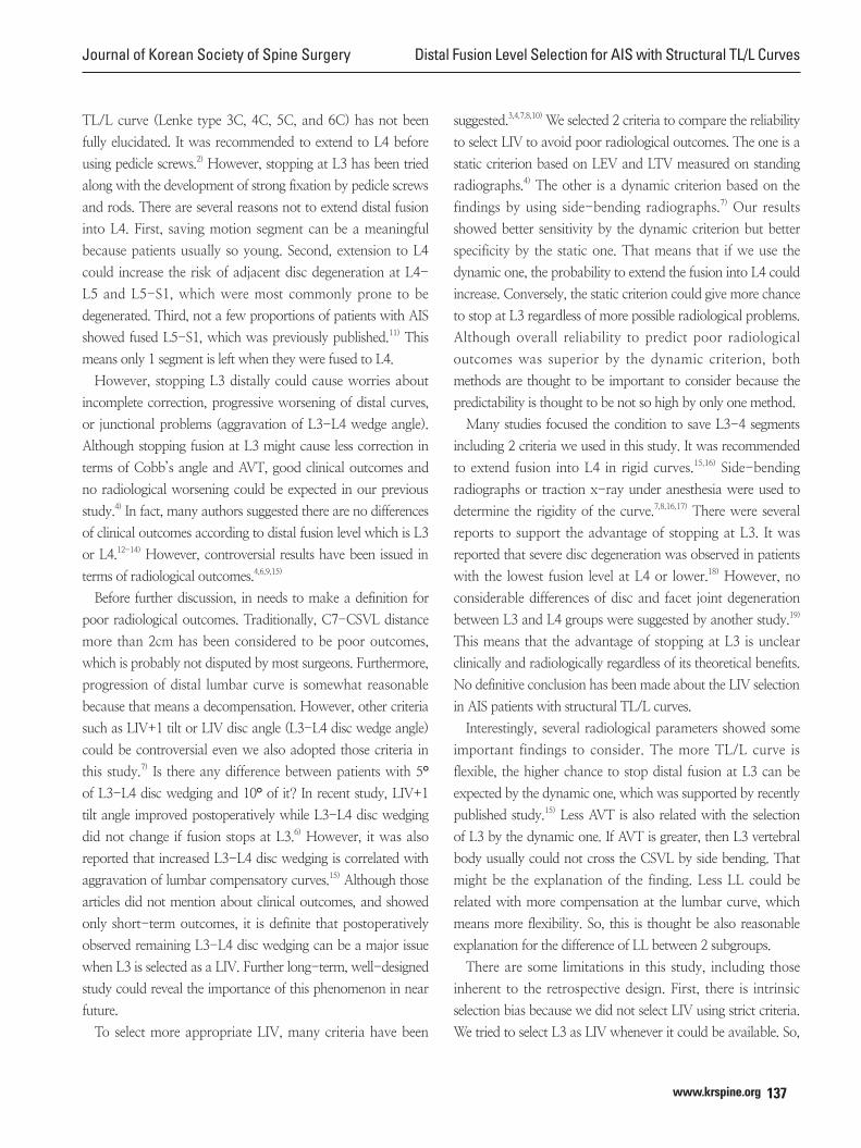
TL/L curve (Lenke type 3C, 4C, 5C, and 6C) has not been
fully elucidated. It was recommended to extend to L4 before
using pedicle screws.2) However, stopping at L3 has been tried
along with the development of strong fixation by pedicle screws
and rods. There are several reasons not to extend distal fusion
into L4. First, saving motion segment can be a meaningful
because patients usually so young. Second, extension to L4
could increase the risk of adjacent disc degeneration at L4-
L5 and L5-S1, which were most commonly prone to be
degenerated. Third, not a few proportions of patients with AIS
showed fused L5-S1, which was previously published.11) This
means only 1 segment is left when they were fused to L4.
However, stopping L3 distally could cause worries about
incomplete correction, progressive worsening of distal curves,
or junctional problems (aggravation of L3-L4 wedge angle).
Although stopping fusion at L3 might cause less correction in
terms of Cobb’s angle and AVT, good clinical outcomes and
no radiological worsening could be expected in our previous
study.4) In fact, many authors suggested there are no differences
of clinical outcomes according to distal fusion level which is L3
or L4.12-14) However, controversial results have been issued in
terms of radiological outcomes.4,6,9,15)
Before further discussion, in needs to make a definition for
poor radiological outcomes. Traditionally, C7-CSVL distance
more than 2cm has been considered to be poor outcomes,
which is probably not disputed by most surgeons. Furthermore,
progression of distal lumbar curve is somewhat reasonable
because that means a decompensation. However, other criteria
such as LIV+1 tilt or LIV disc angle (L3-L4 disc wedge angle)
could be controversial even we also adopted those criteria in
this study.7) Is there any difference between patients with 5° of L3-L4 disc wedging and 10° of it? In recent study, LIV+1
tilt angle improved postoperatively while L3-L4 disc wedging
did not change if fusion stops at L3.6) However, it was also
reported that increased L3-L4 disc wedging is correlated with
aggravation of lumbar compensatory curves.15) Although those
articles did not mention about clinical outcomes, and showed
only short-term outcomes, it is definite that postoperatively
observed remaining L3-L4 disc wedging can be a major issue
when L3 is selected as a LIV. Further long-term, well-designed
study could reveal the importance of this phenomenon in near
future.
To select more appropriate LIV, many criteria have been
suggested.3,4,7,8,10) We selected 2 criteria to compare the reliability
to select LIV to avoid poor radiological outcomes. The one is a
static criterion based on LEV and LTV measured on standing
radiographs.4) The other is a dynamic criterion based on the
findings by using side-bending radiographs.7) Our results
showed better sensitivity by the dynamic criterion but better
specificity by the static one. That means that if we use the
dynamic one, the probability to extend the fusion into L4 could
increase. Conversely, the static criterion could give more chance
to stop at L3 regardless of more possible radiological problems.
Although overall reliability to predict poor radiological
outcomes was superior by the dynamic criterion, both
methods are thought to be important to consider because the
predictability is thought to be not so high by only one method.
Many studies focused the condition to save L3-4 segments
including 2 criteria we used in this study. It was recommended
to extend fusion into L4 in rigid curves.15,16) Side-bending
radiographs or traction x-ray under anesthesia were used to
determine the rigidity of the curve.7,8,16,17) There were several
reports to support the advantage of stopping at L3. It was
reported that severe disc degeneration was observed in patients
with the lowest fusion level at L4 or lower.18) However, no
considerable differences of disc and facet joint degeneration
between L3 and L4 groups were suggested by another study.19)
This means that the advantage of stopping at L3 is unclear
clinically and radiologically regardless of its theoretical benefits.
No definitive conclusion has been made about the LIV selection
in AIS patients with structural TL/L curves.
Interestingly, several radiological parameters showed some
important findings to consider. The more TL/L curve is
flexible, the higher chance to stop distal fusion at L3 can be
expected by the dynamic one, which was supported by recently
published study.15) Less AVT is also related with the selection
of L3 by the dynamic one. If AVT is greater, then L3 vertebral
body usually could not cross the CSVL by side bending. That
might be the explanation of the finding. Less LL could be
related with more compensation at the lumbar curve, which
means more flexibility. So, this is thought be also reasonable
explanation for the difference of LL between 2 subgroups.
There are some limitations in this study, including those
inherent to the retrospective design. First, there is intrinsic
selection bias because we did not select LIV using strict criteria.
We tried to select L3 as LIV whenever it could be available. So,
Jae Hwan Cho et al Volume 26 • Number 4 • December 31 2019
www.krspine.org138
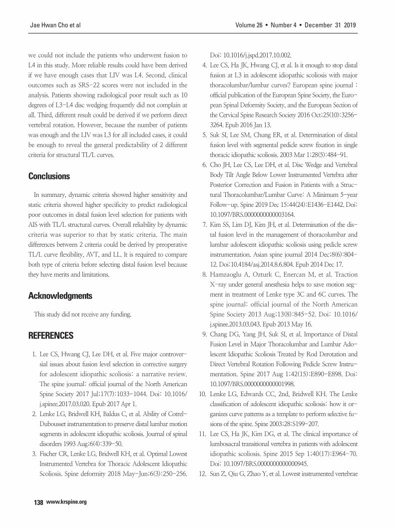
we could not include the patients who underwent fusion to
L4 in this study. More reliable results could have been derived
if we have enough cases that LIV was L4. Second, clinical
outcomes such as SRS-22 scores were not included in the
analysis. Patients showing radiological poor result such as 10
degrees of L3-L4 disc wedging frequently did not complain at
all. Third, different result could be derived if we perform direct
vertebral rotation. However, because the number of patients
was enough and the LIV was L3 for all included cases, it could
be enough to reveal the general predictability of 2 different
criteria for structural TL/L curves.
Conclusions
In summary, dynamic criteria showed higher sensitivity and
static criteria showed higher specificity to predict radiological
poor outcomes in distal fusion level selection for patients with
AIS with TL/L structural curves. Overall reliability by dynamic
criteria was superior to that by static criteria. The main
differences between 2 criteria could be derived by preoperative
TL/L curve flexibility, AVT, and LL. It is required to compare
both type of criteria before selecting distal fusion level because
they have merits and limitations.
Acknowledgments
This study did not receive any funding.
REFERENCES
1. Lee CS, Hwang CJ, Lee DH, et al. Five major controver-
sial issues about fusion level selection in corrective surgery
for adolescent idiopathic scoliosis: a narrative review.
The spine journal: official journal of the North American
Spine Society 2017 Jul;17(7):1033-1044. Doi: 10.1016/
j.spinee.2017.03.020. Epub 2017 Apr 1.
2. Lenke LG, Bridwell KH, Baldus C, et al. Ability of Cotrel-
Dubousset instrumentation to preserve distal lumbar motion
segments in adolescent idiopathic scoliosis. Journal of spinal
disorders 1993 Aug;6(4):339-50.
3. Fischer CR, Lenke LG, Bridwell KH, et al. Optimal Lowest
Instrumented Vertebra for Thoracic Adolescent Idiopathic
Scoliosis. Spine deformity 2018 May-Jun;6(3):250-256.
Doi: 10.1016/j.jspd.2017.10.002.
4. Lee CS, Ha JK, Hwang CJ, et al. Is it enough to stop distal
fusion at L3 in adolescent idiopathic scoliosis with major
thoracolumbar/lumbar curves? European spine journal :
official publication of the European Spine Society, the Euro-
pean Spinal Deformity Society, and the European Section of
the Cervical Spine Research Society 2016 Oct;25(10):3256-
3264. Epub 2016 Jan 13.
5. Suk SI, Lee SM, Chung ER, et al. Determination of distal
fusion level with segmental pedicle screw fixation in single
thoracic idiopathic scoliosis. 2003 Mar 1;28(5):484-91.
6. Cho JH, Lee CS, Lee DH, et al. Disc Wedge and Vertebral
Body Tilt Angle Below Lower Instrumented Vertebra after
Posterior Correction and Fusion in Patients with a Struc-
tural Thoracolumbar/Lumbar Curve: A Minimum 5-year
Follow-up. Spine 2019 Dec 15;44(24):E1436-E1442. Doi:
10.1097/BRS.0000000000003164.
7. Kim SS, Lim DJ, Kim JH, et al. Determination of the dis-
tal fusion level in the management of thoracolumbar and
lumbar adolescent idiopathic scoliosis using pedicle screw
instrumentation. Asian spine journal 2014 Dec;8(6):804-
12. Doi:10.4184/asj.2014.8.6.804. Epub 2014 Dec 17.
8. Hamzaoglu A, Ozturk C, Enercan M, et al. Traction
X-ray under general anesthesia helps to save motion seg-
ment in treatment of Lenke type 3C and 6C curves. The
spine journal: official journal of the North American
Spine Society 2013 Aug;13(8):845-52. Doi: 10.1016/
j.spinee.2013.03.043. Epub 2013 May 16.
9. Chang DG, Yang JH, Suk SI, et al. Importance of Distal
Fusion Level in Major Thoracolumbar and Lumbar Ado-
lescent Idiopathic Scoliosis Treated by Rod Derotation and
Direct Vertebral Rotation Following Pedicle Screw Instru-
mentation. Spine 2017 Aug 1;42(15):E890-E898. Doi:
10.1097/BRS.0000000000001998.
10. Lenke LG, Edwards CC, 2nd, Bridwell KH. The Lenke
classification of adolescent idiopathic scoliosis: how it or-
ganizes curve patterns as a template to perform selective fu-
sions of the spine. Spine 2003;28:S199-207.
11. Lee CS, Ha JK, Kim DG, et al. The clinical importance of
lumbosacral transitional vertebra in patients with adolescent
idiopathic scoliosis. Spine 2015 Sep 1;40(17):E964-70.
Doi: 10.1097/BRS.0000000000000945.
12. Sun Z, Qiu G, Zhao Y, et al. Lowest instrumented vertebrae

Distal Fusion Level Selection for AIS with Structural TL/L CurvesJournal of Korean Society of Spine Surgery
www.krspine.org 139
selection for selective posterior fusion of moderate thora-
columbar/lumbar idiopathic scoliosis: lower-end vertebra
or lower-end vertebra+1? European spine journal : official
publication of the European Spine Society, the European
Spinal Deformity Society, and the European Section of the
Cervical Spine Research Society 2014 Jun;23(6):1251-7.
Doi: 10.1007/s00586-014-3276-0. Epub 2014 Mar 25.
13. Ding R, Liang J, Qiu G, et al. Evaluation of quality of life
in adolescent idiopathic scoliosis with different distal fusion
level: a comparison of L3 versus L4. Journal of spinal disor-
ders & techniques 2014 Jul;27(5):E155-61. Doi: 10.1097/
BSD.0000000000000073
14. Bartie BJ, Lonstein JE, Winter RB. Long-term follow-up of
adolescent idiopathic scoliosis patients who had Harrington
instrumentation and fusion to the lower lumbar vertebrae:
is low back pain a problem? Spine 2019 Mar;178:77-81.
Doi: 10.1016/j.clineuro.2019.02.005. Epub 2019 Feb 5.
15. Chang DG, Suk SI, Song KS, et al. How to avoid distal
adding-on phenomenon for rigid curves in major tho-
racolumbar and lumbar adolescent idiopathic scoliosis?
Identifying the incidence of distal adding-on by the selection
of lowest instrumented vertebra. World neurosurgery 2019
Dec;132:e472-e478. Doi: 10.1016/j.wneu.2019.08.110.
Epub 2019 Aug 27.
16. Qin X, He Z, Yin R, et al. Where to stop distally in Lenke
modifier C AIS with lumbar curve more than 60°: L3 or
L4? Clin Neurol Neurosurg 2019 Mar;178:77-81. Doi:
10.1016/j.clineuro.2019.02.005. Epub 2019 Feb 5.
17. Erdem MN, Karaca S, Korkmaz MF, et al. Criteria for
Ending the Distal Fusion at the L3 Vertebra vs. L4 in Surgi-
cal Treatment of Adolescent Idiopathic Scoliosis Patients
with Lenke Type 3C, 5C, and 6C Curves: Results After
Ten Years of Follow-up. Cureus 2018 May 1;10(5):e2564.
Doi: 10.7759/cureus.2564.
18. Akazawa T, Kotani T, Sakuma T, et al. Spinal fusion on
adolescent idiopathic scoliosis patients with the level of L4
or lower can increase lumbar disc degeneration with sagit-
tal imbalance 35 years after surgery. Spine Surg Relat Res
2017 Dec 20;1(2):72-77. Doi: 10.22603/ssrr.1.2016-0017.
eCollection 2017.
19. Enercan M, Kahraman S, Yilar S, et al. Does It Make a Dif-
ference to Stop Fusion at L3 Versus L4 in Terms of Disc and
Facet Joint Degeneration: An MRI Study With Minimum 5
Years Follow-up. Spine Deform 2016 May;4(3):237-244.
Doi: 10.1016/j.jspd.2015.12.001. Epub 2016 Apr 16.
140
J Korean Soc Spine Surg. 2019 Dec;26(4):132-140. https://doi.org/10.4184/jkss.2019.26.4.140Original Article
© Copyright 2019 Korean Society of Spine Surgery Journal of Korean Society of Spine Surgery. www.krspine.org. pISSN 2093-4378 eISSN 2093-4386 This is an Open Access article distributed under the terms of the Creative Commons Attribution Non-Commercial License (http://creativecommons.org/licenses/by-nc/4.0/) which permits unrestricted non-commercial use, distribution, and reproduction in any medium, provided the original work is properly cited.
구조적 흉요부/요부 만곡 청소년기 특발성 측만증에서 원위부 유합 범위를 결정하는데 있어서 동적 혹은 정
적 기준의 신뢰도 비교조재환 • 황창주 • 박재우 • 이동호 • 이춘성
울산대학교 의과대학 정형외과학교실
연구 계획: 후향적 비교 연구
목적: 구조적 흉요부/요부 만곡 청소년기 특발성 측만증 환자 수술 후 방사선학적 결과를 예측하는 2가지 기준의 신뢰도를 비교하기 위함
선행 연구문헌의 요약: 구조적 흉요부/요부 만곡 청소년기 특발성 측만증의 원위부 유합 부위 결정은 이견이 많음.
대상 및 방법: 원위 유합을 L3까지 수술한 구조적 흉요부/요부 만곡 청소년기 특발성 측만증 환자 131명을 대상으로 하였다. 수술전 전신 척추 기립 자세
와 측부 굽힘 방사선 사진을 촬영하였다. 환자들은 측부 굽힘 사진 상의 기준(동적)과, Last touching vertebra와 lower end vertebra에 따른 기준(정적)에
따라 분류하였고, 술후 방사선학적 결과에 따라 분석하였다. 두 기준에 따른 방사선학적 결과 예측의 신뢰도를 분석하였고, 여러 방사선학적 인자들의 차
이를 분석하였다.
결과: 131명의 환자중 25명이 불량한 결과를 보였다(19.1%). 동적 기준과 정적 기준의 민감도는 각각 0.69와 0.50이었고, 특이도는 0.49와 0.64였다. 전
반적으로는 동적 기준이 우월한 신뢰도를 보여주었다(p=0.03). 하지만, 두 기준에 따른 방사선학적 인자들의 차이는 없었다.
결론: L3에서 유합을 멈췄을 때 동적 기준이 더 불량한 예후를 예측하는데 민감했지만, 특이도는 정적 기준보다 낮았다. 구조적 흉요부/요부 만곡 청소년
기 특발성 측만증 환자에서 원위 유합 범위 결정에 두 기준 모두를 고려하는 것이 좋을 것으로 생각된다.
색인 단어: 청소년기 특발성 측만증, 이중 대만곡, 원위 유합, 하위 고정 척추
약칭 제목: 구조적 흉요부/요부 만곡 청소년기 특발성 측만성 환자의 원위 유합 범위 결정
접수일: 2019년 9월 24일 수정일: 2019년 9월 28일 게재확정일: 2019년 12월 12일
교신저자: 조재환
서울 송파구 올림픽로 43길 88 울산대학교 의과대학 정형외과학교실
TEL: 02-3010-3549 FAX: 02-3010-8555 E-mail: [email protected]
Recommended


![Exercises for adolescent idiopathic scoliosis - …tees.openrepository.com/tees/bitstream/10149/249111/2/249111.pdf[Intervention Review] Exercises for adolescent idiopathic scoliosis](https://img.pdfslide.us/doc/110x75/5aa5e2337f8b9ae7438e1827/exercises-for-adolescent-idiopathic-scoliosis-tees-intervention-review-exercises.jpg)