Isolation and Callus Production from Cotyledon Protoplasts of Cucumis metuliferus William H. McCarthy, Todd C. Wehner, Jiahua Xie, and Margaret E. Daub North Carolina State University, Raleigh, NC 27695-7609 In North Carolina, approximately 8% of the cucumber (Cucumis sativus) yield is lost to root-knot nematodes (Meloidogyne spp.) (Main and Gurtz, 1989). No resistance was found in cucumber after screening of 900 cultigens for resistance to Meloidogyne incognita race 3 (Walters, 1991). African horned cucumber (Cucumis metuliferus) has resistance to M. incognita (Fassuliotis, 1967; Walters, 1991), as well as other diseases such as squash mosaic virus and watermelon mosaic virus (Provvidenti and Robinson, 1974). There would be benefit in transferring nematode resistance into the gene pool of C. sativus from C. metuliferus. However, traditional sexual hybridization techniques have been unsuccessful in producing hybrids between C. metuliferus and cucumber, probably due to differences in chromosome number (Deakin et al., 1971) or incompatibility barriers. In some crops, use of protoplast fusion has allowed barriers to sexual hybridization to be overcome. Within Cucumis, most protoplast isolation and plant regeneration studies have been attempted using C. sativus and Cucumis melo (Wehner et al., 1990). Plant regeneration from protoplasts of C. sativus was first reported by Jia et al. (1986). The successful protocol for asymmetric protoplast fusions of cucumber and melon has been developed (Jarl et al., 1995). However, there were few reports involving protoplast isolation and fusion with C. metuliferus. In one study, a fusion rate of 5 to 12% was reported between protoplasts of C. metuliferus and C. melo. Their attempts to obtain callus from protoplasts of C. metuliferus or from fusion products were unsuccessful (Roig et al., 1986). In a second study, Tang and Punja (1989) reported protoplast fusion of C. metuliferus and C. sativus, and subsequent callus development from hybrid protoplasts. However, they were unable to produce callus from protoplasts of C. metuliferus alone, or to regenerate plants from callus of hybrid protoplast. Development of an efficient system of plant regeneration from C. metuliferus protoplasts may assist in obtaining a procedure to regenerate hybrid
plants from protoplast fusion of C. sativus and C. metuliferus. Therefore, the objective of this study was to develop a reliable procedure for isolation and culture of cotyledon protoplasts of C. metuliferus. Methods. Seeds of C. metuliferus PI 482454 were surface-sterilized with the industrial disinfectant LD (Alcide Corp., Norwalk, Conn.) for 30 minutes on a gyratory shaker at 100 RPM, then were rinsed 5 times with sterilized, distilled water. Seeds were germinated on C1 medium (Table 1) in the dark at 30°C. After 84 hr, seedlings were placed in a culture room maintained at 22°C with a 16 hr photoperiod lights (108 µMol.m-2.s-1). Seedlings were transferred back to 30°C in darkness for 24 hours before protoplast isolation. An enzyme solution was prepared consisting of 0.7 mM KH2PO4, 7 mM CaCl2·2H2O, 0.5 M mannitol, 3 mM MES [2-(N-morpholino) ethanesulfonic acid] (Jia et al., 1986), 2 % (w/v) cellulysin and 0.5 % (w/v) macerase (Calbiochem, La Jolla, California). That solution was mixed at a 1:1 (v/v) ratio with C2 medium having a pH of 5.8 (Table 1), except modified by the addition of 230 mg ▪ L
-1 CaCl2.2H2O as described by Jia et al. (1986). Ten ml of filter-sterilized enzyme C2 solution was added to 0.5 g of whole cotyledons (5 to 7 days old), and vacuum infiltrated at 9.3 kPa for 20 sec. The infiltrated tissue was incubated for 6 hr on a gyratory shaker at 60 rpm at 25°C in the dark. Protoplasts were separated from cell walls and other debris by manually swirling the flasks, and filtering the solution through sterilized Miracloth (Calbiochem, La Jolla, California). Protoplasts were washed 3 times with C2 medium by centrifuging at 100x g for 3 minutes. For all experiments, protoplast density after isolation was estimated using a hemacytometer, and viability was determined using fluroscein diacetate (Widholm, 1972). Protoplasts were cultured in 5 ml of C2 medium at a density of 1x10⁵ protoplasts per ml in 10 x 60 mm petri plates, and incubated in the dark at 25°C. Five days after protoplast isolation, half of the
Cucurbit Genetics Cooperative Report 24:102-106 (2001) 102
plates were transferred to a growth chamber maintained at 30°C in the dark. Seven days after protoplast release, 1 ml of C2 medium with 0.15 M mannitol was added to each plate. Fourteen and 21 days after isolation, 1 ml of C2 medium (with no mannitol) was added to each plate. Protoplast culture plates were swirled 1 min per day to increase aeration. Estimates of protoplast division (PD), calculated as the percentage of protoplasts which had undergone cell division, were made 8 to 10 days after isolation by visual observation of 5 samples per plate with 15 plates per treatment combination (320x magnification). Cell wall regeneration was determined by observed changes in protoplast shape and actual cell division. Using the sample results, total number of divided cells per plate was calculated. This number was then compared to the total number of protoplasts in the plate (5 x 10⁵) to produce an estimate of PD. Approximately 3 weeks after isolation, plating efficiency (PE) was estimated by counting clumps of 8 or more cells which appeared to have originated from 1 protoplast. PE was estimated by counting the number of microcalli in 5 samples per plate with 15 plates per replication (100x magnification), and calculating an approximate number per plate from the random visual counting. Experiment 1 was a randomized complete block design with 4 replications. Treatments consisted of protoplasts cultured in C2 medium at either 25 or 30°C. In experiment 2, protoplasts were isolated and cultured in C2 medium at either 25 or 30°C, using the methods described above. After 3 weeks, 2 ml of microcallus suspension were pipetted onto C4 medium (Table 1) (20 ml in 100 x 15 mm petri dishes) containing varying amounts of 2,4-D, 1H-indo-3-acetic acid (IAA), kinetin and N-(phenylmethyl)-1H-purine-6-amine (BA) (Table 2). Plates were swirled to distribute microcalli on the agar medium, and excess liquid medium was removed. Callus cultures were maintained at 22°C in the dark for 3 weeks before being rated for percentage of each petri plate covered with callus. Callus color was rated 1 to 9 (1=white, 5=yellow, 9=brown). After rating callus, clumps of healthy (white to yellow in color) callus were transferred to
similar media, and to C4 media containing 0.2 mg.L-1 IAA and 5.0 mg ▪ L
-1 kinetin (Jia et al., 1986). Experiment 2 was a randomized complete block design with 4 plates per treatment and 4 replications. Treatments consisted of protoplasts that had been first cultured in C2 medium at 25 or 30°C for 3 weeks cultured on C4 medium containing varying amounts of growth regulators at 22°C (Table 2). In experiment 3, protoplasts were isolated and cultured in C2 medium at either 25 or 30°C, using the methods described in experiment 1. After 3 weeks, protoplasts were transferred to C3 medium for callus induction. Treatments consisted of the same growth regulator combinations as used in experiment 2, using protoplasts which had been cultured at either 25 or 30°C for the first 3 weeks (Table 3). Callus cultures were maintained at 22°C in the dark for 6 weeks before rating each petri plate for percent coverage with callus. Experiment 3 was a randomized complete block design with 3 plates per treatment and 5 replications. Results. In experiment 1, protoplasts cultured at 25°C had a protoplast division percentage (PD) of 4% after 8 to 10 days. Protoplasts cultured at 30°C had a PD of 7%. There was a significant difference between the 2 temperatures for PD. After 3 weeks of culture at 25°C, each plate had a mean of 3970 microcalli per plate (PE of 0.8%). At 30°C, each plate had a mean of 5025 (PE of 1.0%) microcalli per plate. In experiment 2, medium A3 (Table 2) was best for producing a large amount of yellow, friable callus. There were no significant differences in color among media. However, among temperatures, protoplasts cultured at 30°C were significantly whiter (Table 2). Callus color appeared to indicate the potential for continued proliferation because callus with ratings >5 usually had little or no continued growth. Healthy callus transferred to C3 or C4 media containing 0.2 mg ▪ L
-1 IAA and 5.0 mg ▪ L-1 kinetin caused sporadic
greening. Five to 15 roots per plate were observed on callus on media A3 and A4 after 6 weeks. No embryogenesis resulting in plantlet regeneration was observed in this experiment.
Cucurbit Genetics Cooperative Report 24:102-106 (2001) 103
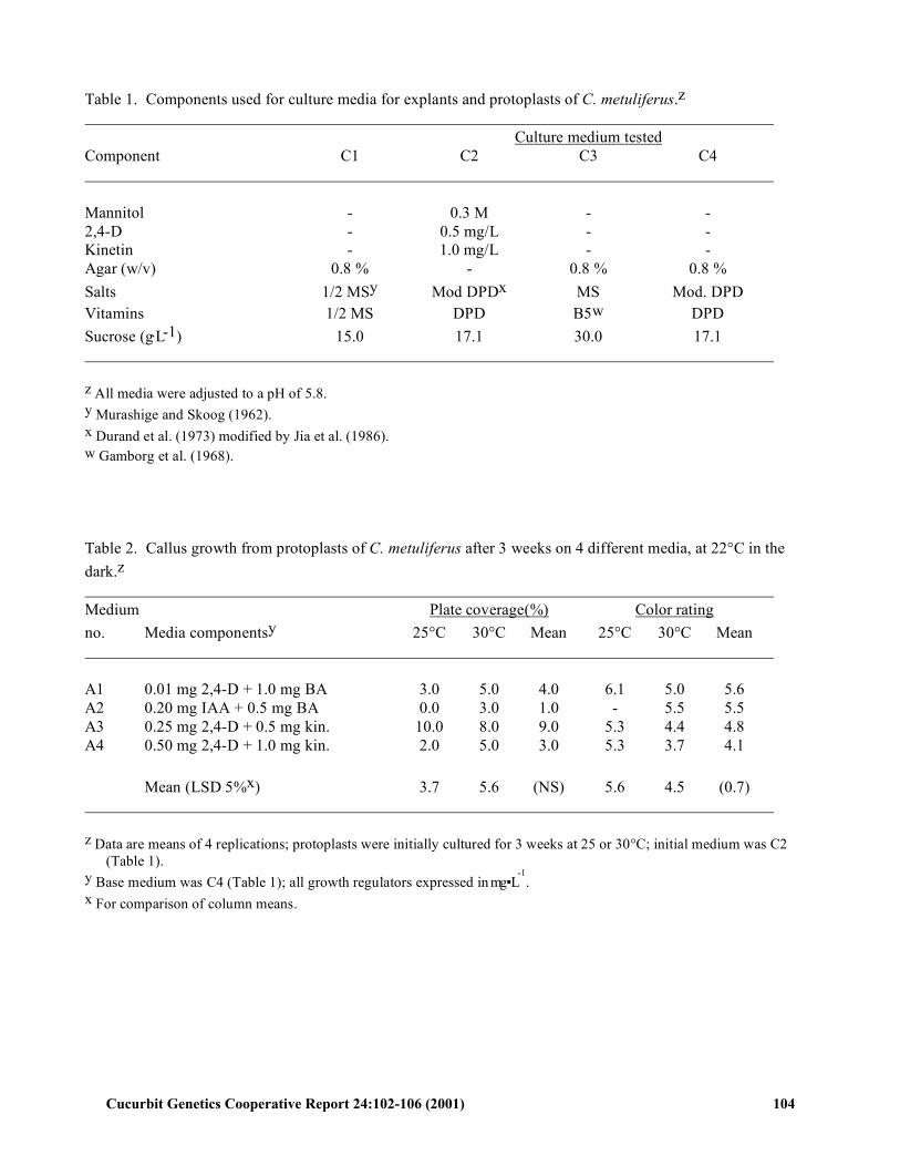
Table 1. Components used for culture media for explants and protoplasts of C. metuliferus.z Culture medium tested Component C1 C2 C3 C4 Mannitol - 0.3 M - - 2,4-D - 0.5 mg/L - - Kinetin - 1.0 mg/L - - Agar (w/v) 0.8 % - 0.8 % 0.8 % Salts 1/2 MSy Mod DPDx MS Mod. DPD Vitamins 1/2 MS DPD B5w DPD Sucrose (g.L-1) 15.0 17.1 30.0 17.1 z All media were adjusted to a pH of 5.8. y Murashige and Skoog (1962). x Durand et al. (1973) modified by Jia et al. (1986). w Gamborg et al. (1968). Table 2. Callus growth from protoplasts of C. metuliferus after 3 weeks on 4 different media, at 22°C in the dark.z Medium Plate coverage(%) Color rating no. Media componentsy 25°C 30°C Mean 25°C 30°C Mean A1 0.01 mg 2,4-D + 1.0 mg BA 3.0 5.0 4.0 6.1 5.0 5.6 A2 0.20 mg IAA + 0.5 mg BA 0.0 3.0 1.0 - 5.5 5.5 A3 0.25 mg 2,4-D + 0.5 mg kin. 10.0 8.0 9.0 5.3 4.4 4.8 A4 0.50 mg 2,4-D + 1.0 mg kin. 2.0 5.0 3.0 5.3 3.7 4.1 Mean (LSD 5%x) 3.7 5.6 (NS) 5.6 4.5 (0.7) z Data are means of 4 replications; protoplasts were initially cultured for 3 weeks at 25 or 30°C; initial medium was C2
(Table 1). y Base medium was C4 (Table 1); all growth regulators expressed in mg ▪ L
-1.
x For comparison of column means.
Cucurbit Genetics Cooperative Report 24:102-106 (2001) 104
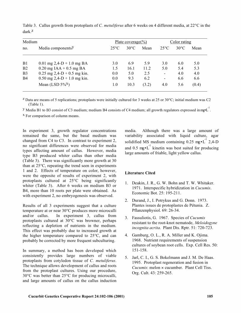
Table 3. Callus growth from protoplasts of C. metuliferus after 6 weeks on 4 different media, at 22°C in the dark.z Medium Plate coverage(%) Color rating no. Media componentsy 25°C 30°C Mean 25°C 30°C Mean B1 0.01 mg 2,4-D + 1.0 mg BA 3.0 6.9 5.9 3.0 6.0 5.0 B2 0.20 mg IAA + 0.5 mg BA 1.5 16.1 11.2 5.0 5.4 5.3 B3 0.25 mg 2,4-D + 0.5 mg kin. 0.0 5.0 2.5 - 4.0 4.0 B4 0.50 mg 2,4-D + 1.0 mg kin. 0.0 9.3 6.2 - 6.6 6.6 Mean (LSD 5%x) 1.0 10.3 (3.2) 4.0 5.6 (0.4) z Data are means of 5 replications; protoplasts were initially cultured for 3 weeks at 25 or 30°C; initial medium was C2
(Table 1). y Media B1 to B3 consist of C3 medium; medium B4 consists of C4 medium; all growth regulators expressed in mg ▪ L
-1.
x For comparison of column means. In experiment 3, growth regulator concentrations remained the same, but the basal medium was changed from C4 to C3. In contrast to experiment 2, no significant differences were observed for media types affecting amount of callus. However, media type B3 produced whiter callus than other media (Table 3). There was significantly more growth at 30 than at 25°C, repeating the trend seen in experiments 1 and 2. Effects of temperature on color, however, were the opposite of results of experiment 2, with protoplasts cultured at 25°C being significantly whiter (Table 3). After 6 weeks on medium B3 or B4, more than 10 roots per plate were obtained. As with experiment 2, no embryogenesis was observed. Results of all 3 experiments suggest that a culture temperature at or near 30°C produces more microcalli and/or callus. In experiment 3, callus from protoplasts cultured at 30°C was browner, perhaps reflecting a depletion of nutrients in the medium. This effect was probably due to increased growth at the higher temperature compared to 25°C, and can probably be corrected by more frequent subculturing. In summary, a method has been developed which consistently provides large numbers of viable protoplasts from cotyledon tissue of C. metuliferus. The technique allows development of callus and roots from the protoplast cultures. Using our procedure, 30°C was better than 25°C for producing microcalli, and large amounts of callus on the callus induction
media. Although there was a large amount of variability associated with liquid culture, agar solidified MS medium containing 0.25 mg ▪ L
-1 2,4-D and 0.5 mg ▪ L
-1 kinetin was best suited for producing large amounts of friable, light yellow callus. Literature Cited 1. Deakin, J. R., G. W. Bohn and T. W. Whitaker.
1971. Interspecific hybridization in Cucumis. Economic Bot. 25: 195-211.
2. Durand, J., I. Potrykus and G. Donn. 1973. Plantes issues de protoplastes de Pétunia. Z. Pflanzenphysiol. 69: 26-34.
3. Fassuliotis, G. 1967. Species of Cucumis resistant to the root-knot nematode, Meloidogyne incognita acrita. Plant Dis. Rptr. 51: 720-723.
4. Gamburg, O. L., R. A. Miller and K. Ojima. 1968. Nutrient requirements of suspension cultures of soybean root cells. Exp. Cell Res. 50: 151-158.
5. Jarl, C. I., G. S. Bokelmann and J. M. De Haas. 1995. Protoplast regeneration and fusion in Cucumis: melon × cucumber. Plant Cell Tiss. Org. Cult. 43: 259-265.
Cucurbit Genetics Cooperative Report 24:102-106 (2001) 105
6. Jia, Shi-rong, You-ying Fu and Yun Lin. 1986. Embryogenesis and plant regeneration from cotyledon protoplast culture of cucumber (Cucumis sativus L.). J. Plant Physiol. 124: 393-398.
7. Main, C. E. and S. K. Gurtz, (eds.). 1989. 1988 estimates of crop losses in North Carolina due to plant diseases and nematodes. Dept. of Plant Path. Spec. Publ. No. 8, North Carolina State University, Raleigh, N.C.
8. Murashige, T. and F. Skoog. 1962. A revised medium for rapid growth and bioassays with tobacco tissue cultures. Physiol. Plant. 15: 473-497.
9. Provvidenti, R. and R. W. Robinson. 1974. Resistance to squash mosaic virus 1 in Cucumis metuliferus. Plant Dis. Rptr. 58: 735-738.
10. Roig, L. A., M. V. Roche, M. C. Orts, L. Zubeldia and V. Moreno. 1986. Isolation and culture of protoplasts from Cucumis metuliferus and Cucurbita martinezii and a method for their fusion with Cucumis melo protoplasts. Cucurbit Genet. Coop. Rpt. 9: 70-73.
11. Tang, F. A. and Z. K. Punja. 1989. Isolation and culture of protoplasts of Cucumis sativus and Cucumis metuliferus and methods for their fusion. Cucurbit Genet. Coop. Rpt. 12: 29-34.
12. Wehner, T. C., R. M. Cade and R. D. Locy. 1990. Cell, tissue and organ culture techniques for genetic improvement of cucurbits, p.367-381. In: R. W. Robinson (ed.). Proc. Intl. Conf. Biol. Chem. Cucurbitaceae.
13. Walters, S. A. 1991. Root knot nematode resistance in Cucumis sativus and Cucumis metuliferus. M.S. Thesis, North Carolina State Univ., Raleigh.
14. Widholm, J. M. 1972. The use of fluroscein diacetate and phenosafranine for determining viability of cultured plant cells. Stain Technol. 47: 189-194.
Cucurbit Genetics Cooperative Report 24:102-106 (2001) 106
Recommended