1
Chapter 1
Introduction
“For every scientist and medical professional hacking at the roots of cancer, there
are tens of thousands hacking at the branches or even studying
the leaves of the tree.” - Henry David Thoreau
1.1 CANCER
Cancer is demarcated as an abnormal growth of cells caused by multiple
changes in gene expression leading to a deregulated balance of cell proliferation and
cell death. This disease is the contributing factor for major mortality on global level
next to cardiovascular diseases.
Cancer is a large group of disease, perhaps even more than hundred. The
development of cancer is a sequential multi-step process, that onset from the elementary
building block of the organs of body – the cells. When the cells continue multiplying
irrespective of requirement, they result in a mass/growth of tissue called a tumor. These
growths are considered either benign or malignant. Benign growth is termed as non-
cancerous and rarely threatens life and do not spread to other parts of the body.
Whereas malignant growth is dubbed as cancerous and cells from malignant tumors
evolve into a population of cells that can invade normal tissues and affect distant sites
by spreading through blood or lymph, causing significant morbidity and mortality. This
systemic process of spreading is termed as metastasis. However, the cancer that
originates from this process is still referred by the organ of origin. For instance, if
cervical cancer spreads to the lungs, it is still called as cervical cancer and not as lung
cancer. Different types of cancer arise from variations in the age of onset, rate of
growth, state of cellular differentiation, diagnostic detectability, invasiveness, metastatic
potential, response to treatment, and prognosis. In many cases causes of cancer are not
clearly defined, but both the external (e.g., environmental chemicals and radiation) and
internal (e.g., immune system defects, genetic predisposition) factors play a major role.
These causal factors may act together or individually to initiate (the initial genetic
insult) and promote (stimulation of growth of initiated cells) carcinogenesis.
2
1.2 PROCESS OF CARCINOGENESIS
Carcinogenesis is a multistep process and characterized by a progression of
changes on cellular and genetic level that ultimately reprogram a cell to undergo
uncontrolled cell division, in which as many as ten distinct mutations may accumulate
before it becomes cancerous.
The cell cycle is regulated by a large number of cellular genes that are
expressed or exhibited at different stages of the cycle. These genes code for/or
determine growth factors, growth-factor receptors and proteins that control gene
functions and cell survival. In all cancers, the process of carcinogenesis starts when
the genes involved in cell cycle control and regulation are malfunctioned due to DNA
damages caused by exposure to chemicals and radiation. Usually the normal cells
with damaged DNA either get repaired or perish. But in the case of cancer, the cell
cycle gets distorted due to activation of oncogenes or by the inactivation of tumor
suppressor genes. Activation of oncogenes drives abnormal, unregulated cell
proliferation and lead to tumor formation, while inactivation of tumor suppressor
genes fails to avert the same. Mutations of the tumor suppressor gene such as p53 are
found in about 50% of the existing human cancers. Viruses also can induce
carcinogenesis by introducing new DNA sequences (oncogenes). Generally, types of
carcinogenesis involve chemical oncogenesis, radiation oncogenesis, viral
oncogenesis, nutritional oncogenesis, hormonal oncogenesis and genetic oncogenesis,
while the latter three are less prevalent than the others.
In a study involving experimental animals, three stages of carcinogenic
processes have been identified. They are: (1) initiation, where DNA is irreversibly
altered; (2) promotion, which is the multiplication of altered cells; and (3)
progression, which involves chromosomal changes, high growth rate, invasiveness
and potential to metastasize. There is usually a long time lag between exposure to a
carcinogen and the occurrence of cancer. The neoplastic changes start with a
reversible increase in cell division caused by an external stimulus, such as hormonal
imbalance or chronic irritation that is termed as hyperplasia. The cells then attain a
stage, termed as dysplasia, where a series of changes get incorporated slowly to give
rise to cancer-like properties to the cells from the properties of normal cells. These
3
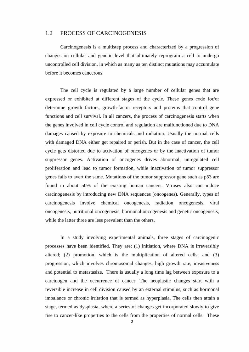
cells are excessively proliferative and are characterized by loss of normal tissue
arrangement and cell structure that upon continuous division and accumulation evolve
as invasive/metastatic cells. The overview of carcinogenic process is pictorially
represented in Fig. 1.1.
Fig. 1.1: Process of carcinogenesis.
1.3 CLASSIFICATIONS OF CANCER BASED ON TISSUE OF
ORIGIN
Cancers are classified by the type of cell that the tumor cells resemble and are
consequently presumed to be the origin of the tumor. Based on the tissue of origin,
tumors are classified into the following four broad categories.
1.3.1 CARCINOMA
The tumors that principally originate from epithelial cells of ectoderm or
endoderm are called carcinoma. The solid tumors in the nerve tissue and the tissues of
body surfaces, or their attached glands are examples of carcinomas. These include
cervical, breast, skin and brain carcinomas. About 85% of cancers are carcinomas.
1.3.2 SARCOMAS
The term sarcoma is used when the tumor principally consists of connective
tissue cells, which are of mesodermal origin. They are solid tumors growing from
connective tissue (cartilage, bone and muscle). Although they account for most of the
cancers studied in laboratory animals, they constitute only about 2% of human cancers.

4
1.3.3 LYMPHOMA
The cancers in which there is an excessive production of lymphocytes by the
lymph nodes and spleen are termed as lymphoma. Hodgkins‟ disease is an example of
a lymphoma. Lymphomas constitute about 5% of human cancers.
1.3.4 LEUKEMIA
Leukemia is a type of cancer of the blood or bone marrow characterized by an
abnormal increase of immature white blood cells called "blasts". They constitute
about 4% of human cancers.
In addition to the types of cancer mentioned above, there might be mixed
malignant tumors, e. g. tumors arising from both ectodermal and mesodermal origin.
1.4 TYPES OF CANCER BASED ON ORGAN OF ORIGIN
The type of cancer is usually referred by the organ of origin and not by the
organ of occurrence. There are over 100 types of cancer affecting various parts of the
body. Each type of cancer is unique with its own causes, symptoms and methods of
treatment. A few important types of cancer based on the organ of origin are:
Blood cancer
Bone cancer
Brain cancer
Breast cancer
Digestive/gastrointestinal cancers
Endocrine cancers
Eye cancer
Genitourinary cancers
Gynaecologic cancers
Head and neck cancer
Respiratory cancers
Skin cancer
5
1.5 CANCER INCIDENCE
Globally, cancer is a major disease with its extensive variations in causative
factors, incidence, disease burden, mortality and survival. As a leading cause of death,
cancer accounted for 12.7 million new cases and 7.6 million deaths in 2008. Out of
which, 70% deaths were in low- and middle-income countries (GLOBOCAN, 2008).
According to Parkin et al. (2005), the number of new cancer cases may increase from
an estimated 10 million cases in 2000 to 15 million cases in 2020 and could reach 24
million cases by 2050. In United States and other western countries, cancer rates
seemed to gradually decrease, but they were found to increase in less developed and
developing countries. The cancer type with high frequency of diagnosis is recognized
as a most common cancer which might vary considerably across countries (Jemal et
al., 2010). Breast cancer in females and lung cancer in males were reported to be the
most common cancers with high incidence and mortality in both economically
developed and developing countries (Jemal et al., 2011). Overall for all cancer types,
the estimated new cases and deaths for 2013 were portrayed in Fig. 1.2.
The incidence rates of cancer in India during 1997 as recorded by National
Cancer Registry Programme (NCRP) varied between 52.9 and 81.5 per 100,000 men;
and between 56.8 and 95.6 per 100,000 women. About half of the cases among men
and one fifth of cases among women were attributable to tobacco use. In India, the
most common female cancers are breast, cervical, stomach, lung and colorectal
cancer; and the most common cancer among men are lung and bronchus, stomach and
hypopharyngeal cancer (NCRP, 1997). The incidence of some sites of cancer like
female breast and oesophagus have shown a significant increase over the years,
whereas others like cancer of the cervix and oral cavity have recorded a decline
(NCRP 2002).

6
Fig. 1.2: The estimated new cases and deaths for all cancer types for 2013 (American
Cancer Society, 2013).
1.6 BREAST CANCER
Breast cancer is the most common cancer in women and it makes up one tenth
of all new cancer diagnoses made worldwide (Ferlay et al., 2004). There was a
transferal in trend from the previous decade during which the most common cause of
cancer death was cervical cancer. Breast cancer accounted for 23% (1.38 million) of all
new cancer cases and 14% (458,400) of all cancer deaths in 2008. Each year in the
United States, more than 226,870 women and about 2,190 men are diagnosed with
breast cancer (NCI, 2012). According to American Cancer Society (2013), 232,340 new
cases of invasive breast cancer are expected among women and 2,240 new cases are
expected in men during 2013. Breast cancer incidence rates were also increased by
140% in Miyagi (Japan) from 1973-1977 to 1998-2002, 40% in Chennai (India) from
1983-1987 to 1998-2002 and 4.5% per year in Kampala (Uganda) from 1991-2006. The
overall incidence of breast cancer among different countries has been graphically
presented in Fig. 1.3. As a result of early detection and improvement the death rates of
breast cancer were found to decrease in North America and several European countries.
In contrast, many African and Asian countries including Uganda, South Korea and
7
India, have observed an increase in mortality due to changes in reproductive patterns,
physical inactivity and obesity. About half of the breast cancer cases and 60% of the
deaths were estimated to occur in economically developing countries (Jemal et al.,
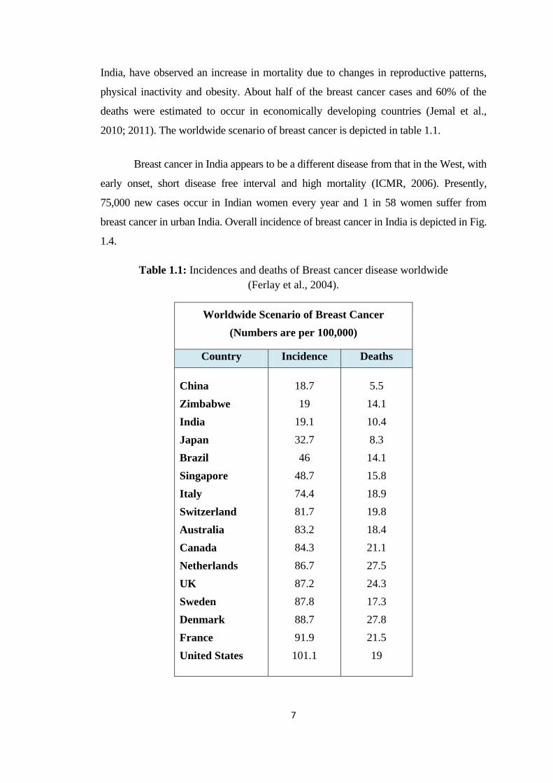
2010; 2011). The worldwide scenario of breast cancer is depicted in table 1.1.
Breast cancer in India appears to be a different disease from that in the West, with
early onset, short disease free interval and high mortality (ICMR, 2006). Presently,
75,000 new cases occur in Indian women every year and 1 in 58 women suffer from
breast cancer in urban India. Overall incidence of breast cancer in India is depicted in Fig.
1.4.
Table 1.1: Incidences and deaths of Breast cancer disease worldwide
(Ferlay et al., 2004).
Worldwide Scenario of Breast Cancer
(Numbers are per 100,000)
Country Incidence Deaths
China
Zimbabwe
India
Japan
Brazil
Singapore
Italy
Switzerland
Australia
Canada
Netherlands
UK
Sweden
Denmark
France
United States
18.7
19
19.1
32.7
46
48.7
74.4
81.7
83.2
84.3
86.7
87.2
87.8
88.7
91.9
101.1
5.5
14.1
10.4
8.3
14.1
15.8
18.9
19.8
18.4
21.1
27.5
24.3
17.3
27.8
21.5
19

8
Fig. 1.3: Trends in female breast cancer incidence rates, 1973-2006
(Jemal et al., 2010).

9
Fig. 1.4: Chart showing incidence of breast cancer in India (NCRP, 2006).
1.6.1 ANATOMY OF THE BREAST
The breast of an adult woman is a tear-shaped, milk producing gland. Each
breast is a hemispheric projection attached to the front of the chest wall on either side of
the sternum by the pectoral muscles and dense connective tissues. These strands of
connective tissue called as the suspensory ligaments of the breast (Cooper‟s ligaments)
run between the skin and deep fascia to support the breast. Each breast has a pigmented
projection called the nipple surrounded by a circular pigmented area named the areola.
The nipple has a series of closely spaced openings of ducts called lactiferous ducts for
the secretion of milk.
Within each breast is a mammary gland that consists of 15 to 20 lobes or
compartments, separated by a variable amount of adipose tissue. In each lobe there are
several smaller compartments called lobules, composed of grapelike clusters of milk-
secreting glands termed as alveoli are embedded in the connective tissue. After
production of milk, it passes from the alveoli into a series of secondary tubules and then
into the lactiferous duct.

10
Fig. 1.5: Anatomy of the breast (Source: siteman.wustl.edu).
The synthesis, secretion and ejection of milk are collectively called as
lactation, which is associated with pregnancy and childbirth. Milk production is
stimulated largely by the hormone prolactin from the anterior pituitary, with
contributions from progesterone and estrogens, and the ejection of milk is stimulated
by oxytocin that is released from the posterior pituitary. Each breast also consists of
many blood vessels and lymph vessels that are connected to the lymph nodes in
surrounding parts of the body. The pictographic anatomy of the breast was given in
Fig. 1.5.
1.6.2 TYPES OF BREAST CANCER
The types of breast cancer broadly fall into five categories depending upon its
origin.
11
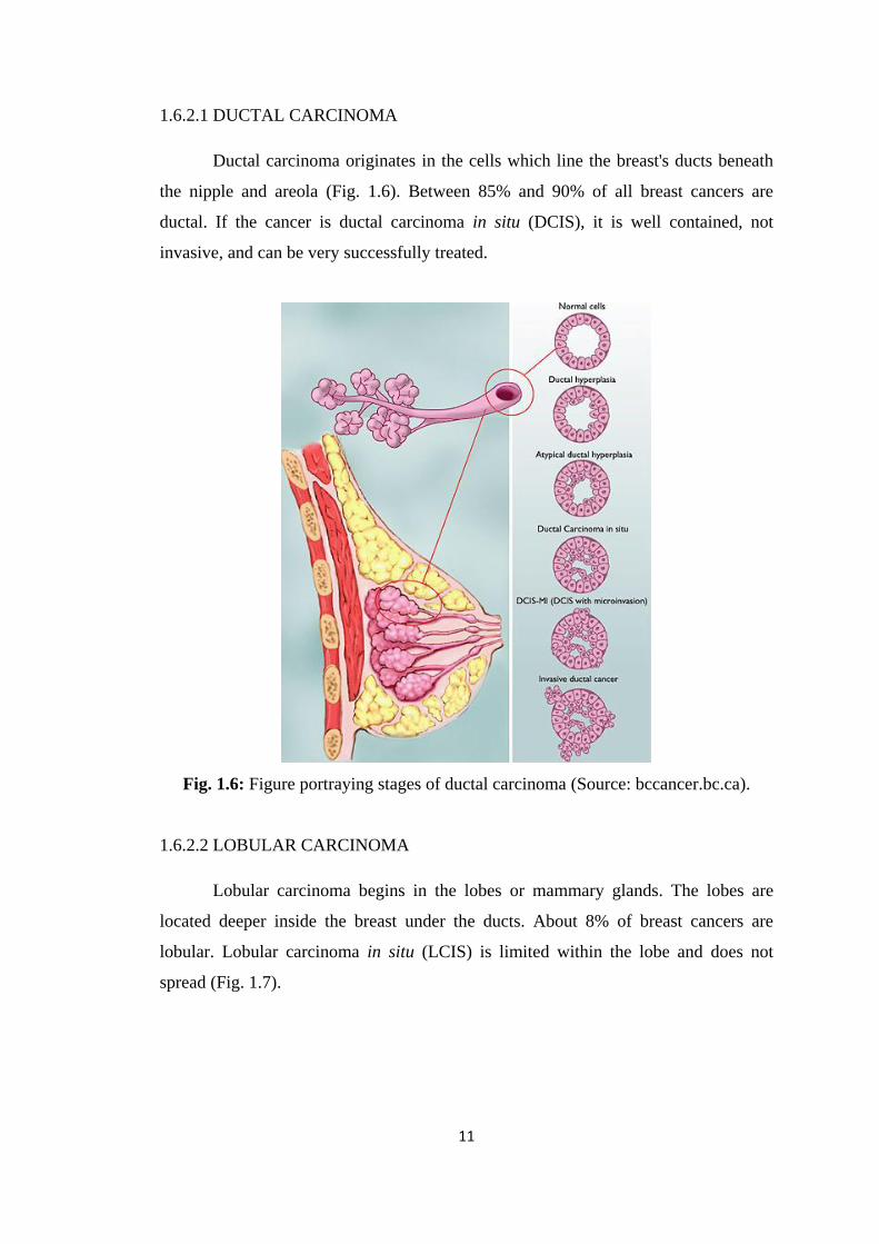
1.6.2.1 DUCTAL CARCINOMA
Ductal carcinoma originates in the cells which line the breast's ducts beneath
the nipple and areola (Fig. 1.6). Between 85% and 90% of all breast cancers are
ductal. If the cancer is ductal carcinoma in situ (DCIS), it is well contained, not
invasive, and can be very successfully treated.
Fig. 1.6: Figure portraying stages of ductal carcinoma (Source: bccancer.bc.ca).
1.6.2.2 LOBULAR CARCINOMA
Lobular carcinoma begins in the lobes or mammary glands. The lobes are
located deeper inside the breast under the ducts. About 8% of breast cancers are
lobular. Lobular carcinoma in situ (LCIS) is limited within the lobe and does not
spread (Fig. 1.7).

12
Fig. 1.7: A depiction of lobular carcinoma in situ (Source: cancer.gov).
Fig. 1.8: Types of invasive carcinoma of the breast (Source: breastcancercare.org.uk).

13
1.6.2.3 INVASIVE (INFILTRATING) BREAST CANCER:
Invasive or infiltrating breast cancer is the second most common group of
breast cancers that invade nearby tissue. It has the potential to spread out of the
original tumor site and invade other parts of the breast and body. There are several
types and subtypes of invasive breast cancer (Fig. 1.8).
1.6.2.4 INFLAMMATORY BREAST CANCER:
Inflammatory breast cancer is the less common, but most aggressive of breast
cancers, taking the form of sheets or nests, instead of lumps. It starts in the soft tissues
of the breast, just under the skin or it appears on the skin (Fig. 1.9).
Fig. 1.9: A representation of inflammatory breast cancer (Source:
virtualmedicalcentre.com).
1.6.2.5 PAGET'S DISEASE OF THE NIPPLE/AREOLA:
Paget's disease is the least common type of breast cancer named after Sir
James Paget, who first noticed the relationship between changes in the nipple and the
underlying breast cancer. Cancer of the nipple often looks like a skin rash or rough
skin. It resembles eczema and can be itchy. The itching and scabs (if scratched) are
signs that cancer may be under the surface of the skin and is breaking through. Paget's
is usually treated with a mastectomy, because the cancer has by then invaded the
nipple, areola and the milk ducts.
14
1.7 CHEMOPREVENTIVES
In spite of advances in clinical treatment of cancer malignancies, the reduction
in rate of such cancer mortality is not satisfactory as per National Cancer Institute
(Sporn and Suh, 2000). Also, to develop therapies suitable for the full gamut of
cancers is depicted as Herculean and almost impossible (Evan and Vousden, 2001).
Such obscurity in developing cancer therapies is due to its molecular complexity.
Recent progress in tumor targeting technology has augmented the likelihood that
cancer prevention relies increasingly on interventions collectively termed
„chemoprevention‟ (Tamimi et al., 2002). Cancer chemoprevention is a
pharmacological approach to intervene the use of agents to inhibit, reverse or retard
carcinogenesis (Surh, 2003) and such agents are termed as chemopreventive agents
(Tamimi et al., 2002). Extensive research in the last few years has revealed that
regular consumption of certain fruits and cruciferous vegetables can reduce the risk of
acquiring specific cancers (Kelloff et al. 1996a). Systematic, molecular target based
evaluation of such dietary agents is one important strategy for discovering better
chemopreventive agents (Kelloff et al. 1996b).
Pre-clinical and clinical studies in chemoprevention field yielded many
valuable data in preventing the onset of disease and suppressing the progress of their
growth, making chemoprevention a challenging and a very rational strategy in future
researches (Meiyanto et al., 2012).
More than 600 potential chemopreventives have been identified and about 40
promising chemopreventive agents and agent combinations, inclusive of abundant
diet-derived agents, have been clinically evaluated for major cancer targets such as
breast, prostrate, lung and colon (Tanaka, 1994; Kelloff et al., 2000). Such
chemopreventive phytochemicals include genistein, resveratrol, diallyl sulfide, S-allyl
cysteine, allicin, lycopene, capsaicin, curcumin, 6-gingerol, ellagic acid, ursolic acid,
silymarin, anethol, catechins and eugenol which might possess several untapped
therapeutic values. These agents have been seen to suppress cancer cell proliferation,
induce apoptosis, suppress the expression of anti-apoptotic proteins, inhibit growth
factor signalling pathways, NF-κB, AP-1 and JAK-STAT activation pathways,

15
angiogenesis and cyclooxygenase-2. These chemopreventive agents also have been
very recently found to reverse chemoresistance and radioresistance in patients
undergoing cancer treatment. Thus, these chemopreventive agents have potential to be
used as adjuncts to current cancer therapies (Dorai and Aggarwal, 2004). The various
molecular targets that aimed at cancer treatments are presented in the Fig. 1.10.
Fig. 1.10: Molecular targets of chemopreventive agents in cancer
(Dorai and Aggarwal, 2004).
Chemopreventive agents can be used not only to prevent cancer, but also to
treat cancer. Because of their pharmacological safety, most chemopreventive agents
can be used in combination with chemotherapeutic agents to enhance the effect at
lower doses and thus minimize chemotherapy-induced toxicity. Tumor cells use
multiple cell survival pathways to prevail, and thus agents that can suppress multiple
pathways have great potential for the treatment of cancer. Selective estrogen receptor
modulators (SERM) are trending as a key factor for breast cancer chemoprevention,
as estrogen exposure levels play a vital role in breast cancer pathogenesis. Tamoxifen,
an antiestrogen, is well known for its effect in reducing breast cancer risk in women
(Fisher et al. 1998). Natural products like, Soy isoflavones are also gaining
importance as SERMs (Kelloff et al., 2000).

16
Chemoprevention by the aid of synthetic agents results in side effects in the
tenure of treatment (Wattanapitayakul et al., 2005). One such example is doxorubicin,
a chemotherapeutic agent commonly used in treatment of breast cancer. It has
exhibited low efficacy thus leading to its resistance and toxicity on normal tissues
(Fimognari et al., 2006). Therefore, the focus of researchers inclines on the natural
metabolites from plant sources in the hunt for a new potent chemopreventives.
1.8 MEDICINAL PLANTS
The use of plants as medicines is probably as old as Human kind itself. The
first written record to the use of plants for its medicinal properties dates back to 2600
BC from the Sumerians and Akkaidians (Samuelsson, 1999), followed by the
Egyptian ebers papyrus of 1500 BC for its mention of plants in treatment of cancer.
The Chinese Materia Medica of 1100 BC has documented the record of more than
600 medicinal plants (Cragg et al., 1997). Susruta and Charaka had discussed the
usage of plants as medicine in their Ayurvedic system of medicine in 1000 BC
(Kappor, 1990). The Greeks have also substantially contributed to the rational
development of the herbal drugs in 100 AD (Samuelsson, 1999).
In recent years, plants are considered as the best sources for drug components,
since it exhibits lesser side effects and believed to offer protection from carcinogenic
exposure. (Krishnaiah et al., 2007; Rates, 2001). There is considerable interest in
screening the plant extracts in modern drug discovery programs, since structurally
novel chemotypes with potent and selective biological activity may be obtained
(Kinghorn, 2000). In USA, under the NCI (National Cancer Institute) program, over
35,000 plants were screened for anti-cancer activity between 1960 and 1986. More
than 1,50,000 plant species have been studied so far and many of them were reported
to contain therapeutic constituents (Ishii et al., 1984; Hoyos et al., 1992).
Drugs have been customarily isolated as single plant extracts/fractions, or
have been mixtures of fractions/extracts from different plants. These drugs
were used subsequent to their evaluation of safety and efficacy in model systems and
humans (Dahiru et al., 2006). Numerous plant products in the form of
17
decoction, tincture, tablets and capsules have been clinically used for the treatment of
different kinds of cancer (Ram and kumari, 2001). This has been scientifically
defined as “the use of non-cytotoxic nutrients or pharmacological agents to
enhance physiological mechanisms that protect the organism against mutant clones of
malignant cells” (Morse and Stoner, 1993; Kinghorn, 2000).
Plant anticancer agents were initially categorized as four structural classes, the
Catharanthus (Vinca) alkaloids (vinblastine, vincristine, vinorelbine), the
epipodophyllotoxins (etoposide, etoposide phosphate, teniposide), the taxanes
(paclitaxel and docetaxel), and the camptothecin derivatives (irinotecan and
topotecan) (Cragg, et al., 1997). Subsequently, several other flavonoids, terpenoids,
alkaloids and phenylpropanoids were isolated from plant sources which were known
to possess anticancer properties (Kintzios, 2006; Park et al., 2008). To prove the
same, most of the plant-derived compounds were known to have undergone a number
of preclinical and clinical trials (Shu, 1998).
Among many recent advances in cancer chemotherapy, plant sources have
played an important role in contributing to the arsenal of approximately 60 cancer
chemotherapeutic drugs on the market (Kinghorn, 2000). Cytotoxic phytochemicals
such as vinca alkaloids or paclitaxel (Taxol) were often used in oncology as highly
potent drugs and/or serve as model for synthetic compounds (Thatte et al., 2000;
Pandi et al., 2011). There have been a plethora of reports in the scientific literature
documenting the chemopreventive potential of phytochemicals such as Lupulone,
Hesperidin and blueberry in various cancer cell lines (Park et al., 2008; Lamy et al.,
2009; Adams et al., 2010). Also, tests in experimental systems (in vitro and in vivo)
have demonstrated that most of the phytochemicals act by interfering with several cell
signalling pathways and lead to cell cycle arrest, apoptosis and/or differentiation
induction in cancer cells (Chathoth et al., 2008).

18
1.9 MEDICINAL PLANTS SELECTED FOR THE STUDY
Medicinal plants selected for the present study were reportedly used as
traditional medicine by diverse tribal and ethnic groups. The collected specimens
were confirmed at Arya Vaidya Pharmacy (Coimbatore) Ltd., Kanjikode, Palakkad,
India and at the University of Calcutta, Kolkata, West Bengal, India.
A brief report of the medicinal plants selected for the study is as follows:
1.9.1 Rheum emodi Wall. ex Meissn.
A) B)
Fig. 1.11. Rheum emodi Wall. ex Meissn, A) plant at natural habitat, B) dried
rhizome.
Rheum species, commonly known as Rhubarbs, has been cultivated for over
5000 years for its medicinal properties and has its origin in the mountains of the
North-western provinces of China and Tibet. It was first mentioned in the Chinese
herbal Pen-King, wherein it has been listed as a purgative and stomachic (Foust,
1992). This genus includes a large number of perennial stout herbs. In India, it is
distributed in the temperate and subtropical regions of Himalaya from Kashmir to
Sikkim, at a height of 2800 and 3800 m above mean sea level (Nautiyal et al., 2003).
The botanical name of Himalayan rhubarb is Rheum emodi Wall. ex Meissn.
(kingdom: Plantae; division: Magnoliophyta; class: Magnoliopsida; order:
Caryophyllales; family: Polygonaceae; genus: Rheum L.) (Sharma, 2009). The
rhizomes of R. emodi have great significance for its traditional use in Ayurvedic,
Unani and folk systems of medicine (Singh et al., 2010). It has also been used as an
ingredient in many polyherbal formulations used for the treatment of various diseases,
19
in particular the regulation of blood fat, hepatitis and cancer, and is officially listed in
the Indian Pharmacopoeia (Singh et al., 2005).
There are also scientific reports subsequent to the traditional use of R. emodi,
where crude extracts of R. emodi were reported to possess hepato-protective activity in
CCl4-treated male rats, and in CCl4-treated cultured primary rat hepatocytes (Ibrahim
et al., 2008). R. emodi was also reported to have an immuno-enhancing effect through
the release of various cytokines (Kounsar and Afzal, 2010), thus playing a role in
chemoprevention. Phytoconstituents from diverse Rheum species have been reported
for its various biological properties (Matsuda et al., 2001). From R. emodi, the polar
extracts have been previously reported for their promising antioxidant, cytotoxic and
apoptotic properties (Babu et al., 2003; Rajkumar et al., 2010a; 2011a). Also, petroleum
ether and chloroform extracts of R. emodi rhizomes have been reported to possess a
moderate degree of antibacterial and antifungal activities with its reported compounds
revandchinone-1, revandchinone-2, revandchinone-3, revandchinone-4 and b-asarone.
Some of the well known compounds such as chrysophanol, physcion and emodin were
also found to be present in R. emodi (Babu et al., 2003).
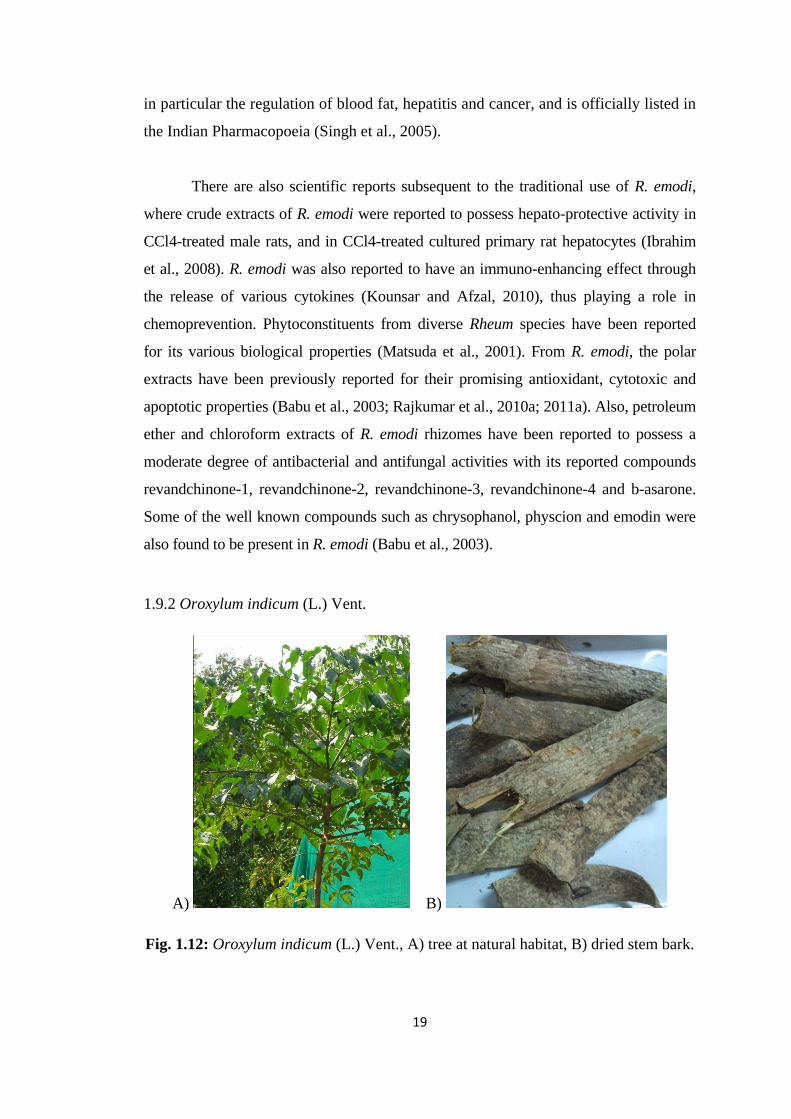
1.9.2 Oroxylum indicum (L.) Vent.
A) B)
Fig. 1.12: Oroxylum indicum (L.) Vent., A) tree at natural habitat, B) dried stem bark.

20
Oroxylum indicum (L.) Vent. (also known as Shivnak, Sonapatha, Shyonaka
or Midnight horror) is a deciduous tree belonging to Bignoniaceae family
characterized with few branches. The bark decoction of O. indicum was previously
reported for its use in treating cancer but lacks specific chemopreventive records
(Mao, 2002). The plant was also reported to be used in the treatment of other ailments
like gastric ulcer, scabies and tonsil pain. O. indicum bark extracts were furthermore
reported to possess antiproliferative/cytotoxic, and apoptotic properties (Lambertini et
al., 2004; Kumar et al., 2010; Rajkumar et al., 2011b). In addition, the bark extracts
were found to possess anti-inflammatory activity (Laupattarakasem et al., 2003) and
anti-microbial activity (Uddin et al., 2003). Previous phytochemical studies of O.
indicum led to the identification of ellagic acid (Maitreyi et al., 2008), 5,7-Dihydroxy
flavone (chrysin) (Babu et al., 2006), 5-hydroxy-8-methoxy-7-0-β-
Dglucopyranuronosyl flavone (Nair and Joshi, 1979), Stigmast-5-en-3-ol (Rasadah et
al., 1998), 5,6,7-trihydroxy flavone (baicalein) (Chen et al., 2003; Chen et al., 2005),
4′,5- Dihydroxy-7-methoxy isoflavone (pratensol) (Polya, 2003), 3-(4- hydroxy
phenyl)-2-propenoic acid and 3, 4′, 5, 7-tetrahydroxy-flavonol (Islam et al., 2010).
1.10 OBJECTIVES OF THE PRESENT STUDY
Traditional medication has been harnessing an intervention of plants for
medicinal use since decades. Most of the medicinal plants mentioned in the ancient
literature lack scientific evidence. There are reports for the use of plant sources in
traditional medicine for its cancer curative properties. Even if the general properties
of such plants have been studied; specific investigations to include its targeted
chemotherapeutic properties are lacking.
Hence, the present study focuses on isolating and identifying a potent plant
compound with target specific chemotherapeutic properties against breast cancer by
testing the following hypothesis:
‘Compound(s) isolated from selected medicinal plants might possess the ability to
activate Caspase-3 in the human breast cancer cells, thus leading the cells towards
apoptotic cell-annihilation.’

21
The objectives of the study include:
Assessment of cytotoxicity of the crude extracts obtained from selected
medicinal plants.
Analysis of the induction of apoptosis by caspase-3 activation by the plant
extracts.
Isolation of compounds from the most efficient extract that activates caspase-3
by TLC and Column chromatography.
Determination of bioactivity (cytotoxicity, apoptosis inductivity and caspase-3
activation potential) of the isolated compounds.
Characterization of the most efficient compound by FTIR, NMR, Mass
spectroscopic analyses.
Recommended