1
Intracellular MHC class II molecules promote TLR-triggered innate
immune responses by maintaining Btk activation
Xingguang Liu, Zhenzhen Zhan, Dong Li, Li Xu, Feng Ma, Peng Zhang, Hangping
Yao and Xuetao Cao
Nature Immunology doi:10.1038/ni.2015

2
Supplementary Figure 1 Normal myeloid development and macrophage
differentiation in MHC class II-deficient mice. (a) Analysis of the percentage of
macrophages (CD11b+ F4/80+) and DCs (CD11c+) in splenocytes from H2-/- or H2+/+
mice by flow cytometry. (b) Analysis of numbers of peritoneal macrophages (pMφ),
bone marrow-derived macrophages (BMDM) and bone marrow-derived DCs (BMDC)
from H2-/- or H2+/+ mice by flow cytometry. (c) Phenotype analysis (CD40, CD80 and
Nature Immunology doi:10.1038/ni.2015

3
CD86) of splenic DCs, immature and mature BMDC from H2-/- or H2+/+ mice by flow
cytometry (isotype control, light-gray line; H2-/-, red line; H2+/+, green line). Data are
shown as mean ± s.e.m. of three independent experiments (a,b) or similar results were
obtained in three independent experiments (c).
Supplementary Figure 2 MHC class II deficiency attenuates TLR-triggered
mRNA expression of proinflammatory cytokines and type I IFN in macrophages.
Quantitative RT-PCR assay of TNF, IL-6, IFN-β and IFN-α4 mRNA expression in
H2-/- or H2+/+ macrophages left unstimulated (Med) or stimulated with LPS (100
ng/ml), CpG ODN (CpG; 0.3μM) or poly(I:C) (10 μg/ml) for 4 h. Quantitative
RT-PCR results are presented as ‘fold induction’ relative to that of H2+/+ cells without
agonist stimulation. * P<0.01. Data are shown as mean ± s.e.m. of three independent
experiments.
Nature Immunology doi:10.1038/ni.2015

4
Supplementary Figure 3 Normal cytokine production triggered by PMA, IL-1β
and MDP and expression of TLRs in macrophages or DCs from MHC class
II-deficient mice. (a) ELISA of TNF and IL-6 production by H2-/- or H2+/+
macrophages left untreated (Med) or treated with PMA (1μg/ml) for 12 h, IL-1β
(100ng/ml) for 12 h or MDP (10μg/ml) for 24 h, respectively. (b,c) Analysis of the
protein (b) or mRNA (c) levels of TLRs in peritoneal macrophages (pMφ) and bone
marrow-derived DCs (BMDC) from H2-/- or H2+/+ mice by flow cytometry (b) or
Nature Immunology doi:10.1038/ni.2015

5
quantitative RT-PCR assay (c) (isotype control, light-gray line; H2-/-, red line; H2+/+,
green line). Data are shown as mean ± s.e.m. of three independent experiments (a,c)
or similar results were obtained in three independent experiments (b).
Supplementary Figure 4 Overexpression of MHC class II molecules does not
affect expression level of TLRs in H2-/- macrophages. Analysis of the protein (a) or
mRNA (b) levels of TLRs in H2-/- macrophages 36 h after transfected with MHC class
II α and β chain expression vectors by flow cytometry or quantitative RT-PCR assay
(isotype control, light-gray line; H2-/-, red line; H2+/+, green line). Similar results were
obtained in three independent experiments (a) or data are shown as mean ± s.e.m. of
three independent experiments (b).
Nature Immunology doi:10.1038/ni.2015

6
Supplementary Figure 5 MHC class II deficiency impairs activation of MAPK,
NF-κB and IRF3 in macrophages stimulated with CpG ODN and poly(I:C).
Immunoblot analysis of phosphorylated or total ERK, JNK, p38, IκBα, IRF3 in
lysates of H2-/- or H2+/+ macrophages left unstimulated (Med) or stimulated with CpG
ODN (CpG; 0.3μM) or poly(I:C) (10 μg/ml) for 30min. Similar results were obtained
in three independent experiments.
Nature Immunology doi:10.1038/ni.2015

7
Supplementary Figure 6 MHC class II deficiency does not impair activation of
MAPK and NF-κB in macrophages stimulated with TNF. Immunoblot analysis of
phosphorylated or total JNK, p38 and IκBα in lysates of H2-/- or H2+/+ macrophages
stimulated with TNF (5ng/ml) for indicated times. Similar results were obtained in
three independent experiments.
Nature Immunology doi:10.1038/ni.2015

8
Supplementary Figure 7 MHC class II molecules maintain TLR-triggered
activation of Btk (a) Immunoblot analysis of phospho-Btk or total Btk in lysates of
H2-/- or H2+/+ peritoneal macrophages stimulated with PMA (1μg/ml). (b,c)
Immunoblot analysis of phospho-Btk or total Btk in lysates of H2-/- peritoneal
macrophages transfected with MHC class II α and β chain expression vectors, and 36
h later left unstimulated (Med) or stimulated with LPS (100 ng/ml) (b), CpG ODN
(CpG; 0.3μM) or poly(I:C) (10 μg/ml) (c), respectively. Similar results were obtained
in three independent experiments.
Nature Immunology doi:10.1038/ni.2015

9
Supplementary Figure 8 Silencing of Btk expression attenuates TLR-triggered
proinflammatory cytokine and type I interferon production in macrophages. (a)
Immunoblot analysis of Btk or β-actin expression in lysates of mouse peritoneal
macrophages 48 h after transfected with control siRNA (Ctrl) or Btk-specific siRNA
(siRNA). (b-d) ELISA of TNF, IL-6 and IFN-β in supernatants of peritoneal
macrophages transfected as in a, and 48 h later stimulated with LPS (100 ng/ml), CpG
ODN (CpG; 0.3μM) or poly(I:C) (10 μg/ml) for 6 h. *P<0.05; **P<0.01. Similar
results were obtained in three independent experiments (a) or data are shown as mean
± s.e.m. of three independent experiments (b-d).
Nature Immunology doi:10.1038/ni.2015

10
Supplementary Figure 9 Blockade of Btk activation attenuates TLR-triggered
proinflammatory cytokine and type I interferon production in macrophages.
ELISA of TNF, IL-6 and IFN-β in the supernatants of peritoneal macrophages
pretreated with LFM-A13 (100 μM) or DMSO for 1 h and then left unstimulated
(Med) or stimulated with LPS (100ng/ml), CpG ODN (CpG; 0.3 μM) or poly(I:C) (10
μg/ml) for 6 h. *P<0.01. Data are shown as mean ± s.e.m. of three independent
experiments.
Nature Immunology doi:10.1038/ni.2015

11
Supplementary Figure 10 No interaction of MHC class II molecules with TLR4,
TLR9, TLR3 in macrophages. (a) Immunoblot analysis of TLR4, TLR9, TLR3 or
MHC class II immunoprecipitated with anti-MHC class II antibody in lysates of
peritoneal macrophages stimulated with LPS, CpG ODN or poly(I:C) for the indicated
times. (b) Immunoblot analysis of MHC class II, TLR4, TLR9 or TLR3
immunoprecipitated with antibody against TLR4, TLR9 or TLR3 in lysates of
peritoneal macrophages treated as in a. Similar results were obtained in three
independent experiments.
Nature Immunology doi:10.1038/ni.2015

12
Nature Immunology doi:10.1038/ni.2015

13
Supplementary Figure 11 MHC class II deficiency does not disrupt the
endosomal trafficking of TLR4, 3, 9 in TLR-activated macrophages. Confocal
analysis of H2+/+ or H2-/-macrophages stimulated with LPS (100ng/ml) (a), poly(I:C)
(10 μg/ml) (b) or CpG ODN (0.3 μM) (c) for 30 min respectively, and then labeled
with antibodies against TLR4, TLR3, TLR9 or EEA1. Similar results were obtained
in three independent experiments.
Nature Immunology doi:10.1038/ni.2015

14
Supplementary Figure 12 Btk interacts with CD40 through its PH domain. (a)
Immunoblot analysis of CD40, MHC class II or Btk immunoprecipitated with
anti-Btk antibody in lysates of cytoplasmic and plasma membrane proteins prepared
from peritoneal macrophages stimulated as indicated. (b) ELISA of TNF production
by Btk-deficient macrophages transfected with hemagglutinin (HA)-tagged wild type
Btk or Btk mutants (ΔPH, PH domain deleted; ΔSH3, SH3 domain deleted; ΔTH, TH
domain deleted; ΔSH2, SH2 domain deleted; ΔKI, kinase domain deleted), and 36 h
later stimulated with LPS for 6 h. (c)Immunoprecipitation and immunoblot analysis of
proteins in peritoneal macrophages transfected with HA-tagged wild-type Btk or Btk
mutants, and 36 h later stimulated with LPS for 15 min. The similar results were
obtained in three independent experiments.
Nature Immunology doi:10.1038/ni.2015

15
Supplementary Figure 13 CD40 deficiency impairs the interaction of
intracellular MHC class II molecules with Btk. Immunoblot analysis of Btk or
MHC class II immunoprecipitated with anti-MHC class II antibody in lysates of
cytoplasmic and plasma membrane proteins prepared from Cd40-/- macrophages
stimulated as indicated. Similar results were obtained in three independent
experiments.
Nature Immunology doi:10.1038/ni.2015

16
Supplementary Figure 14 CD40 deficiency impairs TLR-triggered activation of
tyrosine kinase Btk. Immunoblot analysis of phospho-Btk or total Btk in lysates of
Cd40-/- or Cd40+/+ peritoneal macrophages stimulated with LPS (100 ng/ml) for the
indicated time (a), CpG ODN (CpG; 0.3 μM) or poly(I:C) (10 μg/ml) (b) for 30 min.
Similar results were obtained in three independent experiments.
Nature Immunology doi:10.1038/ni.2015
17
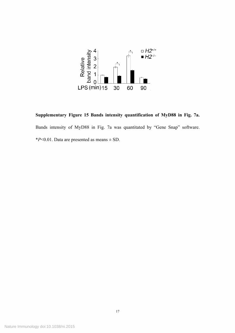
Supplementary Figure 15 Bands intensity quantification of MyD88 in Fig. 7a.
Bands intensity of MyD88 in Fig. 7a was quantitated by “Gene Snap” software.
*P<0.01. Data are presented as means ± SD.
Nature Immunology doi:10.1038/ni.2015

18
Supplementary Figure 16 Working model for intracellular MHC class II
molecules to promote TLR-triggered innate immune responses by maintaining
Btk activation in APCs (a) The classical antigen presentation function of MHC class
II molecules. (b) In MHC class II-expressing APCs, intracellular MHC class II
molecules can interact with Btk via CD40 in endosome and maintain the activation of
Btk upon stimulation with TLR ligands. Activated Btk interacts with MyD88 and
TRIF, promoting the activation of MyD88-dependent and TRIF-dependent pathways
and thus leading to the enhanced production of proinflammatory cytokines and type I
IFNs. Full activation of TLR-triggered innate responses of APCs contributes to the
induction of adaptive immune responses. (c) In MHC class II-deficient APCs,
TLR-triggered Btk activation is impaired, attenuating the activation of
Nature Immunology doi:10.1038/ni.2015

19
MyD88-dependent and TRIF-dependent pathways and leading to the decreased
production of proinflammatory cytokines and type I IFNs.
Nature Immunology doi:10.1038/ni.2015
Recommended
![MANUAL DE USUARIO MÁQUINAS DE HIELO...MANUAL DE USUARIO [AUTOCONTENIDAS Y REMOTAS ] MHC-230/506MA - MHC-235/517MA - MHC-280/625MA - MHC-320/706MA MHC-500/1109MAR - MHC-680/1466MAR](https://img.pdfslide.us/doc/110x75/5e93db5530a5a625c35ecff2/manual-de-usuario-mquinas-de-hielo-manual-de-usuario-autocontenidas-y-remotas.jpg)