Intra-abdominal infections - an everyday surgical challengeD M i S t lliDr Massimo Sartelli
Macerata Hospital (Italy)Department of SurgeryDepartment of Surgery
Chief: I Patrizi
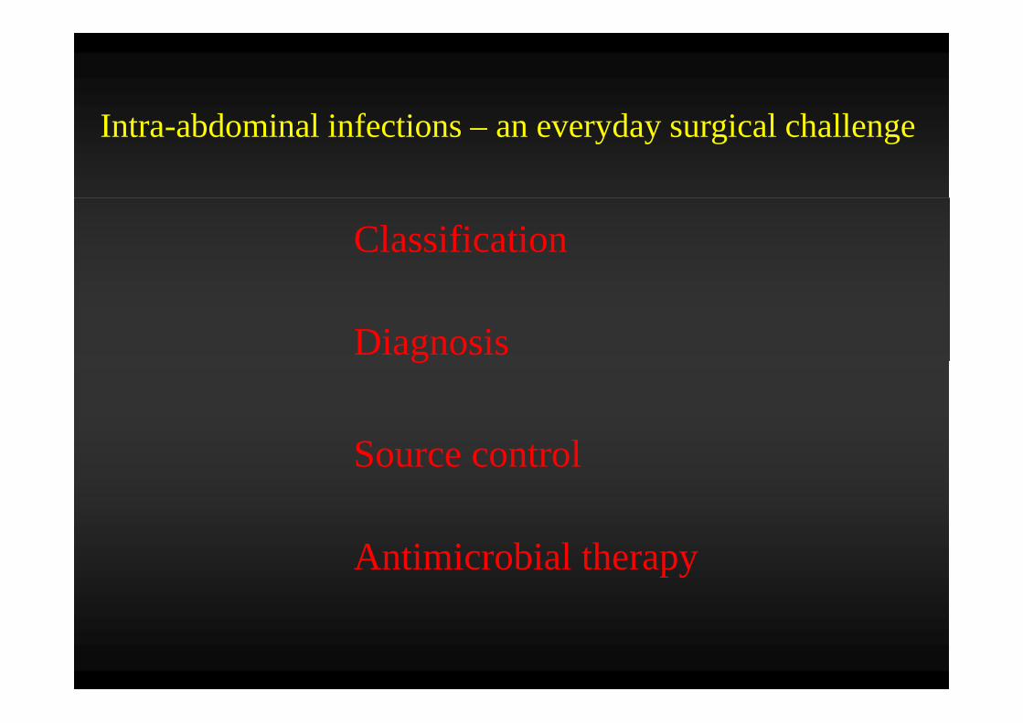
I bd i l i f i d i l h llIntra-abdominal infections – an everyday surgical challenge
Classification
Diagnosisg
S t lSource control
Antimicrobial therapy

Classification
Classification
Intra-abdominal infections (IAIs) include manypathological conditions, ranging from uncomplicatedappendicitis to faecal peritonitis IAIs are classified intoappendicitis to faecal peritonitis. IAIs are classified intouncomplicated and complicated
ClassificationClassification
Uncomplicated IAIsIn uncomplicated IAIs the infectious process only involves asingle organ and does not proceed to peritoneumsingle organ and does not proceed to peritoneum.
Complicated IAIsIn complicated IAIs, the infectious process proceeds beyond theorgan and causes either localized peritonitis or diffuse peritonitis,depending on the ability of the host to contain the process within a
t f th bd i l itpart of the abdominal cavity
ClassificationClassification
Community-acquired IAIs
Hospital-acquired IAIsThey are characterized by increased mortality because of bothunderlying patient health status and increased likelihood ofinfection caused by multi drugs resistant organisms
Peritonitis
P i it iti i diff b t i l i f ti ith t l f i t it f• Primary peritonitis is a diffuse bacterial infection without loss of integrity ofthe gastrointestinal tract. It is rare. It mainly occurs in infancy and earlychildhood and in cirrhotic patients.
• Secondary peritonitis, the most common form of peritonitis, is an acuteperitoneal infection resulting from loss of integrity of the gastrointestinal tractor from infected viscera. It is caused by perforation of the gastrointestinalor from infected viscera. It is caused by perforation of the gastrointestinaltract (e.g. perforated duodenal ulcer) by direct invasion from infected intra-abdominal viscera (e.g. gangrenous appendicitis). Anastomotic dehiscencesare common causes of peritonitis in the postoperative periodare common causes of peritonitis in the postoperative period.
• Tertiary peritonitis is a recurrent infection of the peritoneal cavity that followseither primary or secondary peritonitis.
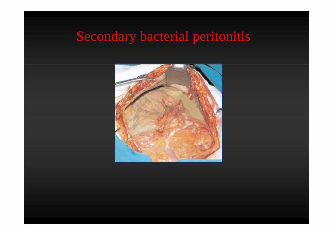
Secondary bacterial peritonitis
Secondary bacterial peritonitis arises as a consequence of injury toan intrabdominal viscus from intrinsic disease or extrinsic trauma.
Marshall JC, Maier RV, Jimenez M, Dellinger EP. Source control in thef i d i h k id b d i C imanagement of severe sepsis and septic shock: an evidence-based review. Crit
Care Med. 2004 Nov;32(11 Suppl):S513-26.
Secondary bacterial peritonitisSecondary bacterial peritonitis
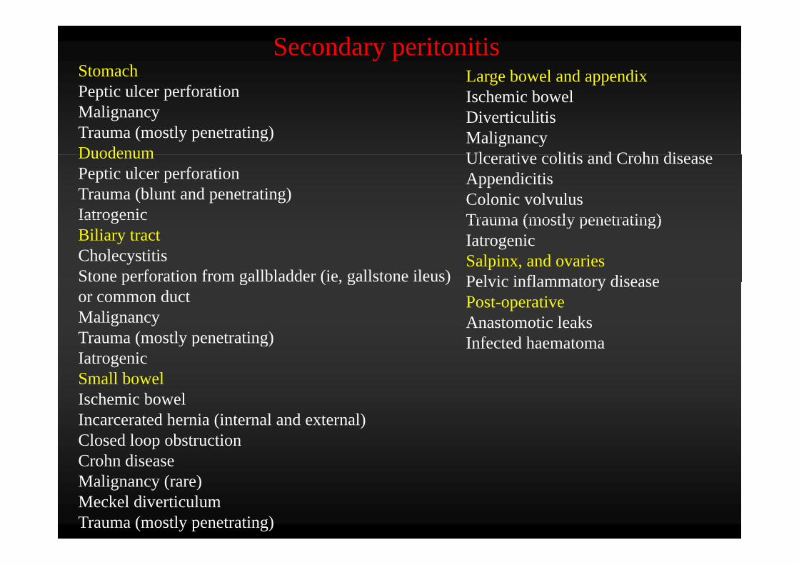
StomachPeptic ulcer perforation
Large bowel and appendixSecondary peritonitis
Peptic ulcer perforationMalignancy Trauma (mostly penetrating)Duodenum
Ischemic bowelDiverticulitisMalignancyUl ti liti d C h diDuodenum
Peptic ulcer perforationTrauma (blunt and penetrating)Iatrogenic
Ulcerative colitis and Crohn diseaseAppendicitis Colonic volvulusTrauma (mostly penetrating)Iatrogenic
Biliary tractCholecystitis Stone perforation from gallbladder (ie, gallstone ileus)
Trauma (mostly penetrating)IatrogenicSalpinx, and ovariesPelvic inflammatory diseasep g ( , g )
or common ductMalignancyTrauma (mostly penetrating)
Pelvic inflammatory diseasePost-operativeAnastomotic leaksInfected haematoma( y p g)
IatrogenicSmall bowelIschemic bowel
Infected haematoma
Incarcerated hernia (internal and external)Closed loop obstructionCrohn disease
li ( )Malignancy (rare)Meckel diverticulum Trauma (mostly penetrating)

Diagnosis
Clinical presentationClinical presentation
Abdominal painIt may be acute or insidious. Initially, the pain may be dull and poorly localized(visceral peritoneum) and often progresses to steady, severe, and more localized(visceral peritoneum) and often progresses to steady, severe, and more localizedpain (parietal peritoneum).
SIRS manifestationsSIRS manifestationsCore body temperature > 38° C or < 36° C, heart rate > 90 beats per minute,respiratory rate > 20 breaths per minute (not ventilated) or PaCO2 < 32 mm Hg(ventilated), WBC > 12,000, < 4,000 or > 10% immature forms (bands).Hypotension and hypoperfusion signs such as lactic acidosis, oliguria, and acutealteration of mental status are indicative of evolution to severe sepsis.p
Abdominal rigidityIt suggests peritonitis and the need for urgent laparotomyIt suggests peritonitis and the need for urgent laparotomy.
Abdominal X-ray
• Look for free gas, bowel obstruction, or subtle signs ofg , , gintestinal ischaemia
• Water-soluble contrast studies can show leaks• Injection of contrast into drains, fistulae or sinus tracts may
help demonstrate anatomy of complex infectios and helpmonitor adequacy of abscess drainage

UltrasoundUltrasound
• Advantage of being portable and almost risk-free • Useful for:
– identifying abscesses and fluid collections – guidance of percutaneous drainage procedures – detection of free fluid – evaluation of biliary tree
• Disadvantages: – operator dependent – difficult to perform in patients who have abdominal
dressings or paralytic ileus
CT abdomenCT abdomenCT of the abdomen and the pelvis, when it is possible to
f it i th di ti t d f h i f i tperform it, remains the diagnostic study of choice for intra-abdominal infections. CT can detect small quantities of fluid,areas of inflammation and other GI tract pathology with aareas of inflammation, and other GI tract pathology, with avery high sensitivity.
A negative CT generally indicates a very low probability of aprocess that can be reversed by surgical intervention, howeverbowel ischaemia cannot be excluded, particularly in the early stages
If the diagnosis of peritonitis is made clinically, a CT scan is notnecessary and generally delays surgical intervention withoutff i li i l d toffering clinical advantage.

CT abdomenCT abdomen
Source controlSource control
Source control
Source control represents a key component of success intherapy of sepsis. It includes drainage of infected fluids,debridement of infected soft tissues, removal of infected devices orf i b di d fi ll d fi it t t t iforeign bodies, and finally, definite measures to correct anatomicderangement resulting in ongoing microbial contamination and torestore optimal functionrestore optimal function.Although highly logical, since source control is the best way toreduce quickly the bacterial inoculum, most recommendationshave, however, low grade due to the difficulty to performappropriate randomized clinical trials in this respect
Marshall JC, Maier RV, Jimenez M, Dellinger EP. Source control in thef i d i h k id b d i C imanagement of severe sepsis and septic shock: an evidence-based review. Crit
Care Med. 2004 Nov;32(11 Suppl):S513-26.
S t lSource control
Source control should be obtained as early as possible after thediagnosis of intra-abdominal sepsis has been confirmed.Inability to control the septic source is associated significantlywith increase in mortality.
Gastro-duodenal perforation
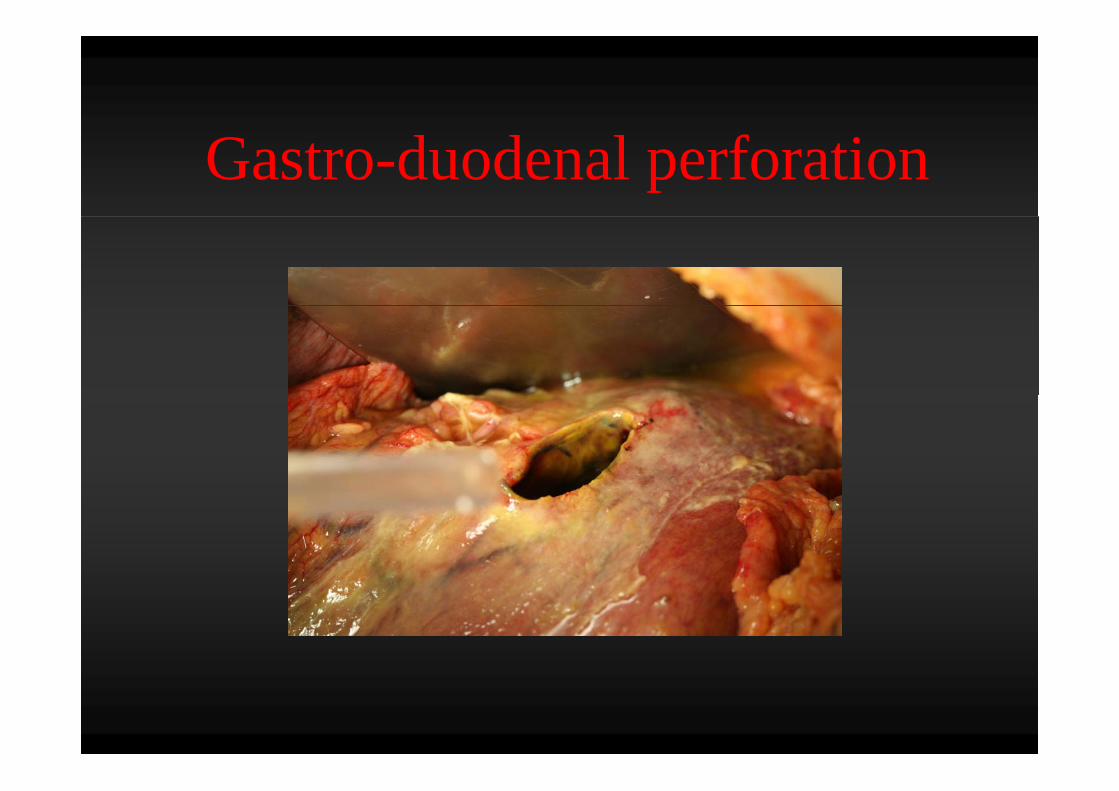
Gastro-duodenal perforationIn perforated peptic ulcer, surgery is the treatment of choice.In selected cases (Pts yunger than 70 ys old, no shock, no peritonitis,lack of spillage of the water-soluble contrast medium atgastroduodenogram) non operative management may be attempted.After initial non operative management no improvement ofAfter initial non operative management, no improvement ofconditions within 24 hours or rapid deterioration are indicationto surgery.g y
Boey J, Lee NW, Koo J, Lam PH, Wong J, Ong GB Immediate definitive surgeryfor perforated duodenal ulcers: a prospective controlled trial Ann Surg 1982; 196:for perforated duodenal ulcers: a prospective controlled trial. Ann Surg 1982; 196:338-344Millat B, Fingerhut A, Borie F Surgical treatment of complicated duodenal ulcers:
ll d i l W ld J S 2000 24 299 306controlled trials. World J Surg 2000; 24:299–306Crisp E. Cases of perforation of the stomach with deductions therefrom relative tothe character and treatment of that lesion. Lancet. 1843;2:639.Wangensteen OH. Nonoperative treatment of localized perforations of theduodenum. Minn Med. 1935;18:477-480
Gastro-duodenal perforationLaparoscopic repair of perforated peptic ulcer is safe andeffective in centers with experienceeffective in centers with experience.The p.o. outcome of laparoscopic approach does not significantlydiffer from that of open surgery, except for lower analgesic p.o.request.In all studies the patients had small ulcers (mean diameter 1cm) and all patients received simple suture, mostly with omental
t h t l i N i i t d ithpatch, or suturless repair; No experience is reported with emergencylaparoscopic resection or laparoscopic repair of large ulcers.
Sanabria A, Villegas MI, Morales Uribe CH. Laparoscopic repair for perforatedpeptic ulcer disease. Cochrane Database of Systematic Reviews 2010 (Mar), Issue4) one meta analysis4), one meta-analysisLau H. Laparoscopic repair of perforated peptic ulcer: a meta-analysis. SurgEndosc. 2004 Jul;18(7):1013-21
Appendicitis
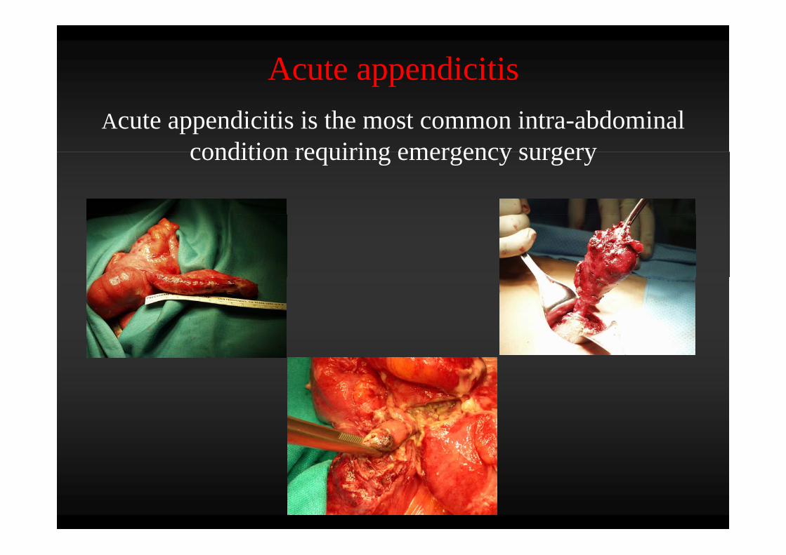
Acute appendicitisAcute appendicitis is the most common intra-abdominal
condition requiring emergency surgerycondition requiring emergency surgery
Uncomplicated appendicitis
Operative intervention for acute, nonperforated appendicitisperformed as soon as is the gold standard.Studies have demonstrated that antibiotics alone may be useful to treat
i i h l f d di i i if h i i kpatients with early, non perforated appendicitis, even if there is a riskof recurrence.
Hansson J, Körner U, Khorram-Manesh A, Solberg A, Lundholm K: Randomized clinical trialf tibi ti th di t i t t t f t di iti iof antibiotic therapy versus appendicectomy as primary treatment of acute appendicitis in
unselected patients. Br J Surg 2009 , 96:473-481.Styrud J, Eriksson S, Nilsson I, Ahlberg G, Haapaniemi S, Neovius G, Rex L, Badume I,Granström L: Appendectomy versus antibiotic treatment in acute appendicitis A prospectiveGranström L: Appendectomy versus antibiotic treatment in acute appendicitis. A prospectivemulticenter randomized controlled trial. World J Surg 2006 , 30:1033-1037.Eriksson S, Granström L: Randomized controlled trial of appendicectomy versus antibiotictherapy for acute appendicitis Br J Surg 1995 82:166-169therapy for acute appendicitis. Br J Surg 1995 , 82:166 169.Varadhan KK, Humes DJ, Neal KR, Lobo DN: Antibiotic therapy versus appendectomy foracute appendicitis: a meta-analysis. World J Surg 2010 , 34:199-209.
L i V d tLaparoscopic Vs open appendectomy
B th d l i h t d tBoth open and laparoscopic approaches to appendectomy areappropriate.Use of either approach should be decided by the surgeon's expertiseUse of either approach should be decided by the surgeon's expertise.The laparoscopic approach is useful for obese patients, elderlypatients and patients whose diagnosis is uncertain, especially womenpatients and patients whose diagnosis is uncertain, especially womenof childbearing age.
Sauerland S, Lefering R, Neugebauer EA: Laparoscopic versus open surgery for suspectedappendicitis. Cochrane Database Syst Rev 2002 :CD001546Faiz O, Clark J, Brown T, Bottle A, Antoniou A, Farrands P, Darzi A, Aylin P: Traditional and, , , , , , , ylaparoscopic appendectomy in adults: outcomes in English NHS hospitals between 1996 and2006. Ann Surg 2008 , 248:800-806.Sporn E, Petroski GF, Mancini GJ, Astudillo JA, Miedema BW, Thaler K: Laparoscopicappendectomy--is it worth the cost? Trend analysis in the US from 2000 to 2005. J Am CollSurg 2009 , 208:179-185.
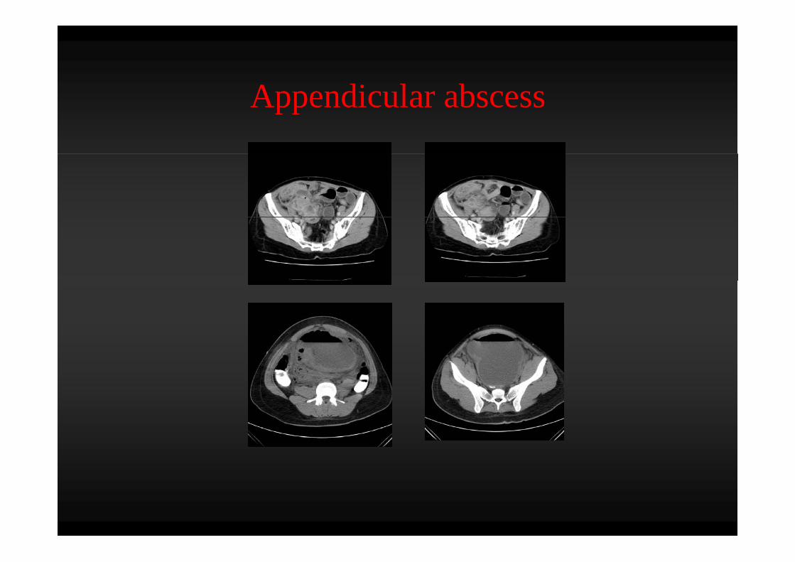
A di l bAppendicular abscess
Appendicular abscess
Although appendix abscess occurs in 10% of patients with acute
pp
appendicitis, its surgical management is surrounded withcontroversy.The traditional management of appendiceal mass has been initialThe traditional management of appendiceal mass has been initialconservative treatment followed by interval appendectomy.Recently interval appendectomy has been questioned, andy pp y q ,there is much controversy whether interval appendectomy isappropriate for adults with an appendicular abscess.
Corfield L: Interval appendicectomy after appendiceal mass or abscess in adults: What is"best practice"? Surg Today 2007, 37(1):1-4.Andersson RE, Petzold MG: Nonsurgical treatment of appendiceal abscess or phlegmon:A systematic review and meta-analysis. Ann Surg 2007, 246(5):741-748.Deakin DE, Ahmed I: Interval appendicectomy after resolution of adult inflammatoryappendix mass--is it necessary? Surgeon 2007 Feb, 5(1):45-50.
Appendicular peritonitispp p
Patients with perforated appendicitis should undergo urgentp pp g gintervention to provide adequate source control
DiverticulitisDiverticulitis
i i li iDiverticulitisN ti t t t ith b l t d tibi ti iNonopeative treatment, with bowel rest and antibiotics, issuggested in patients with uncomplicated diverticulitis.The decision to recommend elective sigmoid colectomy afterThe decision to recommend elective sigmoid colectomy afterrecovery from acute diverticulitis should be made on a case-by-casebasis
Stollman N, Raskin JB: Diverticular disease of the colon. Lancet 2004 , 363:631-639.Jacobs DO: Clinical practice Diverticulitis N Engl J Med 2007 357:2057-2066Jacobs DO: Clinical practice. Diverticulitis. N Engl J Med 2007 , 357:2057-2066.Broderick-Villa G, Burchette RJ, Collins JC, Abbas MA, Haigh PI: Hospitalization foracute diverticulitis does not mandate routine elective colectomy. Arch Surg 2005, 140:576-581.581.Mueller MH, Glatzle J, Kasparek MS, Becker HD, Jehle EC, Zittel TT, Kreis ME: Long-term outcome of conservative treatment in patients with diverticulitis of the sigmoidcolon. Eur J Gastroenterol Hepatol 2005 , 17:649-654.p ,
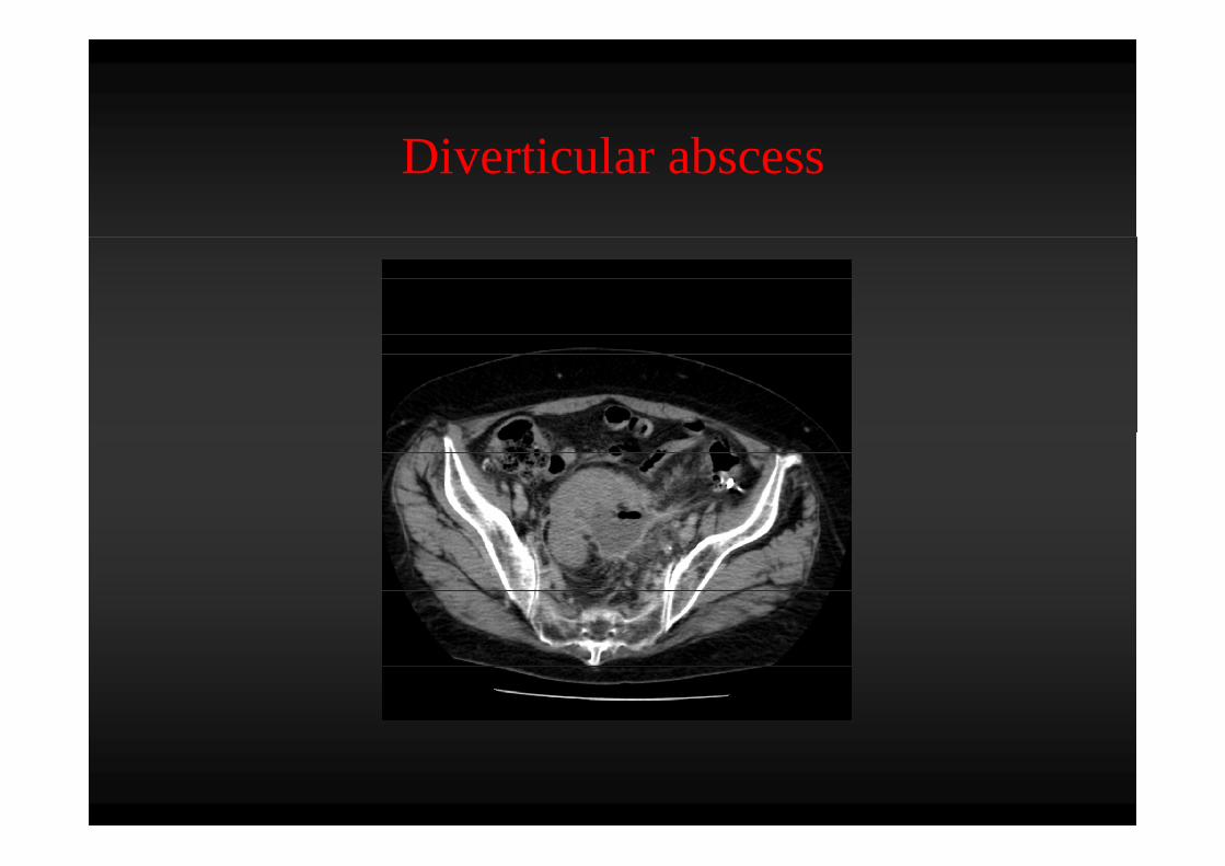
i i l bDiverticular abscess
Diverticular abscessIntravenous antibiotic treatment alone is usually the mostappropriate treatment for patients with a small (<4 cm in diameter)appropriate treatment for patients with a small (<4 cm in diameter)diverticular abscess and image guided percutaneous drainage is forthose with a large (>4 cm in diameter) one.Ambrosetti P, Robert J, Witzig JA, Mirescu D, de Gautard R, Borst F, Rohner A: Incidence,outcome, and proposed management of isolated abscesses complicating acute left-sidedcolonic diverticulitis: a prospective study of 140 patients Dis Colon Rectum 1992 35:1072colonic diverticulitis: a prospective study of 140 patients. Dis Colon Rectum 1992 , 35:1072-1076.Siewert B, Tye G, Kruskal J, Sosna J, Opelka F, Raptopoulos V, Goldberg SN: Impact of CT-guided drainage in the treatment of diverticular abscesses: size matters AJR Am Jguided drainage in the treatment of diverticular abscesses: size matters. AJR Am JRoentgenol 2006 , 186:680-686.Kumar RR, Kim JT, Haukoos JS, Macias LH, Dixon MR, Stamos MJ, Konyalian VR: Factorsaffecting the successful management of intra-abdominal abscesses with antibiotics and theaffecting the successful management of intra abdominal abscesses with antibiotics and theneed for percutaneous drainage. Dis Colon Rectum 2006 , 49:183-189.McKee RF, Deignan RW, Krukowski ZH: Radiological investigation in acute diverticulitis.Br J Surg 1993 , 80:560-565.g ,Padidar AM, Jeffrey RB Jr, Mindelzun RE, Dolph JF: Differentiating sigmoid diverticulitisfrom carcinoma on CT scans: mesenteric inflammation suggests diverticulitis. AJR Am JRoentgenol 1994 , 163:81-83.
Diverticular peritonitis
Urgent operation is required for patients with diffuse peritonitis orfor those who fail non-operative management of acute diverticulitis
.
Ordoñez CA, Puyana JC: Management of peritonitis in the critically ill patient. Surg ClinNorth Am 2006 , 86:1323-1349.Blot S, De Waele JJ: Critical issues in the clinical management of complicated intra-abdominal infections. Drugs 2005 , 65:1611-1620.
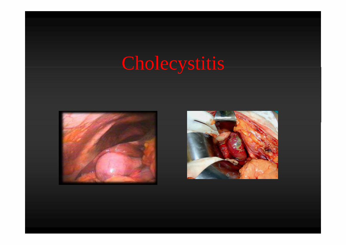
CholecystitisCholecystitis
L i h l t tLaparoscopic cholecystectomy
Laparoscopic cholecystectomy has been accepted as an effectivetreatment for acute cholecystitis
Kiviluoto T, Sirén J, Luukkonen P, Kivilaakso E. Randomised trial of laparoscopic versusopen cholecystectomy for acute and gangrenous cholecystitis Lancet 1998 Jan
.
open cholecystectomy for acute and gangrenous cholecystitis. Lancet. 1998 Jan31;351(9099):321-5.Johansson M, Thune A, Nelvin L, Stiernstam M, Westman B, Lundell L. Randomizedclinical trial of open versus laparoscopic cholecystectomy in the treatment of acuteclinical trial of open versus laparoscopic cholecystectomy in the treatment of acutecholecystitis. Br J Surg. 2005 Jan;92(1):44-9.Kum CK, Goh PMY, Isaac JR, Tekant Y, Ngoi SS. Laparoscopic cholecystectomy for acutecholecystitis. Br J Surg. 1994;81: 1651–1654y g ;
Timing of cholecystectomy
Early cholecystectomy: Surgery + antimicrobial prophylaxis
D l d Ch l t t A ti i bi l th + d l dDelayed Cholecystectomy: Antimicrobial therapy + delayed surgery
.
Timing of cholecystectomy
Evidence suggests that early laparoscopic cholecystectomyEvidence suggests that early laparoscopic cholecystectomyreduces the total length of hospital stay and the risk ofreadmissions attributable to recurrent acute cholecystitis.y
Gurusamy K, Samraj K, Gluud C, Wilson E, Davidson BR. Meta-analysis of randomizedcontrolled trials on the safety and effectiveness of early versus delayed laparoscopiccholecystectomy for acute cholecystitis. Br J Surg. 2010 Feb;97(2):141-50.
.
c o ecystecto y o acute c o ecyst t s. J Su g. 0 0 eb;97( ): 50.Siddiqui T, MacDonald A, Chong PS, Jenkins JT. Early versus delayed laparoscopiccholecystectomy for acute cholecystitis: a meta-analysis of randomized clinical trials. Am JSurg. 2008 Jan;195(1):40-7.g ; ( )Lau H, Lo CY, Patil NG, Yuen WK. Early versus delayed-interval laparoscopiccholecystectomy for acute cholecystitis: a metaanalysis. Surg Endosc. 2006 Jan;20(1):82-7.Papi C, Catarci M, D'Ambrosio L, Gili L, Koch M, Grassi GB, Capurso L. Timing ofcholecystectomy for acute calculous cholecystitis: a meta-analysis. Am J Gastroenterol.2004 Jan;99(1):147-55.
Cholecystostomy in elderly and critically ill y y y ypatients
Some reports (case-series studies) have studied the effectivenessand safety of percutaneous transhepatic gallbladder drainage asthe treatment of first choice for acute cholecystitis in elderlypatients, particularly in those with comorbid conditions, and theyi di t it f lindicate its usefulness.
Avrahami R, Badani E, Watemberg S, Nudelman I, Deutsch AA, Rabin E, et al. The role
.
of percutaneous transhepatic cholecystostomy in the management of acute cholecystitisin high-risk patients. Int Surg. 1995;80:111–14.Davis CA, Landercasper J, Gundersen LH, Lambert PJ. Effective use of percutaneouscholecystostomy in high-risk surgical patients: techniques, tube management, andresults. Arch Surg. 1999;134:727–31.Lee KT, Wong SR, Cheng JS, Ker CG, Sheen PC, Liu YE. Ultrasound-guided
h l i i i l f h l i i i ld lpercutaneous cholecystostomy as an initial treatment for acute cholecystitis in elderlypatients. Dig Surg. 1998;15:328–32.
Gallbladder perforationGallbladder perforation is an unusual initial presentation ofGallbladder perforation is an unusual initial presentation ofgallbladder disease. Early diagnosis of gallbladder perforation andimmediate surgical intervention are of prime importance ing p pdecreasing morbidity and mortality associated with thiscondition.I i l di d i l L i i i iIt is rarely diagnosed preoperatively. Late operative intervention isassociated with increased morbidity, mortality, number of ICUadmissions and long postoperative hospital stays An earlyadmissions, and long postoperative hospital stays. An earlycholecystectomy strategy may lead to improved outcomes but may bedifficult to implement and may not be cost-effective.
.
Derici H, Kara C, Bozdag AD, Nazli O, Tansug T, Akca E. Diagnosis and treatment ofgallbladder perforation. World J Gastroenterol. 2006 Dec 28;12(48):7832-6.Menakuru SR, Kaman L, Behera A, Singh R, Katariya RN. Current management of gallbladder perforations. ANZ J Surg. 2004;74:843–846Roslyn JJ, Thompson JE Jr, Darvin H, DenBesten L. Risk factors for gallbladder perforation.Am J Gastroenterol 1987; 82: 636-640S f idi Si i k i G llbl dd f i i k f dStefanidis D, Sirinek KR, Bingener J. Gallbladder perforation: risk factors and outcome. JSurg Res. 2006 Apr;131(2):204-8. Epub 2006 Jan 18.
Biliary peritonitisBiliary peritonitis
Biliary peritonitisy p
Lavage
Biliary peritonitisy p
Cholecystectomy
Biliary peritonitisy p
Debridement
Biliary peritonitisy p
Drainage
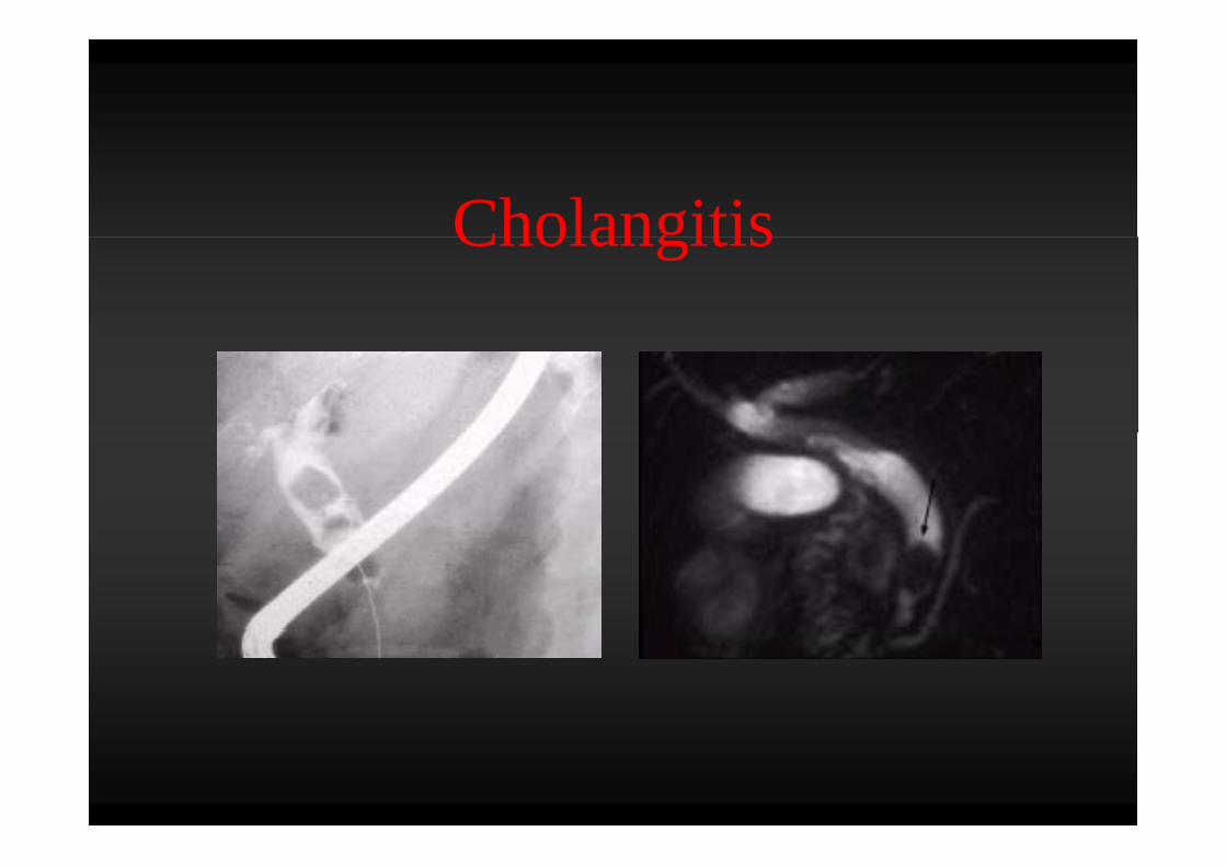
CholangitisCholangitis
Biliary drainageBiliary drainage
Biliary drainage is a radical method to relieve cholestasis, acause of acute cholangitis and takes a central part in thecause of acute cholangitis, and takes a central part in thetreatment of acute cholangitis.Biliary drainage can be achieved by three different procedures:y g y p
• endoscopic,• percutaneous transhepatic• percutaneous transhepatic, • open drainage
Biliary drainageIt h b t d th t h i t bili d iIt has been reported that when no appropriate biliary drainage wasavailable 20–30 years ago, the mortality of acute cholangitis withconservative treatment was extremely high.conservative treatment was extremely high.There has been no randomized controlled trial (RCT) comparingconservative treatment and biliary drainage. However, manypatients with acute cholangitis cannot besaved by conservativetreatment alone.
O’Connor MJ, Schwartz ML, McQuarrie DG, Sumer HW. Acute bacterial cholangitis: ananalysis of clinical manifestation. Arch Surg 1982;117:437–41. 2.Welch JP, Donaldson GA. The urgency of diagnosis and surgical treatment of acutesuppurative cholangitis. Am J Surg 1976;131: 527–32.
Post-operative peritonitisPost operative peritonitis
Post-operative Infections
The most common cause of postoperative peritonitis isp p panastomotic failure/leak.
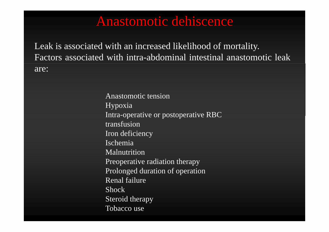
Anastomotic dehiscence
Leak is associated with an increased likelihood of mortality.Factors associated with intra-abdominal intestinal anastomotic leakare:
Anastomotic tensionHypoxiaIntra-operative or postoperative RBCIntra operative or postoperative RBC transfusionIron deficiencyIschemiaIschemiaMalnutritionPreoperative radiation therapyP l d d i f iProlonged duration of operationRenal failureShockSteroid therapyTobacco use
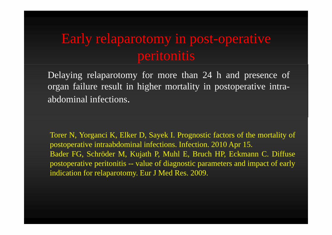
Early relaparotomy in post operativeEarly relaparotomy in post-operative peritonitis
Delaying relaparotomy for more than 24 h and presence oforgan failure result in higher mortality in postoperative intra-g g y p pabdominal infections.
Torer N, Yorganci K, Elker D, Sayek I. Prognostic factors of the mortality ofpostoperative intraabdominal infections Infection 2010 Apr 15postoperative intraabdominal infections. Infection. 2010 Apr 15.Bader FG, Schröder M, Kujath P, Muhl E, Bruch HP, Eckmann C. Diffusepostoperative peritonitis -- value of diagnostic parameters and impact of earlyi di ti f l t E J M d R 2009indication for relaparotomy. Eur J Med Res. 2009.
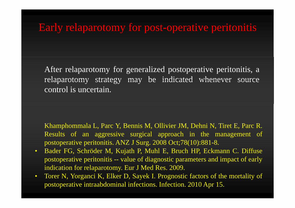
Early relaparotomy for post-operative peritonitisy p y p p p
After relaparotomy for generalized postoperative peritonitis, arelaparotomy strategy may be indicated whenever sourcerelaparotomy strategy may be indicated whenever sourcecontrol is uncertain.
Khamphommala L, Parc Y, Bennis M, Ollivier JM, Dehni N, Tiret E, Parc R.Khamphommala L, Parc Y, Bennis M, Ollivier JM, Dehni N, Tiret E, Parc R.Results of an aggressive surgical approach in the management ofpostoperative peritonitis. ANZ J Surg. 2008 Oct;78(10):881-8.
• Bader FG Schröder M Kujath P Muhl E Bruch HP Eckmann C Diffuse• Bader FG, Schröder M, Kujath P, Muhl E, Bruch HP, Eckmann C. Diffusepostoperative peritonitis -- value of diagnostic parameters and impact of earlyindication for relaparotomy. Eur J Med Res. 2009.T N Y i K Elk D S k I P ti f t f th t lit f• Torer N, Yorganci K, Elker D, Sayek I. Prognostic factors of the mortality ofpostoperative intraabdominal infections. Infection. 2010 Apr 15.
Re-laparotomy strategy
Three methods of local mechanical management of abdominali f ll i i iti l l t f t lsepsis following initial laparotomy for source control are
currently debated:
(1) Open-abdomen(2) planned relaparotomy,(3) on-demand relaparotomy
Re-laparotomy strategy
On demand relaparotomy may be considered the preferred surgicali i i h i i i b i hstrategy in patients with severe peritonitis because it has a
substantial reduction in relaparotomies, health care utilization, andmedical costsmedical costs.
van Ruler O, Mahler CW, Boer KR, Reuland EA, Gooszen HG,Opmeer BC de Graaf PW Lamme B Gerhards MF Steller EP vanOpmeer BC, de Graaf PW, Lamme B, Gerhards MF, Steller EP, vanTill JW, de Borgie CJ, Gouma DJ, Reitsma JB, Boermeester MA;Dutch Peritonitis Study Group. Comparison of on-demand vs plannedrelaparotomy strategy in patients with severe peritonitis: arelaparotomy strategy in patients with severe peritonitis: arandomized trial. JAMA. 2007 Aug 22;298(8):865-72.
Antimicrobial therapyAntimicrobial therapy
Antimicrobial therapy
A i i bi l h l i l l i hAntimicrobial therapy plays an integral role in the managementof intra-abdominal infections. The choice of an inadequateantimicrobial agent is a cause of therapeutic failureantimicrobial agent is a cause of therapeutic failure.
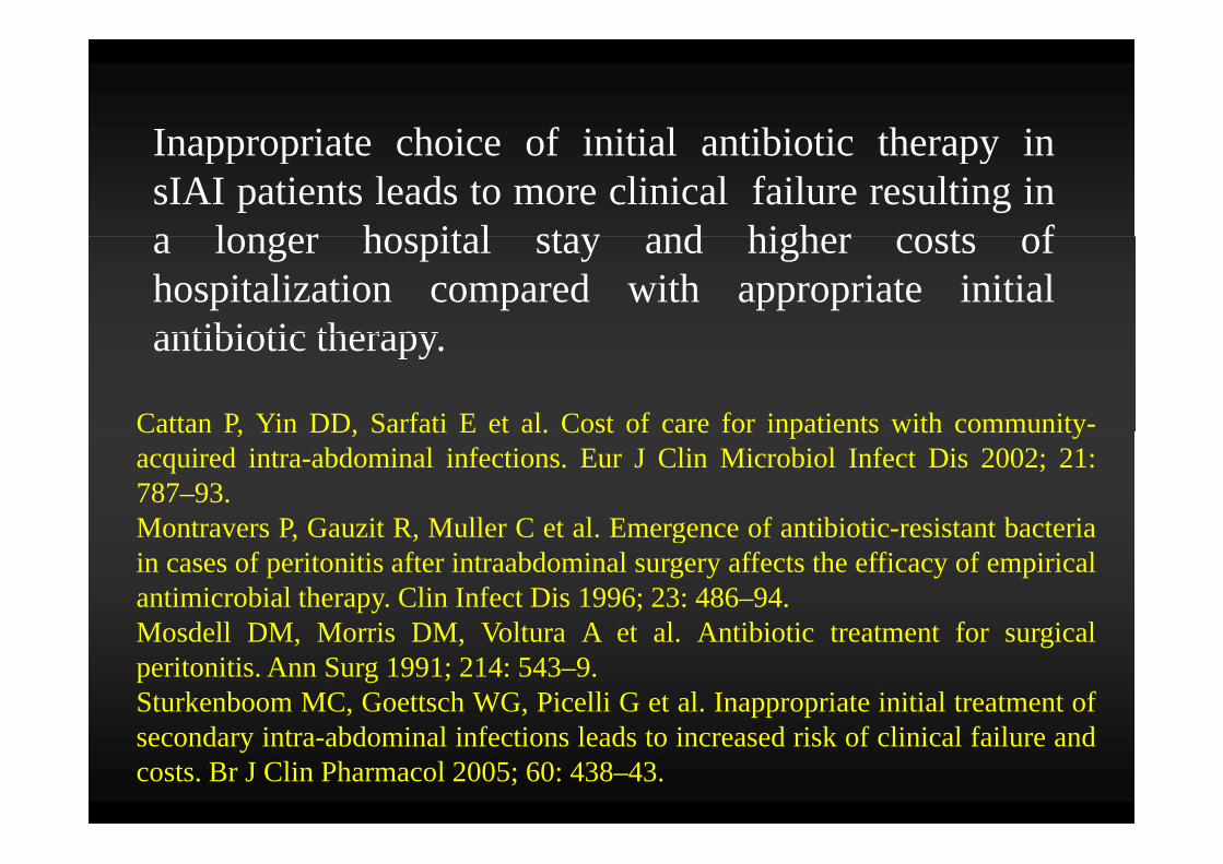
Inappropriate choice of initial antibiotic therapy inInappropriate choice of initial antibiotic therapy insIAI patients leads to more clinical failure resulting ina longer hospital stay and higher costs ofa longer hospital stay and higher costs ofhospitalization compared with appropriate initialantibiotic therapyantibiotic therapy.
Cattan P, Yin DD, Sarfati E et al. Cost of care for inpatients with community-Cattan P, Yin DD, Sarfati E et al. Cost of care for inpatients with communityacquired intra-abdominal infections. Eur J Clin Microbiol Infect Dis 2002; 21:787–93.Montravers P Gauzit R Muller C et al Emergence of antibiotic-resistant bacteriaMontravers P, Gauzit R, Muller C et al. Emergence of antibiotic-resistant bacteriain cases of peritonitis after intraabdominal surgery affects the efficacy of empiricalantimicrobial therapy. Clin Infect Dis 1996; 23: 486–94.M d ll DM M i DM V lt A t l A tibi ti t t t f i lMosdell DM, Morris DM, Voltura A et al. Antibiotic treatment for surgicalperitonitis. Ann Surg 1991; 214: 543–9.Sturkenboom MC, Goettsch WG, Picelli G et al. Inappropriate initial treatment of
d i bd i l i f i l d i d i k f li i l f il dsecondary intra-abdominal infections leads to increased risk of clinical failure andcosts. Br J Clin Pharmacol 2005; 60: 438–43.
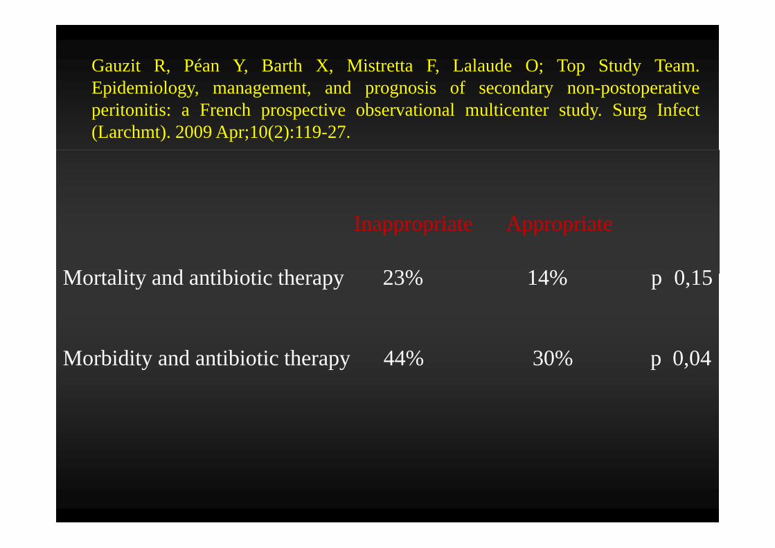
Gauzit R, Péan Y, Barth X, Mistretta F, Lalaude O; Top Study Team.Epidemiology management and prognosis of secondary non postoperativeEpidemiology, management, and prognosis of secondary non-postoperativeperitonitis: a French prospective observational multicenter study. Surg Infect(Larchmt). 2009 Apr;10(2):119-27.
Inappropriate Appropriate
M t lit d tibi ti th 23% 14% 0 15Mortality and antibiotic therapy 23% 14% p 0,15
Morbidity and antibiotic therapy 44% 30% p 0,04
Ri k ifi iRisk stratification
The definition of "risk" in intra-abdominal infections remainsvague.“High risk” is generally intended to describe patients witha high risk for treatment failure. Effective management ofhigh risk patients requires the early use of appropriate,broad spectrum empirical antimicrobial therapybroad-spectrum empirical antimicrobial therapy.
Risk factorsInfection risk factors• Hospital acquired infectionsp q• Recent exposure to antibiotics
Intrinsic risk factors• High severity of illness (APACHE II score >15)• Advanced age• Comorbidity and degree of organ dysfunction• Low albumin level• Poor nutritional status• Immunodepression
P f li• Presence of malignancy
Management risk factorsD l i th i iti l i t ti (>24 h)• Delay in the initial intervention (>24 h)
• Inability to achieve adequate source control
Ri k ifi iRisk stratification
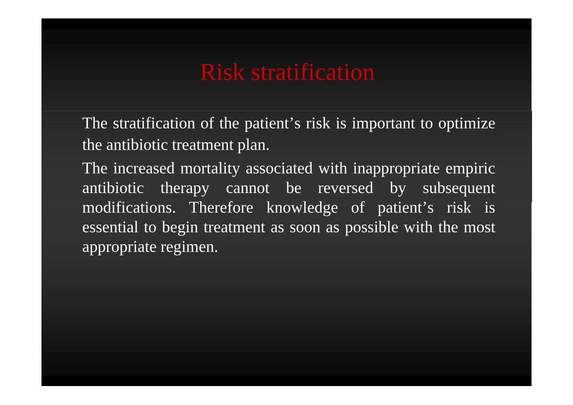
The stratification of the patient’s risk is important to optimizethe antibiotic treatment plan.The increased mortality associated with inappropriate empiricantibiotic therapy cannot be reversed by subsequent
difi i Th f k l d f i ’ i k imodifications. Therefore knowledge of patient’s risk isessential to begin treatment as soon as possible with the mostappropriate regimenappropriate regimen.
Antimicrobial resistanceAntimicrobial resistance
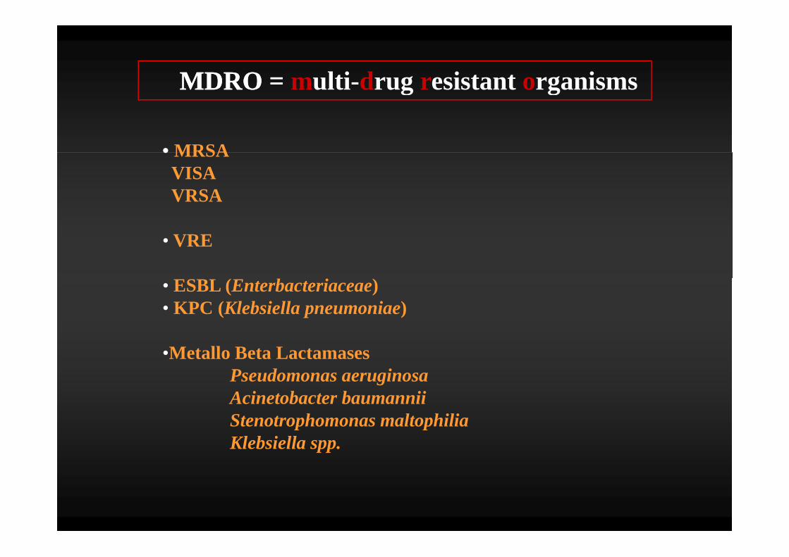
MDROMDRO == multi-drug resistant organismsMDROMDRO multi drug resistant organisms
• MRSA• MRSAVISAVRSA
• VRE
• ESBL (Enterbacteriaceae)• KPC (Klebsiella pneumoniae)
•Metallo Beta LactamasesPseudomonas aeruginosa Acinetobacter baumanniiStenotrophomonas maltophilia Klebsiella spp.
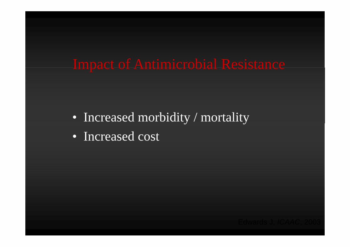
Impact of Antimicrobial ResistanceImpact of Antimicrobial Resistance
• Increased morbidity / mortalityy y• Increased cost
Edwards J, ICAAC, 2003
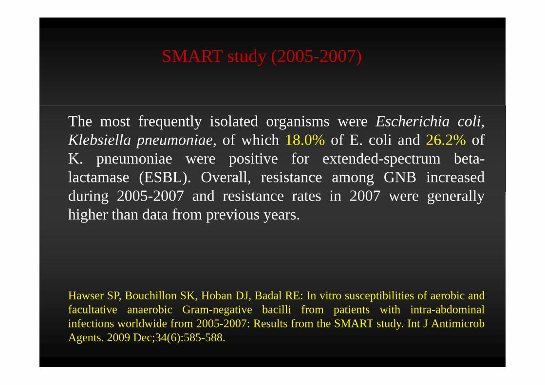
SMART study (2005-2007)SMART study (2005 2007)
The most frequently isolated organisms were Escherichia coli,Klebsiella pneumoniae, of which 18.0% of E. coli and 26.2% ofK. pneumoniae were positive for extended-spectrum beta-lactamase (ESBL). Overall, resistance among GNB increasedd i 2005 2007 d i t t i 2007 llduring 2005-2007 and resistance rates in 2007 were generallyhigher than data from previous years.
Hawser SP, Bouchillon SK, Hoban DJ, Badal RE: In vitro susceptibilities of aerobic andfacultative anaerobic Gram-negative bacilli from patients with intra-abdominalinfections worldwide from 2005 2007: Results from the SMART study Int J Antimicrobinfections worldwide from 2005-2007: Results from the SMART study. Int J AntimicrobAgents. 2009 Dec;34(6):585-588.
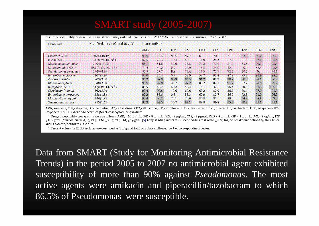
SMART study (2005-2007)
Data from SMART (Study for Monitoring Antimicrobial ResistanceTrends) in the period 2005 to 2007 no antimicrobial agent exhibitedTrends) in the period 2005 to 2007 no antimicrobial agent exhibitedsusceptibility of more than 90% against Pseudomonas. The mostactive agents were amikacin and piperacillin/tazobactam to whichg p p86,5% of Pseudomonas were susceptible.
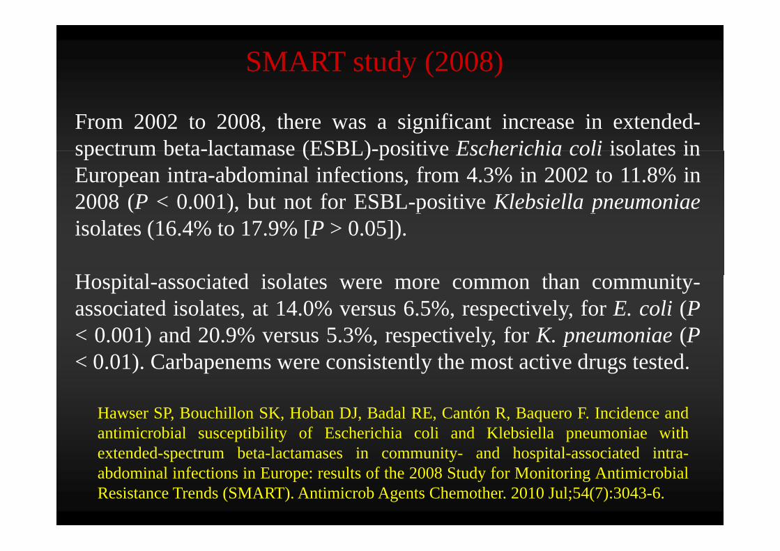
SMART study (2008)
From 2002 to 2008, there was a significant increase in extended-spectrum beta-lactamase (ESBL)-positive Escherichia coli isolates inspectrum beta-lactamase (ESBL)-positive Escherichia coli isolates inEuropean intra-abdominal infections, from 4.3% in 2002 to 11.8% in2008 (P < 0.001), but not for ESBL-positive Klebsiella pneumoniae( ) p pisolates (16.4% to 17.9% [P > 0.05]).
H i l i d i l h iHospital-associated isolates were more common than community-associated isolates, at 14.0% versus 6.5%, respectively, for E. coli (P< 0 001) and 20 9% versus 5 3% respectively for K pneumoniae (P< 0.001) and 20.9% versus 5.3%, respectively, for K. pneumoniae (P< 0.01). Carbapenems were consistently the most active drugs tested.
Hawser SP, Bouchillon SK, Hoban DJ, Badal RE, Cantón R, Baquero F. Incidence andantimicrobial susceptibility of Escherichia coli and Klebsiella pneumoniae withextended-spectrum beta-lactamases in community- and hospital-associated intra-abdominal infections in Europe: results of the 2008 Study for Monitoring AntimicrobialResistance Trends (SMART). Antimicrob Agents Chemother. 2010 Jul;54(7):3043-6.
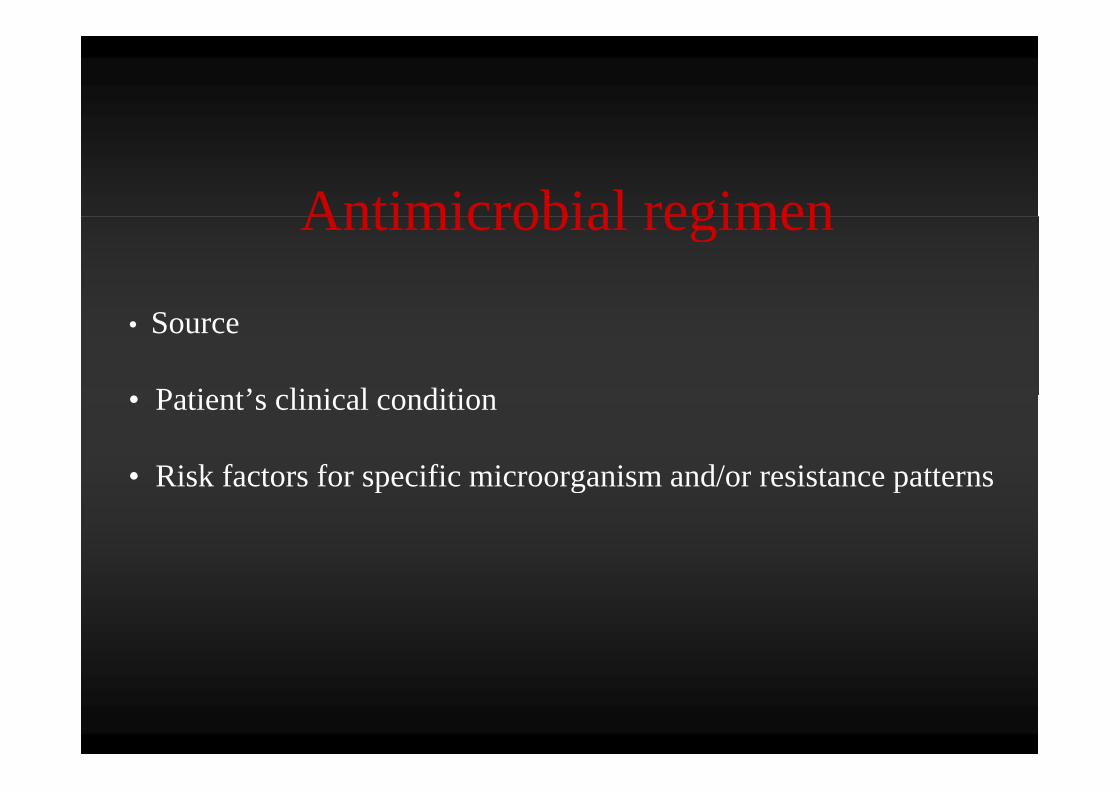
Antimicrobial regimenAntimicrobial regimen
• Source
• Patient’s clinical condition• Patient’s clinical condition
• Risk factors for specific microorganism and/or resistance patternsRisk factors for specific microorganism and/or resistance patterns

Community-acquired extra-biliary IAIs

Community acquired extrabiliary IAINo critically ill patient Ab f i k f f ESBLAbsence of risk factors for ESBL
AMOXICILLIN/CLAVULANATEDaily schedula: 2.2 g every 6 hours (Infusion time 2 hours)
OR (Allergy to beta-lactams)
CIPROFLOXACIN + METRONIDAZOLEDaily schedula: 400 mg every 8 hours Daily schedula: 500 mg every 6 hours
(Infusion time 30 min) (Infusion time 1 hour)
With permission of prof P Viale - Bologna July 2010. WSES congress; ConsensusW t pe ss o o p o V a e o og a Ju y 0 0. WS S co g ess; Co se susConference: First-line management of intra-abdominal infections.
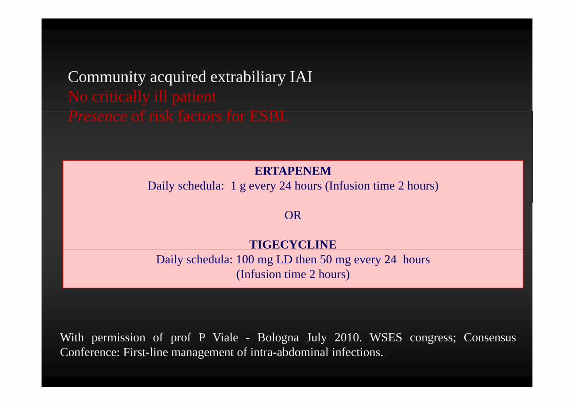
Community acquired extrabiliary IAINo critically ill patient P f i k f f ESBLPresence of risk factors for ESBL
ERTAPENEMDaily schedula: 1 g every 24 hours (Infusion time 2 hours)
OR
TIGECYCLINEDaily schedula: 100 mg LD then 50 mg every 24 hours
(Infusion time 2 hours)
With permission of prof P Viale - Bologna July 2010. WSES congress; ConsensusW t pe ss o o p o V a e o og a Ju y 0 0. WS S co g ess; Co se susConference: First-line management of intra-abdominal infections.
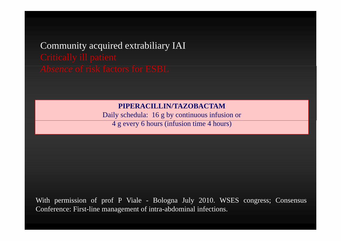
Community acquired extrabiliary IAICritically ill patient Ab f i k f f ESBLAbsence of risk factors for ESBL
PIPERACILLIN/TAZOBACTAMDaily schedula: 16 g by continuous infusion or
4 g every 6 hours (infusion time 4 hours)
With permission of prof P Viale - Bologna July 2010. WSES congress; ConsensusW t pe ss o o p o V a e o og a Ju y 0 0. WS S co g ess; Co se susConference: First-line management of intra-abdominal infections.
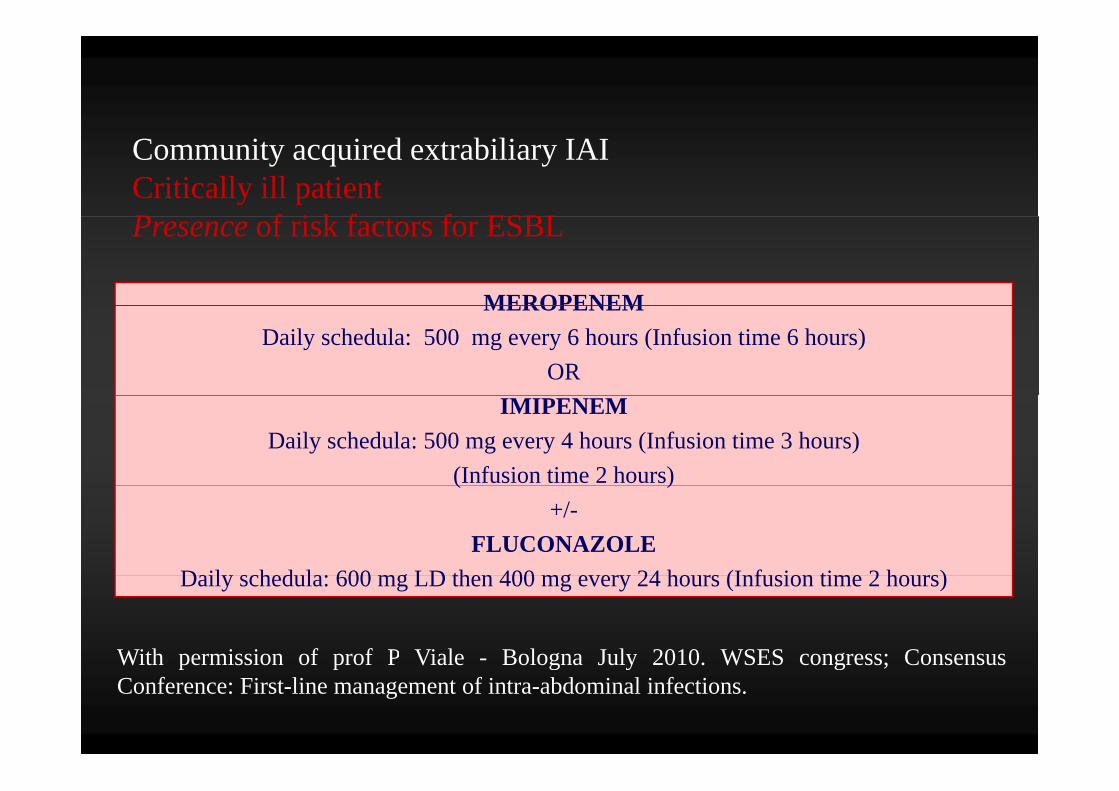
Community acquired extrabiliary IAICritically ill patient P f i k f f ESBL
MEROPENEM
Presence of risk factors for ESBL
MEROPENEMDaily schedula: 500 mg every 6 hours (Infusion time 6 hours)
OR IMIPENEM
Daily schedula: 500 mg every 4 hours (Infusion time 3 hours) (Infusion time 2 hours) ( )
+/-FLUCONAZOLE
D il h d l 600 LD th 400 24 h (I f i ti 2 h )Daily schedula: 600 mg LD then 400 mg every 24 hours (Infusion time 2 hours)
With permission of prof P Viale - Bologna July 2010. WSES congress; ConsensusW t pe ss o o p o V a e o og a Ju y 0 0. WS S co g ess; Co se susConference: First-line management of intra-abdominal infections.
Biliary IAIs
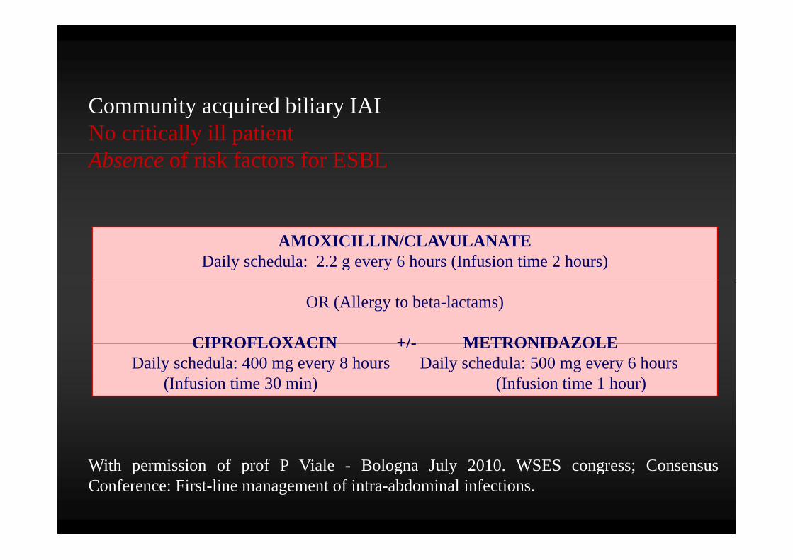
Community acquired biliary IAINo critically ill patient Ab f i k f f ESBLAbsence of risk factors for ESBL
AMOXICILLIN/CLAVULANATEDaily schedula: 2.2 g every 6 hours (Infusion time 2 hours)
OR (Allergy to beta-lactams)
CIPROFLOXACIN +/- METRONIDAZOLECIPROFLOXACIN +/- METRONIDAZOLEDaily schedula: 400 mg every 8 hours Daily schedula: 500 mg every 6 hours
(Infusion time 30 min) (Infusion time 1 hour)
With permission of prof P Viale - Bologna July 2010. WSES congress; ConsensusW t pe ss o o p o V a e o og a Ju y 0 0. WS S co g ess; Co se susConference: First-line management of intra-abdominal infections.
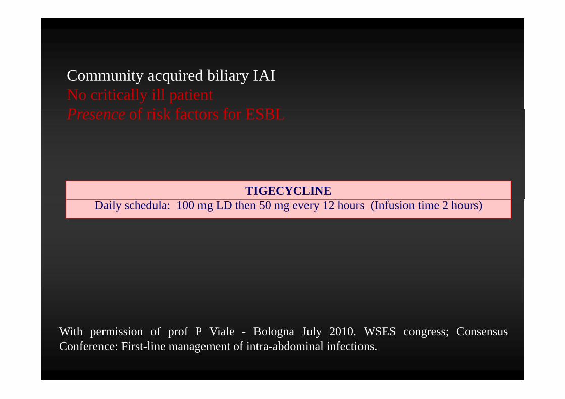
Community acquired biliary IAINo critically ill patient P f i k f f ESBLPresence of risk factors for ESBL
TIGECYCLINEDaily schedula: 100 mg LD then 50 mg every 12 hours (Infusion time 2 hours)
With permission of prof P Viale - Bologna July 2010. WSES congress; ConsensusW t pe ss o o p o V a e o og a Ju y 0 0. WS S co g ess; Co se susConference: First-line management of intra-abdominal infections.
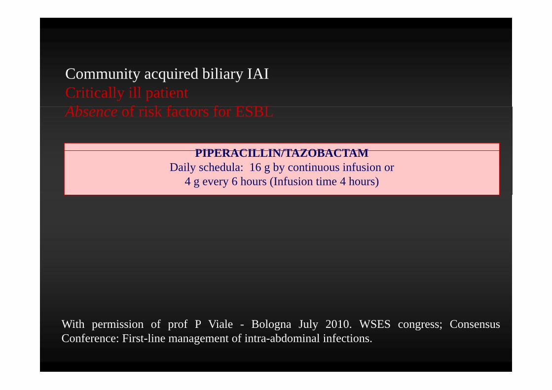
Community acquired biliary IAICritically ill patient Ab f i k f f ESBL
PIPERACILLIN/TAZOBACTAM
Absence of risk factors for ESBL
PIPERACILLIN/TAZOBACTAMDaily schedula: 16 g by continuous infusion or
4 g every 6 hours (Infusion time 4 hours)
With permission of prof P Viale - Bologna July 2010. WSES congress; ConsensusW t pe ss o o p o V a e o og a Ju y 0 0. WS S co g ess; Co se susConference: First-line management of intra-abdominal infections.
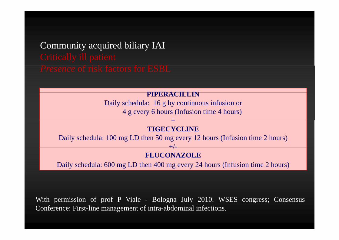
Community acquired biliary IAICritically ill patient P f i k f f ESBL
PIPERACILLIN
Presence of risk factors for ESBL
PIPERACILLINDaily schedula: 16 g by continuous infusion or
4 g every 6 hours (Infusion time 4 hours)++
TIGECYCLINEDaily schedula: 100 mg LD then 50 mg every 12 hours (Infusion time 2 hours)
+/-+/-FLUCONAZOLE
Daily schedula: 600 mg LD then 400 mg every 24 hours (Infusion time 2 hours)
With permission of prof P Viale - Bologna July 2010. WSES congress; ConsensusW t pe ss o o p o V a e o og a Ju y 0 0. WS S co g ess; Co se susConference: First-line management of intra-abdominal infections.

Hospital-acquired extra-biliary IAIs
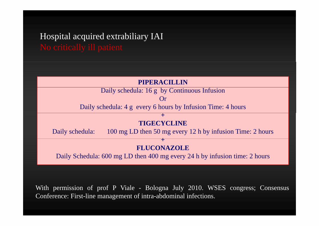
Hospital acquired extrabiliary IAIHospital acquired extrabiliary IAINo critically ill patient
PIPERACILLIN Daily schedula: 16 g by Continuous Infusion
OrDaily schedula: 4 g every 6 hours by Infusion Time: 4 hours
+TIGECYCLINE
Daily schedula: 100 mg LD then 50 mg every 12 h by infusion Time: 2 hours++
FLUCONAZOLE Daily Schedula: 600 mg LD then 400 mg every 24 h by infusion time: 2 hours
With permission of prof P Viale - Bologna July 2010. WSES congress; ConsensusW t pe ss o o p o V a e o og a Ju y 0 0. WS S co g ess; Co se susConference: First-line management of intra-abdominal infections.
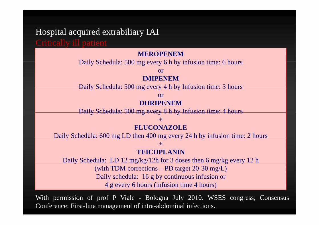
Hospital acquired extrabiliary IAI
MEROPENEMDaily Schedula: 500 mg every 6 h by infusion time: 6 hours
p q yCritically ill patient
Daily Schedula: 500 mg every 6 h by infusion time: 6 hoursor
IMIPENEMDaily Schedula: 500 mg every 4 h by Infusion time: 3 hoursDaily Schedula: 500 mg every 4 h by Infusion time: 3 hours
orDORIPENEM
Daily Schedula: 500 mg every 8 h by Infusion time: 4 hoursDaily Schedula: 500 mg every 8 h by Infusion time: 4 hours+
FLUCONAZOLE Daily Schedula: 600 mg LD then 400 mg every 24 h by infusion time: 2 hoursy g g y y
+TEICOPLANIN
Daily Schedula: LD 12 mg/kg/12h for 3 doses then 6 mg/kg every 12 h y g g g g y(with TDM corrections – PD target 20-30 mg/L)Daily schedula: 16 g by continuous infusion or
4 g every 6 hours (infusion time 4 hours)
With permission of prof P Viale - Bologna July 2010. WSES congress; ConsensusConference: First-line management of intra-abdominal infections.
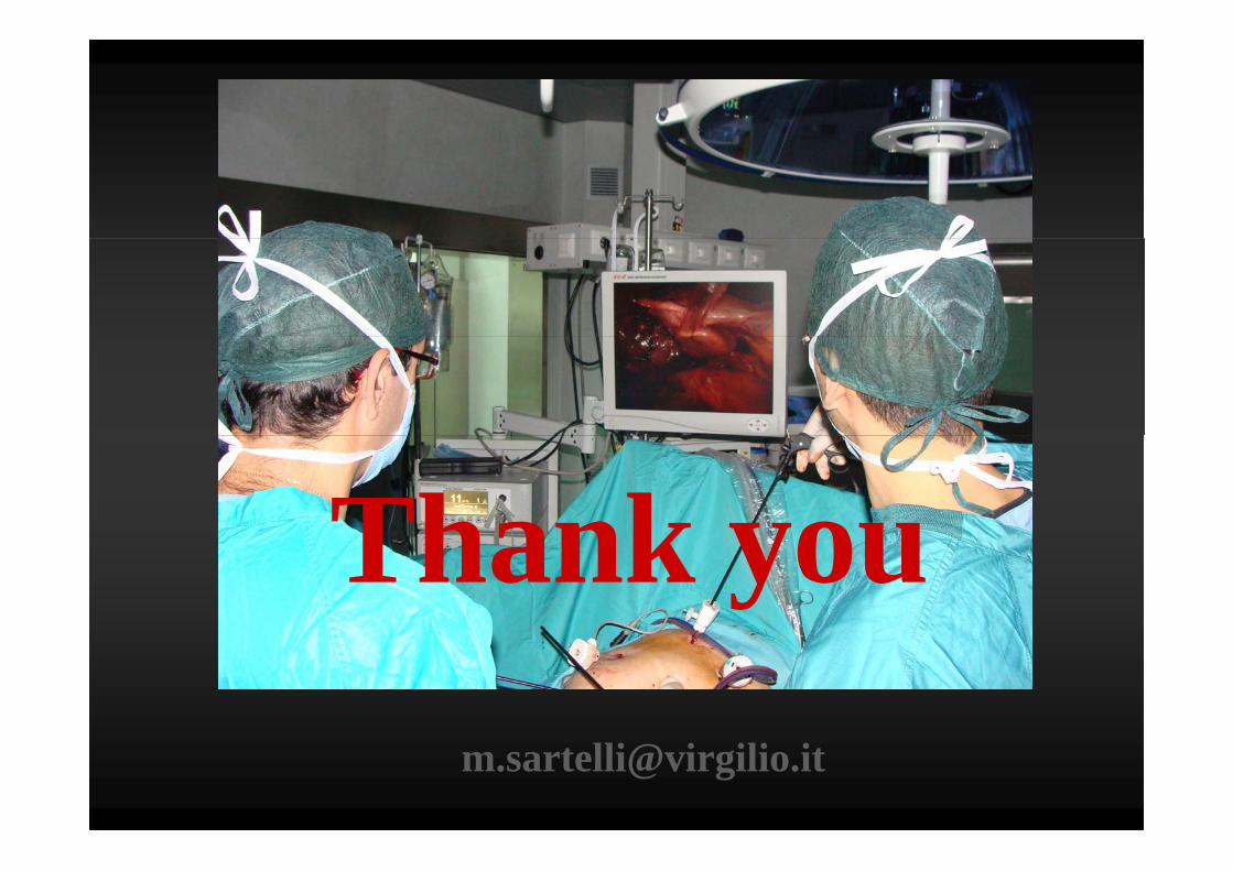
Thank youThank [email protected]
Recommended


![Intra-Abdominal and Abdominal Wall Desmoid Fibromatosis · intra-abdominal and involving the small bowel mesentery [2]. TREATMENT Surgery Margin-negative resection has historically](https://img.pdfslide.us/doc/110x75/5e5a290071d21b380f5b7e74/intra-abdominal-and-abdominal-wall-desmoid-fibromatosis-intra-abdominal-and-involving.jpg)