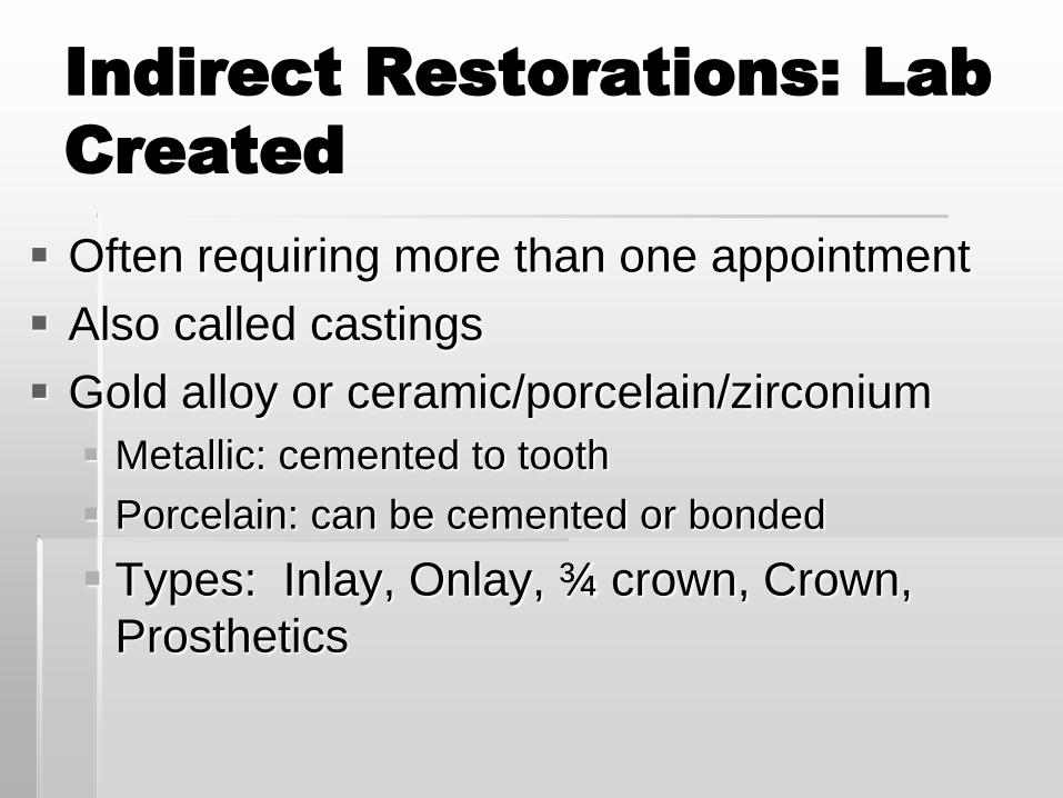
Indirect Restorations: Lab
Created
Often requiring more than one appointment
Also called castings
Gold alloy or ceramic/porcelain/zirconium
Metallic: cemented to tooth
Porcelain: can be cemented or bonded
Types: Inlay, Onlay, ¾ crown, Crown,
Prosthetics
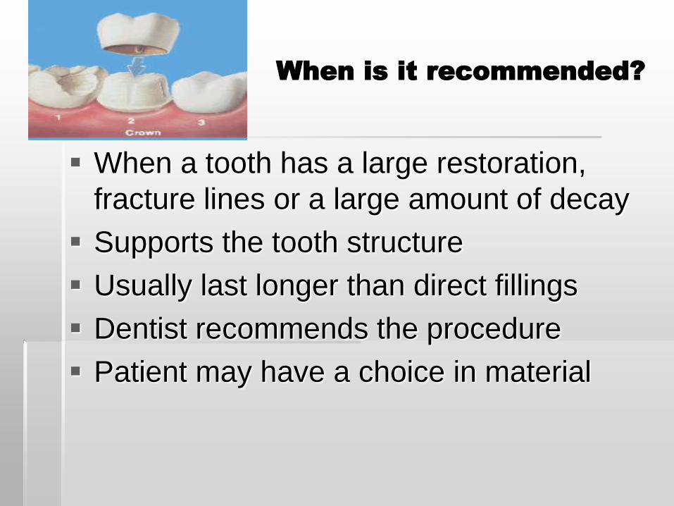
When is it recommended?
When a tooth has a large restoration,
fracture lines or a large amount of decay
Supports the tooth structure
Usually last longer than direct fillings
Dentist recommends the procedure
Patient may have a choice in material
How is it made?
Tooth is prepared by the dentist with a
handpiece and burs
Impression is made
Lab technicians create the restoration
“Cast” the metallic restoration
Patient returns to have restoration placed
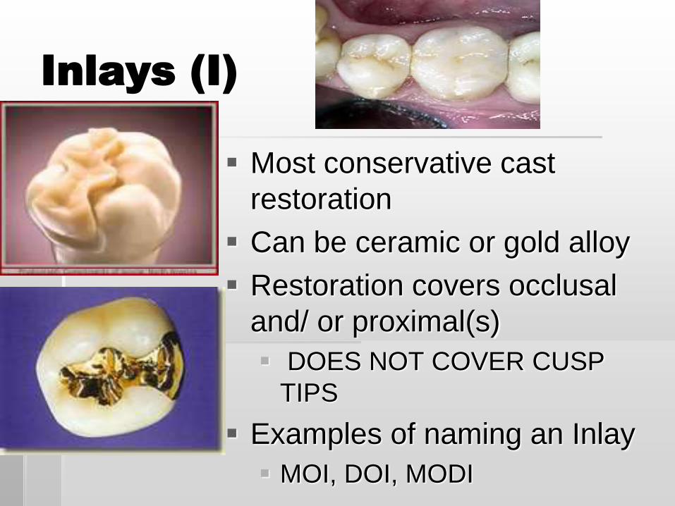
Inlays (I)
Most conservative cast
restoration
Can be ceramic or gold alloy
Restoration covers occlusal
and/ or proximal(s)
DOES NOT COVER CUSP
TIPS
Examples of naming an Inlay
MOI, DOI, MODI
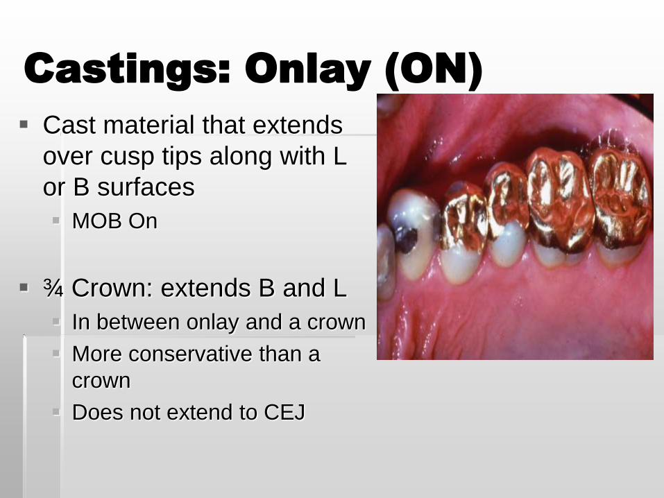
Castings: Onlay (ON)
Cast material that extends
over cusp tips along with L
or B surfaces
MOB On
¾ Crown: extends B and L
In between onlay and a crown
More conservative than a
crown
Does not extend to CEJ
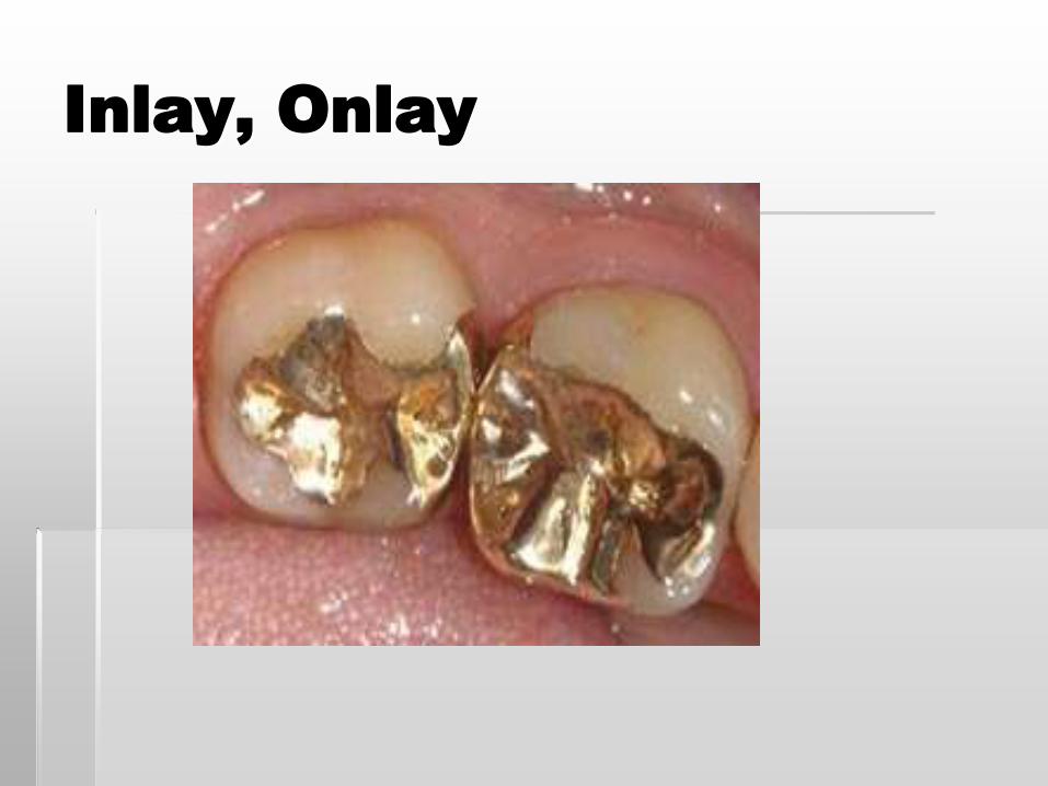
Inlay, Onlay
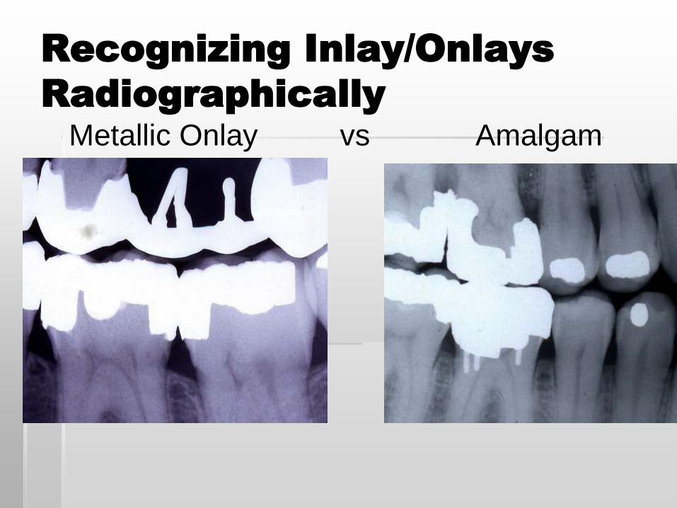
Recognizing Inlay/Onlays
Radiographically
Metallic Onlay vs Amalgam
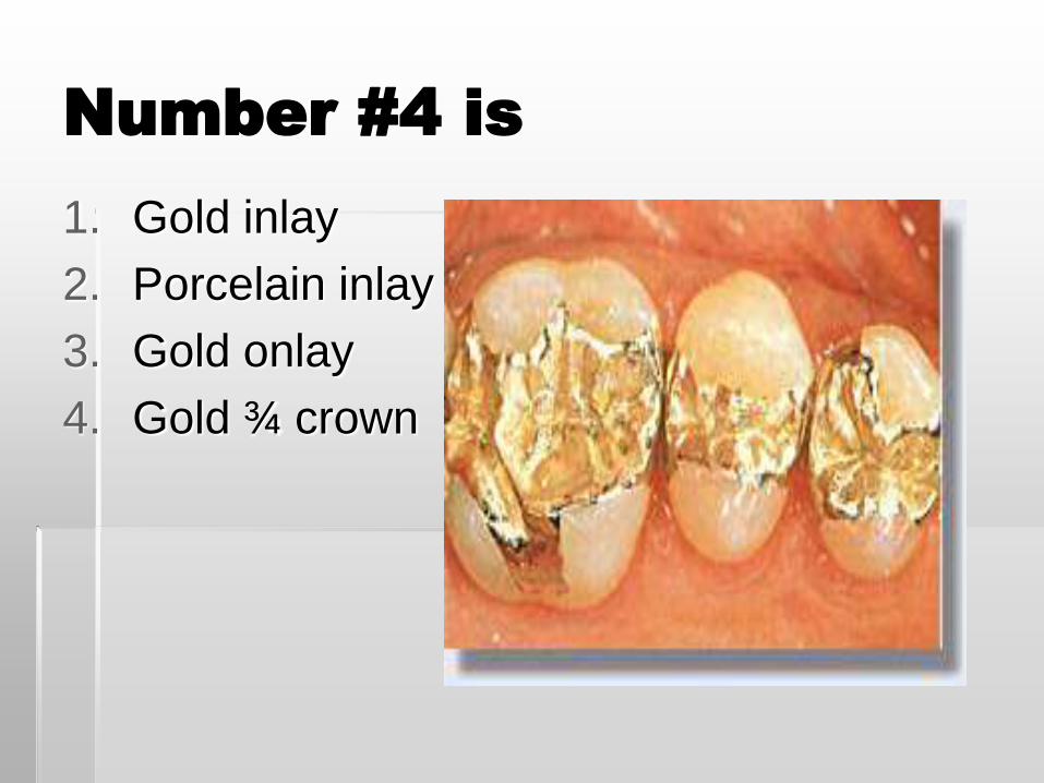
Number #4 is
1. Gold inlay
2. Porcelain inlay
3. Gold onlay
4. Gold ¾ crown
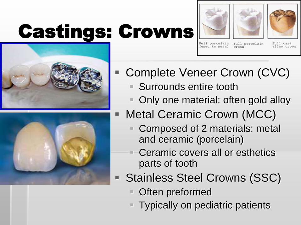
Castings: Crowns
Complete Veneer Crown (CVC) Surrounds entire tooth
Only one material: often gold alloy
Metal Ceramic Crown (MCC) Composed of 2 materials: metal
and ceramic (porcelain)
Ceramic covers all or esthetics parts of tooth
Stainless Steel Crowns (SSC) Often preformed
Typically on pediatric patients
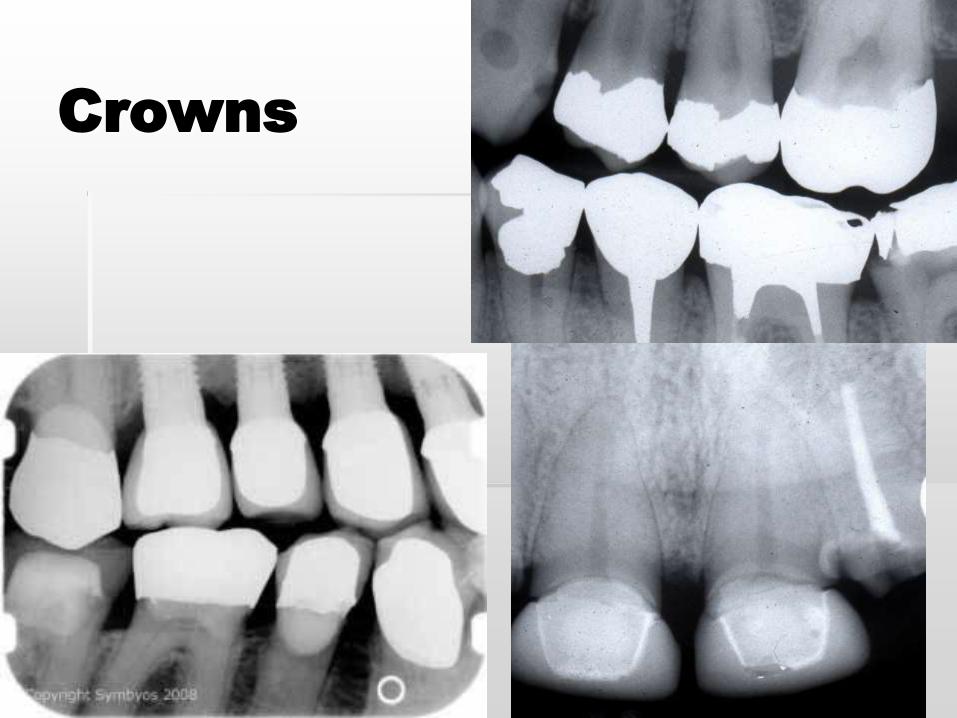
Crowns
Endodontics
Implants
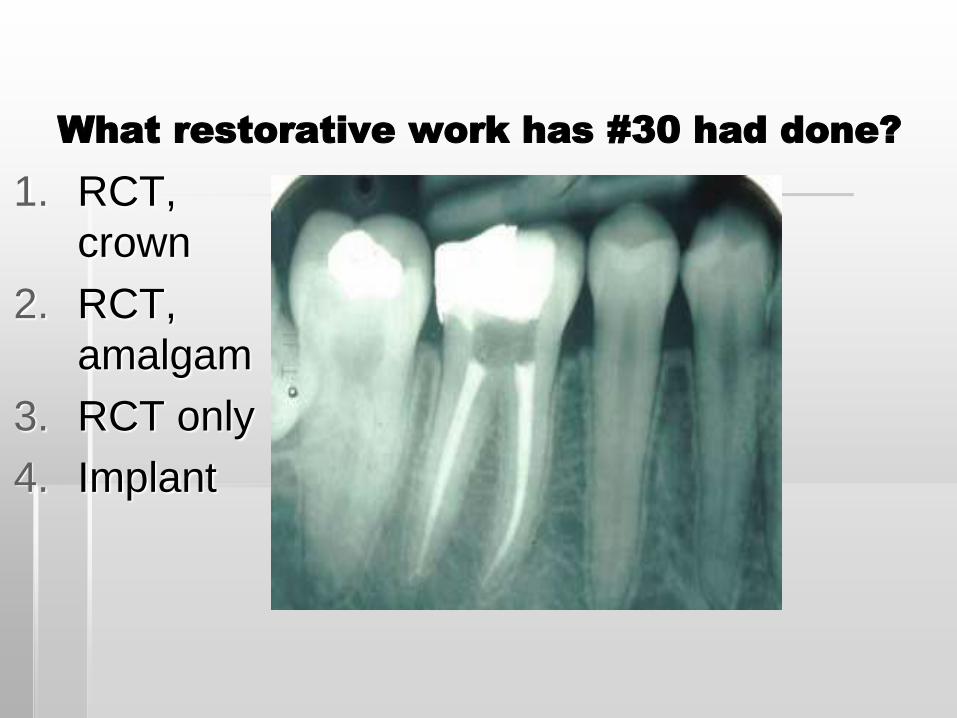
What restorative work has #30 had done?
1. RCT,
crown
2. RCT,
amalgam
3. RCT only
4. Implant
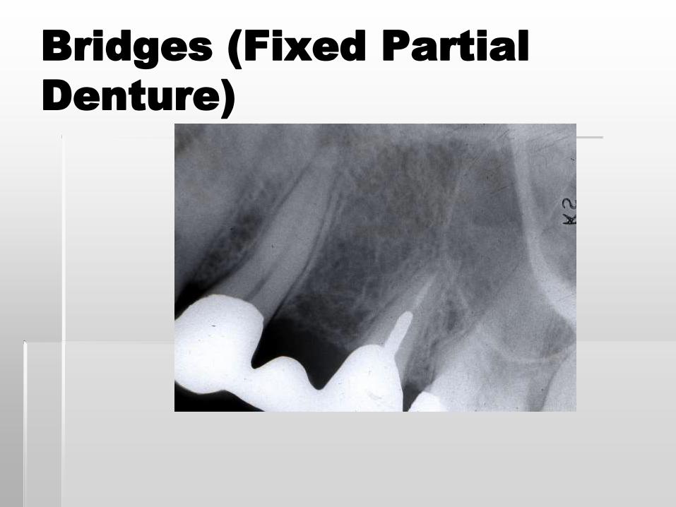
Bridges (Fixed Partial
Denture)
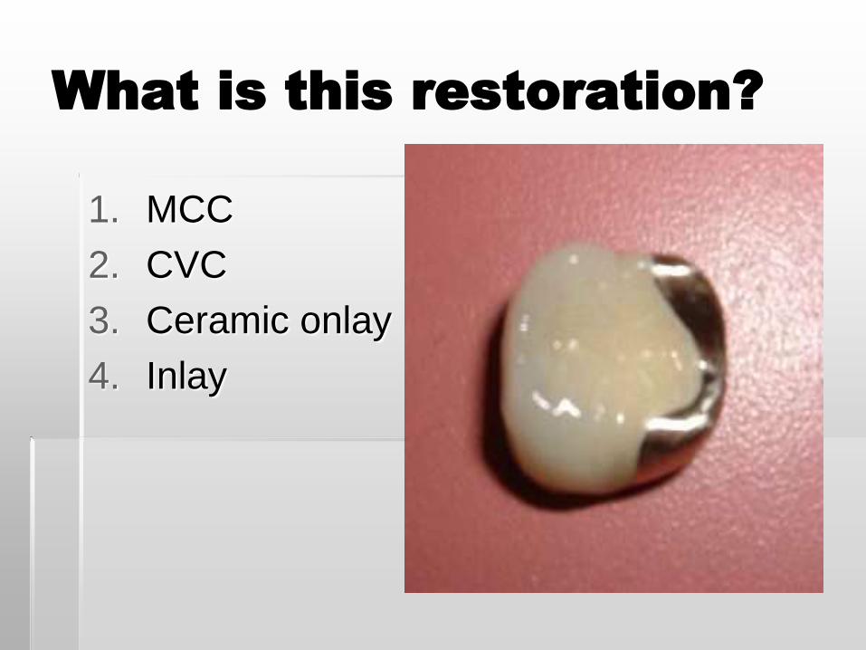
What is this restoration?
1. MCC
2. CVC
3. Ceramic onlay
4. Inlay
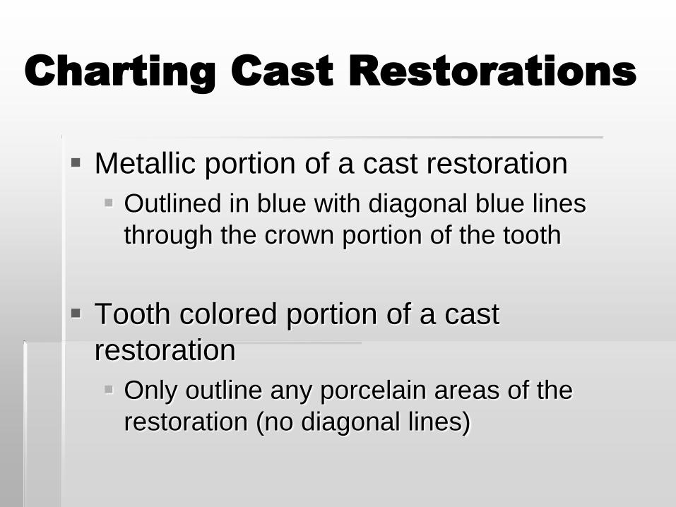
Charting Cast Restorations
Metallic portion of a cast restoration
Outlined in blue with diagonal blue lines
through the crown portion of the tooth
Tooth colored portion of a cast
restoration
Only outline any porcelain areas of the
restoration (no diagonal lines)
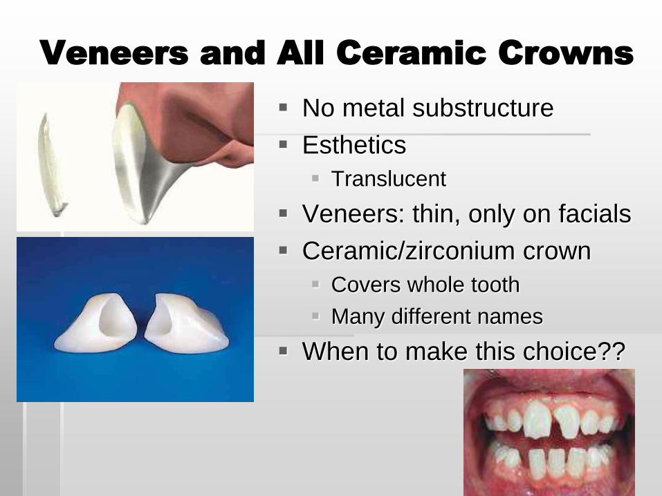
Veneers and All Ceramic Crowns
No metal substructure
Esthetics
Translucent
Veneers: thin, only on facials
Ceramic/zirconium crown
Covers whole tooth
Many different names
When to make this choice??
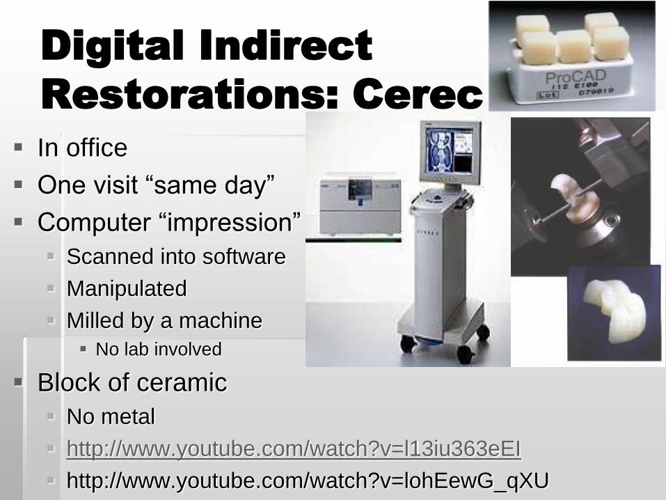
Digital Indirect
Restorations: Cerec
In office
One visit “same day”
Computer “impression”
Scanned into software
Manipulated
Milled by a machine
No lab involved
Block of ceramic
No metal
http://www.youtube.com/watch?v=l13iu363eEI
http://www.youtube.com/watch?v=lohEewG_qXU
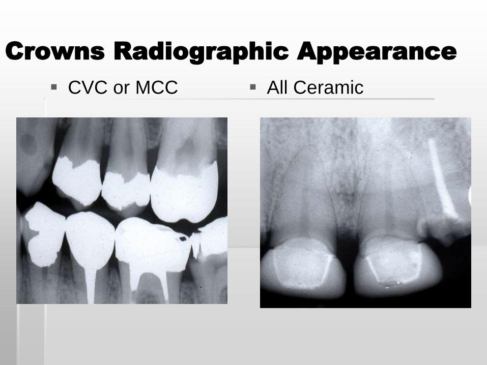
Crowns Radiographic Appearance
CVC or MCC All Ceramic
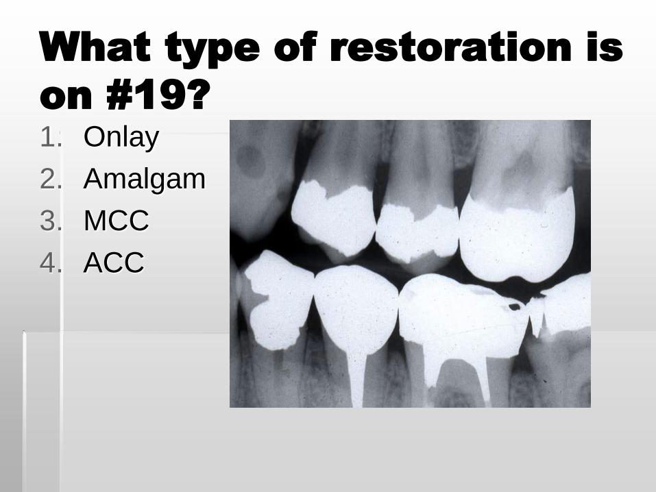
What type of restoration is
on #19?
1. Onlay
2. Amalgam
3. MCC
4. ACC
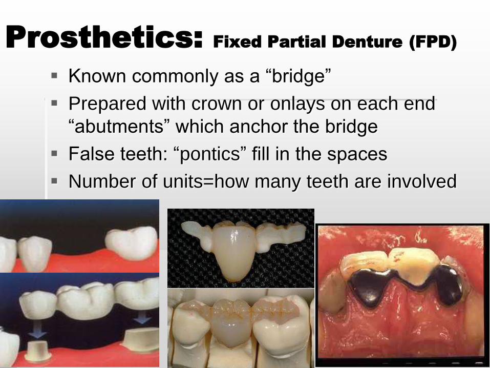
Prosthetics: Fixed Partial Denture (FPD)
Known commonly as a “bridge”
Prepared with crown or onlays on each end
“abutments” which anchor the bridge
False teeth: “pontics” fill in the spaces
Number of units=how many teeth are involved
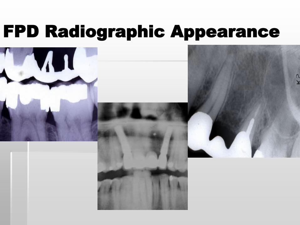
FPD Radiographic Appearance
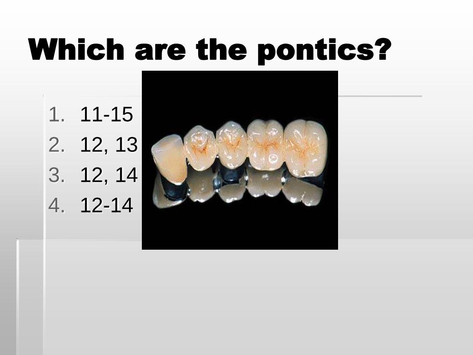
Which are the pontics?
1. 11-15
2. 12, 13
3. 12, 14
4. 12-14
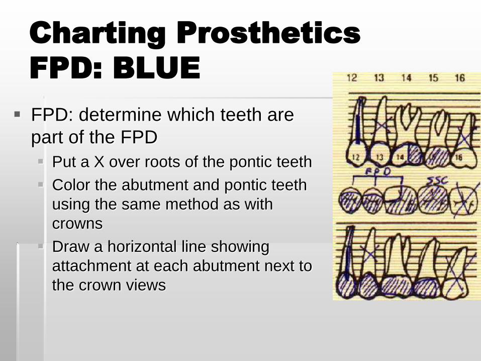
Charting Prosthetics
FPD: BLUE
FPD: determine which teeth are
part of the FPD
Put a X over roots of the pontic teeth
Color the abutment and pontic teeth
using the same method as with
crowns
Draw a horizontal line showing
attachment at each abutment next to
the crown views
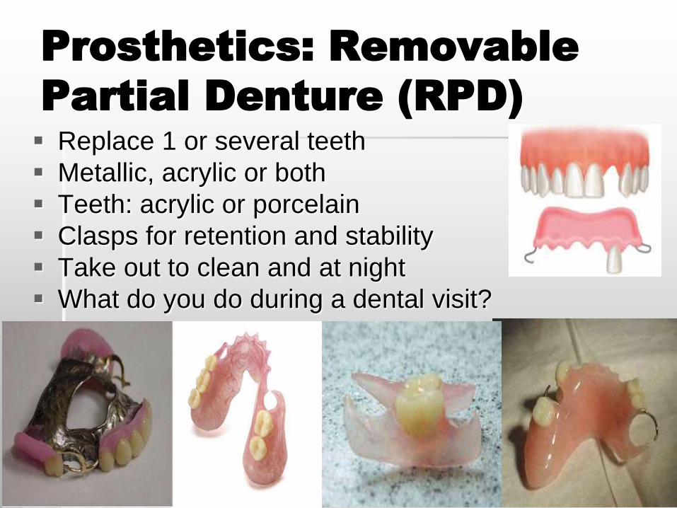
Prosthetics: Removable
Partial Denture (RPD)
Replace 1 or several teeth
Metallic, acrylic or both
Teeth: acrylic or porcelain
Clasps for retention and stability
Take out to clean and at night
What do you do during a dental visit?
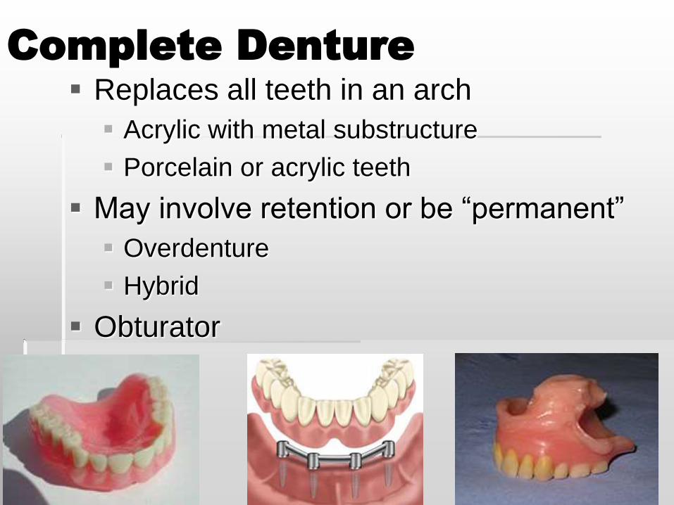
Complete Denture
Replaces all teeth in an arch
Acrylic with metal substructure
Porcelain or acrylic teeth
May involve retention or be “permanent”
Overdenture
Hybrid
Obturator
Charting Prosthetics
RPD and Denture: BLUE
RPD: Determine which teeth are replaced
X out the missing roots in blue
Draw a horizontal line along the occlusals
Draw connecting lines to each tooth replaced by the RPD
Outline each tooth wherever porcelain is visible
Draw diagonal line across wherever metal is visible
Denture
Draw a large X through the entire arch
Draw a blue horizontal line over the occlusal views of
the arch
Write denture
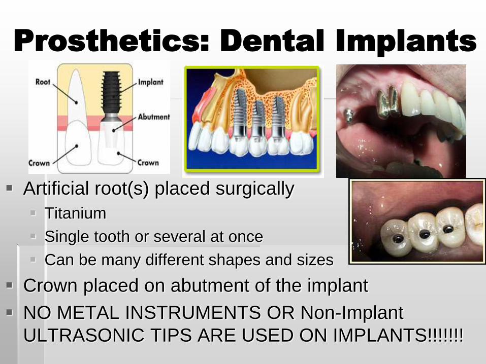
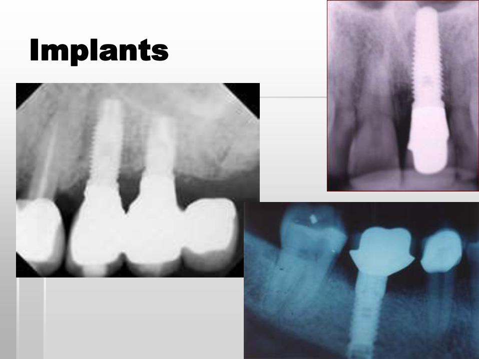
Prosthetics: Dental Implants
Artificial root(s) placed surgically
Titanium
Single tooth or several at once
Can be many different shapes and sizes
Crown placed on abutment of the implant
NO METAL INSTRUMENTS OR Non-Implant
ULTRASONIC TIPS ARE USED ON IMPLANTS!!!!!!!
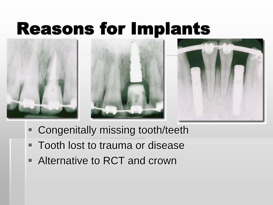
Reasons for Implants
Congenitally missing tooth/teeth
Tooth lost to trauma or disease
Alternative to RCT and crown
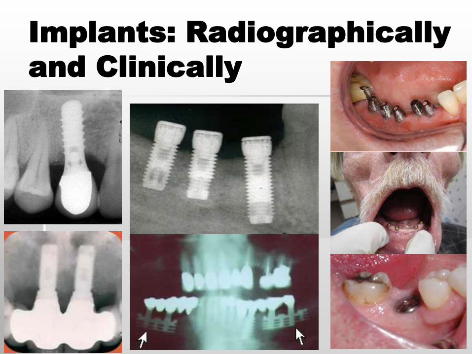
Implants: Radiographically
and Clinically
Charting Prosthetics
Implants
Draw (like a screw) implant body over the root
Write “implant” next to tooth
Use same method as crown charting to chart
implant crown
If implant is part of a larger prosthesis (FPD, RPD,
denture): draw in accordingly
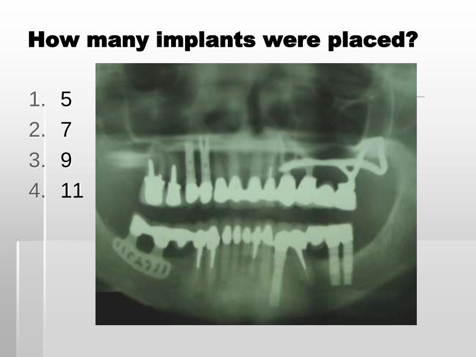
How many implants were placed?
1. 5
2. 7
3. 9
4. 11
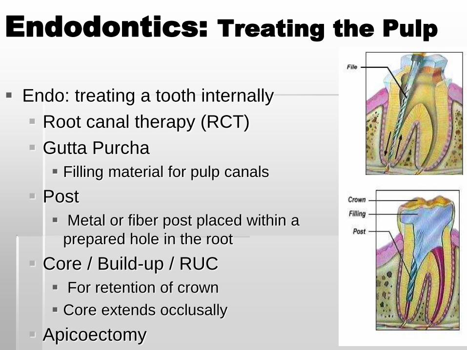
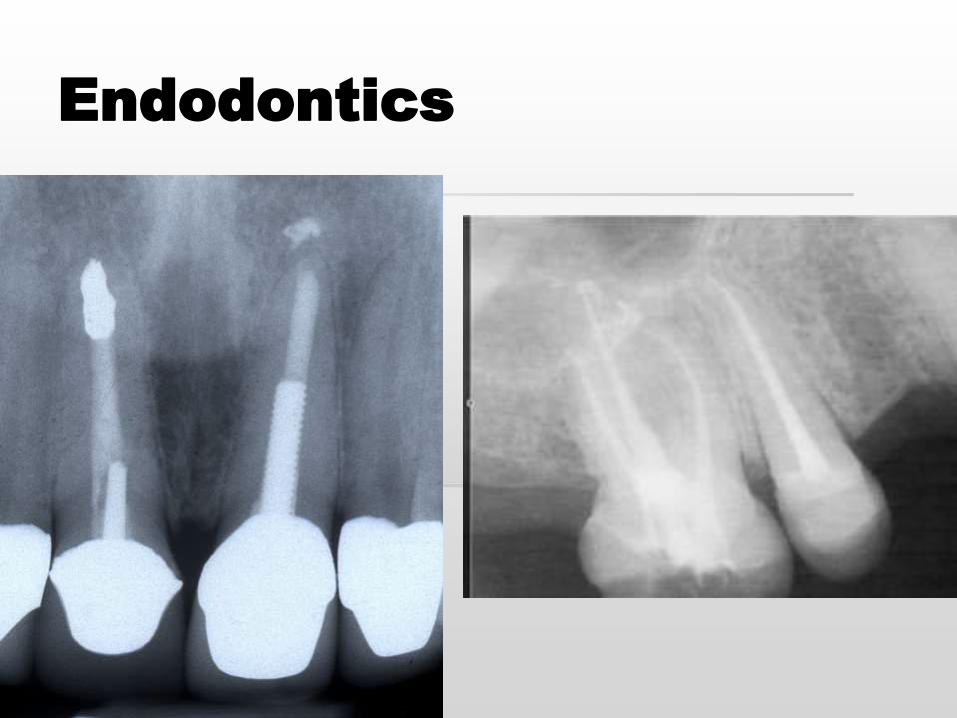
Endodontics: Treating the Pulp
Endo: treating a tooth internally
Root canal therapy (RCT)
Gutta Purcha
Filling material for pulp canals
Post
Metal or fiber post placed within a
prepared hole in the root
Core / Build-up / RUC
For retention of crown
Core extends occlusally
Apicoectomy
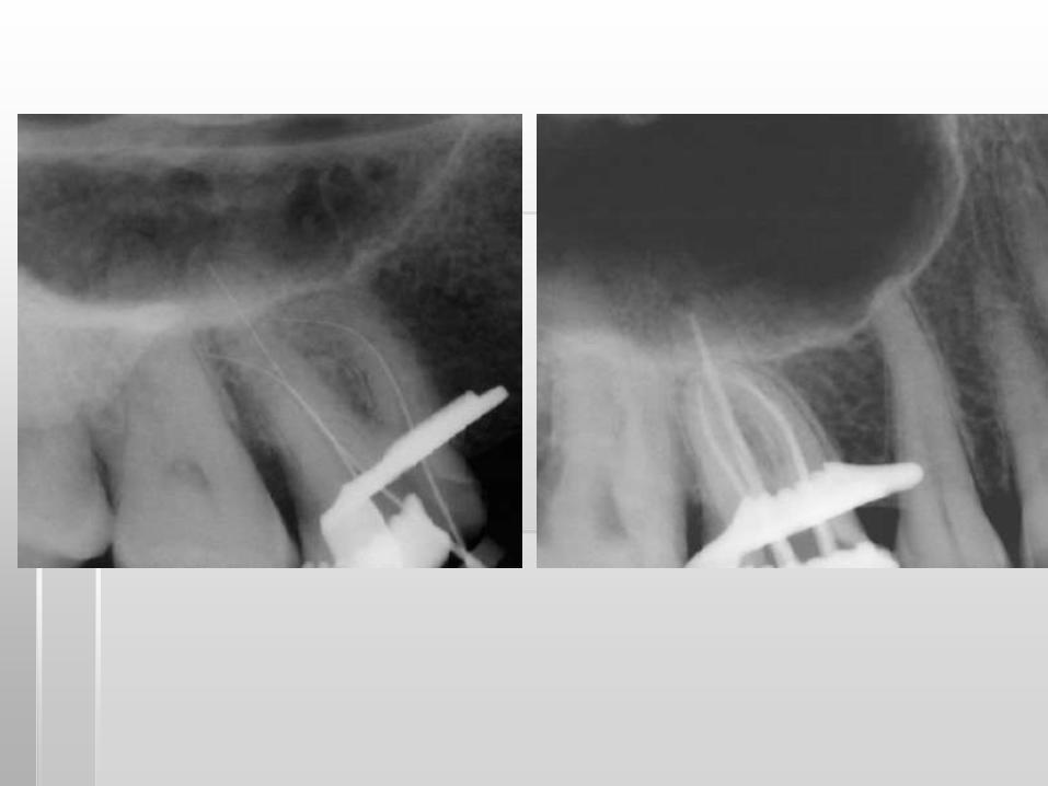
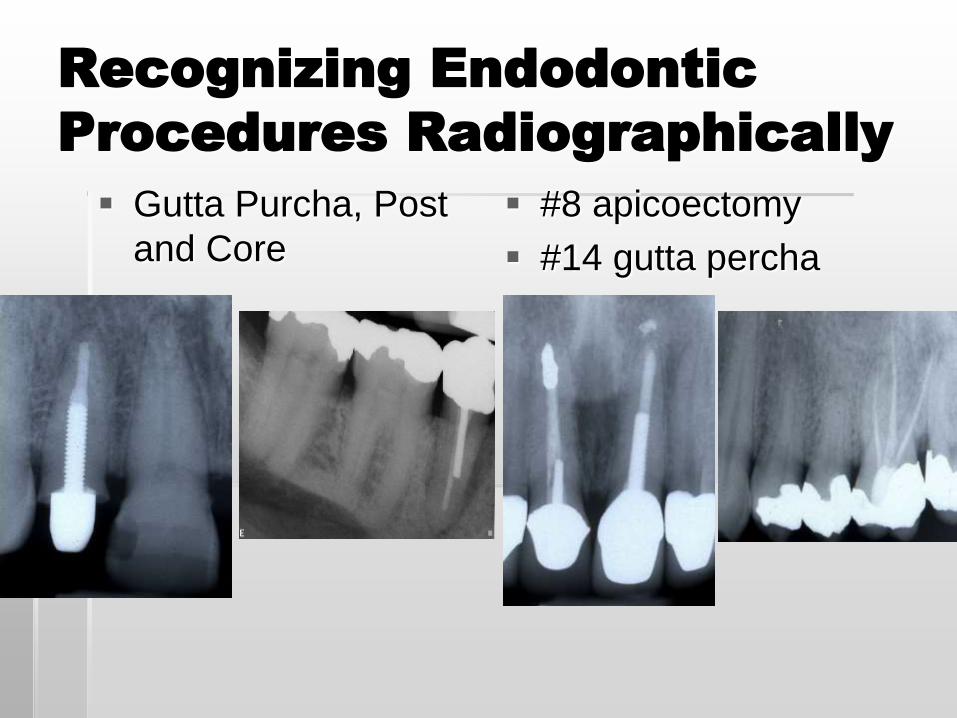
Recognizing Endodontic
Procedures Radiographically
Gutta Purcha, Post
and Core
#8 apicoectomy
#14 gutta percha
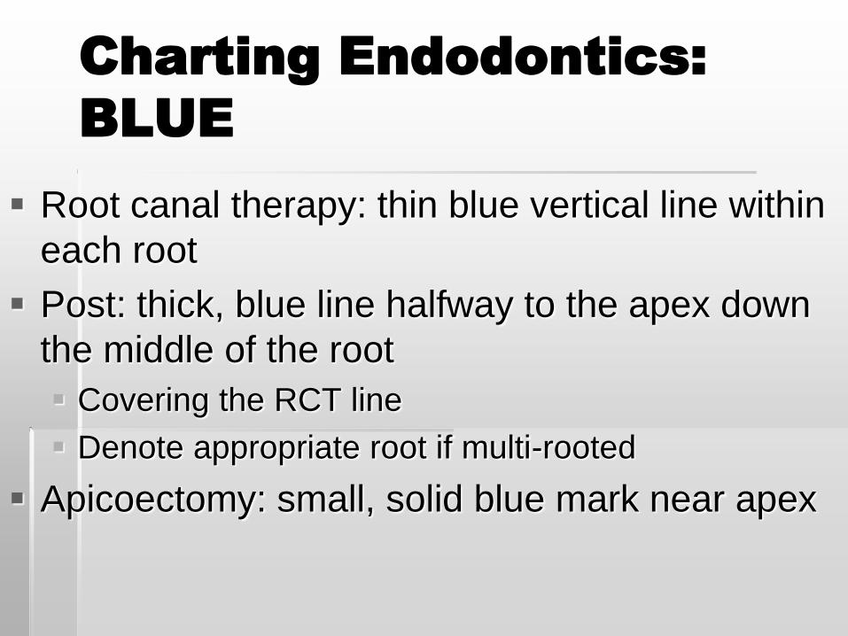
Charting Endodontics:
BLUE
Root canal therapy: thin blue vertical line within
each root
Post: thick, blue line halfway to the apex down
the middle of the root
Covering the RCT line
Denote appropriate root if multi-rooted
Apicoectomy: small, solid blue mark near apex
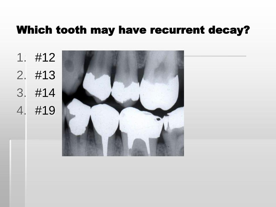
Which tooth may have recurrent decay?
1. #12
2. #13
3. #14
4. #19
Charting Areas of Concern
“BUFF SHEET”: worksheet to write suspicions
and obvious problems
Dr. signs at D & T exam
Chart after confirming with Doctor: RED
Caries: shade in the area of decay
Teeth that need extracted: X out in red
Defective restoration: red arrow
Overhang: OH
Abrasion: circle the area, write “abras”
Attrition: circle the area and write “attrit”
Other defects: use an arrow and comment
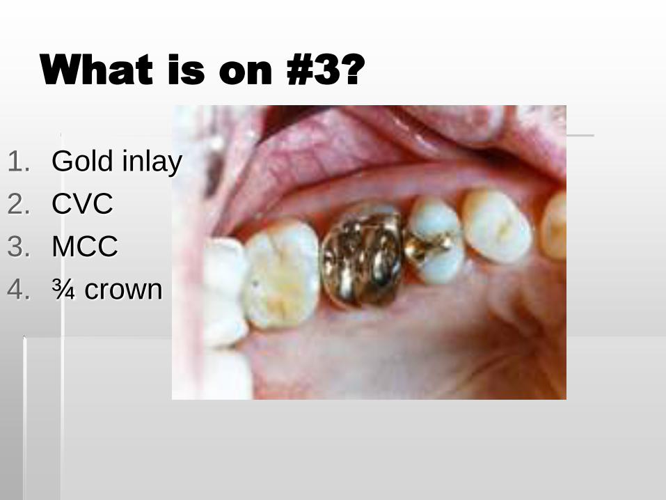
What is on #3?
1. Gold inlay
2. CVC
3. MCC
4. ¾ crown
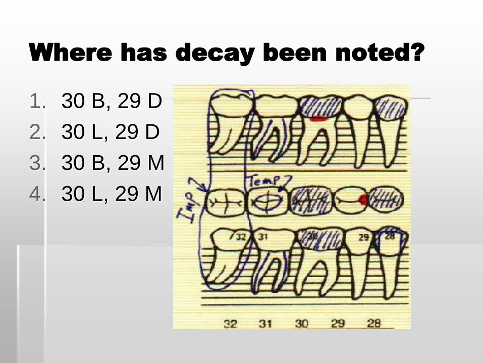
Where has decay been noted?
1. 30 B, 29 D
2. 30 L, 29 D
3. 30 B, 29 M
4. 30 L, 29 M
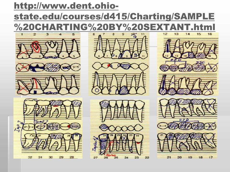
http://www.dent.ohio-
state.edu/courses/d415/Charting/SAMPLE
%20CHARTING%20BY%20SEXTANT.html
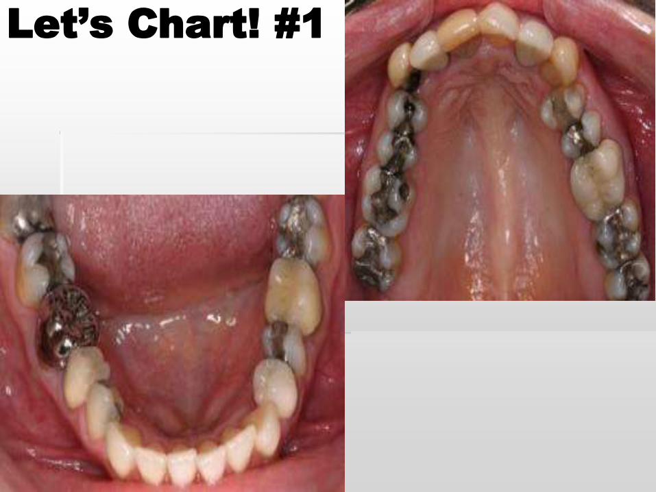
Let’s Chart! #1
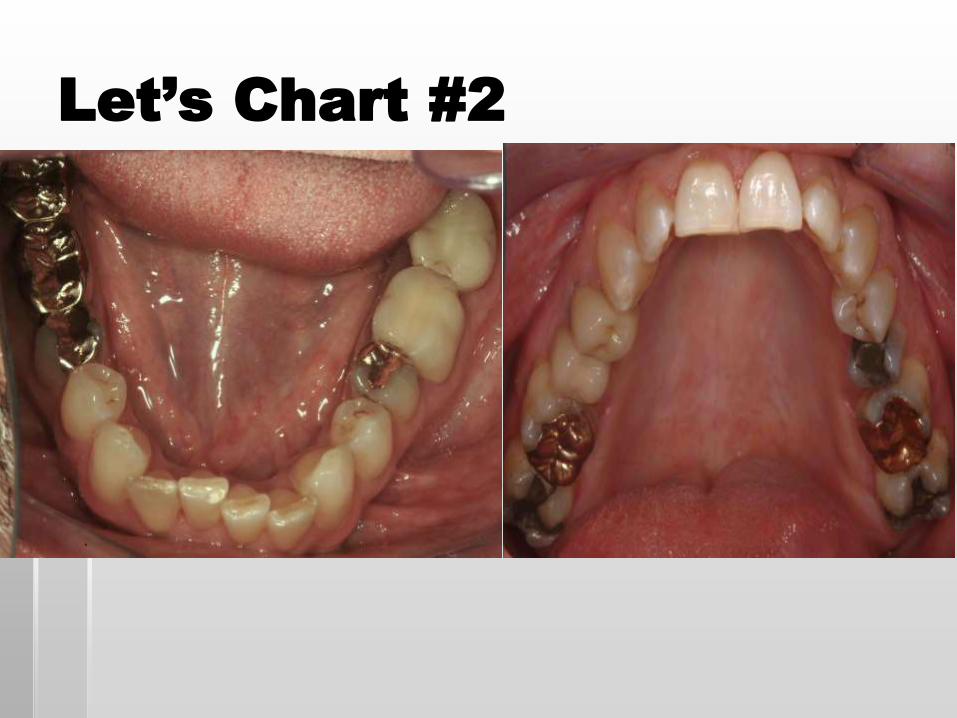
Let’s Chart #2
Recommended