In Vitro Models for Studying AngiogenesisSuparna Sanyal
May 12, 2010
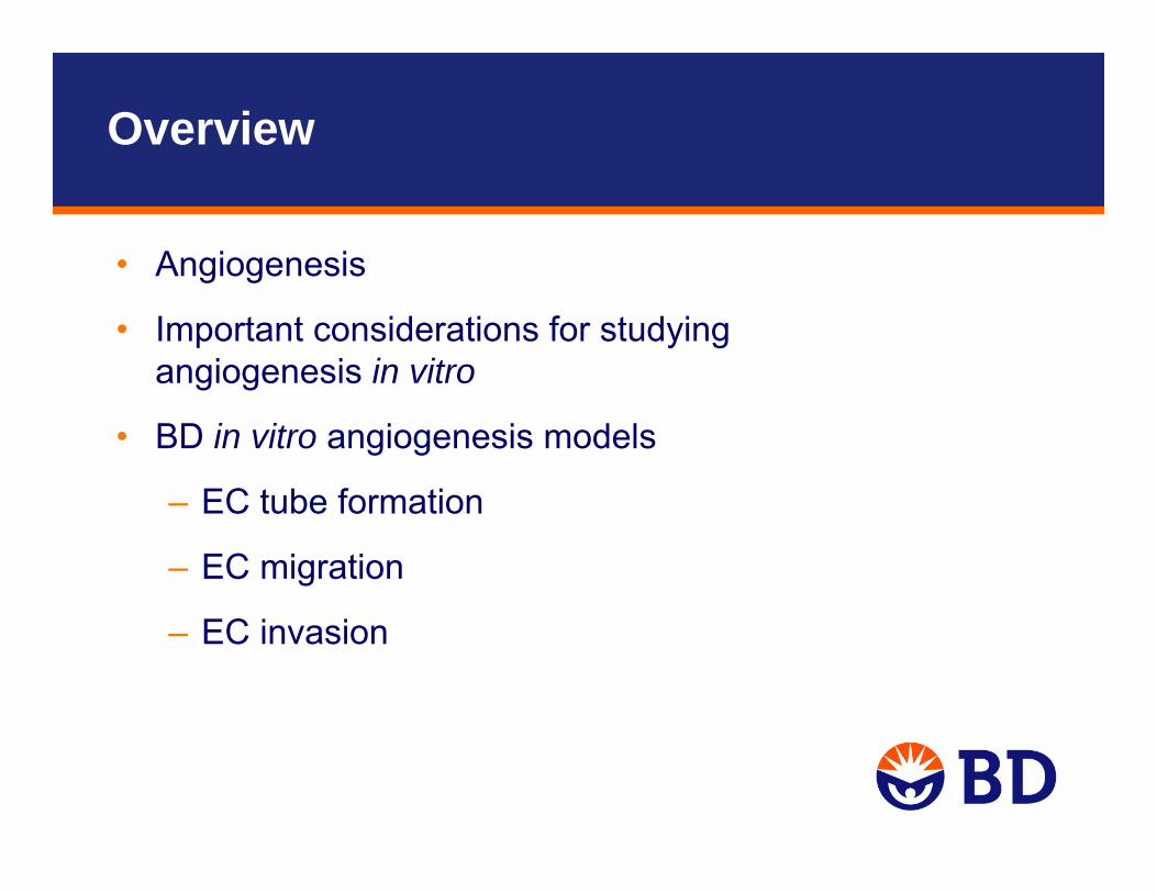
Overview
• Angiogenesis
• Important considerations for studying angiogenesis in vitro
• BD in vitro angiogenesis models
– EC tube formation
– EC migration
– EC invasion
Angiogenesis
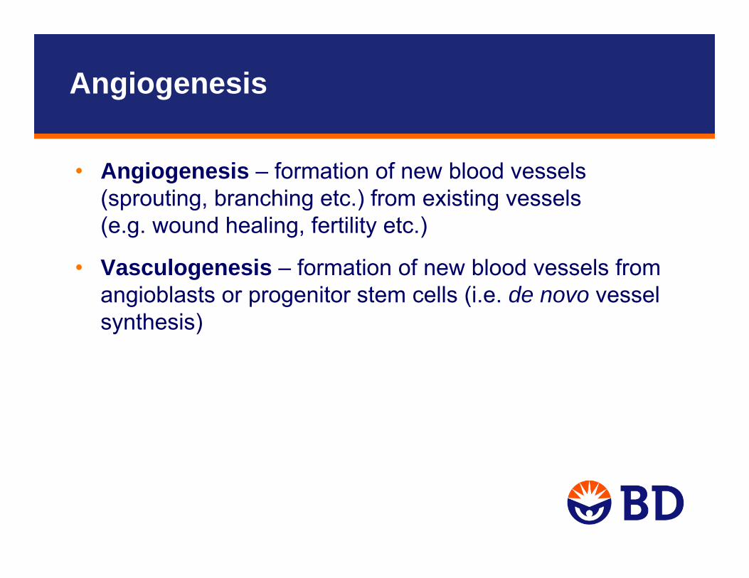
• Angiogenesis – formation of new blood vessels (sprouting, branching etc.) from existing vessels (e.g. wound healing, fertility etc.)
• Vasculogenesis – formation of new blood vessels from angioblasts or progenitor stem cells (i.e. de novo vessel synthesis)

Excessive
Insufficient
ANGIOGENESIS
CancerRheumatoid
Arthritis
Blindness (Diabetic Retinopathy) Psoriasis
Aids Complications
Stroke
Heart Disease
Ulcers
Scleroderma
Infertility
Diseases Associated With Angiogenesis
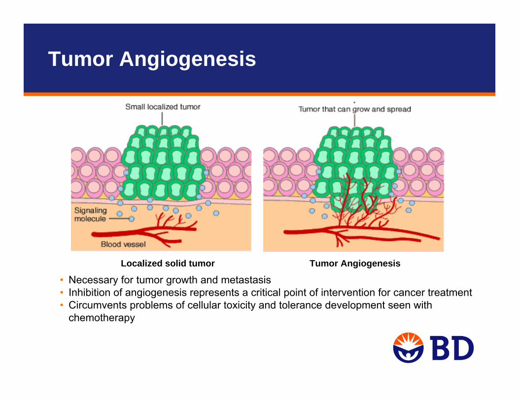
Tumor AngiogenesisLocalized solid tumor
• Necessary for tumor growth and metastasis • Inhibition of angiogenesis represents a critical point of intervention for cancer treatment• Circumvents problems of cellular toxicity and tolerance development seen with
chemotherapy
Tumor Angiogenesis
Angiogenesis Research
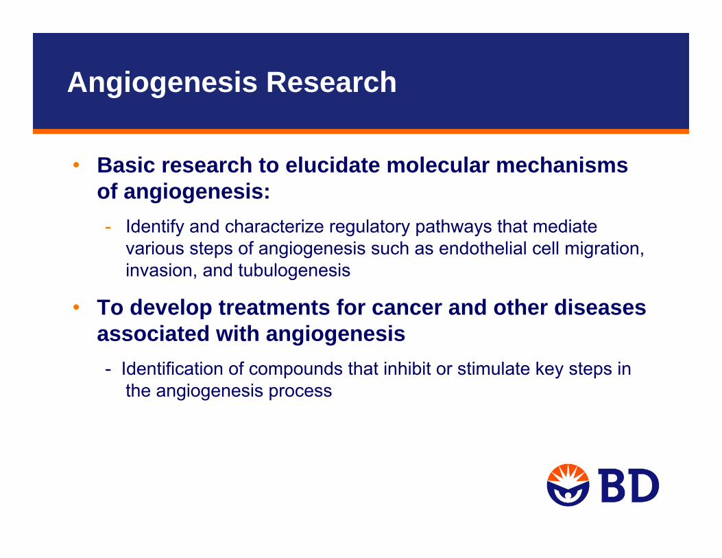
• Basic research to elucidate molecular mechanisms of angiogenesis: - Identify and characterize regulatory pathways that mediate
various steps of angiogenesis such as endothelial cell migration, invasion, and tubulogenesis
• To develop treatments for cancer and other diseases associated with angiogenesis - Identification of compounds that inhibit or stimulate key steps in
the angiogenesis process
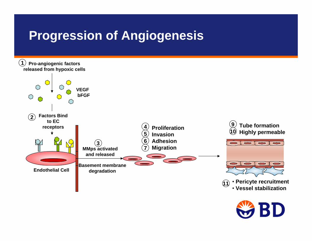
Pro-angiogenic factors released from hypoxic cells
VEGFbFGF
1
Factors Bind to EC
receptors
Endothelial Cell
2Tube formationHighly permeable10
94567
ProliferationInvasionAdhesionMigrationMMps activated
and released
Basement membranedegradation
3
• Pericyte recruitment• Vessel stabilization
11
Progression of Angiogenesis
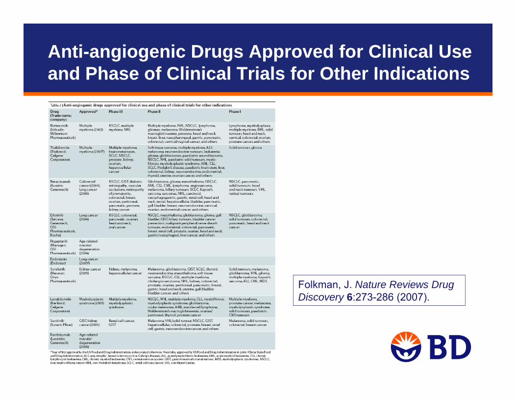
Anti-angiogenic Drugs Approved for Clinical Use and Phase of Clinical Trials for Other Indications
Folkman, J. Nature Reviews Drug Discovery 6:273-286 (2007).
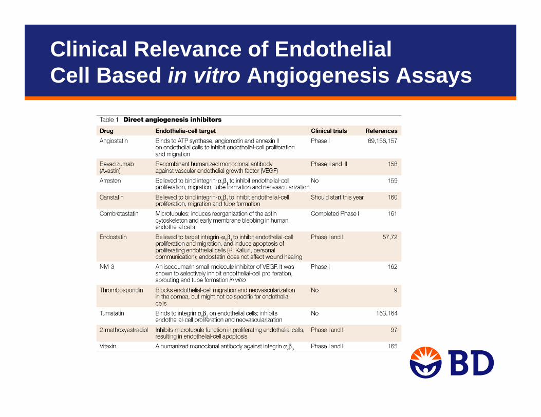
Clinical Relevance of Endothelial Cell Based in vitro Angiogenesis Assays
Important Considerations for Developing Angiogenesis Studies
• Incorporate appropriate extracellular matrix (ECM) protein(s) to facilitate cell functionality and assay outcome
• Choose appropriate endothelial cell source
• Choice of angiogenesis assay
• Establish acceptable dynamic range to measure stimulation and/or inhibition of angiogenesis
Extracellular Matrix
ECM provides a physiological substrate that supports key cellular functions
• Structural organization of cells and tissue
• Cell attachment, survival, and proliferation
• Induction and maintenance of cell differentiation
• Can influence signal transduction and regulation of gene expression
Examples: gelatin, fibronectin, vitronectin, laminin, collagen, BD Matrigel™ matrix
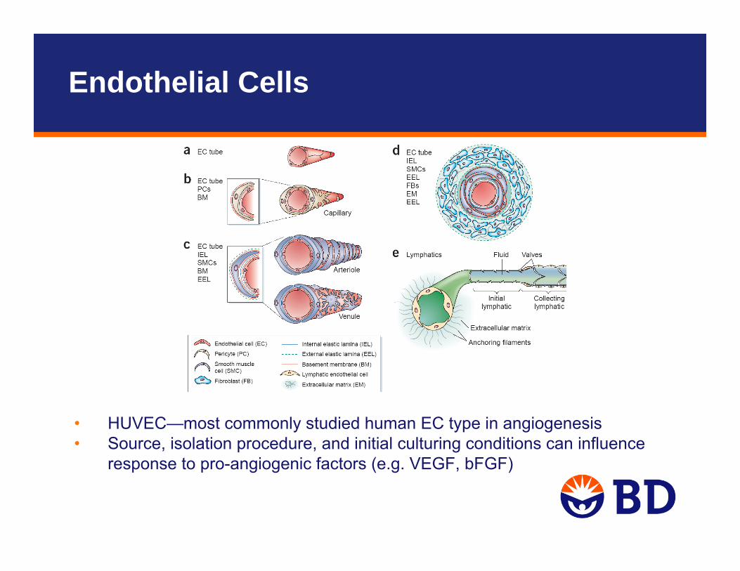
• HUVEC—most commonly studied human EC type in angiogenesis• Source, isolation procedure, and initial culturing conditions can influence
response to pro-angiogenic factors (e.g. VEGF, bFGF)
Endothelial Cells
Sources of Endothelial Cells
• Large vessel– aortic (e.g., HAEC)
– umbilical vein (e.g., HUVEC)
– pulmonary artery
• Microvascular (e.g., HMVEC)– brain
– lung
– dermis (e.g., HDMEC)
– myocardium
Human Umbilical Vein Endothelial Cells
• Most commonly used human EC type for studies of angiogenesis
• Source, isolation procedure, and initial culturing conditions can influence response to pro-angiogenic factors (e.g. VEGF, bFGF)
• BD™ Human Umbilical Vein Endothelial Cells (HUVEC-2)(cat. no. 354151) – Pre-qualified for responsiveness to VEGF in endothelial cell
migration assay – Tested for presence of von Willebrand factor (vWf), CD31,
uptake of Dil-Ac-LDL, and absence of alpha actin
Shipped on dry ice, store at -20ºCDo not store in frost free or -70ºC freezer
BD BioCoat™ Angiogenesis Systems
BD HUVEC-2 cells• Pre-qualified for VEGF responsiveness and for use with endothelial cell
migration assayEndothelial Cell Tube Formation• Composed of a 96-well black/clear plate coated with BD Matrigel matrix
(non-insert system)Endothelial Cell Migration• 24- or 96-Multiwell BD FluoroBlok™ insert (3 μm pore size)• Coated with human fibronectinEndothelial Cell Invasion• 24-Multiwell BD FluoroBlok insert (3 μm pore size)• Coated with BD Matrigel matrix
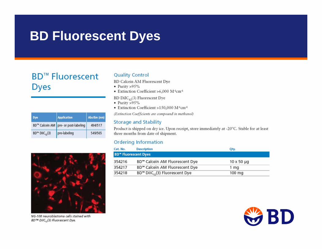
BD Fluorescent Dyes
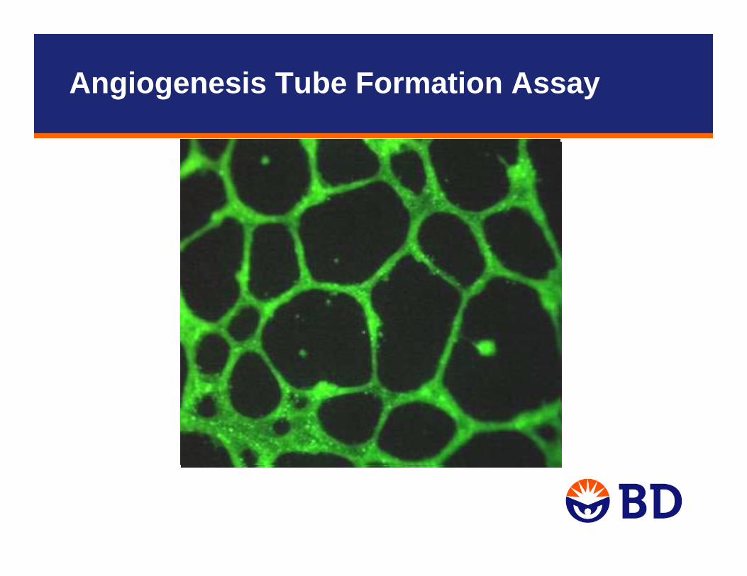
Angiogenesis Tube Formation Assay

1. EC lysis of basement membrane and extracellular matrix
2. EC invasion/migration3. EC proliferation
4. Capillary tube formationand differentiation
Angiogenic StimulatorsVEGF, FGF
Angiogenesis Tube Formation AssaySimplified Steps in Angiogenesis Process
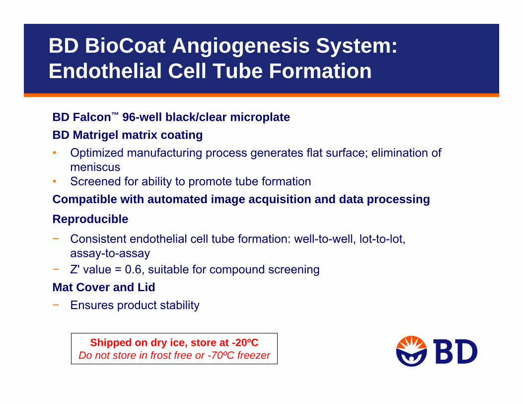
BD BioCoat Angiogenesis System: Endothelial Cell Tube Formation
BD Falcon™ 96-well black/clear microplateBD Matrigel matrix coating• Optimized manufacturing process generates flat surface; elimination of
meniscus • Screened for ability to promote tube formationCompatible with automated image acquisition and data processingReproducible− Consistent endothelial cell tube formation: well-to-well, lot-to-lot,
assay-to-assay− Z' value = 0.6, suitable for compound screeningMat Cover and Lid− Ensures product stability
Shipped on dry ice, store at -20ºCDo not store in frost free or -70ºC freezer
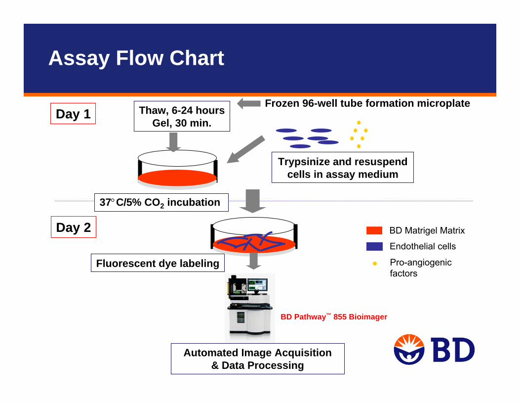
Endothelial cells
Automated Image Acquisition& Data Processing
Day 1
Day 2
37°C/5% CO2 incubation
Fluorescent dye labeling
BD Matrigel Matrix
Frozen 96-well tube formation microplate
Pro-angiogenic factors
Trypsinize and resuspend cells in assay medium
Thaw, 6-24 hoursGel, 30 min.
BD Pathway™ 855 Bioimager
Assay Flow Chart
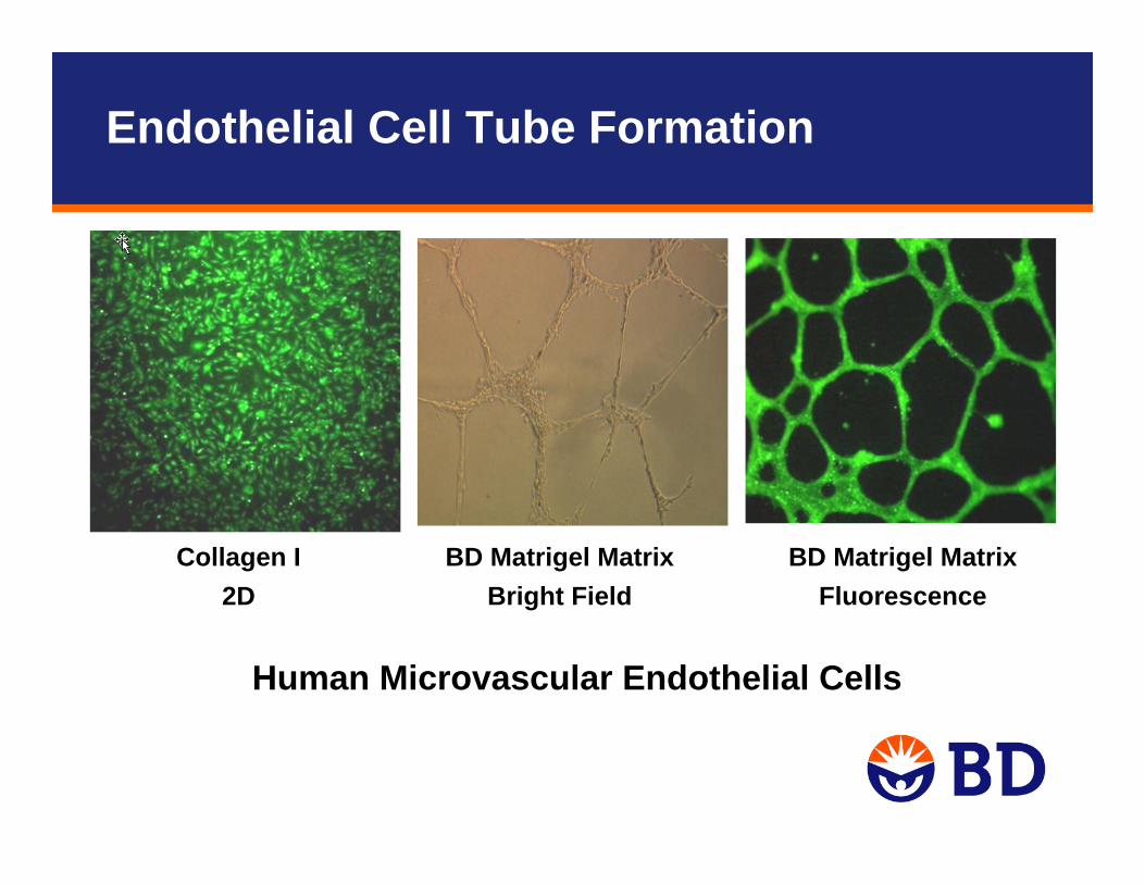
BD Matrigel MatrixBright Field
BD Matrigel MatrixFluorescence
Collagen I2D
Human Microvascular Endothelial Cells
Endothelial Cell Tube Formation
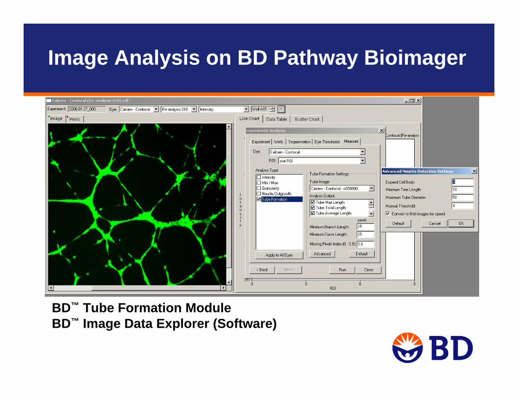
BD™ Tube Formation ModuleBD™ Image Data Explorer (Software)
Image Analysis on BD Pathway Bioimager
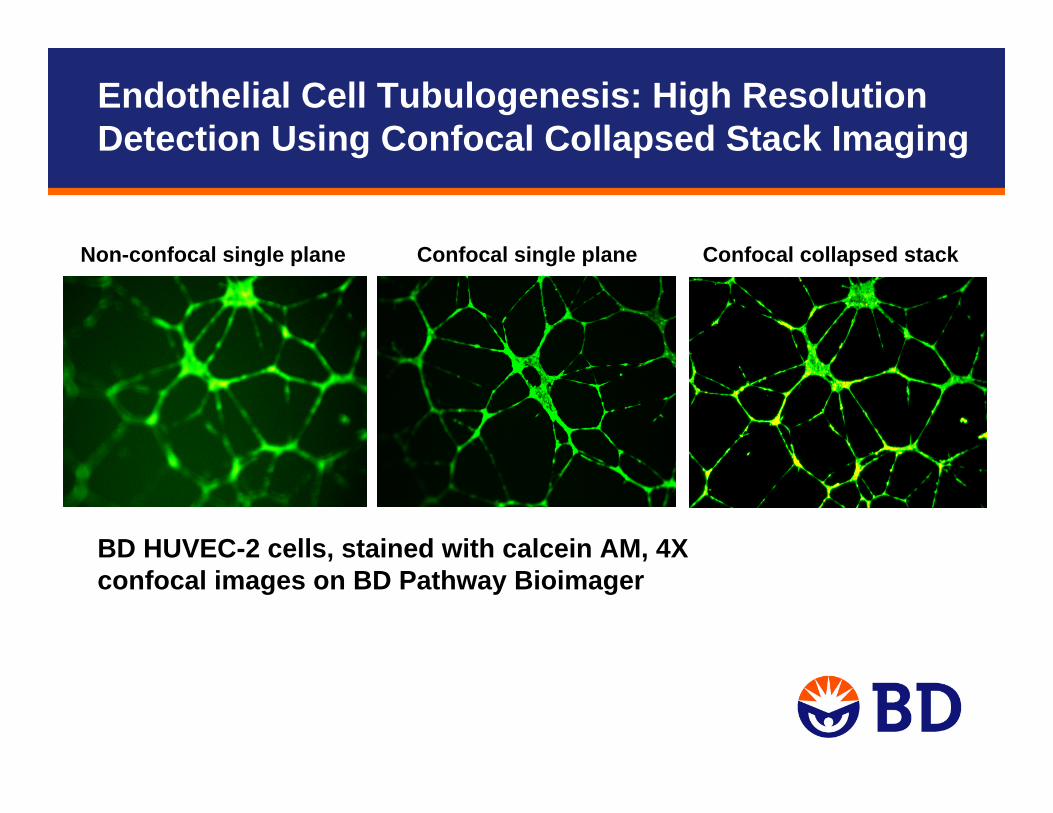
Non-confocal single plane Confocal single plane Confocal collapsed stack
BD HUVEC-2 cells, stained with calcein AM, 4Xconfocal images on BD Pathway Bioimager
Endothelial Cell Tubulogenesis: High Resolution Detection Using Confocal Collapsed Stack Imaging
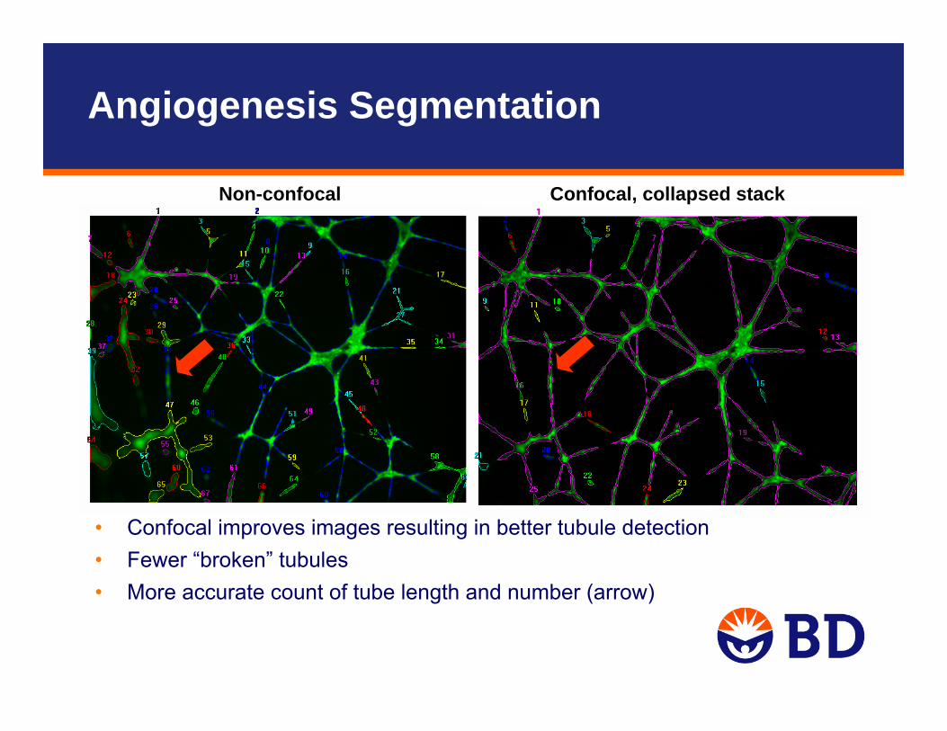
Angiogenesis Segmentation
• Confocal improves images resulting in better tubule detection• Fewer “broken” tubules• More accurate count of tube length and number (arrow)
Non-confocal Confocal, collapsed stack
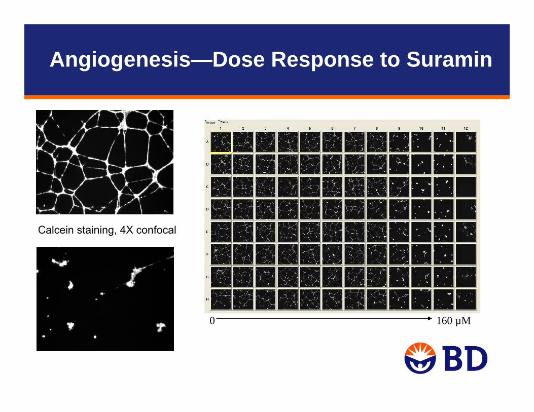
Calcein staining, 4X confocal
0 160 µM
Angiogenesis—Dose Response to Suramin
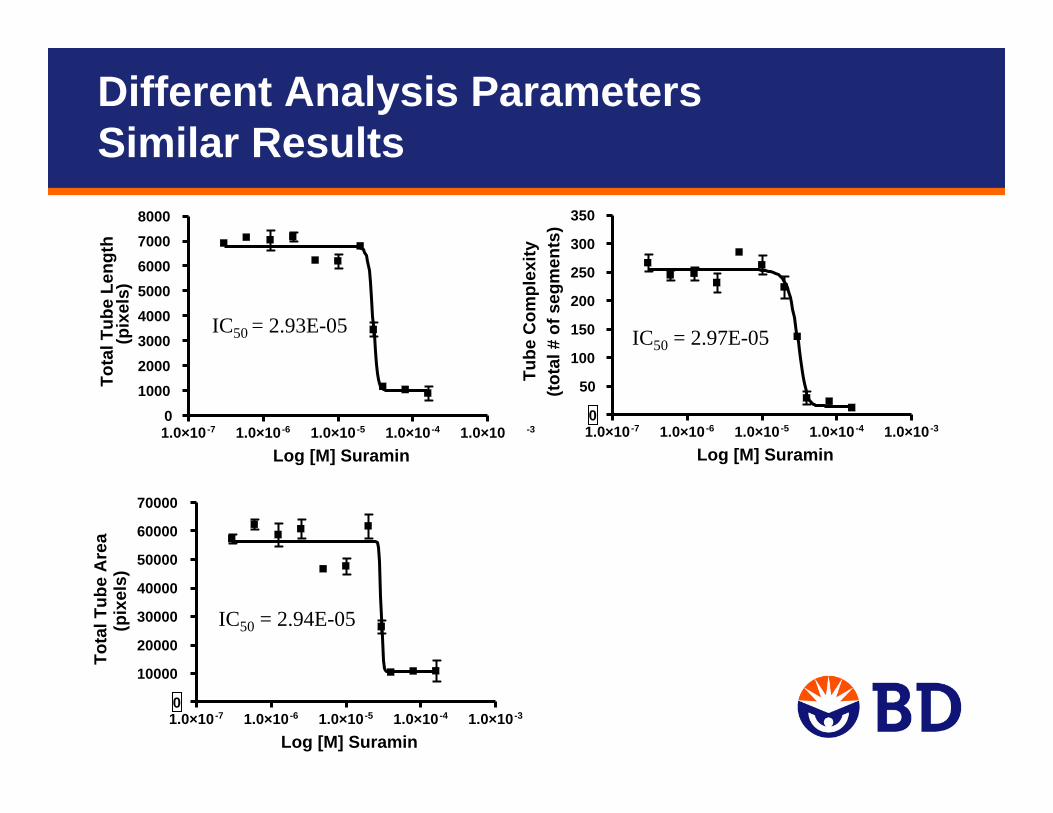
IC50 = 2.93E-05
IC50 = 2.94E-05
IC50 = 2.97E-05
1.0×10⁻⁷ 1.0×10⁻⁶ 1.0×10⁻⁵ 1.0×10⁻⁴ 1.0×10 -30
10002000300040005000600070008000
Log [M] Suramin
Tota
l Tub
e Le
ngth
(pix
els)
1.0×10⁻⁷ 1.0×10⁻⁶ 1.0×10⁻⁵ 1.0×10⁻⁴ 1.0×10⁻³⁰
50
100
150
200
250
300
350
Log [M] Suramin
Tube
Com
plex
ity(to
tal #
of s
egm
ents
)
1.0×10⁻⁷ 1.0×10⁻⁶ 1.0×10⁻⁵ 1.0×10⁻⁴ 1.0×10⁻³⁰
10000
20000
30000
40000
50000
60000
70000
Log [M] Suramin
Tota
l Tub
e A
rea
(pix
els)
Different Analysis ParametersSimilar Results
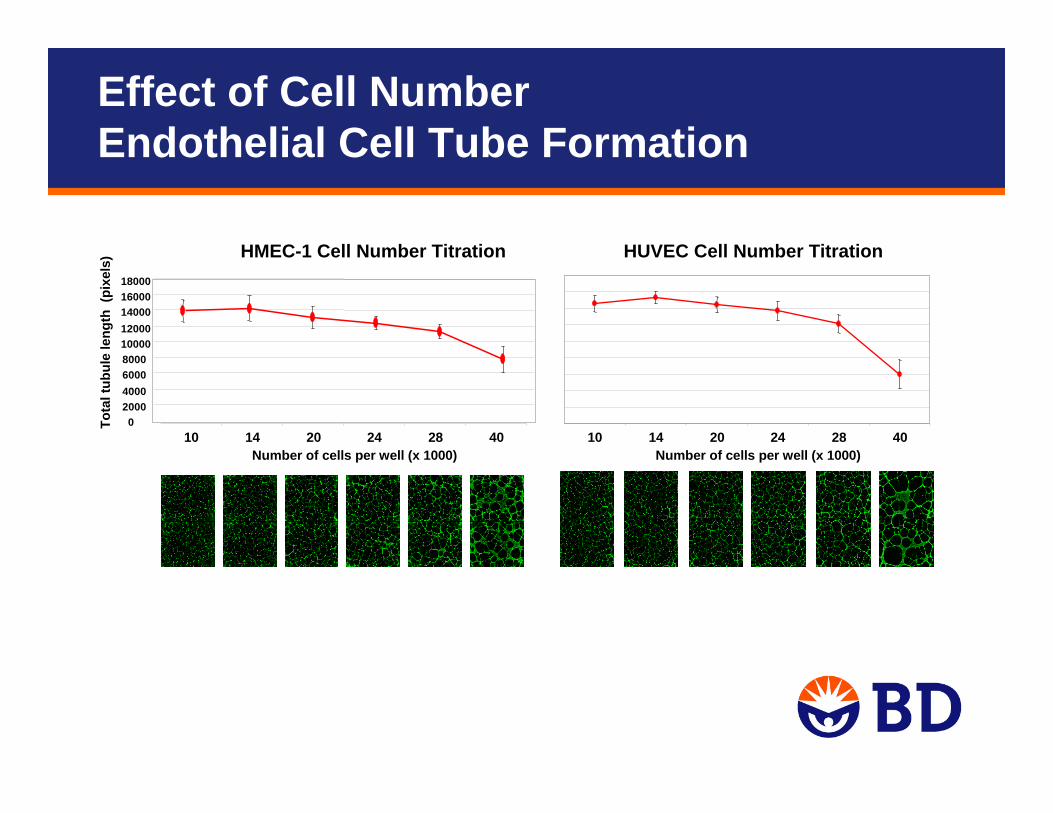
HUVEC Cell Number Titration
10 14 20 24 28 40Number of cells per well (x 1000)
HMEC-1 Cell Number Titration
020004000600080001000012000140001600018000
Tota
l tub
ule
leng
th (
pixe
ls)
10 14 20 24 28 40Number of cells per well (x 1000)
Effect of Cell Number Endothelial Cell Tube Formation
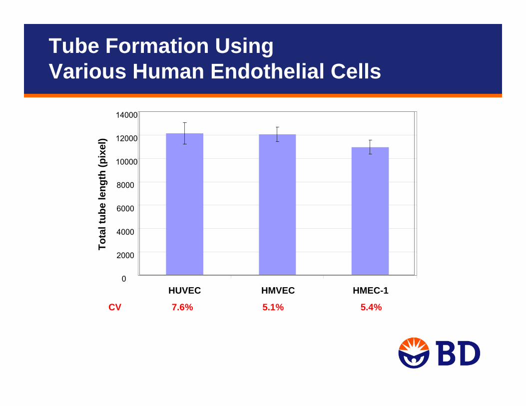
CV 7.6% 5.1% 5.4%
0
2000
4000
6000
8000
10000
12000
14000
HUVEC HMVEC HMEC-1
Tota
l tub
e le
ngth
(pix
el)
Tube Formation UsingVarious Human Endothelial Cells
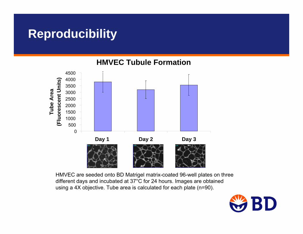
HMVEC are seeded onto BD Matrigel matrix-coated 96-well plates on three different days and incubated at 37°C for 24 hours. Images are obtained using a 4X objective. Tube area is calculated for each plate (n=90).
HMVEC Tubule Formation
0500
10001500200025003000350040004500
Day 1 Day 2 Day 3
Tube
Are
a (F
luor
esce
nt U
nits
)
Reproducibility
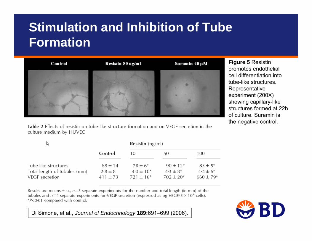
Figure 5 Resistin promotes endothelial cell differentiation into tube-like structures. Representative experiment (200X) showing capillary-like structures formed at 22h of culture. Suramin is the negative control.
Di Simone, et al., Journal of Endocrinology 189:691–699 (2006).
Stimulation and Inhibition of Tube Formation
Endothelial Cell Tube Formation
• Assay setup to data within 24 hours• Validated Protocols• High-throughput: 96-well format• Automated image acquisition and data processing
– Labeling with fluorescent dye– Morphometric analysis– Can use fluorescent microscope
• Reproducible– Consistent endothelial cell tube formation:
• Well-to-well, lot-to-lot, assay-to-assay– Z' value = 0.6, good for screening
BD BioCoat Angiogenesis Systems
Shipped on dry ice, store at -20ºCDo not store in frost free or -70ºC freezer
BD HUVEC-2 cells• Pre-qualified for VEGF responsiveness and for use with endothelial cell
migration assayEndothelial Cell Tube Formation• Composed of a 96-well black/clear plate coated with BD Matrigel matrix
(non-insert system)Endothelial Cell Migration• 24- or 96-Multiwell BD FluoroBlok insert (3 μm pore size)• Coated with human fibronectinEndothelial Cell Invasion• 24-Multiwell BD FluoroBlok insert (3 μm pore size)• Coated with BD Matrigel matrix
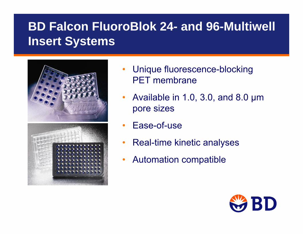
BD Falcon FluoroBlok 24- and 96-Multiwell Insert Systems
• Unique fluorescence-blocking PET membrane
• Available in 1.0, 3.0, and 8.0 µm pore sizes
• Ease-of-use
• Real-time kinetic analyses
• Automation compatible
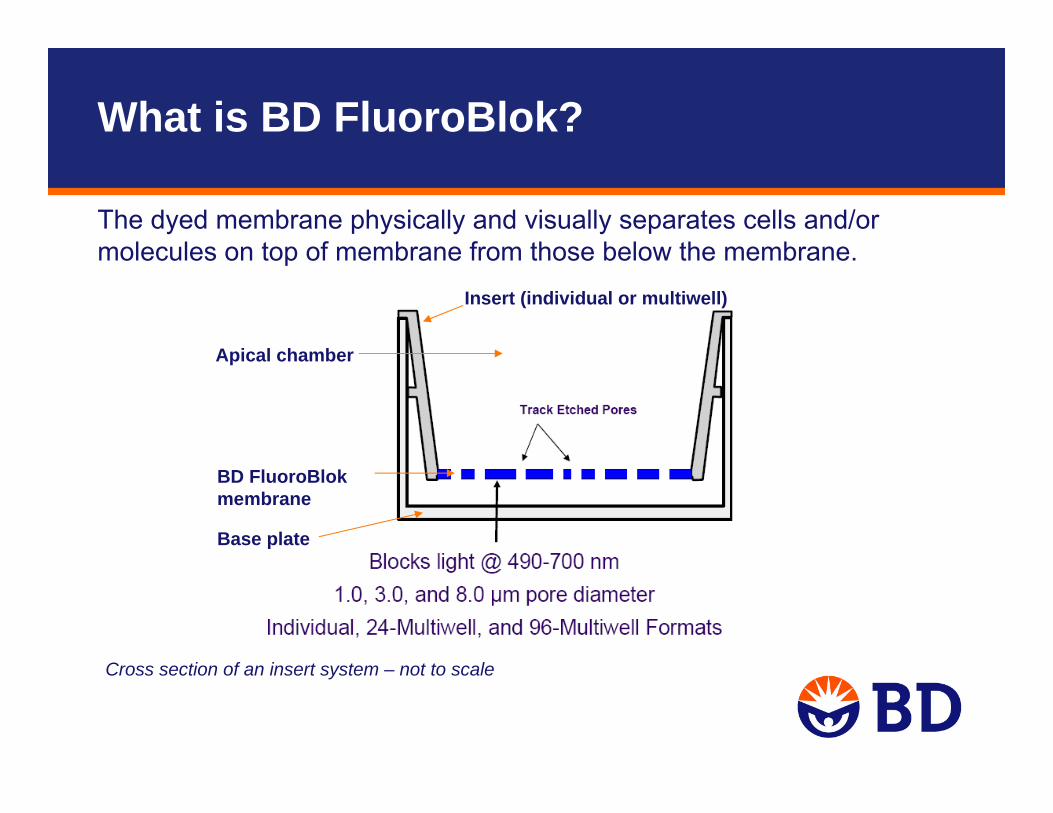
The dyed membrane physically and visually separates cells and/ormolecules on top of membrane from those below the membrane.
What is BD FluoroBlok?
Cross section of an insert system – not to scale
Base plate
BD FluoroBlok membrane
Insert (individual or multiwell)
Apical chamber
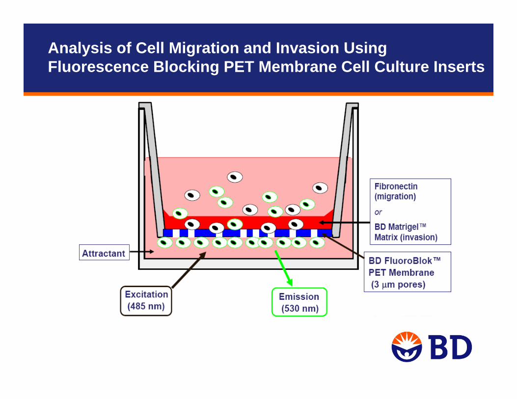
Analysis of Cell Migration and Invasion Using Fluorescence Blocking PET Membrane Cell Culture Inserts
Detection Instrument
• A fluorescent plate reader with bottom-reading capability, and an inverted fluorescent microscope for confirmation and troubleshooting
• A fluorescence imager
• Set Up Guidelines and Dimensional Templates for Fluorescence Plate Readers Used With BD Falcon HTS FluoroBlok Insert Systems and BD BioCoat Multiwell Insert Cell-Based Assays, Technical Bulletin # 436
• http://www.bdbiosciences.com/discovery_labware/technical_resources/pdf/tb436_fluoroblok.pdf
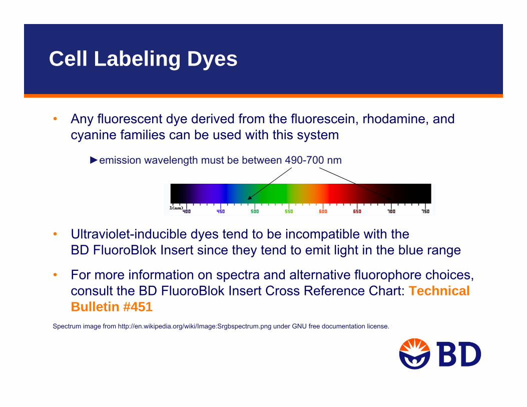
• Any fluorescent dye derived from the fluorescein, rhodamine, and cyanine families can be used with this system
• Ultraviolet-inducible dyes tend to be incompatible with the BD FluoroBlok Insert since they tend to emit light in the blue range
• For more information on spectra and alternative fluorophore choices, consult the BD FluoroBlok Insert Cross Reference Chart: Technical Bulletin #451
Cell Labeling Dyes
►emission wavelength must be between 490-700 nm
Spectrum image from http://en.wikipedia.org/wiki/Image:Srgbspectrum.png under GNU free documentation license.
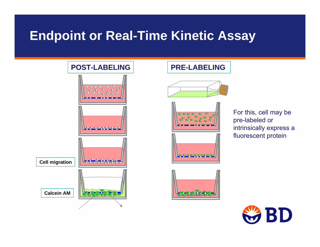
Endpoint or Real-Time Kinetic Assay
POST-LABELING PRE-LABELING
Calcein AM
Cell migration
For this, cell may bepre-labeled or intrinsically express a fluorescent protein
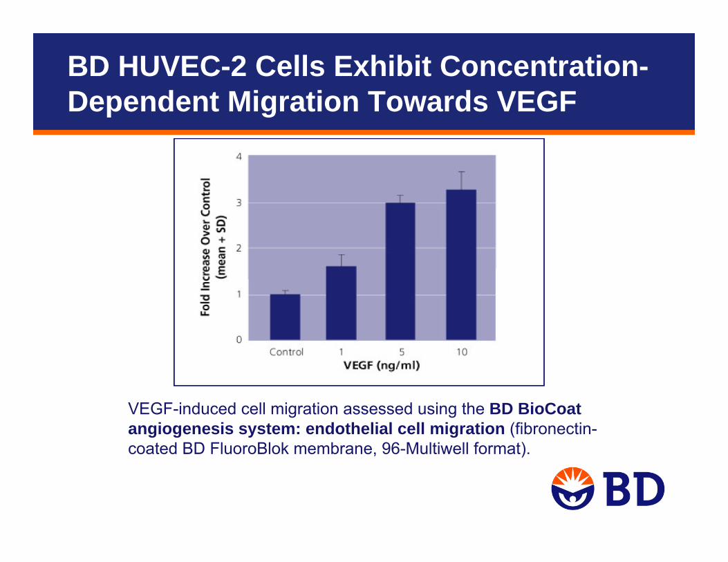
BD HUVEC-2 Cells Exhibit Concentration-Dependent Migration Towards VEGF
VEGF-induced cell migration assessed using the BD BioCoat angiogenesis system: endothelial cell migration (fibronectin-coated BD FluoroBlok membrane, 96-Multiwell format).
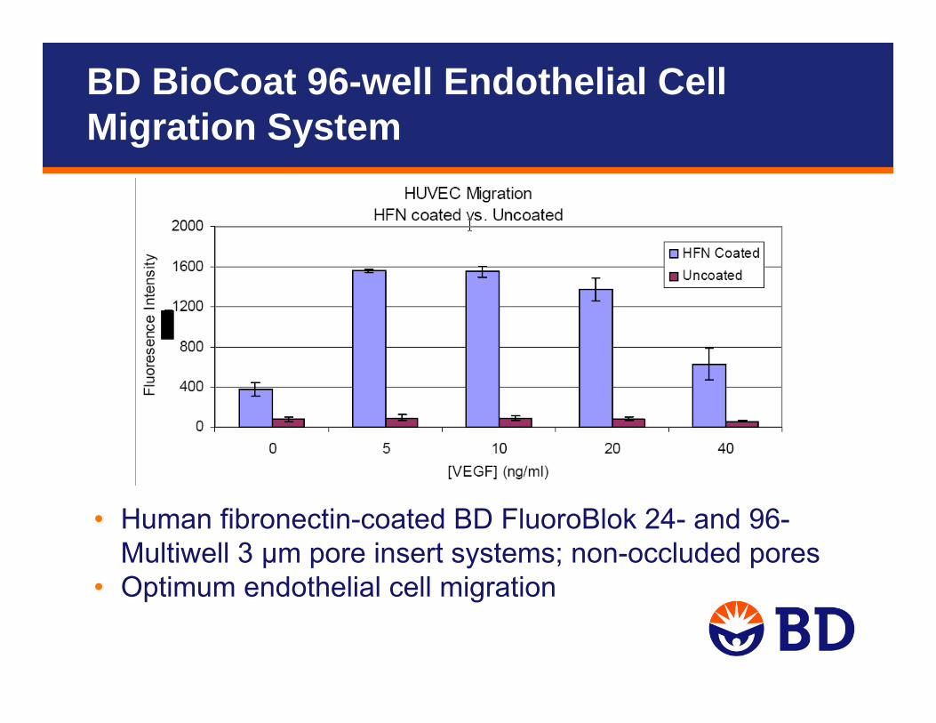
• Human fibronectin-coated BD FluoroBlok 24- and 96-Multiwell 3 μm pore insert systems; non-occluded pores
• Optimum endothelial cell migration
BD BioCoat 96-well Endothelial CellMigration System
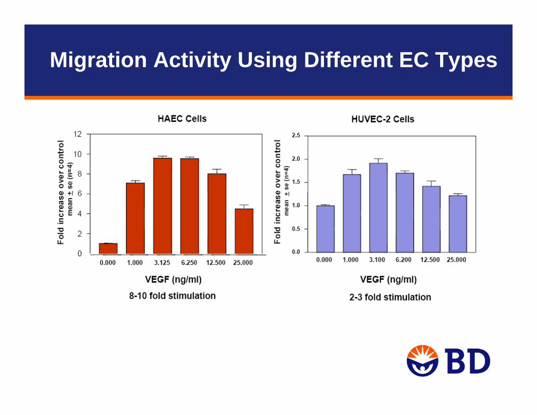
Migration Activity Using Different EC Types
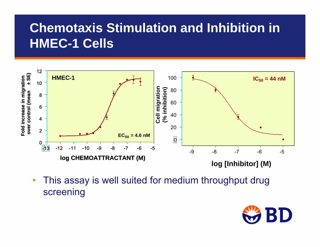
• This assay is well suited for medium throughput drug screening
log [Inhibitor] (M)
-13 -12 -11 -10 -9 -8 -7 -6 -50
2
4
6
8
10
12HMEC-1
EC50 = 4.6 nM
log CHEMOATTRACTANT (M)
Fold
incr
ease
in m
igra
tion
over
con
trol
(mea
n+
SE)
-13 -12 -11 -10 -9 -8 -7 -6 -50
2
4
6
8
10
12HMEC-1
EC50 = 4.6 nM
log CHEMOATTRACTANT (M)
Fold
incr
ease
in m
igra
tion
over
con
trol
(mea
n+
SE)
-9 -8 -7 -6 -5
0
20
40
60
80
100 IC50 = 44 nM
Cel
l mig
ratio
n(%
inhi
bitio
n)
Chemotaxis Stimulation and Inhibition in HMEC-1 Cells
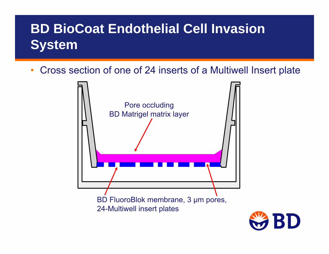
Pore occludingBD Matrigel matrix layer
BD FluoroBlok membrane, 3 µm pores, 24-Multiwell insert plates
• Cross section of one of 24 inserts of a Multiwell Insert plate
BD BioCoat Endothelial Cell Invasion System
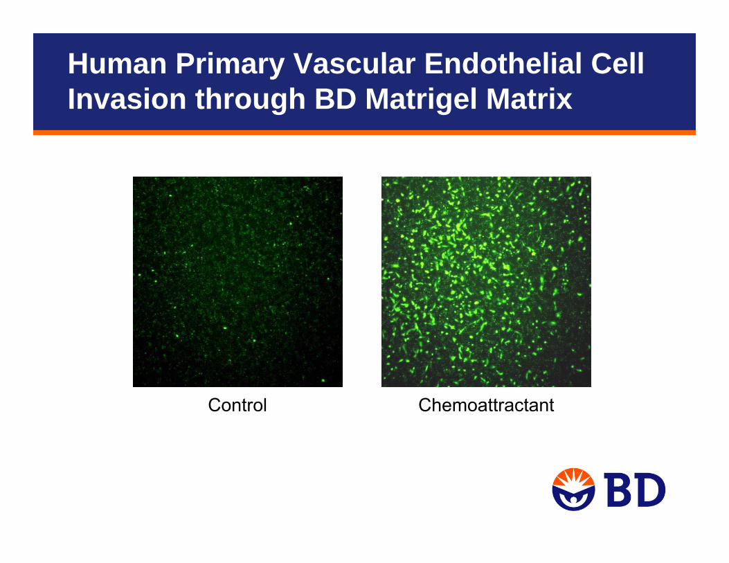
Control Chemoattractant
Human Primary Vascular Endothelial Cell Invasion through BD Matrigel Matrix
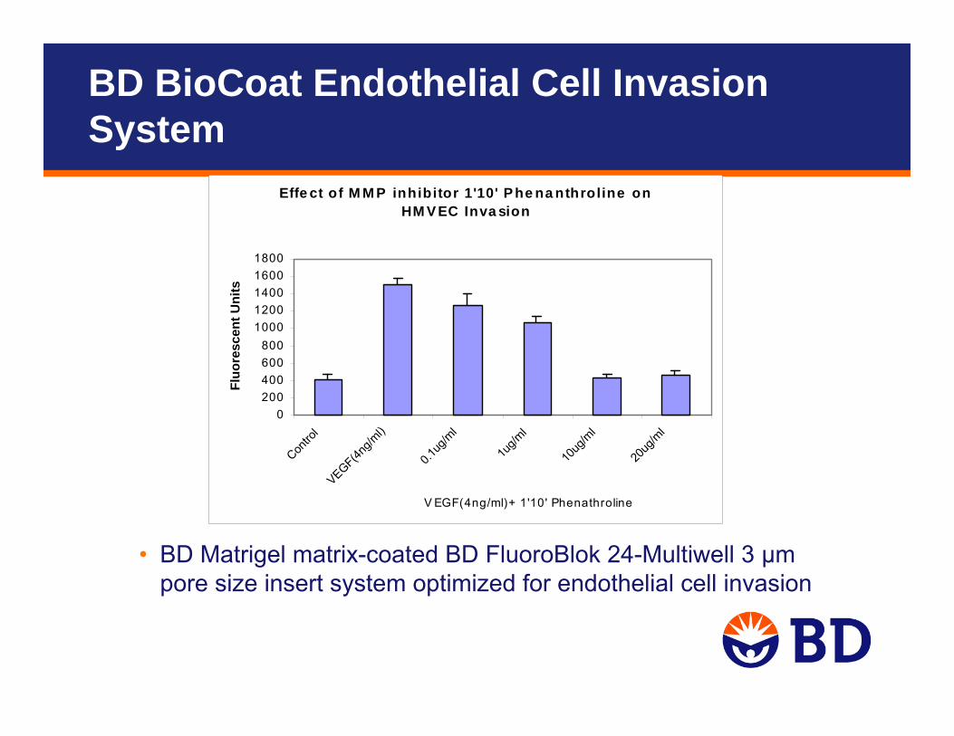
Effe ct of M M P inhibitor 1'10' P he na nthroline on HM V EC Inva sion
0200400600800
10001200140016001800
Contro
lVEGF(4n
g/ml)
0.1ug
/ml
1ug/m
l
10ug
/ml
20ug
/ml
V EGF(4ng/ml)+ 1'10' Phenathroline
Fluo
resc
ent U
nits
• BD Matrigel matrix-coated BD FluoroBlok 24-Multiwell 3 μm pore size insert system optimized for endothelial cell invasion
BD BioCoat Endothelial Cell Invasion System
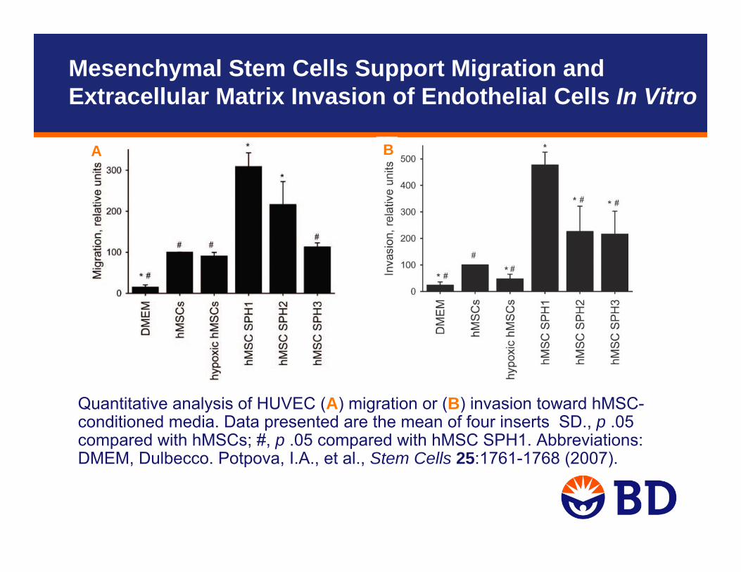
A B
Quantitative analysis of HUVEC (A) migration or (B) invasion toward hMSC-conditioned media. Data presented are the mean of four inserts SD., p .05 compared with hMSCs; #, p .05 compared with hMSC SPH1. Abbreviations: DMEM, Dulbecco. Potpova, I.A., et al., Stem Cells 25:1761-1768 (2007).
Mesenchymal Stem Cells Support Migration and Extracellular Matrix Invasion of Endothelial Cells In Vitro
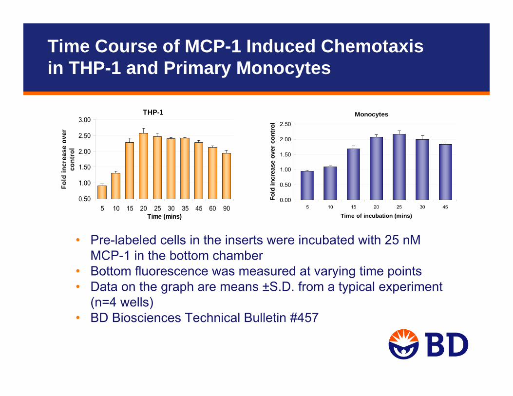
Time Course of MCP-1 Induced Chemotaxisin THP-1 and Primary Monocytes
THP-1
0.50
1.00
1.50
2.00
2.50
3.00
5 10 15 20 25 30 35 45 60 90Time (mins)
Fold
incr
ease
ove
r co
ntro
l
Monocytes
0.00
0.50
1.00
1.50
2.00
2.50
5 10 15 20 25 30 45
Time of incubation (mins)
Fold
incr
ease
ove
r co
ntro
l
• Pre-labeled cells in the inserts were incubated with 25 nMMCP-1 in the bottom chamber
• Bottom fluorescence was measured at varying time points• Data on the graph are means ±S.D. from a typical experiment
(n=4 wells)• BD Biosciences Technical Bulletin #457
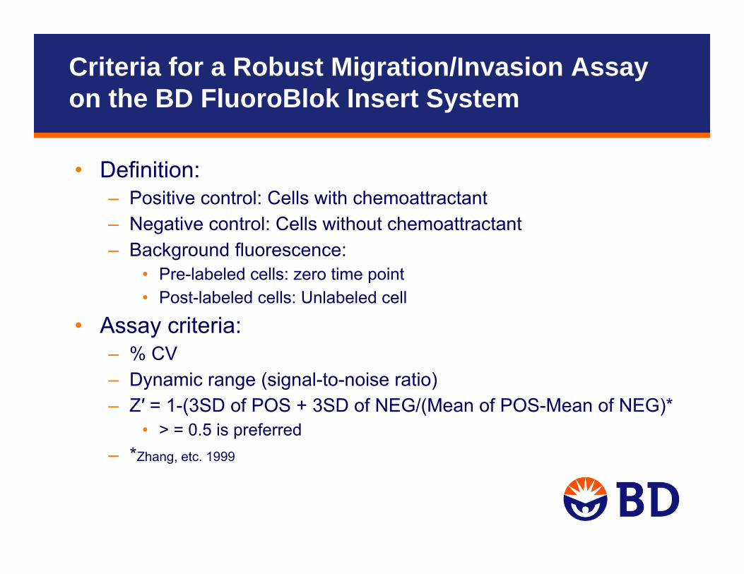
Criteria for a Robust Migration/Invasion Assay on the BD FluoroBlok Insert System
• Definition:– Positive control: Cells with chemoattractant– Negative control: Cells without chemoattractant– Background fluorescence:
• Pre-labeled cells: zero time point • Post-labeled cells: Unlabeled cell
• Assay criteria: – % CV– Dynamic range (signal-to-noise ratio)– Z′ = 1-(3SD of POS + 3SD of NEG/(Mean of POS-Mean of NEG)*
• > = 0.5 is preferred– *Zhang, etc. 1999

References
Angiogenesis1. Potapova, I.A., et al. Mesenchymal stem cells support migration,
extracellular matrix invasion, proliferation, and survival of endothelial cells in vitro. Stem Cells 25:1761-1768 (2007).
2. Di Simone, et al. Resistin regulates human choriocarcinoma cell invasive behaviour and endothelial cell angiogenic processes. Journal of Endocrinology 189:691–699 (2006).
3. Favier, B., et al. Neurophilin-2 interacts with VEGFR-2 and VEGFR-3 and promotes human endothelial cell survival and migration. Blood 108:1243-1250 (2006).
4. Davis, G.E. and Senger, D.R. Endothelial extracellular matrix: biosynthesis, remodeling, and functions during vascular morphogenesis and neovessel stabilization. Circulation Res. 97:1093-1107 (2005).

Questions?
Contact information:Suparna Sanyale-mail: [email protected]
Technical Support:tel: 877.232.8995e-mail: [email protected]/webinarsFor research use only. Not intended for use in diagnostic or therapeutic procedures. BD, BD Logo, and all other trademarks are property of Becton, Dickinson and Company. ©2010 BD
Recommended