Vol. 49, No. 2APPLIED AND ENVIRONMENTAL MICROBIOLOGY, Feb. 1985, p. 4464500099-2240/85/020446-05$02.00/0Copyright © 1985, American Society for Microbiology
Improved Fixation of Cellulose-Acetate Reverse-Osmosis Membranefor Scanning Electron MicroscopyS. M. KUTZ,' D. L. BENTLEY,2 AND N. A. SINCLAIR'*
Department of Microbiology and Immunologyl and Electron Microscope Facility, College ofAgriculture,2 University ofArizona, Tucson, Arizona 85721
Received 6 August 1984/Accepted 15 November 1984
Fixation of cellulose-acetate membranes with either glutaraldehyde-osmium tetroxide or glutaraldehyde-ru-thenium tetroxide resulted in extensive electron beam damage. Beam damage was eliminated and the bacterialsurface structure was preserved, however, when cellulose-acetate membranes were fixed with glutaraldehyde-ruthenium tetroxide and treated successively with thiocarbohydrazide and osmium tetroxide.
Scanning electron microscopy (SEM) is a valuable tool forevaluating biofilm formation and biodegradation of cellulose-acetate (CA) reverse-osmosis membranes used in water andadvanced wastewater treatment. SEM has been used inprevious studies to determine biofilm thickness and compo-sition as well as to reveal chlorine-induced lesions in CAmembranes (8, 9). In addition, SEM allows visualization ofthe microenvironment within reverse-osmosis units and pro-vides information on the characteristics of bacteria present.Glycocalyx networks and bacterial stalks or appendages areindirect evidence of bacterial colonization and adherence.SEM sample preparation often involves the use of glutar-
aldehyde or glutaraldehyde followed by osmium tetroxidefixation (2). The material is then dehydrated in an ethanolseries, critical point dried, and coated with gold or a gold-palladium alloy. CA membranes processed in this way arereadily damaged by an electron beam. Destruction of the CAoccurs even at low accelerating voltages (5 to 10 kV) and lowmagnifications (<5,000x) (8). Optimum focusing and correc-tion for astigmatism are severely limited. Details of bacterialfine structure and the overall physical condition of the CAmembrane surface cannot be discerned at high magnifica-tions (>5,000x) because of electron beam lability. Theapplication of additional coatings of a gold-palladium alloy toCA membranes does not alleviate beam damage.The purpose of this research was to develop a method for
the fixation and preservation of CA membrane integrity forSEM analysis. Membranes fixed with glutaraldehyde andtreated successively with ruthenium tetroxide, thiocarbo-hydrazide, osmium tetroxide, thiocarbohydrazide, and osm-ium tetroxide (RTOTO) were scanned at high and lowmagnifications at accelerating potentials of 10 to 30 kV. Forcomparative purposes, CA membranes were fixed withglutaraldehyde and treated with either ruthenium tetroxideor osmium tetroxide. The electron beam damage to CAmembranes and attached microorganisms was assessed.Three strips (2 by 5 cm) of a low-pressure CA membrane
(degree of substitution, 2.67) were used as the sole carbonsource for naturally occurring well water bacteria inoculatedinto flasks of 0.1% Bacto-Peptone (Difco Laboratories,Detroit, Mich.). Cultures were incubated for 2 weeks at25°C. The CA strips were then fixed overnight in 4%glutaraldehyde (Electron Microscopy Sciences, Ft. Wash-
* Corresponding author.
ington, Pa.) in 0.1 M Millonig buffer (5) at pH 7.2. Sampleswere rinsed three times for 5 minutes each time in Millonigbuffer, followed by three five-minute washes in high-pres-sure-liquid-chromatography (HPLC)-grade water. The firstCA strip was postfixed for 30 min in 2% osmium tetroxide(EMS, Ft. Washington, Pa.) and then rinsed nine times inHPLC-grade water. The second CA strip was postfixed for30 min in 1% ruthenium tetroxide (Polysciences, St. Louis,Mo.) and rinsed nine times in HPLC-grade water. The thirdCA strip was treated like the second but in addition wasincubated for 30 min in a saturated thiocarbohydrazidesolution (Sigma Chemical Co., St. Louis, Mo.). Immediatelybefore use, thiocarbohydrazide was suspended in water andallowed to stand for 1 h at 50°C. The liquid was decanted,and the procedure was repeated until the resulting saturatedsolution was a clear straw color. The solution was allowed tocool to room temperature before use (6). After being treatedwith thiocarbohydrazide, the CA strip was washed ninetimes in HPLC-grade water, incubated in 2% osmium te-troxide for 30 min, and then washed again. Treatments in thethiocarbohydrazide and osmium tetroxide solutions wererepeated once (4). All three samples were dehydrated throughan ethanol series (30 to 100%) and critical point dried withCO2 (1). The samples were sputter coated with 30 nm ofgold-palladium alloy (target composition, 60% Au-40% Pd)with a magnetron sputtering device. Samples were observedwith an International Scientific Instruments DS-130 scanningelectron microscope.
Caution must be used when handling osmium tetroxideand ruthenium tetroxide. Only HPLC-grade water should beused in the preparation of aqueous solutions, and glasswaremust be free from organic compounds to avoid rapid reduc-tion of ruthenium tetroxide.CA membranes conventionally fixed in glutaraldehyde-
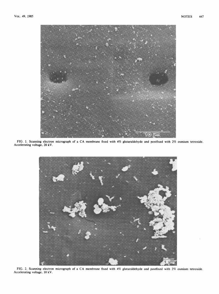
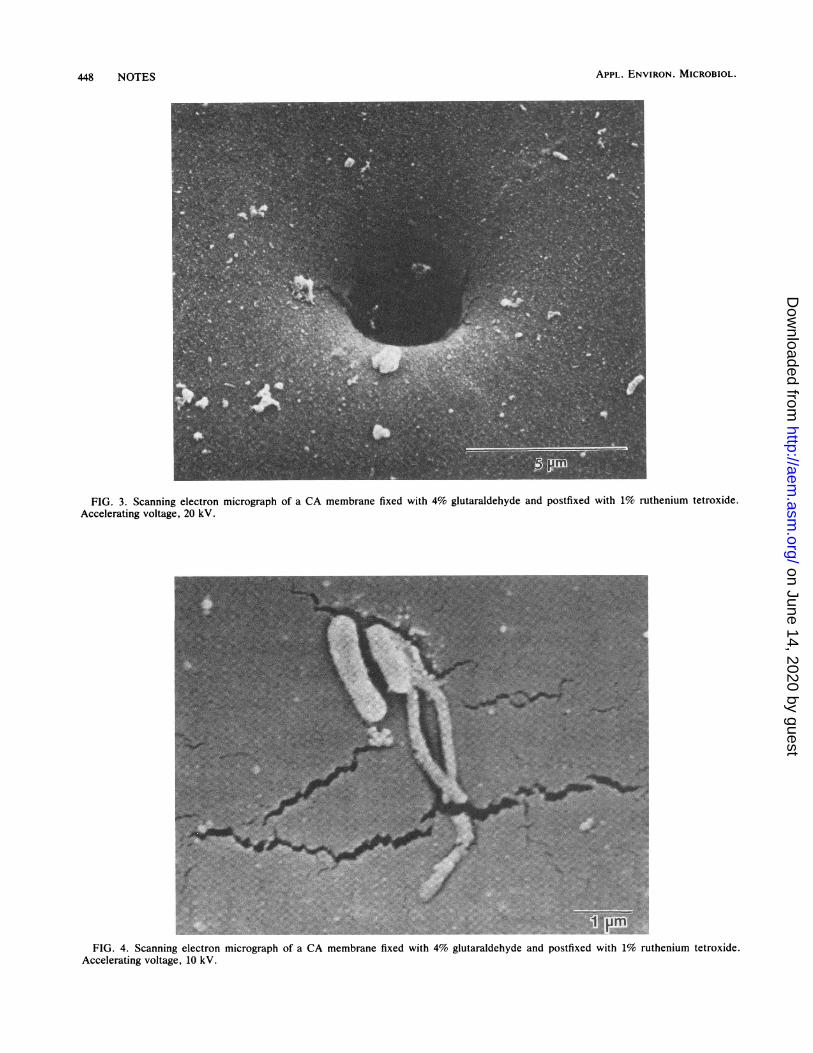
osmium tetroxide (Fig. 1) were labile, as evidenced by rapidelectron beam damage and tearing of the membranes at 20kV. Beam damage to samples occurred in less than 1 min atmagnifications greater than 2,000x and limited the amountof detail visible. Use of a lower accelerating voltage (10 kV)improved the situation somewhat; however, moderate beamdamage still occurred in less than 1 min (Fig. 2). Althoughpostfixation with ruthenium tetroxide increased sample sta-bility, beam damage was nevertheless encountered (Fig. 3and 4). Photographs at magnifications near 5,000x could beattained only by rapid focusing on an adjacent area. Recoat-
446
on June 14, 2020 by guesthttp://aem
.asm.org/
Dow
nloaded from
NOTES 447
FIG. 1. Scanning electron micrograph of a CA membrane fixed with 4% glutaraldehyde and postfixed with 2% osmium tetroxide.Accelerating voltage, 20 kV.
FIG. 2. Scanning electron micrograph of a CA membrane fixed with 4% glutaraldehyde and postfixed with 2% osmium tetroxide.Accelerating voltage, 10 kV.
VOL. 49, 1985
on June 14, 2020 by guesthttp://aem
.asm.org/
Dow
nloaded from
APPL. ENVIRON. MICROBIOL.
FIG. 3. Scanning electron micrograph of a CA membrane fixed with 4% glutaraldehyde and postfixed with 1% ruthenium tetroxide.Accelerating voltage, 20 kV.
FIG. 4. Scanning electron micrograph of a CA membrane fixed with 4% glutaraldehyde and postfixed with 1% ruthenium tetroxide.Accelerating voltage, 10 kV.
448 NOTES
on June 14, 2020 by guesthttp://aem
.asm.org/
Dow
nloaded from
NOTES 449
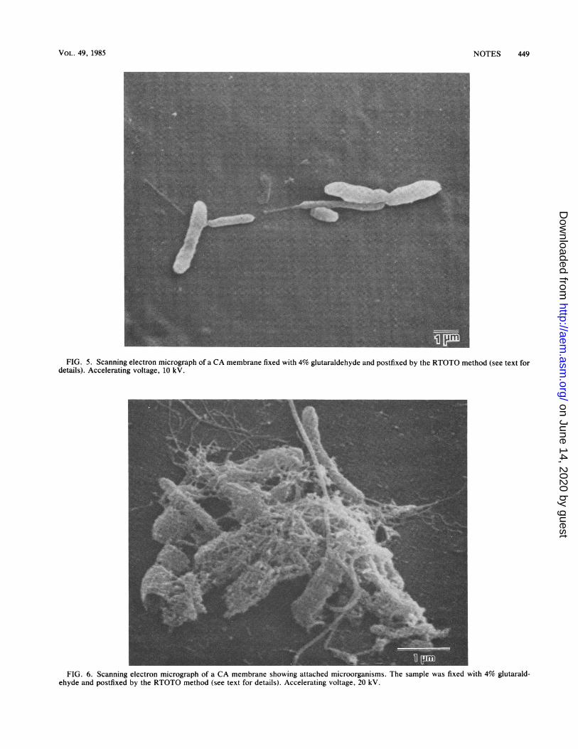
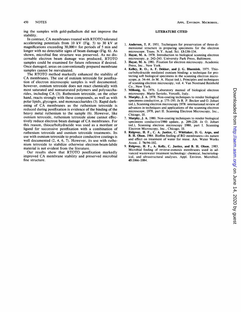
FIG. 5. Scanning electron micrograph of a CA membrane fixed with 4% glutaraldehyde and postfixed by the RTOTO method (see text fordetails). Accelerating voltage, 10 kV.
FIG. 6. Scanning electron micrograph of a CA membrane showing attached microorganisms. The sample was fixed with 4% glutarald-ehyde and postfixed by the RTOTO method (see text for details). Accelerating voltage, 20 kV.
VOL. 49, 1985
on June 14, 2020 by guesthttp://aem
.asm.org/
Dow
nloaded from
APPL. ENVIRON. MICROBIOL.
ing the samples with gold-palladium did not improve thestability.
In contrast, CA membranes treated with RTOTO toleratedaccelerating potentials from 10 kV (Fig. 5) to 30 kV atmagnifications exceeding 30,OOOx for periods of 5 min andlonger with no detectable signs of beam damage (Fig. 6). Asshown, microbial fine structure was preserved. As no dis-cernable electron beam damage was produced, RTOTOsamples could be examined for future reference if desired.Once damaged, areas on conventionally prepared membranesamples cannot be reevaluated.The RTOTO method markedly enhanced the stability of
CA membranes. The use of osmium tetroxide for postfixa-tion of electron microscopic samples is well documented;however, osmium tetroxide does not react chemically withmost saturated and nonsaturated polymers and polysaccha-rides, including CA (3). Ruthenium tetroxide, on the otherhand, reacts strongly with these compounds, as well as withpolar lipids, glycogen, and monosaccharides (3). Rapid dark-ening of CA membranes as the ruthenium tetroxide isreduced during postfixation is evidence of the binding of theheavy metal ruthenium to the sample (6). However, likeosmium tetroxide, ruthenium tetroxide alone cannot effec-tively reduce electron beam damage of CA membranes. Forthis reason, thiocarbohydrazide was used as a mordant orligand for successive postfixation with a combination ofruthenium tetroxide and osmium tetroxide treatments. Itsuse with osmium tetroxide to produce conductive coatings iswell documented (2, 4, 6, 7). However, its use with ruthe-nium tetroxide to stabilize otherwise electron-beam-labilematerial is not evident from the literature.Our results show that RTOTO postfixation markedly
improved CA membrane stability and preserved microbialfine structure.
LITERATURE CITED
1. Anderson, T. F. 1951. Techniques for preservation of three-di-mensional structure in preparing specimens for the electronmicroscope. Trans. N.Y. Acad. Sci. 13:130-134.
2. Hayat, M. A. 1978. Introduction to biological scanning electronmicroscopy, p. 242-243. University Park Press, Baltimore.
3. Hayat, M. A. 1981. Fixation for electron microscopy. AcademicPress, Inc., New York.
4. Kelley, R. O., A. F. Dekker, and J. G. Bluemink. 1975. Thio-carbohydrazide mediated osmium binding: a technique for pro-tecting soft biological specimens in the scanning electron micro-scope, p. 34-44. In M. A. Hayat (ed.), Principles and techniquesof scanning electron microscopy, vol. 4. Van Nostrand ReinholdCo., New York.
5. Millonig, G. 1976. Laboratory manual of biological electronmicroscopy. Mario Saviolo, Vercelli, Italy.
6. Murphy, J. A. 1978. Non-coating techniques to render biologicalspecimens conductive, p. 175-193. In R. P. Becker and 0. Johari(ed.), Scanning electron microscopy 1978: international review ofadvances in techniques and applications of the scanning electronmicroscope, 1978, part II. Scanning Electron Microscopy, Inc.,Chicago, Ill.
7. Murphy, J. A. 1980. Non-coating techniques to render biologicalspecimens conductive/1980 update, p. 209-220. In 0. Johari(ed.), Scanning electron microscopy 1980, part I. ScanningElectron Microscopy, Inc., Chicago, Ill.
8. Ridgway, H. F., C. A. Justice, C. Whittaker, D. G. Argo, andB. H. Olson. 1984. Biofilm fouling of RO membranes-its natureand effect on treatment of water for reuse. Am. Water WorksAssoc. J. 76:94-102.
9. Ridgway, H. F., A. Kelly, C. Justice, and B. H. Olson. 1983.Microbial fouling of reverse-osmosis membranes used in ad-vanced wastewater treatment technology: chemical, bacteriolog-ical, and ultrastructural analyses. Appl. Environ. Microbiol.45:1066-1084.
450 NOTES
on June 14, 2020 by guesthttp://aem
.asm.org/
Dow
nloaded from
Recommended