IgG4-related sclerosing disease
TERUMI KAMISAWA, KENSUKE TAKUMA, NAOTO EGAWA
Department of Internal Medicine
Tokyo Metropolitan Komagome Hospital
3-18-22 Honkomagome, Bunkyo-ku, Tokyo 113-8677, Japan
JAPAN
e-mail: [email protected]
Abstract: Based on histological and immunohistochemical examination of various organs of
autoimmune pancreatitis (AIP) patients, we have found dense infiltration of IgG4-positive
plasma cells and T lymphocytes, as well as fibrosis in the peripancreatic retroperitoneal tissue,
bile duct wall, gallbladder wall, periportal area of the liver, salivary glands, as well as the
pancreas. Furthermore, all of the extrapancreatic lesions associated with AIP, such as sclerosing
cholangitis, sclerosing sialadenitis, and retroperitoneal fibrosis, show infiltration of abundant
IgG4-positive plasma cells. Both the pancreatic and the extrapancreatic lesions of AIP respond
well to steroid therapy. Therefore, we proposed the existence of a novel clinicopathological
entity, an “IgG4-related sclerosing disease”, and suggested that AIP is a pancreatic lesion of this
systemic disease. Some inflammatory pseudotumors may be involved in this disease. In some
cases, only 1 or 2 organs are clinically involved, while in others, 3 or 4 organs are affected. The
disease occurs predominantly in elderly males, is frequently associated with lymphadenopathy,
and responds well to steroid therapy. Serum IgG4 levels and immunostaining with anti-IgG4
antibody are useful in making the diagnosis. The precise pathogenesis and pathophysiology of
IgG4-related sclerosing disease remain unclear. Since malignant tumors are frequently
suspected on initial presentation, IgG4-related sclerosing disease should be considered in the
differential diagnosis to avoid unnecessary surgery.
Key words: IgG4-related sclerosing disease, autoimmune pancreatitis, IgG4, sclerosing
cholangitis, sclerosing sialadenitis, retroperitoneal fibrosis
1 Introduction
Yoshida et al. proposed the concept of
autoimmune pancreatitis (AIP) in 1995 [1],
and AIP has become a distinct entity
recognized worldwide [2-4]. In AIP patients,
serum IgG4 levels are frequently and
significantly elevated, and various
extrapancreatic lesions are present. Based on
histological and immunohistochemical
examination of various organs of AIP patients,
we have found dense infiltration of
IgG4-positive plasma cells and T
WSEAS TRANSACTIONS on BIOLOGY and BIOMEDICINE Terumi Kamisawa, Kensuke Takuma, Naoto Egawa
ISSN: 1109-9518 93 Issue 3, Volume 7, July 2010
lymphocytes, as well as fibrosis in the
peripancreatic retroperitoneal tissue, bile duct
wall, gallbladder wall, periportal area of the
liver, salivary glands, as well as the pancreas.
Furthermore, all of the extrapancreatic
lesions associated with AIP, such as
sclerosing cholangitis, sclerosing sialadenitis,
and retroperitoneal fibrosis, show infiltration
of abundant IgG4-positive plasma cells. Both
the pancreatic and the extrapancreatic lesions
of AIP respond well to steroid therapy.
Therefore, we proposed the existence of a
novel clinicopathological entity, an
“IgG4-related sclerosing disease”, and
suggested that AIP is a pancreatic lesion of
this systemic disease [5-8].
2 IgG4-related sclerosing disease
IgG4-related sclerosing disease is a systemic
disease characterized by extensive
IgG4-positive plasma cell and T lymphocyte
infiltration of various organs. Clinical
manifestations are apparent in organs such as
the pancreas, bile duct, gallbladder, salivary
gland, retroperitoneum, and etc. where tissue
fibrosis with obliterative phlebitis is
pathologically induced. AIP is not simply a
pancreatitis but it is a pancreatic lesion
reflecting an IgG4-related sclerosing disease.
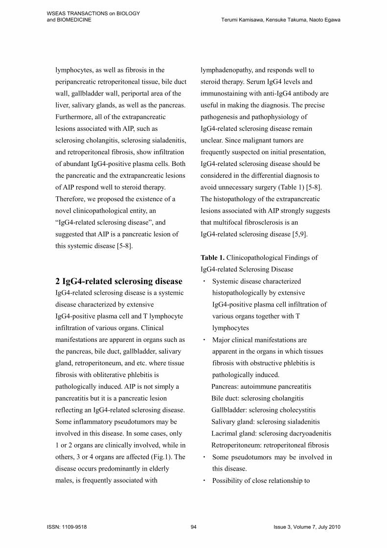
Some inflammatory pseudotumors may be
involved in this disease. In some cases, only
1 or 2 organs are clinically involved, while in
others, 3 or 4 organs are affected (Fig.1). The
disease occurs predominantly in elderly
males, is frequently associated with
lymphadenopathy, and responds well to
steroid therapy. Serum IgG4 levels and
immunostaining with anti-IgG4 antibody are
useful in making the diagnosis. The precise
pathogenesis and pathophysiology of
IgG4-related sclerosing disease remain
unclear. Since malignant tumors are
frequently suspected on initial presentation,
IgG4-related sclerosing disease should be
considered in the differential diagnosis to
avoid unnecessary surgery (Table 1) [5-8].
The histopathology of the extrapancreatic
lesions associated with AIP strongly suggests
that multifocal fibrosclerosis is an
IgG4-related sclerosing disease [5,9].
Table 1. Clinicopathological Findings of
IgG4-related Sclerosing Disease
・ Systemic disease characterized
histopathologically by extensive
IgG4-positive plasma cell infiltration of
various organs together with T
lymphocytes
・ Major clinical manifestations are
apparent in the organs in which tissues
fibrosis with obstructive phlebitis is
pathologically induced.
Pancreas: autoimmune pancreatitis
Bile duct: sclerosing cholangitis
Gallbladder: sclerosing cholecystitis
Salivary gland: sclerosing sialadenitis
Lacrimal gland: sclerosing dacryoadenitis
Retroperitoneum: retroperitoneal fibrosis
・ Some pseudotumors may be involved in
this disease.
・ Possibility of close relationship to
WSEAS TRANSACTIONS on BIOLOGY and BIOMEDICINE Terumi Kamisawa, Kensuke Takuma, Naoto Egawa
ISSN: 1109-9518 94 Issue 3, Volume 7, July 2010
multifocal fibrosclerosis
・ Occasional association with
lymphadenopathy
・ Elderly male preponderance
・ Frequent elevation of serum IgG4 levels
・ Favorite response to steroid therapy
・ Differentiation from malignant tumor is
important.
・ Precise pathogenesis and
pathophysiology remain unclear
/
Fig.1 Schematic illustration of IgG4-related
sclerosing disease
3 Autoimmune pancreatitis
AIP is a unique form of pancreatitis in which
autoimmune mechanisms are suspected to be
involved in the pathogenesis. AIP occurs
more commonly in elderly males. In our 57
AIP Patients, the mean age of the patients
was 66.5 years (range, 25-83 years), and the
male-to-female ratio was 4:1. The major
clinical symptom is obstructive jaundice due
to associated sclerosing cholangitis (70% in
our series). Up to 50% of AIP patients
present with glucose intolerance [10].
Levels of serum IgG4 are particularly high
in AIP. Dense infiltration of IgG4-positive
plasma cells is seen in various organs of AIP
patients. These findings suggest that IgG4
plays a major role in the pathogenesis of AIP,
although the trigger for the IgG4 elevation or
its pathogenetic role in AIP has not been
clearly disclosed [11,12].
It is of utmost importance that AIP be
differentiated from pancreatic cancer, as
some AIP patients in which pancreatic cancer
is suspected undergo unnecessary laparotomy
or pancreatic resection. Since there is
currently no diagnostic serological marker for
AIP, AIP should be diagnosed on the basis of
the presence of a combination of
abnormalities unique to AIP. The Japanese
“Diagnostic Criteria for Autoimmune
Pancreatitis” were revised in 2006 [13]. They
consisted of three items: 1) radiological
imaging showing diffuse or segmental
narrowing of the main pancreatic duct with
irregular wall and diffuse or localized
enlargement of the pancreas; 2) laboratory
data demonstrating abnormally elevated
levels of serum gammaglobulin or IgG, or
IgG4, or the presence of autoantibodies; and
3) histological examination of the pancreas
showing lymphoplasmacytic infiltration and
fibrosis. Diagnosis of AIP is made when
either all 3 criteria are present or criterion 1
together with either criterion 2 or criterion 3
is present.
Radiologically, pancreatic enlargement
is usually hypoechoic, sometimes with
scattered hyperechoic spots on
WSEAS TRANSACTIONS on BIOLOGY and BIOMEDICINE Terumi Kamisawa, Kensuke Takuma, Naoto Egawa
ISSN: 1109-9518 95 Issue 3, Volume 7, July 2010
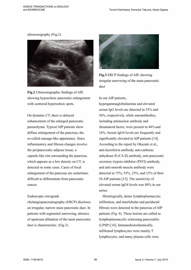
ultrasonography (Fig.2).
Fig.2 Ultrasonographic findings of AIP,
showing hypoechoic pancreatic enlargement
with scattered hyperechoic spots.
On dynamic CT, there is delayed
enhancement of the enlarged pancreatic
parenchyma. Typical AIP patients show
diffuse enlargement of the pancreas, the
so-called sausage-like appearance. Since
inflammatory and fibrous changes involve
the peripancreatic adipose tissue, a
capsule-like rim surrounding the pancreas,
which appears as a low density on CT, is
detected in some cases. Cases of focal
enlargement of the pancreas are sometimes
difficult to differentiate from pancreatic
cancer.
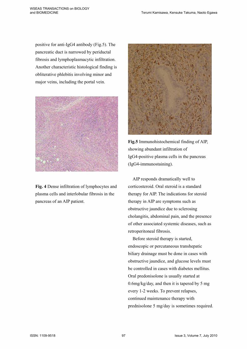
Endoscopic retrograde
cholangiopancreatography (ERCP) discloses
an irregular, narrow main pancreatic duct. In
patients with segmental narrowing, absence
of upstream dilatation of the main pancreatic
duct is characteristic. (Fig.3).
Fig.3 ERCP findings of AIP, showing
irregular narrowing of the main pancreatic
duct
In our AIP patients,
hypergammaglobulinemia and elevated
serum IgG levels are detected in 33% and
56%, respectively, while autoantibodies,
including antinuclear antibody and
rheumatoid factor, were present in 44% and
16%. Serum IgG4 levels are frequently and
significantly elevated in AIP patients [14].
According to the report by Okazaki et al.,
anti-lactoferrin antibody, anti-carbonic
anhydrase-II (CA II) antibody, anti-pancreatic
secretory trypsin inhibitor (PSTI) antibody,
and anti-smooth muscle antibody were
detected in 75%, 55%, 25%, and 15% of their
54 AIP patients [15]. The sensitivity of
elevated serum IgG4 levels was 80% in our
series.
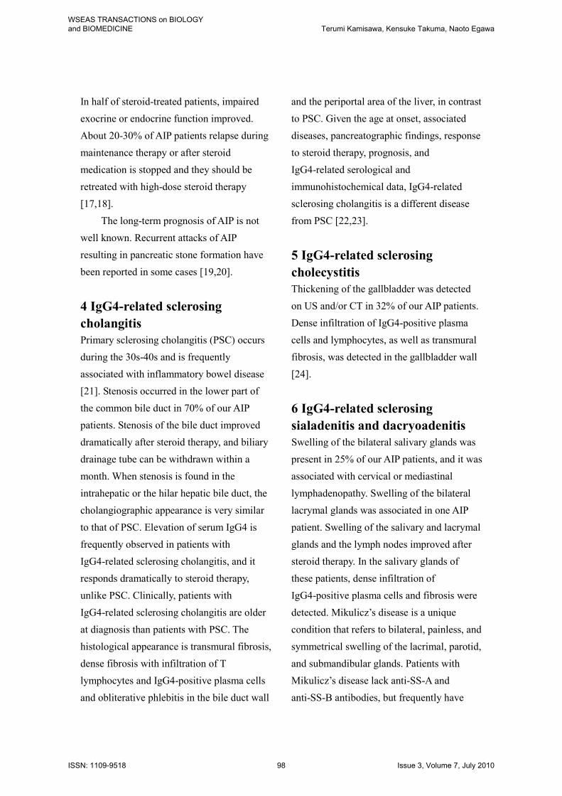
Histologically, dense lymphoplasmacytic
infiltration, and interlobular and periductal
fibrosis were detected in the pancreas of AIP
patients (Fig. 4). These lesions are called as
lymphoplasmacytic sclerosing pancreatitis
(LPSP) [16]. Immunohistochemically,
infiltrated lymphocytes were mainly T
lymphocytes, and many plasma cells were
WSEAS TRANSACTIONS on BIOLOGY and BIOMEDICINE Terumi Kamisawa, Kensuke Takuma, Naoto Egawa
ISSN: 1109-9518 96 Issue 3, Volume 7, July 2010
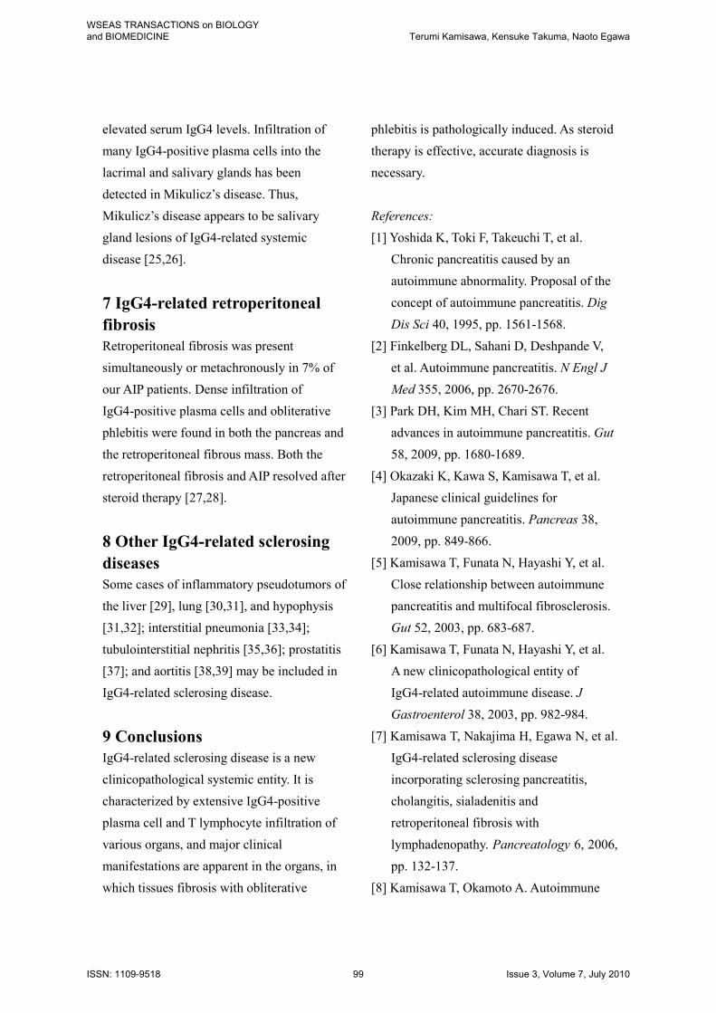
positive for anti-IgG4 antibody (Fig.5). The
pancreatic duct is narrowed by periductal
fibrosis and lymphoplasmacytic infiltration.
Another characteristic histological finding is
obliterative phlebitis involving minor and
major veins, including the portal vein.
Fig. 4 Dense infiltration of lymphocytes and
plasma cells and interlobular fibrosis in the
pancreas of an AIP patient.
Fig.5 Immunohistochemical finding of AIP,
showing abundant infiltration of
IgG4-positive plasma cells in the pancreas
(IgG4-immunostaining).
AIP responds dramatically well to
corticosteroid. Oral steroid is a standard
therapy for AIP. The indications for steroid
therapy in AIP are symptoms such as
obstructive jaundice due to sclerosing
cholangitis, abdominal pain, and the presence
of other associated systemic diseases, such as
retroperitoneal fibrosis.
Before steroid therapy is started,
endoscopic or percutaneous transhepatic
biliary drainage must be done in cases with
obstructive jaundice, and glucose levels must
be controlled in cases with diabetes mellitus.
Oral predonisolone is usually started at
0.6mg/kg/day, and then it is tapered by 5 mg
every 1-2 weeks. To prevent relapses,
continued maintenance therapy with
prednisolone 5 mg/day is sometimes required.
WSEAS TRANSACTIONS on BIOLOGY and BIOMEDICINE Terumi Kamisawa, Kensuke Takuma, Naoto Egawa
ISSN: 1109-9518 97 Issue 3, Volume 7, July 2010
In half of steroid-treated patients, impaired
exocrine or endocrine function improved.
About 20-30% of AIP patients relapse during
maintenance therapy or after steroid
medication is stopped and they should be
retreated with high-dose steroid therapy
[17,18].
The long-term prognosis of AIP is not
well known. Recurrent attacks of AIP
resulting in pancreatic stone formation have
been reported in some cases [19,20].
4 IgG4-related sclerosing
cholangitis
Primary sclerosing cholangitis (PSC) occurs
during the 30s-40s and is frequently
associated with inflammatory bowel disease
[21]. Stenosis occurred in the lower part of
the common bile duct in 70% of our AIP
patients. Stenosis of the bile duct improved
dramatically after steroid therapy, and biliary
drainage tube can be withdrawn within a
month. When stenosis is found in the
intrahepatic or the hilar hepatic bile duct, the
cholangiographic appearance is very similar
to that of PSC. Elevation of serum IgG4 is
frequently observed in patients with
IgG4-related sclerosing cholangitis, and it
responds dramatically to steroid therapy,
unlike PSC. Clinically, patients with
IgG4-related sclerosing cholangitis are older
at diagnosis than patients with PSC. The
histological appearance is transmural fibrosis,
dense fibrosis with infiltration of T
lymphocytes and IgG4-positive plasma cells
and obliterative phlebitis in the bile duct wall
and the periportal area of the liver, in contrast
to PSC. Given the age at onset, associated
diseases, pancreatographic findings, response
to steroid therapy, prognosis, and
IgG4-related serological and
immunohistochemical data, IgG4-related
sclerosing cholangitis is a different disease
from PSC [22,23].
5 IgG4-related sclerosing
cholecystitis
Thickening of the gallbladder was detected
on US and/or CT in 32% of our AIP patients.
Dense infiltration of IgG4-positive plasma
cells and lymphocytes, as well as transmural
fibrosis, was detected in the gallbladder wall
[24].
6 IgG4-related sclerosing
sialadenitis and dacryoadenitis
Swelling of the bilateral salivary glands was
present in 25% of our AIP patients, and it was
associated with cervical or mediastinal
lymphadenopathy. Swelling of the bilateral
lacrymal glands was associated in one AIP
patient. Swelling of the salivary and lacrymal
glands and the lymph nodes improved after
steroid therapy. In the salivary glands of
these patients, dense infiltration of
IgG4-positive plasma cells and fibrosis were
detected. Mikulicz’s disease is a unique
condition that refers to bilateral, painless, and
symmetrical swelling of the lacrimal, parotid,
and submandibular glands. Patients with
Mikulicz’s disease lack anti-SS-A and
anti-SS-B antibodies, but frequently have
WSEAS TRANSACTIONS on BIOLOGY and BIOMEDICINE Terumi Kamisawa, Kensuke Takuma, Naoto Egawa
ISSN: 1109-9518 98 Issue 3, Volume 7, July 2010
elevated serum IgG4 levels. Infiltration of
many IgG4-positive plasma cells into the
lacrimal and salivary glands has been
detected in Mikulicz’s disease. Thus,
Mikulicz’s disease appears to be salivary
gland lesions of IgG4-related systemic
disease [25,26].
7 IgG4-related retroperitoneal
fibrosis
Retroperitoneal fibrosis was present
simultaneously or metachronously in 7% of
our AIP patients. Dense infiltration of
IgG4-positive plasma cells and obliterative
phlebitis were found in both the pancreas and
the retroperitoneal fibrous mass. Both the
retroperitoneal fibrosis and AIP resolved after
steroid therapy [27,28].
8 Other IgG4-related sclerosing
diseases
Some cases of inflammatory pseudotumors of
the liver [29], lung [30,31], and hypophysis
[31,32]; interstitial pneumonia [33,34];
tubulointerstitial nephritis [35,36]; prostatitis
[37]; and aortitis [38,39] may be included in
IgG4-related sclerosing disease.
9 Conclusions
IgG4-related sclerosing disease is a new
clinicopathological systemic entity. It is
characterized by extensive IgG4-positive
plasma cell and T lymphocyte infiltration of
various organs, and major clinical
manifestations are apparent in the organs, in
which tissues fibrosis with obliterative
phlebitis is pathologically induced. As steroid
therapy is effective, accurate diagnosis is
necessary.
References:
[1] Yoshida K, Toki F, Takeuchi T, et al.
Chronic pancreatitis caused by an
autoimmune abnormality. Proposal of the
concept of autoimmune pancreatitis. Dig
Dis Sci 40, 1995, pp. 1561-1568.
[2] Finkelberg DL, Sahani D, Deshpande V,
et al. Autoimmune pancreatitis. N Engl J
Med 355, 2006, pp. 2670-2676.
[3] Park DH, Kim MH, Chari ST. Recent
advances in autoimmune pancreatitis. Gut
58, 2009, pp. 1680-1689.
[4] Okazaki K, Kawa S, Kamisawa T, et al.
Japanese clinical guidelines for
autoimmune pancreatitis. Pancreas 38,
2009, pp. 849-866.
[5] Kamisawa T, Funata N, Hayashi Y, et al.
Close relationship between autoimmune
pancreatitis and multifocal fibrosclerosis.
Gut 52, 2003, pp. 683-687.
[6] Kamisawa T, Funata N, Hayashi Y, et al.
A new clinicopathological entity of
IgG4-related autoimmune disease. J
Gastroenterol 38, 2003, pp. 982-984.
[7] Kamisawa T, Nakajima H, Egawa N, et al.
IgG4-related sclerosing disease
incorporating sclerosing pancreatitis,
cholangitis, sialadenitis and
retroperitoneal fibrosis with
lymphadenopathy. Pancreatology 6, 2006,
pp. 132-137.
[8] Kamisawa T, Okamoto A. Autoimmune
WSEAS TRANSACTIONS on BIOLOGY and BIOMEDICINE Terumi Kamisawa, Kensuke Takuma, Naoto Egawa
ISSN: 1109-9518 99 Issue 3, Volume 7, July 2010
pancreatitis: proposal of IgG4-related
sclerosing disease. J Gastroenterol 41,
2006, pp. 613-625.
[9] Comings DE, Skubi KB, Eyes JV, et al.
Familial multifocal fibrosclerosis. Ann
Intern Med 66, 1967, pp. 884-892.
[10] Kamisawa T, Egawa N, Inokuma S, et al.
Pancreatic endocrine and exocrine
function and salivary gland function in
autoimmune pancreatitis before and after
steroid therapy. Pancreas 27, 2003, pp.
235-238.
[11] Okazaki K, Uchida K, Ohana M, et al.
Autoimmune-related pancreatitis is
associated with antibodies and a
Th1/Th2-type cellular immune response.
Gastroenterology 118, 2000, pp. 573-581.
[12] Zen Y, Fujii T, Harada K, et al. Th2 and
regulatory immune reactions are
increased in immunoglobulin G4-related
sclerosing pancreatitis and cholangitis.
Hepatology 45, 2007, pp. 1538-1546.
[13] Okazaki K, Kawa S, Kamisawa T, et al.
Clinical diagnostic criteria of
autoimmune pancreatitis revised proposal.
J Gastroenterol 41, 2006, pp. 626-631.
[14] Hamano H, Kawa S, Horiuchi A, et al.
High serum IgG4 concentrations in
patients with sclerosing pancreatitis. N
Engl J Med 344, 2001, pp. 732-738.
[15] Okazaki K, Uchida K, Fukui T. Recent
advances in autoimmune pancreatitis:
concept, diagnosis, and pathogenesis. J
Gastroenterol 43, 2008, pp. 409-418.
[16] Kawaguchi K, Koike M, Tsuruta K, et al.
Lymphoplasmacytic sclerosing
pancreatitis with cholangitis: a variant of
primary sclerosing cholangitis
extensively involving pancreas. Hum
Pathol 22, 1991, pp. 387-395.
[17] Kamisawa T, Shimosegawa T, Okazaki
K, et al. Standard steroid therapy for
autoimmune pancreatitis. Gut 58, 2009,
pp. 1504-1507.
[18] Kamisawa T, Okazaki K, Kawa S, et al.
Japanese consensus guidelines for
management of autoimmune pancreatitis:
III. Treatment and prognosis of AIP.
J Gastroenterol 45, 2010, pp. 471-477.
[19] Kamisawa T, Okamoto A. Prognosis of
autoimmune pancreatitis. J
Gastroenterol 42 Suppl 18, 2007, pp.
59-62.
[20] Takayama M, Hamano H, Ochi Y, et al.
Recurrent attacks of autoimmune
pancreatitis result in pancreatic stone
formation. Am J Gastroenterol 99, 2004,
pp. 932-937.
[21] LaRusso NF, Wiesner RH, Ludwig J, et
al. Current concepts. Primary sclerosing
cholangitis. N Eng J Med 310, 1984, pp.
899-903.
[22] Nakazawa T, Ohara H, Sano H, et al.
Clinical differences between primary
sclerosing cholangitis and sclerosing
cholangitis with autoimmune pancreatitis.
Pancreas 30, 2005, pp.20-25.
[23] Kamisawa T, Takuma K, Anjiki H, et al.
Sclerosing cholangitis associated with
autoimmune pancreatitis differs from
primary sclerosing cholangitis. World J
Gastroenterol 15, 2009, pp. 2357-2360.
WSEAS TRANSACTIONS on BIOLOGY and BIOMEDICINE Terumi Kamisawa, Kensuke Takuma, Naoto Egawa
ISSN: 1109-9518 100 Issue 3, Volume 7, July 2010
[24] Kamisawa T, Tu Y, Nakajima H, et al.
Sclerosing cholecystitis associated with
autoimmune pancreatitis. World J
Gastroenterol 12, 2006, pp. 3736-3739.
[25] Yamamoto M, Harada S, Ohara M, et al.
Clinical and pathological differences
between Mikulicz’s disease and Sjogren’s
syndrome. Rheumatology 44, 2005, pp.
227-234.
[26] Kamisawa T, Nakajima H, Hishima T.
Close relationship between chronic
sclerosing sialadenitis and IgG4. Intern
Med J 36, 2006, pp. 527-529.
[27] Kamisawa T, Matsukawa M, Okawa M.
Autoimmune pancreatitis associated with
retroperitoneal fibrosis. JOP 10, 2005, pp.
260-263.
[28] Kamisawa T, Chen PY, Tu Y, et al.
Autoimmune pancreatitis metachronously
associated with retroperitoneal fibrosis
with IgG4-positive plasma cell infiltration.
World J Gastroenterol 12, 2006, pp.
2955-2957.
[29] Kanno A, Satoh K, Kimura K, et al.
Autoimmune pancreatitis with hepatic
inflammatory pseudotumor. Pancreas 31,
2005, pp. 420-423.
[30] Zen Y, Kitagawa S, Minato H, et al.
IgG4-positive plasma cells in
inflammatory pseudotumor (plasma cell
granuloma) of the lung. Hum Pathol 36,
2005, pp. 710-717.
[31] Tsuboi H, Inokuma S, Setoguchi K, et al.
Inflammatory pseudotumors in multiple
organs associated with elevated serum
IgG4 level: recovery by only a small
replacement dose of steroid. Intern Med 47,
2008, 1pp. 139-1142
[32] Wong S, Lam WY, Wong WK, et al.
Hypophysitis presented as inflammatory
pseudotumor in immunoglobulin
G4-related systemic disease. Hum Pathol
38, 2007, pp. 1720-1723.
[33] Hirano K, Kawabe T, Komatsu Y, et al.
High-rate pulmonary involvement in
autoimmune pancreatitis. Intern Med 36,
2006, pp. 58-61.
[34] Takato H, Yasui M, Ichikawa Y, et al.
Nonspecific interstitial pneumonia with
abundant IgG4-positive cells infiltration,
which was thought as pulmonary
involvement of IgG4-related autoimmune
disease. Intern Med 47, 2008, pp. 291-294.
[35] Takeda S, Haratake J, Kasai T, et al.
IgG4-associated idiopathic
tubulointerstitial nephritis complicating
autoimmune pancreatitis. Nephrol Dial
Transplant 19, 2004, pp. 474-476.
[36] Saeki T, Nishi S, Ito T, et al. Renal
lesions in IgG4-related systemic disease.
Intern Med 46, 2007, pp. 1365-1371.
[37] Nishimori I, Kohsaki T, Onishi S, et al.
IgG4-related autoimmune prostatitis: two
cases with or without autoimmune
pancreatitis. Intern Med 46, 2007, pp.
1983-1989.
[38] Sakata N, Tashiro T, Uesugi N, et al.
IgG4-positive plasma cells in
inflammatory abdominal aortic aneurysm:
the possibility of an aortic manifestation
of IgG4-related sclerosing disease. Am J
Surg Pathol 32, 2008, pp. 553-559.
WSEAS TRANSACTIONS on BIOLOGY and BIOMEDICINE Terumi Kamisawa, Kensuke Takuma, Naoto Egawa
ISSN: 1109-9518 101 Issue 3, Volume 7, July 2010
[39] Kasashima S, Zen Y, Kawashima
A,et al. A new clinicopathological
entity of IgG4-related inflammatory
abdominal aortic aneurysm. J Vasc
Surg 49, 2009, pp. 1264-1271.
WSEAS TRANSACTIONS on BIOLOGY and BIOMEDICINE Terumi Kamisawa, Kensuke Takuma, Naoto Egawa
ISSN: 1109-9518 102 Issue 3, Volume 7, July 2010
Recommended