Identifying, Healing and Preventing Pressure Ulcers:
It Takes a Village
Massachusetts Department of Developmental Services September 2017

Speaker Introduction
2
Donna Marie Morrow, RN, WCC, DWC, OMS
Wound Care Manager for Nizhoni Health Somerville, MA Over 30 years nursing and wound
management experience Presented at national conferences and
the National Pressure Ulcer Advisory Panel (NPUAP) Published articles for Today’s Wound
Clinic Ostomy Wound Management
Insert speaker picture if we have it

Today’s Objectives
Identify what pressure ulcers are and what causes them.
Identify those most at risk for a pressure ulcer. Recognizing pressure ulcers, especially in
different skin tones. How to prevent and treat pressure ulcers. Case Studies and Resources
3

A Word of Warning…
Today’s presentation is not meant to be clinical. This webinar is primarily for direct support
professionals and others who work directly with people with disabilities.
We will show some clinical images today of pressure ulcers in different stages.
We’ve limited the images to early stages, but they can be a little graphic.
3

What is a Pressure Ulcer?? Sometimes called “decubitus ulcers” or
“bed sores.” An injury to the skin and underlying tissue.
T wo Main causes: 1) Pressure on one spot of the body for too long 2) Friction on the skin
Stage 1 pressure ulcer, buttocks

Pressure Occurs when someone is lying or sitting in
one position for too long. Pressure of the body against a surface (like a
chair seat) reduces blood flow to the skin and nearby tissue. This stops the flow of oxygen.
Pressure can cause damage to the skin and tissue.
Serious damage to the skin and muscle can occur in as little as 1 hour in a chair and in as little as 2 hours in a bed!

Friction Occurs when skin is
dragged across a surface.
For example: When a person changes position or is moved or repositioned.
If the skin is moist or wet, the friction is worse and can cause significant skin and tissue damage. This can also worsen existing pressure ulcers.

Shearing Occurs when 2 surfaces move in opposite directions.
For example: When a person slides down in bed, their tailbone moves down while the skin over their tailbone stays in place. This pulls the skin in the opposite direction from the movement.

Other Causes of Pressure Sores Any device or object
that sits on the body (pressure) or rubs against the body (friction).
Examples: Splints, rolls, medical tubing, oxygen tubing, CPAP, G-tubes, catheters, socks, clothing, sheets, towels, or draw sheets

The Impact of Pressure Ulcers
At first, the affected skin reddens. Person may complain of pain or itch. If left untreated, the skin can open, become
larger, deeper and more difficult to heal. This causes great pain and
can lead to, bone infection and sepsis.
Can even lead to death.
Stage 2 pressure ulcer

Courtesy of the Center for Disease Control (CDC)

Impact Continued…
As many as 1/3 of hospitalized patients with pressure ulcers die during their
hospitalization. More than half of those who develop a
pressure ulcer in the hospital will die within the next 12 months.
About 60,000 patients die as a direct result of a pressure ulcer each year. This translates to 6.85 deaths every hour of every single day.

Who is most at risk for developing a pressure ulcer?

People at Risk: Are elderly Have lost feeling or sensation in a body part Cannot move themselves (are immobile) Sit or lie down for extended periods of time Have a history of pressure ulcers Have bowel or urinary incontinence

People at Risk Often: Take 8 or more medications Are in poor health and/or have chronic
health conditions, especially diabetes and blood circulation problems
Have poor nutrition or hydration Have fragile skin that tears easily, skin
tears, or chronic skin problems Have excessively dry or moist skin

People with a History of Pressure Ulcers: Are more than five times as likely to
develop another pressure ulcer. The strength of the skin goes down
70% after a pressure ulcer. Even if the skin and muscle heals, it will
never be as strong as it once was.

How to recognize a pressure ulcer

Where do Pressure Ulcers Form? These areas on the body are at higher risk of
breaking down due to pressure and friction:
o Back of the head o Shoulder o Tail bone o Elbow o Hip o Knee o Heel o Ankles

What do Pressure Ulcers Look Like? In the early stages, the skin appears red or inflamed The person often complains of pain or an itch in the
affected area. The skin may be intact, but it is red or spongey. Be aware that pressure ulcers can look different on
different skin colors.
Stage 2 pressure ulcer, buttocks
Stage 1 pressure ulcer, sacrococcygeal

Finding Ulcers: Look, Listen, Feel Look at the person’s skin at close range and at a distance to check for differences in skin tones.
Listen to the person’s reports of soreness or pain.
Feel - touch the person’s skin and check for warmth, coolness, mushiness, and firmness. Skin temperature is often warmer or cooler in a pressure ulcer. The skin will also probably feel firmer or softer.

See Something, Say Something If you suspect someone is at increased
risk for developing a pressure ulcer, say something!
If something looks different or “off”, report your concerns to a nurse or supervisor.

Stages of Pressure Ulcers There are different stages of pressure
ulcers, stage 1 being the least serious and stage 4 being the most serious condition. Medical professionals use stages as a
way to categorize the wound. Some pressure ulcers are “unstageable”
meaning it cannot be determined.

Stage 1 The skin is not broken, but is
inflamed. The area may be red, painful,
firm, soft, warmer, or cooler as compared to surrounding skin.
Source: National Institute of Arthritis and Musculoskeletal and Skin Diseases
Stage 1 pressure ulcer, heel.

Stage 2 The outer layer of skin (epidermis)
and the inner layer of skin (dermis)is damaged or lost.
The wound may be shallow andpinkish or red.
The wound may look like anabrasion, fluid filled blister or a shallow crater.
Source: National Institute of Arthritis and Musculoskeletal and Skin Diseases
Stage 2 pressure ulcer, buttocks.

Stage 3 The loss of skin usually
exposes the fat layer. Bone,tendon, and muscle are notexposed.
The damage may extendbeyond what you see to layersof skin below.
Source: National Institute of Arthritis and Musculoskeletal and Skin Diseases

Stage 4
The pressure ulcer isvery deep, reachinginto muscle and boneand causing extensivedamage.
Damage to deepertissues, tendons, andjoints may occur.
Source: National Institute of Arthritis and Musculoskeletal and Skin Diseases

What do pressure ulcers look like in different skin tones?

Darker vs Lighter Skin Pigmentations The most crucial part of detecting pressure
ulcers is understanding changes in skin color. A pressure ulcer may appear bluish, purplish
or violet in darker pigmented skin. The sameulcer would look redor pink in lighter skin. Stage 1 pressure ulcer, buttoc
,
Stage 1 pressure ulcer, buttocks

How to Check for Pressure Ulcers in Darker Skin Tones Skin may be taut, shiny, or hard.
F eel the area to see if it’s “boggy” or very soft. It will often be a different temperature from surrounding skin.
The person will likely be in pain.It’s important to identify these areas.
Stage 1 pressure ulcers, buttocks.
Source: Dr. Mujahid Zulfiqar Ali http://ispub.com/IJS/13/1/9226

How to prevent pressure ulcers

Early Detection Inspect skin during bathing or daily personal
care. Report any and all changes in skin appearance
or complaints of pain or itching. Do not massage areas that are already red, especially if they are over a boney part of the body (ankle, hip)
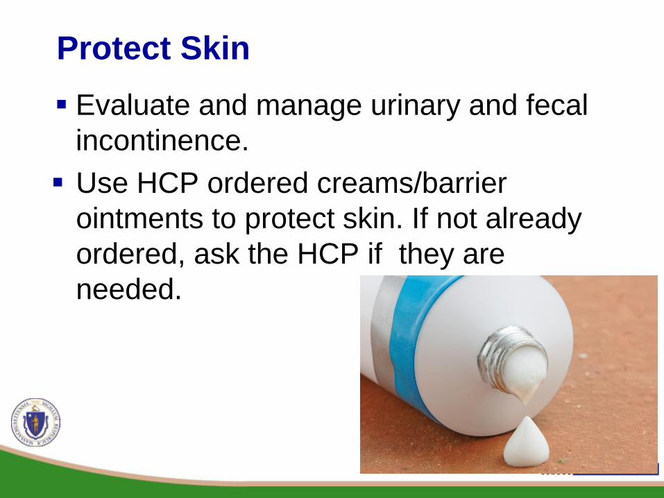
Protect Skin Evaluate and manage urinary and fecal
incontinence. Use HCP ordered creams/barrier
ointments to protect skin. If not alreadyordered, ask the HCP if they are needed.

Good Hydration and Nutrition Maintain good hydration and nutrition. This helps
protect skin and promotes wound healing Drink Enough- good hydration is important to
keeping skin intact Healthy Diet- a diet high in calories, protein,
vitamins, and minerals will promote wound healing

Good Positioning When in bed, maintain the lowest possible
head elevation appropriate for that person to reduce the impact of shear.
Position the patient to
minimize pressure and shearing forces over the heels, elbows, base of head,and ears
Source: MacMed

Reposition
Help the person change positions to relieve pressure on body parts.
People should change position at least every 2 hours and more often if they are at risk for pressure ulcers.
Copyright notice, © TurnAide All Rights Reserved

Repositioning Continued … Use pillows under one side of the back to
offload pressure. Use pillows as cushions/wedges between
and under knees. Place cushion support under feet and
ankles. Follow all health practitioners, physical, and occupational therapists, and nursing orders as prescribed.

Minimize Friction and Shearing Avoid dragging heels, hip, or tail bones
across sheets Use lifting devices such as a draw sheet or
trapeze
© 2017 HealthCraft Products. Image used with permission.

Wheel Chair Repositioning and Assessment Encourage people to shift their weight every 15
minutes while seated. Use tilt feature on chair to reposition every hour. Inspect cushions for signs of wear, proper
inflation, and proper placement. Wheel chairs should be
inspected daily and cleaned to avoid sharp edges or pressure related injury

Provide Good Support Specially ordered air mattresses, wedges,
cushions, or pillows will relieve pressure and provide support both in and out of bed.
This support is needed to offload pressure. These would be recommended by a doctor or
physical or occupational therapist.
© Skil-Care Corporation. Image used with permission.

Skin Care Keep the skin clean and dry. Wash with mild
soap and water, rinse thoroughly, gently pat dry.
Apply lotions as prescribed- to prevent skin breakdown and keep skin intact.
Never massage over an area of skin that is reddened or has skin breakdown.

Pressure Ulcer Management

Pressure Ulcer Management All wound care is to be managed or overseen
by a certified wound and ostomy nurse under the order of a health care provider.
An individualized protocol for the prevention and management of pressure ulcers must be created for any person determined to be at risk.

Braden Scale The Braden scale is
used for predicting pressure ulcer risk. The purpose of the scale is to help health care professionals, especially nurses, to assess a patient’s risk of developing a pressure ulcer.
Copyright, Braden and Bergstrom, 1988. Reprinted with permission. All rights reserved. http://www.bradenscale.com Copyright, Government of South Australia

A Team Approach Planning to provide evidence based
treatment interventions is the responsibility of the whole team. This includes nurses, case managers, family members, and direct care staff.

Pressure Ulcer Treatment

Treatment of Pressure Ulcers Provided by a Wound and Ostomy nurse Your input is important! Let the nurse know if the wound looks,
smells, or feels different to you.

Treatment Monitoring A healthcare professional should evaluate
a pressure ulcer at every dressing change, including: drainage (including type, color, odor, and approximate amount), pain, status of the wound, and a description of wound edges and surrounding tissue. Staff play the most essential role! Staff can notice the beginning of a
pressure ulcer and then report it immediately.

Treatment Goals Open pressure wounds need to be
covered by a dressing. Keep the wound bed MOIST and
the surrounding skin DRY. Protect the ulcer from
contamination. Promote moist wound healing.

Case Examples

Case 1 57 year old active male admitted to the hospital with
pneumonia/sepsis.
Intubated while there and spent 3 days in bed.
Developed a stage 4 pressure ulcer.
Would we normally think that an active 57 year old person would be affected by pressure wounds?
He was immobile while in the hospital (didn’t walk around). The pressure ulcer developed rapidly!
Case 1 Repositioning, good nutrition and
hydration, and frequent skin checks during the hospital stay will help prevent pressure ulcers from developing.
Statistically, hospitalizations and illness increase the chances of every person for developing a pressure ulcer.

Case 2 61 year old male with a history of diabetes, kidney
failure and arterial disease.
Has an amputated leg and wears a prosthetic.
Developed a blister on his right leg above the kneeamputation stump that turned into a stage 2pressure ulcer.
This was caused by wearing his prosthetic devicewhile his stump was edematous (swollen).
Is this a pressure related injury because it is notlocated over where we would normally think apressure wound would develop?

Case 2
A pressure ulcer can develop anywhere the skincomes in contact with a harder surface,including under prosthetic devices, braces andtubing.
When doing daily skin inspections, remember tolook at all skin areas that may come into contactwith any of these devices to detect any earlyredness or discoloration of the skin.

More Information
DDS Guidelines for Managing Pressure Ulcers
Released June 2017 All people with risk factors should be evaluated and
a protocol created for how to manage that risk. All wound care is managed by a Certified Wound
and Ostomy Nurse http://www.mass.gov/eohhs/docs/dmr/reports/fs-pressure-ulcers-protocol.pdf

DDS Signs & Symptoms Sheet
http://www.mass.gov/eohhs/docs/dmr/reports/fs-pressure-ulcers.pdf

Quality Is No Accident Brief
https://shriver.umassmed.edu/sites/shriver.umassmed.edu/files/QINA%20pressure%20ulcers_final-tagged.pdf

57
Resources Berkowitz, D. et al. (2014 October). Preventing pressure ulcers in hospitals: A tool kit for
improving quality care. Agency for Healthcare Research and Quality. Retrieved from: https://www.ahrq.gov/professionals/systems/ hospital/pressureulcertoolkit/index.html
Pressure ulcers: Prevention and management of pressure ulcers. (2014) National Guideline Clearinghouse. Retrieved from: https://www.guideline.gov/search?q=pressure+ulcers
Health care protocol: Pressure ulcer prevention and treatment protocol. (march 2014). Institute for Clinical Systems Improvement. Retrieved from: https://www.icsi.org/_asset/6t7kxy/PressureUlcer.pdf
Park-Lee, E. Caffery, C. (2009, February). Pressure ulcers among nursing home residents: United States, 2004. National Center for Health Statistics. Retrieved from: www.cdc.gov/nchs/products/databriefs/db14.htm
Sandra M. Nettina. (2010). Manual of Nursing Practice [Woltz Kluwer Health] Lippincott Williams & Wilkins · (8th ed.,rev.) Ambler Penn.
Clark, Michael. "Skin Assessment in Dark Pigmented Skin: A Challenge in Pressure Ulcer Prevention." Nursing Times. N.p., 2 Aug. 2010. Web.
"Explore Pressure Sore Statistics." Aquila Corporation. Aquila Corporations, n.d. Web. Sommers, Marilynn S. "Color Awareness: A Must for Patient Assessment." American
Nurse Today. N.p., 10 July 2017. Web.

32
Training produced by the Center for Developmental Disabilities Evaluation & Research (CDDER) on
behalf of the Massachusetts Department of Developmental Services (DDS)
Recommended