HVRA census increta/percreta. 2008-2014: 124 cases
▪ gravid patients at risk for clinically significant adherent placentation.
▪ referred by MFMs who desired second opinion after their own ultrasound studies.
▪ all patients had MR followed by transabdominal, transvaginal color Doppler ultrasound studies.
▪ all patients had MD performed US scanning and MR interpretation by one radiologist (DJC).
▪ delivery, operative and pathology reports reviewed for all patients
1
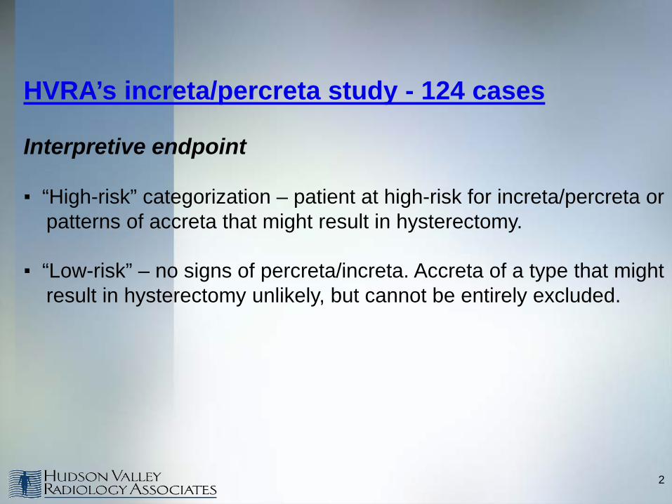
HVRA’s increta/percreta study - 124 cases
Interpretive endpoint
▪ “High-risk” categorization – patient at high-risk for increta/percreta or patterns of accreta that might result in hysterectomy.
▪ “Low-risk” – no signs of percreta/increta. Accreta of a type that might result in hysterectomy unlikely, but cannot be entirely excluded.
2

HVRA's Imaging Census for Placenta Increta/Percreta from January 2008 through May 11, 2014. 124 patients studied.
No. of pts True positive: 34True negative: 78False positive: 8False negative: 4
Sensitivity 89%Specificity 91%Positive Predictive Value 81%Negative Predictive Value 91%
False Positive Rate 9%False Negative Rate 10%
Accuracy 90%
3

4
HVRA’s census of 124 cases
▪ referred by MFMs for MR and second opinion ultrasound
▪ not analyzed as to the predictive attributes of one modality versus the other
▪ personal imaging philosophy
- caution against imaging risk assessment for clinically significant adherent placentation by MR alone without hands-on ultrasound
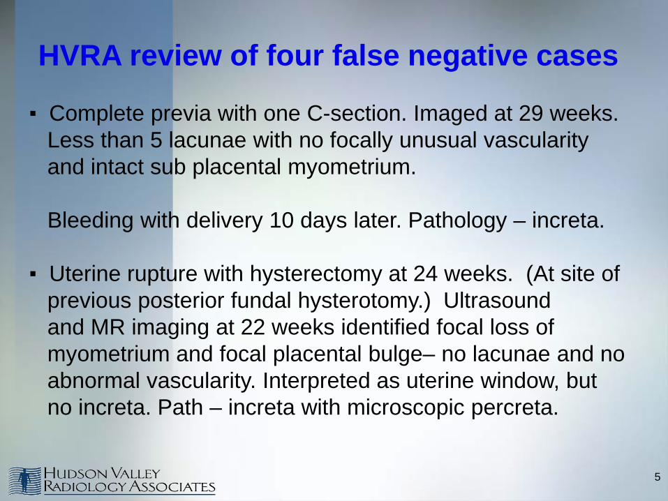
HVRA review of four false negative cases
▪ Complete previa with one C-section. Imaged at 29 weeks. Less than 5 lacunae with no focally unusual vascularityand intact sub placental myometrium.
Bleeding with delivery 10 days later. Pathology – increta.
▪ Uterine rupture with hysterectomy at 24 weeks. (At site of previous posterior fundal hysterotomy.) Ultrasound and MR imaging at 22 weeks identified focal loss of myometrium and focal placental bulge– no lacunae and no abnormal vascularity. Interpreted as uterine window, but no increta. Path – increta with microscopic percreta.
5
Review of HVRA’s false negative cases for increta/percreta
▪ Patient imaged at 32 weeks with placenta previa and two prior C-sections. Path report describes two sites of fundal percreta. Case review – placenta extended from fundus to cervix. Fundus was not imaged on MR and ultrasound.
▪ Patient imaged at 28 weeks demonstrating a focal lower uterine segment placental bulge with myometrial thinning, but no lacunae and no focally unusual vascularity. Case interpretation as no percreta and low-risk for increta. Bleeding at 32 weeks with follow up outside imaging demonstrating increasing lacunae. Delivered at 33 weeks with pathology demonstrating increta.
6

Review of HVRA’s false positive cases, “high-risk” for increta/percreta – 8 cases
Six out of eight false positive cases all demonstrated low-lying placenta or previa with greater than five lacunae, focal placental bulging and myometrial thinning and focally abnormal vascularity.
7

8
Personal perspectives on imaging for clinically significant adherent placentation
▪ time commitment to do it well in a busy practice
▪ depending upon pretest clinical risk factors, MR usually not contributory if trusted ultrasound demonstrates no lacunae, sharply defined basal plate with no focal bulges, intact subplacental myometrium and no focally bizarre angioarchitecturenor Doppler arterio-venous shunting.

9
How does MR help in the imaging risk assessmentfor clinically significant adherent placentation?
•MR performed and reviewed before hands-on ultrasound optimizes multimodality evaluation of suspicious areas.
•MR provides a reassuring complete multiplanar documentation ofplacental-myometrial interface. Especially helpful in assessing patients whose risk factors are other than C-section – myomectomy, Asherman’s, septate uterus.
•Provides the most global pelvic assessment for extent of percreta – abdominal wall, intestine, sidewall vascular encasement.
Placenta MR and Extreme Placental Pathologies▪ “extreme” pathologies
- massive perivillous fibrin deposition - fetal-maternal hemorrhagic vasculopathy- Maternal arterial malperfusion- Villitis of unknown etiology
Clinical indications - unexplained second trimester growth restriction- unexplained second trimester oligohydramnios
Especially if associated with:- unexplained extreme trending of first and/or second trimester
maternal serum biochemical analytes- past history of stillbirth/neonatal death
10

11
Placental MR and placental pathology
▪ research is in its infancy
▪ only a few studies establishing predictive attributes published
Why placental MR for unexplained early growth restriction?
MR is underutilized in the detection of severe placental pathologies associated with early onset growth restriction –
▪ fetal-placental thrombotic (hemorrhagic) vasculopathy▪ massive perivillous fibrin deposition (maternal floor infarction)▪ maternal arterial malperfusion▪ villitis of unknown etiology
Depending upon the specific pathology, significant association with ▪ recurrence risk ▪ preterm delivery ▪ abruption▪ stillbirth and perinatal mortality ▪ cerebral palsy
Prenatal diagnosis of severe placental pathology indicates antenatal causation
12

Placental MR – placental insufficiency.
Pathophysiologic correlations
•decreased T2 signal (dark) placenta correlates with increased fibrosis, infarction, calcification
•diffusion weighted MR pulse sequences detect restricted water diffusion in the extracellular space as measured by decreased apparent diffusion coefficients (ADC)
•decreased ADC values correlate with restricted oxygen diffusion
13
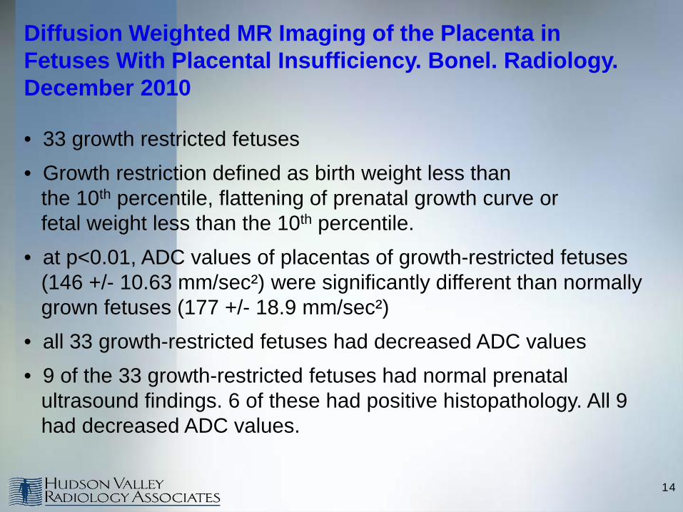
Diffusion Weighted MR Imaging of the Placenta in Fetuses With Placental Insufficiency. Bonel. Radiology. December 2010
• 33 growth restricted fetuses • Growth restriction defined as birth weight less than
the 10th percentile, flattening of prenatal growth curve or fetal weight less than the 10th percentile.
• at p<0.01, ADC values of placentas of growth-restricted fetuses (146 +/- 10.63 mm/sec²) were significantly different than normally grown fetuses (177 +/- 18.9 mm/sec²)
• all 33 growth-restricted fetuses had decreased ADC values • 9 of the 33 growth-restricted fetuses had normal prenatal
ultrasound findings. 6 of these had positive histopathology. All 9 had decreased ADC values.
14
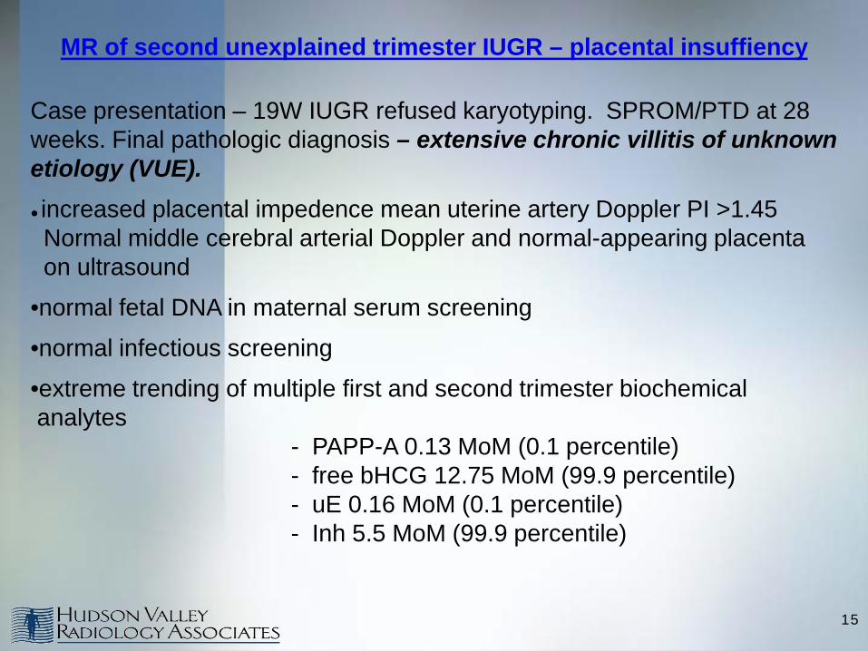
MR of second unexplained trimester IUGR – placental insuffiency
Case presentation – 19W IUGR refused karyotyping. SPROM/PTD at 28 weeks. Final pathologic diagnosis – extensive chronic villitis of unknown etiology (VUE).● increased placental impedence mean uterine artery Doppler PI >1.45 Normal middle cerebral arterial Doppler and normal-appearing placenta on ultrasound
•normal fetal DNA in maternal serum screening
•normal infectious screening
•extreme trending of multiple first and second trimester biochemical analytes
- PAPP-A 0.13 MoM (0.1 percentile)- free bHCG 12.75 MoM (99.9 percentile)- uE 0.16 MoM (0.1 percentile) - Inh 5.5 MoM (99.9 percentile)
15

MR of unexplained second trimester IUGR –placental insuffiency
Why do placental MR?
•patient refused karyotyping – etiology of IUGR uncertain •attempt MR identification of extreme placental pathologies associated with high risk for fetal/neonatal morbidity and mortality.
16
Case presentation – SPROM/PTD at 28 weeks.Chronic villitis of unknown etiology.

MR of unexplained second trimester IUGR – placental insuffiency
Chronic villitis of unknown etiology.
Case presentation – SPROM/PTD at 28 weeks.
MR findings
▪ normal T1 – no macromorphologic hemorrhagic infarct ▪ normal T2 – no placental dark banding to suggest massive perivillous fibrin deposition▪ abnormal diffusion weighting – multiple dark banding with generalized decreased ADC coefficients – restricted fluid motion
17
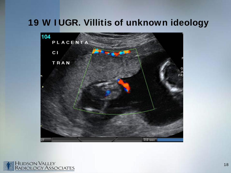
19 W IUGR. Villitis of unknown ideology
18
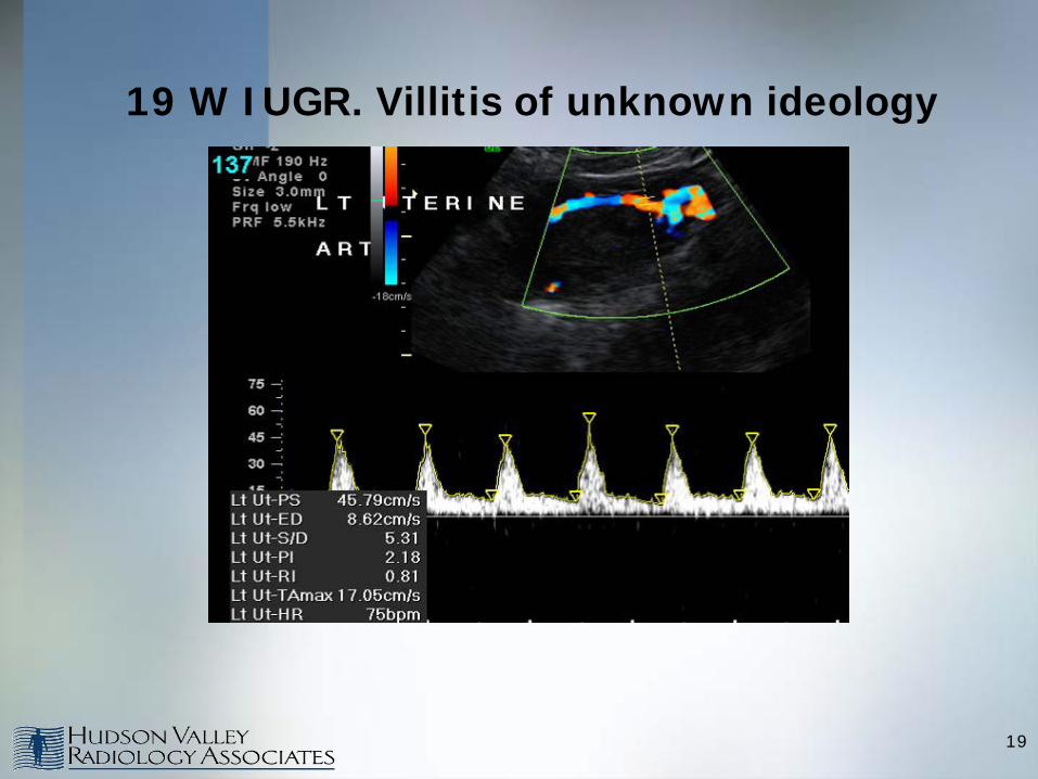
19 W IUGR. Villitis of unknown ideology
19
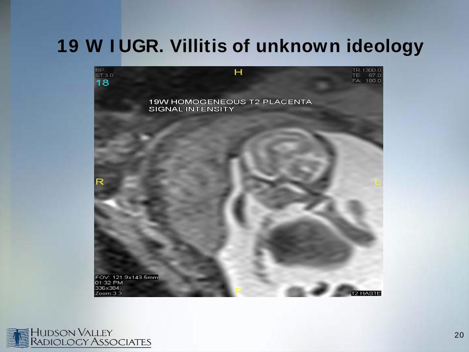
19 W IUGR. Villitis of unknown ideology
20

19 W IUGR. Villitis of unknown ideology
21

MR of unexplained second trimester IUGR – placental insuffiency
Chronic villitis of unknown etiology.
Case presentation – SPROM/PTD at 28 weeks
▪ clinical outcome – euploid neonate 1 lb, 4 oz with no structural malformations nor syndromic stigmata
▪ placental pathology – extensive chronic villitis of unknown etiology (VUE)
- 191 g placenta less than the third percentile for gestational age; 3rd-10th percentile for neonatal BW
- VUE: not part of impaired maternal-fetal perfusion spectrum
22
23
Pathologic diagnosis/clinical correlation
Villitis of unknown etiology
▪ widely believed to be a host – versus – graft response by mother directed at fetal antigens in the villous stroma
▪ major risk factor for CNS injury and stillbirth
▪ significant recurrence risk 20-30%
Redline. Human Pathol. October 2007; 38 (10): 1439-1446.

HISTORY: 31-week gestation whose outside ultrasound demonstrated an enlarged solid right kidney suspiciousfor mesoblastic nephroma. The fetus had recently exhibited ascites that had resolved. Past OB history of 29-week intrauterine fetal demise.
FINDINGS: MR demonstrates abnormal appearing kidneys of heterogeneous solid echotexture and multiple triangular hemorrhagic placental infarcts.
Still birth 2 weeks after MR exam.
AUTOPSY DIAGNOSIS:
Hemorrhagic thrombotic vasculopathy of placenta and fetus.
MR establishes severe placental pathology unable to be seen on ultrasound
24

abnormal right kidney
MR changes diagnosis from neoplasm to vasculopathy.
Hemorragic vasculopathy of fetus and placentaUS: enlarged, heterogenous R kidney.
normal left kidney
25

MR changes diagnosis from neoplasm to vasculopathy.
Hemorrhagic vasculopathy of fetus and placenta
26
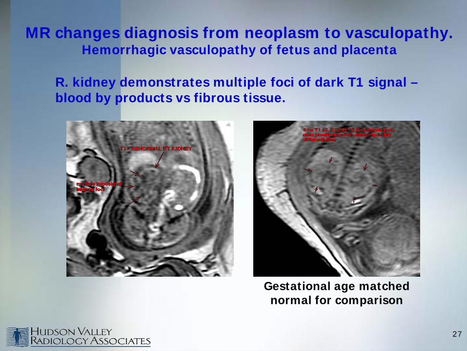
R. kidney demonstrates multiple foci of dark T1 signal –blood by products vs fibrous tissue.
MR changes diagnosis from neoplasm to vasculopathy. Hemorrhagic vasculopathy of fetus and placenta
Gestational age matched normal for comparison
27
Hemorrhagic thrombotic vasculopathy of fetus and placentaKraus. Human Pathology. Vol. 30. 1999
Placental thrombotic vasculopathy indicates significant probability of thrombi in the fetus and represents an underrecognized cause of perinatal mortality and neonatal injuries especially cerebral palsy.
•Amongst 84 perinatal autopsies 19% (16) demonstrated thrombotic vasculopathy of the placenta. 6 of 16 autopsies demonstrated thrombi of fetal brain, lungs, and kidney.
28
MR establishes severe placental pathology unable to be seen on ultrasound Abnormal placenta – IUFD 26w – maternal arterial malperfusion
History: 20w GA referred for unexplained elevated maternal serum AFP (7.2 MoM) with outside MFM ultrasound biometry two weeks smaller than expected. Normal amniocentesis including microarray. Negative maternal workup for thrombophilia, SLE andanticardiolipin antibodies.
MR results: Small placenta demonstrating homogeneous diffuse decreased (dark) signal intensity. Echoplanar diffusion imaging demonstrated decreased apparent diffuse coefficient (ADC).
Uterine artery Doppler demonstrated bilateral early diagnostic notching with markedly elevated resistive indices for gestational age. Umbilical arterial resistive index is 0.92 at the 4th standard deviation higher than expected for gestational age.
29
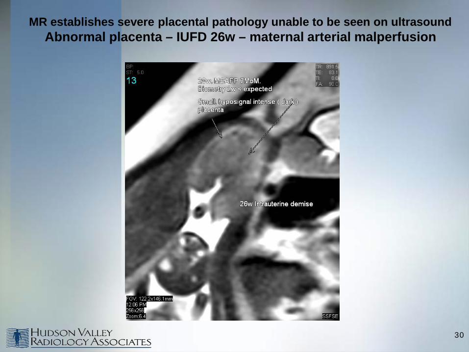
MR establishes severe placental pathology unable to be seen on ultrasound Abnormal placenta – IUFD 26w – maternal arterial malperfusion
30

MR establishes severe placental pathology unable to be seen on ultrasound Abnormal placenta – IUFD 26w – maternal arterial malperfusion
31

Clinical outcome: IUFD at 26w.
Placental pathology: •95 g placenta, less than the 10th percentile for gestational age.
•Diffuse hypoxic-ischemic villous changes.
•Focal villous stromal mineralization.
•Luminal septation within fetal stem blood vessel secondary to intraluminal clot.
•Increased numbers of circulating nucleated red blood cells in fetal circulation consistent with chronic fetal hypoxia secondary to placental vascular lesions.
Teaching point: Diffusion weighted placental imaging may identify abnormalities that corroborate with placental insufficiency. ADC values reflect restricted water diffusion in the extracellular extravascular space suggesting restricted oxygen diffusion.
*Diffusion weighted MR imaging of the placenta in fetuses with placental insufficiency. Radiology Vol. 257:December 2010.
Abnormal placenta – IUFD 26w – maternal arterial malperfusion
32
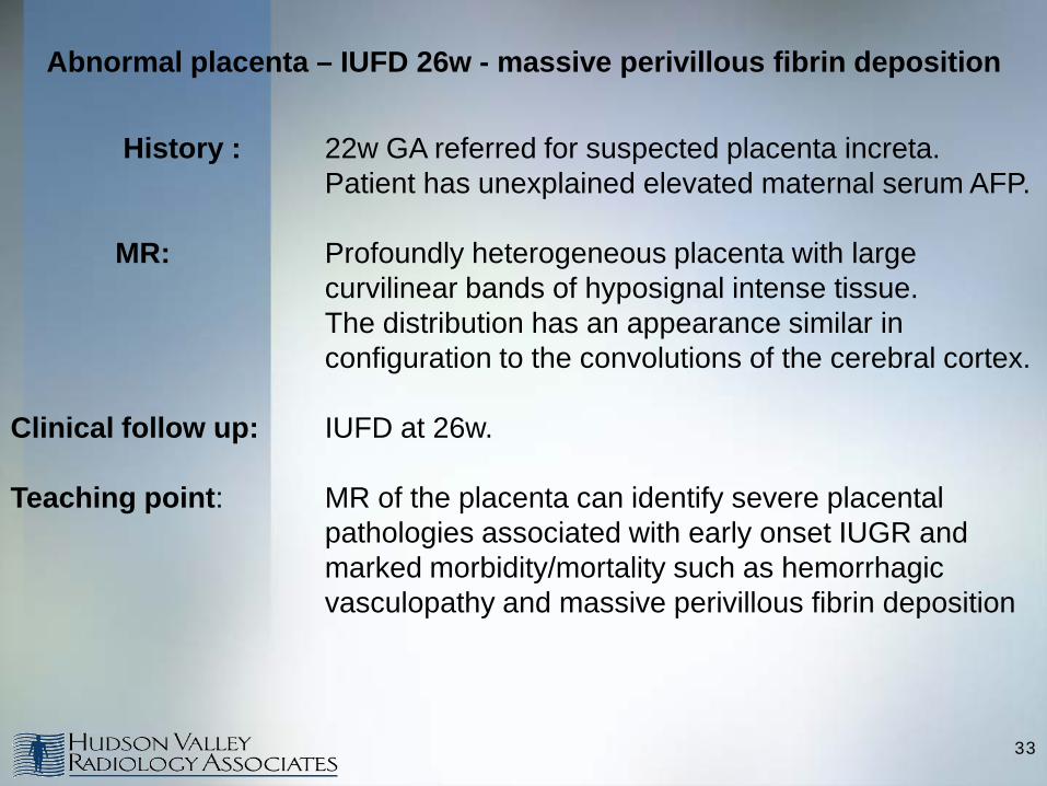
History : 22w GA referred for suspected placenta increta. Patient has unexplained elevated maternal serum AFP.
MR: Profoundly heterogeneous placenta with large curvilinear bands of hyposignal intense tissue. The distribution has an appearance similar inconfiguration to the convolutions of the cerebral cortex.
Clinical follow up: IUFD at 26w.
Teaching point: MR of the placenta can identify severe placental pathologies associated with early onset IUGR andmarked morbidity/mortality such as hemorrhagic vasculopathy and massive perivillous fibrin deposition
Abnormal placenta – IUFD 26w - massive perivillous fibrin deposition
33

MR establishes severe placental pathology unable to be seen on ultrasoundAbnormal placenta – IUFD 26w - massive perivillous fibrin deposition
34
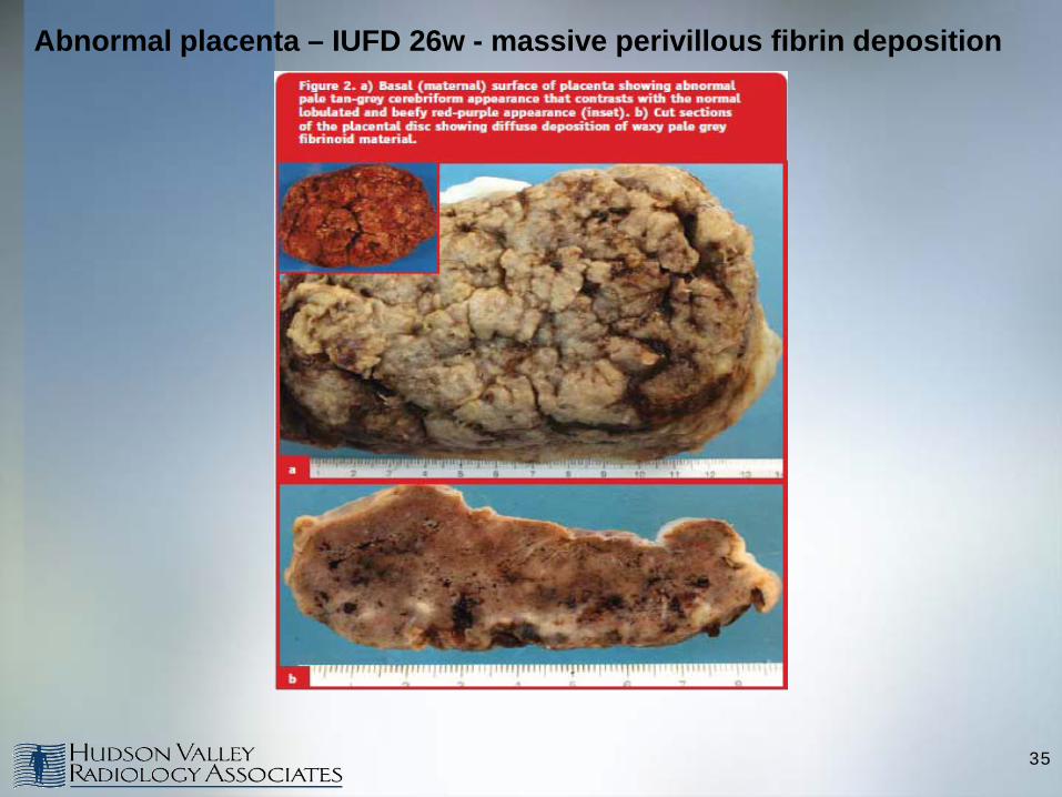
Abnormal placenta – IUFD 26w - massive perivillous fibrin deposition
35
Pathologic diagnosis/clinical correlation
Massive perivillous fibrin deposition (maternal floor infarction)
▪ risk factor for virtually all adverse outcomes from miscarriage to CNS injury
▪ recurrence risk 50-75%
▪ many affected women never achieve a successful pregnancy
Andres. Am J Obstet Gynecol. 1990; 163: 935-938.
36
Recommended