This article is protected by copyright. All rights reserved. 1
Hunting for the function of orphan G protein-coupled receptors –
beyond the search for the endogenous ligand 1
Short title: Function of orphan GPCRs
Raise Ahmad 1,2,3
, Stefanie Wojciech 1,2,3
, and Ralf Jockers1,2,3*
1 Inserm, U1016, Institut Cochin, Paris, France
2 CNRS UMR 8104, Paris, France
3 Univ. Paris Descartes, Paris, France
* Correspondence should be addressed to:
Ralf Jockers, Institut Cochin, 22 rue Méchain, 75014 Paris.
Phone: +331 40 51 64 34; Fax: +331 40 51 64 30; e-mail: [email protected]
This article has been accepted for publication and undergone full peer review but has not been through the
copyediting, typesetting, pagination and proofreading process, which may lead to differences between this
version and the Version of Record. Please cite this article as doi: 10.1111/bph.12942
Acc
epte
d A
rticl
e
This article is protected by copyright. All rights reserved. 2
Abstract
Seven transmembrane-spanning proteins (7TM), also called G protein-coupled receptors
(GPCRs), are among the most versatile and evolutionary successful protein families. Out of
the 400 non-odorant members identified in the human genome, approximately 100 remain
orphans that have not been matched with an endogenous ligand. Apart from the classical
deorphanization strategies, several alternative strategies provided recent new insights into the
function of these proteins, which hold promise for high therapeutic potential. These
alternative strategies consist of the phenotypic characterization of organisms silenced or
overexpressing orphan 7TM proteins, the search for constitutive receptor activity and
formation of protein complexes including 7TM proteins as well as the development of
synthetic, surrogate, ligands. Taken together, a variety of ligand-independent functions can be
attributed to orphan 7TM proteins that range from constitutive activity to complex formation
with other proteins and include “true” orphans for which no ligand exist and “conditional”
orphans that behave like orphans in the absence of ligand and as non-orphans in the presence
of ligand.
Abbreviations:
IUPHAR, international union of basic and clinical pharmacology; 7TM, seven
transmembrane; GPCRs, G protein-coupled receptors; ES, Ewing sarcoma; TNBC, triple
negative breast cancer; VEGF, vascular endothelial growth factor; PGF, placental growth
factor; MMP, matrix metalloproteinase; PTX, pertussis toxin; CRC, colorectal cancer; TCF4,
T-cell factor 4; AMPK, adenosine monophosphate-activated protein kinase; cAMP, cyclic
adenosine monophosphate; Pael-R, parkin-associated endothelin-like receptor; CNS, central
nervous system; PKA, protein kinase A; Ghsr1a, ghrelin receptor; AgRP, agouti-related
protein; PNS, peripheral nervous system; SCs, Schwann cells; AIS, adolescent idiopathic
scoliosis; CREB, cAMP response element-binding protein; SpD, surfactant protein D; BCL,
B-cell lymphoma; LTC, cysteinyl leukotrienes; MC4R, melanocortin receptor; HCMV,
Acc
epte
d A
rticl
e

This article is protected by copyright. All rights reserved. 3
human cytomegalovirus; MSN, medium spiny neurons; FFA, free fatty acid receptor; GABA,
gamma-aminobutyric acid; Mrg, MAS-related G-protein coupled receptor; GnRH,
gonadotropin-releasing-hormone receptor; LGR, leucine-rich repeat containing G protein-
coupled receptor; Erk, Extracellular signal-regulated kinases; CXCR/CCR, cellular
chemokine receptor; KO, knock out; Nogo, neurite outgrowth inhibitor; APP, amyloid
precursor protein; mGluR, metabotropic glutamate receptor; TRPM, transient receptor
potential melastatin; RGS, regulators of G protein signaling; TG, transglutaminase; S1P,
sphingosine 1-phosphate; DPI, diphenyleneiodonium chloride; 2-PCCA,(1R,2R)-2-pyridin-2-
yl-cyclopropane carboxylic acid ((2S,3S)-2-amino-3-methyl-pentyl)-(4′-propylbiphenyl-4-yl)-
amide
Acc
epte
d A
rticl
e
This article is protected by copyright. All rights reserved. 4
Introduction
Seven transmembrane (7TM) domain G protein-coupled receptors (GPCRs) constitute
the largest membrane receptor family. These proteins respond to a wide variety of
extracellular molecules and play a crucial role in cell-to-cell communication by transmitting
extracellular signals into cells (Rosenbaum et al., 2009). Based on sequence homology
different receptor subfamilies have been defined: rhodopsin (class A), secretin, adhesion
(class B), glutamate (class C), frizzled receptors and other 7TM proteins. In contrast to the
latter group, all others are considered to be G protein-coupled. The involvement of GPCRs in
a variety of physiological and pathophysiological processes makes this class of proteins the
most common target of pharmaceutical drugs (Drews, 2000). Genome sequencing projects
indicated that approximately 400 sequences belong to the non-odorant GPCR family in the
human genome (Fredriksson et al., 2003; Joost et al., 2002; Vassilatis et al., 2003). Most of
them have been matched with known ligands using different strategies. However, despite the
vast and longstanding efforts of academic and industrial research to pair 7TM proteins to
potential ligands, 91 non-odorant receptors still remain orphans and another 37 are awaiting
further input to be considered as deorphanized according to the IUPHAR (Davenport et al.,
2013). Deorphanization needs indeed some minimal requirement. Firstly, two or more
refereed papers from independent research groups should demonstrate activity of the ligand at
the receptor, with a potency that is consistent with a physiologic function. In some cases,
although two independent groups have reported a pairing, others have failed to reproduce this
thus requiring further validation (Davenport et al., 2013).
Although deorphanization still remains an important step towards the identification of
the function of orphan 7TM proteins, other, alternative, strategies have become equally
important over the last years to provide new insights into the function of orphan 7TM proteins Acc
epte
d A
rticl
e
This article is protected by copyright. All rights reserved. 5
(Levoye et al., 2008). Among these strategies are the phenotypic characterization of animal
models with modified expression of 7TM proteins of interest (overexpression or silencing),
the characterization of constitutive receptor activity, the association of 7TM proteins with
other proteins such as GPCRs, transporters or enzymes in heteromeric protein complexes and
the identification of synthetic, surrogate ligands (Figure 1). The most recent advances in the
identification of the function of 7TM proteins using alternative strategies will be the focus of
this article.
Patho-Physiological Functions of Orphan 7TM Proteins
Since orphan 7TM proteins represent a potential resource for future drug development,
various approaches, including transgenic and gene knockout approaches in mice, have been
used to decipher their biological roles and their involvement in different patho-physiological
conditions like cancer, metabolism, neurodegenerative disorders and energy metabolism
diseases (see Tables 1-3).
a) Orphan 7TM Proteins in Cancer Metabolism
One of the most highlighted roles of orphan 7TM proteins is in cancer biology. Different
forms of cancer like TNBC (triple negative breast cancer), skin cancer and lung cancer have
been shown to be linked to orphan 7TM proteins (Feigin et al., 2014; Gugger et al., 2008;
Perez-Gomez et al., 2013). The foremost and the most recent among these studies are related
to GPR161, which was found to be overexpressed specifically in TNBC and to correlate with
poor prognosis. Overexpression of GPR161 in human mammary epithelial cells increases cell
proliferation, migration, intracellular accumulation of E-cadherin and formation of
multiacinar structures in 3D cultures. In contrast, knockdown of GPR161 impairs
proliferation of human basal breast cancer cell lines. Therefore, GPR161 is a promising new Acc
epte
d A
rticl
e
This article is protected by copyright. All rights reserved. 6
therapeutic target for TNBC. Another orphan 7TM protein, GPR19, is frequently
overexpressed in tissue samples of lung cancer patients and is therefore considered as a new
potential candidate drug target for the treatment of a subset of lung cancers (Kastner et al.,
2012).
Recent reports are also suggesting the role of adhesion GPCRs in cancer and tumor
development. GPR64, for example, was found to be highly upregulated in Ewing sarcomas
(ES) (Richter et al., 2013). The study suggests that the GPR64 is able to induce invasiveness
and metastasis in ES by orchestrating VEGF receptor 1 ligand placental growth factor (PGF)
and matrix metalloproteinase 1 (MMP1) expression. Given that GPR64 is a membrane-bound,
and thus potentially drugable protein makes it a promising candidate for the development of
novel anti-tumor therapies in the near future. Recent studies showed that GPR48, also known
as LGR4, plays an important role in the development of various organs, cancer development
and progression such as gastric cancer and colorectal cancer (CRC) (Gao et al., 2006; Steffen
et al., 2012). Overexpression of GPR48 in primary CRC and metastatic lymph nodes
correlated with tumor invasion and metastasis. Further, GPR48 increased nuclear β-catenin
accumulation, T-cell factor 4 (TCF4) transcriptional activity and expression of its target genes
including cyclin D1 and c-Myc in CRC cells. Correlation analysis showed that GPR48
expression in CRC tissues was positively associated with β-catenin expression (Wu et al.,
2013).
LGR5 and LGR6 (having ~50% homology at the amino acid level) have also been found
implicated in cancer stem cells and other forms of cancers (Gong et al., 2012; Nakata et al.,
2014). Furthermore, these receptors have been reported to act as a receptor for R-spondins (R-
spondins 1 & 3), which is a specialized group of secreted proteins involved in development
and stem cell growth. LGR5/6 is also able to regulate Wnt/β-catenin signaling in stem cells
during malignant growth (Carmon et al., 2011; Gong et al., 2012) and therefore they are Acc
epte
d A
rticl
e
This article is protected by copyright. All rights reserved. 7
emerging as potential targets for different forms of cancers. With further studies this receptor
family could be utilized as a prognostic biomarker of a broad range of cancers in patients in
the near future. There are other studies suggesting the involvement of different orphan 7TM
proteins in cancer metabolism among which worth mentioning are GPRC5a in breast cancer
(Sokolenko et al., 2014) and GPR34 in lymphoma cell growth (Ansell et al., 2012). Taking
into account these entire studies we can speculate that orphan 7TM proteins could become
biomarkers for different cancer types and could become therapeutic targets for the treatment
of cancer related disorders in the near future justifying the development of synthetic,
surrogate ligands.
b) Orphan 7TM Proteins in Neurodegenerative and Psychiatric Disorders
Most orphan 7TM proteins are abundantly expressed in the brain thus implying a possible role
in brain physiology and neurodegenerative and psychiatric disorders. Two orphan GPCRs that
have attracted particular interest over the past 15 years in neurophysiology are GPR37 and
GPR37L1, a pair of closely related receptors that exhibit distant similarity to endothelin
receptors and other peptide-activated GPCRs (Marazziti et al., 1997; Zeng et al., 1997). They
are found exclusively in the nervous system and are known to be expressed in both neurons
and glia (Imai et al., 2001; Marazziti et al., 1998; Valdenaire et al., 1998). GPR37 was
identified as a substrate of the E3 ubiquitin ligase parkin, earning it the alternative name
“parkin-associated endothelin-like receptor,” or “Pael-R” (Valdenaire et al., 1998) and is
associated with autosomal recessive juvenile Parkinson disease (Obeso et al., 2010; Yang et
al., 2003). The connection between GPR37 and parkin has led to a focus on the dopaminergic
system in GPR37 knockout mice, which exhibited a reduced dopaminergic tone and various
subtle perturbations in dopaminergic signaling in the brain (Imai et al., 2007; Marazziti et al.,
2007). Recently Meyers et al., (Meyer et al., 2013) have reported prosaposin and prosaptide Acc
epte
d A
rticl
e
This article is protected by copyright. All rights reserved. 8
(a peptide derived from prosaposin) as cognate endogenous ligands for GPR37 and
GPR37L1. These ligands bind to GPR37 and GPR37L1, induce receptor internalization, and
stimulate GPR37-and GPR37L1-mediated signaling through PTX-sensitive G proteins.
Furthermore, they are also necessary for mediating endogenous responses in primary cortical
astrocytes. This study becomes more imperative since it is known that both prosaposin and
prosaptide have been reported in numerous studies to exert neuro- and glioprotective effects
(Campana et al., 1998; Li et al., 2010; O'Brien et al., 1994; O'Brien et al., 1995) via the
stimulation of G protein-mediated pathways (Campana et al., 1998; Hiraiwa et al., 1997; Yan
et al., 2000). Thus future studies aiming at the neuroprotective and glioprotective actions of
prosaposin and prosaptide the development of small molecule ligands for these receptors may
provide new therapeutic possibilities for the treatment of Parkinson’s disease and other
neurodegenerative disorders. In the same line findings from Gandia et al., (Gandia et al.,
2013) further highlighted the role of its cysteine rich domain in receptor-mediated
cytotoxicity and improved our understanding regarding its involvement in the patho-
physiology of Parkinson’s disease.
Other promising orphan 7TM proteins having reported to have a role in psychiatric disorders
are GPR50 (Thomson et al., 2005), GPR88 (Del Zompo et al., 2014; Logue et al., 2009),
GPR6 (Oeckl et al., 2014) and GPR52 (Komatsu et al., 2014). Based on genotyping study in
human subjects Thomson et al., (2005) reported a link between polymorphisms in the GPR50
gene and major mental illness while other studies targeted the dopaminergic system in the
striatum and found a possible role of GPR6, GPR52 and GPR88 in related
neurodegenerative/psychiatric disorders. For instance, GPR6 is abundantly expressed in
striatopallidal neurons and its depletion reduces cAMP concentrations in the striatum and
alters the striatal dopaminergic system. Furthermore, knockdown of GPR6 caused an
interesting behavioral phenotype in the form of enhanced motor activity combined with Acc
epte
d A
rticl
e
This article is protected by copyright. All rights reserved. 9
reduced abnormal involuntary movements. These findings could offer an opportunity for the
treatment of Parkinson's disease beyond dopamine replacement (Oeckl et al., 2014). In the
same line, another study based on detailed histological investigation suggests that GPR52
may modulate dopaminergic and glutamatergic transmission in neuronal circuits responsible
for cognitive function and emotion (Komatsu et al., 2014). GPR52 knockout and transgenic
mice exhibited psychosis-related and antipsychotic-like behaviors, respectively. Similarly,
GPR88 has also been the centre of attraction especially in the research of neuropsychiatric
diseases. In rodents, GPR88 is highly expressed in the striatum, with its expression being
limited to dopamine D1 and D2 receptor- containing medium spiny neurons (MSN)
implicated in the pathophysiology of and being modulated by treatments for schizophrenia.
The modulatory role of GPR88 in striatal dopamine function suggests it may be a new target
for the treatment of psychiatric disorders (Logue et al., 2009). Targeted viral expression of
GPR88 in MSNs rescued the molecular and electrophysiological properties and normalized
behavior (Quintana et al., 2012).
There are a couple of other orphan 7TM proteins, which have been observed to modulate
neurogenesis, neurite outgrowth and differentiation in the CNS and its associated patho-
physiologies. GPRC5B is predominantly expressed in neural progenitors in the developing
mouse brain and its depletion in progenitors results in a failure to adopt a neuronal fate.
Further, GPRC5B-mediated signaling affects β-catenin signaling, which is important for the
neuronal differentiation of progenitors during the neurogenic phase (Kurabayashi et al.,
2013).
Interestingly, among those orphan 7TM proteins that constitutively activate Gs proteins,
GPR3, GPR6 and GPR12 have been found to mediate various neurological functions. They
are able to promote neurite outgrowth by constitutively upregulating the cAMP/PKA pathway
(Tanaka et al., 2007). Recently, GPR3 has been reported to inhibit the proliferation of Acc
epte
d A
rticl
e
This article is protected by copyright. All rights reserved. 10
cerebellar granule cells in vitro and to promote survival of neurons by inhibiting their
apoptosis in various physiological conditions (Tanaka et al., 2014; Tanaka et al., 2009).
GPR12 was shown to enhance neurite outgrowth and increase cAMP levels during neurite
extension. Moreover, GPR12 knockout mice showed impaired locomotion, motor function
and learning (swimming) in the Morris Water Maze, suggesting its potential involvement in
learning and memory functions. GPR3 has also been reported to regulate the expression and
development of neuropathic pain and in the analgesia induced by morphine. The genetic
deletion of GPR3 produced hypersensitivity to thermal non-noxious and noxious stimuli
without affecting the spinal inflammatory response associated with sciatic nerve injury and
reduced morphine antinociception (Ruiz-Medina et al., 2011). Furthermore, these mice
showed differences in the locomotor, rewarding and reinforcing effects of cocaine mainly
after acute administration of cocaine, compared to wild-type mice. Taken together GPR3 is
emerging as a new molecular target in neuropathic pain therapy as well as a new component
of the pro-opioid receptor system. With all this information in our hand we can hypothesize
that orphan 7TM proteins are evolving as potential therapeutic targets for CNS-related
diseases.
c) Orphan 7TM Proteins in Energy Metabolism and Diabetes
Orphan 7TM proteins have been proposed to play a noteworthy role in modulating energy
expenditure and metabolism as well as energy homeostasis and related physiological
functions. GPR50 plays a role in the regulation of energy metabolism (Ivanova et al., 2008)
and GPR50 -/-
mice show reduced weight and partial resistance to diet-induced obesity.
Furthermore, GPR50 seems to play an important role in leptin-dependent adaptive
thermogenesis (Bechtold et al., 2012). GPR83 has been suggested to be involved in the
central regulation of energy metabolism as a potential modulator of the hypothalamus–Acc
epte
d A
rticl
e
This article is protected by copyright. All rights reserved. 11
pituitary–adrenal axis (Muller et al., 2013). In the arcuate nucleus, GPR83 colocalizes with
the ghrelin receptor (Ghsr1a) and the agouti-related protein AgRP. The orexigenic and
adipogenic effect of ghrelin is accordingly potentiated in GPR83-deficient mice. GPR83
modulates ghrelin action and regulates systemic metabolism through other ghrelin-
independent pathways. Several other orphan 7TM proteins have also been linked to lipid
metabolism and type 2 diabetes (Bhattacharyya et al., 2006; Chen et al., 2012; Engel et al.,
2011).
GPR82, GPR26, GPR21, GPR27 and GPRC5B have recently been found to be involved in
regulation of diet-induced obesity and insulin sensitivity. As reported by Engel et al. (2011)
GPR 82 -/-
mice show reduced body weight, food intake, triglycerides level and increased
insulin sensitivity and glucose tolerance. In the same line, GPR26 has been reported as a
potent regulator of energy homeostasis by controlling hypothalamic AMP activated protein
kinase (AMPK) activation and its targeted deletion caused hyperphagia and hypometabolism,
which leads to early onset of diet-induced obesity (Chen et al., 2012). Similarly, GPR21-/-
animals are protected from high-fat diet-induced inflammation and reduced insulin sensitivity.
GPR21 is highly expressed in the hypothalamus and macrophages of mice and targeted
deletion of GPR21 in the whole body led to a robust improvement in glucose tolerance and
systemic insulin sensitivity and a modest lean phenotype (Osborn et al., 2012). A study by Ku
et al., (Ku et al., 2012) showed that GPR27 modulates pancreatic β-cell function, insulin
sensitivity and its knockdown in β-cells reduces endogenous mouse insulin promoter activity
and glucose-stimulated insulin secretion. Similarly, GPRC5B also regulates β-cell viability
and insulin secretion and its silencing is associated with increased glucose- and glutamate-
induced insulin secretion. The hyperactivation of GPRC5B contributes to impaired insulin
secretion, a hallmark of type 2 diabetes. Antagonizing GPRC5B activity might represent a
means of restoring normal insulin secretory function in diabetic patients (Soni et al., 2013). Acc
epte
d A
rticl
e
This article is protected by copyright. All rights reserved. 12
Taken together, several orphan 7TM proteins appear to be involved in glucose and lipid
metabolism and might in the future emerge as new drug targets for type 2 diabetes and other
metabolic disorders.
Adhesion Orphan 7TM Proteins and their Physiological Aspects
Adhesion GPCRs have been neglected for a long time but have more recently become the
subject of intense research. In some cases the involvement of adhesion GPCRs in diseases
like cancer, has been established as described above but the majority of studies concentrated
on deciphering there involvement in various physiological processes as discussed in this
chapter (Table 2). Adhesion GPCRs are unique in several aspects. They have a long N-
terminus containing multiple domains, which are linked to the 7TM region through the GPS
(GPCR proteolytic site) domain, which has autocatalytic properties (Baud et al., 1995;
Krasnoperov et al., 1997). Most adhesion GPCRs are orphan 7TM proteins and only for some
of them a natural ligand and a clearly defined function has been proposed. In general,
adhesion GPCRs are implicated in immunological function, synaptic function, planar cell
polarity, tumor progression, and fertility (Davies et al., 2004; Lin et al., 2005; Steinert et al.,
2002; Usui et al., 1999).
Recently, GPR124 and GPR126 have been shown to regulate the development of different
tissues in mammals. GPR124 has been show to affect CNS-specific angiogenesis,
vascularization (Anderson et al., 2011; Cullen et al., 2011; Kuhnert et al., 2010) and is also
required for VEGF-induced tumor angiogenesis (Wang et al., 2014). GPR126 is an important
regulator of embryonic development in mammals and studies on GPR126 -/-
mice have shown
that its disruption leads to fully penetrant embryonic lethality with cardiac abnormality
(Waller-Evans et al., 2010). In another study using a GPR126 knockout mouse line from
Taconic (T-GPR126 -/-
) (Monk et al., 2011), most mutants die in utero, although a few mice Acc
epte
d A
rticl
e
This article is protected by copyright. All rights reserved. 13
survive to postnatal stages. T-GPR126 -/-
mice are characterized both by a lack of myelination
in the peripheral nervous system (PNS) and by multiple defects in peripheral nerves. GPR126
is thought to function in Schwann cells (SCs) through G proteins to control myelination and
differentiation (Mogha et al., 2013). Apart from acting at the CNS, GPR126 is also expressed
in the endocardium during early mouse heart development and its knockout in mice and
knockdown in zebrafish caused hypotrabeculation and affected mitochondrial functions (Patra
et al., 2013). The link with cardiovascular development is further supported by a recent
genetic study by Kou et al., (Kou et al., 2013) revealing its association with the skeletal
disease called adolescent idiopathic scoliosis (AIS). Taken together orphan 7TM proteins like
GPR126 could be a potent target for the CNS related vascular pathologies and developmental
functions.
Another member of this class, GPR97, is highly conserved among species and has been
shown to be a regulator of B-lymphocyte population and to regulate constitutive CREB and
NF-κB activity in mice (Wang et al., 2013). Deletion of GPR97 in mice caused disorganized
spleen architecture with modified primary humoral and secondary immune responses. Further,
Valtcheva et al., (Valtcheva et al., 2013) found GPR97 expressed by lymphatic endothelium
and postulated a role in the development and function of the lymphatic vascular system under
pathological conditions. Therefore, GPR97 as a lymphatic adhesion orphan GPCR might open
new possibilities for the future pharmacological manipulation of lymphangiogenesis.
A very interesting role of GPR116 has been reported in respiratory and lung physiology.
Disruption of the GPR116 gene in mice resulted in progressive accumulation of surfactant
lipids and proteins in the alveolar space, which lead to labored breathing, and reduced
lifespan. Indeed, GPR116 expression in alveolar type II cells is required for maintaining
normal surfactant levels (Yang et al., 2013). GPR116 appears to function as molecular sensor
of alveolar surfactant lipid pool by regulating surfactant secretion (Bridges et al., 2013). Acc
epte
d A
rticl
e
This article is protected by copyright. All rights reserved. 14
Along the same line, Fukuzawa et al., (Fukuzawa et al., 2013) found that GPR116 acts
specifically on surfactant protein D (SpD) in order to regulate synthesis and secretion of
surfactant lipids and proteins and a stimulating effect on recycling (uptake) in response to
elevated levels of Sp-D in the alveolar space. Thus, GPR116 plays an indispensable role in
lung surfactant homeostasis with important ramifications for the understanding and treatment
of lung surfactant disorders.
Constitutively active orphan 7TM proteins
Some of the more than 100 remaining orphan 7TM proteins might have ligand-independent
functions such as a significant degree of constitutive activity to trigger functional responses in
the absence of ligands (Figure 1B). The molecular explanation for constitutive activity is
often based on specific receptor sequences that stabilize the active receptor conformation,
thus allowing the interaction with various proteins that induce cellular signaling events
(Lowther et al., 2013).
Constitutive activity of orphan 7TM proteins improves binding to G proteins even in the
absence of ligand. The constitutive activity of GPR3 (Ledent et al., 2005; Mehlmann et al.,
2004), GPR6 and GPR12 (Tanaka et al., 2007) induces cAMP production resulting from
constitutive coupling to Gαs proteins. For GPR6, it has also been shown in vivo, that its
ablation leads to decreased cAMP levels in the striatum (Oeckl et al., 2014). A constitutive
coupling to Gαi proteins has been demonstrated for GPR20 (Hase et al., 2008). The constant
G protein coupling results in different downstream signaling activities as shown for the Gαs
coupled GPR26 (Jones et al., 2007), which inhibits AMPK activity at the hypothalamic level
in order to prevent adiposity (Chen et al., 2012) and increases CREB phosphorylation in the
amygdale, revealing its possible regulatory role in anxiety (Zhang et al., 2011). GPR12
promotes Erk1/2 phosphorylation and the expression of B-cell lymphoma 2 (BCL2), which is Acc
epte
d A
rticl
e
This article is protected by copyright. All rights reserved. 15
important for its role in supporting proliferation, cell survival (Lu et al., 2012b) and neurite
outgrowth (Lu et al., 2012a).
The degree of constitutive activity of an orphan 7TM protein depends on its expression level,
but also on regulatory proteins as β-arrestin 2 and the G protein–coupled receptor kinase 2
GRK2, that may be implicated in receptor desensitization as shown for GPR3 (Lowther et al.,
2013). An interesting example is GPR17, which has two constitutively active splice variants
GPR17-L and GPR17-S in humans that are expressed in a tissue-specific manner and that
differ in the length of their N-termini and activate Gαi proteins independently from the
putative GPR17 ligands cysteinyl leukotrienes LTC4 and LTD4 (Benned-Jensen et al., 2010).
Constitutive activity has also been observed for orphan adhesion GPCRs, mainly on the level
of the small RhoGTPases RhoA, Cdc42 and Rac1, which are important players in the
regulation of cellular motility (Gupte et al., 2012). Indeed, GPR97, which is expressed in
lymphathic epithelium, is coupled to Gαo and regulates the balance of the active Rho GTPases
Cdc42 and RhoA (Valtcheva et al., 2013; Wang et al., 2013) and GPR56 interferes with
cellular migration of neural cells via the Rho GTPase pathway (Iguchi et al., 2008).
In an effort to provide a molecular explanation for the constitutive activity of orphan 7TM
proteins, deletion of the N-terminal part of GPR61 resulted in the loss of constitutive activity
leading to the hypothesis that constitutive activity of some orphan receptors might be based
on the existence of an N-terminal tethered ligand (Toyooka et al., 2009) (Figure 1C).
Similarly, it has been demonstrated, that the melanocortin receptor MC4R, besides its ligand
induced activation, displays a constitutive activity, which is dependent on its N-terminus
(Ersoy et al., 2012). This constitutive activity can be counteracted by the agouti-related
protein, a natural inverse agonist thus suggesting the interesting possibility that some Acc
epte
d A
rticl
e
This article is protected by copyright. All rights reserved. 16
constitutively active 7TM proteins might be regulated by natural inverse agonists rather than
agonists.
Retroviruses are known to encode proteins with a 7TM structure with sequence homology to
cellular chemokine receptors (Rosenkilde et al., 2008). Several of these proteins show
constitutive activity. Most recent examples are UL33, encoded by the human cytomegalovirus
(HCMV), and BILF, encoded by the Epstein Barr Virus, which both couple promiscuously to
different Gα proteins (Vischer et al., 2014). Their constitutive activity modulates the signaling
in the host cell beneficially for virus-triggered promotion of cellular proliferation and
transformation (Lyngaa et al., 2010). The high level of constitutive activity of virally-encoded
GPCRs might be the result of a less conserved DRY motif, which is responsible for an
inhibitory, inactive conformation of class A GPCRs (Jensen et al., 2012). Thus, some viruses
developed a strategy based on constitutively active 7TM proteins that modify the function of
the host cell in a ligand-independent manner.
Although many GPCRs exhibit a significant level of constitutive activity, it should be
mentioned that in the case of orphan 7TM proteins the possibility always remains that the
apparent constitutive activity might actually reflect the presence of an endogenous ligand that
either is difficult to remove or which is produced by the cell. A study on the former orphan
GPR40 (now called FFA1 for free fatty acid receptor 1) revealed that its apparent constitutive
activity is due to a permanent occupation of the receptor binding site by its endogenous free
fatty acid ligand (Stoddart et al., 2007). The detection of constitutive activity can sometimes
go hand in hand with the disclosure of the identification of a ligand, as recently shown for
GPR174 (Sugita et al., 2013). Constitutively active 7TM proteins present new opportunities
for the design of suitable inverse agonists. Acc
epte
d A
rticl
e
This article is protected by copyright. All rights reserved. 17
Function of Orphan 7TM Proteins in Heterodimeric Complexes with GPCRs
Another function of orphan 7TM proteins relies in their ability to interact with other proteins
providing an allosteric modulation of their activity. At the beginning of the 21st century, the
emerging concept of the formation of heteromeric complexes of GPCRs was extended to
orphan 7TM proteins (Levoye et al., 2006) (Figure 1F). These heteromers are formed
typically between a ligand-dependent GPCR and an orphan 7TM protein that are often
members of the same subfamily. The case of an obligatory heteromeric complex is found in
GABAB1 and GABAB2 receptors. Both proteins constitute a functional complex, with each
one having a specific task: GABAB1 binds the ligand and GABAB2 is responsible for
transducing the signal to the G protein. In this case GABAB2 lacks the GABA binding domain
and behaves thus as the orphan receptor (Kniazeff et al., 2002). Recently this has also been
demonstrated for GPR179, another orphan 7TM protein, which can form a heteromer with the
glutamate receptor subtype mGluR6 in the retina and which is involved in dim light vision
signal transmission from photoreceptors to ON bipolar cells (Orlandi et al., 2013).
Another possibility is a conditional heteromerization of related GPCRs, where heteromer
formation can generate a novel functional unit through allosterism between the two protomers
compared to the corresponding homomers. The pioneer work in this field demonstrated the
heteromer formation of the melatonin MT1 receptor and its orphan family mate GPR50. In
this case, the orphan GPR50 negatively interferes with melatonin-dependent signal
transduction in a manner that is dependent on its long cytosolic C-tail (Levoye et al., 2006).
Another example is the β-alanine binding mas-related receptor MrgD, which can form a
complex with its orphan relative, MrgE. This interaction is associated with potentiation of
signaling and inhibition of internalization of the receptor (Milasta et al., 2006).
The protochordate Ciona intestinalis possesses 4 isoforms of the gonadotropin-releasing-
hormone receptor (GnRH), of which the GnRH R4 is an orphan subtype. The R4 can Acc
epte
d A
rticl
e
This article is protected by copyright. All rights reserved. 18
heteromerize with R1 and potentiate R1-induced Erk1/2 activation and calcium mobilization.
In contrast, heteromers composed of R2 and R4 lead to a shift from Gs to Gi coupling,
resulting in a decrease in cAMP production (Sakai et al., 2012; Sakai et al., 2010). These
results demonstrate that the orphan R4 functions as an allosteric modulator of GnRH
receptors and it will be interesting to see, whether this can be also found in species other than
Ciona intestinalis.
Virally-encoded GPCRs can also form GPCR/orphan 7TM protein complexes as shown for
HCMV-encoded proteins like US28 that promiscuously binds several host cell chemokines,
forming a complex with the orphan 7TM proteins US27, UL33 and UL78. The latter two
complexes have a silencing effect on the NFκB signaling activity of US28, providing several
possibilities to adapt the signaling in the host cell depending on the expression pattern of the
orphan 7TM proteins (Tschische et al., 2011). Interestingly, complex formation can also be
seen between virally-encoded 7TM proteins and GPCRs expressed by the host cell. Indeed,
UL27, UL33 and UL78 can interact with the cellular chemokine receptors CCR5 and CXCR4
and modulate their function including their HIV-coreceptor properties (Arnolds et al., 2013;
Tadagaki et al., 2012).
The constitutively active Epstein Barr Virus 7TM protein BILF1 forms heteromers with
various human chemokine receptors (Vischer et al., 2008). The BILF1-CXCR4 complex
modulates host cell signaling in a competitive mode of action: both receptors signal via Gi
proteins and the complex formation induces a competition of both receptors for the cellular
Gi pool leading to an impairment of the CXCR4-induced Gi signaling cascade (Nijmeijer
et al., 2010). These examples demonstrate that viruses have developed interesting strategies to
use their own orphan 7TM proteins to modulate the signaling of host cell receptors in their
favor. Acc
epte
d A
rticl
e
This article is protected by copyright. All rights reserved. 19
The emerging concept of conditional orphan receptors is further expanding the idea of
ligand-independent functions of 7TM proteins. Conditional orphan receptors can be defined
as proteins with an identified ligand, which can in the absence of ligand behave as orphan
receptors, as recently proposed for the ghrelin receptor GHSR1a. This receptor forms
heteromers with the D2 dopamine receptor in the hypothalamus, a tissue where the ghrelin
peptide produced in the stomach is not supposed to localize. The conditional orphan GHSR1a
modulates D2 receptor activity and is responsible for the anorexigenic properties of dopamine
stimulation. The independence of this interaction of the ghrelin ligand was additionally
demonstrated with the use of ghrelin KO mice (Kern et al., 2012). The modulating activity of
GHSR1a on dopamine signaling can be a consequence of the constitutive ligand-independent
activity of the GHSR1a receptor and it will be interesting to see, whether this concept is also
transferable to other ghrelin heteromers (Schellekens et al., 2013) or other constitutively
active GPCRs.
Function of Orphan 7TM Proteins in Heterodimeric Complexes with Transporters,
Enzymes and other Cellular Proteins
The formation of functional multiprotein complexes is an emerging concept and work
in recent years demonstrated that orphan 7TM proteins can be part of functional complexes
between different protein families and thereby regulate their function or enzymatic activity
(Figure 1E).
Proteomic techniques have been used for uncovering the existence of protein interactions.
GPR50, the orphan member of the melatonin receptor subfamily, has a characteristic long
cytosolic C-tail. Yeast-2-hybrid screens using the C-tail of GPR50 as bait revealed several
cytosolic interactors like Nogo-A and TIP60. Binding of the neurite-outgrowth-inhibitor
Nogo-A to GPR50 was shown to counteract for its negative influence on neurite outgrowth Acc
epte
d A
rticl
e
This article is protected by copyright. All rights reserved. 20
(Grunewald et al., 2009). Complex formation of GPR50 with the transcription factor TIP60
results in the mutual nuclear translocation of the cleaved C-tail of GPR50 and TIP60 and
enhances the nuclear glucocorticoid receptor-dependent gene expression (Li et al., 2011).
The constitutively active GPR3 is involved in the progression of Alzheimer’s pathology
(Thathiah et al., 2009). Elucidating the mechanism has shown the influence of the GPR3 C-
tail and -arrestin 2 binding as intermediates for the increase of production of cleaved
amyloid-beta peptide that can lead to amyloid plaque formation. Both GPR3 and -arrestin 2
enhance γ-secretase activity, probably through a direct interaction (Thathiah et al., 2013).
Demonstration of the in vitro interaction between GPR3 and the amyloid precursor protein
(APP) suggests the formation of a multiprotein complex that is responsible for the generation
of cleaved peptides in the presence of GPR3 (Nelson et al., 2013).
The class C orphan GPR179, that forms a heteromer with mGluR6, seems to be part of a
macromolecular complex at dendrites of ON bipolar neurons in the retina together with the
transient receptor potential melastatin 1 (TRPM1) cation channels and the RGS proteins 7 and
11. The complex is required for appropriate metabotropic signal transmission from
photoreceptors to ON bipolar neurons (Orlandi et al., 2013; Orlandi et al., 2012; Ray et al.,
2014) and depletion of GPR179 leads to stationary night blindness (Orhan et al., 2013;
Peachey et al., 2012).
The adhesion GPCR GPR56 forms a functional complex with the transmembrane tetraspanins
CD81 and CD9 (Little et al., 2004) and binds to the extracellular enzyme transglutaminase
TG2 (Xu et al., 2006). GPR56 can negatively influence the enzymatic activity of TG2 by
promoting its internalisation and degradation, which prohibits TG2-dependent melanoma
formation (Yang et al., 2014).
The cytosolic part of the class C orphan GPRC5B contains several tyrosine residues that can
be phosphorylated by Fyn kinase. GPRC5B and Fyn together form a complex, which induces Acc
epte
d A
rticl
e
This article is protected by copyright. All rights reserved. 21
NFκB signaling and inflammatory signaling activity in adipocytes possibly in a G protein-
independent manner (Kim et al., 2012).
These examples demonstrate that the function of orphan 7TM proteins needn’t be limited to
classical GPCR signaling activity but can be expanded to other functions related to the
formation of multiprotein complexes. Regulation of the formation and activity of such
complexes is expected to be tissue-specific, as the expression of its components will depend
on their specific expression patterns. The multiple techniques for identifying protein
complexes (Daulat et al., 2013) and their application in vivo will surely enable the
identification of more protein complexes and binding partners of orphan 7TM proteins in the
future.
Identification of Synthetic, Surrogate Ligands for Orphan 7TM Proteins
Identification for synthetic surrogate ligands for orphan 7TM proteins presents an
interesting alternative to classical deorphanization approaches to obtain pharmacologic and
therapeutic tools for these 7TM proteins (Figure 1D). It is worth mentioning that small
synthetic molecules may not necessarily have identical functions compared to endogenous
ligands and acts outside of the orthosteric binding site and may modulate the receptor by
binding to an allosteric binding site. The action of allosteric ligands is generally considered to
depend on the presence of the orthosteric ligand. However, several examples demonstrate that
allosteric agonists may have also an effect, which is independent of the presence of the
orthosteric ligand (Schwartz et al., 2007). This is nicely illustrated by two compounds acting
on orphan 7TM proteins. The first compound, CGP7930, allosterically enhances GABA
binding to the GABAB receptor heterodimer. Importantly, CGP7930 can activate the 7TM
domain of the orphan GABAB2 subunit alone (Binet et al., 2004). Similar observations have
been made for taste T1R receptors. Lactisole and cyclamate have been proposed to bind to
the 7TM domain of the orphan T1R3 subunit and thus allosterically regulate ligand binding to Acc
epte
d A
rticl
e
This article is protected by copyright. All rights reserved. 22
the bilobate extracellular orthosteric sites of T1R1 and T1R2 in their respective heterodimers.
Collectively, this indicates that orphan 7TM proteins can be targeted by allosteric agonists
that may be interesting compounds even in the absence of orthosteric ligands for these
proteins.
In addition, the signaling profile of synthetic ligands does not necessarily overlap with that of
the natural agonist raising the question of how reflective of the natural agonist’s action is the
synthetic ligand-activated receptor. Indeed, synthetic ligands, by binding to orthosteric or
allosteric sites, might have biased properties favoring receptor conformations that promote a
different repertoire of downstream signaling responses. Synthetic surrogate ligands therefore
rather complement than substitute for the natural ligand.
When searching for synthetic ligands activating orphan 7TM proteins, a recurrent question
concerns the use of an appropriate functional readout. The recent work from the Kostenis
group may lead to a new way to solve this question as the authors employed a label-free
technology by monitoring dynamic mass redistribution within the cell that occurs as a
consequence of cell stimulation. This non-invasive technique provides a global, integrative
and signaling pathway-unbiased measure of cellular activation. Using this method, the
MDL29,951 compound was identified as a selective agonist for the orphan GPR17, involved
in orchestration of oligodendrocyte differentiation and myelination in the central nervous
system (Hennen et al., 2013). Subsequent pathway analysis confirmed that this compound
triggers a range of downstream signaling molecules including Gi, Gs, Gq and ß-arrestin. The
benefit of having identified a surrogate ligand is further illustrated by this study as
MDL29,951 provided a reliable positive control to monitor GPR17 activation in multiple
assays. Using this positive control, previously proposed GPR17 ligands, uracil nucleotides
and cysteinyl-leukotrienes, failed to be active confirming previous reports putting GPR17
back to the orphan status. Other previously proposed surrogate ligands of GPR17 based on Acc
epte
d A
rticl
e
This article is protected by copyright. All rights reserved. 23
high throughput virtual screening of the GPR17 binding site (Eberini et al., 2011) can now be
validated in parallel with MDL29,951 in the dynamic mass redistribution assay.
The deorphanization of GPR3, a potential new target for Alzheimer’s disease treatment that
also modulates early phases of cocaine reinforcement, is at a similar stage. Although
sphingosine 1-phosphate (S1P) has been reported as a putative ligand for GPR3 (Uhlenbrock
et al., 2002), these results remain controversial as several groups were unable to confirm these
results. In addition, S1P activates many other GPCRs including S1P1-5 receptors, making
S1P unsuitable for further investigation of GPR3-specific signaling and subsequent functions.
Small-molecule surrogate ligands such as diphenyleneiodonium chloride (DPI) represent an
interesting alternative in this context as it specifically promotes GPR3-mediated cAMP
accumulation (Ye et al., 2014).
Even for those 7TM proteins, for which a surrogate ligand has already been identified, further
screening campaigns might be justified. In some cases positive hits have limited use because
of off-target effects by targeting other receptors in a complex biological environment like
tissues. In other cases such ligands may exhibit variable potency across different species.
Confronted by such a situation, several studies have been undertaken to identify surrogate
GPR35 agonists with improved profiles for this receptor that modulates the immune response
and is involved in pain perception (Funke et al., 2013; Neetoo-Isseljee et al., 2013; Thimm et
al., 2013)
Computer-aided pharmacophore modeling based on identified surrogate ligands represents
another strategy to optimize known surrogate ligands or to make predictions about the
structure of putative endogenous ligands. This approach was successfully applied to GPR139,
which led to the proposal that aromatic-containing dipeptides are putative endogenous
agonists for GPR139 (Isberg et al., 2014; Shi et al., 2011). GPR139 is expressed in the brain
and controls locomotor activity. Acc
epte
d A
rticl
e
This article is protected by copyright. All rights reserved. 24
Furthermore, based on the patent literature, the first GPR88 surrogate agonist, 2-PCCA (1R,
2R)-2-pyridin-2-yl-cyclopropane carboxylic acid ((2S,3S)-2-amino-3-methyl-pentyl)-(4′-
propylbiphenyl-4-yl)-amide), has been recently designed. In functional assays inhibition of
cAMP production was observed upon activation of GPR88, an orphan 7TM protein highly
expressed in the striatum and implicated in psychiatric disorders (Jin et al., 2014).
General Perspectives
The repertoire of ligand-independent functions of orphan 7TM proteins is steadily increasing.
Proteomic approaches produce an increasing list of proteins interacting with orphan 7TM
proteins. In addition, formation of heterodimeric GPCR complexes is shown in an increasing
number of cases in vivo. The existence of “conditional” orphan 7TM proteins, typically
consisting in deorphanized GPCRs with identified ligand, which however behave as orphans
in the absence of ligand, largely expands the number of potential orphan 7TM proteins
particularly in the context of receptor heterodimers. Another future focus would be the
identification of further unexplored functions such as the nuclear translocation of orphan 7TM
proteins, either of the entire proteins or part of it, as recently shown for GPR50 (Li et al.,
2011) and GPR158 (Patel et al., 2013), respectively. These examples demonstrate that the
function of orphan 7TM proteins may go far beyond its potential ligand-dependent function
opening new conceptual and therapeutic avenues that may even apply to already deorphanized
GPCRs.
Acc
epte
d A
rticl
e
This article is protected by copyright. All rights reserved. 25
Acknowledgements:
This work was supported by grants from the Agence Nationale de la Recherche (ANR 2011 -
BSV1-012-01 “MLT2D” and ANR-2011-META “MELA-BETES”, ANR-12-RPIB-0016
“MED-HET-REC-2”), the Fondation de la Recherche Médicale (Equipe FRM
DEQ20130326503, to R.J.), the Fondation ARC (N°SFI20121205906), Institut National de la
Santé et de la Recherche Médicale (INSERM) and Centre National de la Recherche
Scientifique (CNRS).
Author contributions:
RA, SW and RJ wrote the review together.
Competing Interests’ Statement: None
References
Anderson KD, Pan L, Yang XM, Hughes VC, Walls JR, Dominguez MG, et al. (2011).
Angiogenic sprouting into neural tissue requires Gpr124, an orphan G protein-coupled
receptor. Proc Natl Acad Sci U S A 108: 2807-2812.
Ansell SM, Akasaka T, McPhail E, Manske M, Braggio E, Price-Troska T, et al. (2012).
t(X;14)(p11;q32) in MALT lymphoma involving GPR34 reveals a role for GPR34 in tumor
cell growth. Blood 120: 3949-3957.
Arnolds KL, Lares AP, Spencer JV (2013). The US27 gene product of human
cytomegalovirus enhances signaling of host chemokine receptor CXCR4. Virology 439: 122-
131.
Baud V, Chissoe SL, Viegas-Pequignot E, Diriong S, N'Guyen VC, Roe BA, et al. (1995).
EMR1, an unusual member in the family of hormone receptors with seven transmembrane
segments. Genomics 26: 334-344.
Bechtold DA, Sidibe A, Saer BR, Li J, Hand LE, Ivanova EA, et al. (2012). A role for the
melatonin-related receptor GPR50 in leptin signaling, adaptive thermogenesis, and torpor.
Curr Biol 22: 70-77.
Acc
epte
d A
rticl
e
This article is protected by copyright. All rights reserved. 26
Benned-Jensen T, Rosenkilde MM (2010). Distinct expression and ligand-binding profiles of
two constitutively active GPR17 splice variants. Br J Pharmacol 159: 1092-1105.
Bhattacharyya S, Luan J, Challis B, Keogh J, Montague C, Brennand J, et al. (2006).
Sequence variants in the melatonin-related receptor gene (GPR50) associate with circulating
triglyceride and HDL levels. J Lipid Res 47: 761-766.
Binet V, Brajon C, Le Corre L, Acher F, Pin JP, Prezeau L (2004). The heptahelical domain
of GABA(B2) is activated directly by CGP7930, a positive allosteric modulator of the
GABA(B) receptor. J Biol Chem 279: 29085-29091.
Bridges JP, Ludwig MG, Mueller M, Kinzel B, Sato A, Xu Y, et al. (2013). Orphan G
protein-coupled receptor GPR116 regulates pulmonary surfactant pool size. Am J Respir Cell
Mol Biol 49: 348-357.
Campana WM, Hiraiwa M, O'Brien JS (1998). Prosaptide activates the MAPK pathway by a
G-protein-dependent mechanism essential for enhanced sulfatide synthesis by Schwann cells.
FASEB J 12: 307-314.
Carmon KS, Gong X, Lin Q, Thomas A, Liu Q (2011). R-spondins function as ligands of the
orphan receptors LGR4 and LGR5 to regulate Wnt/beta-catenin signaling. Proc Natl Acad Sci
U S A 108: 11452-11457.
Chen D, Liu X, Zhang W, Shi Y (2012). Targeted inactivation of GPR26 leads to hyperphagia
and adiposity by activating AMPK in the hypothalamus. PLoS One 7: e40764.
Cullen M, Elzarrad MK, Seaman S, Zudaire E, Stevens J, Yang MY, et al. (2011). GPR124,
an orphan G protein-coupled receptor, is required for CNS-specific vascularization and
establishment of the blood-brain barrier. Proc Natl Acad Sci U S A 108: 5759-5764.
Daulat A, Maurice P, Jockers R (2013). Techniques for the discovery of GPCR-associated
protein complexes. Methods Enzymol 521: 329-345.
Davenport AP, Alexander SP, Sharman JL, Pawson AJ, Benson HE, Monaghan AE, et al.
(2013). International Union of Basic and Clinical Pharmacology. LXXXVIII. G protein-
coupled receptor list: recommendations for new pairings with cognate ligands. Pharmacol
Rev 65: 967-986.
Davies B, Baumann C, Kirchhoff C, Ivell R, Nubbemeyer R, Habenicht UF, et al. (2004).
Targeted deletion of the epididymal receptor HE6 results in fluid dysregulation and male
infertility. Mol Cell Biol 24: 8642-8648.
Del Zompo M, Deleuze JF, Chillotti C, Cousin E, Niehaus D, Ebstein RP, et al. (2014).
Association study in three different populations between the GPR88 gene and major
psychoses. Mol Genet Genomic Med 2: 152-159.
Drews J (2000). Drug discovery: a historical perspective. Science 287: 1960-1964.
Acc
epte
d A
rticl
e
This article is protected by copyright. All rights reserved. 27
Eberini I, Daniele S, Parravicini C, Sensi C, Trincavelli ML, Martini C, et al. (2011). In silico
identification of new ligands for GPR17: a promising therapeutic target for neurodegenerative
diseases. J Comput Aided Mol Des 25: 743-752.
Engel KM, Schrock K, Teupser D, Holdt LM, Tonjes A, Kern M, et al. (2011). Reduced food
intake and body weight in mice deficient for the G protein-coupled receptor GPR82. PLoS
One 6: e29400.
Ersoy BA, Pardo L, Zhang S, Thompson DA, Millhauser G, Govaerts C, et al. (2012).
Mechanism of N-terminal modulation of activity at the melanocortin-4 receptor GPCR. Nat
Chem Biol 8: 725-730.
Feigin ME, Xue B, Hammell MC, Muthuswamy SK (2014). G-protein-coupled receptor
GPR161 is overexpressed in breast cancer and is a promoter of cell proliferation and invasion.
Proc Natl Acad Sci U S A 111: 4191-4196.
Fredriksson R, Lagerstrom MC, Lundin LG, Schioth HB (2003). The G-protein-coupled
receptors in the human genome form five main families. Phylogenetic analysis, paralogon
groups, and fingerprints. Mol Pharmacol 63: 1256-1272.
Fukuzawa T, Ishida J, Kato A, Ichinose T, Ariestanti DM, Takahashi T, et al. (2013). Lung
surfactant levels are regulated by Ig-Hepta/GPR116 by monitoring surfactant protein D. PLoS
One 8: e69451.
Funke M, Thimm D, Schiedel AC, Muller CE (2013). 8-Benzamidochromen-4-one-2-
carboxylic acids: potent and selective agonists for the orphan G protein-coupled receptor
GPR35. J Med Chem 56: 5182-5197.
Gandia J, Fernandez-Duenas V, Morato X, Caltabiano G, Gonzalez-Muniz R, Pardo L, et al.
(2013). The Parkinson's disease-associated GPR37 receptor-mediated cytotoxicity is
controlled by its intracellular cysteine-rich domain. J Neurochem 125: 362-372.
Gao Y, Kitagawa K, Shimada M, Uchida C, Hattori T, Oda T, et al. (2006). Generation of a
constitutively active mutant of human GPR48/LGR4, a G-protein-coupled receptor. Hokkaido
Igaku Zasshi 81(2): 101-105, 107, 109.
Gong X, Carmon KS, Lin Q, Thomas A, Yi J, Liu Q (2012). LGR6 is a high affinity receptor
of R-spondins and potentially functions as a tumor suppressor. PLoS One 7: e37137.
Grunewald E, Kinnell HL, Porteous DJ, Thomson PA (2009). GPR50 interacts with neuronal
NOGO-A and affects neurite outgrowth. Mol Cell Neurosci 42: 363-371.
Gugger M, White R, Song S, Waser B, Cescato R, Riviere P, et al. (2008). GPR87 is an
overexpressed G-protein coupled receptor in squamous cell carcinoma of the lung. Dis
Markers 24(1): 41-50.
Gupte J, Swaminath G, Danao J, Tian H, Li Y, Wu X (2012). Signaling property study of
adhesion G-protein-coupled receptors. FEBS Lett 586: 1214-1219.
Acc
epte
d A
rticl
e
This article is protected by copyright. All rights reserved. 28
Hase M, Yokomizo T, Shimizu T, Nakamura M (2008). Characterization of an orphan G
protein-coupled receptor, GPR20, that constitutively activates Gi proteins. J Biol Chem 283:
12747-12755.
Hennen S, Wang H, Peters L, Merten N, Simon K, Spinrath A, et al. (2013). Decoding
signaling and function of the orphan G protein-coupled receptor GPR17 with a small-
molecule agonist. Sci Signal 6: ra93.
Hiraiwa M, Campana WM, Martin BM, O'Brien JS (1997). Prosaposin receptor: evidence for
a G-protein-associated receptor. Biochem Biophys Res Comm 240(2): 415-418.
Iguchi T, Sakata K, Yoshizaki K, Tago K, Mizuno N, Itoh H (2008). Orphan G protein-
coupled receptor GPR56 regulates neural progenitor cell migration via a G alpha 12/13 and
Rho pathway. J Biol Chem 283: 14469-14478.
Imai Y, Inoue H, Kataoka A, Hua-Qin W, Masuda M, Ikeda T, et al. (2007). Pael receptor is
involved in dopamine metabolism in the nigrostriatal system. Neurosci Res 59: 413-425.
Imai Y, Soda M, Inoue H, Hattori N, Mizuno Y, Takahashi R (2001). An unfolded putative
transmembrane polypeptide, which can lead to endoplasmic reticulum stress, is a substrate of
Parkin. Cell 105: 891-902.
Isberg V, Andersen KB, Bisig C, Dietz GP, Brauner-Osborne H, Gloriam DE (2014).
Computer-aided discovery of aromatic l-alpha-amino acids as agonists of the orphan G
protein-coupled receptor GPR139. J Chem Inf Model 54: 1553-1557.
Ivanova EA, Bechtold DA, Dupre SM, Brennand J, Barrett P, Luckman SM, et al. (2008).
Altered metabolism in the melatonin-related receptor (GPR50) knockout mouse. Am J Physiol
Endocrinol Metab 294: E176-182.
Jensen AS, Sparre-Ulrich AH, Davis-Poynter N, Rosenkilde MM (2012). Structural diversity
in conserved regions like the DRY-motif among viral 7TM receptors-A consequence of
evolutionary pressure? Adv Virol 2012: 231813.
Jin C, Decker AM, Huang XP, Gilmour BP, Blough BE, Roth BL, et al. (2014). Synthesis,
pharmacological characterization, and structure-activity relationship studies of small
molecular agonists for the orphan GPR88 receptor. ACS Chem Neurosci 5: 576-587.
Jones PG, Nawoschik SP, Sreekumar K, Uveges AJ, Tseng E, Zhang L, et al. (2007). Tissue
distribution and functional analyses of the constitutively active orphan G protein coupled
receptors, GPR26 and GPR78. Biochim Biophys Acta 1770: 890-901.
Joost P, Methner A (2002). Phylogenetic analysis of 277 human G-protein-coupled receptors
as a tool for the prediction of orphan receptor ligands. Genome Biol 3: RESEARCH0063.
Kastner S, Voss T, Keuerleber S, Glockel C, Freissmuth M, Sommergruber W (2012).
Expression of G protein-coupled receptor 19 in human lung cancer cells is triggered by entry
into S-phase and supports G(2)-M cell-cycle progression. Mol Cancer Res 10: 1343-1358.
Acc
epte
d A
rticl
e
This article is protected by copyright. All rights reserved. 29
Kern A, Albarran-Zeckler R, Walsh HE, Smith RG (2012). Apo-ghrelin receptor forms
heteromers with DRD2 in hypothalamic neurons and is essential for anorexigenic effects of
DRD2 agonism. Neuron 73: 317-332.
Kim YJ, Sano T, Nabetani T, Asano Y, Hirabayashi Y (2012). GPRC5B activates obesity-
associated inflammatory signaling in adipocytes. Sci Signal 5: ra85.
Kniazeff J, Galvez T, Labesse G, Pin JP (2002). No ligand binding in the GB2 subunit of the
GABA(B) receptor is required for activation and allosteric interaction between the subunits. J
Neurosci 22: 7352-7361.
Komatsu H, Maruyama M, Yao S, Shinohara T, Sakuma K, Imaichi S, et al. (2014).
Anatomical transcriptome of G protein-coupled receptors leads to the identification of a novel
therapeutic candidate GPR52 for psychiatric disorders. PLoS One 9: e90134.
Kou I, Takahashi Y, Johnson TA, Takahashi A, Guo L, Dai J, et al. (2013). Genetic variants
in GPR126 are associated with adolescent idiopathic scoliosis. Nat Genet 45: 676-679.
Krasnoperov VG, Bittner MA, Beavis R, Kuang Y, Salnikow KV, Chepurny OG, et al.
(1997). alpha-Latrotoxin stimulates exocytosis by the interaction with a neuronal G-protein-
coupled receptor. Neuron 18: 925-937.
Ku GM, Pappalardo Z, Luo CC, German MS, McManus MT (2012). An siRNA screen in
pancreatic beta cells reveals a role for Gpr27 in insulin production. PLoS Genet 8: e1002449.
Kuhnert F, Mancuso MR, Shamloo A, Wang HT, Choksi V, Florek M, et al. (2010). Essential
regulation of CNS angiogenesis by the orphan G protein-coupled receptor GPR124. Science
330: 985-989.
Kurabayashi N, Nguyen MD, Sanada K (2013). The G protein-coupled receptor GPRC5B
contributes to neurogenesis in the developing mouse neocortex. Development 140: 4335-
4346.
Ledent C, Demeestere I, Blum D, Petermans J, Hamalainen T, Smits G, et al. (2005).
Premature ovarian aging in mice deficient for Gpr3. Proc Natl Acad Sci U S A 102: 8922-
8926.
Levoye A, Dam J, Ayoub MA, Guillaume JL, Jockers R (2006). Do orphan G-protein-
coupled receptors have ligand-independent functions? New insights from receptor
heterodimers. EMBO Rep 7: 1094-1098.
Levoye A, Jockers R (2008). Alternative drug discovery approaches for orphan GPCRs. Drug
Discov Today 13: 52-58.
Li J, Hand LE, Meng QJ, Loudon AS, Bechtold DA (2011). GPR50 interacts with TIP60 to
modulate glucocorticoid receptor signalling. PLoS One 6: e23725.
Li N, Sarojini H, An J, Wang E (2010). Prosaposin in the secretome of marrow stroma-
derived neural progenitor cells protects neural cells from apoptotic death. J Neurochem 112:
1527-1538. Acc
epte
d A
rticl
e
This article is protected by copyright. All rights reserved. 30
Lin HH, Faunce DE, Stacey M, Terajewicz A, Nakamura T, Zhang-Hoover J, et al. (2005).
The macrophage F4/80 receptor is required for the induction of antigen-specific efferent
regulatory T cells in peripheral tolerance. J Exp Med 201: 1615-1625.
Little KD, Hemler ME, Stipp CS (2004). Dynamic regulation of a GPCR-tetraspanin-G
protein complex on intact cells: central role of CD81 in facilitating GPR56-Galpha q/11
association. Mol Biol Cell 15: 2375-2387.
Logue SF, Grauer SM, Paulsen J, Graf R, Taylor N, Sung MA, et al. (2009). The orphan
GPCR, GPR88, modulates function of the striatal dopamine system: a possible therapeutic
target for psychiatric disorders? Mol Cell Neurosci 42: 438-447.
Lowther KM, Uliasz TF, Gotz KR, Nikolaev VO, Mehlmann LM (2013). Regulation of
constitutive GPR3 signaling and surface localization by GRK2 and beta-arrestin-2
overexpression in HEK293 cells. PLoS One 8: e65365.
Lu X, Zhang N, Dong S, Hu Y (2012a). Involvement of GPR12 in the induction of neurite
outgrowth in PC12 cells. Brain Res Bull 87: 30-36.
Lu X, Zhang N, Meng B, Dong S, Hu Y (2012b). Involvement of GPR12 in the regulation of
cell proliferation and survival. Mol Cell Biochem 366: 101-110.
Lyngaa R, Norregaard K, Kristensen M, Kubale V, Rosenkilde MM, Kledal TN (2010). Cell
transformation mediated by the Epstein-Barr virus G protein-coupled receptor BILF1 is
dependent on constitutive signaling. Oncogene 29: 4388-4398.
Marazziti D, Gallo A, Golini E, Matteoni R, Tocchini-Valentini GP (1998). Molecular
cloning and chromosomal localization of the mouse Gpr37 gene encoding an orphan G-
protein-coupled peptide receptor expressed in brain and testis. Genomics 53: 315-324.
Marazziti D, Golini E, Gallo A, Lombardi MS, Matteoni R, Tocchini-Valentini GP (1997).
Cloning of GPR37, a gene located on chromosome 7 encoding a putative G-protein-coupled
peptide receptor, from a human frontal brain EST library. Genomics 45: 68-77.
Marazziti D, Mandillo S, Di Pietro C, Golini E, Matteoni R, Tocchini-Valentini GP (2007).
GPR37 associates with the dopamine transporter to modulate dopamine uptake and behavioral
responses to dopaminergic drugs. Proc Natl Acad Sci U S A 104: 9846-9851.
Mehlmann LM, Saeki Y, Tanaka S, Brennan TJ, Evsikov AV, Pendola FL, et al. (2004). The
Gs-linked receptor GPR3 maintains meiotic arrest in mammalian oocytes. Science 306: 1947-
1950.
Meyer RC, Giddens MM, Schaefer SA, Hall RA (2013). GPR37 and GPR37L1 are receptors
for the neuroprotective and glioprotective factors prosaptide and prosaposin. Proc Natl Acad
Sci U S A 110: 9529-9534.
Milasta S, Pediani J, Appelbe S, Trim S, Wyatt M, Cox P, et al. (2006). Interactions between
the Mas-related receptors MrgD and MrgE alter signalling and trafficking of MrgD. Mol
Pharmacol 69: 479-491. Acc
epte
d A
rticl
e
This article is protected by copyright. All rights reserved. 31
Mogha A, Benesh AE, Patra C, Engel FB, Schoneberg T, Liebscher I, et al. (2013). Gpr126
functions in Schwann cells to control differentiation and myelination via G-protein activation.
J Neurosci 33: 17976-17985.
Monk KR, Oshima K, Jors S, Heller S, Talbot WS (2011). Gpr126 is essential for peripheral
nerve development and myelination in mammals. Development 138: 2673-2680.
Muller TD, Muller A, Yi CX, Habegger KM, Meyer CW, Gaylinn BD, et al. (2013). The
orphan receptor Gpr83 regulates systemic energy metabolism via ghrelin-dependent and
ghrelin-independent mechanisms. Nat Commun 4: 1968.
Nakata S, Phillips E, Goidts V (2014). Emerging role for leucine-rich repeat-containing G-
protein-coupled receptors LGR5 and LGR4 in cancer stem cells. Cancer Manag Res 6: 171-
180.
Neetoo-Isseljee Z, MacKenzie AE, Southern C, Jerman J, McIver EG, Harries N, et al.
(2013). High-throughput identification and characterization of novel, species-selective GPR35
agonists. J Pharmacol Exp Ther 344: 568-578.
Nelson CD, Sheng M (2013). Gpr3 stimulates Abeta production via interactions with APP
and beta-arrestin2. PLoS One 8: e74680.
Nijmeijer S, Leurs R, Smit MJ, Vischer HF (2010). The Epstein-Barr virus-encoded G
protein-coupled receptor BILF1 hetero-oligomerizes with human CXCR4, scavenges Galphai
proteins, and constitutively impairs CXCR4 functioning. J Biol Chem 285: 29632-29641.
O'Brien JS, Carson GS, Seo HC, Hiraiwa M, Kishimoto Y (1994). Identification of
prosaposin as a neurotrophic factor. Proc Natl Acad Sci U S A 91: 9593-9596.
O'Brien JS, Carson GS, Seo HC, Hiraiwa M, Weiler S, Tomich JM, et al. (1995).
Identification of the neurotrophic factor sequence of prosaposin. FASEB J 9: 681-685.
Obeso JA, Rodriguez-Oroz MC, Goetz CG, Marin C, Kordower JH, Rodriguez M, et al.
(2010). Missing pieces in the Parkinson's disease puzzle. Nat Med 16: 653-661.
Oeckl P, Hengerer B, Ferger B (2014). G-protein coupled receptor 6 deficiency alters striatal
dopamine and cAMP concentrations and reduces dyskinesia in a mouse model of Parkinson's
disease. Exp Neurol 257C: 1-9.
Orhan E, Prezeau L, El Shamieh S, Bujakowska KM, Michiels C, Zagar Y, et al. (2013).
Further insights into GPR179: expression, localization, and associated pathogenic
mechanisms leading to complete congenital stationary night blindness. Invest Ophthalmol Vis
Sci 54: 8041-8050.
Orlandi C, Cao Y, Martemyanov KA (2013). Orphan receptor GPR179 forms
macromolecular complexes with components of metabotropic signaling cascade in retina ON-
bipolar neurons. Invest Ophthalmol Vis Sci 54: 7153-7161.
Acc
epte
d A
rticl
e
This article is protected by copyright. All rights reserved. 32
Orlandi C, Posokhova E, Masuho I, Ray TA, Hasan N, Gregg RG, et al. (2012). GPR158/179
regulate G protein signaling by controlling localization and activity of the RGS7 complexes. J
Cell Biol 197: 711-719.
Osborn O, Oh DY, McNelis J, Sanchez-Alavez M, Talukdar S, Lu M, et al. (2012). G protein-
coupled receptor 21 deletion improves insulin sensitivity in diet-induced obese mice. J Clin
Invest 122: 2444-2453.
Patel N, Itakura T, Gonzalez JM, Jr., Schwartz SG, Fini ME (2013). GPR158, an orphan
member of G protein-coupled receptor Family C: glucocorticoid-stimulated expression and
novel nuclear role. PLoS One 8: e57843.
Patra C, van Amerongen MJ, Ghosh S, Ricciardi F, Sajjad A, Novoyatleva T, et al. (2013).
Organ-specific function of adhesion G protein-coupled receptor GPR126 is domain-
dependent. Proc Natl Acad Sci U S A 110: 16898-16903.
Peachey NS, Ray TA, Florijn R, Rowe LB, Sjoerdsma T, Contreras-Alcantara S, et al. (2012).
GPR179 is required for depolarizing bipolar cell function and is mutated in autosomal-
recessive complete congenital stationary night blindness. Am J Hum Genet 90: 331-339.
Perez-Gomez E, Andradas C, Flores JM, Quintanilla M, Paramio JM, Guzman M, et al.
(2013). The orphan receptor GPR55 drives skin carcinogenesis and is upregulated in human
squamous cell carcinomas. Oncogene 32: 2534-2542.
Quintana A, Sanz E, Wang W, Storey GP, Guler AD, Wanat MJ, et al. (2012). Lack of
GPR88 enhances medium spiny neuron activity and alters motor- and cue-dependent
behaviors. Nat Neurosci 15: 1547-1555.
Ray TA, Heath KM, Hasan N, Noel JM, Samuels IS, Martemyanov KA, et al. (2014).
GPR179 is required for high sensitivity of the mGluR6 signaling cascade in depolarizing
bipolar cells. J Neurosci 34: 6334-6343.
Richter GH, Fasan A, Hauer K, Grunewald TG, Berns C, Rossler S, et al. (2013). G-Protein
coupled receptor 64 promotes invasiveness and metastasis in Ewing sarcomas through PGF
and MMP1. J Pathol 230: 70-81.
Rosenbaum DM, Rasmussen SG, Kobilka BK (2009). The structure and function of G-
protein-coupled receptors. Nature 459: 356-363.
Rosenkilde MM, Smit MJ, Waldhoer M (2008). Structure, function and physiological
consequences of virally encoded chemokine seven transmembrane receptors. Br J Pharmacol
153 Suppl 1: S154-166.
Ruiz-Medina J, Ledent C, Valverde O (2011). GPR3 orphan receptor is involved in
neuropathic pain after peripheral nerve injury and regulates morphine-induced
antinociception. Neuropharmacology 61: 43-50.
Sakai T, Aoyama M, Kawada T, Kusakabe T, Tsuda M, Satake H (2012). Evidence for
differential regulation of GnRH signaling via heterodimerization among GnRH receptor
paralogs in the protochordate, Ciona intestinalis. Endocrinology 153: 1841-1849. Acc
epte
d A
rticl
e
This article is protected by copyright. All rights reserved. 33
Sakai T, Aoyama M, Kusakabe T, Tsuda M, Satake H (2010). Functional diversity of
signaling pathways through G protein-coupled receptor heterodimerization with a species-
specific orphan receptor subtype. Mol Biol Evol 27: 1097-1106.
Schellekens H, Dinan TG, Cryan JF (2013). Taking two to tango: a role for ghrelin receptor
heterodimerization in stress and reward. Front Neurosci 7: 148.
Schwartz TW, Holst B (2007). Allosteric enhancers, allosteric agonists and ago-allosteric
modulators: where do they bind and how do they act? Trends Pharmacol Sci 28: 366-373.
Shi F, Shen JK, Chen D, Fog K, Thirstrup K, Hentzer M, et al. (2011). Discovery and SAR of
a series of agonists at orphan G protein-coupled receptor 139. ACS Med Chem Lett 2: 303-
306.
Sokolenko AP, Bulanova DR, Iyevleva AG, Aleksakhina SN, Preobrazhenskaya EV, Ivantsov
AO, et al. (2014). High prevalence of GPRC5A germline mutations in BRCA1-mutant breast
cancer patients. Int J Cancer 134: 2352-2358.
Soni A, Amisten S, Rorsman P, Salehi A (2013). GPRC5B a putative glutamate-receptor
candidate is negative modulator of insulin secretion. Biochem Biophys Res Comm 441: 643-
648.
Steffen JS, Simon E, Warneke V, Balschun K, Ebert M, Rocken C (2012). LGR4 and LGR6
are differentially expressed and of putative tumor biological significance in gastric carcinoma.
Virchows Arch 461: 355-365.
Steinert M, Wobus M, Boltze C, Schutz A, Wahlbuhl M, Hamann J, et al. (2002). Expression
and regulation of CD97 in colorectal carcinoma cell lines and tumor tissues. Am J Pathol 161:
1657-1667.
Stoddart LA, Brown AJ, Milligan G (2007). Uncovering the pharmacology of the G protein-
coupled receptor GPR40: high apparent constitutive activity in guanosine 5'-O-(3-
[35S]thio)triphosphate binding studies reflects binding of an endogenous agonist. Mol
Pharmacol 71: 994-1005.
Sugita K, Yamamura C, Tabata K, Fujita N (2013). Expression of orphan G-protein coupled
receptor GPR174 in CHO cells induced morphological changes and proliferation delay via
increasing intracellular cAMP. Biochem Biophys Res Comm 430: 190-195.
Tadagaki K, Tudor D, Gbahou F, Tschische P, Waldhoer M, Bomsel M, et al. (2012). Human
cytomegalovirus-encoded UL33 and UL78 heteromerize with host CCR5 and CXCR4
impairing their HIV coreceptor activity. Blood 119: 4908-4918.
Tanaka S, Ishii K, Kasai K, Yoon SO, Saeki Y (2007). Neural expression of G protein-
coupled receptors GPR3, GPR6, and GPR12 up-regulates cyclic AMP levels and promotes
neurite outgrowth. J Biol Chem 282: 10506-10515.
Acc
epte
d A
rticl
e
This article is protected by copyright. All rights reserved. 34
Tanaka S, Miyagi T, Dohi E, Seki T, Hide I, Sotomaru Y, et al. (2014). Developmental
expression of GPR3 in rodent cerebellar granule neurons is associated with cell survival and
protects neurons from various apoptotic stimuli. Neurobiol Dis 68C: 215-227.
Tanaka S, Shaikh IM, Chiocca EA, Saeki Y (2009). The Gs-linked receptor GPR3 inhibits the
proliferation of cerebellar granule cells during postnatal development. PLoS One 4: e5922.
Thathiah A, Horre K, Snellinx A, Vandewyer E, Huang Y, Ciesielska M, et al. (2013). beta-
arrestin 2 regulates Abeta generation and gamma-secretase activity in Alzheimer's disease.
Nat Med 19: 43-49.
Thathiah A, Spittaels K, Hoffmann M, Staes M, Cohen A, Horre K, et al. (2009). The orphan
G protein-coupled receptor 3 modulates amyloid-beta peptide generation in neurons. Science
323: 946-951.
Thimm D, Funke M, Meyer A, Muller CE (2013). 6-Bromo-8-(4-[(3)H]methoxybenzamido)-
4-oxo-4H-chromene-2-carboxylic Acid: a powerful tool for studying orphan G protein-
coupled receptor GPR35. J Med Chem 56: 7084-7099.
Thomson PA, Wray NR, Thomson AM, Dunbar DR, Grassie MA, Condie A, et al. (2005).
Sex-specific association between bipolar affective disorder in women and GPR50, an X-
linked orphan G protein-coupled receptor. Mol Psychiatry 10: 470-478.
Toyooka M, Tujii T, Takeda S (2009). The N-terminal domain of GPR61, an orphan G-
protein-coupled receptor, is essential for its constitutive activity. J Neurosci Res 87: 1329-
1333.
Tschische P, Tadagaki K, Kamal M, Jockers R, Waldhoer M (2011). Heteromerization of
human cytomegalovirus encoded chemokine receptors. Biochem Pharmacol 82: 610-619.
Uhlenbrock K, Gassenhuber H, Kostenis E (2002). Sphingosine 1-phosphate is a ligand of the
human gpr3, gpr6 and gpr12 family of constitutively active G protein-coupled receptors. Cell
Signal 14: 941-953.
Usui T, Shima Y, Shimada Y, Hirano S, Burgess RW, Schwarz TL, et al. (1999). Flamingo, a
seven-pass transmembrane cadherin, regulates planar cell polarity under the control of
Frizzled. Cell 98: 585-595.
Valdenaire O, Giller T, Breu V, Ardati A, Schweizer A, Richards JG (1998). A new family of
orphan G protein-coupled receptors predominantly expressed in the brain. FEBS Lett 424:
193-196.
Valtcheva N, Primorac A, Jurisic G, Hollmen M, Detmar M (2013). The orphan adhesion G
protein-coupled receptor GPR97 regulates migration of lymphatic endothelial cells via the
small GTPases RhoA and Cdc42. J Biol Chem 288(50): 35736-35748.
Vassilatis DK, Hohmann JG, Zeng H, Li F, Ranchalis JE, Mortrud MT, et al. (2003). The G
protein-coupled receptor repertoires of human and mouse. Proc Natl Acad Sci U S A 100:
4903-4908.
Acc
epte
d A
rticl
e
This article is protected by copyright. All rights reserved. 35
Vischer HF, Nijmeijer S, Smit MJ, Leurs R (2008). Viral hijacking of human receptors
through heterodimerization. Biochem Biophys Res Comm 377: 93-97.
Vischer HF, Siderius M, Leurs R, Smit MJ (2014). Herpesvirus-encoded GPCRs: neglected
players in inflammatory and proliferative diseases? Nat Rev Drug Discov 13: 123-139.
Waller-Evans H, Promel S, Langenhan T, Dixon J, Zahn D, Colledge WH, et al. (2010). The
orphan adhesion-GPCR GPR126 is required for embryonic development in the mouse. PLoS
One 5: e14047.
Wang JJ, Zhang LL, Zhang HX, Shen CL, Lu SY, Kuang Y, et al. (2013). Gpr97 is essential
for the follicular versus marginal zone B-lymphocyte fate decision. Cell Death Dis 4: e853.
Wang Y, Cho SG, Wu X, Siwko S, Liu M (2014). G-protein coupled receptor 124 (GPR124)
in endothelial cells regulates vascular endothelial growth factor (VEGF)-induced tumor
angiogenesis. Curr Mol Med 14: 543-554.
Wu J, Xie N, Xie K, Zeng J, Cheng L, Lei Y, et al. (2013). GPR48, a poor prognostic factor,
promotes tumor metastasis and activates beta-catenin/TCF signaling in colorectal cancer.
Carcinogenesis 34: 2861-2869.
Xu L, Begum S, Hearn JD, Hynes RO (2006). GPR56, an atypical G protein-coupled
receptor, binds tissue transglutaminase, TG2, and inhibits melanoma tumor growth and
metastasis. Proc Natl Acad Sci U S A 103: 9023-9028.
Yan L, Otero DA, Hiraiwa M, O'Brien JS (2000). Prosaptide D5 reverses hyperalgesia:
inhibition of calcium channels through a pertussis toxin-sensitive G-protein mechanism in the
rat. Neurosci Lett 278: 120-122.
Yang L, Friedland S, Corson N, Xu L (2014). GPR56 inhibits melanoma growth by
internalizing and degrading its ligand TG2. Cancer Res 74: 1022-1031.
Yang MY, Hilton MB, Seaman S, Haines DC, Nagashima K, Burks CM, et al. (2013).
Essential regulation of lung surfactant homeostasis by the orphan G protein-coupled receptor
GPR116. Cell Rep 3: 1457-1464.
Yang Y, Nishimura I, Imai Y, Takahashi R, Lu B (2003). Parkin suppresses dopaminergic
neuron-selective neurotoxicity induced by Pael-R in Drosophila. Neuron 37: 911-924.
Ye C, Zhang Z, Wang Z, Hua Q, Zhang R, Xie X (2014). Identification of a novel small-
molecule agonist for human g protein-coupled receptor 3. J Pharmacol Exp Ther 349: 437-
443.
Zeng Z, Su K, Kyaw H, Li Y (1997). A novel endothelin receptor type-B-like gene enriched
in the brain. Biochem Biophys Res Comm 233: 559-567.
Zhang LL, Wang JJ, Liu Y, Lu XB, Kuang Y, Wan YH, et al. (2011). GPR26-deficient mice
display increased anxiety- and depression-like behaviors accompanied by reduced
phosphorylated cyclic AMP responsive element-binding protein level in central amygdala.
Neuroscience 196: 203-214. Acc
epte
d A
rticl
e
This article is protected by copyright. All rights reserved. 36
Figure legend:
Figure 1: Functions of orphan 7TM proteins.
Orphan 7TM proteins can have various cellular functions. (A) They can depend on a yet-to-be
identified natural ligand(s). (B) Orphan 7TM proteins can also display constitutive activity,
mainly based on constitutive coupling to G proteins and engaging different downstream
signaling pathways. (C) This constitutive activity can be maintained by the presence of
intramolecular N-terminal tethered ligands. (D) Medicinal chemistry allows the design and
synthesis of suitable surrogate ligands that can modulate the activity of 7TM proteins. Orphan
7TM proteins can also exert their function in complex with other proteins (E-F) with different
cellular localizations as extracellular (not shown, example GPR56 and TG 2), transmembrane
or intracellular proteins and modulate their function or enzymatic activity. Heteromer
formation with other GPCRs is a specific complex-dependent action of orphan 7TM proteins
that occurs between different cellular GPCRs and are found in different species as for
heteromers between viral orphan 7TM proteins and host cell GPCRs. Additionally,
deorphanized GPCRs can behave in distinct cellular contexts as conditional orphans and
allosterically modulate the function of their binding partner in the absence of their natural
ligands.
Acc
epte
d A
rticl
e
This article is protected by copyright. All rights reserved. 37
Table references:
Adams JW, Wang J, Davis JR, Liaw C, Gaidarov I, Gatlin J, et al. (2008). Myocardial
expression, signaling, and function of GPR22: a protective role for an orphan G protein-
coupled receptor. Am J Physiol Heart Circ Physiol 295: H509-521.
Cartwright A, Schmutz C, Askari A, Kuiper JH, Middleton J (2014). Orphan receptor
GPR15/BOB is up-regulated in rheumatoid arthritis. Cytokine 67: 53-59.
Hirano M, Zang L, Oka T, Ito Y, Shimada Y, Nishimura Y, et al. (2006). Novel reciprocal
regulation of cAMP signaling and apoptosis by orphan G-protein-coupled receptor GPRC5A
gene expression. Biochem Biophys Res Commun 351: 185-191.
Hoshii T, Takeo T, Nakagata N, Takeya M, Araki K, Yamamura K (2007). LGR4 regulates
the postnatal development and integrity of male reproductive tracts in mice. Biol Reprod 76:
303-313.
Kiene M, Rethi B, Jansson M, Dillon S, Lee E, Lantto R, et al. (2014). Toll-like receptor 3
signalling up-regulates expression of the HIV co-receptor G-protein coupled receptor 15 on
human CD4+ T cells. PloS one 9: e88195.
Kim SV, Xiang WV, Kwak C, Yang Y, Lin XW, Ota M, et al. (2013). GPR15-mediated
homing controls immune homeostasis in the large intestine mucosa. Science 340: 1456-1459.
Lahl K, Sweere J, Pan J, Butcher E (2014). Orphan chemoattractant receptor GPR15 mediates
dendritic epidermal T-cell recruitment to the skin. Eur J Immunol doi: 10.1002/eji.201444628
Leung T, Humbert JE, Stauffer AM, Giger KE, Chen H, Tsai HJ, et al. (2008). The orphan G
protein-coupled receptor 161 is required for left-right patterning. Dev Biol 323: 31-40.
Levoye A, Dam J, Ayoub MA, Guillaume JL, Couturier C, Delagrange P, et al. (2006). The
orphan GPR50 receptor specifically inhibits MT1 melatonin receptor function through
heterodimerization. EMBO J 25: 3012-3023.
Liebscher I, Muller U, Teupser D, Engemaier E, Engel KM, Ritscher L, et al. (2011). Altered
immune response in mice deficient for the G protein-coupled receptor GPR34. J Biol Chem
286: 2101-2110.
Low K, Aebischer P (2012). Use of viral vectors to create animal models for Parkinson's
disease. Neurobiol Dis 48: 189-201.
Lum AM, Wang BB, Beck-Engeser GB, Li L, Channa N, Wabl M (2010). Orphan receptor
GPR110, an oncogene overexpressed in lung and prostate cancer. BMC cancer 10: 40.
Matsumoto M, Straub RE, Marenco S, Nicodemus KK, Matsumoto S, Fujikawa A, et al.
(2008). The evolutionarily conserved G protein-coupled receptor SREB2/GPR85 influences Acc
epte
d A
rticl
e
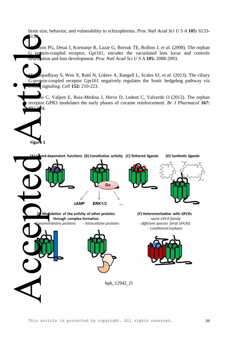
This article is protected by copyright. All rights reserved. 38
brain size, behavior, and vulnerability to schizophrenia. Proc Natl Acad Sci U S A 105: 6133-
6138.
Matteson PG, Desai J, Korstanje R, Lazar G, Borsuk TE, Rollins J, et al. (2008). The orphan
G protein-coupled receptor, Gpr161, encodes the vacuolated lens locus and controls
neurulation and lens development. Proc Natl Acad Sci U S A 105: 2088-2093.
Mukhopadhyay S, Wen X, Ratti N, Loktev A, Rangell L, Scales SJ, et al. (2013). The ciliary
G-protein-coupled receptor Gpr161 negatively regulates the Sonic hedgehog pathway via
cAMP signaling. Cell 152: 210-223.
Tourino C, Valjent E, Ruiz-Medina J, Herve D, Ledent C, Valverde O (2012). The orphan
receptor GPR3 modulates the early phases of cocaine reinforcement. Br J Pharmacol 167:
892-904.
bph_12942_f1
Acc
epte
d A
rticl
e
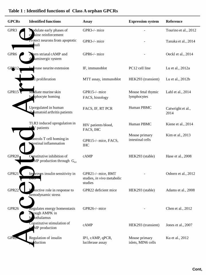
GPCRs Identified functions Assay Expression system Reference
GPR3
Modulate early phases of
cocaine reinforcement
Protect neurons from apoptotic
stimuli
GPR3-/- mice
GPR3-/- mice
-
-
Tourino et al., 2012
Tanaka et al., 2014
GPR6
Alters striatal cAMP and
dopaminergic system
GPR6-/- mice - Oeckl et al., 2014
GPR12
Increase neurite extension
Cell proliferation
IF, immunoblot
MTT assay, immunoblot
PC12 cell line
HEK293 (transient)
Lu et al., 2012a
Lu et al., 2012b
GPR15
Mediate murine skin
lymphocyte homing
Upregulated in human
rheumatoid arthritis patients
TLR3 induced upregulation in
HIV patients
Controls T cell homing in
intestinal inflammation
GPR15-/- mice
FACS, histology
FACS, IF, RT PCR
HIV patients blood,
FACS, IHC
GPR15-/- mice, FACS,
IHC
Mouse fetal thymic
lymphocytes
Human PBMC
Human PBMC
Mouse primary
intestinal cells
Lahl et al., 2014
Catwright et al.,
2014
Kiene et al., 2014
Kim et al., 2013
GPR20 Constitutive inhibition of
cAMP production through Gai
cAMP HEK293 (stable) Hase et al., 2008
GPR21 Improves insulin sensitivity in
DIO
GPR21-/- mice, BMT
studies, in vivo metabolic
studies
- Osborn et al., 2012
GPR22 Protective role in response to
hemodynamic stress
GPR22 deficient mice HEK293 (stable)
Adams et al., 2008
GPR26
Regulates energy homeostasis
through AMPK in
hypothalamus
Constitutive stimulation of
cAMP production
GPR26-/- mice
cAMP
-
HEK293 (transient)
Chen et al., 2012
Jones et al., 2007
GPR27 Regulation of insulin
production
IP1, cAMP, qPCR,
luciferase assay
Mouse primary
islets, MIN6 cells
Ku et al., 2012
Table 1 : Identified functions of Class A orphan GPCRs
Cont,
Acc
epte
d A
rticl
e
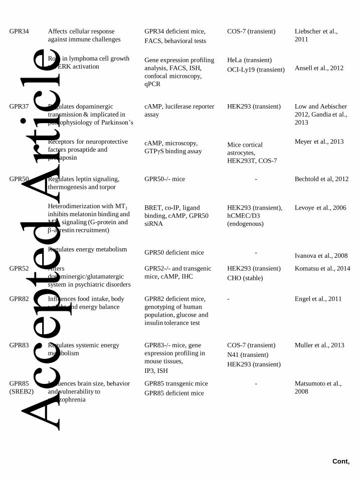
GPR34 Affects cellular response
against immune challenges
Role in lymphoma cell growth
via ERK activation
GPR34 deficient mice,
FACS, behavioral tests
Gene expression profiling
analysis, FACS, ISH,
confocal microscopy,
qPCR
COS-7 (transient)
HeLa (transient)
OCI-Ly19 (transient)
Liebscher et al.,
2011
Ansell et al., 2012
GPR37 Regulates dopaminergic
transmission & implicated in
pathophysiology of Parkinson’s
Receptors for neuroprotective
factors prosaptide and
prosaposin
cAMP, luciferase reporter
assay
cAMP, microscopy,
GTPγS binding assay
HEK293 (transient)
Mice cortical
astrocytes,
HEK293T, COS-7
Low and Aebischer
2012, Gandia et al.,
2013
Meyer et al., 2013
GPR50 Regulates leptin signaling,
thermogenesis and torpor
Heterodimerization with MT1
inhibits melatonin binding and
MT1 signaling (G-protein and
b-arrestin recruitment)
Regulates energy metabolism
GPR50-/- mice
BRET, co-IP, ligand
binding, cAMP, GPR50
siRNA
GPR50 deficient mice
-
HEK293 (transient),
hCMEC/D3
(endogenous)
-
Bechtold et al, 2012
Levoye et al., 2006
Ivanova et al., 2008
GPR52 Alters
dopaminergic/glutamatergic
system in psychiatric disorders
GPR52-/- and transgenic
mice, cAMP, IHC
HEK293 (transient)
CHO (stable)
Komatsu et al., 2014
GPR82 Influences food intake, body
weight and energy balance
GPR82 deficient mice,
genotyping of human
population, glucose and
insulin tolerance test
- Engel et al., 2011
GPR83 Regulates systemic energy
metabolism
GPR83-/- mice, gene
expression profiling in
mouse tissues,
IP3, ISH
COS-7 (transient)
N41 (transient)
HEK293 (transient)
Muller et al., 2013
GPR85
(SREB2)
Influences brain size, behavior
and vulnerability to
schizophrenia
GPR85 transgenic mice
GPR85 deficient mice
- Matsumoto et al.,
2008
Cont,
Acc
epte
d A
rticl
e
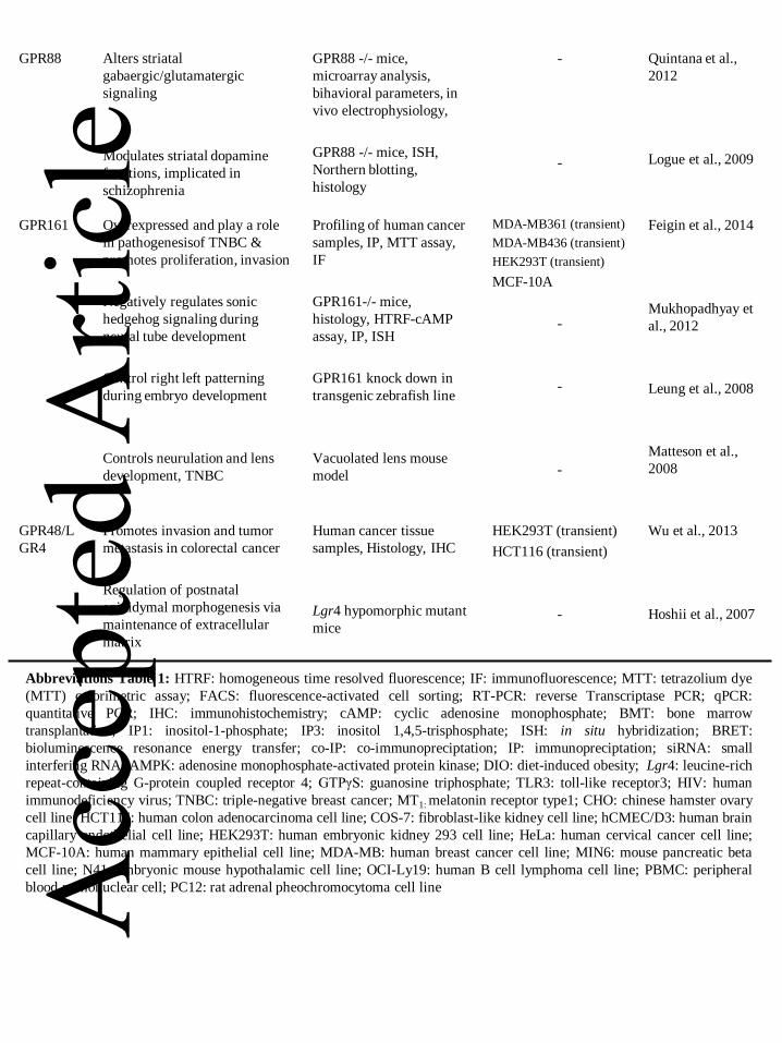
GPR88 Alters striatal
gabaergic/glutamatergic
signaling
Modulates striatal dopamine
functions, implicated in
schizophrenia
GPR88 -/- mice,
microarray analysis,
bihavioral parameters, in
vivo electrophysiology,
GPR88 -/- mice, ISH,
Northern blotting,
histology
-
-
Quintana et al.,
2012
Logue et al., 2009
GPR161 Overexpressed and play a role
in pathogenesisof TNBC &
promotes proliferation, invasion
Negatively regulates sonic
hedgehog signaling during
neural tube development
Control right left patterning
during embryo development
Controls neurulation and lens
development, TNBC
Profiling of human cancer
samples, IP, MTT assay,
IF
GPR161-/- mice,
histology, HTRF-cAMP
assay, IP, ISH
GPR161 knock down in
transgenic zebrafish line
Vacuolated lens mouse
model
MDA-MB361 (transient)
MDA-MB436 (transient)
HEK293T (transient)
MCF-10A
-
-
-
Feigin et al., 2014
Mukhopadhyay et
al., 2012
Leung et al., 2008
Matteson et al.,
2008
GPR48/L
GR4
Promotes invasion and tumor
metastasis in colorectal cancer
Regulation of postnatal
epididymal morphogenesis via
maintenance of extracellular
matrix
Human cancer tissue
samples, Histology, IHC
Lgr4 hypomorphic mutant
mice
HEK293T (transient)
HCT116 (transient)
-
Wu et al., 2013
Hoshii et al., 2007
Abbreviations Table 1: HTRF: homogeneous time resolved fluorescence; IF: immunofluorescence; MTT: tetrazolium dye
(MTT) colorimetric assay; FACS: fluorescence-activated cell sorting; RT-PCR: reverse Transcriptase PCR; qPCR:
quantitative PCR; IHC: immunohistochemistry; cAMP: cyclic adenosine monophosphate; BMT: bone marrow
transplantation; IP1: inositol-1-phosphate; IP3: inositol 1,4,5-trisphosphate; ISH: in situ hybridization; BRET:
bioluminescence resonance energy transfer; co-IP: co-immunopreciptation; IP: immunopreciptation; siRNA: small
interfering RNA; AMPK: adenosine monophosphate-activated protein kinase; DIO: diet-induced obesity; Lgr4: leucine-rich
repeat-containing G-protein coupled receptor 4; GTPγS: guanosine triphosphate; TLR3: toll-like receptor3; HIV: human
immunodeficiency virus; TNBC: triple-negative breast cancer; MT1: melatonin receptor type1; CHO: chinese hamster ovary
cell line; HCT116: human colon adenocarcinoma cell line; COS-7: fibroblast-like kidney cell line; hCMEC/D3: human brain
capillary endothelial cell line; HEK293T: human embryonic kidney 293 cell line; HeLa: human cervical cancer cell line;
MCF-10A: human mammary epithelial cell line; MDA-MB: human breast cancer cell line; MIN6: mouse pancreatic beta
cell line; N41: embryonic mouse hypothalamic cell line; OCI-Ly19: human B cell lymphoma cell line; PBMC: peripheral
blood mononuclear cell; PC12: rat adrenal pheochromocytoma cell line Acc
epte
d A
rticl
e
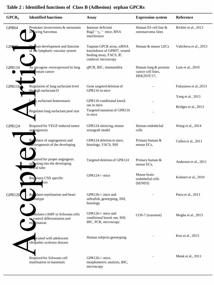
GPCRS Identified functions Assay Expression system Reference
GPR64 Promotes invasiveness & metastasis
in Ewing Sarcomas
Immune deficient
Rag2−/−γC−/− mice, RNA
interference
Human ES cell line &
osteosarcoma lines
Richter et al., 2013
GPR97 Mediate development and function
of the lymphatic vascular system
Taqman GPCR array, siRNA
knockdown of GPR97, wound
healing assay, FACS, IF,
confocal microscopy
Human & mouse LECs Valtcheva et al., 2013
GPR110 As oncogene overexpressed in lung
& prostate cancer
qPCR, IHC, immunoblot Human lung & prostate
cancer cell lines,
HEK293T/17,
Lum et al., 2010
GPR116 Regulation of lung surfactant level
through surfactant D
Lung surfactant homeostasis
Regulates lung surfactant pool size
Gene targeted deletion of
GPR116 in mice
GPR116 conditional knock
out in mice
Targeted mutation of GPR116
in mice
-
-
-
Fukuzawa et al.,2013
Yang et al., 2013
Bridges et al., 2013
GPR124 Required for VEGF-induced tumor
angiogenesis
Regulator of angiogenesis and
barriergenesis of the developing
CNS
Required for proper angiogenic
sprouting into the developing
neural tube
Regulates CNS specific
angiogenesis
GPR124 silencing, mouse
xenograft model
GPR124 deletion in mice,
histology, FACS, ISH
Targeted deletion of GPR124
GPR124-/- mice
Human endothelial
cells
Primary human &
mouse ECs,
Primary human &
mouse ECs,
Mouse brain
endothelial cells
(bEND3)
Wang et al., 2014
Cullen et al., 2011
Anderson et al., 2011
Kuhnert et al., 2010
GPR126 Regulates myelination and heart
phenotype
Modulates cAMP in Schwann cells
to control differentiation and
myelination
Associated with adolescent
idiopathic scoliosis disease
Required for Schwann cell
myelination in mammals
GPR126-/- mice and
zebrafish, genotyping, ISH,
histology
GPR126-/- mice and
conditional knock out, ISH,
IHC, PCR, microscopy
Human subjects genotyping
GPR126-/- mice,
morphometric analysis, IHC,
microscopy
-
COS-7 (transient)
-
-
Patra et al., 2013
Mogha et al., 2013
Kou et al., 2013
Monk et al., 2011
Table 2 : Identified functions of Class B (Adhesion) orphan GPCRs
Acc
epte
d A
rticl
e

Abbreviations Table 2: IF: immunofluorescence; FACS: fluorescence-activated cell sorting; IHC:
immunohistochemistry; cAMP: cyclic adenosine monophosphate; ISH: in situ hybridization; qPCR: quantitative PCR;
siRNA: small interfering RNA; CNS: central nervous system; VEGF: vascular endothelial growth factor; Rag2:
Recombination-activating genes; LECs: cultured lymphatic endothelial cells; ECs: endothelial cells; bEND3: mouse
brain endothelial cell line; ES: embryonic stem cells; CHO: chinese hamster ovary cell line; COS-7: fibroblast-like
kidney cell line; HEK293: human embryonic kidney 293 cell line
Acc
epte
d A
rticl
e
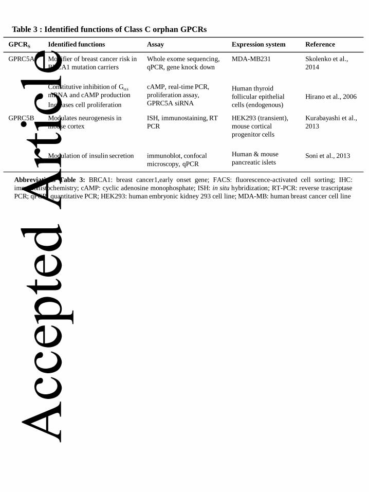
GPCRS Identified functions Assay Expression system Reference
GPRC5A Modifier of breast cancer risk in
BRCA1 mutation carriers
Constitutive inhibition of Gas
mRNA and cAMP production
Increases cell proliferation
Whole exome sequencing,
qPCR, gene knock down
cAMP, real-time PCR,
proliferation assay,
GPRC5A siRNA
MDA-MB231
Human thyroid
follicular epithelial
cells (endogenous)
Skolenko et al.,
2014
Hirano et al., 2006
GPRC5B Modulates neurogenesis in
mouse cortex
Modulation of insulin secretion
ISH, immunostaining, RT
PCR
immunoblot, confocal
microscopy, qPCR
HEK293 (transient),
mouse cortical
progenitor cells
Human & mouse
pancreatic islets
Kurabayashi et al.,
2013
Soni et al., 2013
Table 3 : Identified functions of Class C orphan GPCRs
Abbreviations Table 3: BRCA1: breast cancer1,early onset gene; FACS: fluorescence-activated cell sorting; IHC:
immunohistochemistry; cAMP: cyclic adenosine monophosphate; ISH: in situ hybridization; RT-PCR: reverse trascriptase
PCR; qPCR: quantitative PCR; HEK293: human embryonic kidney 293 cell line; MDA-MB: human breast cancer cell line
Acc
epte
d A
rticl
e
Recommended