© 2019, IJSRPAS All Rights Reserved 55
International Journal of Scientific Research in ______________________________ Research Paper . Physics and Applied Sciences
Vol.7, Issue.2, pp.55-61, April (2019) E-ISSN: 2348-3423
DOI: https://doi.org/10.26438/ijsrpas/v7i2.5561
High Electrical Conductivity, Thermal Stability and Gas Sensing Studies
on Silver-Polyaniline Nanocomposite
Sanjib Baglari1* and D. Sarkar
2
1,2
Department of Physics, Gauhati University, Guwahati, India
*Corresponding author Email: [email protected]
Available online at: www.isroset.org
Received: 09/Mar/2019, Accepted: 04/Apr/2019, Online: 30/Apr/2019
Abstract- This paper reports the synthesis of Polyaniline-Silver (Pani/Ag) nanocomposites by an in-situ chemical
polymerization method using ammonium persulphate (APS) as an oxidizing agent. The prepared Pani/Ag nanocomposite were
characterized by XRD, EDAX, FESEM, TEM, TGA, UV-vis spectroscopy. The XRD patterns indicated that the crystalline
phase of Ag is cubic with average crystallite size of 26 nm. UV–vis spectroscopy analysis indicated that the Ag nanoparticles
have some effect on the Pani matrix. The TGA result showed that thermal stability of the nanocomposite was increased after
incorporation of Ag nanoparticles. Pani/Ag nanocomposite showed superior DC conductivity when compared to pure Pani. The
Pani/Ag nanocomposite also showed excellent chloroform sensing behaviour than pure Pani, which might be due to increase in
the surface area of Pani in the presence of Ag nanoparticles.
Keywords: Polyaniline, Nanocomposite, DC Conductivity, Gas Sensing, Thermal Stability, UV Vis.
I. INTRODUCTION
Conducting polymer exhibit both conducting and semi
conducting properties. Recently, conducting polymers has
received much attentions in research because of their
promising optical properties, controllable chemical and
electrochemical properties, easy processability, ease of
synthesis, inexpensive monomer and environmental stability
[1-4]. Among the conducting polymers, polyaniline (PANI)
has been studied extensively worldwide for its various
applications in the design of sensor [5-6], optoelectronics
device [7], secondary batteries, fuel cell [8], super capacitor
[9] and catalytic properties [10].
In recent years, composites of polyaniline with different
metal oxide for gas sensing applications has received great
attention worldwide because of its higher sensitivity,
reversible response, quick response time and inexpensive
monomer [11]. It has been reported that incorporation of
metal nanoparticle to polyaniline exhibits excellent sensing
properties with large effective surface area for chemical
interaction and hence improve catalytic efficiency [12-13]. It
was also reported that incorporation of metal nanoparticle
effectively increased electrical, optical and dielectric
properties of the polyaniline composite [14-15]. Satish
Sharma et al [16] reported that the chemically synthesized
Copper/Polyaniline nanocomposite gas sensor exhibited
higher sensitivity, faster response time and recovery rates to
chloroform vapour than those of a pure PANI sensor.
Mudassir Hasan et al [17] showed that PANI/Au
nanocomposite exhibited a better ammonia sensing and
recovery response than pure PANI. Athawale et al [18]
demonstrated the potential application of the PANI/Pd
nanocomposite as a selective methanol sensor. They found
that sensor characteristics such as the response time and
long-term stability of the PANI/Pd nanocomposite sensor
were superior to pure PANI sensor. Therefore, it is hopeful
to obtain PANI/Ag nanocomposites would be a novel
material in the area of gas sensor with excellent gas sensing
behaviour. To the best of our knowledge, the PANI/Ag
nanocomposite as chloroform sensing materials have not
been reported elsewhere. Chloroform is released into the
environment through several anthropogenic sources where
industrial sites using it in the production line. Exposure to 50
ppm of chloroform can damage human health [19].
Therefore, the reliable and quick detection of chloroform gas
is needed.
The aim of this study was to prepare a Polyaniline/Ag
nanocomposite for chloroform gas sensing. The combination
of PANI with silver nanoparticle composite is expected to
produce synergism between the constituents, which could
effectively improve sensitivity and recovery behaviour for
chloroform sensing application.
In this present work, PANI/Ag nanocomposites were
prepared by an in-situ chemical polymerization method
using APS as an oxidizing agent and hence explore the
Int. J. Sci. Res. in Physics and Applied Sciences Vol.7(2), Apr 2019, E-ISSN: 2348-3423
© 2019, IJSRPAS All Rights Reserved 56
possibility of improving of conductivity and gas sensing
property of Pani by doping it with silver nanoparticles.
II. MATERIALS AND METHOD
A. Materials
Aniline monomer, silver nitrate (AgNO3) and sodium
borohydride (NaBH4) were purchased from Sigma Aldrich
(India) as analytical grade. APS, hydrochloric acid (HCl)
and Chloroform were obtained from Merck India Ltd. as
reagent grade. All solutions of reacting materials were
prepared in Deionized (DI) water.
B. Synthesis of Ag nanoparticles
Silver nanoparticles were synthesized by chemical reduction
of AgNO3 using NaBH4 by a previously reported method
[20]. In a typical experiment, Silver nitrate solution was
separately prepared by adding different mole concentration,
viz., 0.1, 0.2 and 0.3M of AgNO3 in DI water. The details of
the method are as follows:
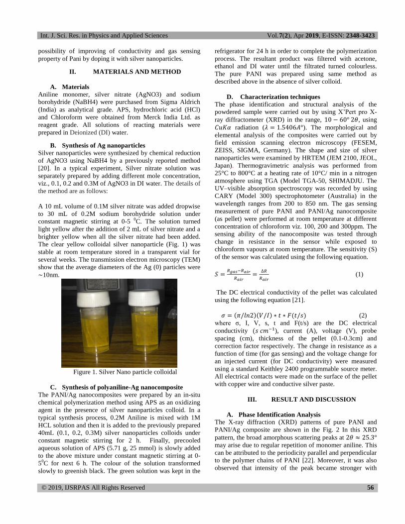
A 10 mL volume of 0.1M silver nitrate was added dropwise
to 30 mL of 0.2M sodium borohydride solution under
constant magnetic stirring at 0-5 0C. The solution turned
light yellow after the addition of 2 mL of silver nitrate and a
brighter yellow when all the silver nitrate had been added.
The clear yellow colloidal silver nanoparticle (Fig. 1) was
stable at room temperature stored in a transparent vial for
several weeks. The transmission electron microscopy (TEM)
show that the average diameters of the Ag (0) particles were
∼10nm.
Figure 1. Silver Nano particle colloidal
C. Synthesis of polyaniline-Ag nanocomposite
The PANI/Ag nanocomposites were prepared by an in-situ
chemical polymerization method using APS as an oxidizing
agent in the presence of silver nanoparticles colloid. In a
typical synthesis process, 0.2M Aniline is mixed with 1M
HCL solution and then it is added to the previously prepared
40mL (0.1, 0.2, 0.3M) silver nanoparticles colloids under
constant magnetic stirring for 2 h. Finally, precooled
aqueous solution of APS (5.71 g, 25 mmol) is slowly added
to the above mixture under constant magnetic stirring at 0-
50C for next 6 h. The colour of the solution transformed
slowly to greenish black. The green solution was kept in the
refrigerator for 24 h in order to complete the polymerization
process. The resultant product was filtered with acetone,
ethanol and DI water until the filtrated turned colourless.
The pure PANI was prepared using same method as
described above in the absence of silver colloid.
D. Characterization techniques
The phase identification and structural analysis of the
powdered sample were carried out by using X’Pert pro X-
ray diffractometer (XRD) in the range, , using
radiation ( ). The morphological and
elemental analysis of the composites were carried out by
field emission scanning electron microscopy (FESEM,
ZEISS, SIGMA, Germany). The shape and size of silver
nanoparticles were examined by HRTEM (JEM 2100, JEOL,
Japan). Thermogravimetric analysis was performed from
25°C to 800°C at a heating rate of 10°C/ min in a nitrogen
atmosphere using TGA (Model TGA-50, SHIMADJU. The
UV–visible absorption spectroscopy was recorded by using
CARY (Model 300) spectrophotometer (Australia) in the
wavelength ranges from 200 to 850 nm. The gas sensing
measurement of pure PANI and PANI/Ag nanocomposite
(as pellet) were performed at room temperature at different
concentration of chloroform viz. 100, 200 and 300ppm. The
sensing ability of the nanocomposite was tested through
change in resistance in the sensor while exposed to
chloroform vapours at room temperature. The sensitivity (S)
of the sensor was calculated using the following equation.
(1)
The DC electrical conductivity of the pellet was calculated
using the following equation [21].
( )( ) ( ) (2)
where σ, I, V, s, t and F(t/s) are the DC electrical
conductivity ( ), current (A), voltage (V), probe
spacing (cm), thickness of the pellet (0.1-0.3cm) and
correction factor respectively. The change in resistance as a
function of time (for gas sensing) and the voltage change for
an injected current (for DC conductivity) were measured
using a standard Keithley 2400 programmable source meter.
All electrical contacts were made on the surface of the pellet
with copper wire and conductive silver paste.
III. RESULT AND DISCUSSION
A. Phase Identification Analysis
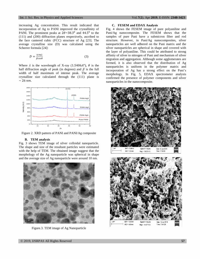
The X-ray diffraction (XRD) patterns of pure PANI and
PANI/Ag composite are shown in the Fig. 2 In this XRD
pattern, the broad amorphous scattering peaks at may arise due to regular repetition of monomer aniline. This
can be attributed to the periodicity parallel and perpendicular
to the polymer chains of PANI [22]. Moreover, it was also
observed that intensity of the peak became stronger with
Int. J. Sci. Res. in Physics and Applied Sciences Vol.7(2), Apr 2019, E-ISSN: 2348-3423
© 2019, IJSRPAS All Rights Reserved 57
increasing Ag concentration. This result indicated that
incorporation of Ag to PANI improved the crystallinity of
PANI. The prominent peaks at = and to the
(111) and (200) diffraction planes respectively, ascribed to
the face cantered cubic (FCC) structure of Ag [23]. The
average crystalline size (D) was calculated using the
Scherrer formula [24]:
(3)
Where is the wavelength of X-ray ( ), is the
half diffraction angle of peak (in degrees) and is the full
width of half maximum of intense peak. The average
crystalline size calculated through the (111) plane is
.
10 20 30 40 50 60
0
200
400
600
800
1000
1200
1400
1600
1800
Inte
nsity
(A
.U)
2
(111)
(200)
Pure PANI
PANI-Ag1
PANI-Ag2
PANI-Ag3
Figure 2. XRD pattern of PANI and PANI/Ag composite
B. TEM analysis
Fig. 3 shows TEM image of silver colloidal nanoparticle.
The shape and size of the resultant particles were estimated
with the help of TEM. The obtained image suggest that the
morphology of the Ag nanoparticle was spherical in shape
and the average size of Ag nanoparticle were around 10 nm.
Figure.3. TEM image of Ag Nanoparticle
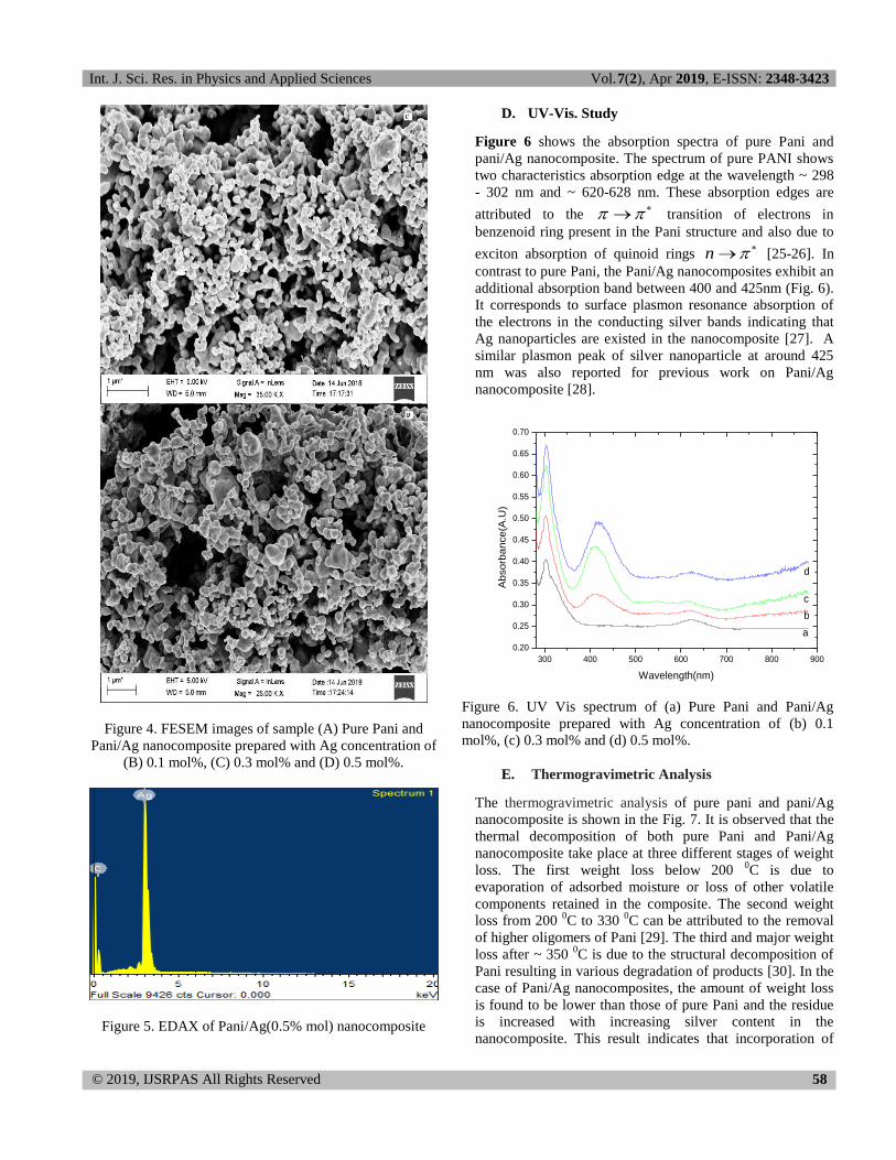
C. FESEM and EDAX Analysis
Fig. 4 shows the FESEM image of pure polyaniline and
Pani/Ag nanocomposite. The FESEM shows that the
samples of pure Pani have a submicron fibre and rod
structure. However, in Pani/Ag nanocomposites, silver
nanoparticles are well adhered on the Pani matrix and the
silver nanoparticles are spherical in shape and covered with
the layer of polyaniline. This could be attributed to strong
affinity of silver to nitrogen of Pani and mechanism of silver
migration and aggregation. Although some agglomerates are
formed, it is also observed that the distribution of Ag
nanoparticles is uniform in the polymer matrix and
incorporation of Ag has a strong effect on the Pani’s
morphology. In Fig. 5, EDAX spectrometer analysis
confirmed the presence of polymer components and silver
nanoparticles in the nanocomposite.
Int. J. Sci. Res. in Physics and Applied Sciences Vol.7(2), Apr 2019, E-ISSN: 2348-3423
© 2019, IJSRPAS All Rights Reserved 58
Figure 4. FESEM images of sample (A) Pure Pani and
Pani/Ag nanocomposite prepared with Ag concentration of
(B) 0.1 mol%, (C) 0.3 mol% and (D) 0.5 mol%.
Figure 5. EDAX of Pani/Ag(0.5% mol) nanocomposite
D. UV-Vis. Study
Figure 6 shows the absorption spectra of pure Pani and
pani/Ag nanocomposite. The spectrum of pure PANI shows
two characteristics absorption edge at the wavelength ~ 298
- 302 nm and ~ 620-628 nm. These absorption edges are
attributed to the transition of electrons in
benzenoid ring present in the Pani structure and also due to
exciton absorption of quinoid rings n [25-26]. In
contrast to pure Pani, the Pani/Ag nanocomposites exhibit an
additional absorption band between 400 and 425nm (Fig. 6).
It corresponds to surface plasmon resonance absorption of
the electrons in the conducting silver bands indicating that
Ag nanoparticles are existed in the nanocomposite [27]. A
similar plasmon peak of silver nanoparticle at around 425
nm was also reported for previous work on Pani/Ag
nanocomposite [28].
300 400 500 600 700 800 900
0.20
0.25
0.30
0.35
0.40
0.45
0.50
0.55
0.60
0.65
0.70
Ab
so
rba
nce
(A.U
)
Wavelength(nm)
a
b
c
d
Figure 6. UV Vis spectrum of (a) Pure Pani and Pani/Ag
nanocomposite prepared with Ag concentration of (b) 0.1
mol%, (c) 0.3 mol% and (d) 0.5 mol%.
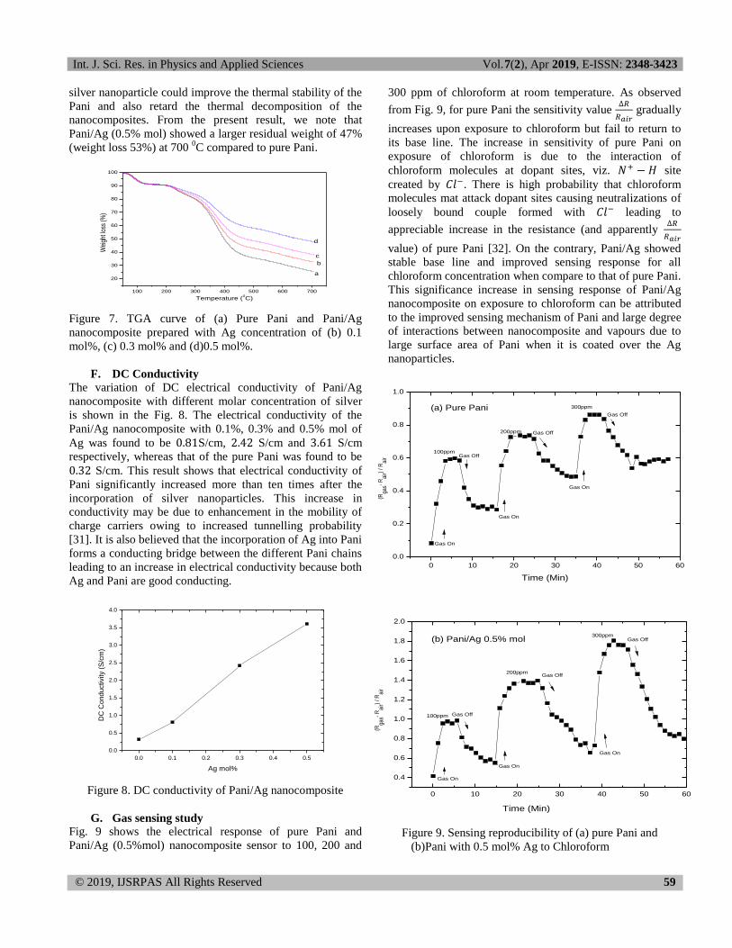
E. Thermogravimetric Analysis
The thermogravimetric analysis of pure pani and pani/Ag
nanocomposite is shown in the Fig. 7. It is observed that the
thermal decomposition of both pure Pani and Pani/Ag
nanocomposite take place at three different stages of weight
loss. The first weight loss below 200 0C is due to
evaporation of adsorbed moisture or loss of other volatile
components retained in the composite. The second weight
loss from 200 0C to 330
0C can be attributed to the removal
of higher oligomers of Pani [29]. The third and major weight
loss after ~ 350 0C is due to the structural decomposition of
Pani resulting in various degradation of products [30]. In the
case of Pani/Ag nanocomposites, the amount of weight loss
is found to be lower than those of pure Pani and the residue
is increased with increasing silver content in the
nanocomposite. This result indicates that incorporation of
Int. J. Sci. Res. in Physics and Applied Sciences Vol.7(2), Apr 2019, E-ISSN: 2348-3423
© 2019, IJSRPAS All Rights Reserved 59
silver nanoparticle could improve the thermal stability of the
Pani and also retard the thermal decomposition of the
nanocomposites. From the present result, we note that
Pani/Ag (0.5% mol) showed a larger residual weight of 47%
(weight loss 53%) at 700 0C compared to pure Pani.
100 200 300 400 500 600 700
20
30
40
50
60
70
80
90
100
Wei
ght l
oss
(%)
Temperature (0C)
a
b
d
c
Figure 7. TGA curve of (a) Pure Pani and Pani/Ag
nanocomposite prepared with Ag concentration of (b) 0.1
mol%, (c) 0.3 mol% and (d)0.5 mol%.
F. DC Conductivity
The variation of DC electrical conductivity of Pani/Ag
nanocomposite with different molar concentration of silver
is shown in the Fig. 8. The electrical conductivity of the
Pani/Ag nanocomposite with 0.1%, 0.3% and 0.5% mol of
Ag was found to be S/cm, S/cm and S/cm
respectively, whereas that of the pure Pani was found to be
S/cm. This result shows that electrical conductivity of
Pani significantly increased more than ten times after the
incorporation of silver nanoparticles. This increase in
conductivity may be due to enhancement in the mobility of
charge carriers owing to increased tunnelling probability
[31]. It is also believed that the incorporation of Ag into Pani
forms a conducting bridge between the different Pani chains
leading to an increase in electrical conductivity because both
Ag and Pani are good conducting.
0.0 0.1 0.2 0.3 0.4 0.5
0.0
0.5
1.0
1.5
2.0
2.5
3.0
3.5
4.0
DC
Co
nd
uctivity (
S/c
m)
Ag mol%
Figure 8. DC conductivity of Pani/Ag nanocomposite
G. Gas sensing study
Fig. 9 shows the electrical response of pure Pani and
Pani/Ag (0.5%mol) nanocomposite sensor to 100, 200 and
300 ppm of chloroform at room temperature. As observed
from Fig. 9, for pure Pani the sensitivity value
gradually
increases upon exposure to chloroform but fail to return to
its base line. The increase in sensitivity of pure Pani on
exposure of chloroform is due to the interaction of
chloroform molecules at dopant sites, viz. site
created by . There is high probability that chloroform
molecules mat attack dopant sites causing neutralizations of
loosely bound couple formed with leading to
appreciable increase in the resistance (and apparently
value) of pure Pani [32]. On the contrary, Pani/Ag showed
stable base line and improved sensing response for all
chloroform concentration when compare to that of pure Pani.
This significance increase in sensing response of Pani/Ag
nanocomposite on exposure to chloroform can be attributed
to the improved sensing mechanism of Pani and large degree
of interactions between nanocomposite and vapours due to
large surface area of Pani when it is coated over the Ag
nanoparticles.
0 10 20 30 40 50 60
0.0
0.2
0.4
0.6
0.8
1.0
Gas Off
Gas Off
Gas On
Gas On
100ppm
200ppm
300ppm
Gas Off
Gas On
(R
gas-
Rai
r) /
Rai
r
Time (Min)
(a) Pure Pani
0 10 20 30 40 50 60
0.4
0.6
0.8
1.0
1.2
1.4
1.6
1.8
2.0
Gas On
Gas On
Gas Off
Gas Off
100ppm
200ppm
300ppm
Gas Off
Gas On
(Rga
s- R
air)
/ Rai
r
Time (Min)
(b) Pani/Ag 0.5% mol
Figure 9. Sensing reproducibility of (a) pure Pani and
(b)Pani with 0.5 mol% Ag to Chloroform
Int. J. Sci. Res. in Physics and Applied Sciences Vol.7(2), Apr 2019, E-ISSN: 2348-3423
© 2019, IJSRPAS All Rights Reserved 60
IV. CONCLUSIONS
In this paper, Polyaniline-Silver (Pani/Ag) nanocomposite
were synthesis by an in-situ chemical polymerization
method. The structural morphology, thermal stability, DC
electrical conductivity and chloroform-sensing mechanism
of the nanocomposite were examined using different
techniques. Thermal stability of Polyaniline significantly
improved after the incorporation of Ag nanoparticles to pure
Polyaniline. The DC conductivity of Pani/Ag nanocomposite
was recorded ten time higher than pure Pani. Pani/Ag
nanocomposite showed better ammonia sensitivity and
recovery for chloroform when compare with pure Pani.
ACKNOWLEDGEMENTS
The authors sincerely acknowledge UGC, India for the
financial support. The author would like to acknowledge
NEIST Jorhat, IIT Guwahati and Tezpur University for
providing the necessary facilities for sample
characterisation.
REFERENCES
[1]. Gordana, Ćirić-Marjanović, “Recent advances in polyaniline
composites with metals, metalloids and nonmetals”, Synthetic
Metals, Vol 170, pp. 31– 56, 2013.
[2]. Manoj K. Sharma, M. K. Ambolikar, A. S.; Aggarwal, S. K, “In
situ synthesis of gold–polyaniline composite in nanopores of
polycarbonate membrane” J. Mater. Sci.,Vol.46, pp. 5715, 2011.
[3]. Naseri M, Fotouhi L, Ehsani A, “Recent Progress in the
Development of Conducting Polymer-Based Nanocomposites for
Electrochemical Biosensors Applications: A Mini-Review”,
Chem. Rec., Vol. 18, Issue 6, pp. 599-618, 2018.
[4]. A. Kitani, T. Akashi, K. Sugimoto, and S. Ito, “Electrocatalytic
oxidation of methanol on platinum modified polyaniline
electrodes” Synth. Met. Vol. 121, Issue 1-3, 1301, 2001.
[5]. M L Rozemarie, B Andrei, H Liliana, R Cramariuc, O Cramariuc,
“Electrospun Based Polyaniline Sensors – A Review”, IOP
Conference Series: Materials Science and Engineering, Vol. 209,
pp. 012063, 2017.
[6]. S Panday, “Highly sensitive and selective chemiresistor gas/vapor
sensors based on polyaniline nanocomposite: A comprehensive
review”, Journal of Science: Advanced Materials and Devices,
Vol. 1, pp. 431-453, 2016.
[7]. Y. Xia, K. Sun, J. Ouyang, “Solution-processed metallic
conducting polymer films as transparent electrode of
optoelectronic devices”, Adv. Mater, Vol. 24, pp. 2436–2440,
2012.
[8]. Blinova, N. V.; Stejskal, J.; Trchov_a, M.; Sapurina, I.; Ciric-
Marjanovic, G, “Polyaniline–silver composites prepared by the
oxidation of aniline with silver nitrate in acetic acid solutions”,
Polym. Int, Vol. 59, pp. 437–446, 2010.
[9]. Sun, L.; Shi, Y.; Li, B.; Chu, L.; He, Z.; Liu, J, “Synthesis and
characterization of polypyrrole/Au nanocomposites by
microemulsion polymerization”, Colloids Surf. A, Vol. 397, pp. 8-
11, 2012.
[10]. D K. Dutta, S. Das, D. Rana, P. P. Kundu, “Enhancements of
Catalyst Distribution and Functioning Upon Utilization of
Conducting Polymers as Supporting Matrices in DMFC: A
Review”, Polym. Review, Vol. 55, Issue 1, pp. 1-56. 2015.
[11]. S. Z. Wu, F. Zeng, F. X. Li, and Y. L. Zhu, “Ammonia sensitivity
of polyaniline films via emulsion polymerization”, Eur. Polym.
Journal, Vol. 36, Issue 4, pp. 679-683, 2000.
[12]. G. D. Khuspe, S. T. Navale, D. K. Bandgar, R. D. Sakhare, M. A.
Chougule, and V. B. Patil, “SnO2nanoparticles-modified
polyaniline films as highly selective, sensitive, reproducible and
stable ammonia sensors”, Electron. Mater. Lett., Vol. 10, Issue 1,
pp. 191-197, 2014.
[13]. Roopa J, Muniraj R, Magaraj T M, B S Satya Narayana, K S
Geetha, “Development of a Conducting Polymer [polyaniline]
based gas sensor”, International Conference on Electrical,
Electronics, Signals, Communication and Optimization (EESCO)
(IEEE), 2015.
[14]. T.K. Sarma, D. Chowdhury, A. Paul, A. Chattopadhyay,
“Synthesis of Au nanoparticle-conductive polyaniline composite
using H2O2 a oxidising as well as reducing agent”, Chem.
Commun., Vol.14 pp.1048–1049, 2002.
[15]. W. Xue, H. Qiu, K. Fang, J. Li, J. Zhao, M. Li, “Electrical
properties of the composite pellets containing DBSA-doped
polyaniline and Fe nanoparticles”, Synth. Met., Vol. 156 833-837,
2006.
[16]. S. Sharma, C. Nirkhe, S. Pethkar, A.A. Athawale, “Chloroform
vapour sensor based on copper/polyaniline nanocomposite”, Sens.
Actuators, B: Chem., Vol. 85, pp. 31–136, 2002.
[17]. Mudassir Hasan, Mohd Omaish Ansari, Moo Hwan Cho,
Moonyong Lee, “Electrical Conductivity, Optical Property and
Ammonia Sensing Studies on HCl Doped Au@Polyaniline
Nanocomposites”, Electron. Mater. Lett., Vol. 11, Issue 1 pp. 1-6,
2015.
[18]. A.A. Athawale, S.V. Bhagwat, P.P. Katre, “Nanocomposite of
Pd–polyaniline as a selective methanol sensor”, Sens. Actuators,
B: Chem., Vol. 114, pp. 263–267, 2006.
[19]. Irma Zulayka Mohamad Ahada, Sulaiman Wadi Harunb, Seng
Neon Gan, Sook Wai Phang, “Polyaniline (PAni) optical sensor in
chloroform detection”, Sensors and Actuators B, Vol. 261, pp.
97–105, 2018.
[20]. Lorraine Mulfinger, “Synthesis and Study of Silver
Nanoparticles”, Journal of Chemical Education, Vol. 84, Issue 2,
pp. 322- 327, 2007.
[21]. F. M. Smits, "Measurement of Sheet Resistivities With the Four-
Point Probe”, The Bell System Technical Journal, pp. 711-718,
1958.
[22]. Qingming Jia, Shaoyun Shan, Lihong Jiang, Yaming Wang,
“One-Step Synthesis of Polyaniline Nanofibers Decorated with
Silver”, Appl. Plym. Sci., Vol. 115, 26–31, 2010.
[23]. K. Gupta, P. C. Jana and A. K. Meikap, “Optical and Electrical
Transport Properties of Polyaniline-Silver Nanocomposite,”
Synthetic Metals, Vol. 160, Issue 13-14, pp. 1566-1573, 2010.
[24]. B.D. Cullity, Elements of X-ray Diffraction, M.A. Reading,
Addison-Wesley, 1978.
Int. J. Sci. Res. in Physics and Applied Sciences Vol.7(2), Apr 2019, E-ISSN: 2348-3423
© 2019, IJSRPAS All Rights Reserved 61
[25]. E. Erdem, M. Karakisla, M. Sacak, “The chemical synthesis of
conductive polyaniline doped with dicarboxylic acids”, Eur.
Polym. J., Vol. 40, pp. 785-791, 2004.
[26]. D. Shihai, M. Hui, W. Zhang, “Fabrication of DBSA‐doped
polyaniline nanorods by interfacial polymerization”, J. Appl.
Polym. Sci., Vol. 109, pp.2842-2847, 2008.
[27]. Junhu Zhang et al, “Thin Films of Ag Nanoparticles Prepared
from the Reduction of AgI Nanoparticles in Self-Assembled
Films”, Journal of Colloid and Interface Science, Vol. 255, pp.
115–118, 2002.
[28]. Arup Choudhury, “Polyaniline/silver nanocomposites: Dielectric
properties and ethanol vapour sensitivity”, Sensors and Actuators
B, Vol. 138, pp. 318–325, 2009.
[29]. G. Chakraborty, S. Ghatak, A. K. Meikap, T. Woods, R. Babu,
and W. J. Blau, “Characterization and electrical transport
properties of polyaniline and multiwall carbon nanotube
composites”, J. Polym. Sci. Part B: Polym. Phy. Vol. 48, pp.
1767-1775, 2010.
[30]. M.O. Ansari, F. Mohammad, “Thermal stability, electrical
conductivity and ammonia sensing studies on p-toluenesulfonic
acid doped polyaniline:titanium dioxide (pTSA/Pani:TiO2)
nanocomposites”, Sens. Actuators B, Vol.157, pp. 122-129, 2011.
[31] M. Amrithesh, S. Aravind, S. Jayalekshmi, R.S. Jayasree,
“Enhanced luminescence observed in polyaniline–
polymethylmethacrylate composites”, J. Alloys Compd., Vol. 449
pp. 176-179, 2008.
[32]. Satish Sharma, ChetanNirkhe, SushamaPethkar, Anjali
A.Athawale, “chloroform vapour sensor based on
copper/polyaniline nanocomposite”, Senors and Actuators B, Vol.
85, Isuue 1-2, pp. 131-136, 2002.
Recommended

![Ultrasonic preparation, stability and thermal conductivity ... · effect on thermal conductivity at different particle concentrations has been recorded [10–14] . Nanofluids containing](https://img.pdfslide.us/doc/110x75/60b98fecd489ad698b5f3b45/ultrasonic-preparation-stability-and-thermal-conductivity-effect-on-thermal.jpg)