1
GLP-1 REGULATES PROLIFERATION OF GLP-1 –
SECRETING CELLS THROUGH A FEEDBACK
MECHANISM
Master of Science Degree project, 30 credits point
Accomplished at the
Department of Clinical Science and Education, Södersjukhuset
Unit for Diabetes Research, Karolinska Institutet, Stockholm, Sweden
Author: Mohamed Abdullahi
2010
Supervisor: Senior researchers, Qimin Zhang: Karolinska Institutet
Examinator: Senior Lecturer, Sven Hamp: Märladalen University
2
CONTENTS
Abstract ..................................................................................................................................... 3
List of abbreviations ................................................................................................................... 4
Introduction .............................................................................................................................. 5
Diabetes mellitus ........................................................................................................................ 5
Incretins ...................................................................................................................................... 5
GIP ............................................................................................................................................. 5
GLP-1 ......................................................................................................................................... 6
GLP-1 Receptor .......................................................................................................................... 7
Physiological action of GLP-1 ................................................................................................... 8
GLP-1 in diabetes treatment ...................................................................................................... 8
The incretin effect of GLP-1 in type 2 diabetes .......................................................................... 9
GLP-1 and its stable analogues ................................................................................................. 9
Aim of the study ........................................................................................................................ 10
Materials and methods ............................................................................................................ 11
The Cells ................................................................................................................................... 11
Experimental methods .............................................................................................................. 11
Anti-diabetic agents .................................................................................................................. 11
3H-thymidine incorporation ..................................................................................................... 11
Immunocytochemistry with Ki67 staining ................................................................................ 11
Immunolabeling ........................................................................................................................ 12
Protein measurement BCA-assay ............................................................................................. 12
Western blot .............................................................................................................................. 12
Cell lysate preparation ............................................................................................................. 12
SDS-PAGE and Western blotting ............................................................................................. 12
Statistic methods ....................................................................................................................... 12
Results and Discussion ............................................................................................................ 14
Conclusion and future work ................................................................................................... 18
Acknowledgement .................................................................................................................... 18
References ................................................................................................................................ 19
3
Abstract
Background and aim: Diabetes mellitus (DM) is a chronic and progressive illness that
affects all type of populations and ages. According to World health organization (WHO) by
2030 it will be 366 million people effected world wild. Many new drugs are Glucagon-like
peptide-1 (GLP-1) based therapy for treatment of type 2diabetes. GLP-1 is released from the
intestinal L-cells, and is a potent stimulator of glucose-dependent insulin secretion. The aim
of this study was to investigate the effect of GLP-1 and its stable analogs on cell proliferation
of GLP-1 secreting GLUTag cells.
Material and methods: GluTag cells were incubated for 48h in DMEM medium containing
(0.5% fetal bovine serum and 100 IU/ml penicillin and 100 μg/ml streptomycin and 3mM
glucose concentration) in the present or absence of the agents. DNA synthesis was measured
using 3H- thymidine incorporation and Ki67 antigen staining. Western blot were performed to
investigate the present of GLP-1 receptor in GLUTag cells.
Results/conclusions: GLP-1(7-36) increased cell proliferation in GLUTag cells, an effect
which was blocked by the GLP-1 receptor antagonist exendin(9-39). The GLP-1 receptor was
expressed in GluTag cells.
These results suggest that GLP-1 regulates proliferation of the GLP-1-secreting cell
through a feedback mechanism via its receptor. Since serum GLP-1 levels are decreased in
type 2 diabetic patients, the effect of GLP-1 on the GLP-1-secreting cell proliferation
suggested here provides a novel beneficial long-term effect of the incretin-based drugs in
clinical practice i.e. through increase of the GLP-1-secreting cell mass, augmenting the
incretin effect. In addition, the feedback mechanism action of GLP-1 reveals a new insight in
regulation manner of the L-cell proliferation.
Keywords: Incretin hormone, GLP-1, GLP-1 receptor, Exendin-4, Diabetes
4
List of abbreviations
BSA Bovine Serum Albumin
DPP-IV dipeptidyl peptidase-4
DM Diabetes mellitus
DAPI 4',6-diamidino-2-phenylindole
DMEM Dulbecco's Modified Eagle Medium
Ex4 Exendin4
FBS Fetal bovine serum
GLP-1 Glucagon-like peptide 1
GLP-1R Glucagon-like peptide 1 receptor
GIP Gastric inhibitory polypeptide
TBS Tris Buffered Saline
PBS-T Phosphate Buffered Saline/Tween 20
PBS Phosphate Buffered Saline
Pest Penicillin/streptomycin
5
Introduction
Diabetes mellitus
Diabetes mellitus (DM) is a chronic and progressive illness that affects all type of
populations and ages. According to World health organization (WHO) the prevalence of
diabetes for all age-groups worldwide which was estimated to be 2.8% in 2000 will increase
to 4.4% by 2030. This means that the total number of people with diabetes is projected to rise
from 171 million in 2000 to 366 million in 2030 [1].
There are different types of diabetes such as gestational diabetes, diabetes that are
associated with hormonal disorders, but the two main type of diabetes are type1 and type 2.
Type 1 is accounted for 5-10% of all diagnosed cases, while type 2 accounts for 85-90% of all
patients with DM [2]. DM is a metabolic disorder where the cells of the body cannot utilize
glucose properly. In type 1 DM, the pancreatic beta cells are gradually destroyed by the
immune system resulting in reduced insulin production and an increased peripheral resistance
to insulin. The blood glucose level rises (hyperglycemia) due to lack of insulin required for
systemic glucose metabolism and glycogen synthesis in the liver [3]. Type 2 diabetes results
from a combination of genetic and acquired factors that impair beta-cell function and tissue
insulin sensitivity [4]. There are at least three factors contribute to hyperglycemia in type 2
diabetes (1) peripheral insulin resistance, (2) impaired insulin secretion, and (3) increased
liver (hepatic) glucose production. One of the first sign that a metabolic abnormality is present
in type 2 DM patient is the presence of insulin resistance which presents as an overproduction
of glucose by the liver, impaired peripheral glucose utilization and increased breakdown of fat
(5).
In the beginning as insulin resistance develops the pancreas compensates by increasing
insulin secretion, as a result blood glucose levels remain normal. But over time the pancreas
fails to produce enough insulin to meet the body´s demands resulting in insulin resistance and
glucose intolerance progressing to type 2 DM [6, 7]. Hyperglycemia is only evident many
years after insulin resistance begins, and as result tissue cells and the vasculature can be
exposed to the negative impact of insulin resistance, hyperglycemia and hyperlipidemia for
years before there is clinical evidence of a problem.
Incretins
Incretins are hormones that are released from the gut into the bloodstream in response to
ingestion of food, thereby modulating insulin secretory response. The incretin effect is
described as the ability of the gastrointestinal hormones such as GLP-1, released in response
to food intake, to stimulate insulin release from the endocrine pancreas. The incretin effect
accounts for 20 to 60 % of the overall postprandial insulin secretion in healthy subjects [8].
The two most important increatin hormones are glucose-dependent insulintropic peptide
(GIP) and glucagon-like peptide 1 (GLP-1).
GIP
GIP is a 42 amino acid peptide produced by the K- cells and they are often found in the
duodenum but also in the small intestine, the active form is GIP (3-42). The main stimulators
for the secretion of GIP are fat and carbohydrate rich meals [9]. GIP secretion reaches peak
6
concentration within 15-30 min after intake of a meal [10]. Shortly after GIP (3-42) peptide is
released into the circulation it is cleaved by the enzyme dipeptidyl-peptidase IV(DPP IV) at
the NH2 - terminal part resulting in GIP(3-42). In type 2 DM, the response of the pancreatic β-
cell to GIP is reduced. The reduced β-cell response to GIP remains unclear. It has been
proposed that hyperglycemia alters the physiological response as a result of down-regulation
of GIP receptor [11]. Therefore GLP-1 becomes more important target for treating type 2
DM.
GLP-1
GLP-1 is a 30 amino acid peptide hormone product in the L-cells,which are distributed in
the small and lager intestine, with the majority of L-cells are localized to the ileum and
colon[12,13,14]. It is produced by posttranslational cleavage of proglucagon, which is
processed by the coexpressed prohormone convertase PC1/3 in the L-cells [15, 16, 17]. The
L- cells is endocrine cells. Ingestion of meal, particularly rich in carbohydrates and fat, is the
primary stimulator for GLP-1 secretion, although the sizes of the meal and the gastric
emptying rate have an impact on the secretion [12].
In human, GLP-1 is release rapidly into the circulation after oral nutrient ingestion and oral
glucose administration but not by intravenous glucose administration, and its secretion occurs
in two phases starting with an early phase within 10-15min, followed by a longer 30-60 min
secondary phase[18.19].Even though little is known about the exact mechanisms where
nutrients stimulate the GLP-1 secretion the early phase of GLP-1 secretion may be due to the
L-cells located in more proximal regions of the small intestine, or the autonomic nervous
systems neurotransmitters and GIP [14,20 ,21].
There are several forms of GLP-1 secreted in vivo such as GLP-1(1-37) and GLP-1(1-36)-
NH2 which are thought to be biologically inactive, but the essential and biologically active
forms of GLP-1 are GLP-1(7-37) and GLP-1(7-36)NH2. Both of them appear to have the
similar potency in their ability to stimulate insulin secretion [17, 21, 22]. The majority of
GLP-1 that is in circulation in humans are GLP-1(7-36)NH2, the addition of the amide group
to GLP-1(7-36)NH2 is believed mediated by the enzyme peptidylglycine α-amidating
monooxygenase and may enhance the survival of GLP-1 in plasma [23].
GLP-1 is extremely vulnerable to the catalytic activity of the dipeptidyl peptidase-4(DPP-
IV), which cleaves off the two NH2-terminal amino acid, resulting in the formation of the
metabolites GLP-1(9-37) or GLP-1(9-36)NH2 [17]. DPP-IV is widely expressed and can be
found in multiple tissues and cell type, including the kidney, lung, liver, CNS, and pancreas.
But also on the luminal surface of endothelial cells those lining blood vessels that drain the
intestinal mucosa which are positioned directly next to the sites of GLP-1 secretion. This
leads to more than half of the GLP-1 that enters the portal circulation already has been cleave
by DPP-IV before entering into the systemic circulation [21, 24, 25,]. Numerous of studies in
both animals and humans have demonstrated that inhibition of DPP-IV activity prolongs the
half-life of intact biologically active GLP-1, therefore a number of inhibitors has been
developed or are still in a development phase. The major route for GLP-1 eliminations is
through the kidney and involves a mechanism that includes glomerular filtration, uptake and
catabolism [26].
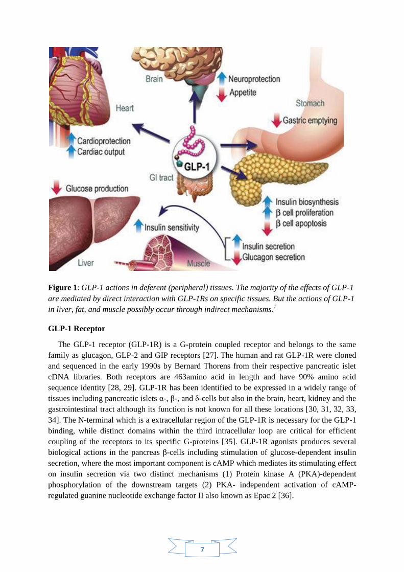
7
Figure 1: GLP-1 actions in deferent (peripheral) tissues. The majority of the effects of GLP-1
are mediated by direct interaction with GLP-1Rs on specific tissues. But the actions of GLP-1
in liver, fat, and muscle possibly occur through indirect mechanisms.1
GLP-1 Receptor
The GLP-1 receptor (GLP-1R) is a G-protein coupled receptor and belongs to the same
family as glucagon, GLP-2 and GIP receptors [27]. The human and rat GLP-1R were cloned
and sequenced in the early 1990s by Bernard Thorens from their respective pancreatic islet
cDNA libraries. Both receptors are 463amino acid in length and have 90% amino acid
sequence identity [28, 29]. GLP-1R has been identified to be expressed in a widely range of
tissues including pancreatic islets α-, β-, and δ-cells but also in the brain, heart, kidney and the
gastrointestinal tract although its function is not known for all these locations [30, 31, 32, 33,
34]. The N-terminal which is a extracellular region of the GLP-1R is necessary for the GLP-1
binding, while distinct domains within the third intracellular loop are critical for efficient
coupling of the receptors to its specific G-proteins [35]. GLP-1R agonists produces several
biological actions in the pancreas β-cells including stimulation of glucose-dependent insulin
secretion, where the most important component is cAMP which mediates its stimulating effect
on insulin secretion via two distinct mechanisms (1) Protein kinase A (PKA)-dependent
phosphorylation of the downstream targets (2) PKA- independent activation of cAMP-
regulated guanine nucleotide exchange factor II also known as Epac 2 [36].
8
Physiological action of GLP-1
The most important action of GLP-1 is its incretin function. The incretin effects imply the
amplification of insulin secretion caused by the incretin hormones. GLP-1 enhances both the
biosynthesis and secretion of insulin from pancreatic β-cells. The incretin effect is estimated
to account for approximately 50%–70% of the total insulin secreted after oral glucose
administration [37]. The circulating GLP-1 concentration is low in the fasting state, but rises
within 15 min after ingestion normally peaking within 30-45 min and returning to basal levels
over 2-3 hours [38]. The incretin effect is demonstrated by comparing insulin responses to
oral and intravenous administration glucose, where the intravenous infusion is adjusted so it
results in the same plasma glucose concentration [27, 39]. The outcome of a healthy person,
oral administration causes a three to fourfold lager insulin response comparing with the
intravenous method. Similar increases are revealed by measuring the insulin´s C-peptide,
ruling out the possibility that the differences caused by a liver insulin uptake, since the C-
peptide is not taken up by the liver. This proves that the increase in insulin secretion in oral
administration is mainly due to the actions of insulinotropic gut hormones where the two most
important are GLP-1 and GIP [27, 40, 41, 42]. In addition its incretin effects, GLP-1 is
involved in the maintenance of pancreatic β -cell mass by stimulating β -cell proliferation and
inhibiting β -cell apoptosis [43,44]. In the gastrointestinal tract the GLP-1 effects include
inhibition of meal-stimulated gastric acid secretion and gastric emptying. The inhibition of
gastric empting reduce the increases in meal associated blood glucose levels by slowing the
transit of nutrients from the stomach to the small intestinal, and therefore contributes to the
normalization of blood glucose levels.
GLP-1 also has several other effects such as
suppressing liver glucose production, inhibit glucagon secretion and increasing insulin
sensitivity and secretion [45, 46, 47, 48, 49].
GLP-1 has neural effects since both GLP-1 and it´s receptors are present in regions of the
CNS that regulate many functions including feeding behavior resulting in loss of appetite,
gastric motility and cardiovascular function. It also has a neuroprotective effects and has been
proposed as a new therapeutic agent for neurodegenerative diseases. The cardiac effects
associated with GLP-1 are for example an increase in cardiac output and other cardio
protections [17, 45].
GLP-1 in diabetes treatment
While other insulinotropic agents such as sulfonylureas have an increasing effect on
pancreatic β-cell apoptosis. Studies in different animal models have provided evidence that
GLP-1 can delay or even reverse the loss of β-cell mass by inhibiting apoptosis even in
human β-cells [49, 50, 51]. Even do β-cell replications rarely occur in human inhibitions of
apoptosis may be important for the treatment of diabetes, since the normal number of β-cells
is maintained in a balance between apoptosis and proliferation [49, 52, 53]. Furthermore
GLP-1 stimulates the transcription of genes coding for β-cell components involved in the
process of glucose sensing and insulin synthesis and secretion [54]. GLP-1 has an effect on
appetite and food intake therefore reducing the body weight and benefiting the treatment of
the disease [55].
9
Incretin effect of GLP-1 in type 2 diabetes
The incretin effect seems to be absent or at least reduced in patient with type 2diabetes,
since, unlike a healthy person, there were no significant differences in C-peptide
concentration between the experiments where oral and intravenous glucose was given
[56].GLP-1 has been shown to have a significantly reduction in type 2 diabetes, although the
insulin response to GLP-1 is retained [57,58,59 ].Both defects in the secretion and action of
incretin hormones seems be responsible for the reduced incretin effects on diabetes type 2
patients even though the molecular mechanisms underlying this is currently unknown.
GLP-1 and its stable analogues
There are several reasons way GLP-1 is used for treatment of diabetes, the fact that insulin
secretion can be restored to a normal level by administration of GLP-1 is the most important
one. The major challenge for development of GLP-1 based drugs has been that the hormone is
rapidly inactivated by DPP-IV. There are now two approaches used to overcome this
problem. The first consist in the development of GLP-1 analogs, also called incretin mimetics
that bind to the GLP-1 receptors with the same affinity as GLP-1 but resist the degradation by
DPP-IV. The second is to design drugs that inhibit the action of DPP-IV so called incretin
enhancers witch prolong the effect of native GLP-1 and increase their serum levels. The two
available incertin enhancers are sitagliptin(Januvia) and vildagliptin (Galvus).
Exenatide (synthetic exendin 4) was isolated from the lizard venom, Heloderma suspectum
and shares approximately 50% of its amino acid sequence with mammalian GLP-1
[60, 61]. Exendin-4 is a GLP-1 receptor agonist and unlike GLP-1 it is not degraded by DPP-
IV. It was approved for diabetes treatment in 2005 and is available under the name Byetta.
Exendin 4 has several incretin effect which include lowering blood glucose without a risk for
hypoglycaemia, increases insulin secretion (beta-cell function), and lowers glucagon release
[58, 62, 63, 64]. Exendin (9-39) which is an N-terminally reduced peptide copied of the
Lizard GLP-1R exendin(9-39) , binds to the GLP-1R and functions as a specific GLP-1
antagonist[65].
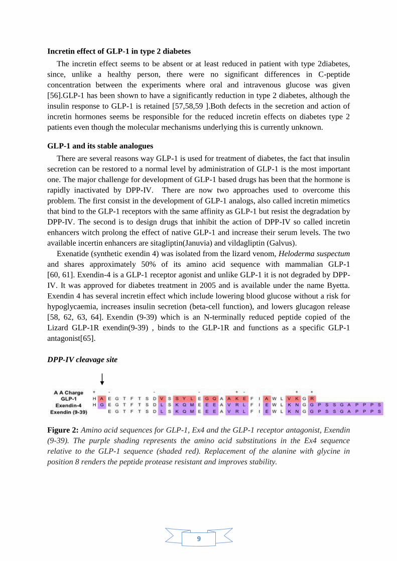
DPP-IV cleavage site
Figure 2: Amino acid sequences for GLP-1, Ex4 and the GLP-1 receptor antagonist, Exendin
(9-39). The purple shading represents the amino acid substitutions in the Ex4 sequence
relative to the GLP-1 sequence (shaded red). Replacement of the alanine with glycine in
position 8 renders the peptide protease resistant and improves stability.
10
The limitations of older therapies for diabetes have intensified the request for new
medications and better understanding of both the physiology and pathophysiology of glucose
metabolism. The effect of GLP-1 and its agonists on proliferation of the GLP-1-secreting cells
are unknown. In this study, we have studied the role of GLP-1 on proliferation of the GLP-1-
producing cell GluTag.
Aim of the study
To investigate the roles of anti-diabetic drugs GLP-1 (7-36), exendin-4, and its
metabolite GLP-1 (9-36) on proliferation of the GLP-1 secreting GluTag -cells.
To study the involvement of the GLP-1 receptor on GLP-1/exendin-4-induced
proliferation of GluTag cells.
11
Materials and methods
Cells
The cells used for all experiments were GLUTag cells which are a stable immortalized
murine enteroendocrine cell line that secretes GLP-1. This cell line was isolated from a
glucagon producing entroendocrine cell tumour that occurs in glucagon gene SV40 T-antigen
transgenic mice. The GLUTag cells were grown in DMEM medium containing 5.5mM
glucose concentration, 10% (vol/vol) fetal bovine serum (FBS ) and 1% penicillin/streptomycin
(Pest) at 37°C in a humidified (5% CO2 , 95% air) atmosphere. The medium was changed every 2days.
Experimental methods
Anti-diabetic agents
The concentration of the drugs during the incubation was GLP1-1(7-36) 10nM
(Polypeptide Group), GLP-1(9-36) 10nM (PolyPeptide Laboratories, Torrance, CA),
Exendin4 1nM (Sigma Aldrich, St Louis, MO) Exendin (9-39) 100nM (PolyPeptide
Laboratories, Torrance, CA). Due to the degradation of GLP-1(7-36) was added every 12 h
during the 48 h incubation.
3H-thymidine incorporation
400,000-450 000 Glutag cells was grown in 6-well culture plate with 2 ml medium/well
comntaining DMEM, supplimentated with 10% FBS and 5,5 mM glucose, and allowed to
reach 80-90% confluence. The cells was then incubated overnight with starvation medium
(DMEM 0,5%FBS and 5.5 mM glucose), followed by an incubation for 48 hours with the
starvation medium containing the different anti-diabetic agents. Two wells of cells were used
as controls where no anti-diabetic agent was present. Six hours before the end of the
incubation, all wells were added with 1µCi/ml 3H-thymidine (Amersham Biosciences
#TRKQ7090). At the end of the incubation, the cells were trypsinized, centrifuged and the
supernatant was discarded, the cells were washed three times in1 ml PBS . 3H-thymidine
incorporated into DNA was measured using microplate scintillation and luminescence counter
(Wallac MicroBeta® Trilux 1450-024, PerkinElmer with software: MicroBeta windows
workstation version: 3.1) in the presence of 200µl scintillation fluid (PerkinElmer). The result
was normalized with the protein concentration of the sample determined using BCA Protein
Assay kit.
Immunocytochemistry with Ki67 staining
Approximately 100 000-150 000 cells/well was transfer to 24-well plate containing 1 ml
DMEM medium with 10%FBS and 5,5mM glucose. The cells were grown to 80-90%
confluence. The medium was then change to the starvation medium with or without anti-
diabetic agents and the cells were incubated for 48 hours in an incubator. At the end of the
incubation, the medium was removed and the cells were rinsed twice with PBS before fixed
with ice cold 3.7% formaldehyde (Sigma, F8775) in PBS for 10 minutes at room temperature.
The cells were then washed three times with PBS before proceeding with immunolabeling.
12
Immunolabeling
The fixed cells were blocked in PBS-0.1% Tween 20 (PBS-T) with 10% rabbit serum for 2
hours at 4o
C. Immunolabeling was performed by incubation of the fixed cells with the
primary rabbit- polyclonal anti-Ki67 antibody (Leica Biosystems #NCL-Ki67p, 1:500 in
PBS-T, 200 μl/well) for 1 hour. The cells was washed four time with PBS-T before a two-
hour incubation with the secondary antibody anti-rabbit FITC-conjugated (Invitrogen ,
#21206, 1:200 in PBS-T/10% rabbit serum) The cells were washed four times in PBS-T.
Nuclear was stained with the fluorescent marker 4',6-diamidino-2-phenylindole (DAPI)
(SIGMA #D8417, 1: 50000, 5 min). The cells were washed several times with PBS-T before
images were captured on an Olympus microscope (CKX41 with a KX-85 Olympus camera
and software: Cell^A Olympus).
Protein measurement - BCA-assay
The protein content in each sample was determined using a BCA Protein Assay (Micro
BCA Protein Assay Kit Thermo Scintific #23235) following the manufacturer instructions.
Briefly, a standard curve was prepared as follows, 6 ml PBS solution was diluted with 315 µl
lysis buffer and bovine serum albumin (BSA) with the concentration 2 mg/ml to obtain a
series of dilutions (0,0,5, 1, 2.5, 5, 10, 20, and 40 μg/ml). 20 µl from each cell samples was
also diluted with 380 µl PBS solution and both standards and samples were transferred to the
microplate, subsequently, 150 µl reagent mixture from the kit were added to each well. The
mixture was then allowed to incubate at 37° C in a humidified (5% CO2 , 95% air) atmosphere
for 2-3 hours. Absorbance was measured at 562 nm using a microplate reader (Labsystems
iEMS Reader MF, Finland with software: Ascent software version 2.6)
Western blotting
Cell lysate preparation
Two culture flasks containing GLuTag and MIN6 cells with a confluence of 80-90% were
harvested by trypsinization.. The cells were washed twice with PBS and lysed in lysis buffer
(PBS 1%Tritorn, 2.5µl 200 µM PMSF and protease inhibitor cocktail). After 30 minute
incubation on ice, the cell lysate was centrifuged at 5000 g for 20 min, at 4° C. The
supernatant was collected into fresh tubes and stored at -80° C pending analysis. Ten µl of the
supernatant was taken for protein content determination. The protein content in each sample
was determined using BCA protein assay.
SDS-PAGE and Western blotting
A gel for SDS-PAGE containing 7.5% polyacrylamide was prepared.Same amount of
protein i.e. 20 µg from each of the two samples was loaded to the gel and run for approx 1 h
at 150 V. Separated proteins in the gels were electrophoretically transferred onto
nitrocellulose membranes (100 V for 1 hour). The membrane was rinced with TBS-T(20 mM
Tris base,137 mM NaCL pH 7.6, with, 0.05% Tween20) and blocked overnight in 5% non-
fat-milk(Bio-Rad Laboratories #1706405) in TBS-T. The membrane was washed again with
TBS-T and further incubated overnight with the primary anti-GLP-1 receptor antibody (Santa
13
Cruz Biotechnology #Sc-66911) diluted 1:500 in TBS-T containing 1% BSA. The membrane
was washed 6 times on a plate shaker. The nitrocellulose membrane was then incubated for 1
hour with the secondary antibody goat anti-rabbit IG-HRP (Santa Cruz Biotechnology #Sc-
2030) diluted 1:10000 in TBS-T-1%BSA. The membrane was washed in TBS-T six times and
protein bands were visualized by ECL (Amersham, GE Healthcare UK # RPN2132). In order
to normalize the band densities, the membrane was stripped and re-blotted with monoclonal
β-actin mouse antibody (SIGMA #A1978) diluted 1: 2000 in TBS-T and 1%BSA. The band
densities were measured using densitometry (Bio-Rad Laboratories Segrate, Milano, Italy Id
no: 101865-0 software : Quantiy one 4.6.3).
Statistic methods
Statistical analysis was conducted using ANOVA or Student´s t test. The result were
expressed as mean ±Standard error (SEM) it was considered to be statistically significant at
P<0.05.
14
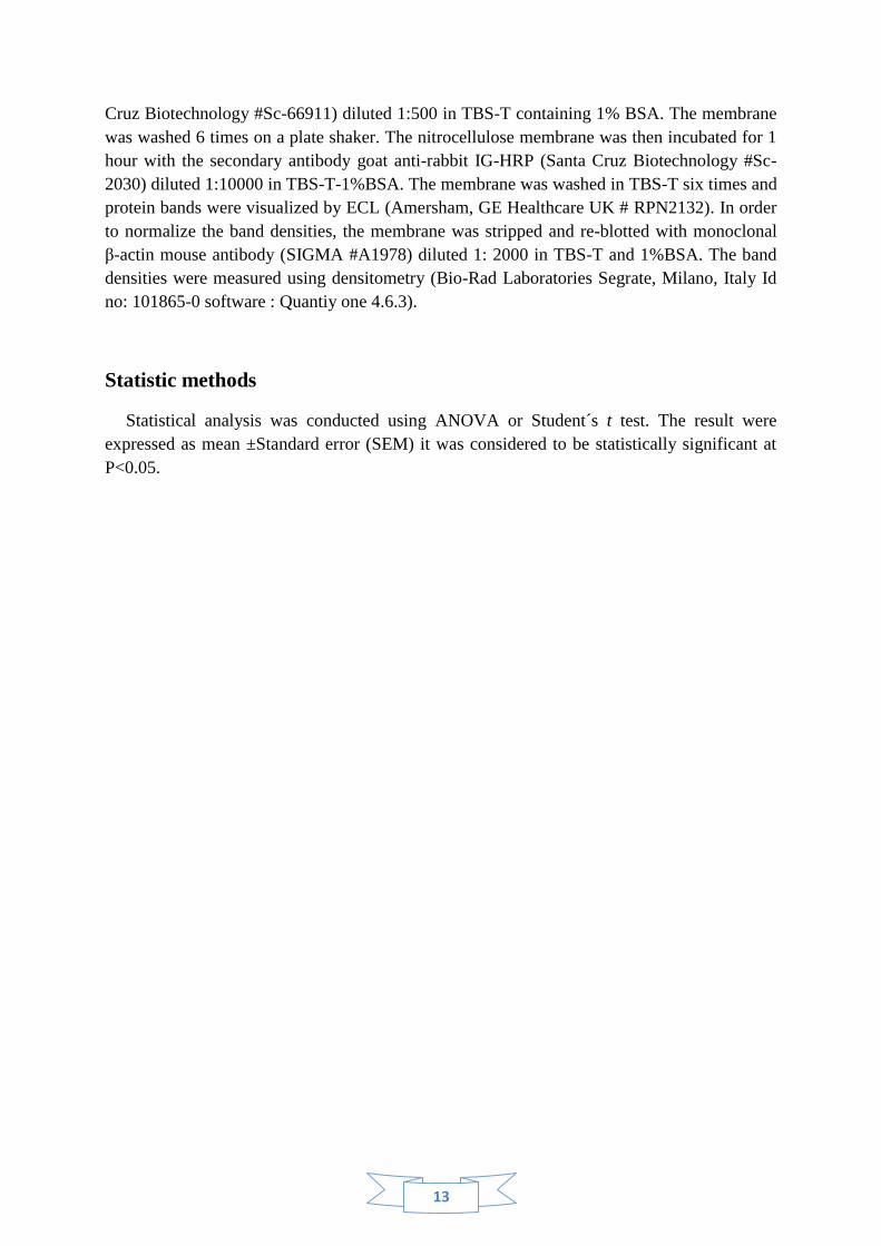
Results and Discussion
1/. GLP-1 (7-36), but not its metabolite GLP-1(9-36) and exendin-4, stimulates
proliferation of the GLP-1-secreting cell GluTag.
Effect on cell proliferation was measured using two different methods that are widely used
for studying cell proliferation. 3H- thymidine incorporation is standard method to monitor
rates of DNA synthesis and cell proliferation, were Ki67 is used as a proliferation marker
since the polyclonal antibody labels Ki67 antigen in the granular components of the nucleolus
during proliferation stages.
C GLP-1(9-36)
3H
-Th
ym
idie
nin
co
rpo
ration
0
50
100
A B
C GLP-1(7-36) Ex-4
0
100
200
300
400
3H
-Thym
idie
nin
corp
ora
tion
(%of
co
ntro
l)
3H
-Thym
idie
nin
corp
ora
tion
(%of
co
ntro
l)
*
Figure1: Cell was incubated for 48 h in DMEM medium contaning 3mM glucose, .5% FBS in
the present or absent of the agents. Cells were pulsed with 3H –thymidine 6 h prior to the end
of the incubation. The result were normalized by protein content in each dish and expressed
as percentage of control. They are from three independent duplicate experiments (* P<0.05).
GLP-1(7-36) significantly enhanced thymidine incorporation into cells. At the
concentration of 10 nM, GLP-1(7-36) caused approximately 300% of increase in 3H-
thymidine incorporation, compared to the controls, indicating a stimulatory effect of the drugs
on cell proliferation. In contrast, Exendin4 did not show any effect on cell proliferation.
(Figure 1 A) In addition, incubation of the cells with GLP-1(9-36), the major GLP-1
metabolite did not result in any changes in thymidine incorporation compared to control.
(Figure 1B)
15
GLP
-1(7
-36)
Ki6
7sta
ine
dcell
(%)
0
50
100
150
*
C
GLP
-1(9
-36)
GLP
-1/E
x(9-
39)
A B C D
Figure 2: Cell was incubated for 48 h in DMEM medium contaning 3mM glucose, 0.5% FBS
in the present or absent of the agents. Immunocytochemistry was then performed to detect
cells in proliferation state. Result are from three independent duplicate experiments (*
P<0.05)
To further verify the observed effect of GLP-1(7-36) on proliferation of GluTag cells, Ki67
staining was performed. The result shows that GLP-1(7-36) casued a 50% increase in
proliferation compared to control. Consistent with the results from 3H-thymidine
incorporation, GLP-1(9-36) did not show any significant changes in the Ki67 staining
(Figures 2). This result further indicates a role of GLP-1(7-36), but not its metabolite, in
promoting proliferation of the cells.
The involvement of the GLP-1 receptor in the GLP-1(7-36)-induced proliferation was
evaluated by co-incubation of the cells with the GLP-1 receptor antagonist Exendin (9-39).
Ata concentration of 100 nM , exendin(9-39) completely blocked the GLP-1(7-36)- induced
proliferation.
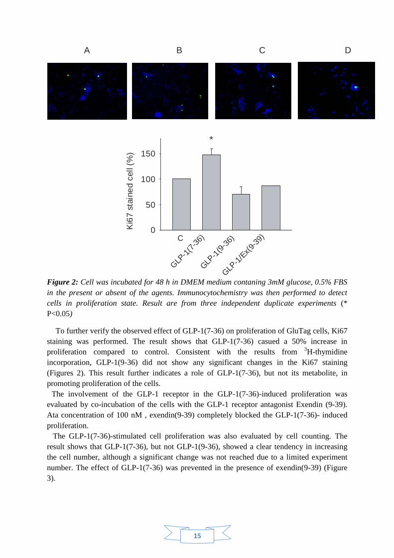
The GLP-1(7-36)-stimulated cell proliferation was also evaluated by cell counting. The
result shows that GLP-1(7-36), but not GLP-1(9-36), showed a clear tendency in increasing
the cell number, although a significant change was not reached due to a limited experiment
number. The effect of GLP-1(7-36) was prevented in the presence of exendin(9-39) (Figure
3).
16
Cel
lnum
ber
(%co
ntro
l)
0
50
100
150
*
GLP-1
(7-3
6)C
GLP-1
(9-3
6)
GLP-1
/Ex(9
-39)
Figure 3: The cell number from Ki67 stained cells presented as percentage of control, the
result are from three experiments (* P<0.05).
The above results indicate that GLP-1(7-36), secreted from the GLP-1-secreting cells is able
to stimulate the cell proliferation by a feedback mechanism. Such feedback action manner has
been seen in other cells, such as the pancreatic β-cell, where insulin stimulates insulin gene
expression and secretion. The present results suggest that such an autocrine/paracrine action
may also be functioning in the GLP-1 secreting cells in regulation of cell proliferation.
Considering that the GLP-1 metabolite GLP-1(9-36), which has poor affinity to the GLP-1
receptor, was without effect on the cell proliferation; and that the effect of GLP-1(7-36) could
be blocked by the GLP-1 receptor antagonist, the effect of GLP-1(7-36) on the cell
proliferation is likely to be mediated through the GLP-1 receptor.
The lack of effect by exendin-4 may suggest different action manner of GLP-1 and
exendin-4 [22, 23, 29]. In addition, the presence of distinct exendin receptor has been
proposed [28]. This finding raised a question: whether the GLP-1 receptor is expressed in the
GLP-1-secreting cells?
17
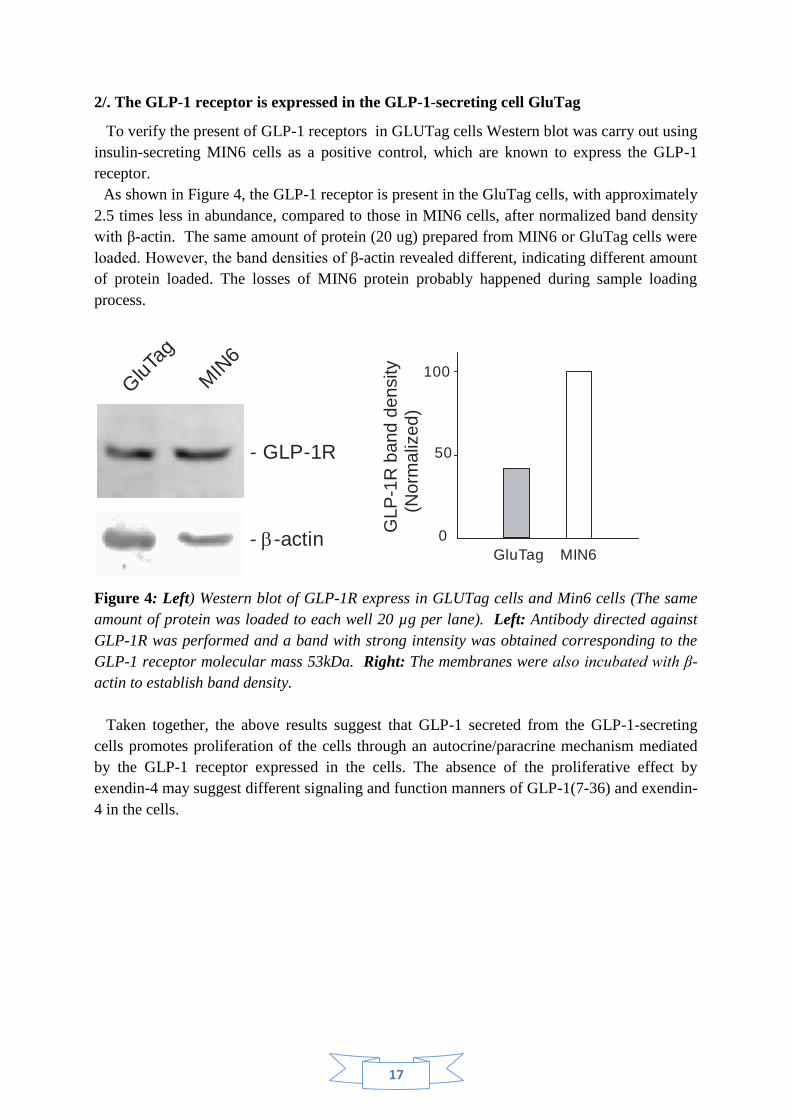
2/. The GLP-1 receptor is expressed in the GLP-1-secreting cell GluTag
To verify the present of GLP-1 receptors in GLUTag cells Western blot was carry out using
insulin-secreting MIN6 cells as a positive control, which are known to express the GLP-1
receptor.
As shown in Figure 4, the GLP-1 receptor is present in the GluTag cells, with approximately
2.5 times less in abundance, compared to those in MIN6 cells, after normalized band density
with β-actin. The same amount of protein (20 ug) prepared from MIN6 or GluTag cells were
loaded. However, the band densities of β-actin revealed different, indicating different amount
of protein loaded. The losses of MIN6 protein probably happened during sample loading
process.
- GLP-1R
GluTa
g
MIN
6
- -actinb 0
50
100
GLP
-1R
band d
ensity
(N
orm
aliz
ed)
GluTag MIN6
Figure 4: Left) Western blot of GLP-1R express in GLUTag cells and Min6 cells (The same
amount of protein was loaded to each well 20 µg per lane). Left: Antibody directed against
GLP-1R was performed and a band with strong intensity was obtained corresponding to the
GLP-1 receptor molecular mass 53kDa. Right: The membranes were also incubated with β-
actin to establish band density.
Taken together, the above results suggest that GLP-1 secreted from the GLP-1-secreting
cells promotes proliferation of the cells through an autocrine/paracrine mechanism mediated
by the GLP-1 receptor expressed in the cells. The absence of the proliferative effect by
exendin-4 may suggest different signaling and function manners of GLP-1(7-36) and exendin-
4 in the cells.
18
Conclusion and future work
The result obtained suggests:
1. GLUTag cells express GLP-1 recepors
2. GLP-1(7-36) enhances cell proliferation in GLUtag cells
3. Exendin-4 and GLP-1(9-36) do not have effect on cell proliferation
4. The GLP-1(7-36)-induced proliferation of GluTag cells is blocked by Ex4(9-36) in
GLUTag cells.
Future work:
In order to further validate these finding, further studies are required. After having
investigated the agent’s effect on cell proliferation, the mechanism underlying this effect
needs to be studied. This includes studying whether GLP-1 receptor actually is activated in
GLUTag cells, by investigate the signal transduction pathways activated by GLP-1(7-36) and
the post- receptor activations in this event. In addition the requirement of GLP-1 receptor in
the GLP-1(7-36)-induced proliferation of GluTag cells should also be studied, in cells with
GLP-1 receptor knockdown using siRNA.
Acknowledgement
First and foremost I owe a great debt and gratitude to my supervisor Md. Qimin Zhang for
giving me the opportunity of doing my MSc thesis here at Forskningcentrum Karolinska
Institutet Södersjukhuset and more importantly, for all support, knowledge and guidance
during my project.
I would also like to extend my gratitude to PhD-student Camilla Kappe for supporting my
project with cells and demonstrating all the techniques. This work should not have been
possible without all your help.
I also want to thanks the Head of the Unit for Diabetes Research, Åke Sjöholm for given
me the opportunity to do my thesis in his group. I also want to acknowledge all the other PhD-
students in Åke Sjöholm group and rest of the staff at Forskningcentrum.
Finally I also want to thank my examiner Sven Hamp from Märladalen University.
19
References
1. Wild S, Roglic G, Green A, Sicree R, King H. Global prevalence of diabetes: estimates
for the year 2000 and projections for 2030 Diabetes Care. 2004 May;27(5):1047-53.
2. Kumar P, Clark M Clinical Medicine. 6th edn. WB Saunders, London (2005)
3. Cohn RM, Roth KS Biochemistry and Disease. Williams and Wilkins, London (1996)
4. Stumvoll M, Goldstein BJ, Von Haeften TW. Type 2 diabetes: principles of
pathogenesis and therapy. Lencet, 365:1333-1346,
5. John C. Pickup, Williams Gareth , 1997 Textbook of Diabetes Volume 1 Blackwell
Science Ltd, Oxford
6. American Diabetes Association. Standards of medical care for patients with diabetes
mellitus. Diabetes Care. 2002;25(1):213Y229
7. The Expert Committee on the Diagnosis and Classification of Diabetes Mellitus.
Follow-up report on the diagnosis of diabetes mellitus. Diabetes Care.
2003;26(11):3160Y3167
8. Meier JJ, Nauck Ma, Glucagon-like peptide-1 (GLP-1) in biology and pathology.
Diabetes Metab Res Rev 21:91-117,2005
9. Dupre J, Ross SA, Watson D et al. Stimulation of insulin secretion by gastric inhibitory
polypeptide in man. The Journal of Clinical Endocrinology and Metabolism 1973; 37:
826–828
10. Cataland S, Crockett SE, Brown JC, Mazzaferri EL. Gastric inhibitory polypeptide
(GIP) stimulation by oral glucose in man. J Clin Endocrinol Metab. 1974
Aug;39(2):223-8.
11. Lynn FC et al., Diabetes 50:1004–1011, 2001; Zhou J et al., Am J Physiol Endocrinol
Metab 293:E538–E547, 2007
12. Vilsboll T, Krarup T, Sonne J, Madsbad S, Volund A, Juul AG, Holst JJ. Incretin
secretion in relation to meal size and body weight in healthy subjects and people with
type 1 and type 2 diabetes mellitus. J Clin Endocrinol Metab 88: 2706–2713, 2003
13. Miholic J, Orskov C, Holst JJ, Kotzerke J, Meyer HJ. Emptying of the gastric
substitute, glucagon-like peptide-1 (GLP-1), reactive hypoglycemia after total
gastrectomy. Dig Dis Sci 36: 1361–1370, 1991.
14. Mortensen K, Christensen LL, Holst JJ, Orskov C. GLP-1 and GIP re colocalized in a
subset of endocrine cells in the small intestine. Regul Pept 2003;114:189–196
15. Ugleholdt R, Zhu X, Deacon CF, Orskov C, Steiner DF, HolstJJ. Impaired intestinal
proglucagon processing in mice lacking prohormone convertase 1. Endocrinology 145:
1349–1355, 2004
16. Zhu X, Zhou A, Dey A, Norrbom C, Carroll R, Zhang C, LaurentV, Lindberg I,
Ugleholdt R, Holst JJ, Steiner DF. Disruption of PC1/3 expression in mice causes
dwarfism and multiple neuroendocrine peptide processing defects. Proc Natl Acad Sci
USA 99: 10293–10298, 2002.
17. JENS JUUL HOLST.The Physiology of Glucagon-like Peptide 1 Physiol Rev. 2007
Oct;87(4):1409-39
20
18. Unger RH, Ohneda A, Valverde I, Eisentraut AM, Exton J. Characterization of the
responses of circulating glucagon-like immunoreactivity to intraduodenal and
intravenous administration of glucose. J Clin Invest 1968;47:48–65.
19. Hermann C, Goke R, Richter G, Fehmann HC, Arnold R, Goke B. Glucagon-like
peptide-1 and glucose-dependent insulin-releasing polypeptide plasma levels in
response to nutrients. Digestion 1995;56:117–126.
20. Theodorakis MJ, Carlson O, Michopoulos S, Doyle ME, Juhaszova M, Petraki K, Egan
JM. Human duodenal enteroendocrine cells: source of both incretin peptides, GLP-1
and GIP. Am J Physiol 2006;290:E550–E559.
21. Baggio LL, Drucker DJ. GLP-1 and GIP Gastroenterology.Biology of incretins: 2007
May;132(6):2131-57.
22. Orskov C, Wettergren A, Holst JJ. Biological effects and metabolic rates of
glucagonlike peptide-1 7-36 amide and glucagon like peptide-1 7-37 in healthy
subjects are indistinguishable.Diabetes 1993;42:658–661
23. Orskov C, Rabenhoj L, Wettergren A, Kofod H, Holst, JJ. Tissue and plasma
concentrations of amidated and glycine-extended glucagon-like peptide I in humans.
Diabetes 1994;43:535– 539.
24. Deacon CF, Johnsen AH, Holst JJ. Degradation of glucagon-like peptide-1 by human
plasma in vitro yields an N-terminally truncated peptide that is a major endogenous
metabolite in vivo. J Clin Endocrinol Metab 1995;80:952–957
25. Hansen L, Deacon CF, Orskov C, Holst JJ. Glucagon-like peptide-1-(7-36)amide is
transformed to glucagon-like peptide-1-(9-36)amide by dipeptidyl peptidase IV in the
capillaries supplying the L cells of the porcine intestine. Endocrinology 1999,
140:5356-5363.
26. Ruiz-Grande C, Alarcon C, Alcantara A, Castilla C, Lopez Novoa JM, Villanueva-
Penacarrillo ML, Valverde I. Renal catabolism of truncated glucagon-like peptide 1.
Horm Metab Res 1993;25: 612–616
27. Mayo KE, Miller LJ, Bataille D, Dalle S, Goke B, Thorens B,Drucker DJ. The
Glucagon Receptor Family. Pharmacol Rev 55: 167–194, 2003 International Union of
Pharmacology. XXXV
28. Thorens B. Expression cloning of the pancreatic beta cell receptor for the gluco-
incretin hormone glucagon-like peptide 1. Proc Natl Acad Sci USA 89: 8641–8645,
1992.
29. Thorens B, Porret A, Buhler L, Deng SP, Morel P, Widmann C. Cloning and functional
expression of the human islet GLP-1 receptor. Demonstration that exendin-4 is an
agonist and exendin- (9–39) an antagonist of the receptor. Diabetes 42: 1678–1682,
1993
30. Alvarez E, Martinez MD, Roncero I, Chowen JA, Garcia-Cuartero B, Gispert JD, Sanz
C, Vazquez P, Maldonado A, de CJ, Desco M, Pozo MA, Blazquez E. The expression
of GLP-1receptor mRNA and protein allows the effect of GLP-1 on glucose metabolism
in the human hypothalamus and brainstem. J Neurochem 92: 798–806, 2005
31. Bullock BP, Heller RS, Habener JF. Tissue distribution of messenger Ribonucleic acid
encoding the rat glucagon-like peptide-1 receptor. Endocrinology 137: 2968–2978,
1996.
21
32. Campos RV, Lee YC, Drucker DJ. Divergent tissue-specific and developmental
expression of receptors for glucagon and glucagonlike peptide-1 in the mouse.
Endocrinology 134: 2156–2164, 1994.
33. Wei Y, Mojsov S. Tissue-specific expression of the human receptor for glucagon-like
peptide-I: brain, heart and pancreatic forms have the same deduced amino acid
sequences. FEBS Lett 358: 219–224, 1995.
34. Wei Y, Mojsov S. Distribution of GLP-1 and PACAP receptors in human tissues. Acta
Physiol Scand 157: 355–357, 1996.
35. Hallbrink M, Holmqvist T, Olsson M, Ostenson CG, Efendic S, Langel U.
Different domains in the third intracellular loop of the GLP-1 receptor are responsible
for Galpha(s) and Galpha(i)/ Galpha(o) activation. Biochim Biophys Acta
2001;1546:79–86.
36. Holz GG. Epac: a new cAMP-binding protein in support of glucagon-like peptide-1
receptor-mediated signal transduction in the pancreatic beta-cell. Diabetes 53: 5–13,
2004.
37. Baggio LL Gastroenterology 2007;132:2131–2157).
38. Deacon CF. What do we know about the secretion and degradation of incretin
hormones? Regulatory Peptides 2005; 128:117–124.
39. Perley M, Kipnis DM. Plasma insulin responses to oral and intravenous glucose:
studies in normal and diabetic subjects. J Clin Invest 46: 1954–1962, 1967.
40. Nauck MA, Homberger E, Siegel EG, Allen RC, Eaton RP, Ebert R, Creutzfeldt W.
Incretin effects of increasing glucose loads in man calculated from venous insulin and
C-peptide responses. J Clin Endocrinol Metab. 1986 Aug;63(2):492-8
41. McIntyre N, Holdsworth CD, Turner DS. Intestinal factors in the control of insulin
secretion. Secretion.J Clin Endocrinol Metab 25: 1317–1324, 1965.
42. Vilsboll T, Holst JJ. Incretins, insulin secretion and Type 2 diabetes mellitus.
Diabetologia 47: 357–366, 2004.
43. Buteau J, Foisy S, Joly E, Prentki M, Glucagon-like Peptide-1 Induces Pancreatic β-
cell proliferation via transactivation of the epidermal growth factor receptor. Diabetes
2003, 52:124-132
44. Buteau J El-Assaad W, Rhodes CJ, Rosenberg L, Joly E, Glucagon-like peptide-1
prevents beta cell glucolipotoxicity. Diabetologia 2004, 47:806-815
45. During MJ, Cao L, Zuzga DS, Francis JS, Fitzsimons HL, Jiao X, Bland RJ, Klugmann
M, Banks WA, Drucker DJ, Haile CN. Glucagon-like peptide-1 receptor is involved in
learning and neuroprotection. Nat Med 9: 1173–1179, 2003.
46. Willms B, Werner J, Holst JJ, Orskov C, Creutzfeldt W, Nauck MA.
Gastric emptying, glucose responses, and insulin secretion after a liquid test meal:
effects of exogenous glucagon-like peptide-1 (GLP-1)-(7-36) amide in type 2
(noninsulin-dependent) diabetic patients. J Clin Endocrinol Metab 1996;81:327–332.
47. Meier JJ, Gallwitz B, Salmen S, Goetze O, Holst JJ, Schmidt WE, Nauck MA.
Normalization of glucose concentrations and deceleration of gastric emptying after
solid meals during intravenous glucagon-like peptide 1 in patients with type 2 diabetes.
J Clin Endocrinol Metab 2003;88:2719–2725.
22
48. Fehmann HC, Goke R, Goke B. Cell and molecular biology of the incretin hormones
glucagon-like peptide-I and glucose-dependent insulin releasing polypeptide. Endocr
Rev 16: 390–410, 1995.
49. Butler AE, Janson J, Bonner-Weir S, Ritzel R, Rizza RA, Butler PC.
Beta-cell deficit and increased beta-cell apoptosis in humans with type 2 diabetes.
Diabetes 2003; 52: 102–110.
50. Buteau J, El-Assaad W, Rhodes CJ, Rosenberg L, Joly E, Prentki M. Glucagon-like
peptide-1 prevents beta cell glucolipotoxicity. Diabetologia 47: 806–815, 2004.
51. Farilla L, Bulotta A, Hirshberg B, Li CS, Khoury N, Noushmehr H, Bertolotto
C, Di MU, Harlan DM, Perfetti R. Glucagon-like peptide 1 inhibits cell apoptosis and
improves glucose responsiveness of freshly isolated human islets. Endocrinology 144:
5149–5158, 2003.
52. Bonner-Weir S. Beta-cell turnover: its assessment and implications. Diabetes 50 Suppl
1: S20–S24, 2001.
53. Tyrberg B, Ustinov J, Otonkoski T, Andersson A. Stimulated endocrine cell
proliferation and differentiation in transplanted human pancreatic islets: effects of the
ob gene and compensatory growth of the implantation organ. Diabetes 2001; 50: 301–
307
54. Wang Y, Egan JM, Raygada M, Nadiv O, Roth J, Montrose Rafizadeh C. Glucagon-
like peptide-1 affects gene transcription and messenger ribonucleic acid stability of
components of the insulin secretory system in RIN 1046-38 cells. Endocrinology 1995;
136: 4910–4917.
55. Hu FB, Manson JE, Stampfer MJ, et al. Diet, lifestyle, and the risk of type 2 diabetes
mellitus in women. New Engl J Med 2001; 345: 790–797
56. Nauck M, Sto¨ckmann F, Ebert R et al. Reduced incretin effect in type 2 (non-insulin-
dependent) diabetes. Diabetologia 1986; 29: 46–54.
57. Nauck MA, Heimesaat MM, Orskov C, Holst JJ, Ebert R, Creutzfeldt W.
Preserved incretin activity of glucagon-like peptide 1 [7–36 amide] but not of synthetic
human gastric inhibitory polypeptide in patients with type-2 diabetes mellitus. J Clin
Invest 91: 301–307, 1993.
58. Toft-Nielsen MB, Damholt MB, Madsbad S, Hilsted LM, Hughes TE, Michelsen BK,
Holst JJ. Determinants of the impaired secretion of glucagon-like peptide-1 in type 2
diabetic patients. J Clin Endocrinol Metab 86: 3717–3723, 2001
59. Vilsboll T, Krarup T, Deacon CF, Madsbad S, Holst JJ. Reduced postprandial
concentrations of intact biologically active glucagon-like peptide 1 in type 2 diabetic
patients. Diabetes 50: 609–613, 2001.
60. ChenYE, Drucker DJ. Tissue-specific expression of unique mRNAs that encod
proglucagon-derived peptides or exendin 4 in the lizard. J Biol Chem 1997; 272:
4108–15.
61. Thorens B, Porret A, Buhler L, Deng SP, Morel P, Widmann C. Cloning and functional
expression of the human islet GLP-1 receptor. Demonstration that exendin-4 is an
agonist and exendin- (9–39) an antagonist of the receptor. Diabetes 42: 1678–1682,
1993
23
62. Vilsboll T, Krarup T, Madsbad S, Holst JJ. Defective amplification of the late phase
insulin response to glucose by GIP in obese Type II diabetic patients.
Diabetologia 45: 1111–1119, 2002
63. Kendall, D.M., et al., Effects of exenatide (exendin-4) on glycemic control over 30
weeks in patients with type 2 diabetes treated with metformin and a sulfonylurea.
Diabetes Care, 2005. 28(5): p. 1083-91.
64. Egan, J.M., A.R. Clocquet, and D. Elahi, The insulinotropic effect of acute exendin-4
administered to humans: comparison of nondiabetic state to type 2 diabetes. J Clin
Endocrinol Metab, 2002. 87(3): p. 1282-90.
65. Thorens B, Porret A, Buhler L, Deng SP, Morel P, Widmann C.Cloning and functional
expression of the human islet GLP-1 receptor. Demonstration that exendin-4 is an
agonist and exendin- (9–39) an antagonist of the receptor. Diabetes 42: 1678–1682,
1993
Recommended