Structure900
� sheet. The fold of the enzyme is a variant of the ribo- Nicholas O’Toole and Miroslaw CyglerBiotechnology Research Institute, NRCkinase fold with some differences in chain connectivity
in the N-terminal segment of the protein. The proteins 6100 Royalmount AvenueMontreal, Quebec H4P 2R2classified within the SCOP database as having the ribo-
kinase-like fold include several kinases as well as pep- Canadatide ligases from the Mur family. All of them utilize ATP
Selected Readingas a phosphate donor. PPCS on the other hand utilizesATP for an adenylation reaction. The structure of human
Abiko, Y. (1975). In Metabolic Pathways, D.M. Greenberg, ed. (NewPPCS demonstrates that the ribokinase-like fold accom- York: Academic Press), pp. 1–25.modates a wider range of reactions and ATP binding Albert, A., Martinez-Ripoll, M., Espinosa-Ruiz, A., Yenush, L., Culia-modes than previously thought. nez-Macia, F.A., and Serrano, R. (2000). Structure 8, 961–969.
Manoj et al. have also derived a model for the quater- Daugherty, M., Polanuyer, B., Farrell, M., Scholle, M., Lykidis, A.,nary structure of the bifunctional bacterial PPCS/ Crecy-Lagard, V., and Osterman, A. (2002). J. Biol. Chem. 277,
21431–21439.PPCDC enzymes. The bifunctional enzyme from E. coliwas earlier demonstrated to be a dodecamer based on Gerdes, S.Y., Scholle, M.D., D’Souza, M., Bernal, A., Baev, M.V.,
Farrell, M., Kurnasov, O.V., Daugherty, M.D., Mseeh, F., Polanuyer,gel filtration measurements (Kupke et al., 2000). TheB.M., et al. (2002). J. Bacteriol. 184, 4555–4572.oligomeric states of the PPCS and PPCDC domainsIzard, T., and Geerlof, A. (1999). EMBO J. 18, 2021–2030.when expressed alone, in combination with the knownKupke, T., Uebele, M., Schmid, D., Jung, G., Blaesse, M., andstructures of homologous proteins to these domainsSteinbacher, S. (2000). J. Biol. Chem. 275, 31838–31846.from humans (the present structure) and A. thaliana (Al-Manoj, N., Strauss, E., Begley, T.P., and Ealick, S.E. (2003). Structurebert et al., 2000), suggest that the bifunctional enzyme11, this issue, 927–936.consists of a PPCDC core surrounded by an outer layerObmolova, G., Teplyakov, A., Bonander, N., Eisenstein, E., Howard,of PPCS dimers. In the model presented, the two activeA.J., and Gilliland, G.L. (2001). J. Struct. Biol. 136, 119–125.
sites are near each other, joined by an internal channel,O’Toole, N., Barbosa, J.A., Li, Y., Hung, L.W., Matte, A., and Cygler,
and neither is obstructed by the dodecamer formation. M. (2003). Protein Sci. 12, 327–336.Verification of this predicted quaternary assembly awaits
Yun, M., Park, C.G., Kim, J.Y., Rock, C.O., Jackowski, S., and Park,the experimental determination of a bacterial PPCS/ H.W. (2000). J. Biol. Chem. 275, 28093–28099.PPCDC structure, which would also guide efforts into Zhao, L., Allanson, N.M., Thomson, S.P., Maclean, J.K., Barker, J.J.,structure-based drug discovery for this antibacterial Primrose, W.U., Tyler, P.D., and Lewendon, A. (2003). Eur. J. Med.
Chem. 38, 345–349.target.
Structure, Vol. 11, August, 2003, 2003 Elsevier Science Ltd. All rights reserved. DOI 10.1016/S0969-2126(03)00164-3
the enzyme moves farther along the gene, phosphatesGetting a Gripare increasingly found on S2 residues of the CTD repeats,on the CTD of Pol II and the S2-phosphorylated CTD is known to bind cleav-age/polyadenylation factor subunits, such as Pcf11(e.g., Licatalosi et al., 2002). However, because not muchis known about the structure of the CTD, whether free
The first structure of a pre-mRNA processing factor or factor bound, phosphorylated or unphosphorylated,bound to heptad repeats from the C-terminal domain very little is known about the molecular details of CTD-of RNA polymerase II is revealed in a crystal of capping protein interactions. A recent paper in Molecular Cellguanylyltransferase complexed with a four-repeat provides a major boost to our knowledge in this area,phosphopeptide. revealing how the phospho-CTD interacts with one of
its better-characterized binding partners, the guanylyl-transferase component of pre-mRNA capping enzymeThe C-terminal repeat domain (CTD) of RNA polymerase(Fabrega et al., 2003).II, composed of up to 52 repeats of the consensus hepta-
To date, only one structure showing a CTD repeat haspeptide Y1S2P3T4S5P6S7, contributes to the functional or-been solved, that of mammalian Pin1 complexed withganization of the nucleus by mediating interactions be-a canonical heptapeptide carrying Ser-PO4 at both posi-tween RNAP II and a lengthening list of nuclear factors.tions 2 and 5 (Verdecia et al., 2000). The Pin1 structureActively transcribing RNAP II carries a highly phosphory-provided a valuable first look at one CTD repeat boundlated CTD, which is known to recruit or tether certainto a protein, but it also left many questions unanswered.RNA processing proteins to sites of transcription, posi-In that structure, the single CTD repeat binds as antioning them to act on the nascent transcript (reviewedextended coil, with 5 residues making contact with thein Bentley, 2002). As the polymerase begins to transcribeprotein (P3T4S5pP6S7). The phosphate on Ser5 makes sev-a gene, the CTD carries phosphates mostly on S5; thiseral contacts with the protein, which contribute signifi-form of the phospho-CTD efficiently binds 5� end cap-cantly to the binding affinity. In contrast, the phosphateping components, such as capping guanylyltransfer-
ases (e.g., Komarnitsky et al., 2000; Pei et al., 2001). As on Ser2 points away from Pin1 into solution, not making
Previews901
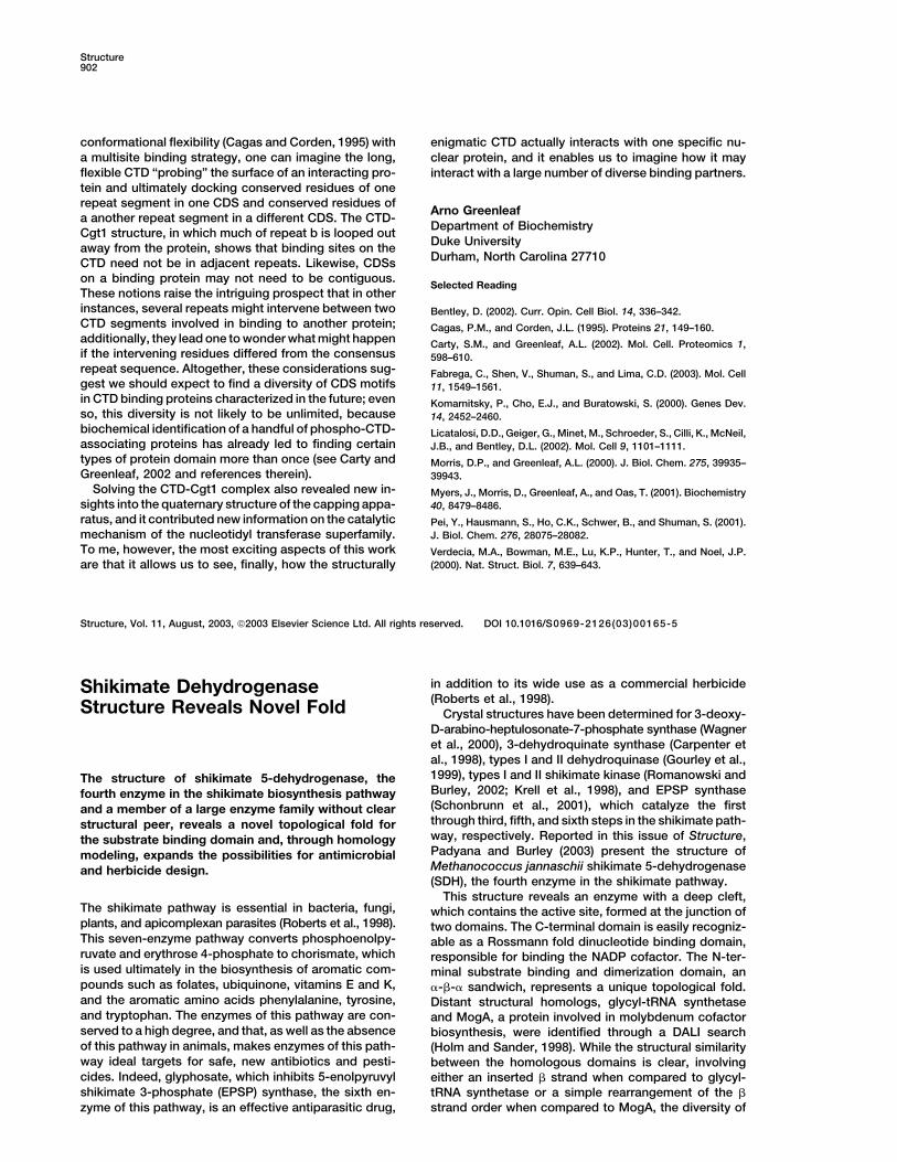
Figure 1. Interactions between CTD Phosphopeptide and Cgt1
The four-repeat, 28 residue peptide, numbered as in Fabrega et al., 2003, is shown; the repeats are referred to as a–d, and each Ser5-PO4 isin bold. A single line above the peptide indicates residues that were structured in the crystal (monomer A and monomer B forms), and doublelines below it indicate residues contacting the two CTD-docking sites (CDS) in Cgt1 (monomer B). A wavy double line below the peptideindicates residues involved in a roughly 90� bend.
contact with any part of Pin1; this result was surprising drophobic interactions in between. It should be notedthat extensive genetic tests (which monitored in vivoin view of the finding that the yeast homolog of Pin1
preferentially binds repeat peptides with more than one effects of changing amino acid side chains in the CTDrepeats or in the Cgt1) are entirely consistent with thephosphate (Myers et al., 2001). Additionally, the Tyr resi-
due (Y1) appears to make no contribution to the binding. interactions observed in the crystal (Fabrega et al., 2003and references therein).The studies on Pin1 raise a number of questions, among
which are the following: (1) will the coiled conformation We see in this structure answers to the above ques-tions. (1) In contrast to the Pin1 results, the currentof the CTD repeat in the Pin1 complex be found in other
complexes? (2) Will CTD segments composed of multi- structure shows the CTD peptide in contact with Cgt1 isnot in a coiled form, but rather in an extended, nonhelicalple repeats bind differently from a single repeat? (3) In
other CTD-protein complexes, will the Tyr of the CTD conformation. (2) Binding of the several-repeat peptideto Cgt1 is very different from binding of a single repeatrepeats contribute to the binding interactions?
All of these questions have been answered and a great to Pin1. The phospho-CTD-interacting region of Cgt1 iscomposed of two distinct “docking sites,” each of whichdeal of additional information generated in work from
the Lima and Shuman labs, reported in the June issue contacts a distinct 6-7 residue region of the CTD pep-tide. Because the contact region in each CTD extendsof Molecular Cell (Fabrega et al., 2003). These workers
solved the structure of the Candida albicans capping across canonical repeat boundaries, it is clear that asingle canonical repeat peptide must bind differentlyguanylyltransferase (Cgt1, residues 1–395) crystallized
as a complex with a CTD phosphopeptide composed from a multiple-repeat peptide (note that the CTD se-quence is circularly permuted, and it is likely that func-of four heptapeptide repeats in which every Ser5 was
phosphorylated (Figure 1). In the better characterized tional units within the CTD will include parts of morethan one canonical repeat [e.g., TSPSYS, PSYSPT, etc.];of the two forms observed (monomer B), a stretch of 17
amino acids of the CTD peptide is ordered in the crystal a particular 7 residue peptide thus may or may not con-tain a natural binding determinant). (3) In the Cgt1-CTDand interacts with numerous residues lining a saddle-
like channel in the enzyme more than 40 A long (Figure complex, the conserved Y1 residues play crucial roles inthe peptide’s interactions with the protein. The aromatic1B; Figure 2 of Fabrega et al., 2003). Parts of three
canonical repeats are involved in the binding (residues ring of Y1b makes intimate contacts with aromatic andhydrophobic residues on the floor of the cleft in theT4a–P6c in Figure 1).
The N-terminal-most peptide residue making intimate enzyme, while Y1c makes both VDW contacts and hydro-gen bonds (via the ring hydroxyl) to residues in CDS2;contact with Cgt1 is the Ser5-PO4 of the first repeat
(repeat a, residue S5pa), which inserts into a positively interestingly, the bend and looping out of residues inrepeat b of the peptide bring Y1c within about 5 A of Y1b.charged pocket. Following along the peptide in the
C-terminal direction, the next 3 residues are in an ex- The phospho-CTD-Cgt1 structure, in addition to pro-viding a detailed picture of one particular complex, helpstended �-like conformation and make van der Waals
(VDW) contacts with the enzyme, P6a and Y1b contacts to explain certain features of known CTD-protein inter-actions and also provides a basis for speculating aboutappearing especially important. These contacts make
up the “CTD-docking site 1” (CDS1). Then, the peptide properties of CTD-protein complexes to be solved inthe future. It seems likely that the conformational reper-makes a roughly 90� turn, which involves looping several
residues out away from Cgt1 (notably the next Ser-PO4, toire of the CTD bound to its many partners will be large;this feature helps explain how it is that sets of CTDS5pb), and resumes close contacts with the protein
through the next Tyr residue, Y1c. Repeat c makes addi- binding partners bear “no obvious structural related-ness…” (Fabrega et al., 2003, p. 1549). Also, as bothtional contacts along the enzyme cleft to define CDS2,
finally making another strong ionic interaction via S5pc, the phosphopeptide and the Cgt1 protein employ twobinding sites for their interaction, we see here anotherwhich is buried in another positive pocket. Thus, the
binding of the phosphopeptide depends strongly on two example of multisite binding involving the CTD (Morrisand Greenleaf, 2000); in this binding mode, several weakelectrostatic interactions at either end of the binding
region, involving Ser5-PO4 residues separated by two interactions combine synergistically to create a muchstronger overall affinity. Considering together the CTD’scomplete (noncanonical) repeats, and several hy-
Structure902
conformational flexibility (Cagas and Corden, 1995) with enigmatic CTD actually interacts with one specific nu-a multisite binding strategy, one can imagine the long, clear protein, and it enables us to imagine how it mayflexible CTD “probing” the surface of an interacting pro- interact with a large number of diverse binding partners.tein and ultimately docking conserved residues of onerepeat segment in one CDS and conserved residues of
Arno Greenleafa another repeat segment in a different CDS. The CTD-
Department of BiochemistryCgt1 structure, in which much of repeat b is looped outDuke Universityaway from the protein, shows that binding sites on theDurham, North Carolina 27710CTD need not be in adjacent repeats. Likewise, CDSs
on a binding protein may not need to be contiguous.Selected Reading
These notions raise the intriguing prospect that in otherinstances, several repeats might intervene between two Bentley, D. (2002). Curr. Opin. Cell Biol. 14, 336–342.CTD segments involved in binding to another protein; Cagas, P.M., and Corden, J.L. (1995). Proteins 21, 149–160.additionally, they lead one to wonder what might happen Carty, S.M., and Greenleaf, A.L. (2002). Mol. Cell. Proteomics 1,if the intervening residues differed from the consensus 598–610.repeat sequence. Altogether, these considerations sug- Fabrega, C., Shen, V., Shuman, S., and Lima, C.D. (2003). Mol. Cellgest we should expect to find a diversity of CDS motifs 11, 1549–1561.in CTD binding proteins characterized in the future; even Komarnitsky, P., Cho, E.J., and Buratowski, S. (2000). Genes Dev.so, this diversity is not likely to be unlimited, because 14, 2452–2460.biochemical identification of a handful of phospho-CTD- Licatalosi, D.D., Geiger, G., Minet, M., Schroeder, S., Cilli, K., McNeil,associating proteins has already led to finding certain J.B., and Bentley, D.L. (2002). Mol. Cell 9, 1101–1111.types of protein domain more than once (see Carty and Morris, D.P., and Greenleaf, A.L. (2000). J. Biol. Chem. 275, 39935–Greenleaf, 2002 and references therein). 39943.
Solving the CTD-Cgt1 complex also revealed new in- Myers, J., Morris, D., Greenleaf, A., and Oas, T. (2001). Biochemistrysights into the quaternary structure of the capping appa- 40, 8479–8486.ratus, and it contributed new information on the catalytic Pei, Y., Hausmann, S., Ho, C.K., Schwer, B., and Shuman, S. (2001).mechanism of the nucleotidyl transferase superfamily. J. Biol. Chem. 276, 28075–28082.To me, however, the most exciting aspects of this work Verdecia, M.A., Bowman, M.E., Lu, K.P., Hunter, T., and Noel, J.P.
(2000). Nat. Struct. Biol. 7, 639–643.are that it allows us to see, finally, how the structurally
Structure, Vol. 11, August, 2003, 2003 Elsevier Science Ltd. All rights reserved. DOI 10.1016/S0969-2126(03)00165-5
in addition to its wide use as a commercial herbicideShikimate Dehydrogenase(Roberts et al., 1998).Structure Reveals Novel Fold Crystal structures have been determined for 3-deoxy-D-arabino-heptulosonate-7-phosphate synthase (Wagneret al., 2000), 3-dehydroquinate synthase (Carpenter etal., 1998), types I and II dehydroquinase (Gourley et al.,1999), types I and II shikimate kinase (Romanowski andThe structure of shikimate 5-dehydrogenase, theBurley, 2002; Krell et al., 1998), and EPSP synthasefourth enzyme in the shikimate biosynthesis pathway(Schonbrunn et al., 2001), which catalyze the firstand a member of a large enzyme family without clearthrough third, fifth, and sixth steps in the shikimate path-structural peer, reveals a novel topological fold forway, respectively. Reported in this issue of Structure,the substrate binding domain and, through homologyPadyana and Burley (2003) present the structure ofmodeling, expands the possibilities for antimicrobialMethanococcus jannaschii shikimate 5-dehydrogenaseand herbicide design.(SDH), the fourth enzyme in the shikimate pathway.
This structure reveals an enzyme with a deep cleft,The shikimate pathway is essential in bacteria, fungi, which contains the active site, formed at the junction ofplants, and apicomplexan parasites (Roberts et al., 1998). two domains. The C-terminal domain is easily recogniz-This seven-enzyme pathway converts phosphoenolpy- able as a Rossmann fold dinucleotide binding domain,ruvate and erythrose 4-phosphate to chorismate, which responsible for binding the NADP cofactor. The N-ter-is used ultimately in the biosynthesis of aromatic com- minal substrate binding and dimerization domain, anpounds such as folates, ubiquinone, vitamins E and K, �-�-� sandwich, represents a unique topological fold.and the aromatic amino acids phenylalanine, tyrosine, Distant structural homologs, glycyl-tRNA synthetaseand tryptophan. The enzymes of this pathway are con- and MogA, a protein involved in molybdenum cofactorserved to a high degree, and that, as well as the absence biosynthesis, were identified through a DALI searchof this pathway in animals, makes enzymes of this path- (Holm and Sander, 1998). While the structural similarityway ideal targets for safe, new antibiotics and pesti- between the homologous domains is clear, involvingcides. Indeed, glyphosate, which inhibits 5-enolpyruvyl either an inserted � strand when compared to glycyl-shikimate 3-phosphate (EPSP) synthase, the sixth en- tRNA synthetase or a simple rearrangement of the �
strand order when compared to MogA, the diversity ofzyme of this pathway, is an effective antiparasitic drug,
Recommended