Genome-based identification and analysis of
collagen-related structural motifs in bacterial and
viral proteins
By: Magnus Rasmussen1*, Micael Jacobsson2, and Lars Björck1.
1From the Department of Cell and Molecular Biology, Section for Molecular
1
Copyright 2003 by The American Society for Biochemistry and Molecular Biology, Inc.
JBC Papers in Press. Published on June 3, 2003 as Manuscript M304709200 by guest on M
arch 26, 2018http://w
ww
.jbc.org/D
ownloaded from
Pathogenesis, Lund University, Lund, Sweden and 2 Biovitrum AB, Stockholm, Sweden
and the Department of Medical Chemistry, Uppsala University, Uppsala, Sweden
*Corresponding author. Mailing address: Section for Molecular Pathogenesis,
Department of Cell and Molecular Biology, Lund University, BMC, B14, TornavÀgen
10, S-221 84 Lund, Sweden. Tel. : +46-46-2224489. Fax: +46-46-157756.
E-mail: [email protected]
2
by guest on March 26, 2018
http://ww
w.jbc.org/
Dow
nloaded from
Summary
Collagens are extended trimeric proteins composed of the repetitive sequence glycine-
X-Y. A collagen-related structural motif (CSM) containing glycine-X-Y repeats is also
found in numerous proteins often referred to as collagen-like proteins. Little is known
about CSMs in bacteria and viruses, but the occurrence of such motifs has recently been
demonstrated. Moreover, bacterial CSMs form collagen-like trimers regardless that these
organisms can not synthesize hydroxyproline, a critical residue for the stability of the
collagen triple helix. Here we present 100 novel proteins of bacteria and viruses
(including bacteriophages) containing CSMs identified by in silico analyses of genomic
sequences. These CSMs differ significantly from human collagens in amino acid content
and distribution; bacterial and viral CSMs having a lower proline content and a
preference for proline in the X position of GXY triplets. Moreover, the CSMs identified
contained more threonine than collagens, and in 17 out of 53 bacterial CSMs threonine
was the dominating amino acid in the Y position. Molecular modeling suggests that
threonines in the Y position make direct hydrogen bonds to neighboring backbone
carbonyls and thus substitute for hydroxyproline in the stabilization of the collagen-like
triple-helix of bacterial CSMs. The majority of the remaining CSMs were either rich in
proline or rich in charged residues. The bacterial proteins containing a CSM that could be
functionally annotated were either surface structures or spore components whereas the
viral proteins generally could be annotated as structural component of the viral particle.
The limited occurrence of CSMs in eubacteria and lower eukaryotes and the absence of
CSMs in archaebacteria, suggest that DNA encoding CSMs has been transferred
3
by guest on March 26, 2018
http://ww
w.jbc.org/
Dow
nloaded from
horizontally, possibly from multicellular organisms to bacteria.
4
by guest on March 26, 2018
http://ww
w.jbc.org/
Dow
nloaded from
Introduction
Collagens, present in most multicellular organisms, are helical proteins composed of
three extended polyproline type II-like chains (1). Best studied are the fibrillar collagens,
constituting important components of the extracellular matrix. However, also non-
fibrillar collagens as well as proteins with shorter collagen-like regions exist, and a
collagen-related structural motif (CSM) has been identified in proteins of bacteria,
bacteriophages, and viruses (2-5). Bacteria and viruses are generally believed to be
unable to synthesize hydroxyproline, a residue regarded essential for the stabilization of a
triple-helical structure. However, the absolute requirement of hydroxyproline for triple-
helix formation has lately been challenged (6,7), and alternative means for the
stabilization of triple-helical collagen have been proposed (8,9). Importantly, it has been
shown that three different bacterial CSMs trimerize, despite the lack of hydroxyprolines
(2,10).
This study was undertaken to investigate the occurrence of proteins containing CSMs in
bacteria and viruses through an in silico approach. We found that CSMs are encoded by a
minority of bacteria and bacteriophages and that these CSMs differs from human
collagens in several important aspects. A novel mechanism for the stabilization of
bacterial CSMs is presented. We also propose that horizontal gene transfer has
contributed to the evolution of CSMs.
Experimental procedures
5
by guest on March 26, 2018
http://ww
w.jbc.org/
Dow
nloaded from

The CSMs were identified by down-loading 56 bacterial proteomes from the TIGR.org
homepage using the batch download function and pattern searches in-house, using
MacVector (Oxford Molecular Ltd., version 6.5.3). The pattern used was (GPP)(7) with
12 allowed mismatches. Matches containing G in the P positions were excluded. The
CSMs were then used in unfiltered tBLASTn searches from the NCBI microbial genome
home-page against 136 eubacterial genomes, 15 archeabacterial genomes, 30 genomes of
lower eukaryotes, and against available viral genomes. Obtained hits were analyzed with
the pattern in-house. Open reading frames flanking the bacterial CSM-encoding genes
were analyzed by BLASTp to determine if the gene was phage-encoded.
Homology models of proline-rich CSMs were constructed on a trimeric [(GPP)10]3
template from a 1.3 ≈ X-ray structure (11). The models were built by global energy
minimization using the template as constraint. After the model structures had been
minimized, conformations of all threonine side-chains were sampled through an
exhaustive systematic search of the χ-angles of said side-chains. All molecular
mechanics calculations were carried out using the ICM software (12).
All statistical analyses for differences were performed using Fisher’s exact test.
Results and discussion
Proteins with collagen-related structural motifs in bacteria and viruses- The
characteristic primary structure of collagen (glycine-X-Y repeats) and the high content
of proline allowed us to construct a simple pattern ((GPP)(7)) for in silico detection of
CSMs. The pattern was applied to 58 bacterial proteomes to detect such sequences. In
6
by guest on March 26, 2018
http://ww
w.jbc.org/
Dow
nloaded from

addition, BLAST searches against 137 eubacterial genomes, 15 archaebacterial genomes,
30 genomes of lower eukarya, and against viral genomes, with the CSMs obtained in the
proteome screen, identified additional proteins containing CSMs. In total 103 CSMs were
identified. The results, which are summarized in figure 1A, show that proteins with a
CSM are present in a minority of genomes analyzed. No CSMs was detected in the 15
archaebacterial genomes, while 53 CSMs were detected in the 137 eubacterial genomes
analyzed. In eubacteria, CSMs are mostly found in the firmicute group (including
mycobacteria and Gram-positive bacteria) and on several occasions a single genome
encodes more than one such protein. The viral CSMs detected were mostly from
bacteriophages (37/43) and some of them represent allelic variants. A given viral or
bacteriophage genome only encodes one CSM. In the lower eukarya group, CSMs were
only detected in proteins from three different strains of Plasmodium falciparum. These
CSMs were omitted from further analysis as they were to few to allow any statistical
analysis.
Characteristics of bacterial and viral CSMs- The CSMs were generally dissimilar in
primary structure, except for the periodicity of the glycines and a relatively high proline
content. The length of the CSM varied from seven (the detection-limit of the pattern) to
745 continuos GXY repeats, with a mean number of 76 GXY repeats. In 18 of the 103
CSM-containing proteins identified, more than one CSM was identified. Both COOH-
and NH2-terminal to the CSM, other regions of varying length and sequence were found
in all proteins. A position-specific analysis of the amino acid content of the identified
7
by guest on March 26, 2018
http://ww
w.jbc.org/
Dow
nloaded from

CSMs revealed several differences compared to human collagens (Fig 1B). As no
statistically significant differences were noted between bacteriophage CSMs and CSMs
from other viruses these were pooled for the purpose of subsequent analyses. Perhaps
most striking is the difference in proline content and the preference for proline in position
X in the CSMs from bacteria and viruses. The proline content is significantly lower in
these CSMs as compared to human collagens (p=3.7x10-147 for the bacterial CSMs and
p=2.8x10⁻¹⁶ for the viral CSMs). In bacterial and viral CSMs the fraction of prolines
found in the Y position was significantly lower as compared this fraction in human
collagens (p=3.2x10-77 for bacterial and p=1.2x10-59 for the viral CSMs). Prolines in
position Y are generally hydroxylated in human collagen, thus, the relative absence of
prolines in this position among CSMs from bacteria and viruses probably reflects their
inability to synthesize hydroxyproline. In this context it should be mentioned that
bacterial homologues of eukaryotic prolyl hydroxylases could not be identified despite
extensive similarity searches.
The CSMs identified contained significantly higher proportion of threonine (p=7.6x10-
206 for bacterial CSMs and p=1.9x10-36 for viral CSMs) and glutamine (p=2.1x10⁻¹²
for bacteria and p=4.1x10⁻¹⁴ for viruses) in the Y position as compared to human
collagen. A minority of the bacterial CSMs (17 out of 53) had more than 50 % of Y
positions occupied by threonine (mean 94 %). In these threonine-rich CSMs, the X
position is typically occupied by prolines, alanines, or serines, whereas charged residues
are less common than in the other bacterial CSMs and in human collagen. The proteins
8
by guest on March 26, 2018
http://ww
w.jbc.org/
Dow
nloaded from
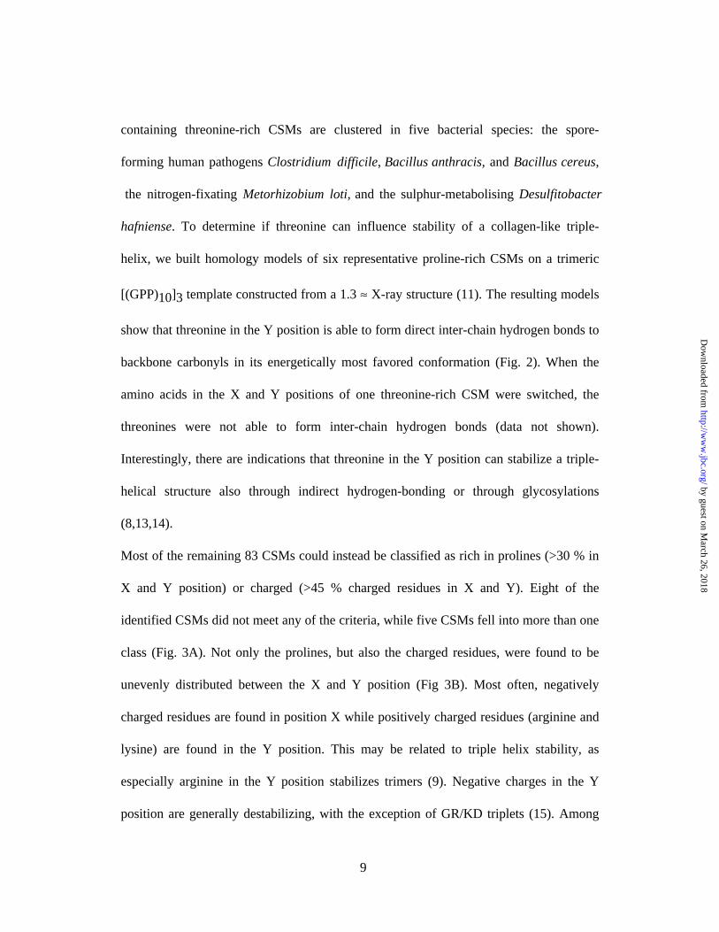
containing threonine-rich CSMs are clustered in five bacterial species: the spore-
forming human pathogens Clostridium difficile, Bacillus anthracis, and Bacillus cereus,
the nitrogen-fixating Metorhizobium loti, and the sulphur-metabolising Desulfitobacter
hafniense. To determine if threonine can influence stability of a collagen-like triple-
helix, we built homology models of six representative proline-rich CSMs on a trimeric
[(GPP)10]3 template constructed from a 1.3 ≈ X-ray structure (11). The resulting models
show that threonine in the Y position is able to form direct inter-chain hydrogen bonds to
backbone carbonyls in its energetically most favored conformation (Fig. 2). When the
amino acids in the X and Y positions of one threonine-rich CSM were switched, the
threonines were not able to form inter-chain hydrogen bonds (data not shown).
Interestingly, there are indications that threonine in the Y position can stabilize a triple-
helical structure also through indirect hydrogen-bonding or through glycosylations
(8,13,14).
Most of the remaining 83 CSMs could instead be classified as rich in prolines (>30 % in
X and Y position) or charged (>45 % charged residues in X and Y). Eight of the
identified CSMs did not meet any of the criteria, while five CSMs fell into more than one
class (Fig. 3A). Not only the prolines, but also the charged residues, were found to be
unevenly distributed between the X and Y position (Fig 3B). Most often, negatively
charged residues are found in position X while positively charged residues (arginine and
lysine) are found in the Y position. This may be related to triple helix stability, as
especially arginine in the Y position stabilizes trimers (9). Negative charges in the Y
position are generally destabilizing, with the exception of GR/KD triplets (15). Among
9
by guest on March 26, 2018
http://ww
w.jbc.org/
Dow
nloaded from
the charged CSMs with a net negative charge in the Y position, aspartate is more
common than glutamate and the aspartate is in 50 % of the cases preceded by a positively
charged residue. In addition to stabilization, charges in collagen are important for the
interactions between collagen and other macromolecules (16).
Function of the CSM- The function of CSMs in bacteria and viruses is not well
understood. The few CSMs studied have been shown to mediate trimerization or
elongation (2,10,17,18) of proteins found at bacterial or bacteriophage surfaces (2,18-
25). Furthermore, the CSM of one protein from Streptococcus sanguis has been shown to
mediate aggregation of platelets (26), while it is less clear whether the CSMs from two
Streptococcus pyogenes proteins contribute to the adhesive properties of these proteins
(21,24). The viral proteins containing CSMs identified in this study were mainly encoded
by bacteriophages and had to a large extent been annotated or could easily be annotated
by similarity searches. The majority of these proteins were tail fiber or host specificity
proteins, involved in the interactions between bacteriophages and bacteria (see e g (27)).
Only a minority of the bacterial proteins containing a CSM could be annotated. Three
proteins had previously been described as surface or spore associated (20-25) and from
pattern and similarity searches, an additional 13 proteins could be classified as cell wall-
attached (28) or transmembrane proteins. The non collagen-related parts of the proteins
showed surprisingly little sequence similarity to other proteins, making a functional
classification of the bacterial proteins difficult. The presence of CSMs in proteins with so
little sequence similarity indicate that the CSM is indeed a structural motif to elongate or
10
by guest on March 26, 2018
http://ww
w.jbc.org/
Dow
nloaded from

trimerize a variety of different, mainly extracellular proteins. The possibility remains,
however, that some CSMs may mediate binding of bacterial or bacteriophage proteins to
other macromolecules.
Evolutionary aspects- Since so few bacterial and unicellular eucaryotic genomes encode
CSMs and archaebacteria completely lack such sequences, it seems unlikely that CSMs
have evolved before the diversification of archaea, bacteria, and eukaryotes. It appears
more plausible that genes encoding CSMs have moved horizontally or arisen on several
occasions during evolution rather than selective gene loss among archaea, bacteria, and
lower eukaryotes. Horizontal gene transfer between bacteria and vertebraea has been
proposed to be relatively common (29,30), although this view has also been questioned
(31). If horizontal gene transfers of CSMs have occurred, we believe that the direction of
transfer has been from eukaryotes to bacteria. After horizontal transfer of sequences
encoding a CSM, bacteriophages may have promoted further horizontal transfer within
the bacterial kingdom leading to the ”patchy” distribution seen for CSMs among
bacteria. We have no evidence for a role of viruses or bacteriophages in the putative
horizontal transfer between eukaryotes and bacteria, but this can not be excluded. After
the putative horizontal transfer, extensive rearrangements seem to have occurred in the
bacterial CSMs, since there are limited sequence similarities except for the spacing of
glycines in the bacterial CSMs. The limited sequence similarities also made phylogenetic
analyses unmeaningful to perform. Interestingly, the threonine-rich CSMs are very
dissimilar in amino acid composition from human collagens and from the other bacterial
11
by guest on March 26, 2018
http://ww
w.jbc.org/
Dow
nloaded from
CSMs. Instead, the threonine-rich CSMs have their closest homologues in hypothermal
vent worm cuticle collagens (14). This suggests that several events of horizontal gene
transfer of the genetic material encoding CSMs could have occurred during evolution. An
alternative evolutionary explanation to the threonine-rich CSMs is that these have arisen
de novo in bacteria, followed by horizontal spread to a few other bacterial species.
Especially the threonine-rich CSMs are highly repetitive and could have evolved from
duplications of a short DNA segment.
The presence of CSMs in bacteria and viruses underlines the importance of collagen as a
structural motif in nature. This work also suggest that alternative means for triple-helix
stabilization probably operate in bacteria and viruses, and future elucidation of the
structure and function of bacterial and viral CSMs represents an interesting scientific
task.
Acknowledgments
TIGR and other generous providers of sequence data are acknowledged. We thank Dr.
Robert Janulczyk for constructive comments. This work was supported by grants from
the Swedish Research Council (projects 7480 and 14379); the Medical Faculty, Lund
University; the Foundations of Kock and Österlund; and Hansa Medical AB.
12
by guest on March 26, 2018
http://ww
w.jbc.org/
Dow
nloaded from
References
1. Beck, K., and Brodsky, B. (1998) J Struct Biol 122, 17-29
2. Charalambous, B. M., Keen, J. N., and McPherson, M. J. (1988) EMBO J. 7, 2903-
2909
3. Bamford, D. H., and Bamford, J. K. (1990) Nature 344, 497
4. Medveczky, M. M., Geck, P., Vassallo, R., and Medveczky, P. G. (1993) Virus Genes
7, 349-365
5. Smith, M. C., Burns, N., Sayers, J. R., Sorrell, J. A., Casjens, S. R., and Hendrix, R.
W. (1998) Science 279, 1834
6. Ruggiero, F., Exposito, J. Y., Bournat, P., Gruber, V., Perret, S., Comte, J., Olagnier,
B., Garrone, R., and Theisen, M. (2000) FEBS Lett 469, 132-136
7. Perret, S., Merle, C., Bernocco, S., Berland, P., Garrone, R., Hulmes, D. J. S., Theisen,
M., and Ruggiero, F. (2001) J. Biol. Chem 276, 43693-43698
8. Bann, J. G., Peyton, D. H., and Bachinger, H. P. (2000) FEBS Lett 473, 237-240
9. Yang, W., Chan, V. C., Kirkpatrick, A., Ramshaw, J. A., and Brodsky, B. (1997) J
Biol Chem 272, 28837-28840
10. Xu, Y., Keene, D. R., Bujnicki, J. M., Höök, M., and Lukomski, S. (2002) J Biol
Chem 277, 27312-27318
11. Berisio, R., Vitagliano, L., Mazzarella, L., and Zagari, A. (2002) Prot. Sci. 11, 262-
270
12. Abagyan, R. A., and Totrov, M. M. (1994) J. Mol. Biol. 235, 983-1002
13. Kramer, R. Z., Bella, J., Mayville, P., Brodsky, B., and Berman, H. M. (1999) Nat
13
by guest on March 26, 2018
http://ww
w.jbc.org/
Dow
nloaded from
Struct Biol 6, 454-457
14. Mann, K., Mechling, D. E., Bachinger, H. P., Eckerskorn, C., Gaill, F., and Timpl, R.
(1996) J Mol Biol 261, 255-266
15. Chan, V. C., Ramshaw, J. A. M., Kirkpatrick, A., Beck, K., and Brodsky, B. (1997) J.
Biol. Chem. 272, 31441-31446
16. Doi, T., Higashino, K., Kurihara, Y., Wada, Y., Miyazaki, T., Nakamura, H., Uesugi,
S., Imanishi, T., Kawabe, Y., and Itakura, H. (1993) J. Biol. Chem. 268, 2126-2133
17. Sylvestre, P., Couture-Tosi, E., and Mock, M. (2003) J. Bacteriol. 185, 1555-1563
18. Caldentey, J., Tuma, R., and Bamford, D. H. (2000) Biochemistry 39, 10566-10573
19. Erickson, P. R., and Herzberg, M. C. (1987) J. Immunol. 138, 3360-3366
20. Rasmussen, M., Edén, A., and Björck, L. (2000) Infect. Immun. 68, 6370-6377
21. Lukomski, S., Nakashima, K., Abdi, I., Cipriano, V. I., Ireland, R. M., Reid, S. D.,
Adams, G. G., and Musser, J. M. (2000) Infect. Immun. 68, 6542-6553
22. Whatmore, A. (2001) Microbiology 147, 419-429
23. Lukomski, S., Nakashima, K., Abdi, I., Cipriano, V. J., Shelvin, B. J., Graviss, E. A.,
and Musser, J. M. (2001) Infect. Immun. 69, 1729-1738
24. Rasmussen, M., and Björck, L. (2001) Mol. Microbiol. 40, 1427-1438
25. Sylvestre, P., Couture-Tosi, E., and Mock, M. (2002) Mol Microbiol 45, 169-178
26. Erickson, P. R., and Herzberg, M. C. (1993) J. Biol. Chem. 268, 1646-1649
27. Duplessis, M., and Moineau, S. (2001) Mol. Microbiol. 41, 325-336
28. Janulczyk, R., and Rasmussen, M. (2001) Infect. Immun. 69, 4019-4026
29. Lander, E. S., Linton, L. M., Birren, B., Nusbaum, C., Zody, M. C., Baldwin, J.,
14
by guest on March 26, 2018
http://ww
w.jbc.org/
Dow
nloaded from
Devon, K., Dewar, K., Doyle, M., FitzHugh, W., Funke, R., Gage, D., Harris, K.,
Heaford, A., Howland, J., Kann, L., Lehoczky, J., LeVine, R., McEwan, P., McKernan,
K., Meldrim, J., Mesirov, J. P., Miranda, C., Morris, W., Naylor, J., Raymond, C.,
Rosetti, M., Santos, R., Sheridan, A., Sougnez, C., Stange-Thomann, N., Stojanovic, N.,
Subramanian, A., Wyman, D., Rogers, J., Sulston, J., and Ainscough, R. (2001) Nature
409, 860-921
30. Syvanen, M. (2002) Trends in Genetics 18, 245-248
31. Stanhope, M. J., Lupas, A., Italia, M. J., Koretke, K. K., Volker, C., and Brown, J. R.
(2001) Nature 411, 940-944
32. Ramshaw, J. A. M., Shah, N. K., and Brodsky, B. (1998) J. Struct. Biol. 122, 86-91
33. Bella, J., Eaton, M., Brodsky, B., and Berman, H. M. (1994) Science 266, 75-81
15
by guest on March 26, 2018
http://ww
w.jbc.org/
Dow
nloaded from
Legends to figures
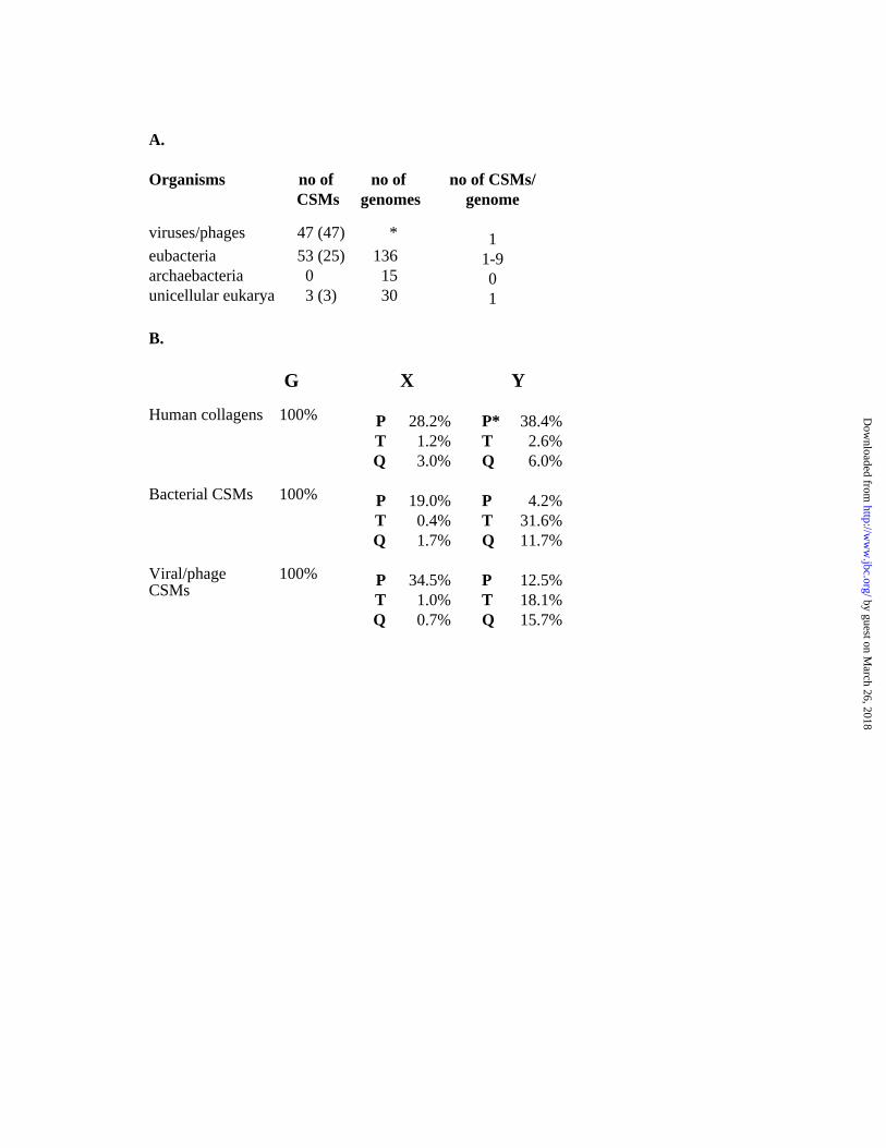
FIG 1. Occurrence and composition of CSMs
A: The number of proteins containing a CSM in the various organisms is given, and the
number of genomes by which these CSMs are encoded are indicated within parenthesis.
The asterisk indicates that searches were made against all entries containing viral
sequences. The number of genomes analyzed and the number of proteins containing
CSMs in a given genome are also shown.
B: The position-specific distribution of glycines, prolines, glutamines, and threonines of
the bacterial and viral CSMs and human collagens is given. The values represent the
percentage of a given amino acid in a specific position of the GXY triplet. The asterisk
indicate that prolines in the Y position of human collagens are most often hydroxylated.
Data on amino acid frequency for human collagens were from a previous report (32).
FIG 2. Detailed view of the homology model of a threonine-rich CSM from B. anthracis.
The conformation of threonine side-chains was determined by an exhaustive systematic
search procedure. Hydrogen bonds are shown in black. Part of the hydroxyproline-
containing triple-helical structure in PDB entry 1cag (33) is shown in cyan for
comparison.
FIG 3. Amino acid composition of bacterial and viral CSMs
A: The CSMs identified in bacterial and viral proteins (53 and 47 respectively) were
16
by guest on March 26, 2018
http://ww
w.jbc.org/
Dow
nloaded from
categorized into three groups. The percentage of CSMs falling into each group is given
The threonine-rich CSMs have more than 50 % of Y positions occupied by threonine, the
proline-rich have more than 30 % of X and Y positions occupied with proline, whereas
charged CSMs have more than 45 % charged residues in the X and Y positions. Some
CSMs can not be classified into one of these groups, whereas some meet more than one
criterion.
B: The skew in charge-distribution between the X and Y position of GXY triplets from
charged CSMs is shown. For every charged CSM, the net charge (K/R=+1, D/E=-1) of
residues in the Y position minus the net charge of residues in the X position was divided
by the total number of charged residues. Thus, a CSM with all positively charged
residues in the Y position and all negatively charged residues in the X position will obtain
a value of +1, whereas a CSM with all positively charged residues in the X position and
all negatively charged residues in the Y position will obtain a value of -1. Each dot in the
diagram represents one CSM.
17
by guest on March 26, 2018
http://ww
w.jbc.org/
Dow
nloaded from
A.
Organisms no of CSMs
no of genomes
no of CSMs/genome
viruses/phages 47 (47) * 1eubacteria 53 (25) 136 1-9 archaebacteria 0 15 0unicellular eukarya 3 (3) 30 1
B.
G X Y
Human collagens 100% PTQ
28.2%1.2%3.0%
P*TQ
38.4%2.6%6.0%
Bacterial CSMs 100% PTQ
19.0%0.4%1.7%
PTQ
4.2%31.6%11.7%
Viral/phageCSMs
100% PTQ
34.5%1.0%0.7%
PTQ
12.5%18.1%15.7%
by guest on March 26, 2018
http://ww
w.jbc.org/
Dow
nloaded from
Magnus Rasmussen, Micael Jacobsson and Lars Björckbacterial and viral proteins
Genome-based identification and analysis of collagen-related structural motifs in
published online June 3, 2003J. Biol. Chem.
10.1074/jbc.M304709200Access the most updated version of this article at doi:
Alerts:
When a correction for this article is posted•
When this article is cited•
to choose from all of JBC's e-mail alertsClick here
by guest on March 26, 2018
http://ww
w.jbc.org/
Dow
nloaded from
Recommended