Genetic approaches to discover novel oncogenes in
human cancer
By
Mr. Prajish Sundaram Iyer
[LIFE09201204001]
Tata Memorial Centre, Navi Mumbai
A thesis submitted to the
Board of Studies in Life Sciences
in partial fulfilment of requirements
for the Degree of
DOCTOR OF PHILOSOPHY
of
HOMI BHABHA NATIONAL INSTITUTE
November, 2017
STATEMENT BY AUTHOR
This dissertation has been submitted in partial fulfillment of requirements for an advanced
degree at Homi Bhabha National Institute (HBNI) and is deposited in the Library to be made
available to borrowers under rules of the HBNI.
Brief quotations from this dissertation are allowable without special permission, provided that
accurate acknowledgement of source is made. Requests for permission for extended quotation
from or reproduction of this manuscript in whole or in part may be granted by the Competent
Authority of HBNI when in his or her judgment the proposed use of the material is in the
interests of scholarship. In all other instances, however, permission must be obtained from the
author.
Navi Mumbai Prajish Sundaram Iyer
Date

DECLARATION
I, hereby declare that the investigation presented in the thesis has been carried out by me. The
work is original and has not been submitted earlier as a whole or in part for a degree /
diploma at this or any other Institution / University.
Navi Mumbai Prajish Sundaram Iyer
PUBLICATIONS
List of Publications arising from the thesis
Journal
Iyer P, Barreto SG, Sahoo B, Chandrani P, Ramadwar MR, Shrikhande SV, Dutt A. Non-
typhoidal Salmonella DNA traces in gallbladder cancer. Infectious agents and cancer. 2016;
11:12. (Thesis Work)
Chapters in books and lecture Notes : None
Conferences
1. P.Iyer, N.Gardi, M.Ranjan, Bikram Sahoo, P.Chandrani, P.Upadhyay, M.R.Ramadwar,
S.V.Shrikhande, A.Dutt Abstract 4397: Deciphering the Diversity of Somatic Alterations and
Salmonella Infection in Gallbladder Cancer by Whole Exome Sequencing (April 1st – 5th,
2017) Proceedings of the 108th Annual Meeting of the American Association for Cancer
Research, Washington D.C(Poster Presentation).
2. P.Iyer,N.Gardi,M.Ranjan,P.Chandrani,P.Upadhyay,M.R.Ramadwar,S.V.Shrikhande, A.Dutt
AbstractK002: Discovery of somatic ERBB2 alterations in human gallbladder cancer New
Ideas in Cancer – Challenging Dogmas (26th- 28th Feb 2016), European Journal of
Cancer,10.1016/S0959-8049(16)31953-0 (Poster Presentation)
3. P.Iyer, N.Gardi, M.Ranjan, Bikram Sahoo,P.Chandrani, P.Upadhyay, M.R.Ramadwar,
S.V.Shrikhande, A.Dutt Deciphering the Diversity of Somatic Alterations and Salmonella
Infection in Gallbladder Cancer by Whole Exome Sequencing (April 8th – 10th, 2016)
35th Annual Convention of Indian Association for Cancer Research (Oral Presentation
under award category)
4. P.Iyer, N.Gardi, M.Ranjan, Bikram Sahoo, P.Chandrani, P.Upadhyay, M.R.Ramadwar,
S.V.Shrikhande, A.Dutt Landscape of somatic alterations and identification of non-typhoidal
Salmonella traces in gallbladder cancer (January 29th – 30th 2016) MOSCON conference.
(Poster Presentation)
PUBLICATIONS
5. P.Iyer, N.Gardi, M.Ranjan, Bikram Sahoo, P.Chandrani, P.Upadhyay, M.R.Ramadwar,
S.V.Shrikhande, A.Dutt Deciphering the Diversity of Somatic Alterations and Salmonella
Infection in Gallbladder Cancer by Whole Exome Sequencing (October 3rd - 5th, 2016) 2016
NextGen Genomics and Bioinformatics (Nature reviews Best Poster award)
6. P.Iyer, N.Gardi, M.Ranjan, Bikram Sahoo, P.Chandrani, P.Upadhyay, M.R.Ramadwar,
S.V.Shrikhande, A.Dutt Deciphering the Diversity of Somatic Alterations and Salmonella
Infection in Gallbladder Cancer by Whole Exome Sequencing (December 7th – 11th, 2016)
India International Science Festival (Poster Presentation)-Selected among 8000 posters in
the Life science category
7. Chandrani P*, Kulkarni V*, Iyer P, Upadhyay P, Chaubal R, Das P, Mulherkar R, Singh R,
and Dutt A. HPVDetector : A tool to detect HPV and their integration sites using next
generation sequencing data (November 17th- 19th , 2014.) 2014 NextGen Genomics and
Bioinformatics (Nature reviews Best Poster award)
8. P Upadhyay, P Chandrani, R Chaubal, N Oak, M Shetty, K Karve, R Prasad, P Iyer and
A.Dutt “Integrated Genomic Characterization of Head & Neck Cancer Cell Lines Derived
from Indian Patients” at The Global Cancer Genomics Consortium Second Annual
Symposium: Genomics Medicine in Cancer Research, 2012, ACTREC, India.(Best Poster
award)
9. R Chaubal, P Iyer, V.Kulkarni, I Mittra, A.Dutt A Next Generation Sequencing approach to
determine traces of human genetic material in NIH3T3 cells transformed by chromatin &
DNA fragments extracted from human cancer patients at The Global Cancer Genomics
Consortium Second Annual Symposium: Genomics Medicine in Cancer Research, 2012,
ACTREC, India (Best Poster award)
PUBLICATIONS
Others
1. Chandrani P, Prabhash K, Prasad R, Sethunath V, Ranjan M, Iyer P, Aich J, Dhamne H,
Iyer DN, Upadhyay P, Mohanty B, Chandna P, Kumar R, Joshi A, Noronha V, Patil V,
Ramaswamy A, Karpe A, Thorat R, Chaudhari P, Ingle A, Choughule A, Dutt A. Drug-
sensitive FGFR3 mutations in lung adenocarcinoma. Annals of oncology : official journal of
the European Society for Medical Oncology. 2017; 28(3):597-603.
2. Upadhyay P, Gardi N, Desai S, Sahoo B, Singh A, Togar T, Iyer P, Prasad R, Chandrani P,
Gupta S, Dutt A. TMC-SNPdb: an Indian germline variant database derived from whole
exome sequences. Database : the journal of biological databases and curation. 2016; 2016.
3. Chandrani P, Upadhyay P, Iyer P, Tanna M, Shetty M, Raghuram GV, Oak N, Singh A,
Chaubal R, Ramteke M, Gupta S, Dutt A. Integrated genomics approach to identify
biologically relevant alterations in fewer samples. BMC genomics. 2015; 16:936.
4. Chandrani P, Kulkarni V, Iyer P, Upadhyay P, Chaubal R, Das P, Mulherkar R, Singh R,
Dutt A. NGS-based approach to determine the presence of HPV and their sites of integration
in human cancer genome. British journal of cancer. 2015; 112(12):1958-65.
5. Choughule A, Sharma R, Trivedi V, Thavamani A, Noronha V, Joshi A, Desai S,
Chandrani P, Sundaram P, Utture S, Jambhekar N, Gupta S, Aich J, Prabhash K, Dutt A.
Coexistence of KRAS mutation with mutant but not wild-type EGFR predicts response to
tyrosine-kinase inhibitors in human lung cancer. British journal of cancer. 2014;
111(11):2203-4.
Prajish Sundaram Iyer
ACKNOWLEDGMENTS
ACKNOWLEDGEMENTS
One of the obivious transistion from my master’s program was to do a Ph.D. Though it was not my
obvious choice to do Ph.D in India, but after I joined Dutt laboratory at ACTREC, it was totally a
different ball game. In this journey of five years, there are so many experiences that cannot be shared
in a single piece of acknowledgment. Today, I would like to thank all of you on this occasion.
Firstly, I would like to express my deepest and earnest gratitude for my mentor, Dr. Amit Dutt, for his
constant guidance, motivation and support in all aspects of my career throughout my tenure. It was his
aggression and infectious enthusiasm for the science which made me doing science as a passion. His
constructive, sharp and extremely critical supervision has always encouraged me to perform
interdisciplinary research in cancer and develop deep scientific temperament. Throughout my tenure,
he was a sharp critic but a caring mentor. His words would echo in my body language which
transformed me to a great extent in my scientific pursuits. His throat cutting remarks has made me
stronger to face the brutal world of science.
I am highly privileged to you for allowing me to actively participating in such an endeavour. I am
thankful to doctoral committee members, Dr. Sorab N. Dalal (chairperson), Dr. Prasanna
Venkataraman and Dr Shailesh Shrikhande for their continuous critical advice and constructive
suggestion during doctoral committee meetings. Advices made during DC meetings were invaluable
to my research work and has allowed me to progress in focused manner. I am also thankful to Dr. Rita
Mulherkar (ex-chairperson), for their advice in my thesis work. I would like to thank Dr Harsha
Gowda for providing gallbladder cancer cell lines at a crucial period of my tenure.
It was privileged to utilize the excellent infrastructure and facilities at ACTREC and Tata
MemorialCentre for which I would like to thank Dr. Rajendra Badwe (Director, TMC), Dr. Shubhada
Chiplunkar(Director, ACTREC), Dr. Sudeep Gupta (Deputy Director, ACTREC), Dr. Rajiv Sarin
(Ex-Director,ACTREC), Dr. Surekha Zingde (Ex-Deputy Director, ACTREC). I would also like to
thank ACTREC for providing me regular fellowship, Wellcome Trust/DBT India Alliance,
Department of Biotechnology, India and Tata Memorial Centre for funding my research work. I am
appreciative to Homi Bhabha National Institute (HBNI) and Tata Memorial Centre (TMC) - Sam
Mistry foundation for proving financial support to present my work at an international meeting.
I am grateful to my clinical collaborators Dr. Shailesh Shrikhande, Dr. Mukta Ramadwar, Mr Anand
Deshpande, Dr Vikram Chaudhary, Ms.Manisha Kulkarni for their dedication and active support for
providing the gallbladder tissue samples and well annotated clinical information of patients.
I would like to express my deepest appreciation to Dr. Shilpee Dutt for her valuable advice during lab
meetings as well as doctoral committee meetings, which helped me widen my research interests.
I am exceptionally grateful to all the patients for agreeing to be a part of my thesis work and helping
me to conduct research.

ACKNOWLEDGMENTS
I would like to thank the various ACTREC facilities staffs for their assistance. I am thankful to Mr.
Dandekar sir for his dexterous assistance in common instrument facility. Additionally, help from staff
in sequencing, microscopy, flow cytometry, sequencing, IT, library, steno-pool, administration,
account, canteen, and engineering and security personals. It’s due to their efforts I could get all the
facilities running smoothly and organized manner. A special thank you to Mr. Naresh, Sharda Mam,
Anand sir, Tanuja, Jayraj, M.A. Sharma, Chitra, Aruna Mam, Karan sir, Arun Shetty for always being
ready to help in administrative work.
I express my humble thank you to all the past and present members of Shilpee Laboratory for
providing such a nurturing atmosphere and contributing to my personal and professional growth. A
special thanks to Jacinth, Anagha, Sameer, Ekjot, Jyoti, Priya, Shailesh, Tejal, Swati, Samreen, Neha,
Aloka, Saket, Poorvaja for creating a friendly atmosphere and being a part of my scientific and non-
scientific stuff. Special thanks to Sameer for experimental work. Special thanks to Smita mam for
supporting and encouraging me at all times.
The Dutt laboratory members have immensely contributed to my personal and professional growth by
providing an amazing fertile ground and creating an interdisciplinary research ecosystem in the lab. I
am thankful to Malika, Nilesh, Rohan, Bikram, Trupti, Pratik, Mukul, Ratnam, Asim, Sanket, Prachi,
Vidya, Mayur for their constant support in my work and motivation during difficult times. Special
thanks to Malika for working really hard on the project and turning the project into a goldstone.
Special thanks to Rohan, Nilesh, Bikram and Pratik for helping me with computational work. I am
especially thankful to Pawan for constant support during my ups and downs. Thanks to Trupti for
helping me during this time of thesis. Special thanks to Ratnam and Renu for being supportive
backbone of lab by managing the IRB and administrative stuffs. I would also like to thank Kanishka,
Ankita, Sunil, Reshma, Sharan, Rashmi, Renu, Vaibhav, Deepak Iyer, Kunal for being great friends
and assisting me at various occasions during my tenure. Special thanks to Sunil, for the conversations
that we used to share in central park. I really indebted to your constant support and meaningful
interactions in and out of the lab. I am thankful to Post-doctoral fellows at Dutt Laboratory, Dr.
Kuldeep, Dr. Jyoti, Dr.Hemant, Dr. Manoj for training me in several experimental stuffs, constant
advice, feedback in my research work.
I am thankful to members of lab, Bhaskar and Neelima for their timely help for my experiments.
Special thanks to Asim for timely help for my cell culture experiments. Special thanks to newbie
Hitesh for taking out time to check the draft. Special thanks to Sanket for helping me with cell line
whole exome sequencing. I am thankful to Deepak Amburle, Deepak Chauhan, Mr. Rane for being
providing endless technical support and being exceedingly friendly. Special thanks to Dhananjay sir
and Shailesh sir for cheering me up and making me feel like a king. Special thanks to Deepak for
accompanying me for tea, helping me with media, trypsin and all the non-scientific discussions during
all this tenure.

ACKNOWLEDGMENTS
I am thankful to my Ph.D batch mates, Gopal, Bhavik, Saujanya, Pratik, Arunabha for being
supportive in all phases of tenure. We all shared several memorable moments together in an out of
ACTREC campus which I will remember lifetime. Special thanks to my labmate Mukul for being a
caring and a tolerant labmate. I would remember the fun times we have shared together in the lab.
Special thanks to all company representatives who have been my friends and helped me during
difficult times.
Last but not the least, I am immensely thankful to my parents who have unconditionally helped me in
all my endeavours and being a strong pillar in my life. Special thanks to my papa for always cheering
me up with astrological advices. And my moma, praying always for me. I am very thankful to my
brother Prashant anna who always encouraged me in all parts of my tenure.
Lastly, I don’t believe much in god. But what I know is hardwork, perseverance, honesty and belief in
oneself are all different forms I would see god. And only when the time is right these things work.
Waiting for the right time………
Thank you
TABLE OF CONTENTS
TABLE OF CONTENTS
SYNOPSIS OF Ph.D. THESIS ................................................................................................. I
LIST OF FIGURES ..............................................................................................................XVI
LIST OF TABLES ............................................................................................................ XVIII
ABBREVIATIONS ..............................................................................................................XIX
SUMMARY ............................................................................................................................ XX
1. CHAPTER1: INTRODUCTION AND REVIEW OF LITERATURE ..................... 22
1.1 Human cancer and genomics ............................................................................................... 22
1.2 Genomics of rare cancer types ............................................................................................. 23
1.3 Gallbladder Cancer .............................................................................................................. 23
1.3.1 Definition and Epidemiology of GBC ..................................................................... 23
1.3.2 Epidemiology of gallbladder cancer ........................................................................ 24
1.3.3 Unmet need to treat gallbladder cancer in India ...................................................... 25
1.3.4 Application of next generation sequencing in gallbladder cancer .......................... 26
1.3.5 The landscape of known genomic alterations in gallbladder cancer. ...................... 27
1.3.6 Risk factors for gallbladder cancer .......................................................................... 29
1.3.6.1 Gallstones and cholecystitis ................................................................................. 29
1.3.6.2 Porcelain gallbladder ............................................................................................ 30
1.3.6.3 Age and Gender .................................................................................................... 31
1.3.6.4 Diet and Obesity ................................................................................................... 31
1.3.6.5 Bacterial infections ............................................................................................... 31
1.3.6.6 Genetic polymorphisms in gallbladder cancer ..................................................... 32
1.3.6.7 Environmental effects ........................................................................................... 33
1.4 Molecular pathology of gallbladder cancer ................................................................ 33
1.5 Research objective ...................................................................................................... 36
Objectives ......................................................................................................................... 36
CHAPTER 2: NON TYPHOIDAL SALMONELLA TRACES IN GALLBLADDER
CANCER (an excerpt; as published in BMC Infectious Agents and Cancer (2016);11:1237
Abstract .................................................................................................................................... 37
2.1 Introduction ......................................................................................................................... 38
TABLE OF CONTENTS
2.1.1 Infections in cancer ................................................................................................. 38
2.1.2 Infections in gallbladder cancer .............................................................................. 38
2.2 Materials and Methods ........................................................................................................ 39
2.2.1 Patient information: ................................................................................................. 39
2.2.2 PCR analysis for Salmonella isolates ...................................................................... 40
2.2.3 Sequencing and analysis .......................................................................................... 40
2.3 Results ................................................................................................................................. 41
2.3.1 HPVDetector pipeline identifies Salmonella sequence present in gallbladder cancer
samples ............................................................................................................................. 41
2.3.2 Annotation of the Salmonella reads found in gallbladder cancer samples .............. 42
2.3.4 Sanger validation of Salmonella reads identified in gallbladder cancer samples ... 44
2.4 Discussion ........................................................................................................................... 45
3.1 Introduction ....................................................................................................................... 47
3.2 Materials and Methods ........................................................................................................ 49
3.2.1Patient Information: .................................................................................................. 49
3.2.2 DNA extraction ....................................................................................................... 49
3.2.3 Exome sequencing capture, library construction, and sequencing .......................... 50
3.2.4 Copy number analysis from Exome sequencing data ............................................. 50
3.2.5 Cell culture and reagents ......................................................................................... 51
3.2.6 Soft Agar assay ........................................................................................................ 51
3.2.7 Virus production ...................................................................................................... 51
3.2.8 Growth Curve .......................................................................................................... 52
3.2.9 MTT assay ............................................................................................................... 52
3.2.10 Western blotting .................................................................................................... 52
3.2.11 Receptor tyrosine kinase proteome array .............................................................. 52
3.2.12 Invasion assay ........................................................................................................ 53
3.2.13 Wound healing assay ............................................................................................. 53
3.2.14 Survival analysis .................................................................................................... 53
TABLE OF CONTENTS
3.3 Results ................................................................................................................................. 54
3.3.1 Integrated genomics and proteomics approach identify aberrant alterations in
members of the EGFR family in gallbladder cancer ........................................................ 54
3.3.2 ERBB2 and EGFR are essential for the survival of gallbladder cancer cells not
harboring KRAS G12V mutant allele ................................................................................ 58
3.3.3 Irreversible EGFR inhibitors block proliferation and survival of gallbladder cancer
cell lines ............................................................................................................................ 60
3.3.4 Clinical correlation of TP53 and EGFR family mutations in GBC patients ........... 62
III- Additional supporting data: ................................................................................................. 70
3.1 Comparison of significantly altered genes from whole exome sequencing of
gallbladder cancer samples ............................................................................................... 70
4.CHAPTER 4: SUMMARY AND CONCLUSION ............................................................ 71
4.1 Mutant KRAS predicts response to EGFR inhibitors in gallbladder cancer cell lines. ....... 71
4.2 Association of non-typhoidal Salmonella with gallbladder cancer .................................... 72
5.CHAPTER 5: BIBLIOGRAPHY ........................................................................................ 74
6.CHAPTER 6: APPENDIX .................................................................................................. 80
6.1 APPENDIX I: List of variants identified in whole exome sequencing of 27 samples ....... 80
6.2 APPENDIX 2: List of variants identified in whole exome sequencing of 5 gallbladder
cancer cell lines. ........................................................................................................................ 80
6.3 APPENDIX 3: Detailed annotation table of read sequences of different salmonella species
identified across gallbladder cancer patient samples................................................................. 80
7.REPRINTS OF PUBLICATIONS………………………………………………….81 ONWARDS
SYNOPSIS
I | P a g e
Homi Bhabha National Institute
SYNOPSIS OF Ph.D. THESIS
Name of the Student: Prajish sundaram Iyer
1. Name of the Constituent Institution: Tata Memorial Centre, ACTREC
2. Enrolment No. and Date of Enrolment: LIFE09201204001, 31st July 2012
3. Title of the Thesis: Genetic approaches to discover novel oncogenes in human cancer
4. Board of Studies: Life Science
1. INTRODUCTION
Cancer is a disease of the genome. Recent developments in high-throughput genetic mutation
profiling facilitate to do a comprehensive analysis of the cancer genome using next-
generation sequencing technologies[1]. Large-scale profiling projects have revealed a
landscape of the cancer genome that includes a diverse variety of cancer genomic alterations
such as point mutations, copy number variation, and translocations. Cancer genome and
transcriptome sequencing have revealed additional clinically relevant novel gene fusions in
solid tumors[2]. The developments in the technologies have helped to characterize exomes,
genomes, and epigenomes of various cancers. Despite developments in next-generation
sequencing technologies not much is understood regarding the rare cancer types such as
biliary tract cancers due to its low prevalence in the western countries[3].
Biliary tract cancers are a group of heterogeneous cancers that arise either in intra-or
extrahepatic bile ducts or the gallbladder. These cancers are presented at late advanced stages
hence have a poor prognosis. Among the biliary tract cancers, gallbladder cancer is one of the
most aggressive cancer with poor prognosis. Surgical resection has been preferred as an
SYNOPSIS
II | P a g e
option for resectable gallbladder cancer and offers a potential cure. The major risk factor for
gallbladder cancer is gallstones however there are other minor risk factors such as female
gender, obesity, Salmonella infections, cholangitis and gallbladder polyps[4]. The median
survival is less than a year and is less than 5%[4]. The adjuvant chemotherapy given to
gallbladder cancer patients consists of gemcitabine and platinum-based therapy. There is
enough evidence that suggests the resistance of the cancer type to cytotoxic agents [5, 6].
Hence there is an unmet need to identify potential targeted therapies in gallbladder cancer. In
India, highest incidence has been reported in Delhi and Bhopal in women (6.6 and 5.2 per
1,00,000 respectively) which is far more higher than south India(0.6 – Chennai and 0.8 –
Bangalore)[7]. Comprehensive genomic characterization of gallbladder cancer in India is still
dismal despite high prevalence. There are few candidate gene-based studies from India that
have identified few targets but these targets are still not in clinical practice [8-10]. There are
few reports from China and the west to characterize the gallbladder cancer genome using
next-generation sequencing technologies [11-13]. However, the molecular mechanism of the
cancer is still poorly understood. Hence more sequencing studies with larger number of
samples are required to identify candidate targets in gallbladder cancer.
Mutations in the EGFR family members ( EGFR, ERBB2, ERBB3 ) have been recently
known to be altered at a frequency of 10-15% in gallbladder cancers and the signaling
pathway has been shown to be altered in the pathogenesis of gallbladder cancer[11, 14] Some
of these alterations in the EGFR family have already been shown to be sensitive to tyrosine
kinase inhibitors in NSCLC and breast cancers[15, 16]. The EGFR receptor family consists
of EGFR/ErbB1, ErbB2 (HER2), ErbB3 (HER3) and ERbB4 (HER4). All receptors except
HER2 bind specific ligands via extracellular domain. Upon ligand binding, these receptors
use HER2 as a preferential dimerization partner. Homo/Heterodimerization of receptors
results in phosphorylation of residues in the intracellular domain resulting in activation of
several signaling pathways such as Ras/Raf/MAPK and the PI3K-Akt pathways[17]. Reports
SYNOPSIS
III | P a g e
suggest that though EGF does not bind HER2, implying that EGFR may be involved in
HER2 signal transduction. Oncogenic transformation by EGFR or HER2 requires high
receptor expression while moderate levels are sufficient to cause transformation. Also,
reports suggest that down-regulation of normally expressed EGFR suppressed the ability of
overexpressed HER2 suggesting HER2 requires EGFR for sustained signaling and
transforming potential [18]. This synergistic activity of EGFR-HER2 heterodimerization may
be particularly significant as these receptors are upregulated concomitantly in breast and
other tumors[19] [20-22]. Hence it is pivotal to study EGFR signaling in gallbladder cancer
from the Indian gallbladder cancer perspective.
Pathogenic infections have been associated with cancer worldwide. About 18-20% of the
malignancies have been attributed to infections[23]. Among the infection-related cancers,
cancers of the stomach, cervix, and liver detain the highest incidence figures and are largely
attributable to Helicobacter pylori, human papillomaviruses and hepatitis B & C viruses
respectively[24]. In the case of viruses as carcinogens, the critical part of the virus is
integrated into the cancer cell resulting in the expression of viral oncogenes that disrupt cell
cycle check-points, inhibit apoptosis and contribute to cell immortalization[25]. Other
organisms such as H.pylori, Fusobacterium the chronic persistent infection leads to
inflammation which in turn lead to the release of chemokines, cytokines which can result in
deregulation of the immune system and promote neovascularization[26, 27]. Among the
various risk factors for gallbladder cancer infections with enteric organisms like Salmonella
typhi are of core importance. The presence of gallstones and the chronic typhoid carrier state
might co-operate in the pathogenesis of gallbladder carcinoma, however, the cause and effect
relationship is still needed to be ascertained. There is increasing evidence that products of
degradation of bile salts by intestinal bacteria may contribute to tumorigenic process however
exact causal role needs to be determined[28, 29]. A Recent report by Scanu et al provided a
mechanistic role of chronic Salmonella infection in host triggering cell transformation
SYNOPSIS
IV | P a g e
pathways [30]. Most of the studies have focussed on association studies with only the
typhoidal Salmonella species while no systematic studies have been done to find the
association of non-typhoidal species in gallbladder cancer. Few studies have shown the
presence of H.pylori species in gallbladder samples by PCR-based methods however
mechanistic studies are still dismal[31]. However large epidemiological studies and better
detection methods at a higher resolution are needed to understand the role of H.pylori/S.typhi
in gallbladder cancer.
2. OBJECTIVES
The objective of this research proposal is to identify novel oncogenic mutations and
pathogenic sequences in gallbladder cancer using genomic approaches. The identification of
such oncogenic mutations could be a useful step towards the development of novel targeted
therapies. We intend to accomplish this objective as follows:
1) Apply next-generation sequencing analysis of gallbladder cancer to identify pathogenic
sequences in gallbladder cancer using computational subtraction method.
2) Apply next-generation sequencing analysis of gallbladder cancer to identify genes whose
somatic genomic alterations suggest the properties of driver oncogenes. In a more directed
approach, we will sequence exome, from gallbladder tumors of Indian origin cases.
3) We will test candidate oncogenes identified in Objective 2 by gain-of-function assays for
cellular transformation and activation of known oncogenic signaling pathways.
Objective 1- Apply next-generation sequencing analysis of gallbladder cancer to identify
pathogenic sequences in gallbladder cancer using computational subtraction method.
Specific objective 1a: Detection of Salmonella sequences from exome sequencing data.
SYNOPSIS
V | P a g e
Epidemiological findings support and indicate the association of Salmonella with gallbladder
cancer. However, the reports exist only for the typhoidal Salmonella, while no reports exist
for the association of non-typhoidal Salmonella with gall bladder cancer which has been
associated only with a systemic illness that triggers an inflammatory response. So we propose
to identify the presence of Salmonella sequences in gallbladder exome sequencing data using
HPVDetector [32] with the addition of Salmonella genome as a reference genome in addition
to HPV genome.
Whole exome data for these 26 samples were analyzed to detect Salmonella traces using
HPVDetector pipeline, modified to include additional genome sequence of 6 common
Salmonella isolates. The computational approach, in brief, subtracts all reads that align with
the human genome and aligns remaining reads to HPV and Salmonella reference database
from NCBI. While HPV16 was detected in 1 gallbladder sample, Salmonella isolates were
found across multiple samples: S. typhi Ty2 (3 samples), S. typhi CT18 (6 samples), S.
typhimurium LT2 (10 samples), S. choleraesuis SCB67 (5 samples), S. paratyphi TCC (3
samples), and S. paratyphi SPB7 (7 samples). In total, Salmonella reads were found in 19 of
26 gallbladder tissues (tumor as well as adjacent normal tissues).Typhoidal Salmonella
species were present in 11 of 26 gallbladder cancer samples, consistent with as known earlier.
In addition, we present the first evidence to support the association of even non-typhoidal
Salmonella species in 12 of 26 gallbladder cancer with 6 samples co-infected with typhoidal
and non-typhoidal isolates. To test the specificity of our assay we re-analysed the whole
exome data by taking the reverse of the exome data and did not find any spurious Salmonella
reads. To test the sensitivity of the assay, we downsampled our raw fastq data from 100X to
1X of one of the sample using Downsample Sam (http://broadinstitute.github.io/picard/), the
function of Picard Toolkit. Distinct Salmonella reads were detected at as low as 10X whole
exome coverage that increased linearly.
SYNOPSIS
VI | P a g e
Specific objective 1b: Validation of Salmonella sequences identified from exome sequencing
data.
Confirmation of the true identity of Salmonella sequences identified using HPVDetector by
PCR amplification of read sequences from tumor samples and further to be confirmed by
Sanger sequencing.
Further, we confirmed the presence of Salmonella sequences using PCR by amplifying 150bp
read sequence from 4 samples and Sanger sequencing to validate the true identity of
sequences discovered by reference modified HPVDetector.
2) Apply next-generation sequencing analysis of gallbladder cancer to identify genes whose
somatic genomic alterations suggest the properties of driver oncogenes. In a more directed
approach, we will sequence exome, from gallbladder tumors of Indian origin cases.
Specific objective 2a -Exome sequencing of gallbladder tumor samples
Sample collection - We collected 26 fresh frozen gallbladder tumor samples for whole exome
sequencing and 98 FFPE samples for extended validation. We extracted DNA from fresh
frozen samples and processed for whole exome sequencing. Out of 98 FFPE blocks, 27 FFPE
blocks were suitable for our study.
Whole exome sequencing and analysis-
To investigate the somatic mutation spectrum of Indian gall bladder cancer genome we have
analyzed 17 tumors (10 tumor-matched normal paired and 7 unpaired tumors using whole
exome sequencing approach. The average coverage for sequencing these samples was
around >100X which was suitable for variant calling. Using various steps of filtering in
bioinformatics pipeline, we identify 383 somatic alterations across 17 tumors, which includes
an average 112 synonymous, 245 missense, 8 nonsense, 8 indels and 8 splice site changes.
The average mutation rate considering the paired tumors is about 7.7 mutations/Mb. We
SYNOPSIS
VII | P a g e
further extended the analysis by comparing our study with COSMIC (Catalogue of somatic
alterations in cancer – Gallbladder cancer) and recent exome sequencing in gall bladder
cancer.[11] We identified 18 genes that were common in our study and these studies. We
found TP53 (35.2%), ERBB2 (17.6%), SF3B1 (17.6%), ATM (17.6%) and AKAP11 (17.6%)
mutations in more than two samples. We validated some of the alterations identified in TP53,
ERBB2, ERBB3, SMAD4 and CTNNB1 In the recent exome study ERBB pathway related
genes were significantly mutated[11], we extended the discovery of three different activating
mutations of ERBB2 and single mutation in ERBB3 in our study to an independent validation
sample set of 27 FFPE (Formalin fixed paraffin embedded tissues) tumor-samples. We
validated 1/3 of the ERBB2 alterations identified by exome sequencing in the independent set
of samples by Sanger sequencing. Out of the two kinase domain mutations of ERBB2, V777L
was recurrently mutated in 6 out of 44 samples(13% overall mutation frequency ) while
I767M was found only in a single sample. ERBB2 (V777L and I767M) has been shown to be
activating in ERBB2 amplification negative breast cancer cell lines and NIH3T3 cell line by
colony forming and 3D matrigel assays. A recent report with 9 gall bladder cancer patients
identified one patient with the ERBB2 V777L mutation who showed a mixed response to
lapatinib[14]. We identified a C-terminal novel alteration in ERBB3 (ERBB3 R1127H) which
is not reported in the literature and it further warrants functional validation. To gain insight
into the mutation spectrum of gallbladder cancer cell lines, we performed whole exome
sequencing of 5 gallbladder cancer cell lines(OCUG1, NOZ, G415, TGBC2TKB, and
SNU308) at an average coverage of >100X. Using several steps of filtering in exome
sequencing pipeline we identified a total of 2154 alterations comprising of 1930 missense
mutations, 65 nonsense mutations, 70 splice site mutations, 83 silent mutations, 4 start codon
SNP and 2 Non-stop mutations. We did not observe any hotspot alteration in ERBB2 in any
of the cell lines. We identified KRAS alterations KRAS G12V in the NOZ cell line and KRAS
G13D in the G415 cells as opposed to primary tumors where we did not observe KRAS
SYNOPSIS
VIII | P a g e
alterations. We identified a polymorphism in ERBB2 I655V in SNU308 which is a non-
activating alteration in ERBB2. We validated some of the variants identified in these cell lines
by Sanger sequencing to confirm true positive variant discovery by whole exome sequencing.
Specific objective 2b-Functional validation in gallbladder cancer cell lines
To investigate the phosphorylation status of ERBB family of proteins in gallbladder cancer
cell lines we used a Phospho-RTK array (R&D systems) which would identify the
phosphorylation status of 49 RTKs in an array format spotted in duplicates on a nitrocellulose
membrane using a pan anti-phospho-tyrosine antibody conjugated to horseradish peroxidase.
Out of the four cell lines analyzed (OCUG1, G415, NOZ, and TGBC2TKB), we observed
hyperphosphorylation of EGFR in all gallbladder cancer cell lines while the mild amount of
phosphorylation of HER2 was observed in two of the cell lines. On treating these cell lines
with an inhibitor such as BIBW-2992(a known ERBB2 and EGFR inhibitor) OCUG1 was
found to be highly sensitive to BIBW-2992 than other cell lines in MTT based experiments.
Further, we checked downstream components of ERBB2 in OCUG1, p-MAPK levels
decreased with increased concentration of the drug. Soft agar colony formation also
decreased with increased concentration of drug with maximum inhibition observed at 1 µM
and 10µM. Also, wound healing assay in gallbladder cell lines (OCUG1, G415) indicated
that cell migration was inhibited in presence of the inhibitor as compared to the control.
We performed experiments in the presence and absence of EGF and treatment with the
BIBW-2992. On treatment with BIBW-2992, in presence of EGF, there was the complete
abolishment of p-MAPK levels indicating the cell proliferation was efficiently inhibited by
BIBW-2992. Also, we checked p-EGFR and p-HER2 levels in the treated cells, we observed
that there was the complete abolishment of phosphorylation of EGFR and HER2 in the
treated cell lines. We performed stable knockdown of ERBB2 in OCUG1, G415,
TGBC2TKB, and NOZ cells with five shRNA constructs. Efficient knockdown was
SYNOPSIS
IX | P a g e
observed with sh1, sh3, sh4 and sh5 as analyzed by western blot analysis. Soft agar colony
formation assay of the knockdown clones in the three cell lines (OCUG1, G415, and
TGBC2TKB) indicated that colony formation decreased in the shRNA clones as compared to
scrambled control with the strongest inhibition observed in the sh1 and sh3 clone. However,
NOZ cell line did not show much difference in soft agar colony formation assay. Growth
curve analysis of shRNA clones in the three cells (OCUG1, G415, and TGBC2TKB)
indicated that cell proliferation was affected in a time-dependent manner. However, NOZ
cells did not show any much difference in the growth pattern in the knockdown clones.
Invasive behavior of gallbladder cancer cells was reduced in the knockdown clone (sh1 clone)
of OCUG1, G415, and NOZ as compared to the scrambled control indicated by Transwell
cell Invasion assay.
3. CONCLUSION
Gall bladder cancer has the highest incidence among the biliary tract cancers. Despite this
high incidence rate, coupled with a comparable mortality rate, the genomic causality
underlying this disease remains unexplored. Using a highly sensitive methodology that
resolves the genome of the disease at base pair resolution( whole exome sequencing), we set
out to identify somatic aberrations (mutations and copy number) and infections using
computational subtraction methods which may play a causal role in disease pathogenesis.
We specifically identified recurrent, actionable HER2 alterations as well as copy number
changes in EGFR which we show to be sensitive to a pan-HER2 inhibitor. We further
observed a differential response of gallbladder cancer cell lines to the pan-HER2 inhibitor,
which was primarily based on the presence of different KRAS mutations (codon 12 and codon
13 alterations). Similar observations in colorectal cancer have been reported wherein patients
with KRAS (G13D) mutations respond better to anti-EGFR therapy than KRAS (G12V)
mutations. These findings may have a clinical relevance in gallbladder cancer and allow
SYNOPSIS
X | P a g e
patient stratification and could preclude gallbladder cancer patients from anti-EGFR therapy
on the basis of KRAS mutational status.
Further studies with larger number samples would be required to have greater insights into
the mutation spectrum of Indian gallbladder cancer genome, and confirm these findings. Thus,
our discovery introduces a hitherto unknown modality of targetted therapeutic intervention in
this disease, which may change the current therapeutic regimen in gallbladder cancer, and
introduce scope for precision medicine in the clinics for this dreaded disease.
Additionally, using next-generation sequencing, we identified the presence of DNA
sequences of infectious agents (non-typhoidal Salmonella) in gallbladder cancer patient tissue,
which may be associated with disease progression. Our study identifies a new association of
non-typhoidal Salmonella with gallbladder cancer. We propose a hypothesis that the presence
of non-typhoidal Salmonella species in our study along with typhoidal species, provides the
inflammatory stimulus required for carcinogenesis. Our study extends the current scope of
treatment and provides a basis for treating the non-typhoidal species, along with typhoidal
species, for reducing chronic infection due to Salmonella in gallbladder cancer. Further, we
observe co-occurrence of TP53 alterations and Salmonella infections in gallbladder cancer
patients. Detection of the Salmonella bacteria using molecular approaches may allow better
management of the disease in the current treatment regimen for gallbladder cancer. Further
studies would be required to attribute causality of the disease to Salmonella infections in
gallbladder cancer.
Taken together, identification of EGFR family alterations and Salmonella infections in
gallbladder cancer may allow better treatment and management of the disease.
References
1. Qu, K., et al., Meta-signature of mutated genes in gallbladder cancer: evidence based high
throughput screening assays. Ann Transl Med, 2016. 4(11): p. 229.
2. Vogelstein, B., et al., Cancer genome landscapes. Science, 2013. 339(6127): p. 1546-58.
SYNOPSIS
XI | P a g e
3. Chan, E. and J. Berlin, Biliary tract cancers: understudied and poorly understood. J Clin
Oncol, 2015. 33(16): p. 1845-8.
4. Hundal, R. and E.A. Shaffer, Gallbladder cancer: epidemiology and outcome. Clin Epidemiol,
2014. 6: p. 99-109.
5. Vikhanskaya, F., et al., Mechanism of resistance to cisplatin in a human ovarian-carcinoma
cell line selected for resistance to doxorubicin: possible role of p53. Int J Cancer, 1997. 72(1):
p. 155-9.
6. Lowe, S.W., et al., p53-dependent apoptosis modulates the cytotoxicity of anticancer agents.
Cell, 1993. 74(6): p. 957-67.
7. Kapoor, V.K. and A.J. McMichael, Gallbladder cancer: an 'Indian' disease. Natl Med J India,
2003. 16(4): p. 209-13.
8. Srivastava, K., et al., Candidate gene studies in gallbladder cancer: a systematic review and
meta-analysis. Mutat Res, 2011. 728(1-2): p. 67-79.
9. Kumari, N., et al., Mutation profiling in gallbladder cancer in Indian population. Indian J
Pathol Microbiol, 2014. 57(1): p. 9-12.
10. Kimura, Y., et al., Frequent beta-catenin alteration in gallbladder carcinomas. J Exp Clin
Cancer Res, 2003. 22(2): p. 321-8.
11. Li, M., et al., Whole-exome and targeted gene sequencing of gallbladder carcinoma identifies
recurrent mutations in the ErbB pathway. Nat Genet, 2014. 46(8): p. 872-6.
12. Javle, M., et al., Biliary cancer: Utility of next-generation sequencing for clinical
management. Cancer, 2016.
13. Javle, M., et al., Molecular characterization of gallbladder cancer using somatic mutation
profiling. Hum Pathol, 2014. 45(4): p. 701-8.
14. Javle, M., et al., HER2/neu-directed therapy for biliary tract cancer. J Hematol Oncol, 2015. 8:
p. 58.
15. Lynch, T.J., et al., Activating mutations in the epidermal growth factor receptor underlying
responsiveness of non-small-cell lung cancer to gefitinib. N Engl J Med, 2004. 350(21): p.
2129-39.
16. Kancha, R.K., et al., Differential sensitivity of ERBB2 kinase domain mutations towards
lapatinib. PLoS One, 2011. 6(10): p. e26760.
17. Fiszman, G.L. and M.A. Jasnis, Molecular Mechanisms of Trastuzumab Resistance in HER2
Overexpressing Breast Cancer. Int J Breast Cancer, 2011. 2011: p. 352182.
18. Zhou, X. and Y.M. Agazie, The signaling and transformation potency of the overexpressed
HER2 protein is dependent on the normally-expressed EGFR. Cell Signal, 2012. 24(1): p.
140-50.
19. Brennan, P.J., et al., HER2/neu: mechanisms of dimerization/oligomerization. Oncogene,
2000. 19(53): p. 6093-101.
20. Lee, J.W., et al., Somatic mutations of ERBB2 kinase domain in gastric, colorectal, and
breast carcinomas. Clin Cancer Res, 2006. 12(1): p. 57-61.
SYNOPSIS
XII | P a g e
21. Cancer Genome Atlas, N., Comprehensive molecular portraits of human breast tumours.
Nature, 2012. 490(7418): p. 61-70.
22. Stephens, P.J., et al., The landscape of cancer genes and mutational processes in breast cancer.
Nature, 2012. 486(7403): p. 400-4.
23. de Martel, C. and S. Franceschi, Infections and cancer: established associations and new
hypotheses. Crit Rev Oncol Hematol, 2009. 70(3): p. 183-94.
24. Pagano, J.S., et al., Infectious agents and cancer: criteria for a causal relation. Semin Cancer
Biol, 2004. 14(6): p. 453-71.
25. zur Hausen, H., Oncogenic DNA viruses. Oncogene, 2001. 20(54): p. 7820-3.
26. Ljungh, A. and T. Wadstrom, The role of microorganisms in biliary tract disease. Curr
Gastroenterol Rep, 2002. 4(2): p. 167-71.
27. Vandeven, N. and P. Nghiem, Pathogen-driven cancers and emerging immune therapeutic
strategies. Cancer Immunol Res, 2014. 2(1): p. 9-14.
28. Aries, V., et al., Bacteria and the aetiology of cancer of the large bowel. Gut, 1969. 10(5): p.
334-5.
29. Samaras, V., et al., Chronic bacterial and parasitic infections and cancer: a review. J Infect
Dev Ctries, 2010. 4(5): p. 267-81.
30. Scanu, T., et al., Salmonella Manipulation of Host Signaling Pathways Provokes Cellular
Transformation Associated with Gallbladder Carcinoma. Cell Host Microbe, 2015. 17(6): p.
763-74.
31. de Martel, C., et al., Helicobacter species in cancers of the gallbladder and extrahepatic
biliary tract. Br J Cancer, 2009. 100(1): p. 194-9.
32. Chandrani, P., et al., NGS-based approach to determine the presence of HPV and their sites
of integration in human cancer genome. Br J Cancer, 2015. 112(12): p. 1958-65.
Publications
a.Published
Prajish Iyer, Savio George Barreto, Bikram Sahoo, Pratik Chandrani, Mukta R
Ramadwar, Shailesh V Shrikhande, Amit Dutt(2016) ‘Non-typhoidal Salmonella DNA traces
in gallbladder cancer” BMC Infectious Agents and cancer(Thesis Work)
P. Chandrani, K. Prabhash, A. Choughule, R. Prasad, V. Sethunath, M. Ranjan, P.
Iyer, J. Aich, H. Dhamne, D. N. Iyer, P. Upadhyay, B. Mohanty, P. Chandna, R. Kumar,
A. Joshi, V. Noronha, V. Patil, A. Ramaswamy, A. Karpe, R. Thorat, P. Chaudhari, A.
Ingle, A. Dutt(2016) ‘Drug Sensitive FGFR3 mutations in lung adenocarcinoma Ann Onc
Chandrani P*, Kulkarni V*, Iyer P, Upadhyay P, Chaubal R, Das P, Mulherkar R, Singh
R, and Dutt A. (2015). “NGS Based Approach to Determine the Presence of HPV and Their
Sites of Integration in Human Cancer Genome.” British Journal of Cancer
SYNOPSIS
XIII | P a g e
Pratik Chandrani*, Pawan Upadhyay*, Prajish Iyer, Mayur Tanna, Madhur Shetty,
Gorantala Venkata Raghuram, Ninad Oak, Ankita Singh, Rohan Chaubal, Manoj Ramteke,
Sudeep Gupta and Amit Dutt(2015) ”Integrated genomics approach to identify biologically
relevant alterations in fewer samples.” BMC Genomics
A Choughule, R Sharma, V Trivedi, A Thavamani, V Noronha, A Joshi, S Desai, P
Chandrani, P Sundaram, S Utture, N Jambhekar, S Gupta, J Aich, K Prabhash, A
Dutt.(2014)” Coexistence of KRAS mutation with mutant but not wild-type EGFR predicts
response to tyrosine-kinase inhibitors in human lung cancer British Journal of Cancer
Pawan Upadhyay, Nilesh Gardi, Sanket Desai, Bikram Sahoo, Ankita Singh, Trupti Togar,
Prajish Iyer, Ratnam Prasad, Pratik Chandrani, Sudeep Gupta, Amit Dutt (2016) TMC-
SNPdb: an Indian germline database derived from whole exome sequences Database (Oxford)
b. Accepted: NA
c. Communicated: NA
d. Other Publications: NA
Conference abstracts
P.Iyer, N.Gardi, M.Ranjan, Bikram Sahoo, P.Chandrani, P.Upadhyay, M.R.Ramadwar,
S.V.Shrikhande, A.Dutt Abstract 4397: Deciphering the Diversity of Somatic Alterations and
Salmonella Infection in Gallbladder Cancer by Whole Exome Sequencing (April 1st – 5th,
2017) Proceedings of the 108th Annual Meeting of the American Association for Cancer
Research, Washington D.C.
P.Iyer,N.Gardi,M.Ranjan,P.Chandrani,P.Upadhyay,M.R.Ramadwar,S.V.Shrikhande, A.Dutt
AbstractK002: Discovery of somatic ERBB2 alterations in human gallbladder cancer New
Ideas in Cancer – Challenging Dogmas (26th- 28th Feb 2016), European Journal of
Cancer,10.1016/S0959-8049(16)31953-0 (Poster Presentation)
P.Iyer, N.Gardi, M.Ranjan, Bikram Sahoo,P.Chandrani, P.Upadhyay, M.R.Ramadwar,
S.V.Shrikhande, A.Dutt Deciphering the Diversity of Somatic Alterations and Salmonella
Infection in Gallbladder Cancer by Whole Exome Sequencing (April 8th – 10th, 2016)
35th Annual Convention of Indian Association for Cancer Research (Oral Presentation under
award category)
P.Iyer, N.Gardi, M.Ranjan, Bikram Sahoo, P.Chandrani, P.Upadhyay, M.R.Ramadwar,
S.V.Shrikhande, A.Dutt Landscape of somatic alterations and identification of non-typhoidal
Salmonella traces in gallbladder cancer (January 29th – 30th 2016) MOSCON conference.
(Poster Presentation)
P.Iyer, N.Gardi, M.Ranjan, Bikram Sahoo, P.Chandrani, P.Upadhyay, M.R.Ramadwar,
S.V.Shrikhande, A.Dutt Deciphering the Diversity of Somatic Alterations and Salmonella
Infection in Gallbladder Cancer by Whole Exome Sequencing (October 3rd - 5th, 2016) 2016
NextGen Genomics and Bioinformatics (Nature reviews Best Poster award)
P.Iyer, N.Gardi, M.Ranjan, Bikram Sahoo, P.Chandrani, P.Upadhyay, M.R.Ramadwar,
S.V.Shrikhande, A.Dutt Deciphering the Diversity of Somatic Alterations and Salmonella
Infection in Gallbladder Cancer by Whole Exome Sequencing (December 7th – 11th, 2016)

SYNOPSIS
XIV | P a g e
India International Science Festival (Poster Presentation)-Selected among 8000 posters in
the Life science category
Chandrani P*, Kulkarni V*, Iyer P, Upadhyay P, Chaubal R, Das P, Mulherkar R, Singh R,
and Dutt A. HPVDetector : A tool to detect HPV and their integration sites using next
generation sequencing data (November 17th- 19th , 2014.) 2014 NextGen Genomics and
Bioinformatics (Nature reviews Best Poster award)
P Upadhyay, P Chandrani, R Chaubal, N Oak, M Shetty, K Karve, R Prasad, P Iyer and
A.Dutt “Integrated Genomic Characterization of Head & Neck Cancer Cell Lines Derived
from Indian Patients” at The Global Cancer Genomics Consortium Second Annual
Symposium: Genomics Medicine in Cancer Research, 2012, ACTREC, India.(Best Poster
award)
SYNOPSIS
XV | P a g e

LIST OF FIGURES
LIST OF FIGURES
I-Figure 1:Overall worldwide variation in incidence of gallbladder cancer………………….3
I-Figure 2:Incidence of gallbladder cancer and gallstones in different ethnicities…………...9
I-Figure 3:Worldwide incidence of typhoid and gallbladder cancer…………………………11
I-Figure 4:Dysplasia to invasive carcinoma model of GBC involving sequential
histopathological and molecular changes associated with gallstones and inflammation…….13
I-Figure 5:Gallbladder carcinogenesis and dissemination model……………………………14
II-Figure 1: Profiling the occurrence of 143 HPV types and 6 Salmonella isolates across 26
gallbladder cancer patients ....................................................................................................... 41
II-Figure 2: Detailed annotation of read sequences of different Salmonella species identified
across gallbladder cancer patient samples ............................................................................... 43
II-Figure 3: Specificity and sensitivity for detection of Salmonella reads in whole exome
sequencing of gallbladder samples .......................................................................................... 44
II-Figure 4: Sanger validation of Salmonella read sequences in gallbladder cancer samples 44
III-Figure 1: Distribution and features of variants identified in whole exome sequence of
tumor samples and cell lines………………….……………………………………………...37
III-Figure 2: Integrated genome analysis of 17 primary tumor samples, 5 cell lines and
tyrosine kinase protein array of 5 GBC cell lines…………………………………………… 38
III-Figure 3 Validation of alterations in primary tumors identified by whole exome seq…...39
III- Figure 4 Validation of tyrosine kinase array using western blot analysis in
GBC cell lines……………………………………………………………………………….39
III-Figure 5 Validation of alterations in cell lines identified by whole exome
sequencing……………………………………………………………………………………41

LIST OF FIGURES
III-Figure 6: Effect of shERBB2 on ERBB2 expression, colony formation, cell invasion,
migration and proliferation in gallbladder cancer cell lines………………………………….42
III-Figure 7 Effect of shEGFR on EGFR expression, colony formation, invasion and
proliferation…………………………………………………………………………………..43
III-Figure 8: Biochemical and phenotypic effects of BIBW on EGFR-HER2 pathway in
gallbladder cell lines…………………………………………………………………………44
III-Figure 9: Overall survival of GBC patients with respect to TP53 and EGFR
Mutations……………………………………………………………………………………..45

LIST OF TABLES
LIST OF TABLES
II-Table 1: Primer sequences for Salmonella validation ......................................................... 46
III-Table 1: Clinical characteristics of the primary tumor samples ......................................... 65
III -Table 2: Exome sequencing quality control and statistics of primary tumor samples ...... 66
III-Table 3 :Statistics of alterations sample-wise in exome sequencing of primary tumor
samples ..................................................................................................................................... 67
III-Table 4: Statistics of alterations sample-wise in exome sequencing of cell lines .............. 68
III-Table 5: Primers used for validation of mutations ............................................................. 69
ABBREVIATIONS
XIX | P a g e
ABBREVIATIONS
ACTREC Advanced Centre for Treatment Research and Education in Cancer
COSMIC Catalogue of Somatic Mutations in Cancer
dbSNP Single Nucleotide Polymorphism Database
DNA Deoxyribonucleic acid
EGFR Epidermal Growth Factor Receptor
FFPE Formalin-Fixed, Paraffin-Embedded
HBNI Homi Bhabha National Institute
HPV Human Papillomavirus
ICGC International Cancer Genome Consortium
IRB Institutional Review Board
KRAS Kirsten rat sarcoma viral oncogene homolog
mL Millilitre
TCGA The Cancer Genome Atlas
TMC Tata Memorial Centre
TMH Tata Memorial Hospital
TMC-SNPdb Tata Memorial Centre - Single Nucleotide Polymorphism Database
TSCC Tongue Squamous Cell Carcinoma
NGS Next-Generation Sequencing
SNP Single Nucleotide Polymorphism
GBC Gallbladder cancer
LDL Low Density Lipoprotein
ApoE Apolipoprotein E
ApoB Apolipoprotein B
ERBB2 erbb receptor tyrosine kinase 2
HER2 Human epidermal growth factor receptor 2
APBDJ Anomalous junction of the pancreaticobiliary duct
CDKN2A Cyclin dependent kinase inhibitor 2A
BMI Body mass index
CASP8 Caspase 8
PTGS2 Prostaglandin endopreoxide synthase 2
TLR Toll like receptor
SHBG Sex Hormone binding globulin
NAT2 N-acetyl trasnferase 2
XRCC1 X-Ray Repair Cross Complementing 1
IDH1 Isocitrate dehydrogenase 1
COX2 Cyclooxygenase 2
FHIT Fragile histidine triad
SUMMARY
XX | P a g e
SUMMARY
Gallbladder cancer is a rare neoplasm. In India, gallbladder cancer is a major problem in the
northern part of the country with its highest incidence of 22/1,00,000 women and risk factors
such as gallstones, female gender, and genetic alterations. Genome-wide studies are far in
dismal. There is an unmet need to understand the genomic landscape of Indian gallbladder
cancer genome. I interrogated the coding region of gallbladder cancer genome of 27
samples (10 paired and 7 unpaired tumors) using whole exome sequencing at an average
coverage of 100X and above. First, I analyzed the exome sequencing data
for identifying Salmonella sequences as well as the presence of 143 HPV types using
computational subtraction based HPVDetector tool. I found an interesting association of
typhoidal Salmonella strains in 11 of 26 gallbladder cancer samples and non-
typhoidal Salmonella species in 12 of 26 samples, 6 samples were co-infected with both. I
observed co-occurrence of TP53 alterations in 4 of 16 Salmonella positive samples while I
did not observe TP53 alterations in Salmonella negative samples.
Secondly, my analysis of the whole exome data led to the identification of 383 somatic
alterations across 17 tumors, which includes an average 112 synonymous, 245 missense,
8 nonsense, 8 indels and 8 splice site changes. I found recurrent alterations in TP53,
CTNNB1, SF3B1, ATM, AKAP11 and other genes by exome sequencing analysis. Of specific
mention, my work has led to the discovery of a recurrent activating ERBB2 V777L mutation
in 6 of 44 gallbladder cancer samples with an overall mutation frequency of 13%;
along with KRAS G12V and G13D mutation in 2 of 4 gallbladder cancer cell lines. I
demonstrated that treatment of these cells with either ERBB2-specific or EGFR-specific
shRNA or with irreversible EGFR inhibitor BIBW-2992 inhibits transformation and survival
along with migration and invasion characteristics of gallbladder cancer cells with wild-
type KRAS or those harboring KRAS(G13D) but not KRAS(G12V) mutation.
SUMMARY
XXI | P a g e
In overall, I present the first landscape of somatic alterations in Indian gallbladder cancer
genome and identification of non-typhoidal Salmonella species along with co-occurrence
of TP53 alterations that could aid in the treatment of gallbladder cancer. More importantly,
my study implicates ERBB2 as a novel therapeutic target in gallbladder cancer, and puts
forward the first evidence that the presence of KRAS G12V but not KRAS G13D mutation
may preclude patients to respond to anti-EGFR treatment in gallbladder cancer, similar to the
clinical algorithm commonly practiced to stratify patients for anti-EGFR treatment in
colorectal cancer.
INTRODUCTION AND REVIEW OF LITERATURE
22 | P a g e
1. CHAPTER1: INTRODUCTION AND REVIEW OF LITERATURE
1.1 Human cancer and genomics
For the past two decades, we have witnessed a tremendous advancement in understanding of
the pathogenesis of cancer. The process of carcinogenesis arises through a multistep,
mutagenic process whereby cancer cells acquire common properties such as unlimited growth
potential, self-sufficiency in growth signals, and resistance to antiproliferative cues and
apoptotic cues[2]. Many of these traits have been bought by series of accumulating genetic
alterations that involve gain-of-function mutations, amplification, and/or overexpression of
key oncogenes together with the loss of function mutation, deletion and/or epigenetic
silencing of key tumor suppressors[3]. 60% of cancer deaths are constituted by malignancies
of five organs i.e. lung, liver, stomach, head & neck and colon worldwide [4]. India also
matches the global pattern of these cancer types however there is a higher proportion of head
& neck and cervical cancer in India (GLOBOCAN, 2012; http://globocan.iarc.fr).
Chemotherapy, surgery, and radiation are the most common conventional treatment options
available to the patients. However, with increasing resistance to conventional therapies, there
is unmet need to identify molecular targets that could help in designing better treatment
strategies for patients.
The development of technologies in analyzing nucleic acids together with advanced
computational approaches has facilitated the study of cancer in a way which was previously
not possible[5]. Cancer is a disease of the genome characterized by a diversity of genetic and
epigenetic alterations[6]. The early efforts in the cancer genome analysis have helped in
identification of new targets for cancer therapy and new insights into the relationship between
specific genetic mutations and their clinical response as well as new approaches for
diagnosis[5, 7]. The rapid pace of development of sequencing technologies such as next-
generation sequencing technologies (NGS) has impacted the field of cancer genomics while
dramatically reducing the cost of data production[6]. These developments have further
INTRODUCTION AND REVIEW OF LITERATURE
23 | P a g e
motivated largescale coordinated cancer genomic efforts (TCGA, ICGC) to perform
comprehensive profiling of tumors and enable genome-informed personalized cancer
medicine[8].
1.2 Genomics of rare cancer types
The large-scale genome characterization efforts have been focused on most common cancer
types such as brain, lung, head and neck, breast and so on. Very few genomic efforts have
been concentrated on rare cancer types. One of the rare cancer types is a group of cancers of
the biliary tract that arise from the biliary epithelium. The biliary tract cancers are further
classified into three major types as intrahepatic (intrahepatic cholangiocarcinoma),
(extrahepatic cholangiocarcinoma) and gallbladder carcinoma. These cancers are generally
very aggressive in nature. Patients present their cases in later stages and systematic
chemotherapeutic regimens generally have dismal response rates. Hence, the treatment
strategies are often palliative in nature[9]. Due to the rarity of these malignancies worldwide
except for few regions, the treatment strategies for these cancers has been identical. With
recent developments in the molecular techniques and NGS, it has been demonstrated that
each tumor type has a unique genomic landscape[10]. Among the tumor types of the biliary
tract, gallbladder cancer is one the most common and aggressive biliary tract cancer. The
genomic landscape of gallbladder cancer is not well characterized[11]. As a result,
identification of molecular targets may be important for genomics-guided precision medicine
approaches as well as biomarker-driven clinical trial design.
1.3 Gallbladder Cancer
1.3.1 Definition and Epidemiology of GBC
Gallbladder cancer is one of the most common malignancies of the biliary tract and is ranked
fifth among the gastrointestinal cancers worldwide. Gallbladder cancer (GBC) is female
gender biased and mostly affects at advanced ages[12, 13]. GBC is regarded as highly lethal
INTRODUCTION AND REVIEW OF LITERATURE
24 | P a g e
diseases of the biliary tract with 5-year survival estimates less than 5%. The global
occurrence of gallbladder cancer varies with different regions and ethnicities, reaching
epidemic levels in some regions and ethnicities. The basis of this variability could be
attributed to different geographical conditions, environmental exposures and genetic
predisposition to carcinogenesis[1]. GBC develops over a period of 5 to 15 years with
metaplasia to dysplasia, carcinoma-in-situ and then, invasive cancer. The prognosis of GBC
is dismal and surgical resection is a current curative option for patients with GBC. However,
less than 10% of the patients are presented at the resectable stage, while 50% of patients have
lymph node metastasis[14]. Epidemiologically, mortality rates of gallbladder cancer are
higher in countries with higher incidence.
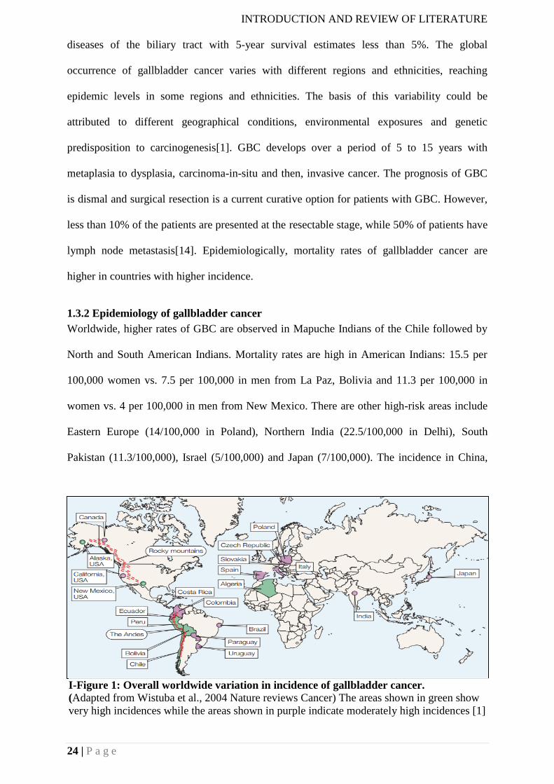
1.3.2 Epidemiology of gallbladder cancer
Worldwide, higher rates of GBC are observed in Mapuche Indians of the Chile followed by
North and South American Indians. Mortality rates are high in American Indians: 15.5 per
100,000 women vs. 7.5 per 100,000 in men from La Paz, Bolivia and 11.3 per 100,000 in
women vs. 4 per 100,000 in men from New Mexico. There are other high-risk areas include
Eastern Europe (14/100,000 in Poland), Northern India (22.5/100,000 in Delhi), South
Pakistan (11.3/100,000), Israel (5/100,000) and Japan (7/100,000). The incidence in China,
I-Figure 1: Overall worldwide variation in incidence of gallbladder cancer.
(Adapted from Wistuba et al., 2004 Nature reviews Cancer) The areas shown in green show
very high incidences while the areas shown in purple indicate moderately high incidences [1]
INTRODUCTION AND REVIEW OF LITERATURE
25 | P a g e
especially in Shanghai, have doubled over the years[15]. GBC is relatively low in the United
States and Mediterranean countries (UK, France, and Norway)[1, 16]. In the United States,
Hispanic women and men have a higher incidence of GBC than non-Hispanic men[1].
A retrospective study carried out in North Central India during 2007-2008 identified
gallbladder cancer to be at a fourth position after head and neck, breast and cervical cancer.
Within the Indian population highest incidence has been reported in northern cities(3.7 per
100 000 for male and 8.9 per 100 000 for female and in Bhopal it is 1.6, 2.5 per 100 000 for
male and female, respectively) as compared to southern cities ( eg in Chennai, the incidence
is 0.5 per 100 000 for male and 0.8 per 100 000 for 100 000 for female and in Bangalore,
incidence for male is 0.6 per 100 000 and for female it is 0.7 per 100 000 population) female
and in Bangalore, (incidence for male is 0.6 per 100 000 and for female it is 0.7 per 100 000
population)[12, 17].
1.3.3 Unmet need to treat gallbladder cancer in India
Gallbladder cancer is very common in the northern and north-eastern states of India. The
mean survival rate with advanced stages of cancer is 6 months with a 5-year survival rate of
less than 5%[1]. Since early diagnosis of the cancer is difficult, most of the gallbladder
cancers (95%) are detected at advanced stages where curative resection is not possible. Of the
remaining 5% who have stage I or II diseases, cholecystectomy is performed for symptomatic
gallstones. Chemotherapy and radiotherapy are given for unresectable cancers, however, the
survival frequencies are low in such cases. Few studies from India have shown the
association of ABC transporter genes like ABCB4, ABCB11, CYP7A1, ApoB, ApoE and LDL
receptor polymorphisms in gallstone diseases is also implicated in gallbladder cancer[18].
Gallstone disease with typhoidal infections is an important risk factor for gallbladder cancer,
is also common in northern India[19]. However, secondary prevention by prophylactic
cholecystectomy is controversial, as there is no evidence to support it[20]. There are different
studies in India investigating the role of pesticides, trace elements, bacteria in bile, bile
INTRODUCTION AND REVIEW OF LITERATURE
26 | P a g e
composition, chronic typhoid carriage, hormonal factors, and genetic factors like KRAS
alterations in the causation of gallbladder cancer[21-25]. However, these studies are limited
by the fewer number of samples and systematic genome-wide studies are dismal. Lack of
systematic clinical trials in India investigating the role of target therapies in gallbladder
cancer. Hence there is an unmet need to study the cancer type in a systematic and
comprehensive at a genome-wide scale.
1.3.4 Application of next generation sequencing in gallbladder cancer
The recent developments in sequencing technologies have helped in molecular
characterization of several rare cancer types. Biliary tract cancers are one of the rare cancer
types which are comprised of intrahepatic cholangiocarcinoma, extrahepatic
cholangiocarcinoma, and the gallbladder carcinoma. Genomic profiling of gallbladder
cancers using mass spectrometry and targeted sequencing technologies identified mutations
in IDH1 and TP53 as the most recurrently altered genes in gallbladder cancer[26]. Another
comprehensive study of 57 samples by whole exome sequencing and ultra-deep sequencing
of cancer-related genes identified mutations in TP53(47%), KRAS(7.8%), ERBB2(9.8%),
ERBB3(11.8%) and also the authors identified ERBB pathway as the most recurrently
mutated pathway in gallbladder cancer affecting up to 36.8% of GBC samples. Further using
multivariate analysis the authors show that the cases with ERBB pathway alterations have the
worse prognosis[27]. Another study using ion torrent based amplicon sequencing of 46 genes
in 9 gallbladder cancer samples identified one patient with activating ERBB2 alteration and
rest of the other samples with ERBB2 amplification. Patient with the ERBB2 mutation had a
mixed response to the pan-HER2 inhibitor[28]. Using targeted sequencing of 236 cancer-
related genes of 9 gallbladder cancer patients, recurrent mutations were observed in TP53,
ARID1A, and KRAS. Transcriptome sequencing of 8 gallbladder cancer patients and 3 normal
samples identified 519 genes to be differentially expressed and identified liver X receptors
and farnesoid receptors to be top canonical pathways to be deregulated in gall bladder
INTRODUCTION AND REVIEW OF LITERATURE
27 | P a g e
cancer[29]. Another RNA-seq study of 3 tumors and adjacent normal samples identified 161
differentially expressed genes and the authors observed enrichment of genes related to
pathways such as cell cycle, enzyme modulators, and pathways in cancer[30]. Despite the
higher prevalence in India, no genome-wide studies have been done using next-generation
sequencing technologies.
1.3.5 The landscape of known genomic alterations in gallbladder cancer.
The most common alteration reported in gallbladder cancer which occurs earlier in the
dysplasia to carcinoma sequence is p53 alterations. The most common alterations are in exon
5 and exon 8. Most of the p53 alterations are missense alterations that increase the stability of
the protein. The frequency of alterations reported is above 50%. Loss of heterozygosity of
p53 occurred earlier and more frequently than protein overexpression[31]. The frequency of
KRAS alterations is quite variable in gallbladder cancer ranging from 39-59%. Most of the
KRAS alterations have been reported in codon 12. Higher frequency of KRAS alterations has
been reported in patients having the anomalous junction of the pancreaticobiliary
duct(APBDJ) suggesting reflux of pancreatic juice might contribute to carcinogenic process[1,
31, 32]. Inactivation of CDKN2A has been observed in half of the GBC cases that occur by a
combination of mutations, deletion, and abnormal hypermethylation. Increased expression of
CDK4 and cyclin D1 detected by immunostaining in 41-60% of samples has been noted in
the progression of gallbladder cancer[33]. Cyclooxygenase 2 (COX2) overexpression has
been observed to occur earlier in the cascade of gallbladder carcinogenesis[34]. Loss of
heterozygosity and SNPs have been observed in DCC (deleted in colorectal carcinoma) gene
in gallbladder cancer and are considered as an early event in the cascade of gallbladder
carcinogenesis[35]. Reduction of FHIT (Fragile Histidine Triad) expression has been
observed in the progression of gallbladder cancer from dysplasia to invasive carcinoma[36].
LOH of 3p and 9p has been related to the progression of gallbladder cancer. Also, increasing
LOH proportions has been observed on chromosomes 3p, 9q, 8p, and 22q in normal,
INTRODUCTION AND REVIEW OF LITERATURE
28 | P a g e
dysplasia and malignant tissue[37]. Mismatch repair gene alterations are frequently reported
in gallbladder cancer. High-frequency microsatellite instability (MIN-H) has been reported in
early and late gallbladder cancers[31]. HER2 alterations have been reported in Chinese and
Japanese population. Overexpression of HER2 has been reported in 30-60% of GBC cases
and gene amplification is found in 70% of the cases[38]. In a mouse model system (BK5-
ERBB2 mice model) overexpression of HER2 in the basal layer of the biliary tract,
epithelium leads to the development of gallbladder carcinoma by 3 months of age. However,
the mouse gallbladder tumors were different from human tumors characterized by adenoma
precursors and papillary structures that filled the gallbladder lumen[39]. Expression levels of
HER2 varied depending on the increasing grade of the tumor.
1.3.6 Targeted therapy in gallbladder cancer
The conventional mode of treatment for gallbladder cancer is surgery for resectable cancers
and there are gemcitabine and fluoropyrimidine-based chemotherapeutic regimens for
unresectable cancers. Few reports have evaluated the effect of targeted therapies along with
the conventional treatment. Some studies suggest the benefit from blockade of EGFR by oral
tyrosine kinase inhibitor erlotinib or anti-EGFR monoclonal antibody cetuximab [40, 41].
Another study evaluated the benefit of the efficacy of bevacizumab, a monoclonal antibody
targeting VEGF along with erlotinib in a phase II trial. Overall, there was a partial response
among nine patients with six patients sustained beyond three weeks, and with an overall
median response of 8.4 months. The other half of the patients had a stable disease[42].
Likewise, other inhibitors such as sunitinib and sorafenib have their modest benefit in biliary
tract cancers[43, 44]. Hence, targeted therapy has shown some promise in gallbladder cancer,
however with better screening of patients with alterations the response rates may be improved
in the treatment of gallbladder cancer.
INTRODUCTION AND REVIEW OF LITERATURE
29 | P a g e
1.3.6 Risk factors for gallbladder cancer
The major risk factors for gallbladder cancer are chronic inflammation, geographical and
ethnic variation, increasing age, female gender, low socioeconomic status, genetic
predisposition, infections, low cholecystectomy cases and exposure to certain chemicals.
Some of the risk factors are discussed below:
1.3.6.1 Gallstones and cholecystitis
Gallstones are the major risk factor for gallbladder cancer accounting for 60-90% cases in
different regions of the world. A very common phenomenon is chronic inflammation due to
gallstone irritation of the gallbladder wall, which is observed in 85% of the cases. Patients
with gallstone have a higher incidence of gallbladder cancer[15]. The association between
with cholelithiasis and cancer may explain why female gender, multiparity and increased
body mass indices (also a risk for gallstone formation) are associated with developing
carcinoma of the gallbladder. The size of the gallstones (>3cm) and duration of stones in the
gallbladder have a stronger association with a pathogenesis that culminates in cancer [45, 46].
Though gallstones are associated risk factor, which is likely facultative rather than causative
prophylactic cholecystectomy is not favored for clinically silent gallstones except for large
stones and elderly patients with cholethiasis[47]. The decrease in incidence and mortality of
gallbladder cancer began decades before the introduction of laparoscopic cholecystectomy
and apparently stabilized in the past decade. There is no temporal relationship known to exist
between laparoscopic cholecystectomy rate and the incidence and mortality rates of
gallbladder cancer.
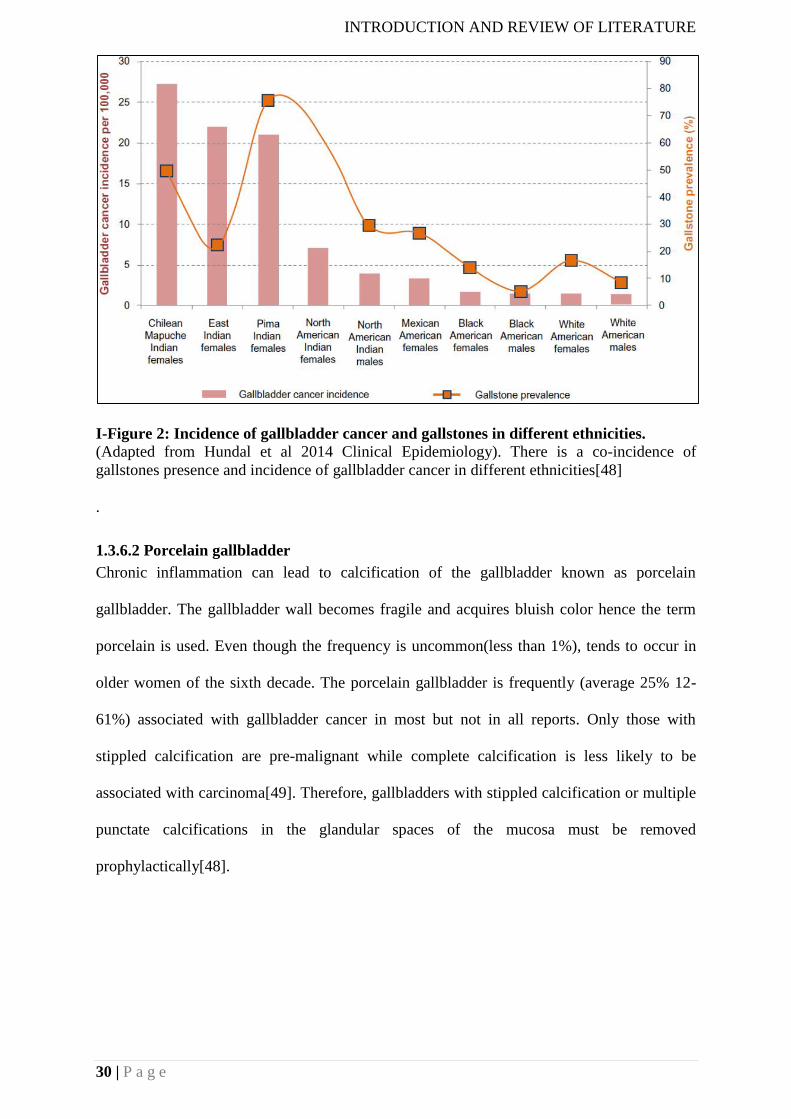
INTRODUCTION AND REVIEW OF LITERATURE
30 | P a g e
I-Figure 2: Incidence of gallbladder cancer and gallstones in different ethnicities.
(Adapted from Hundal et al 2014 Clinical Epidemiology). There is a co-incidence of
gallstones presence and incidence of gallbladder cancer in different ethnicities[48]
.
1.3.6.2 Porcelain gallbladder
Chronic inflammation can lead to calcification of the gallbladder known as porcelain
gallbladder. The gallbladder wall becomes fragile and acquires bluish color hence the term
porcelain is used. Even though the frequency is uncommon(less than 1%), tends to occur in
older women of the sixth decade. The porcelain gallbladder is frequently (average 25% 12-
61%) associated with gallbladder cancer in most but not in all reports. Only those with
stippled calcification are pre-malignant while complete calcification is less likely to be
associated with carcinoma[49]. Therefore, gallbladders with stippled calcification or multiple
punctate calcifications in the glandular spaces of the mucosa must be removed
prophylactically[48].
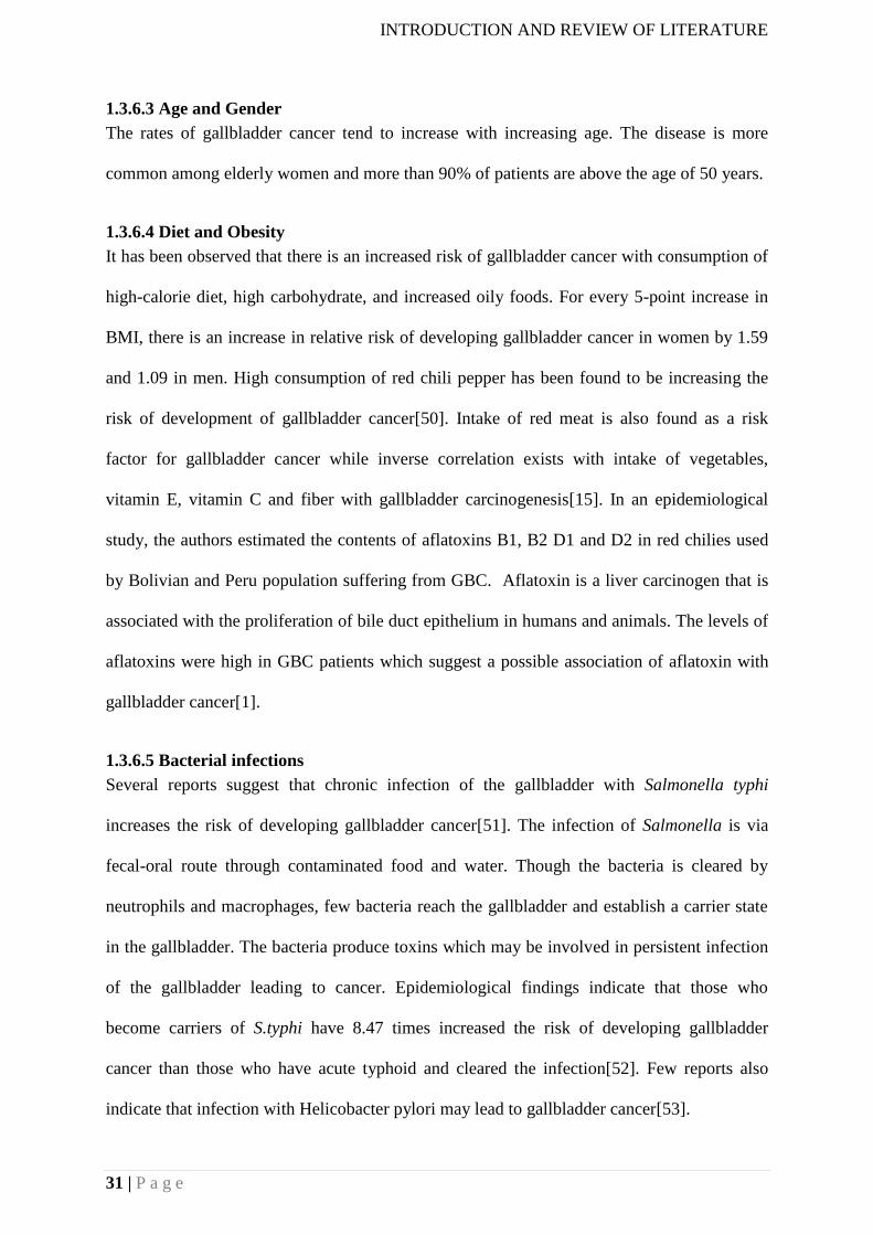
INTRODUCTION AND REVIEW OF LITERATURE
31 | P a g e
1.3.6.3 Age and Gender
The rates of gallbladder cancer tend to increase with increasing age. The disease is more
common among elderly women and more than 90% of patients are above the age of 50 years.
1.3.6.4 Diet and Obesity
It has been observed that there is an increased risk of gallbladder cancer with consumption of
high-calorie diet, high carbohydrate, and increased oily foods. For every 5-point increase in
BMI, there is an increase in relative risk of developing gallbladder cancer in women by 1.59
and 1.09 in men. High consumption of red chili pepper has been found to be increasing the
risk of development of gallbladder cancer[50]. Intake of red meat is also found as a risk
factor for gallbladder cancer while inverse correlation exists with intake of vegetables,
vitamin E, vitamin C and fiber with gallbladder carcinogenesis[15]. In an epidemiological
study, the authors estimated the contents of aflatoxins B1, B2 D1 and D2 in red chilies used
by Bolivian and Peru population suffering from GBC. Aflatoxin is a liver carcinogen that is
associated with the proliferation of bile duct epithelium in humans and animals. The levels of
aflatoxins were high in GBC patients which suggest a possible association of aflatoxin with
gallbladder cancer[1].
1.3.6.5 Bacterial infections
Several reports suggest that chronic infection of the gallbladder with Salmonella typhi
increases the risk of developing gallbladder cancer[51]. The infection of Salmonella is via
fecal-oral route through contaminated food and water. Though the bacteria is cleared by
neutrophils and macrophages, few bacteria reach the gallbladder and establish a carrier state
in the gallbladder. The bacteria produce toxins which may be involved in persistent infection
of the gallbladder leading to cancer. Epidemiological findings indicate that those who
become carriers of S.typhi have 8.47 times increased the risk of developing gallbladder
cancer than those who have acute typhoid and cleared the infection[52]. Few reports also
indicate that infection with Helicobacter pylori may lead to gallbladder cancer[53].
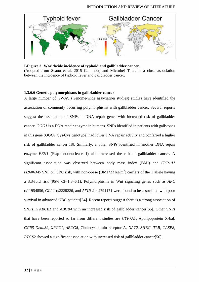
INTRODUCTION AND REVIEW OF LITERATURE
32 | P a g e
I-Figure 3: Worldwide incidence of typhoid and gallbladder cancer.
(Adopted from Scanu et al, 2015 Cell host, and Microbe) There is a close association
between the incidence of typhoid fever and gallbladder cancer.
1.3.6.6 Genetic polymorphisms in gallbladder cancer
A large number of GWAS (Genome-wide association studies) studies have identified the
association of commonly occurring polymorphisms with gallbladder cancer. Several reports
suggest the association of SNPs in DNA repair genes with increased risk of gallbladder
cancer. OGG1 is a DNA repair enzyme in humans. SNPs identified in patients with gallstones
in this gene (OGG1 Cys/Cys genotype) had lower DNA repair activity and conferred a higher
risk of gallbladder cancer[18]. Similarly, another SNPs identified in another DNA repair
enzyme FEN1 (Flap endonuclease 1) also increased the risk of gallbladder cancer. A
significant association was observed between body mass index (BMI) and CYP1A1
rs2606345 SNP on GBC risk, with non-obese (BMI<23 kg/m2) carriers of the T allele having
a 3.3-fold risk (95% CI=1.8–6.1). Polymorphisms in Wnt signaling genes such as APC
rs11954856, GLI-1 rs2228226, and AXIN-2 rs4791171 were found to be associated with poor
survival in advanced GBC patients[54]. Recent reports suggest there is a strong association of
SNPs in ABCB1 and ABCB4 with an increased risk of gallbladder cancer[55]. Other SNPs
that have been reported so far from different studies are CYP7A1, Apolipoprotein X-baI,
CCR5 Delta32, XRCC1, ABCG8, Cholecystokinin receptor A, NAT2, SHBG, TLR, CASP8,
PTGS2 showed a significant association with increased risk of gallbladder cancer[56].
INTRODUCTION AND REVIEW OF LITERATURE
33 | P a g e
1.3.6.7 Environmental effects
Though there is no conclusive evidence to support the association of environmental pollutants
with gallbladder cancer. Few reports suggest the higher biliary concentration of cadmium,
chromium, and lead were found in the bile of cancer patients than patients with gallstones.
Increased expression of metallothionein has been observed in GBC patients which may play a
role in gallbladder carcinogenesis.
1.4 Molecular pathology of gallbladder cancer
There are proposed two pathways in the pathogenesis of gallbladder pathogenesis viz
dysplasia-carcinoma sequence and adenoma-invasive carcinoma. In the first model, normal
epithelium changes to dysplasia due to chronic irritation or inflammation thus progressing to
carcinoma in situ and eventually leading to invasive cancer. In the second model, gallbladder
polyp is formed by the initial glandular proliferation of the epithelium, malignant
transformation occurs within this initially formed an initial benign mass[31]. In addition to
the previously proposed models, the updated model “gallbladder carcinogenesis and
dissemination model incorporates the course of disease after the development of invasive
carcinoma and addition of new molecular markers that could be used for diagnosis or
response to therapy[31]. Chronic inflammation plays an important role in gallbladder cancer.
There is a reduction in expression of inflammatory markers such as COX2, EGFR and other
markers at advanced stages of cancer. The protective influence of the expression of markers
in the advanced stages is still needed to be ascertained. Most dysplasias and carcinoma in situ
are observed after cholecystectomy when the entire lesion is removed and evidence suggest
that progression could occur from precursor lesions to infiltrating carcinoma. About 90% of
the gallbladder cancers are adenocarcinoma, of which 10-37% of carcinomas cannot be
identified with certainty on gross examination since the macroscopic findings are similar to
chronic cholecystitis[57]. The majority of the carcinomas originate in the fundus (60%), body
(30%) and neck (10%)[58]. Most of the gallbladder cancers are well to moderately
differentiated adenocarcinomas. Few previously reported histological studies have identified
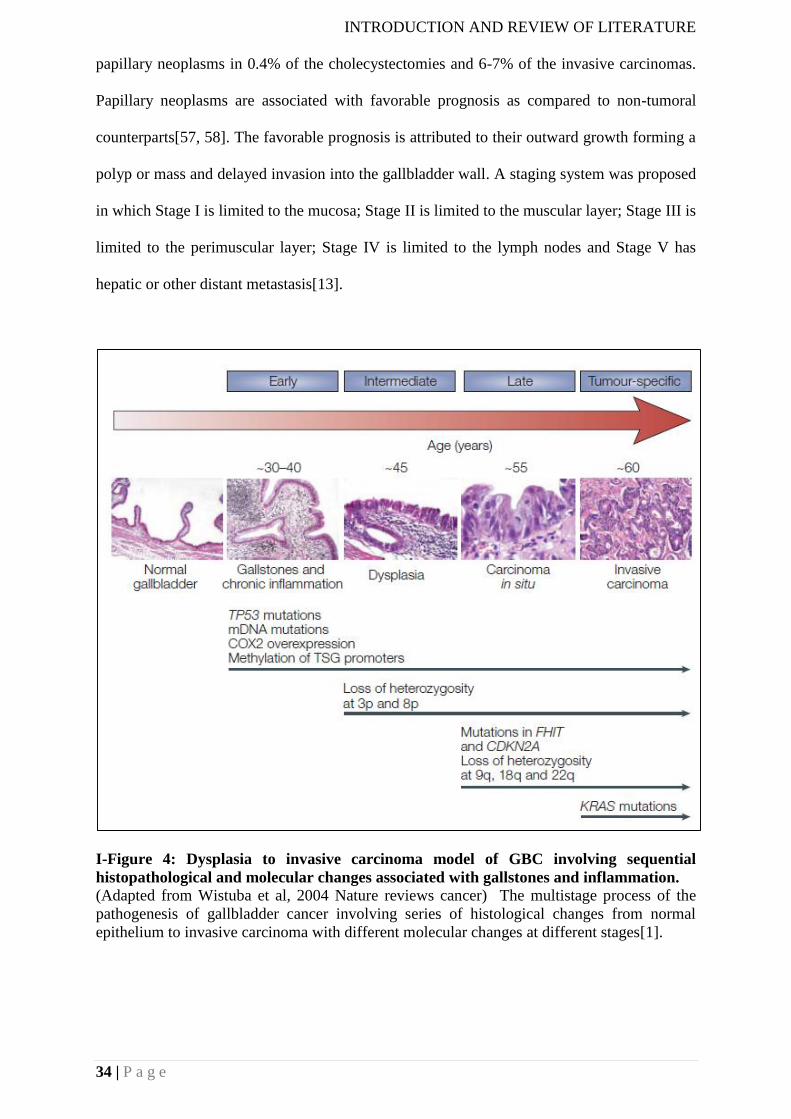
INTRODUCTION AND REVIEW OF LITERATURE
34 | P a g e
papillary neoplasms in 0.4% of the cholecystectomies and 6-7% of the invasive carcinomas.
Papillary neoplasms are associated with favorable prognosis as compared to non-tumoral
counterparts[57, 58]. The favorable prognosis is attributed to their outward growth forming a
polyp or mass and delayed invasion into the gallbladder wall. A staging system was proposed
in which Stage I is limited to the mucosa; Stage II is limited to the muscular layer; Stage III is
limited to the perimuscular layer; Stage IV is limited to the lymph nodes and Stage V has
hepatic or other distant metastasis[13].
I-Figure 4: Dysplasia to invasive carcinoma model of GBC involving sequential
histopathological and molecular changes associated with gallstones and inflammation.
(Adapted from Wistuba et al, 2004 Nature reviews cancer) The multistage process of the
pathogenesis of gallbladder cancer involving series of histological changes from normal
epithelium to invasive carcinoma with different molecular changes at different stages[1].
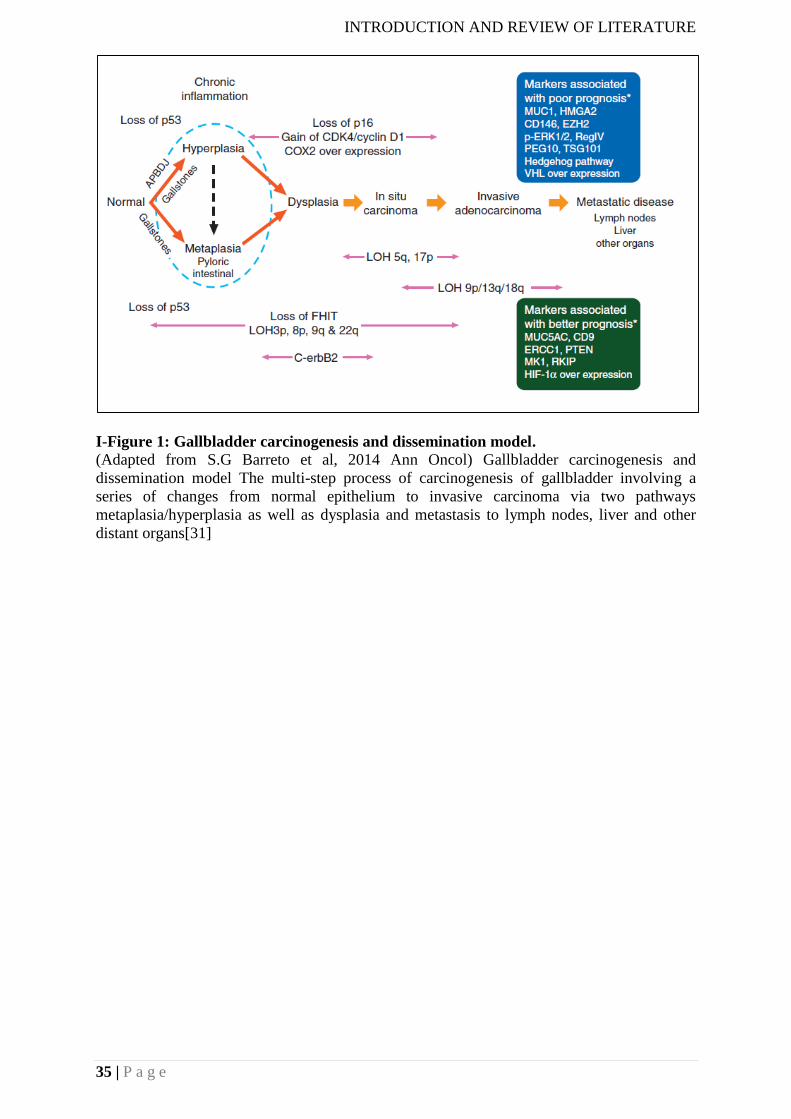
INTRODUCTION AND REVIEW OF LITERATURE
35 | P a g e
I-Figure 1: Gallbladder carcinogenesis and dissemination model.
(Adapted from S.G Barreto et al, 2014 Ann Oncol) Gallbladder carcinogenesis and
dissemination model The multi-step process of carcinogenesis of gallbladder involving a
series of changes from normal epithelium to invasive carcinoma via two pathways
metaplasia/hyperplasia as well as dysplasia and metastasis to lymph nodes, liver and other
distant organs[31]
INTRODUCTION AND REVIEW OF LITERATURE
36 | P a g e
1.5 Research objective
Rationale
Gallbladder cancer is an aggressive disease with poor prognosis. Currently, surgery is the
only curative mode of treatment for the disease. Chemotherapy and radiotherapy are used as
adjuvants; however, there is very little effect on survival of the patients. Moreover, molecular
targets have not been identified yet. My research proposal takes the advantage of combining
genomic approaches followed by functional validation of the alterations discovered to build
up a molecular framework of therapeutically relevant alterations thereby benefit the patients
with the deadly disease. The objectives of the thesis are listed below:
Objectives
1) Apply next-generation sequencing analysis of gallbladder cancer to identify pathogenic
sequences in gallbladder cancer using computational subtraction method.
2) Apply next-generation sequencing analysis of gallbladder cancer to identify genes whose
somatic genomic alterations suggest the properties of driver oncogenes. In a more directed
approach, we will sequence exome, from gallbladder tumors of Indian origin cases.
3) We will test candidate oncogenes identified in Aim 2 by gain-of-function assays for
cellular transformation and activation of known oncogenic signaling pathways.
NON TYPHOIDAL SALMONELLA TRACES IN GALLBLADDER CANCER
37 | P a g e
CHAPTER 2: NON TYPHOIDAL SALMONELLA TRACES IN GALLBLADDER
CANCER (an excerpt; as published in BMC Infectious Agents and Cancer (2016);11:12
Abstract
Background
We earlier proposed a genetic model for gallbladder carcinogenesis and its dissemination
cascade. However, the association between gallbladder cancer and ‘inflammatory stimulus’ to
drive the initial cascade in the model remained unclear. A recent study suggested infection
with Salmonella can lead to changes in the host signalling pathways in gallbladder cancer.
Findings
We examined the whole exomes of 26 primary gallbladder tumour and paired normal
samples for presence of 143 HPV (Human papillomavirus) types along with 6
common Salmonella serotypes (S. typhi Ty2, S. typhi CT18, S. typhimurium LT2, S.
choleraesuis SCB67, S. paratyphi TCC, and S. paratyphi SPB7) using a computational
subtraction pipeline based on the HPVDetector, we recently described. Based on our
evaluation of 26 whole exome gallbladder primary tumors and matched normal samples:
association of typhoidal Salmonella species were found in 11 of 26 gallbladder cancer
samples, and non-typhoidal Salmonella species in 12 of 26 gallbladder cancer, with 6
samples were found co-infected with both.
Conclusions
We present the first evidence to support the association of non-typhoidal Salmonella species
along with typhoidal strains in gallbladder cancer. Salmonella infection in the chronic carrier
state fits the role of the ‘inflammatory stimulus’ in the genetic model for gallbladder
carcinogenesis that may play a role in gallbladder cancer analogous to Helicobacter pylori in
gastric cancer.
NON TYPHOIDAL SALMONELLA TRACES IN GALLBLADDER CANCER
38 | P a g e
2.1 Introduction
2.1.1 Infections in cancer
Worldwide, it has been estimated that 20% of cancers are attributed to infectious agents. It’s
known that viral and bacterial pathogens have been postulated to play an important role in the
development of cancer [59, 60]. Among the pathogens, viruses account for the majority of
malignancies from a universal perspective. There are seven oncogenic viruses (hepatitis B
and C (HBV and HCV), human papillomavirus (HPV), Epstein-Barr virus (EBV) and human
T cell lymphoma virus 1(HTLV-1), Merkel cell polyomavirus, Kaposi’s sarcoma virus, one
oncogenic bacterium (Helicobacter pylori) play a tumorigenic role in the development of
cancer[61]. The vital portion of the viral genome can be found in a cancer cell resulting in the
expression of viral genes disrupt cell cycle, inhibit apoptosis thus contributing to the cellular
process of immortalization. In contrast pathogens like bacteria (H.pylori, O.viverrini, and
S.typhi) produce a chronic inflammatory state that leads to the production of cytokines,
prostaglandins which can result in deregulation of the immune system and
neovascularization[60]. All these pathogens have been associated with the carcinogenic
process, however, the molecular mechanism has not been elucidated so far.
2.1.2 Infections in gallbladder cancer
Gallbladder cancer is one of the most common cancers of the biliary tract. One of the major
risk factors is infections with Salmonella bacteria[51]. Several epidemiological studies from
India, especially from the northern part of the country have reported a chronic carriage of
typhoid and gallbladder cancer[62]. Typhoid is caused by Salmonella enterica serovar Typhi.
Salmonella typhi enters the bloodstream through contaminated food or water. After crossing
the intestinal epithelial barrier, Salmonella is phagocytosed and are systematically spread to
produce acute disease. About 3-5% of typhoid patients become chronic carriers, with
gallbladder providing the niche for its persistence[63]. Chronic carriers are symptomatic and
have an approximately 8-fold risk of developing gallbladder cancer than the non-carriers[64].
NON TYPHOIDAL SALMONELLA TRACES IN GALLBLADDER CANCER
39 | P a g e
Few reports suggest the co-operative relationship between gallstones and Salmonella
typhoidal infections in the carcinogenesis of gallbladder cancer, however, the cause and
effect relationship is not ascertained[65]. The proposed mechanism of tumorigenicity by
Salmonella bacteria is the production of b-glucuronidase that result in deconjugation of toxins
and bile acids which in turn lead to carcinogenesis[66]. Other evidence indicates that
products of degradation of bile acids by the bacteria may contribute to tumorigenesis[67, 68].
Recent evidence shows that Salmonella enterica infection induces transformation in
predisposed murine gallbladder organoids, fibroblasts with TP53 mutations and c-MYC
amplification by activation of MAPK and AKT pathways[19]. The typhoidal Salmonella was
strongly associated with gallbladder cancer however non-typhoidal Salmonella species (S.
Typhimurium and S. Choleraesius) that elicits a stronger immune response is linked with the
systemic illness (gastroenteritis) have as yet not associated with gallbladder cancer[69]. We
proposed a gallbladder cancer carcinogenesis model based on current understanding of tumor
biology[31]. However, the lacunae in the model is the driving force behind inflammation-
related changes is not ascertained. Here we examined the exomes of primary gallbladder
tumor and paired normal samples for the presence of 6 common Salmonella serotypes with
available genome information (S. typhi Ty2, S. typhi CT18, S. typhimurium LT2, S.
choleraesuis SCB67, S. paratyphi TCC, and S. paratyphi SPB7) using a computational
subtraction pipeline based on the HPVDetector tool.
2.2 Materials and Methods
2.2.1 Patient information:
Twenty six fresh frozen primary tumor and matched normal tissues were obtained from the
tissue repository of Tata Memorial Hospital (TMH). The Institutional Review Board (IRB)
and the Ethics Committee (EC) of Tata Memorial Centre (TMC) - Advanced Centre for
Treatment, Research and Education in Cancer (ACTREC) (Mumbai, India) approved the
NON TYPHOIDAL SALMONELLA TRACES IN GALLBLADDER CANCER
40 | P a g e
project (#104). Since this was a retrospective analysis, the IRB and the EC waived the need
for an informed consent. Patients were randomly selected based on the availability of fresh
frozen tissues. The patient characteristics including age, gender, gallstone status and
histopathology were recorded.
2.2.2 PCR analysis for Salmonella isolates
The PCR method used for Salmonella detection has been previously described [12]. Nested
PCR was carried out in a 25µl volume containing 10 µl KAPA 2X ready mix master-mix
(Kapa Biosystems catalog no-KK1024), 10pmol primer and 100 ng of genomic DNA.
Following the first round of PCR (94 °C for 1 min, 55 °C for 1 min 15 s,72 °C for 3 min – 40
cycles) with ST1 and ST2 primers,5 µl PCR product was used as template for nested PCR
using ST3 and ST4 primers (94 °C for 1 min, 68 °C for 1 min 15 s, 72 °C for 3 min – 40
cycles). We also performed validation of Salmonella sequences using read specific primers.
The PCR conditions - 94 °C for 1 min, 59 °C for 30s, 72 °C for 45 s – for 30 cycles.
2.2.3 Sequencing and analysis
Exome capture and library preparation were performed using Agilent Sure select in-solution
(low-input capture500 ng) target enrichment technology. Genomic DNA was sheared and
size selected (150–200 bp) and ligated to adaptors and run on Illumina Nextseq 500 platform
to generate 150 bp paired-end reads at a coverage of 100X and above. To detect Salmonella
traces, the HPVDetector pipeline was used, as described previously [11]. Briefly, reads were
aligned against six known Salmonella species genomes in addition of the HPVDetector
dataset of 143 HPV types, as downloaded from the National Centre for Biotechnology
Information (NCBI), using BWA (Burrows wheeler algorithm) aligner (v0.6.2). All reference
sequences were annotated and concatenated to compose multi-fasta sequences using bio-perl
modules. The alignment files were parsed using UNIX shell program to detect the types of
Salmonella represented by at least one read that aligned to a particular Salmonella type with
high confidence.
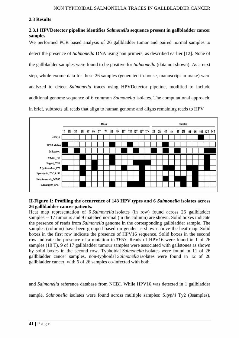
NON TYPHOIDAL SALMONELLA TRACES IN GALLBLADDER CANCER
41 | P a g e
2.3 Results
2.3.1 HPVDetector pipeline identifies Salmonella sequence present in gallbladder cancer
samples
We performed PCR based analysis of 26 gallbladder tumor and paired normal samples to
detect the presence of Salmonella DNA using pan primers, as described earlier [12]. None of
the gallbladder samples were found to be positive for Salmonella (data not shown). As a next
step, whole exome data for these 26 samples (generated in-house, manuscript in make) were
analyzed to detect Salmonella traces using HPVDetector pipeline, modified to include
additional genome sequence of 6 common Salmonella isolates. The computational approach,
in brief, subtracts all reads that align to human genome and aligns remaining reads to HPV
II-Figure 1: Profiling the occurrence of 143 HPV types and 6 Salmonella isolates across
26 gallbladder cancer patients.
Heat map representation of 6 Salmonella isolates (in row) found across 26 gallbladder
samples -- 17 tumours and 9 matched normal (in the column) are shown. Solid boxes indicate
the presence of reads from Salmonella genome in the corresponding gallbladder sample. The
samples (column) have been grouped based on gender as shown above the heat map. Solid
boxes in the first row indicate the presence of HPV16 sequence. Solid boxes in the second
row indicate the presence of a mutation in TP53. Reads of HPV16 were found in 1 of 26
samples (10 T). 9 of 17 gallbladder tumour samples were associated with gallstones as shown
by solid boxes in the second row. Typhoidal Salmonella isolates were found in 11 of 26
gallbladder cancer samples, non-typhoidal Salmonella isolates were found in 12 of 26
gallbladder cancer, with 6 of 26 samples co-infected with both.
and Salmonella reference database from NCBI. While HPV16 was detected in 1 gallbladder
sample, Salmonella isolates were found across multiple samples: S.typhi Ty2 (3samples),
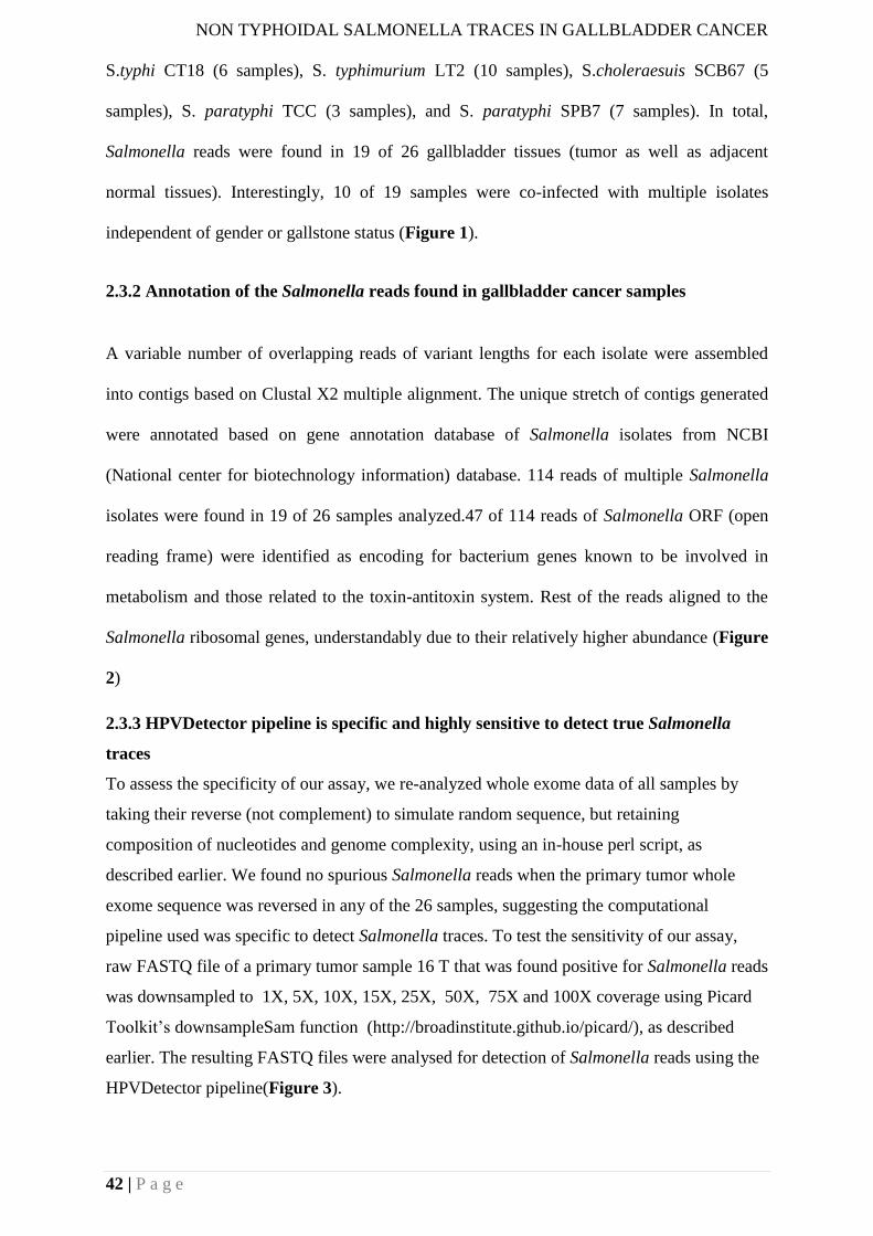
NON TYPHOIDAL SALMONELLA TRACES IN GALLBLADDER CANCER
42 | P a g e
S.typhi CT18 (6 samples), S. typhimurium LT2 (10 samples), S.choleraesuis SCB67 (5
samples), S. paratyphi TCC (3 samples), and S. paratyphi SPB7 (7 samples). In total,
Salmonella reads were found in 19 of 26 gallbladder tissues (tumor as well as adjacent
normal tissues). Interestingly, 10 of 19 samples were co-infected with multiple isolates
independent of gender or gallstone status (Figure 1).
2.3.2 Annotation of the Salmonella reads found in gallbladder cancer samples
A variable number of overlapping reads of variant lengths for each isolate were assembled
into contigs based on Clustal X2 multiple alignment. The unique stretch of contigs generated
were annotated based on gene annotation database of Salmonella isolates from NCBI
(National center for biotechnology information) database. 114 reads of multiple Salmonella
isolates were found in 19 of 26 samples analyzed.47 of 114 reads of Salmonella ORF (open
reading frame) were identified as encoding for bacterium genes known to be involved in
metabolism and those related to the toxin-antitoxin system. Rest of the reads aligned to the
Salmonella ribosomal genes, understandably due to their relatively higher abundance (Figure
2)
2.3.3 HPVDetector pipeline is specific and highly sensitive to detect true Salmonella
traces
To assess the specificity of our assay, we re-analyzed whole exome data of all samples by
taking their reverse (not complement) to simulate random sequence, but retaining
composition of nucleotides and genome complexity, using an in-house perl script, as
described earlier. We found no spurious Salmonella reads when the primary tumor whole
exome sequence was reversed in any of the 26 samples, suggesting the computational
pipeline used was specific to detect Salmonella traces. To test the sensitivity of our assay,
raw FASTQ file of a primary tumor sample 16 T that was found positive for Salmonella reads
was downsampled to 1X, 5X, 10X, 15X, 25X, 50X, 75X and 100X coverage using Picard
Toolkit’s downsampleSam function (http://broadinstitute.github.io/picard/), as described
earlier. The resulting FASTQ files were analysed for detection of Salmonella reads using the
HPVDetector pipeline(Figure 3).
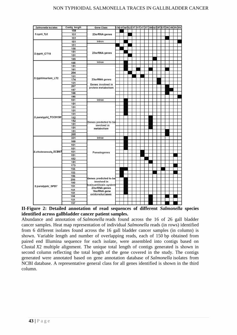
NON TYPHOIDAL SALMONELLA TRACES IN GALLBLADDER CANCER
43 | P a g e
II-Figure 2: Detailed annotation of read sequences of different Salmonella species
identified across gallbladder cancer patient samples.
Abundance and annotation of Salmonella reads found across the 16 of 26 gall bladder
cancer samples. Heat map representation of individual Salmonella reads (in rows) identified
from 6 different isolates found across the 16 gall bladder cancer samples (in column) is
shown. Variable length and number of overlapping reads, each of 150 bp obtained from
paired end Illumina sequence for each isolate, were assembled into contigs based on
Clustal X2 multiple alignment. The unique total length of contigs generated is shown in
second column reflecting the total length of the gene covered in the study. The contigs
generated were annotated based on gene annotation database of Salmonella isolates from
NCBI database. A representative general class for all genes identified is shown in the third
column.
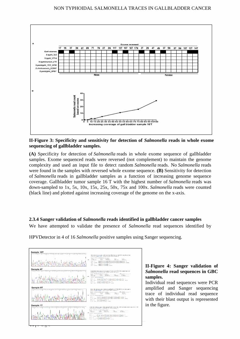
NON TYPHOIDAL SALMONELLA TRACES IN GALLBLADDER CANCER
44 | P a g e
(A) Specificity for detection of Salmonella reads in whole exome sequence of gallbladder
samples. Exome sequenced reads were reversed (not complement) to maintain the genome
complexity and used an input file to detect random Salmonella reads. No Salmonella reads
were found in the samples with reversed whole exome sequence. (B) Sensitivity for detection
of Salmonella reads in gallbladder samples as a function of increasing genome sequence
coverage. Gallbladder tumor sample 16 T with the highest number of Salmonella reads was
down-sampled to 1x, 5x, 10x, 15x, 25x, 50x, 75x and 100x. Salmonella reads were counted
(black line) and plotted against increasing coverage of the genome on the x-axis.
2.3.4 Sanger validation of Salmonella reads identified in gallbladder cancer samples
We have attempted to validate the presence of Salmonella read sequences identified by
HPVDetector in 4 of 16 Salmonella positive samples using Sanger sequencing.
II-Figure 3: Specificity and sensitivity for detection of Salmonella reads in whole exome
sequencing of gallbladder samples.
II-Figure 4: Sanger validation of
Salmonella read sequences in GBC
samples.
Individual read sequences were PCR
amplified and Sanger sequencing
trace of individual read sequence
with their blast output is represented
in the figure.
NON TYPHOIDAL SALMONELLA TRACES IN GALLBLADDER CANCER
45 | P a g e
2.4 Discussion
We examined Salmonella and HPV DNA sequences in gallbladder tumors and paired normal,
the high incidence of Salmonella sequence found in 16 of 26 samples analyzed in the study
suggests a possible role of Salmonella infection in gallbladder cancer analogous to
Helicobacter pylori in gastric cancer and Fusobacterium in colon cancer[70, 71]. We
demonstrate the presence of typhoidal Salmonella species in 11 of 26 gallbladder cancer
samples, consistent with as known earlier. In addition, we present the first evidence to
support the association of even non-typhoidal Salmonella species in 12 of 26 gallbladder
cancer, with 6 of 26 samples co-infected with typhoidal as well as non-typhoidal Salmonella
isolates. Systemic inflammation is known to be associated with a poor prognosis in
gallbladder cancer[72]. Owing to the ability of Salmonella infection to stimulate a host
response, it is likely that these bacteria are able to provide the continued ‘inflammatory
stimulus’ necessary for carcinogenesis. Recent reports suggest that Salmonella infections
promote malignant transformation in genetically predisposed mice, murine gall bladder
organoids and fibroblasts with TP53 mutations[19]. We observed 4 of 16 Salmonella positive
samples harbored TP53 mutations while we did not observe TP53 mutations in Salmonella
negative samples. Salmonella isolates in the chronic carrier state thus fits the role of the
‘inflammatory stimulus’ in the genetic model for gallbladder carcinogenesis and its
dissemination cascade, which may trigger transformation through chronic inflammation, but
not for maintenance of tumorigenesis[31].The focus of treatment in typhoid-endemic
countries such as India has historically been solely on eliminating typhoidal Salmonella
species often underestimating the contribution of the non-typhoidal isolates that show an
inherent higher resistance to the standard antibiotics[69] resulting in their ability to lead to
chronic carrier state in humans. The presence of non-typhoidal Salmonella species in our
study highlights that in typhoid as well as gallbladder cancer endemic countries such as India
and other similar countries, efforts must be directed not only at treating typhoid fever, but
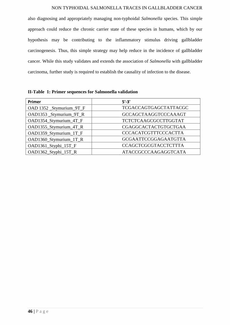
NON TYPHOIDAL SALMONELLA TRACES IN GALLBLADDER CANCER
46 | P a g e
also diagnosing and appropriately managing non-typhoidal Salmonella species. This simple
approach could reduce the chronic carrier state of these species in humans, which by our
hypothesis may be contributing to the inflammatory stimulus driving gallbladder
carcinogenesis. Thus, this simple strategy may help reduce in the incidence of gallbladder
cancer. While this study validates and extends the association of Salmonella with gallbladder
carcinoma, further study is required to establish the causality of infection to the disease.
II-Table 1: Primer sequences for Salmonella validation
Primer 5'-3'
OAD 1352 _Stymurium_9T_F TCGACCAGTGAGCTATTACGC
OAD1353 _Stymurium_9T_R GCCAGCTAAGGTCCCAAAGT
OAD1354_Stymurium_4T_F TCTCTCAAGCGCCTTGGTAT
OAD1355_Stymurium_4T_R CGAGGCACTACTGTGCTGAA
OAD1359_Stymurium_1T_F CCCACATCGTTTCCCACTTA
OAD1360_Stymurium_1T_R GCGAATTCCGGAGAATGTTA
OAD1361_Styphi_15T_F CCAGCTCGCGTACCTCTTTA
OAD1362_Styphi_15T_R ATACCGCCCAAGAGGTCATA
EGFR AND KRAS ALTERATIONS MEDIATE RESPONSE TO EGFR INHIBITOR
47 | P a g e
3. CHAPTER 3:
ERBB2 and KRAS Alterations Mediate Response to EGFR Inhibitors in early stage
Gallbladder Cancer
Abstract
The uncommonness of gallbladder cancer in the developed world has contributed to the
generally poor understanding of the disease. The development of new and effective treatment,
therefore, has been and continues to be a major public health imperative. We report mutation
analysis of 44 predominantly early-staged gallbladder tumors and 5-gallbladder cancer cell
lines by a combination of whole exome and directed sequencing. We discover recurrent
activating ERBB2 (V777L) somatic mutation in 6 of 44 gallbladder primary tumors with an
overall mutation frequency of 13%; along with KRAS activating mutations in 3 cancer
samples. Consistent with whole exome findings, phospho-proteomic array profile of 49-
tyrosine kinase revealed constitutive phosphorylation of ERBB2 and EGFR that were found
to heterodimerize. We demonstrate that treatment with ERBB2-specific, EGFR-specific
shRNA or with a covalent EGFR family inhibitor BIBW-2992 inhibits transformation,
survival, migration and invasion characteristics of gallbladder cancer cells harboring wild-
type or KRAS (G13D) but not KRAS (G12V) mutation. In summary, our studies implicate
ERBB2 as an important therapeutic target in early-stage gallbladder cancer. We also present
the first evidence that the presence of KRAS (G12V), but not KRAS (G13D) mutation, may
preclude gallbladder cancer patients to respond to anti-EGFR treatment, similar to the clinical
algorithm commonly practiced to opt for anti-EGFR treatment in colorectal cancer.
3.1 Introduction
Genomically matched therapies targeting activated tyrosine kinases have shown promise
across multiple cancer types [73]. The success of tyrosine kinase inhibitors (TKIs) such as
imatinib, a BCR-ABL fusion protein inhibitor [74]; vemurafenib, a RAF inhibitor [75];
lapatinib, an inhibitor of ERBB2 [76]; erlotinib and crizotinib, inhibitors of EGFR and ALK,
respectively [77-79]; and, others have provided a powerful validation for precision cancer
medicine. Although these treatments offer great promise, selective genomic profiling of
tumors tend to impede broader implementation of genome-based cancer care [80]. For
example, an inadequacy to account for multiple relevant genetic alterations likely resulted in
comparable outcomes in a recently performed randomized trial where multiple cancer type
patients were profiled for selected driver alterations and randomized to receive genomically-
EGFR AND KRAS ALTERATIONS MEDIATE RESPONSE TO EGFR INHIBITOR
48 | P a g e
matched versus conventional therapy [81]. Such important clinical studies underscore the
need for convergence of information for multiple genetic alterations to ensure the success of
future clinical trial designs, with specific emphasis for consideration of co-occurring
alterations that could potentially render tumors unlikely to benefit from genomically-matched
treatments. Some prototypical examples include KRAS, NRAS, and BRAF mutations in
colorectal cancers or secondary EGFR mutations in lung cancer against anti- EGFR targeted
therapies [82].
The EGFR family of receptor tyrosine kinases (RTK) consists of EGFR, HER2, HER3 and
HER4 (human EGFR-related- 2, -3, and -4). A ligand-bound EGFR family member forms a
homo- or hetero-dimer to activate the PI3K-AKT-mTOR or RAS-RAF-MAPK downstream
signaling pathway to evade apoptosis and enhance cell proliferation [83-85]. Interestingly, of
all EGFR family members, HER2 lacks a ligand binding domain and forms preferred partner
for other members to heterodimerize even in the absence of ligand [83]. Deregulation of
EGFR family RTK-signaling network endows tumor cells with attributes to sustain their
malignant behavior and survival, as is frequently observed in breast cancer, lung cancer,
pancreatic cancer, head and neck cancer and colorectal cancer [86]. Interfering with the
EGFR pathway thus forms the basis for the development of targeted anticancer therapies such
as RTK-targeted antibodies (Cetuximab and Herceptin) and small-molecule inhibitors of
RTK kinase (Erlotinib, Lapatinib, Afatinib, etc.) that have shown dramatic clinical response
[86]. In such responses, however, the co-occurrence of a KRAS mutation – a downstream
component of the pathway-- preclude patients from anti-EGFR treatment in colorectal cancer,
wherein KRAS codon 12, but not codon 13 mutations are associated with poor outcomes [87-
89], underscoring their prognostic impact.
Gallbladder cancer, the most common malignancy of biliary tract, is a rare form of cancer in
the world where chemotherapy and other palliative treatments have little effect on overall
survival of patients [14, 90]. The poor understanding of gallbladder cancer due to its
uncommonness in the western world but high prevalence in Chile and the Indian subcontinent
lends itself to the need for further research [1, 31, 91]. The 5-year survival rate of an early
stage T1 gallbladder carcinoma is nearly 100%, however, of advanced stage, T3/T4 is less
than 15% [90, 92]. A hope for longer-term survival has specifically been promising for an
early staged T2 carcinomas with an intermediate 5-year survival [93]. Literature suggests
HER2 overexpression in 12–15 % of advanced stage gallbladder cancers with a favorable
EGFR AND KRAS ALTERATIONS MEDIATE RESPONSE TO EGFR INHIBITOR
49 | P a g e
response to HER2 directed therapy [26, 28, 38, 94]. Moreover, three recent studies analyzed
whole exome sequence of advanced stage gallbladder tumors among non-Indian populations
with consistent findings [10, 27, 95]. In order to understand the landscape of somatic
alterations among a clinically distinct early staged pT1/pT2 Indian gallbladder cancer patients,
we performed whole exome sequencing of 17 early staged tumor-normal paired gallbladder
samples, 5 gallbladder cancer cell lines followed by validation in 27 additional tumor samples.
Here, we report novel somatic mutations of ERBB2 in gallbladder cancer, and its therapeutic
implication in presence and absence of KRAS (G12V) and (G13D) mutations
3.2 Materials and Methods
3.2.1Patient Information:
A total of 27 fresh frozen samples (10 tumor-normal paired and 7 orphan tumors) were
utilized for whole exome sequencing. An additional set of 27 FFPE samples were utilized as
a validation set. Tumor-normal paired samples were collected at Tata Memorial Hospital and
Advanced Centre for Treatment, Research and Education in Cancer (ACTREC), Mumbai.
Sample set and (ACTREC-TMC) Internal Review Board (IRB) --IRB Project Number # 104-
- approved study protocols. Formalin-fixed paraffin-embedded tissue blocks were collected
from the tissue repository of Tata memorial hospital (TMH-TTR) in compliance with the
guidelines. These tissues were examined for tumor content and the tumor content was in the
range of 40-90%. Patient samples and characteristics are provided in Table 1.
3.2.2 DNA extraction
Genomic DNA was extracted from fresh frozen samples by using Qiagen Blood and Cell
culture DNA kit. The extracted DNA yield and quality were assessed using Nanodrop
ND2000 (Thermo scientific). The extracted DNA (about 1µg) from the fresh-frozen tissue
specimens were sent to Genotypic Technology Pvt Ltd, Bangalore for exome sequencing.
Genomic DNA from FFPE blocks was extracted using Qiagen QiAmp DNA FFPE Tissue kit
as per manufacturer instructions. The extracted DNA yield and quality were assessed using
Nanodrop ND2000 (Thermo scientific). These samples were further checked for integrity by
PCR amplification of GAPDH (96bp). These samples were used for extended Sanger
EGFR AND KRAS ALTERATIONS MEDIATE RESPONSE TO EGFR INHIBITOR
50 | P a g e
validation of identified variants in exome sequencing. The primers used for validation are
listed in Table 5.
3.2.4 Exome sequencing capture, library construction, and sequencing
Exome capture and sequencing were performed as described previously [96]. Briefly, Agilent
Sure select in-solution (low-input capture-500ng) were used to capture ~62Mb region of
human genome comprising of ~201,121 exons representing ~20,974 gene sequences,
including 5’UTR, 3’UTR, microRNAs and other non-coding RNA. Sequencing was run with
150bp paired-end reads to achieve coverage of 100X and was performed according to
Illumina standard protocol.
3.2.5 Exome analysis pipeline and somatic mutation calling
The variant analysis was performed as described previously [96, 97] and detailed in
supplementary material and methods. MutSigCV v2.0 [98] and IntOgen [99] were used for
identification of the significantly mutated gene and p value ≤0.05 was considered as the
threshold for significance. The variants were excluded if they were present in exclusively in
dbSNP, TMC-SNPdb or both. Also, we removed variants that were identified in all three
databases – COSMIC (v68) [100], dbSNP (v142) [101] and TMC-SNPdb database [102]. The
annotated cancer-associated variants were annotated using Oncotator (v1.1.6.0) [103] and
restricted our further analysis to only coding variants. Intogen
(https://www.intogen.org/search) was used to calculate the significance of frequently mutated
gene in our cohort. Since our dataset was inherently not suitable for above tools due to a
limited number of tumor samples (n=17), we have also performed extensive functional
prediction tool based analysis for non-synonymous variants using nine different tools as
described earlier [97]. Total number of identified somatic substitutions in exome sequencing
were extracted from MutSigCV output and were processed to calculate the number and
frequency distribution of various transitions and transversions.
3.2.6 Copy number analysis from Exome sequencing data
Control-FREEC [104] was used for copy number analysis from BAM files of variant calling
analysis. Genes with Segments-of-Gain-Or-Loss (SGOL) score ≥4 were defined as amplified
genes and ≤-2 as deleted genes by cghMCR package of R
(http://bioconductor.org/packages/release-/bioc/html/cghMCR.html). The validation of
somatic copy number changes was performed as described previously [96].
EGFR AND KRAS ALTERATIONS MEDIATE RESPONSE TO EGFR INHIBITOR
51 | P a g e
3.2.7 Cell culture and reagents
Human GBC cell lines (OCUG1, SNU308, TGBC2TKB, NOZ, and G415) obtained as a kind
gift from Dr. Akhilesh Pandey (IOB, Bangalore) were cultured in DMEM media containing
10% FBS, 100 units/ml penicillin, and 100mg/ml streptomycin and amphotericin. All cell
lines were incubated at 37oC with 5% CO2 . The cell lines were authenticated by DNA short
tandem repeat (STR) profiling using Promega Geneprint 10 system in conjunction with
GeneMarker HID software tool. All cell lines were made mycoplasma free if necessary with
EZKill Mycoplasma removal reagent (HiMedia).
3.2.8 Soft Agar assay
All experiments were performed in triplicates as described earlier [105]. Briefly, anchorage-
independent growth was assessed for the knockdown clones of ERBB2 and EGFR along with
respective scrambled control. About 1ml of 2X DMEM supplemented with 20% FBS
containing (1ml of 1.6% agar) to obtain 0.8% agar was added to the six-well plate as bottom
agar and was allowed to solidify. Next, 5 * 103 cells were supplemented with 1ml of 2X
DMEM containing 0.8% agar to obtain 0.4% agar and were added to the bottom agar as top
agar. The cells were incubated for 2 weeks at 37oC and 5% CO2 .Colonies were counted
under a microscope with a magnification of 10X.
3.2.9 Virus production
293FT cells were seeded in 6 well plates one day before transfection and each of the lentiviral
constructs along with packaging plasmids -pPAX helper vector and pVSVG were transfected
using Lipofectamine 3000 reagent (Invitrogen) as per manufacturer’s protocol. The viral soup
was collected 48 and 72 hrs post-transfection, passed through the 0.45µM filter and stored at
4OC. Respective cells for transduction were seeded one day before infection in a six-well
plate and allowed to grow to reach 50-60% confluency. One ml of the virus soup (1:1 dilution)
and 8ug/ml of polybrene (Sigma) was added to cells and incubated for six hours. Cells were
selected with puromycin (Sigma) (2µg/ml) selection for 2 days as further described earlier
[96].
EGFR AND KRAS ALTERATIONS MEDIATE RESPONSE TO EGFR INHIBITOR
52 | P a g e
3.2.10 Growth Curve
Growth curve assay was performed on a 24 well plate format with a cell density of
20000cells/well. Cell growth was assessed post 48hr and 96hr by counting the cells using a
hemocytometer and was recorded. Cell proliferation was calculated as percentage
proliferation normalized to scrambled control. All the experiments were performed in
triplicates.
3.2.11 MTT assay
1000 cells per well were seeded in 96 well plate followed by incubation with the drug for 72
hours and six replicate per concentration and subsequently incubated with MTT (0.5 mg/ml)
for 4 hours and then MTT assay was performed and data was acquired at 570nm using
Microplate reader. Percentage cell viability was calculated against vehicle treated.
3.2.12 Western blotting
Cells were lysed in RIPA buffer and protein concentration was estimated using BCA (MP
Biomedical) method. 50 μg protein was separated on 10% SDS-PAGE gel, the transfer was
verified using Ponceau S (Sigma), transferred to nitrocellulose membrane and blocked in
Tris-buffered saline containing 5% BSA (Sigma) and 0.05% Tween-20(Sigma). The primary
antibody against Total HER2 (sc-33684 Dilution 1:500), Total EGFR (1005) (sc-03 Dilution
1:500), Total ERK2(C-14) (sc- 154 Dilution 1:500) and β-Actin(I-19)-R (sc-1616-R Dilution
1:3000) were obtained from Santa Cruz biotechnology. The primary antibodies Phospho-
HER2 (Tyr1248) (AP0152 Dilution 1:500) from Abclonal and Phospho-p44/42 (T202/Y204)
MAPK (#4370) Dilution 1:1000), Phospho-EGFR (Y1068) (#2234 Dilution 1:500) were
obtained from Cell signaling technology respectively. Thiazolyl blue tetrazolium bromide
(MTT, TC191) was obtained from Hi-Media.
3.2.13 Receptor tyrosine kinase proteome array
The relative amount of 49 tyrosine kinases were evaluated using Proteome Profiler Human
Phospho- RTK array kit (ARY001B – Proteome Profiler, R&D systems) and the protocols
were followed as per manufacturer’s recommendation. Briefly, cells were harvested, washed
with 1X PBS and lysed after which 400µg of protein was mixed with a buffer and incubated
EGFR AND KRAS ALTERATIONS MEDIATE RESPONSE TO EGFR INHIBITOR
53 | P a g e
with pre-blocked nitrocellulose membrane at 4oC. Subsequently, the membranes were probed
using detection antibodies and probed using streptavidin-HRP, after which signals were
developed using the chemi-reagents provided with the kit. The Pixel density of each spot in
the array in duplicate was quantified using Image J macro-Protein array analyzer plug in. The
average pixel density of the duplicate spots for each of the kinases was subtracted from the
negative density and was plotted, as detailed earlier [106].
3.2.14 Invasion assay
Invasion ability of the cells was assessed in Transwell system using cell culture inserts for 24
well plates with 8µm pores (BD Biosciences, NJ). The upper side of the cell culture insert
was coated with Matrigel (BD Biosciences, San Jose, CA). GBC cells were seeded at a
density of 2 * 104 on the upper side of the coated Matrigel in presence of serum-free DMEM.
Complete DMEM media with 10% FBS was added to the lower side of the insert and were
incubated at 37oC in 5% CO2 incubator for 12-14hrs. Post incubation the non-migratory cells
on the lower side of the cell culture insert were removed using a cotton swab. The transwell
chambers were fixed and stained with 0.1% crystal violet. The invasion ability was estimated
by counting the cells that have migrated to the lower side of the cell culture insert. Cells in
visual field with a magnification of 20X were counted in each Transwell chamber in
triplicates.
3.2.15 Wound healing assay
Confluent monolayers in 6 well plate are subjected to scratch with a sterile pipette tip. After
this, cells are washed with 1X PBS to remove debris and subsequently incubated with media.
Cell migration at the wound surface was measured during a period of 20h under an
inverted .microscope. The quantification of cell migration was done using Cell Profiler
wound healing pipeline for three independent wounds in 3 independent experiments.
3.2.16 Survival analysis
Survival analysis was assessed using statistical package SPSS statistics v20. Overall survival
could be calculated from the date of registration and the endpoint was taken as the date of
EGFR AND KRAS ALTERATIONS MEDIATE RESPONSE TO EGFR INHIBITOR
54 | P a g e
death with censoring implied at last date of follow-up. Correlation analysis was assessed
using descriptive statistics of SPSS statistics v20.
3.3Results
3.3.1 Integrated genomics and proteomics approach identify aberrant alterations in
members of the EGFR family in gallbladder cancer
We performed whole-exome sequencing on paired tumor and germline DNA samples from
17 patients with gallbladder cancer and 5 gallbladder cancer cell lines (Table 1 and 2). We
achieved >100-fold mean sequence coverage of targeted exonic regions. The average non-
synonymous mutation rate was found to be 7.7 mutations per megabase (Table 3), which is
significantly higher than as reported for other populations [27]. The nucleotide mutation
pattern was observed to be enriched for C>T transition followed by A>G transition (Figure 1),
consistent with previous reports [27]. A total of 5060 somatic variants found across 17
tumors consisted of 3239 missense, 1449 silent, 131 nonsense, 135 indels and 106 splice site
mutations. Somatic mutations in genes previously reported to be altered in gallbladder cancer,
including recurrent mutations in TP53 (35.2%), ERBB2, SF3B1, ATM, and AKAP11 at 17.6%
each were found to be mutated at comparable frequencies [27] (Figure 2). For validation of a
few TP53, ERBB2, ERBB3, SMAD4 and CTNNB1 mutations, sanger-based sequencing was
carried out in a subset of patients (Figure 3). Among set novel alterations, we observed
significant somatic mutations in chromatin modifier genes such as SF3B1, ATRX, CREBBP
and EZH2 that are known to play a significant role in other cancer types [107-110]. In
addition, we also found two tumor samples that harbored known activating kinase domain
mutations in ERBB2, (V777L) and (I767M) [111, 112]; while two samples harbored EGFR
(I1005V) and ERBB3 (R112H) mutation. We identified 5 more samples with ERBB2
mutations harboring (V777L) mutations in an additional set of 27 gallbladder cancer samples
(Figure 2B). Interestingly, copy number analysis using cghMCR software identified EGFR
amplification with a highest Segment Gain Or Loss (SGOL) score of 18 (Figure 2A), as
reported earlier [10, 28, 113]. In overall, we observed genomic amplification in EGFR, CDK4,
MDM4, CCND1, CCNE1, MYC, STK11 and BRD3, and deletion in FHIT, SMAD4, TRIM33
and APC.
Next, to correlate differential activation of signaling molecules with their genomic alterations,
we performed phospho-proteomic profile of four gallbladder cell lines for 49 receptor
tyrosine kinases using a phospho-RTK array. Consistent with whole exome findings, we
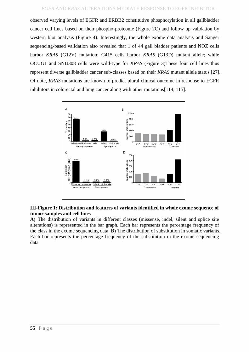
EGFR AND KRAS ALTERATIONS MEDIATE RESPONSE TO EGFR INHIBITOR
55 | P a g e
observed varying levels of EGFR and ERBB2 constitutive phosphorylation in all gallbladder
cancer cell lines based on their phospho-proteome (Figure 2C) and follow up validation by
western blot analysis (Figure 4). Interestingly, the whole exome data analysis and Sanger
sequencing-based validation also revealed that 1 of 44 gall bladder patients and NOZ cells
harbor KRAS (G12V) mutation; G415 cells harbor KRAS (G13D) mutant allele; while
OCUG1 and SNU308 cells were wild-type for KRAS (Figure 3)These four cell lines thus
represent diverse gallbladder cancer sub-classes based on their KRAS mutant allele status [27].
Of note, KRAS mutations are known to predict plural clinical outcome in response to EGFR
inhibitors in colorectal and lung cancer along with other mutations[114, 115].
III-Figure 1: Distribution and features of variants identified in whole exome sequence of
tumor samples and cell lines
A) The distribution of variants in different classes (missense, indel, silent and splice site
alterations) is represented in the bar graph. Each bar represents the percentage frequency of
the class in the exome sequencing data. B) The distribution of substitution in somatic variants.
Each bar represents the percentage frequency of the substitution in the exome sequencing
data
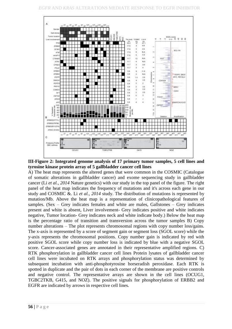
EGFR AND KRAS ALTERATIONS MEDIATE RESPONSE TO EGFR INHIBITOR
56 | P a g e
III-Figure 2: Integrated genome analysis of 17 primary tumor samples, 5 cell lines and
tyrosine kinase protein array of 5 gallbladder cancer cell lines
A) The heat map represents the altered genes that were common in the COSMIC (Catalogue
of somatic alterations in gallbladder cancer) and exome sequencing study in gallbladder
cancer (Li et al., 2014 Nature genetics) with our study in the top panel of the figure. The right
panel of the heat map indicates the frequency of mutations and it's across each gene in our
study and COSMIC &. Li et al., 2014 study. The distribution of mutations is represented by
mutation/Mb. Above the heat map is a representation of clinicopathological features of
samples. (Sex – Grey indicates females and white are males, Gallstones – Grey indicates
present and white is absent, Liver involvement- Grey indicates positive and white indicates
negative, Tumor location- Grey indicates neck and white indicate body.) Below the heat map
is the percentage ratio of transition and transversion across the tumor samples B) Copy
number alterations – The plot represents chromosomal regions with copy number loss/gains.
The x-axis is represented by a score of segment gain or segment loss (SGOL score) while the
y-axis represents the chromosomal positions. Copy number gain is indicated by red with
positive SGOL score while copy number loss is indicated by blue with a negative SGOL
score. Cancer-associated genes are annotated in their representative amplified regions. C)
RTK phosphorylation in gallbladder cancer cell lines Protein lysates of gallbladder cancer
cell lines were incubated on RTK arrays and phosphorylation status was determined by
subsequent incubation with anti-phosphotyrosine horseradish peroxidase. Each RTK is
spotted in duplicate and the pair of dots in each corner of the membrane are positive controls
and negative control. The representative arrays are shown in the cell lines (OCUG1,
TGBC2TKB, G415, and NOZ). The positive signals for phosphorylation of ERBB2 and
EGFR are indicated by arrows in respective cell lines.
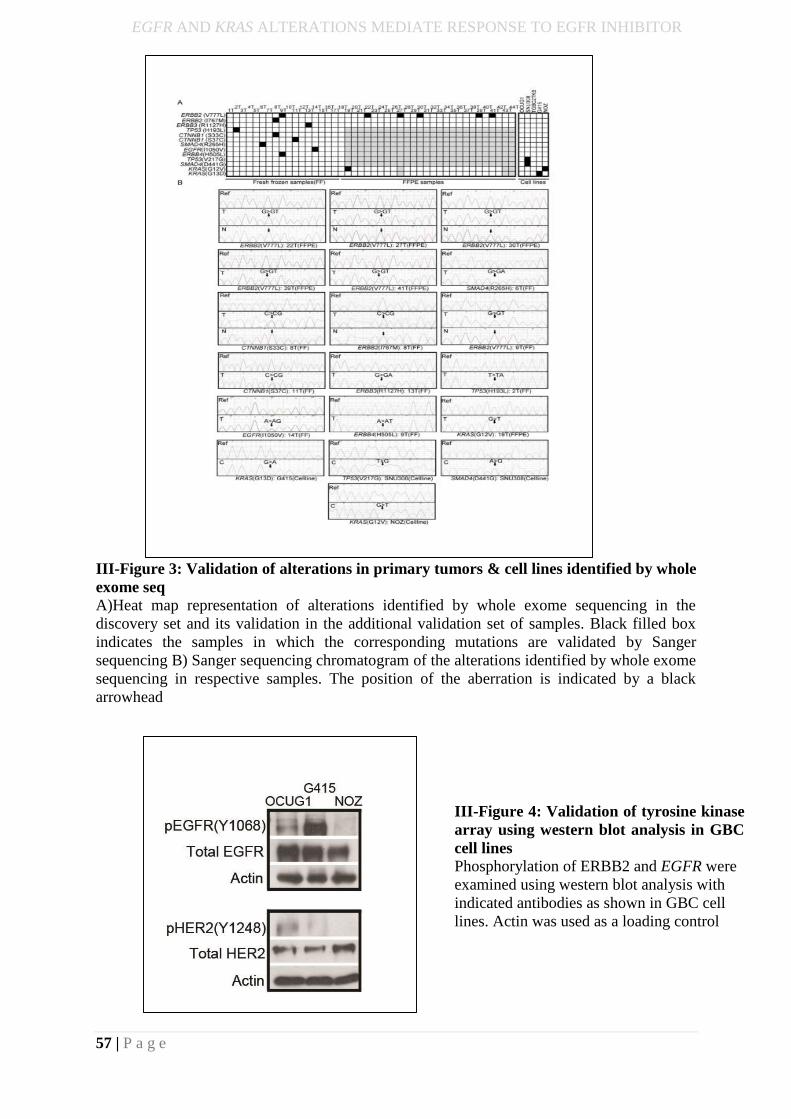
EGFR AND KRAS ALTERATIONS MEDIATE RESPONSE TO EGFR INHIBITOR
57 | P a g e
III-Figure 3: Validation of alterations in primary tumors & cell lines identified by whole
exome seq
A)Heat map representation of alterations identified by whole exome sequencing in the
discovery set and its validation in the additional validation set of samples. Black filled box
indicates the samples in which the corresponding mutations are validated by Sanger
sequencing B) Sanger sequencing chromatogram of the alterations identified by whole exome
sequencing in respective samples. The position of the aberration is indicated by a black
arrowhead
III-Figure 4: Validation of tyrosine kinase
array using western blot analysis in GBC
cell lines
Phosphorylation of ERBB2 and EGFR were
examined using western blot analysis with
indicated antibodies as shown in GBC cell
lines. Actin was used as a loading control
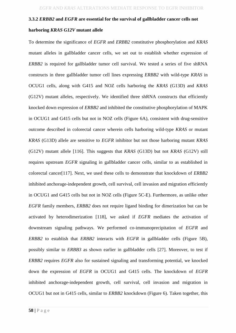
EGFR AND KRAS ALTERATIONS MEDIATE RESPONSE TO EGFR INHIBITOR
58 | P a g e
3.3.2 ERBB2 and EGFR are essential for the survival of gallbladder cancer cells not
harboring KRAS G12V mutant allele
To determine the significance of EGFR and ERBB2 constitutive phosphorylation and KRAS
mutant alleles in gallbladder cancer cells, we set out to establish whether expression of
ERBB2 is required for gallbladder tumor cell survival. We tested a series of five shRNA
constructs in three gallbladder tumor cell lines expressing ERBB2 with wild-type KRAS in
OCUG1 cells, along with G415 and NOZ cells harboring the KRAS (G13D) and KRAS
(G12V) mutant alleles, respectively. We identified three shRNA constructs that efficiently
knocked down expression of ERBB2 and inhibited the constitutive phosphorylation of MAPK
in OCUG1 and G415 cells but not in NOZ cells (Figure 6A), consistent with drug-sensitive
outcome described in colorectal cancer wherein cells harboring wild-type KRAS or mutant
KRAS (G13D) allele are sensitive to EGFR inhibitor but not those harboring mutant KRAS
(G12V) mutant allele [116]. This suggests that KRAS (G13D) but not KRAS (G12V) still
requires upstream EGFR signaling in gallbladder cancer cells, similar to as established in
colorectal cancer[117]. Next, we used these cells to demonstrate that knockdown of ERBB2
inhibited anchorage-independent growth, cell survival, cell invasion and migration efficiently
in OCUG1 and G415 cells but not in NOZ cells (Figure 5C-E). Furthermore, as unlike other
EGFR family members, ERBB2 does not require ligand binding for dimerization but can be
activated by heterodimerization [118], we asked if EGFR mediates the activation of
downstream signaling pathways. We performed co-immunoprecipitation of EGFR and
ERBB2 to establish that ERBB2 interacts with EGFR in gallbladder cells (Figure 5B),
possibly similar to ERBB3 as shown earlier in gallbladder cells [27]. Moreover, to test if
ERBB2 requires EGFR also for sustained signaling and transforming potential, we knocked
down the expression of EGFR in OCUG1 and G415 cells. The knockdown of EGFR
inhibited anchorage-independent growth, cell survival, cell invasion and migration in
OCUG1 but not in G415 cells, similar to ERBB2 knockdown (Figure 6). Taken together, this
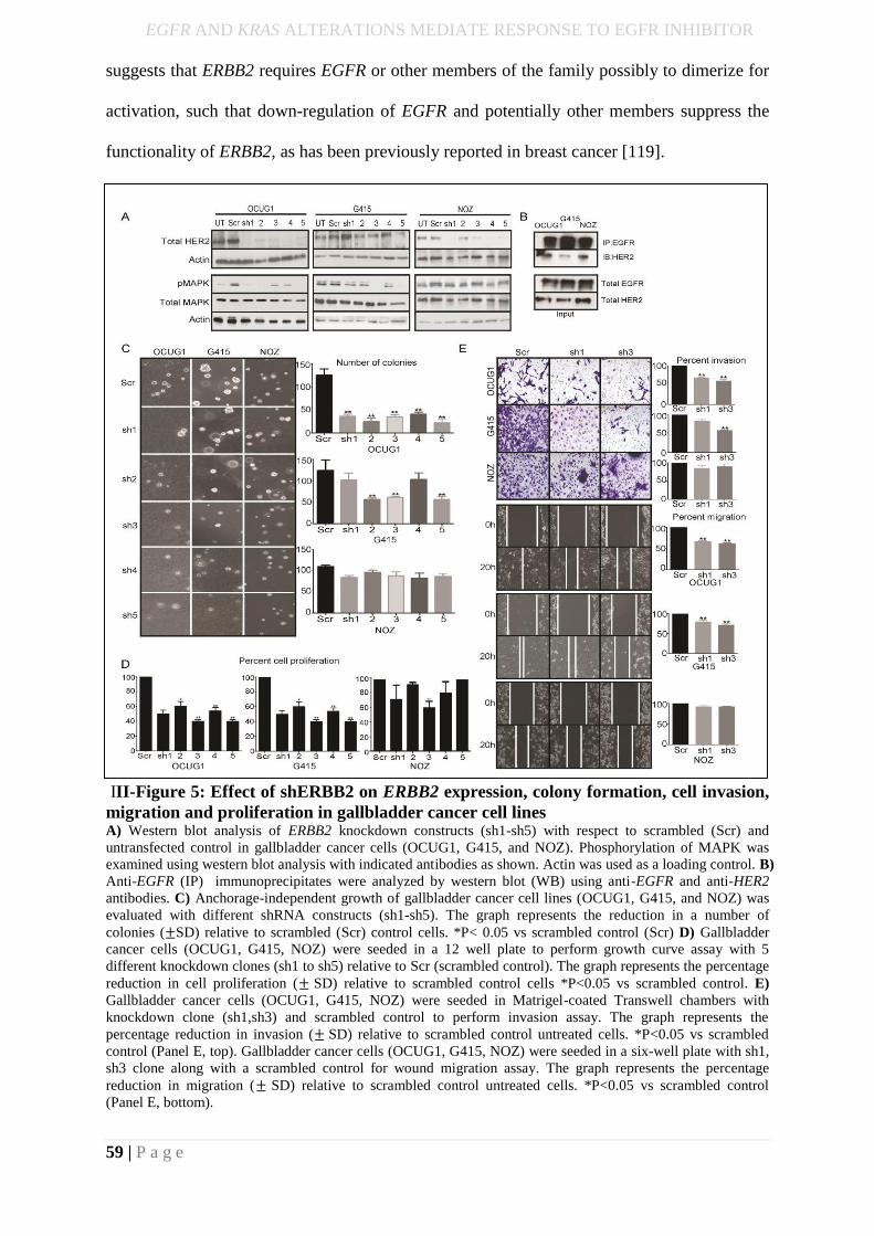
EGFR AND KRAS ALTERATIONS MEDIATE RESPONSE TO EGFR INHIBITOR
59 | P a g e
suggests that ERBB2 requires EGFR or other members of the family possibly to dimerize for
activation, such that down-regulation of EGFR and potentially other members suppress the
functionality of ERBB2, as has been previously reported in breast cancer [119].
III-Figure 5: Effect of shERBB2 on ERBB2 expression, colony formation, cell invasion,
migration and proliferation in gallbladder cancer cell lines A) Western blot analysis of ERBB2 knockdown constructs (sh1-sh5) with respect to scrambled (Scr) and
untransfected control in gallbladder cancer cells (OCUG1, G415, and NOZ). Phosphorylation of MAPK was
examined using western blot analysis with indicated antibodies as shown. Actin was used as a loading control. B)
Anti-EGFR (IP) immunoprecipitates were analyzed by western blot (WB) using anti-EGFR and anti-HER2
antibodies. C) Anchorage-independent growth of gallbladder cancer cell lines (OCUG1, G415, and NOZ) was
evaluated with different shRNA constructs (sh1-sh5). The graph represents the reduction in a number of
colonies (±SD) relative to scrambled (Scr) control cells. *P< 0.05 vs scrambled control (Scr) D) Gallbladder
cancer cells (OCUG1, G415, NOZ) were seeded in a 12 well plate to perform growth curve assay with 5
different knockdown clones (sh1 to sh5) relative to Scr (scrambled control). The graph represents the percentage
reduction in cell proliferation (± SD) relative to scrambled control cells *P<0.05 vs scrambled control. E)
Gallbladder cancer cells (OCUG1, G415, NOZ) were seeded in Matrigel-coated Transwell chambers with
knockdown clone (sh1,sh3) and scrambled control to perform invasion assay. The graph represents the
percentage reduction in invasion (± SD) relative to scrambled control untreated cells. *P<0.05 vs scrambled
control (Panel E, top). Gallbladder cancer cells (OCUG1, G415, NOZ) were seeded in a six-well plate with sh1,
sh3 clone along with a scrambled control for wound migration assay. The graph represents the percentage
reduction in migration (± SD) relative to scrambled control untreated cells. *P<0.05 vs scrambled control
(Panel E, bottom).
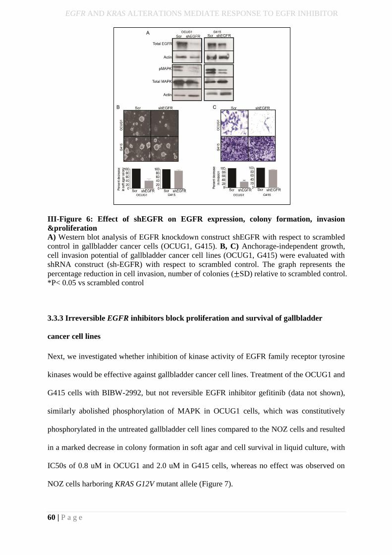
EGFR AND KRAS ALTERATIONS MEDIATE RESPONSE TO EGFR INHIBITOR
60 | P a g e
III-Figure 6: Effect of shEGFR on EGFR expression, colony formation, invasion
&proliferation
A) Western blot analysis of EGFR knockdown construct shEGFR with respect to scrambled
control in gallbladder cancer cells (OCUG1, G415). B, C) Anchorage-independent growth,
cell invasion potential of gallbladder cancer cell lines (OCUG1, G415) were evaluated with
shRNA construct (sh-EGFR) with respect to scrambled control. The graph represents the
percentage reduction in cell invasion, number of colonies (±SD) relative to scrambled control.
*P< 0.05 vs scrambled control
3.3.3 Irreversible EGFR inhibitors block proliferation and survival of gallbladder
cancer cell lines
Next, we investigated whether inhibition of kinase activity of EGFR family receptor tyrosine
kinases would be effective against gallbladder cancer cell lines. Treatment of the OCUG1 and
G415 cells with BIBW-2992, but not reversible EGFR inhibitor gefitinib (data not shown),
similarly abolished phosphorylation of MAPK in OCUG1 cells, which was constitutively
phosphorylated in the untreated gallbladder cell lines compared to the NOZ cells and resulted
in a marked decrease in colony formation in soft agar and cell survival in liquid culture, with
IC50s of 0.8 uM in OCUG1 and 2.0 uM in G415 cells, whereas no effect was observed on
NOZ cells harboring KRAS G12V mutant allele (Figure 7).
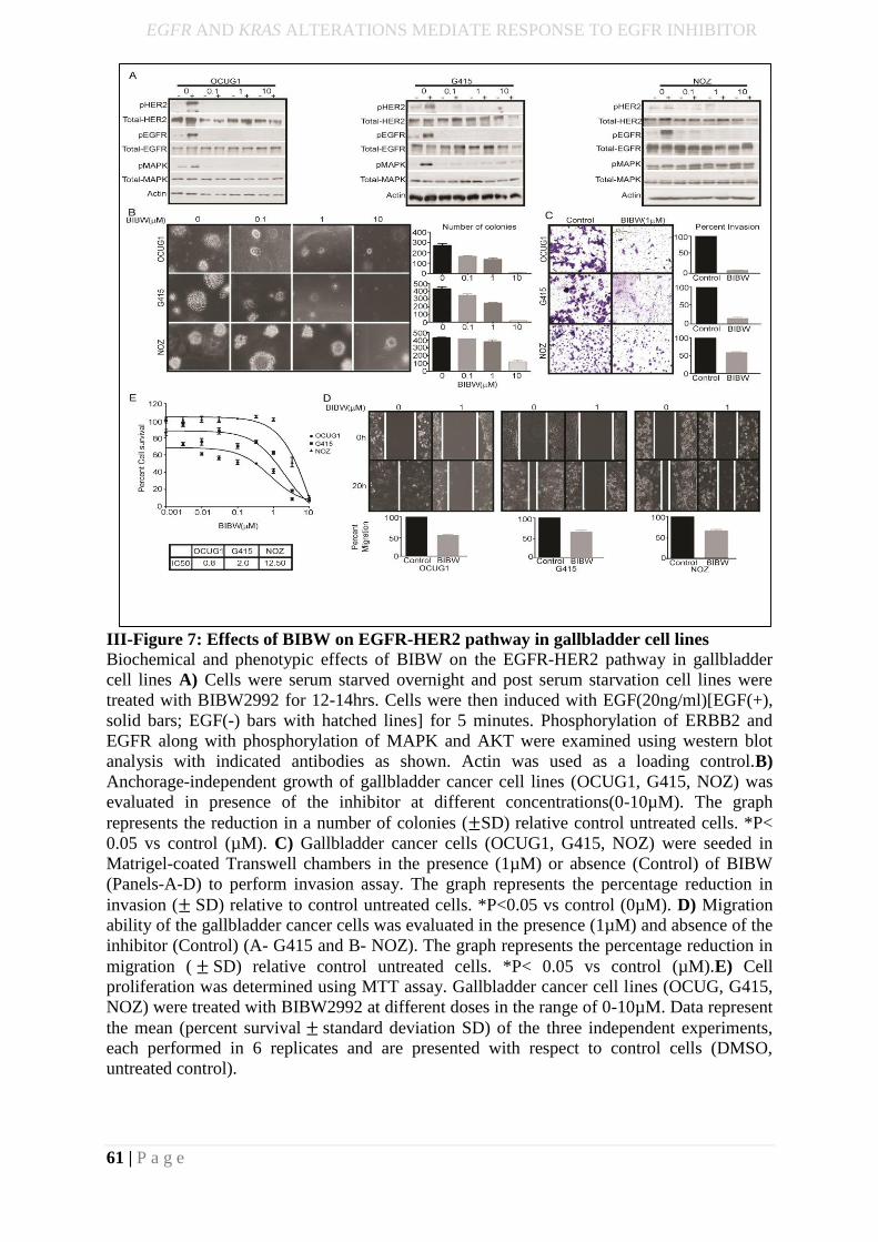
EGFR AND KRAS ALTERATIONS MEDIATE RESPONSE TO EGFR INHIBITOR
61 | P a g e
III-Figure 7: Effects of BIBW on EGFR-HER2 pathway in gallbladder cell lines
Biochemical and phenotypic effects of BIBW on the EGFR-HER2 pathway in gallbladder
cell lines A) Cells were serum starved overnight and post serum starvation cell lines were
treated with BIBW2992 for 12-14hrs. Cells were then induced with EGF(20ng/ml)[EGF(+),
solid bars; EGF(-) bars with hatched lines] for 5 minutes. Phosphorylation of ERBB2 and
EGFR along with phosphorylation of MAPK and AKT were examined using western blot
analysis with indicated antibodies as shown. Actin was used as a loading control.B)
Anchorage-independent growth of gallbladder cancer cell lines (OCUG1, G415, NOZ) was
evaluated in presence of the inhibitor at different concentrations(0-10µM). The graph
represents the reduction in a number of colonies (±SD) relative control untreated cells. *P<
0.05 vs control (µM). C) Gallbladder cancer cells (OCUG1, G415, NOZ) were seeded in
Matrigel-coated Transwell chambers in the presence (1µM) or absence (Control) of BIBW
(Panels-A-D) to perform invasion assay. The graph represents the percentage reduction in
invasion (± SD) relative to control untreated cells. *P<0.05 vs control (0µM). D) Migration
ability of the gallbladder cancer cells was evaluated in the presence (1µM) and absence of the
inhibitor (Control) (A- G415 and B- NOZ). The graph represents the percentage reduction in
migration ( ± SD) relative control untreated cells. *P< 0.05 vs control (µM).E) Cell
proliferation was determined using MTT assay. Gallbladder cancer cell lines (OCUG, G415,
NOZ) were treated with BIBW2992 at different doses in the range of 0-10µM. Data represent
the mean (percent survival ± standard deviation SD) of the three independent experiments,
each performed in 6 replicates and are presented with respect to control cells (DMSO,
untreated control).
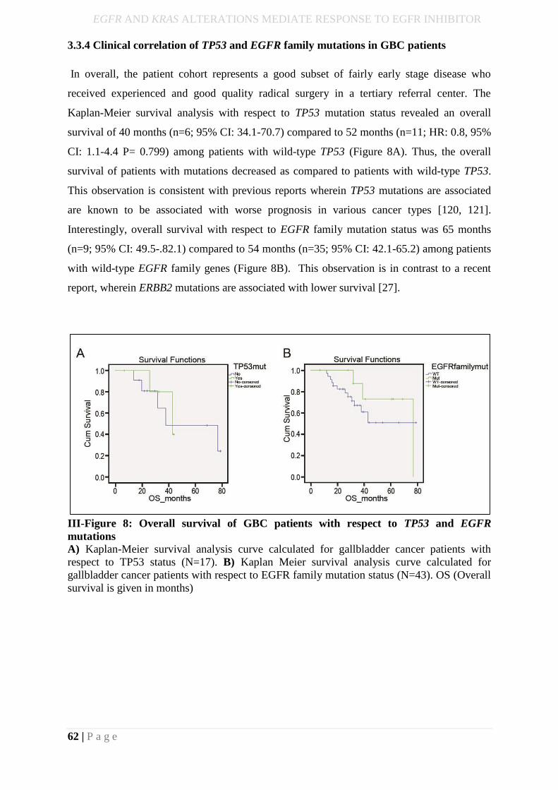
EGFR AND KRAS ALTERATIONS MEDIATE RESPONSE TO EGFR INHIBITOR
62 | P a g e
3.3.4 Clinical correlation of TP53 and EGFR family mutations in GBC patients
In overall, the patient cohort represents a good subset of fairly early stage disease who
received experienced and good quality radical surgery in a tertiary referral center. The
Kaplan-Meier survival analysis with respect to TP53 mutation status revealed an overall
survival of 40 months (n=6; 95% CI: 34.1-70.7) compared to 52 months (n=11; HR: 0.8, 95%
CI: 1.1-4.4 P= 0.799) among patients with wild-type TP53 (Figure 8A). Thus, the overall
survival of patients with mutations decreased as compared to patients with wild-type TP53.
This observation is consistent with previous reports wherein TP53 mutations are associated
are known to be associated with worse prognosis in various cancer types [120, 121].
Interestingly, overall survival with respect to EGFR family mutation status was 65 months
(n=9; 95% CI: 49.5-.82.1) compared to 54 months (n=35; 95% CI: 42.1-65.2) among patients
with wild-type EGFR family genes (Figure 8B). This observation is in contrast to a recent
report, wherein ERBB2 mutations are associated with lower survival [27].
III-Figure 8: Overall survival of GBC patients with respect to TP53 and EGFR
mutations
A) Kaplan-Meier survival analysis curve calculated for gallbladder cancer patients with
respect to TP53 status (N=17). B) Kaplan Meier survival analysis curve calculated for
gallbladder cancer patients with respect to EGFR family mutation status (N=43). OS (Overall
survival is given in months)

EGFR AND KRAS ALTERATIONS MEDIATE RESPONSE TO EGFR INHIBITOR
63 | P a g e
Discussion
This study represents the first genomic landscape of Indian gallbladder cancer that reveals
somatic mutations in TP53, ERBB2, ATM, AKAP11, SMAD4, and CTNNB1 similar to as
reported in Chinese and Caucasian population. Our mutation pattern analysis revealed an
enrichment for C>T transition followed by A>G transition, a signature which suggests an
underlying chronic inflammation leading to GC to AT polyclonal transition [122], as reported
earlier [27, 123]. We also observed significant somatic mutations in chromatin modifier
genes such as SF3B1, ATRX, CREBBP and EZH2 that have not been reported earlier in
gallbladder cancer, indicating potential therapeutic options among the Indian population.
Analyzing the potential effects of somatic alterations on survival of gallbladder cancer
patients, we observed a trend among patients with wild-type TP53 to survive longer than
patients with TP53 mutations, which is known to predict failure of chemotherapy in several
cancer types [124] and is consistent with previous reports observed in gallbladder cancer
[125].
Comparison of whole exome sequencing analysis of gallbladder cancer samples of Japanese
origin with our study suggests four genes (TP53, ARID2, EGFR and SMAD4) to be
significantly altered, with frequency as shown in III-Additional Supporting Table-1 [10]. Of
note, consistent with a recent report that described alterations in ERBB2 and ERBB3 at a
frequency of 9.8% and 11.8% respectively among Chinese gallbladder cancer [27], we found
recurrent activating ERBB2 (V777L) mutation in 6 of 44 gallbladder cancer samples with an
overall mutation frequency of 13%, in addition to ERBB3 (R112H) and EGFR (I1005V)
mutation occurring at 2%, each in our sample set. The (V777L) alteration has been shown to
be sensitive to lapatinib in biliary tract cancer, breast cancer cell lines and other isogenic
systems overexpressing the alteration [28, 111, 112]. Functional studies performed using
gallbladder cell lines establish that ERBB2 and EGFR are essential for the survival of
gallbladder cancer cells. Given that ERBB2 lacks the ligand binding domain, the co-
immunoprecipitation experiments suggest that ERBB2 dimerize with EGFR, and possibly
with other members, to constitutively activate the pathway. Interestingly, genetic or
pharmacological ablation of ERBB2 and EGFR function, using EGFR small-molecule
irreversible inhibitor BIBW-2992, diminishes the survival, anchorage-independent growth,
migration and invasion characteristics of gallbladder cancer cell lines, suggesting members of
the EGFR family as an effective therapeutic target.
Furthermore, while KRAS mutations in gallbladder cancer have been reported to occur at a
frequency from 3% to 30 % [126], some co-occurring with activating ERBB3 mutation [30],
we observed KRAS (G12V) and (G13D) mutation in 1 of 44 primary gallbladder tumors and
EGFR AND KRAS ALTERATIONS MEDIATE RESPONSE TO EGFR INHIBITOR
64 | P a g e
2 of 5 gallbladder cancer cell lines that are known to be associated with differential clinical
outcome in response to anti-EGFR therapy in colorectal cancer [127-129]. The biological
characteristics of KRAS mutation are known to vary by cancer types as those found in
pancreatic and non-small cell lung cancers are predominantly at codon 12, while in colorectal
and gallbladder mutations appears to be in codon 12 and codon 13 [130]. Moreover, the
clinical response among patients and studies with isogenic colon cell line indicate KRAS
(G13D) mutation as sensitive but (G12V) as resistant to anti-EGFR therapy suggesting codon
13 mutations are still dependent on inductive upstream EGFR signaling and exhibit weaker in
vitro transforming activity than codon 12 mutations [89, 128].
In summary, besides suggesting adoption of anti-EGFR therapy as a therapeutic option in
gallbladder cancer based on ERBB2 alteration, we present the first evidence that presence of
KRAS (G12V) but not KRAS (G13D) mutation may preclude such patients to respond to the
treatment, similar to the clinical algorithm commonly practiced based on EGFR alteration in
colorectal cancer. Due to smaller sample size, this study remains underpowered to reach
statistical significance for patients harboring alterations in ERBB2 mutations. However, due
to a low prevalence rate of the disease, target accrual in clinical trials has been a bottleneck in
gallbladder cancer. This study thus forms the basis to test the efficacy of ERBB2 inhibitors in
gallbladder cancer and would help inform design a randomized clinical trial by considering
the inclusion of gallbladder patients under basket clinical trials such as the NCI–Molecular
Analysis for Therapy Choice (NCI-MATCH) trial that are genomically matched [131].
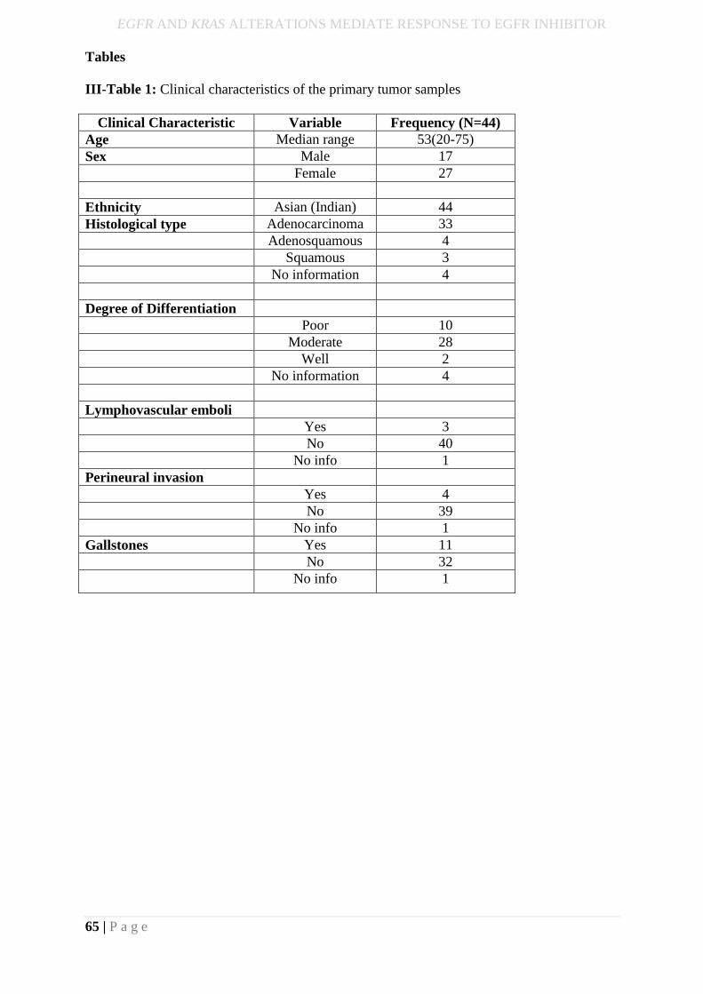
EGFR AND KRAS ALTERATIONS MEDIATE RESPONSE TO EGFR INHIBITOR
65 | P a g e
Tables
III-Table 1: Clinical characteristics of the primary tumor samples
Clinical Characteristic Variable Frequency (N=44)
Age Median range 53(20-75)
Sex Male 17
Female 27
Ethnicity Asian (Indian) 44
Histological type Adenocarcinoma 33
Adenosquamous 4
Squamous 3
No information 4
Degree of Differentiation
Poor 10
Moderate 28
Well 2
No information 4
Lymphovascular emboli
Yes 3
No 40
No info 1
Perineural invasion
Yes 4
No 39
No info 1
Gallstones Yes 11
No 32
No info 1
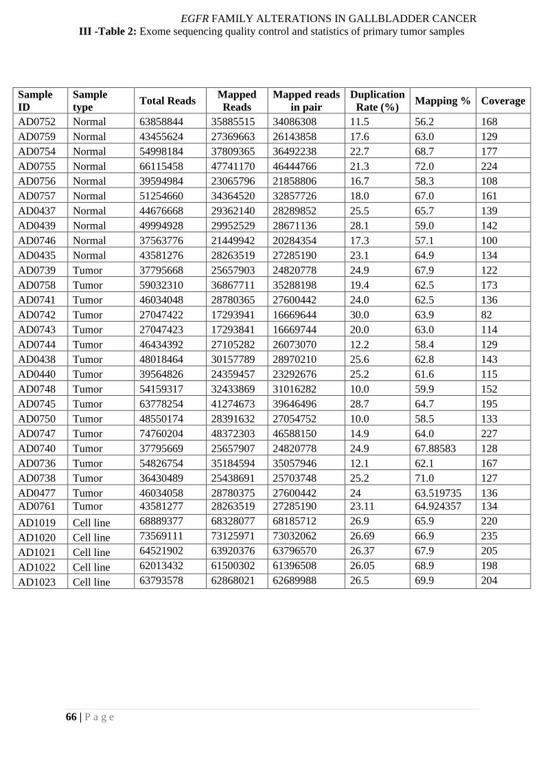
EGFR FAMILY ALTERATIONS IN GALLBLADDER CANCER
66 | P a g e
III -Table 2: Exome sequencing quality control and statistics of primary tumor samples
Sample
ID
Sample
type Total Reads
Mapped
Reads
Mapped reads
in pair
Duplication
Rate (%) Mapping % Coverage
AD0752 Normal 63858844 35885515 34086308 11.5 56.2 168
AD0759 Normal 43455624 27369663 26143858 17.6 63.0 129
AD0754 Normal 54998184 37809365 36492238 22.7 68.7 177
AD0755 Normal 66115458 47741170 46444766 21.3 72.0 224
AD0756 Normal 39594984 23065796 21858806 16.7 58.3 108
AD0757 Normal 51254660 34364520 32857726 18.0 67.0 161
AD0437 Normal 44676668 29362140 28289852 25.5 65.7 139
AD0439 Normal 49994928 29952529 28671136 28.1 59.0 142
AD0746 Normal 37563776 21449942 20284354 17.3 57.1 100
AD0435 Normal 43581276 28263519 27285190 23.1 64.9 134
AD0739 Tumor 37795668 25657903 24820778 24.9 67.9 122
AD0758 Tumor 59032310 36867711 35288198 19.4 62.5 173
AD0741 Tumor 46034048 28780365 27600442 24.0 62.5 136
AD0742 Tumor 27047422 17293941 16669644 30.0 63.9 82
AD0743 Tumor 27047423 17293841 16669744 20.0 63.0 114
AD0744 Tumor 46434392 27105282 26073070 12.2 58.4 129
AD0438 Tumor 48018464 30157789 28970210 25.6 62.8 143
AD0440 Tumor 39564826 24359457 23292676 25.2 61.6 115
AD0748 Tumor 54159317 32433869 31016282 10.0 59.9 152
AD0745 Tumor 63778254 41274673 39646496 28.7 64.7 195
AD0750 Tumor 48550174 28391632 27054752 10.0 58.5 133
AD0747 Tumor 74760204 48372303 46588150 14.9 64.0 227
AD0740 Tumor 37795669 25657907 24820778 24.9 67.88583 128
AD0736 Tumor 54826754 35184594 35057946 12.1 62.1 167
AD0738 Tumor 36430489 25438691 25703748 25.2 71.0 127
AD0477 Tumor 46034058 28780375 27600442 24 63.519735 136
AD0761 Tumor 43581277 28263519 27285190 23.11 64.924357 134
AD1019 Cell line 68889377 68328077 68185712 26.9 65.9 220
AD1020 Cell line 73569111 73125971 73032062 26.69 66.9 235
AD1021 Cell line 64521902 63920376 63796570 26.37 67.9 205
AD1022 Cell line 62013432 61500302 61396508 26.05 68.9 198
AD1023 Cell line 63793578 62868021 62689988 26.5 69.9 204
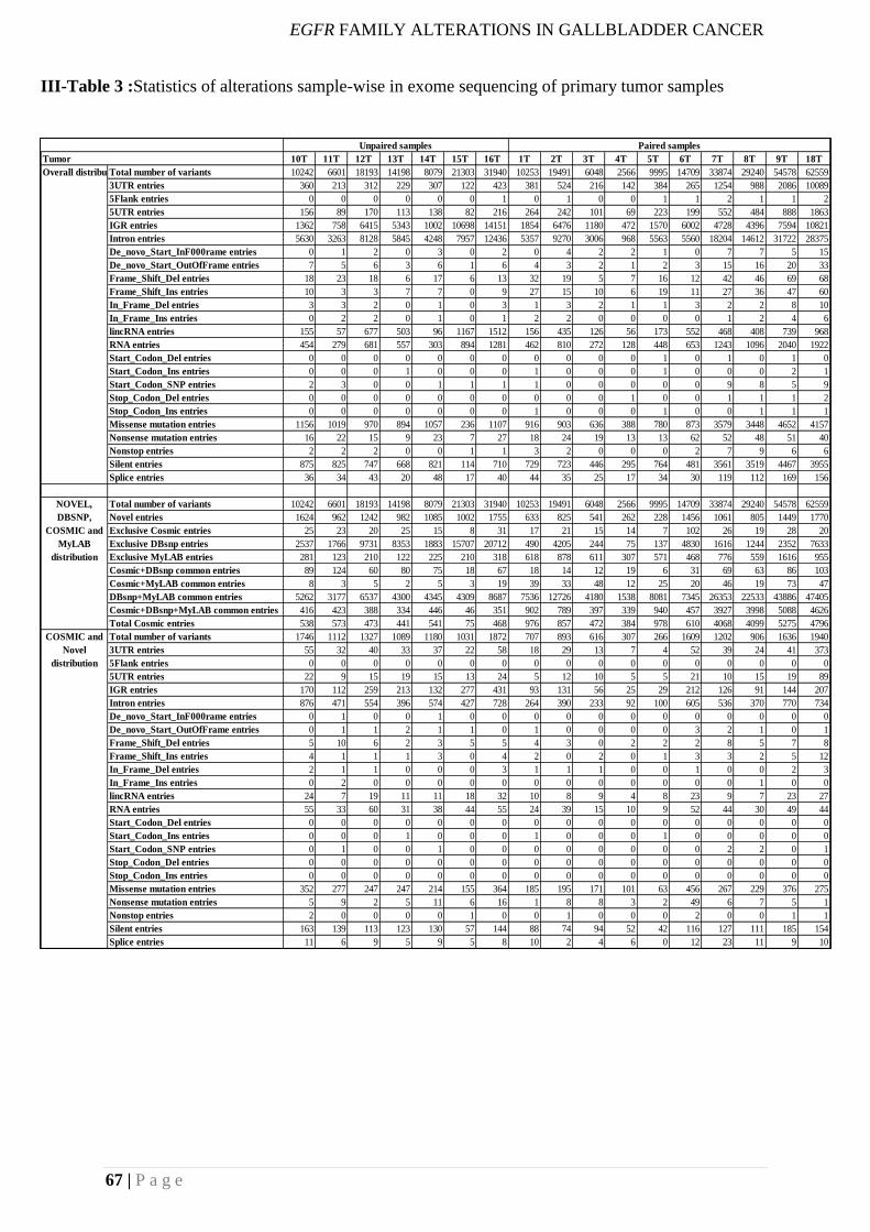
EGFR FAMILY ALTERATIONS IN GALLBLADDER CANCER
67 | P a g e
10T 11T 12T 13T 14T 15T 16T 1T 2T 3T 4T 5T 6T 7T 8T 9T 18T
Total number of variants 10242 6601 18193 14198 8079 21303 31940 10253 19491 6048 2566 9995 14709 33874 29240 54578 62559
3UTR entries 360 213 312 229 307 122 423 381 524 216 142 384 265 1254 988 2086 10089
5Flank entries 0 0 0 0 0 0 1 0 1 0 0 1 1 2 1 1 2
5UTR entries 156 89 170 113 138 82 216 264 242 101 69 223 199 552 484 888 1863
IGR entries 1362 758 6415 5343 1002 10698 14151 1854 6476 1180 472 1570 6002 4728 4396 7594 10821
Intron entries 5630 3263 8128 5845 4248 7957 12436 5357 9270 3006 968 5563 5560 18204 14612 31722 28375
De_novo_Start_InF000rame entries 0 1 2 0 3 0 2 0 4 2 2 1 0 7 7 5 15
De_novo_Start_OutOfFrame entries 7 5 6 3 6 1 6 4 3 2 1 2 3 15 16 20 33
Frame_Shift_Del entries 18 23 18 6 17 6 13 32 19 5 7 16 12 42 46 69 68
Frame_Shift_Ins entries 10 3 3 7 7 0 9 27 15 10 6 19 11 27 36 47 60
In_Frame_Del entries 3 3 2 0 1 0 3 1 3 2 1 1 3 2 2 8 10
In_Frame_Ins entries 0 2 2 0 1 0 1 2 2 0 0 0 0 1 2 4 6
lincRNA entries 155 57 677 503 96 1167 1512 156 435 126 56 173 552 468 408 739 968
RNA entries 454 279 681 557 303 894 1281 462 810 272 128 448 653 1243 1096 2040 1922
Start_Codon_Del entries 0 0 0 0 0 0 0 0 0 0 0 1 0 1 0 1 0
Start_Codon_Ins entries 0 0 0 1 0 0 0 1 0 0 0 1 0 0 0 2 1
Start_Codon_SNP entries 2 3 0 0 1 1 1 1 0 0 0 0 0 9 8 5 9
Stop_Codon_Del entries 0 0 0 0 0 0 0 0 0 0 1 0 0 1 1 1 2
Stop_Codon_Ins entries 0 0 0 0 0 0 0 1 0 0 0 1 0 0 1 1 1
Missense mutation entries 1156 1019 970 894 1057 236 1107 916 903 636 388 780 873 3579 3448 4652 4157
Nonsense mutation entries 16 22 15 9 23 7 27 18 24 19 13 13 62 52 48 51 40
Nonstop entries 2 2 2 0 0 1 1 3 2 0 0 0 2 7 9 6 6
Silent entries 875 825 747 668 821 114 710 729 723 446 295 764 481 3561 3519 4467 3955
Splice entries 36 34 43 20 48 17 40 44 35 25 17 34 30 119 112 169 156
Total number of variants 10242 6601 18193 14198 8079 21303 31940 10253 19491 6048 2566 9995 14709 33874 29240 54578 62559
Novel entries 1624 962 1242 982 1085 1002 1755 633 825 541 262 228 1456 1061 805 1449 1770
Exclusive Cosmic entries 25 23 20 25 15 8 31 17 21 15 14 7 102 26 19 28 20
Exclusive DBsnp entries 2537 1766 9731 8353 1883 15707 20712 490 4205 244 75 137 4830 1616 1244 2352 7633
Exclusive MyLAB entries 281 123 210 122 225 210 318 618 878 611 307 571 468 776 559 1616 955
Cosmic+DBsnp common entries 89 124 60 80 75 18 67 18 14 12 19 6 31 69 63 86 103
Cosmic+MyLAB common entries 8 3 5 2 5 3 19 39 33 48 12 25 20 46 19 73 47
DBsnp+MyLAB common entries 5262 3177 6537 4300 4345 4309 8687 7536 12726 4180 1538 8081 7345 26353 22533 43886 47405
Cosmic+DBsnp+MyLAB common entries 416 423 388 334 446 46 351 902 789 397 339 940 457 3927 3998 5088 4626
Total Cosmic entries 538 573 473 441 541 75 468 976 857 472 384 978 610 4068 4099 5275 4796
Total number of variants 1746 1112 1327 1089 1180 1031 1872 707 893 616 307 266 1609 1202 906 1636 1940
3UTR entries 55 32 40 33 37 22 58 18 29 13 7 4 52 39 24 41 373
5Flank entries 0 0 0 0 0 0 0 0 0 0 0 0 0 0 0 0 0
5UTR entries 22 9 15 19 15 13 24 5 12 10 5 5 21 10 15 19 89
IGR entries 170 112 259 213 132 277 431 93 131 56 25 29 212 126 91 144 207
Intron entries 876 471 554 396 574 427 728 264 390 233 92 100 605 536 370 770 734
De_novo_Start_InF000rame entries 0 1 0 0 1 0 0 0 0 0 0 0 0 0 0 0 0
De_novo_Start_OutOfFrame entries 0 1 1 2 1 1 0 1 0 0 0 0 3 2 1 0 1
Frame_Shift_Del entries 5 10 6 2 3 5 5 4 3 0 2 2 2 8 5 7 8
Frame_Shift_Ins entries 4 1 1 1 3 0 4 2 0 2 0 1 3 3 2 5 12
In_Frame_Del entries 2 1 1 0 0 0 3 1 1 1 0 0 1 0 0 2 3
In_Frame_Ins entries 0 2 0 0 0 0 0 0 0 0 0 0 0 0 1 0 0
lincRNA entries 24 7 19 11 11 18 32 10 8 9 4 8 23 9 7 23 27
RNA entries 55 33 60 31 38 44 55 24 39 15 10 9 52 44 30 49 44
Start_Codon_Del entries 0 0 0 0 0 0 0 0 0 0 0 0 0 0 0 0 0
Start_Codon_Ins entries 0 0 0 1 0 0 0 1 0 0 0 1 0 0 0 0 0
Start_Codon_SNP entries 0 1 0 0 1 0 0 0 0 0 0 0 0 2 2 0 1
Stop_Codon_Del entries 0 0 0 0 0 0 0 0 0 0 0 0 0 0 0 0 0
Stop_Codon_Ins entries 0 0 0 0 0 0 0 0 0 0 0 0 0 0 0 0 0
Missense mutation entries 352 277 247 247 214 155 364 185 195 171 101 63 456 267 229 376 275
Nonsense mutation entries 5 9 2 5 11 6 16 1 8 8 3 2 49 6 7 5 1
Nonstop entries 2 0 0 0 0 1 0 0 1 0 0 0 2 0 0 1 1
Silent entries 163 139 113 123 130 57 144 88 74 94 52 42 116 127 111 185 154
Splice entries 11 6 9 5 9 5 8 10 2 4 6 0 12 23 11 9 10
Paired samples
Overall distribution
NOVEL,
DBSNP,
COSMIC and
MyLAB
distribution
COSMIC and
Novel
distribution
Tumor
Unpaired samples
III-Table 3 :Statistics of alterations sample-wise in exome sequencing of primary tumor samples
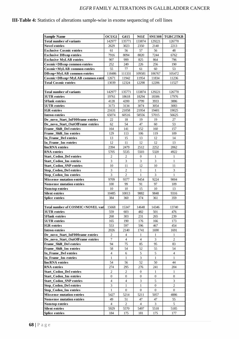
EGFR FAMILY ALTERATIONS IN GALLBLADDER CANCER
68 | P a g e
Sample Name OCUG1 G415 NOZ SNU308 TGBC2TKB
Total number of variants 142977 135771 133874 129221 126770
Novel entries 2629 3023 2350 2140 2213
Exclusive Cosmic entries 61 56 57 56 48
Exclusive DBsnp entries 7916 8094 8820 7244 6762
Exclusive MyLAB entries 907 999 825 864 796
Cosmic+DBsnp common entries 252 249 226 256 190
Cosmic+MyLAB common entries 55 77 61 60 53
DBsnp+MyLAB common entries 118486 111331 109581 106767 105472
Cosmic+DBsnp+MyLAB common entries12671 11942 11954 11834 11236
Total Cosmic entries 13039 12324 12298 12206 11527
Total number of variants 142977 135771 133874 129221 126770
3UTR entries 19761 18618 18294 18306 17976
5Flank entries 4128 4200 3799 3933 3886
5UTR entries 3173 3134 3074 3054 3083
IGR entries 21631 21058 21954 19401 19025
Intron entries 65074 60516 58556 57015 56625
De_novo_Start_InF000rame entries 22 18 19 19 27
De_novo_Start_OutOfFrame entries 62 54 47 60 53
Frame_Shift_Del entries 164 141 152 160 157
Frame_Shift_Ins entries 129 113 106 119 109
In_Frame_Del entries 13 15 13 12 14
In_Frame_Ins entries 12 11 12 12 13
lincRNA entries 2394 2479 2512 2252 2062
RNA entries 5705 5535 5503 5320 4922
Start_Codon_Del entries 2 2 0 1 1
Start_Codon_Ins entries 3 3 3 3 1
Start_Codon_SNP entries 10 11 12 10 11
Stop_Codon_Del entries 3 2 1 1 3
Stop_Codon_Ins entries 3 2 1 3 1
Missense mutation entries 9709 9377 9454 9224 9004
Nonsense mutation entries 100 99 91 97 109
Nonstop entries 10 10 15 10 13
Silent entries 10485 10013 9882 9848 9316
Splice entries 384 360 374 361 359
Total number of COSMIC+NOVEL variants15668 15347 14648 14346 13740
3UTR entries 559 603 482 501 476
5Flank entries 268 303 231 265 239
5UTR entries 165 190 176 166 173
IGR entries 553 597 596 467 454
Intron entries 2026 2140 1741 1690 1691
De_novo_Start_InF000rame entries 2 4 1 1 1
De_novo_Start_OutOfFrame entries 7 4 4 3 2
Frame_Shift_Del entries 94 75 85 95 83
Frame_Shift_Ins entries 58 54 52 55 54
In_Frame_Del entries 4 6 5 5 4
In_Frame_Ins entries 1 1 3 1 1
lincRNA entries 54 56 52 50 44
RNA entries 274 295 276 241 204
Start_Codon_Del entries 2 2 0 1 1
Start_Codon_Ins entries 0 0 0 0 0
Start_Codon_SNP entries 4 4 3 5 3
Stop_Codon_Del entries 3 1 1 0 2
Stop_Codon_Ins entries 1 0 0 0 0
Missense mutation entries 5427 5214 5211 5057 4886
Nonsense mutation entries 49 51 47 47 55
Nonstop entries 4 2 4 3 5
Silent entries 5929 5570 5497 5518 5185
Splice entries 184 175 181 175 177
III-Table 4: Statistics of alterations sample-wise in exome sequencing of cell lines
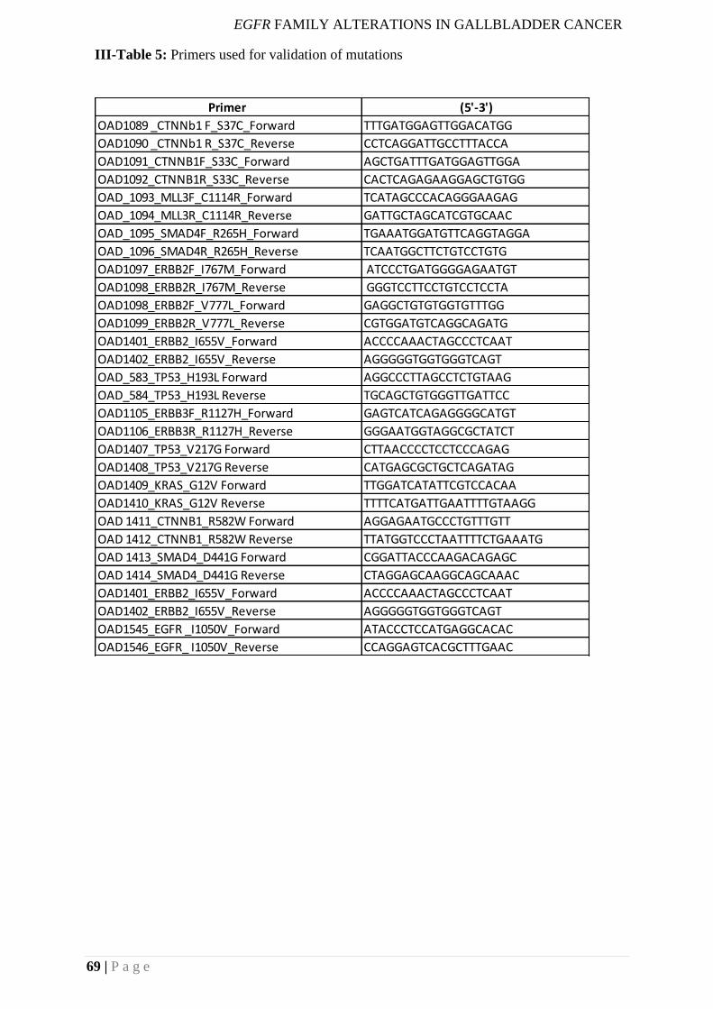
EGFR FAMILY ALTERATIONS IN GALLBLADDER CANCER
69 | P a g e
Primer (5'-3')
OAD1089 _CTNNb1 F_S37C_Forward TTTGATGGAGTTGGACATGG
OAD1090 _CTNNb1 R_S37C_Reverse CCTCAGGATTGCCTTTACCA
OAD1091_CTNNB1F_S33C_Forward AGCTGATTTGATGGAGTTGGA
OAD1092_CTNNB1R_S33C_Reverse CACTCAGAGAAGGAGCTGTGG
OAD_1093_MLL3F_C1114R_Forward TCATAGCCCACAGGGAAGAG
OAD_1094_MLL3R_C1114R_Reverse GATTGCTAGCATCGTGCAAC
OAD_1095_SMAD4F_R265H_Forward TGAAATGGATGTTCAGGTAGGA
OAD_1096_SMAD4R_R265H_Reverse TCAATGGCTTCTGTCCTGTG
OAD1097_ERBB2F_I767M_Forward ATCCCTGATGGGGAGAATGT
OAD1098_ERBB2R_I767M_Reverse GGGTCCTTCCTGTCCTCCTA
OAD1098_ERBB2F_V777L_Forward GAGGCTGTGTGGTGTTTGG
OAD1099_ERBB2R_V777L_Reverse CGTGGATGTCAGGCAGATG
OAD1401_ERBB2_I655V_Forward ACCCCAAACTAGCCCTCAAT
OAD1402_ERBB2_I655V_Reverse AGGGGGTGGTGGGTCAGT
OAD_583_TP53_H193L Forward AGGCCCTTAGCCTCTGTAAG
OAD_584_TP53_H193L Reverse TGCAGCTGTGGGTTGATTCC
OAD1105_ERBB3F_R1127H_Forward GAGTCATCAGAGGGGCATGT
OAD1106_ERBB3R_R1127H_Reverse GGGAATGGTAGGCGCTATCT
OAD1407_TP53_V217G Forward CTTAACCCCTCCTCCCAGAG
OAD1408_TP53_V217G Reverse CATGAGCGCTGCTCAGATAG
OAD1409_KRAS_G12V Forward TTGGATCATATTCGTCCACAA
OAD1410_KRAS_G12V Reverse TTTTCATGATTGAATTTTGTAAGG
OAD 1411_CTNNB1_R582W Forward AGGAGAATGCCCTGTTTGTT
OAD 1412_CTNNB1_R582W Reverse TTATGGTCCCTAATTTTCTGAAATG
OAD 1413_SMAD4_D441G Forward CGGATTACCCAAGACAGAGC
OAD 1414_SMAD4_D441G Reverse CTAGGAGCAAGGCAGCAAAC
OAD1401_ERBB2_I655V_Forward ACCCCAAACTAGCCCTCAAT
OAD1402_ERBB2_I655V_Reverse AGGGGGTGGTGGGTCAGT
OAD1545_EGFR _I1050V_Forward ATACCCTCCATGAGGCACAC
OAD1546_EGFR_ I1050V_Reverse CCAGGAGTCACGCTTTGAAC
III-Table 5: Primers used for validation of mutations
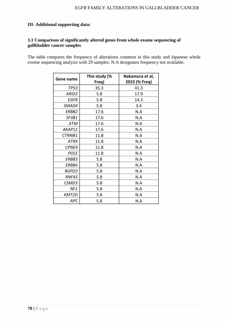
EGFR FAMILY ALTERATIONS IN GALLBLADDER CANCER
70 | P a g e
III- Additional supporting data:
3.1 Comparison of significantly altered genes from whole exome sequencing of
gallbladder cancer samples
The table compares the frequency of alterations common in this study and Japanese whole
exome sequencing analysis with 29 samples. N.A designates frequency not available.
Gene name This study (%
Freq) Nakamura et al,
2015 (% Freq)
TP53 35.3 41.3
ARID2 5.8 17.9
EGFR 5.8 14.3
SMAD4 5.8 3.4
ERBB2 17.6 N.A
SF3B1 17.6 N.A
ATM 17.6 N.A
AKAP11 17.6 N.A
CTNNB1 11.8 N.A
ATRX 11.8 N.A
CPNE4 11.8 N.A
POLE 11.8 N.A
ERBB3 5.8 N.A
ERBB4 5.8 N.A
RGPD3 5.8 N.A
RNF43 5.8 N.A
CSMD3 5.8 N.A
NF1 5.8 N.A
KMT2D 5.8 N.A
APC 5.8 N.A
SUMMARY AND CONCLUSION
71 | P a g e
4.CHAPTER 4: SUMMARY AND CONCLUSION
Gallbladder cancer is an aggressive disease with poor prognosis. The disease has been least
studied from the functional genomics perspective due to the rarity of the disease worldwide.
The cancer is treated mostly by surgery without lymph node involvement[49]. Currently,
gem-cis(gemcitabine-cisplatin) or gem-ox(gemcitabine oxaliplatin) based treatment is given
to advanced cases without any targeted therapy which offers a very modest survival
benefit[12]. Few reports suggest the efficacy of the combination of cetuximab and
gemcitabine in gallbladder cancer, thus demonstrating some promise for targeted therapy in
gallbladder cancer[41]. Hence, there is an unmet need for better therapeutically relevant
targets in GBC especially in a country like India, where the cancer is highly prevalent in the
northern part of the country. Another aspect of gallbladder cancer is an association of
infections with gallbladder cancer. Epidemiological studies suggest a strong association of
typhoidal Salmonella infections with gallbladder cancer, however, the other group of
Salmonella - non-typhoidal Salmonella was not associated with gallbladder cancer [51, 69].
These reports suggest an only association, no causal relationship has been established. It has
been proposed that gallbladder infections with Salmonella trigger host manipulation
pathways under predisposed of condition thus leading to cancer [19]. These factors led to the
origin of my thesis. The major focus of my thesis is to identify therapeutically relevant
alterations in gallbladder cancer and understanding the association of infections in
gallbladder cancer using integrated approaches.
4.1 Mutant KRAS predicts response to EGFR inhibitors in gallbladder cancer cell lines.
Mutations in KRAS are predictive biomarkers of anti-EGFR therapy in colorectal cancer [88].
Similarly, KRAS alterations are also known in gallbladder cancer[132]. However, not much
has been studied neither at the genomic or functional level to understand the overall biology
of gallbladder cancer. The disease has been associated with worse outcomes. There are very
few genome-wide studies regarding gallbladder cancer from the Chinese and Caucasian[26,
SUMMARY AND CONCLUSION
72 | P a g e
27] and from India, there are only candidate gene-based studies[18]. Hence, using a sensitive
methodology such as whole exome sequencing, I set out to identify somatic aberrations
(mutations and copy number) in primary tumors and tyrosine kinase proteome profiler to
check the expression of activated tyrosine kinases in gallbladder cancer cell lines.
Using the whole exome sequencing approach, I identified alterations in known cancer-
associated genes such as TP53, ERBB2, SMAD4, and β-catenin which are already known in
the literature[22] for gallbladder cancer. Further, I specifically identified recurrent, actionable
ERBB2 alterations as well as copy number changes in EGFR which we show to be sensitive
to EGFR family inhibitor. Using proteome profiler in cell lines, I observed higher
phosphorylation of EGFR as compared to other tyrosine kinases in gallbladder cancer cell
lines. Also, using biochemical approaches, I further observed a differential response of
gallbladder cancer cell lines to the EGFR family inhibitor, which was primarily based on the
presence of different KRAS mutations (codon 12 and codon 13 alterations). Similar
observations in colorectal cancer have been reported wherein patients with KRAS (G13D)
mutations respond better to anti-EGFR therapy than KRAS (G12V) mutations[133]. These
findings in gallbladder cancer cell lines may have a clinical relevance in gallbladder cancer
and allow patient stratification and could preclude gallbladder cancer patients from anti-
EGFR therapy on the basis of KRAS mutational status. Our findings may change the current
treatment regimen in gallbladder cancer and introduce the scope of the genome-guided
precision medicine in the clinics for this disease.
4.2 Association of non-typhoidal Salmonella with gallbladder cancer
Epidemiological reports suggest a strong association of chronic typhoidal Salmonella
infections with gallbladder cancer[51]. However, non-typhoidal Salmonella infections have
been associated with systemic illness such as gastrointestinal infections[69]. Using a
modified HPVDetector tool for computational subtraction, I identified the presence of non-
typhoidal DNA sequences in gallbladder patients using whole exome sequencing data, which
may be associated with disease progression. Further, I observed co-occurrence of TP53
SUMMARY AND CONCLUSION
73 | P a g e
alteration with Salmonella infections in gallbladder cancer patients. Recent reports suggest
Salmonella infections induce manipulation of host metabolic pathways in TP53 mutant and
c-Myc amplified predisposed mice, murine gallbladder organoids, and fibroblasts[19].
Infections with typhoidal and the non-typhoidal Salmonella provides the necessary
inflammatory stimulus for gallbladder cancer[123]. These findings expand the current
understanding of infections of Salmonella with gallbladder cancer. Also, the findings may
suggest better management of the disease by detection of Salmonella using molecular
approaches.
In overall, my thesis puts forward the first landscape of somatic alterations in Indian
gallbladder cancer genome and leads to the identification of non-
typhoidal Salmonella species along with co-occurrence of TP53 alterations that could
potentially aid in better management of gallbladder cancer. More importantly, my
study implicates ERBB2 as a novel therapeutic target in gallbladder cancer and puts
forward the first evidence that the presence of KRAS G12V but not KRAS G13D mutation
may preclude patients to respond to anti-EGFR treatment in gallbladder cancer, as reported in
colorectal cancer.
BIBILIOGRAPHY
74 | P a g e
CHAPTER 5: BIBLIOGRAPHY
1. Wistuba, II and A.F. Gazdar, Gallbladder cancer: lessons from a rare tumour. Nat Rev Cancer, 2004. 4(9): p. 695-706.
2. Luo, J., N.L. Solimini, and S.J. Elledge, Principles of cancer therapy: oncogene and non-oncogene addiction. Cell, 2009. 136(5): p. 823-37.
3. Macconaill, L.E. and L.A. Garraway, Clinical implications of the cancer genome. J Clin Oncol, 2010. 28(35): p. 5219-28.
4. Mortality, G.B.D. and C. Causes of Death, Global, regional, and national age-sex specific all-cause and cause-specific mortality for 240 causes of death, 1990-2013: a systematic analysis for the Global Burden of Disease Study 2013. Lancet, 2015. 385(9963): p. 117-71.
5. Chin, L., et al., Making sense of cancer genomic data. Genes Dev, 2011. 25(6): p. 534-55. 6. Mardis, E.R. and R.K. Wilson, Cancer genome sequencing: a review. Hum Mol Genet, 2009.
18(R2): p. R163-8. 7. Garraway, L.A. and E.S. Lander, Lessons from the cancer genome. Cell, 2013. 153(1): p. 17-37. 8. International Cancer Genome, C., et al., International network of cancer genome projects.
Nature, 2010. 464(7291): p. 993-8. 9. Sicklick, J.K., et al., Genomics of gallbladder cancer: the case for biomarker-driven clinical
trial design. Cancer Metastasis Rev, 2016. 35(2): p. 263-75. 10. Nakamura, H., et al., Genomic spectra of biliary tract cancer. Nat Genet, 2015. 47(9): p. 1003-
10. 11. Shaffer, E.A., Gallbladder cancer: the basics. Gastroenterol Hepatol (N Y), 2008. 4(10): p.
737-41. 12. Kapoor, V.K. and A.J. McMichael, Gallbladder cancer: an 'Indian' disease. Natl Med J India,
2003. 16(4): p. 209-13. 13. Lazcano-Ponce, E.C., et al., Epidemiology and molecular pathology of gallbladder cancer. CA
Cancer J Clin, 2001. 51(6): p. 349-64. 14. Rakic, M., et al., Gallbladder cancer. Hepatobiliary Surg Nutr, 2014. 3(5): p. 221-6. 15. Stinton, L.M. and E.A. Shaffer, Epidemiology of gallbladder disease: cholelithiasis and cancer.
Gut Liver, 2012. 6(2): p. 172-87. 16. Randi, G., S. Franceschi, and C. La Vecchia, Gallbladder cancer worldwide: geographical
distribution and risk factors. Int J Cancer, 2006. 118(7): p. 1591-602. 17. Barbhuiya, M.A., et al., Gallbladder cancer incidence in Gwalior district of India: Five-year
trend based on the registry of a regional cancer center. Indian J Cancer, 2015. 52(3): p. 430-7. 18. Srivastava, K., et al., Candidate gene studies in gallbladder cancer: a systematic review and
meta-analysis. Mutat Res, 2011. 728(1-2): p. 67-79. 19. Scanu, T., et al., Salmonella Manipulation of Host Signaling Pathways Provokes Cellular
Transformation Associated with Gallbladder Carcinoma. Cell Host Microbe, 2015. 17(6): p. 763-74.
20. Sheth, S., A. Bedford, and S. Chopra, Primary gallbladder cancer: recognition of risk factors and the role of prophylactic cholecystectomy. Am J Gastroenterol, 2000. 95(6): p. 1402-10.
21. Basu, S., et al., Heavy and trace metals in carcinoma of the gallbladder. World J Surg, 2013. 37(11): p. 2641-6.
22. Kumari, N., et al., Mutation profiling in gallbladder cancer in Indian population. Indian J Pathol Microbiol, 2014. 57(1): p. 9-12.
23. Saranga Bharathi, R., et al., Female sex hormone receptors in gallbladder cancer. J Gastrointest Cancer, 2015. 46(2): p. 143-8.
24. Sharma, V., et al., Role of bile bacteria in gallbladder carcinoma. Hepatogastroenterology, 2007. 54(78): p. 1622-5.
25. Shukla, V.K., et al., Organochlorine pesticides in carcinoma of the gallbladder: a case-control study. Eur J Cancer Prev, 2001. 10(2): p. 153-6.
26. Javle, M., et al., Molecular characterization of gallbladder cancer using somatic mutation profiling. Hum Pathol, 2014. 45(4): p. 701-8.
27. Li, M., et al., Whole-exome and targeted gene sequencing of gallbladder carcinoma identifies recurrent mutations in the ErbB pathway. Nat Genet, 2014. 46(8): p. 872-6.
BIBILIOGRAPHY
75 | P a g e
28. Javle, M., et al., HER2/neu-directed therapy for biliary tract cancer. J Hematol Oncol, 2015. 8: p. 58.
29. Zuo, M., et al., RNA sequencing-based analysis of gallbladder cancer reveals the importance of the liver X receptor and lipid metabolism in gallbladder cancer. Oncotarget, 2016. 7(23): p. 35302-12.
30. Gu, X., et al., RNA sequencing reveals differentially expressed genes as potential diagnostic and prognostic indicators of gallbladder carcinoma. Oncotarget, 2015. 6(24): p. 20661-71.
31. Barreto, S.G., A. Dutt, and A. Chaudhary, A genetic model for gallbladder carcinogenesis and its dissemination. Ann Oncol, 2014. 25(6): p. 1086-97.
32. Hanada, K., et al., Gene mutations of K-ras in gallbladder mucosae and gallbladder carcinoma with an anomalous junction of the pancreaticobiliary duct. Am J Gastroenterol, 1999. 94(6): p. 1638-42.
33. Feng, Z., et al., The risk factor of gallbladder cancer: hyperplasia of mucous epithelium caused by gallstones associates with p16/CyclinD1/CDK4 pathway. Exp Mol Pathol, 2011. 91(2): p. 569-77.
34. Legan, M., et al., Expression of cyclooxygenase-2 is associated with p53 accumulation in premalignant and malignant gallbladder lesions. World J Gastroenterol, 2006. 12(21): p. 3425-9.
35. Wistuba, II, et al., Allele-specific mutations involved in the pathogenesis of endemic gallbladder carcinoma in Chile. Cancer Res, 1995. 55(12): p. 2511-5.
36. Koda, M., et al., Expression of Fhit, Mlh1, and P53 protein in human gallbladder carcinoma. Cancer Lett, 2003. 199(2): p. 131-8.
37. Wistuba, II, et al., High resolution chromosome 3p, 8p, 9q and 22q allelotyping analysis in the pathogenesis of gallbladder carcinoma. Br J Cancer, 2002. 87(4): p. 432-40.
38. Roa, I., et al., Overexpression of the HER2/neu Gene: A New Therapeutic Possibility for Patients With Advanced Gallbladder Cancer. Gastrointest Cancer Res, 2014. 7(2): p. 42-8.
39. Wu, Q., et al., Therapeutic effect of rapamycin on gallbladder cancer in a transgenic mouse model. Cancer Res, 2007. 67(8): p. 3794-800.
40. Lee, J., et al., Gemcitabine and oxaliplatin with or without erlotinib in advanced biliary-tract cancer: a multicentre, open-label, randomised, phase 3 study. Lancet Oncol, 2012. 13(2): p. 181-8.
41. Gruenberger, B., et al., Cetuximab, gemcitabine, and oxaliplatin in patients with unresectable advanced or metastatic biliary tract cancer: a phase 2 study. Lancet Oncol, 2010. 11(12): p. 1142-8.
42. Lubner, S.J., et al., Report of a multicenter phase II trial testing a combination of biweekly bevacizumab and daily erlotinib in patients with unresectable biliary cancer: a phase II Consortium study. J Clin Oncol, 2010. 28(21): p. 3491-7.
43. El-Khoueiry, A.B., et al., SWOG 0514: a phase II study of sorafenib in patients with unresectable or metastatic gallbladder carcinoma and cholangiocarcinoma. Invest New Drugs, 2012. 30(4): p. 1646-51.
44. Yi, J.H., et al., A phase II study of sunitinib as a second-line treatment in advanced biliary tract carcinoma: a multicentre, multinational study. Eur J Cancer, 2012. 48(2): p. 196-201.
45. Andrea, C. and A. Enzo, Cholesterol Gallstones Larger Than 3 cm Appear to Be Associated With Gallbladder Cancer: Identification of a High Risk Group of Patients That Could Benefit From Preventive Cholecystectomy. Ann Surg, 2016. 263(3): p. e56.
46. Csendes, A., et al., Number and size of stones in patients with asymptomatic and symptomatic gallstones and gallbladder carcinoma: a prospective study of 592 cases. J Gastrointest Surg, 2000. 4(5): p. 481-5.
47. Shrikhande, S.V., et al., Cholelithiasis in gallbladder cancer: coincidence, cofactor, or cause! Eur J Surg Oncol, 2010. 36(6): p. 514-9.
48. Hundal, R. and E.A. Shaffer, Gallbladder cancer: epidemiology and outcome. Clin Epidemiol, 2014. 6: p. 99-109.
49. Kanthan, R., et al., Gallbladder Cancer in the 21st Century. J Oncol, 2015. 2015: p. 967472.
BIBILIOGRAPHY
76 | P a g e
50. Serra, I., et al., Association of chili pepper consumption, low socioeconomic status and longstanding gallstones with gallbladder cancer in a Chilean population. Int J Cancer, 2002. 102(4): p. 407-11.
51. Kumar, S., S. Kumar, and S. Kumar, Infection as a risk factor for gallbladder cancer. J Surg Oncol, 2006. 93(8): p. 633-9.
52. Samaras, V., et al., Chronic bacterial and parasitic infections and cancer: a review. J Infect Dev Ctries, 2010. 4(5): p. 267-81.
53. de Martel, C., et al., Helicobacter species in cancers of the gallbladder and extrahepatic biliary tract. Br J Cancer, 2009. 100(1): p. 194-9.
54. Yadav, A., et al., Association of Wnt signaling pathway genetic variants in gallbladder cancer susceptibility and survival. Tumour Biol, 2016. 37(6): p. 8083-95.
55. Mhatre, S., et al., Common genetic variation and risk of gallbladder cancer in India: a case-control genome-wide association study. Lancet Oncol, 2017. 18(4): p. 535-544.
56. Liebe, R., et al., Modifiable Factors and Genetic Predisposition Associated with Gallbladder Cancer. A Concise Review. J Gastrointestin Liver Dis, 2015. 24(3): p. 339-48.
57. Lai, C.H. and W.Y. Lau, Gallbladder cancer--a comprehensive review. Surgeon, 2008. 6(2): p. 101-10.
58. Gore, R.M. and R.P. Shelhamer, Biliary tract neoplasms: diagnosis and staging. Cancer Imaging, 2007. 7 Spec No A: p. S15-23.
59. de Martel, C. and S. Franceschi, Infections and cancer: established associations and new hypotheses. Crit Rev Oncol Hematol, 2009. 70(3): p. 183-94.
60. Selgrad, M., et al., The role of viral and bacterial pathogens in gastrointestinal cancer. J Cell Physiol, 2008. 216(2): p. 378-88.
61. Vandeven, N. and P. Nghiem, Pathogen-driven cancers and emerging immune therapeutic strategies. Cancer Immunol Res, 2014. 2(1): p. 9-14.
62. Shukla, V.K., et al., Carcinoma of the gallbladder--is it a sequel of typhoid? Dig Dis Sci, 2000. 45(5): p. 900-3.
63. Mager, D.L., Bacteria and cancer: cause, coincidence or cure? A review. J Transl Med, 2006. 4: p. 14.
64. Caygill, C.P., et al., The association between typhoid carriage, typhoid infection and subsequent cancer at a number of sites. Eur J Cancer Prev, 1995. 4(2): p. 187-93.
65. Dutta, U., et al., Typhoid carriers among patients with gallstones are at increased risk for carcinoma of the gallbladder. Am J Gastroenterol, 2000. 95(3): p. 784-7.
66. Kinoshita, N. and H.V. Gelboin, beta-Glucuronidase catalyzed hydrolysis of benzo(a)pyrene-3-glucuronide and binding to DNA. Science, 1978. 199(4326): p. 307-9.
67. Chipman, J.K., Bile as a source of potential reactive metabolites. Toxicology, 1982. 25(2-3): p. 99-111.
68. Aries, V., et al., Bacteria and the aetiology of cancer of the large bowel. Gut, 1969. 10(5): p. 334-5.
69. Crump, J.A., et al., Epidemiology, Clinical Presentation, Laboratory Diagnosis, Antimicrobial Resistance, and Antimicrobial Management of Invasive Salmonella Infections. Clin Microbiol Rev, 2015. 28(4): p. 901-37.
70. Kostic, A.D., et al., Genomic analysis identifies association of Fusobacterium with colorectal carcinoma. Genome Res, 2012. 22(2): p. 292-8.
71. Marshall, B.J. and J.R. Warren, Unidentified curved bacilli in the stomach of patients with gastritis and peptic ulceration. Lancet, 1984. 1(8390): p. 1311-5.
72. Wu, X.S., et al., Evaluation of two inflammation-based prognostic scores in patients with resectable gallbladder carcinoma. Ann Surg Oncol, 2014. 21(2): p. 449-57.
73. Krause, D.S. and R.A. Van Etten, Tyrosine kinases as targets for cancer therapy. N Engl J Med, 2005. 353(2): p. 172-87.
74. Druker, B.J., et al., Activity of a specific inhibitor of the BCR-ABL tyrosine kinase in the blast crisis of chronic myeloid leukemia and acute lymphoblastic leukemia with the Philadelphia chromosome. N Engl J Med, 2001. 344(14): p. 1038-42.
75. Flaherty, K.T., et al., Inhibition of mutated, activated BRAF in metastatic melanoma. N Engl J Med, 2010. 363(9): p. 809-19.
BIBILIOGRAPHY
77 | P a g e
76. Rusnak, D.W., et al., The effects of the novel, reversible epidermal growth factor receptor/ErbB-2 tyrosine kinase inhibitor, GW2016, on the growth of human normal and tumor-derived cell lines in vitro and in vivo. Mol Cancer Ther, 2001. 1(2): p. 85-94.
77. Lynch, T.J., et al., Activating mutations in the epidermal growth factor receptor underlying responsiveness of non-small-cell lung cancer to gefitinib. N Engl J Med, 2004. 350(21): p. 2129-39.
78. Christensen, J.G., et al., Cytoreductive antitumor activity of PF-2341066, a novel inhibitor of anaplastic lymphoma kinase and c-Met, in experimental models of anaplastic large-cell lymphoma. Mol Cancer Ther, 2007. 6(12 Pt 1): p. 3314-22.
79. Paez, J.G., et al., EGFR mutations in lung cancer: correlation with clinical response to gefitinib therapy. Science, 2004. 304(5676): p. 1497-500.
80. McGranahan, N. and C. Swanton, Biological and therapeutic impact of intratumor heterogeneity in cancer evolution. Cancer Cell, 2015. 27(1): p. 15-26.
81. Le Tourneau, C., et al., Randomised proof-of-concept phase II trial comparing targeted therapy based on tumour molecular profiling vs conventional therapy in patients with refractory cancer: results of the feasibility part of the SHIVA trial. Br J Cancer, 2014. 111(1): p. 17-24.
82. De Roock, W., et al., Effects of KRAS, BRAF, NRAS, and PIK3CA mutations on the efficacy of cetuximab plus chemotherapy in chemotherapy-refractory metastatic colorectal cancer: a retrospective consortium analysis. Lancet Oncol, 2010. 11(8): p. 753-62.
83. Spivak-Kroizman, T., et al., Heterodimerization of c-erbB2 with different epidermal growth factor receptor mutants elicits stimulatory or inhibitory responses. J Biol Chem, 1992. 267(12): p. 8056-63.
84. Graus-Porta, D., et al., ErbB-2, the preferred heterodimerization partner of all ErbB receptors, is a mediator of lateral signaling. EMBO J, 1997. 16(7): p. 1647-55.
85. Garner, A.P., et al., An antibody that locks HER3 in the inactive conformation inhibits tumor growth driven by HER2 or neuregulin. Cancer Res, 2013. 73(19): p. 6024-35.
86. Tebbutt, N., M.W. Pedersen, and T.G. Johns, Targeting the ERBB family in cancer: couples therapy. Nat Rev Cancer, 2013. 13(9): p. 663-73.
87. Imamura, Y., et al., Specific mutations in KRAS codons 12 and 13, and patient prognosis in 1075 BRAF wild-type colorectal cancers. Clin Cancer Res, 2012. 18(17): p. 4753-63.
88. Yoon, H.H., et al., KRAS codon 12 and 13 mutations in relation to disease-free survival in BRAF-wild-type stage III colon cancers from an adjuvant chemotherapy trial (N0147 alliance). Clin Cancer Res, 2014. 20(11): p. 3033-43.
89. Morelli, M.P. and S. Kopetz, Hurdles and complexities of codon 13 KRAS mutations. J Clin Oncol, 2012. 30(29): p. 3565-7.
90. Zhu, A.X., et al., Current management of gallbladder carcinoma. Oncologist, 2010. 15(2): p. 168-81.
91. Shrikhande, S., et al., Cholelithiasis in gallbladder cancer: coincidence, cofactor, or cause! Eur J Surg Oncol, 2010. 36(6): p. 514-519.
92. Dai, M., Y. Fong, and A. Lowy, Treatment of T3 gallbladder cancer. J Gastrointest Surg, 2009. 13(11): p. 2040-2.
93. Miller, G. and W.R. Jarnagin, Gallbladder carcinoma. Eur J Surg Oncol, 2008. 34(3): p. 306-12. 94. Wolff, A.C., et al., Recommendations for human epidermal growth factor receptor 2 testing
in breast cancer: American Society of Clinical Oncology/College of American Pathologists clinical practice guideline update. J Clin Oncol, 2013. 31(31): p. 3997-4013.
95. Jiao, Y., et al., Exome sequencing identifies frequent inactivating mutations in BAP1, ARID1A and PBRM1 in intrahepatic cholangiocarcinomas. Nat Genet, 2013. 45(12): p. 1470-1473.
96. Upadhyay, P., et al., Notch pathway activation is essential for maintenance of stem-like cells in early tongue cancer. Oncotarget, 2016. 7(31): p. 50437-50449.
97. Chandrani, P., et al., Drug-sensitive FGFR3 mutations in lung adenocarcinoma. Ann Oncol, 2017. 28(3): p. 597-603.
98. Lawrence, M.S., et al., Mutational heterogeneity in cancer and the search for new cancer-associated genes. Nature, 2013. 499(7457): p. 214-8.
BIBILIOGRAPHY
78 | P a g e
99. Gonzalez-Perez, A., et al., IntOGen-mutations identifies cancer drivers across tumor types. Nat Methods, 2013. 10(11): p. 1081-2.
100. Forbes, S.A., et al., The Catalogue of Somatic Mutations in Cancer (COSMIC). Curr Protoc Hum Genet, 2008. Chapter 10: p. Unit 10 11.
101. Sherry, S.T., et al., dbSNP: the NCBI database of genetic variation. Nucleic Acids Res, 2001. 29(1): p. 308-11.
102. Upadhyay, P., et al., TMC-SNPdb: an Indian germline variant database derived from whole exome sequences. Database (Oxford), 2016. 2016.
103. Ramos, A.H., et al., Oncotator: cancer variant annotation tool. Hum Mutat, 2015. 36(4): p. E2423-9.
104. Boeva, V., et al., Control-FREEC: a tool for assessing copy number and allelic content using next-generation sequencing data. Bioinformatics, 2012. 28(3): p. 423-5.
105. Chandrani, P., et al., Integrated genomics approach to identify biologically relevant alterations in fewer samples. BMC Genomics, 2015. 16: p. 936.
106. Godbole, M., et al., Progesterone suppresses the invasion and migration of breast cancer cells irrespective of their progesterone receptor status - a short report. Cell Oncol (Dordr), 2017. 40(4): p. 411-417.
107. Mullighan, C.G., et al., CREBBP mutations in relapsed acute lymphoblastic leukaemia. Nature, 2011. 471(7337): p. 235-9.
108. Bodor, C., et al., EZH2 Y641 mutations in follicular lymphoma. Leukemia, 2011. 25(4): p. 726-9.
109. Lovejoy, C.A., et al., Loss of ATRX, genome instability, and an altered DNA damage response are hallmarks of the alternative lengthening of telomeres pathway. PLoS Genet, 2012. 8(7): p. e1002772.
110. Yoshida, K. and S. Ogawa, Splicing factor mutations and cancer. Wiley Interdiscip Rev RNA, 2014. 5(4): p. 445-59.
111. Kancha, R.K., et al., Differential sensitivity of ERBB2 kinase domain mutations towards lapatinib. PLoS One, 2011. 6(10): p. e26760.
112. Bose, R., et al., Activating HER2 mutations in HER2 gene amplification negative breast cancer. Cancer Discov, 2013. 3(2): p. 224-37.
113. Nakazawa, K., et al., Amplification and overexpression of c-erbB-2, epidermal growth factor receptor, and c-met in biliary tract cancers. J Pathol, 2005. 206(3): p. 356-65.
114. Tural, D., et al., Association KRAS G13D tumor mutated outcome in patients with chemotherapy refractory metastatic colorectal cancer treated with cetuximab. Hepatogastroenterology, 2013. 60(125): p. 1035-40.
115. Choughule, A., et al., Coexistence of KRAS mutation with mutant but not wild-type EGFR predicts response to tyrosine-kinase inhibitors in human lung cancer. Br J Cancer, 2014. 111(11): p. 2203-4.
116. Osumi, H., et al., Cetuximab treatment for metastatic colorectal cancer with KRAS p.G13D mutations improves progression-free survival. Mol Clin Oncol, 2015. 3(5): p. 1053-1057.
117. Kumar, S.S., et al., KRAS G13D Mutation and Sensitivity to Cetuximab or Panitumumab in a Colorectal Cancer Cell Line Model. Gastrointest Cancer Res, 2014. 7(1): p. 23-6.
118. Linggi, B. and G. Carpenter, ErbB receptors: new insights on mechanisms and biology. Trends Cell Biol, 2006. 16(12): p. 649-56.
119. Zhou, X. and Y.M. Agazie, The signaling and transformation potency of the overexpressed HER2 protein is dependent on the normally-expressed EGFR. Cell Signal, 2012. 24(1): p. 140-50.
120. Kandioler-Eckersberger, D., et al., TP53 mutation and p53 overexpression for prediction of response to neoadjuvant treatment in breast cancer patients. Clin Cancer Res, 2000. 6(1): p. 50-6.
121. Kandioler-Eckersberger, D., et al., The TP53 genotype but not immunohistochemical result is predictive of response to cisplatin-based neoadjuvant therapy in stage III non-small cell lung cancer. J Thorac Cardiovasc Surg, 1999. 117(4): p. 744-50.
122. Yanagisawa, N., et al., Sporadic TP53 transition mutations in chronic cholecystitis are possibly linked to gallbladder carcinogenesis. Anticancer Res, 2010. 30(11): p. 4443-9.
BIBILIOGRAPHY
79 | P a g e
123. Iyer, P., et al., Non-typhoidal Salmonella DNA traces in gallbladder cancer. Infect Agent Cancer, 2016. 11: p. 12.
124. Petitjean, A., et al., TP53 mutations in human cancers: functional selection and impact on cancer prognosis and outcomes. Oncogene, 2007. 26(15): p. 2157-65.
125. Doval, D.C., et al., Expression of epidermal growth factor receptor, p53, Bcl2, vascular endothelial growth factor, cyclooxygenase-2, cyclin D1, human epidermal receptor-2 and Ki-67: Association with clinicopathological profiles and outcomes in gallbladder carcinoma. J Carcinog, 2014. 13: p. 10.
126. Muller, B.G., X. De Aretxabala, and M. Gonzalez Domingo, A review of recent data in the treatment of gallbladder cancer: what we know, what we do, and what should be done. Am Soc Clin Oncol Educ Book, 2014: p. e165-70.
127. Guerrero, S., et al., K-ras codon 12 mutation induces higher level of resistance to apoptosis and predisposition to anchorage-independent growth than codon 13 mutation or proto-oncogene overexpression. Cancer Res, 2000. 60(23): p. 6750-6.
128. De Roock, W., et al., Association of KRAS p.G13D mutation with outcome in patients with chemotherapy-refractory metastatic colorectal cancer treated with cetuximab. JAMA, 2010. 304(16): p. 1812-20.
129. Li, W., et al., Colorectal carcinomas with KRAS codon 12 mutation are associated with more advanced tumor stages. BMC Cancer, 2015. 15: p. 340.
130. Prior, I.A., P.D. Lewis, and C. Mattos, A comprehensive survey of Ras mutations in cancer. Cancer Res, 2012. 72(10): p. 2457-67.
131. Do, K., G. O'Sullivan Coyne, and A.P. Chen, An overview of the NCI precision medicine trials-NCI MATCH and MPACT. Chin Clin Oncol, 2015. 4(3): p. 31.
132. Roa, J.C., et al., [K-ras gene mutation in gallbladder carcinoma]. Rev Med Chil, 2004. 132(8): p. 955-60.
133. Chen, J., et al., Association between KRAS codon 13 mutations and clinical response to anti-EGFR treatment in patients with metastatic colorectal cancer: results from a meta-analysis. Cancer Chemother Pharmacol, 2013. 71(1): p. 265-72.
79 | P a g e

REPRINTS OF PUBLICATIONS
80 | P a g e
6.CHAPTER 6: APPENDIX
(Attached as a google drive link due to huge size of the document)
6.1 APPENDIX I: List of variants identified in whole exome sequencing of 27 samples
6.2 APPENDIX 2: List of variants identified in whole exome sequencing of 5 gallbladder
cancer cell lines.
6.3 APPENDIX 3: Detailed annotation table of read sequences of different salmonella
species identified across gallbladder cancer patient samples

REPRINTS OF PUBLICATIONS
81 | P a g e
7. CHAPTER 7: REPRINTS OF PUBLICATIONS
Recommended