GENE THERAPY FOR PHENYLKETONURIA: DOMINANT-NEGATIVE
INTERFERENCE IN A RECESSIVE DISEASE
By
CATHERINE ELISABETH CHARRON
A DISSERTATION PRESENTED TO THE GRADUATE SCHOOL OF THE UNIVERSITY OF FLORIDA IN PARTIAL FULFILLMENT
OF THE REQUIREMENTS FOR THE DEGREE OF DOCTOR OF PHILOSOPHY
UNIVERSITY OF FLORIDA
2005

Copyright 2005
by
Catherine Elisabeth Charron

I dedicate this work to my parents. If not for their sacrifice, I would not be here today writing and working in a language that I did not learn from birth. They gave me the
opportunity to lead my life with endless possibilities in sight and see the world as an open road to explore.

iv
ACKNOWLEDGMENTS
The past four years would not have been so successful without the help and support
of many. First I would like to thank all of my committee members, Drs. Byrne, Laipis,
Lewin and Petersen, for their continued support, positive attitude and broad knowledge of
science. Our meetings were always a source of inspiration to work harder and increase
my general knowledge in sciences. I especially thank my mentor, Dr. Philip Laipis, for
his support, friendship, and trust: without it I would not have had the courage and the
patience to persevere during rough times. I thank him for his continued belief in my
abilities, allowing me to grow as a scientist and as a person.
Many people have come through our laboratory in the past four years, and I am
grateful to all for their friendship and help. Nenita Cortez, Heather Steele, and Wen-Tao
Deng deserve many thanks for helping me begin my work in the laboratory and teaching
me the skills that were indispensable to this project. I thank Jon-Michael Knapp and Ken
Ross for their friendship as I began my work in the lab. Andreas Zori and Dawn Young, I
thank for their eagerness to learn and their patience with me as I strive to become a better
teacher everyday. I give special thanks to Dr Jennifer Embury, without whose expert
knowledge and countless hours spent analyzing our mice, this project would not have
been complete. Her perseverance and generosity are an inspiration and an example that I
will endeavor to follow throughout my career. I am very grateful to Mandy Blackburn
and Brian O’Donnell for their essential technical assistance in the lab. I am deeply

v
indebted to Stacy Porvasnik for her help with surgeries. Her skills and generosity allowed
for much saved time, giving me the opportunity to finish this project in so few years.
I would like to give special thanks to Dr. Omaththage P. Perera. He is a great
example as a scientist, parent and person, and I am deeply grateful to have met him and
learned so much from him during his short time in the lab.
Appreciation is extended to the Pathology Animal Care Facility and to the Vecor
Core for the support they provide. I also acknowledge the staff in both the Genetics and
Biochemistry Departments, and in the Interdisciplinary Progam’s main office.
I would like to thank my family for their love and support during all this time. I
would not be here today if not for their continuing encouragement. Finally, I would like
to thank my husband Sean Lewis for his love and patience. I could not imagine doing this
without him; he is my strength, my love and my best friend.
vi
TABLE OF CONTENTS page
ACKNOWLEDGMENTS .............................................................................................. iv
LIST OF TABLES ......................................................................................................... ix
LIST OF FIGURES.........................................................................................................x
ABSTRACT..................................................................................................................xii
CHAPTER
1 INTRODUCTION....................................................................................................1
Phenylketonuria........................................................................................................1 History ..............................................................................................................1 Clinical Features................................................................................................3
Classic phenotype.......................................................................................3 Phenotype of early-treated patients .............................................................5 Maternal phenylketonuria syndrome...........................................................6
Genetics ............................................................................................................8 The Phenylalanine Metabolic Pathway ............................................................ 10 Animal Models for PKU.................................................................................. 13 Alternative Therapies ...................................................................................... 14
Gene Therapy Vectors Based on Adeno-Associated Virus...................................... 16 Adeno-Associated Virus Biology..................................................................... 17 Current Trends and Applications of rAAV....................................................... 18
RNA and DNA as Therapeutic Agents ................................................................... 19 RNA Interference ............................................................................................ 20 Ribozymes....................................................................................................... 21
2 MATERIALS AND METHODS............................................................................ 27
In Vitro Ribozyme Analysis.................................................................................... 27 Deprotection of RNA Oligos ........................................................................... 27 Target End-Labelling....................................................................................... 28 Time Course of Cleavage Reactions ................................................................ 28 In Vitro Transcription ...................................................................................... 29 Full-Length Transcript Cleavage Reaction....................................................... 30 Multiple Turnover Kinetic Analysis................................................................. 30
vii
Molecular Cloning Protocols .................................................................................. 32 Cloning of Ribozyme Vectors.......................................................................... 32 Construction of CB-mPAH-F263S .................................................................. 33 Construction of CB-mPAH-Hd........................................................................ 33 Construction of tRNA-RzI209 ......................................................................... 36
Cell Culture Protocols ............................................................................................ 38 Transient Cell Transfection with CaPO4 .......................................................... 38 Transient Transfections using Superfect .......................................................... 39
Phenylalanine Hydroxylase Activity Assay ............................................................ 39 Protein Concentration Assay................................................................................... 40 Western Blotting .................................................................................................... 40 Northern Blotting ................................................................................................... 41 Recombinant Adeno Associated Virus Packaging................................................... 41 Animal Procedures ................................................................................................. 42
Growth Rate Analysis...................................................................................... 42 Blood Collection ............................................................................................. 42 Microplate Serum Phenylalanine Assay........................................................... 43 Food Consumption Measurement .................................................................... 44 Portal Vein Injections ...................................................................................... 44 Phenylalanine Loading .................................................................................... 45 Sacrifice and Tissue Collection........................................................................ 45
RNase Protection Assays ........................................................................................ 46 Southern Blotting ................................................................................................... 47 RNA Interference Protocols.................................................................................... 48
Generation of siRNA Cassettes........................................................................ 48 Reverse Transcriptase Reaction and Polymerase Chain Reaction ..................... 48
3 ANIMAL MODEL ANALYSIS............................................................................. 50
General Sex Dimorphism in BTBR Pahenu2 Mice.................................................... 50 Growth Curve Analysis ................................................................................... 50 Serum Phenylalanine Levels............................................................................ 53 Food Consumption .......................................................................................... 54 Lifespan Analysis ............................................................................................ 55
Phenylalanine Hydroxylase in BTBR Pahenu2 Mice ................................................ 56 Liver PAH....................................................................................................... 56
Message levels.......................................................................................... 56 Protein levels............................................................................................ 57 Activity levels .......................................................................................... 57
Kidney PAH.................................................................................................... 59 Discussion.............................................................................................................. 59
4 DOMINANT-NEGATIVE INTERFERENCE IN PHENYLKETONURIA ............ 68
Gene Therapy for Phenylketonuria: Divergent Results by Sex in BTBR Pahenu2 .... 69 Liver PAH: Evidence of Dominant-Negative Interference ..................................... 70 In vitro Cell Transfection Studies with Normal and Mutant PAH............................ 71
viii
Discussion.............................................................................................................. 74
5 DESIGNING A HAMMERHEAD RIBOZYME AGAINST PHENYLALANINE HYDROXYLASE.................................................................................................. 83
Hammerhead Ribozyme Design for mPAH ............................................................ 83 In Vitro Ribozyme Tests......................................................................................... 83 Cloning RzI209 into p21-nhp and Designing a Ribozyme-Resistant mPAH............ 85 Ribozyme I209 Is Active In Vivo............................................................................ 86 Ribozyme I209 Can Overcome Dominant-Negative Interference............................ 88 Discussion.............................................................................................................. 89
6 GENE THERAPY FOR PHENYLKETONURIA................................................. 103
Dose-Response in BTBR Pahenu2 Males to rAAV2-CB-mPAH-Hd-WPRE........... 103 Combining an Ineffective Dose of rAAV2-CB-mPAH-Hd-WPRE with Increasing
rAAV2-CB-RzI209-(-SalI) Doses.................................................................... 109 Gene Therapy with a Mildly Effective Dose of rAAV2-CB-mPAH-Hd-WPRE
and Increasing Amounts of rAAV2-CB-RzI209-(-SalI).................................... 110 Discussion............................................................................................................ 111
7 DEVELOPMENT OF A SINGLE VECTOR CARRYING THE MOUSE PAH GENE AND RIBOZYME I209 ............................................................................ 123
Design and Cloning of a Dual rAAV Vector......................................................... 124 Cell Transfection Experiments with CB-mPAH-Hd-tRNA-RzI209....................... 125 In Vivo Experiments with CB-mPAH-Hd-tRNA-RzI209 ...................................... 126 Discussion............................................................................................................ 127
8 DEVELOPMENT OF SHORT INTERFERING RNAS FOR MURINE PAH ...... 134
Short Interfering RNA Site Selection.................................................................... 134 siRNA Cell Culture Tests ..................................................................................... 135 Discussion............................................................................................................ 137
9 SUMMARY, CONCLUSION AND FUTURE DIRECTIONS ............................. 141
General Significance ............................................................................................ 141 Summary and Conclusion..................................................................................... 141 Future Directions.................................................................................................. 143 Dual Gene Replacement and Antisense Technology Approaches for the Treatment
of Genetic Diseases.......................................................................................... 145
GLOSSARY ............................................................................................................... 148
LIST OF REFERENCES............................................................................................. 151
BIOGRAPHICAL SKETCH ....................................................................................... 165
ix
LIST OF TABLES
Table page 2-1 Multiple turnover kinetic analysis reaction set-up................................................. 31
2-2 PCR mutagenesis primers for mPAH-F263S construction. ................................... 34
2-3 PCR mutagenesis primers for mPAH-Hd construction.......................................... 35
2-4 Oligos for tRNA-RzI209 construction. ................................................................. 37
3-1 Unpaired t-test analysis of litter weights............................................................... 52
3-2 Results of ANOVA analysis of adult weights. ...................................................... 53
3-3 Serum phenylalanine values in BTBR Pahenu2. ..................................................... 54
3-4 Lifespan analysis.................................................................................................. 56
3-5 PAH activity in liver samples. .............................................................................. 58
5-1 Ribozyme I209 kinetic properties. ........................................................................ 85
6-1 rAAV2-CB-mPAH-Hd-WPRE vector titers. ...................................................... 104
6-2 Serum phenylalanine levels for timed bleeds in male Pahenu2 mice. .................... 106
8-1 Short interfering RNAs for mouse PAH. ............................................................ 136
x
LIST OF FIGURES
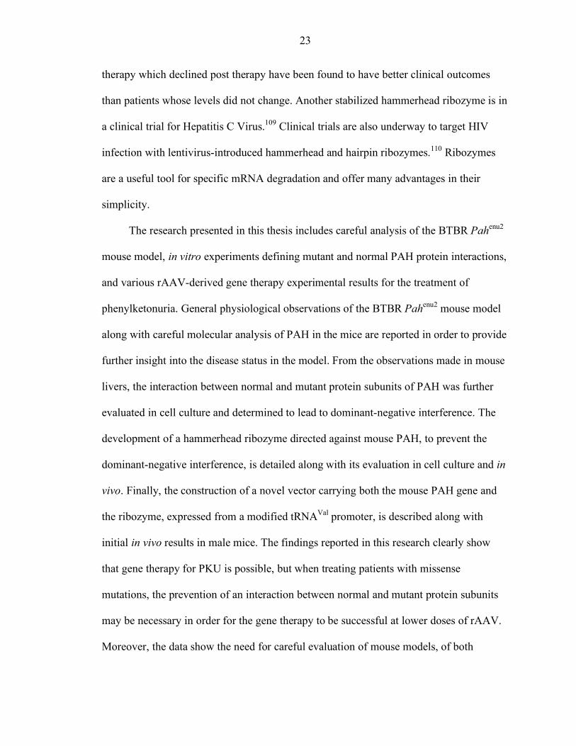
Figure page 1-1 Phenylalanine conversion to tyrosine.................................................................... 25
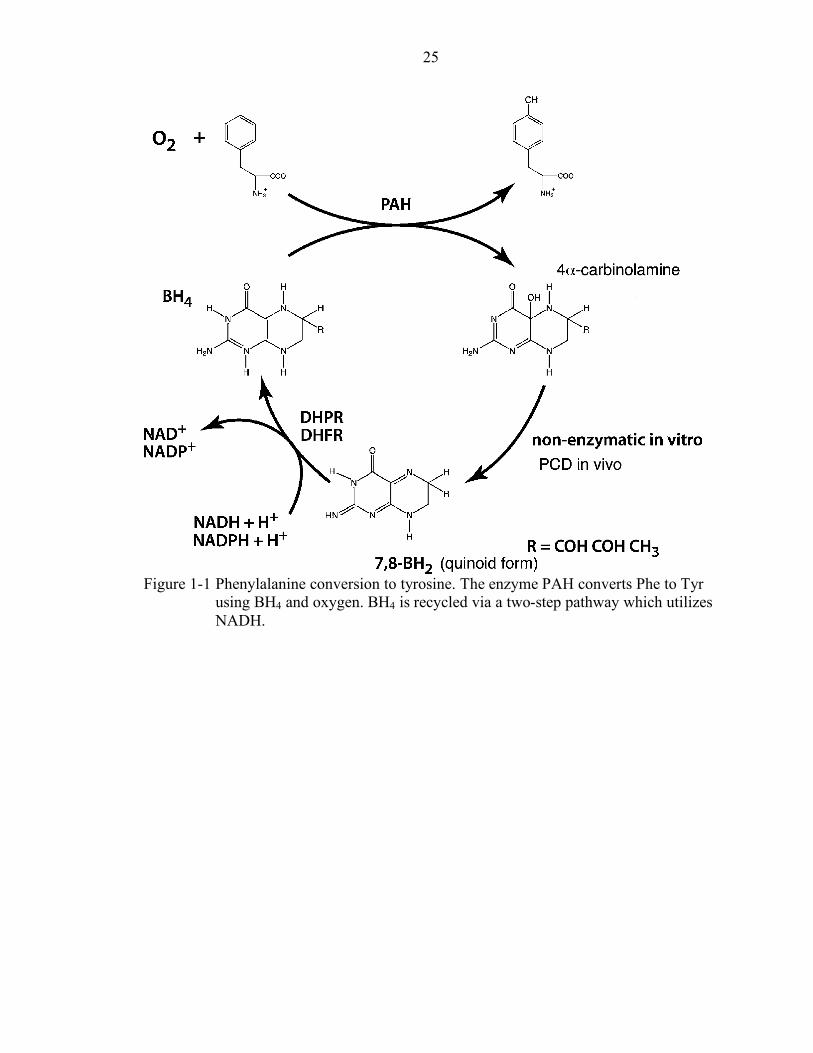
1-2 Hammerhead ribozyme structure. ......................................................................... 26
3-1 BTBR Pahenu2 mouse model................................................................................. 61
3-2 BTBR mice growth curves. .................................................................................. 62
3-3 Male and female weight differences. .................................................................... 63
3-4 Average daily food consumption. ......................................................................... 64
3-5 Northern blot of mouse PAH................................................................................ 65
3-6 Western blot of mouse PAH................................................................................. 66
3-7 Kidney PAH amounts........................................................................................... 67
4-1 rAAV vector maps. .............................................................................................. 76
4-2 Serum phenylalanine levels after gene therapy wirh rAAV2................................. 77
4-3 PAH amounts in mouse liver................................................................................ 78
4-4 Cloning strategy for construction of CB-mPAH-F263S. ....................................... 79
4-5 Test transfections with CB-mPAH and CB-mPAH-F263S. .................................. 80
4-6 Mixed transient transfection results. ..................................................................... 81
4-7 Western blot of native PAGE with mixed transfection samples. ........................... 82
5-1 Mouse PAH ribozyme designs. ............................................................................ 91
5-2 Time Course analyses with ribozymes at 20mM MgCl2........................................ 92
5-3 Time Course analysis of ribozyme I209 at 5mM MgCl2........................................ 93
5-4 Ribozyme I209 kinetic analysis. ........................................................................... 94
xi
5-5 Long target cleavage analysis. .............................................................................. 95
5-6 CB-RzI209........................................................................................................... 96
5-7 Cloning strategy for the construction of a ribozyme-resistant mPAH clone........... 97
5-8 CB-RzI209 stably expresses RzI209 in 293 cells. ................................................. 98
5-9 CB-mPAH-Hd is resistant to the ribozyme. .......................................................... 99
5-10 Ribozyme I209 successfully prevents dominant-negative interference in 293 cells. .............................................................................................................. 100
5-11 Null ribozyme designs........................................................................................ 101
5-12 The null ribozymes do not prevent dominant-negative interference. ................... 102
6-1 Dose response to rAAV2-CB-mPAH-Hd-WPRE. .............................................. 114
6-2 Phenylalanine loading experiment. ..................................................................... 115
6-3 RNase protection assay with dose-response animals. .......................................... 116
6-4 Southern blot of dose response animals. ............................................................. 117
6-5 Phenylalanine hydroxylase activity in gene therapy-treated animals. .................. 118
6-6 Serum phenylalanine levels after dual vector injections. ..................................... 119
6-7 RNase protection assay for co-injected animals. ................................................. 120
6-8 Southern blot detection of two rAAV vectors. .................................................... 121
6-9 Serum phenylalanine levels after co-injection of mildly effective CB-mPAH-Hd-WPRE dose and increasing amounts of CB-RzI209-(-SalI). ......................... 122
7-1 tRNA-RzI209 design.......................................................................................... 129
7-2 Cloning strategy for construction of tRNA-RzI209 cassette................................ 130
7-3 Results of transient cell transfections with CB-mPAH-Hd-tRNA-RzI209........... 131
7-4 tRNA-RzI209 activity and expression in HEK-293 cells. ................................... 132
7-5 In vivo results with CB-mPAH-Hd-tRNA-RzI209. ............................................. 133
8-1 Cell culture siRNA working concentration determination................................... 139
8-2 Mouse PAH siRNA test results. ......................................................................... 140
xii
Abstract of Dissertation Presented to the Graduate School of the University of Florida in Partial Fulfillment of the Requirements for the Degree of Doctor of Philosophy
GENE THERAPY FOR PHENYLKETONURIA: DOMINANT-NEGATIVE INTERFERENCE IN A RECESSIVE DISEASE
By
Catherine Elisabeth Charron
August 2005
Chair: Philip J. Laipis Major Department: Biochemistry and Molecular Biology
Phenylketonuria (PKU) is an autosomal recessive disease where phenylalanine
accumulates in the blood; high brain levels of phenylalanine often lead to mental
retardation. The enzyme phenylalanine hydroxylase (PAH), which converts
phenylalanine to tyrosine, is the mutated gene for over 97% of patients. Dietary
restriction of phenylalanine is the only form of therapy for PKU and is recommended for
life. Unfortunately patients often go off diet during adolescence, and this has led to a rise
in maternal PKU syndrome, the increased incidence of birth defects in children born to
phenylketonuric women. Gene therapy for phenylketonuria would cure the
hyperphenylalaninemia (HPA) and help prevent maternal PKU syndrome.
Using recombinant adeno-associated virus serotype 2 (rAAV2), we have
successfully delivered the mouse PAH gene to male mice and cured the HPA. While
successful, the doses needed in the Pahenu2 mouse model are 5 to 10 times higher than
those used to cure hemophilia A in a mouse model. The Pahenu2 mouse model has a
xiii
missense mutation in PAH rendering the enzyme inactive, and we found that PAH is
present in the liver at 30 percent of normal levels. Since the enzyme is a homotetramer,
dominant-negative interference after gene therapy could explain the need for high rAAV
doses to cure HPA. Using transient transfections we confirmed that mutant and normal
monomer interact together and reduce total PAH activity.
To prevent the dominant-negative interference, we developed a ribozyme that
cleaves the endogenous PAH message. When both ribozyme and resistant PAH gene
were delivered in separate rAAV vectors, no improvement in the effectiveness of the
therapy was observed. Endogenous PAH message was reduced in liver samples
confirming ribozyme activity in vivo. A single vector was constructed to contain the
resistant PAH gene and the ribozyme expressed by a modified tRNAVal promoter. The
novel vector was delivered to male Pahenu2 mice and normalization of serum
phenylalanine levels was achieved with four fold lower doses than with the original CB-
mPAH vector, confirming the dominant-negative interference hypothesis. This
observation of dominant-negative interference to gene therapy in a classic recessive
disorder may prove quite common in many human genetic diseases.
1
CHAPTER 1 INTRODUCTION
Phenylketonuria
Phenylketonuria (PKU) is one of the most commonly inherited human genetic
diseases with an incidence in the United States (US) around 1 in 15,000 births. The gene
affected in the majority (97%) of patients is phenylalanine hydroxylase (PAH), and the
disease is inherited as an autosomal recessive disorder. Accumulation of phenylalanine
(Phe) in the blood, brain and other organs is the cause of the disease, classically
characterized by severe mental retardation. Since the 1960s, severe (>1mM) or milder
(0.36-1mM) hyperphenylalaninemia (HPA) have been detected in the neonatal period,
and treated by the dietary restriction of phenylalanine. If blood Phe levels are kept within
a nontoxic range throughout childhood, brain and cognitive development are near normal.
Unfortunately, the diet is both expensive and unpleasant, and is now recommended for
life by physicians. This chapter presents a summary of the current knowledge on
phenylketonuria including a discussion of the issues associated with maternal
phenylketonuria syndrome.
History
The classic phenylketonuria phenotype originally described by Folling in 1934 is
characterized by severe mental retardation, microcephaly, delayed speech, seizures,
eczema and behavior abnormalities.1 When Folling discovered that two of his patients
presenting with the same symptoms were related, he quickly realized that this form of
mental retardation was inherited in a recessive pattern. After chemical analysis, he
2
determined that the patients excreted phenylpyruvic acid in their urine: he had discovered
a new inborn error of metabolism, the first mental retardation to have a recognized
chemical feature.2 In 1937 the disease was renamed phenylketonuria to emphasize this
biochemical feature.2
Penrose, in the United Kingdom (UK), and Jervis, in the US, studied the known
patients extensively because of the interest generated by this new inborn error of
metabolism and its effect on intelligence. They very quickly observed varying degrees of
severity in terms of the quantitative trait and described patients (using the common terms
at the time) as imbeciles, idiots or simple morons. Since sex chromosome linkage was
found to be negative, the disease was known to be autosomal and suspected of having
undetermined phenotype-influencing factors either environmental or genetic. They
reported a higher number of cases in white populations and calculated the carrier
frequency to be approximately 1 in 100 for both the US and the UK. Penrose used PKU
as a medical example to challenge eugenics since for eliminating PKU from the
population eugenic proponents would have to sterilize one percent of the population:
“Only a lunatic would advocate such a procedure to prevent the occurrence of a handful
of harmless imbeciles.” 2: 198 He also theorized on how altering body metabolism could
influence the psychiatric manifestation of the disease, accurately predicting the future
success of treating PKU by dietary therapy.
Hyperphenylalaninemia (HPA) was found to be the cause of the disease by Jervis
in 1947, and the defective enzyme was determined to be liver PAH in 1953 by
Udenfriend and Cooper.3 In the same year, Bickel demonstrated the possibility of
improving the mental retardation by using a Phe-restricted diet. The need to identify PKU
3
patients early became obvious: at the time it is estimated that one percent of the
population in mental institutions had PKU. In 1957 the ferric chloride “diaper test” was
tested in some California well-baby clinics, but the test proved to be unreliable during the
first month of life. Four years later a reliable assay was developed to screen blood-spots
from newborns for hyperphenylalaninemia.4 This made neonatal screening possible and
allowed for the Phe-restricted diet to be started before one month of life. During the next
two decades neonatal screening was instituted throughout the Western World and
thousands of PKU patients have been placed on diets shortly after birth and given the
opportunity to develop normally.
Clinical Features
Classic phenotype
Although mental retardation is the main feature of the untreated patient, the
mechanism by which phenylalanine causes the disease is still not known. Recent studies
display the potential risks associated with high Phe concentration in cerebrospinal fluid
(CSF). Patch-clamp experiments show that by competing for binding sites on NMDA and
non-NMDA receptors, Phe depresses glutamate receptor function in hippocampal and
cerebrocortical cultured neurons.5 The glutamate receptor is associated with formation of
synapses during early development and in dendritic spine changes in adult tissue, and
thus it is involved in memory performance and learning. In vivo the glutamate receptor is
not saturated by its substrate and thus could effectively be inhibited by higher CSF Phe
levels, most likely leading to memory and learning dysfunctions. The BTBR Pahenu2
mouse model brain (see later section) shows an up-regulation of the density of NMDA
receptors as determined by radioactive ligand binding and western blotting for specific
subunits of NMDA receptors.6 AMPA receptor subunits (a non-NMDA glutamate
4
receptor) are also found to be elevated as compared to the heterozygote forebrain
samples.
A study measuring Na+, K+-ATPase activity in erythrocyte membranes from treated
PKU patients has shown that there is a negative correlation between ATPase activity and
the serum Phe concentration in the patients.7 The patients who had serum Phe levels
above 0.30mM had decreased Na+, K+-ATPase activity; this correlates with the
observation that ATPase activity is reduced in the cortex of rats subjected to experimental
PKU. The same isozyme of the ATPase is present in the brain, and loss of its activity
occurs in neurodegenerative disorders. Direct inhibition of the Na+, K+-ATPase is
associated with glutamate release. However, it is unknown at this point if the decreased
activity in erythrocytes is similar in the brain, and if this is somehow related to the
increase in NMDA receptors observed in the brains of Pahenu2 mice. Creatine kinase
activity, important in maintaining energy homeostasis in the brain, and dopamine
synthesis have been found to be reduced in hyperphenylalaninemic mouse or rat brains,
adding to the complexity of the phenotype.8,9
An earlier study in a mouse model with inducible hyperphenylalaninemia by
administration of Phe in the drinking water examined adult mice brains. Statistically
significant decreases in the muscarinic acetylcholine receptors in the hippocampus and
cerebral cortex were observed.10 Phe has been shown to inhibit ATP-sulfurylase
decreasing the synthesis of sulfatides which are myelin-associated lipids. The decrease in
sulfatides results in lower protection levels of myelin and higher myelin turnover not
compensated by higher myelin synthesis. Low myelination was observed in the brain
autopsy of severe untreated PKU patients and in the Pahenu2 brains.8,11 The brains in the
5
induced HPA study showed loss of the acetylcholine receptor in a [Phe]- and time of
exposure-dependent manner in a region of the brain associated with acquisition and long-
term storage of information. The association of specific neuronal receptors and possible
permanent brain damage with HPA supports the need for lifelong therapy.
Phenotype of early-treated patients
Once neonatal detection of hyperphenylalaninemia was possible, patients were
placed, within one month after birth, on a phenylalanine-free diet. This diet prevents the
elevation of serum Phe levels and the neuropsychological phenotype is averted. The diet
consists of a mixture of free amino acids or modified protein hydrolysates and is ingested
as a drink after dilution in water. The commercial products currently available have
improved since the early 1960s in terms of overall nutritional qualities and vitamin
balance, and have been shown to lead to normal physical growth in children.12 However,
the taste and smell of the products are poor, and make compliance to the diet difficult.
Phenylketonuric children must be closely followed throughout their childhood by a clinic
to monitor serum Phe levels, growth parameters and diet intake.
In the 1960s, a few reports came out suggesting that termination of diet in early
childhood would not lead to any side effects. Unfortunately, the conclusion was
premature. In 1978, and in a follow up study in 1991, Smith et al. showed that
termination or relaxation of the diet can lead to loss of intelligence quotient (IQ)
points.13,14 Poor dietary control in early and continuously-treated PKU patients (10.8
years-old) affects short-term memory, selective attention, behavioral inhibition and rule-
based behavior as compared to well-controlled PKU patients with Phe levels below
400µmol/l and age- and IQ-matched normal control subjects.15 In the same study, the
better controlled patients had significant, but mild, impairments in planning and sustained
6
attention as compared to the normal subjects. Again, this emphasizes the need to find a
better cure for PKU.
Maternal phenylketonuria syndrome
Maternal PKU syndrome refers to the increased range of birth defects seen in
children born of hyperphenylalaninemic mothers on a poorly controlled diet. Growth
retardation, psychomotor handicaps and other birth defects have been reported.16 High
phenylalanine in the mother was first noted to be teratogenic to the fetus by Dent in 1956
and Mabry in 1963.17 The first reports noted mental retardation in the non-PKU offspring
of PKU mothers, but before the end of the decade reports on microcephaly, intrauterine
growth retardation, and high frequency of congenital heart defects were published.17
Since diet was not recommended for life after its early institution in the 1960s and the
1970s, the rise in maternal PKU syndrome came about as the first early-treated patients
reached childbearing age. The extent of the syndrome was not fully understood until the
report from Lenke and Levy, compiling data from a wide range of metabolic centers
across the world, was published in the New England Journal of Medicine in 1980.18
Mental retardation in the children born to women with Phe at 20mg/dL (untreated classic
PKU) was found to occur in 92% of cases, microcephaly in 73%, congenital heart defects
in 12% and low birth weight, below 2.5kg, in 40% of births. These risks were shown to
increase as the mother’s Phe levels increased. In the US, two thirds of phenylketonuric
women are not on diet when they become pregnant.19 The benefits of treating PKU
patients from infancy could be erased if this increase in birth defects, an unforeseen side-
effect of the prior success with PKU, is not addressed.20
The Maternal PKU Collaborative Study was started in 1984 to examine the effects
of phenylalanine-control during gestation on pregnancy outcomes. The international
7
study enrolled 382 women with 574 pregnancies.21 The women were monitored during
pregnancy, and the children followed until 6-7 years of age to measure cognitive
development.22 The frequency of abnormalities in the children was found to be directly
related to maternal phenylalanine levels during pregnancy.16
The range of birth defects attributable to maternal PKU syndrome includes prenatal
growth retardation, microcephaly, congenital heart disease and facial dysmorphias.23
While fetal loss for PKU women is comparable to the normal averages, increases in these
birth defects are always related to phenylalanine levels and length of exposure during
gestation.23 Control prior to conception and control below 360µmol/L achieved by 10
weeks of gestation will lead to a normal or near normal outcome both at birth and in IQ at
follow up. Congenital heart disease is not strictly related to Phe concentration, but its
frequency is increased when poor control with inadequate protein and vitamin intake
occurs during the first trimester.22 Postnatal growth retardation is inversely correlated to
phenylalanine control during the gestation period; IQ goes down significantly in the same
manner. Women with IQ less than 85 need special support since their adherence to the
diet is not as easily achieved: currently in the US the status of care and support is not
adequate to allow for proper control and better pregnancy outcomes in these women.
Other factors besides phenylalanine levels are thought to affect pregnancy outcome
and the child’s IQ at 6 to 7 years of age. These include age of the mother, socioeconomic
status, parental IQ, and home characteristics.24 Home characteristics and parental IQ can
explain most of the lower than expected IQ scores in the children; more than three of the
known risk factors for one pregnancy also can lead to poorer than expected outcome.
Nonetheless, nine women with classic PKU observing late diet control had children who
8
demonstrated higher than expected IQs at 6 years.24 A common feature between maternal
PKU syndrome, fetal alcohol syndrome and pyruvate dehydrogenase deficiency is a
potent inhibition of pyruvate dehydrogenase: since modifier genes are known to prevent
toxicity in fetal alcohol syndrome, the possibility of modifier genes for PKU is an
attractive explanation for the variance in the results observed in the late-treated group.24
Genetics
The incidence of the disease in the US varies from 1 in 13,500 to 1 in 19,000 births.
For non-PKU hyperphenylalaninemia, the estimate is 1 in 48,000 births.25 The prevalence
of PKU is higher in white and Native American than in black, hispanic and asian
populations. Much allelic diversity has been reported at the locus (>450 known
mutations); an extensive database containing all of the known mutations is located at
http://www.pahdb.mcgil.ca.26 This diversity leads to much phenotypic variability even
amongst patients with the same PAH genotype. Other genetic and environmental factors
probably influence the clinical phenotype but have yet to be elucidated.
PAH is located on the human chromosome 12 at position q22-q24.1.27 The first
human cDNA clone was isolated in 1985. The protein is 451 amino acids or 51,672
Daltons.28 The protein was isolated from the rat as a dimer, and thought to be made up of
two identical subunits.29 PAH contains 13 exons over 90kb of DNA.30 The average exon
length is 114 bases, ranging from 57 to 892 bases. Phenylalanine hydroxylase is strongly
homologous to tyrosine hydroxylase, and this homology is greatest in the C-terminal two-
thirds of the protein. Interestingly, for PAH this corresponds to the last 1698 bases of the
mRNA which is coded in 16kb of DNA, while the most divergent parts of the protein
correspond to 567 bases of mRNA coded in 72kb of DNA. The largest intron, between
exons 3 and 4, is 23kb and falls between amino acids 117 and 118 where the homology to
9
tyrosine hydroxylase begins. This suggests that functional and tissue-specific regulators
could be contained within that intron or at least within the 72kb of divergent DNA.30 Rat
and human PAH share 96% homology at the amino acid level, and 89% at the nucleotide
level, with 82% of the differing nucleotides as silent codon changes.28
Transcription of PAH has been shown to be regulated by a 9kb fragment situated
upstream of the human gene. As other housekeeping genes, it does not have a TATA box,
and uses multiple transcription initiation start sites both in humans and in rodents.31 The
5’ region of human PAH contains two half sites of the glucocorticoid response element
(GRE), two consensus sites for activator protein 2 (AP2) and one partial site for cAMP
response element (CRE).32 A 1.7kb region situated from position -3.5kb to -5.2kb
contains 2 hepatocyte nuclear factor 1 (HNF1) binding sites.33 HNF1 was shown to
activate the 9kb promoter region in a dose-dependent manner, and can be enhanced by its
dimerization cofactor DCoH. Interestingly DCoH is also the enzyme pterin-4-α-
carbinolamine dehydratase (PCD), responsible for converting 4-α-carbinolamine-
tetrahydrobiopterin to 7,8-dihydrobiopterin quinoid form in the recycling pathway of BH4
(see Figure 1-1 and later section). Both DCoH and PAH can be found on the same operon
in Pseudomonas aeruginosa, suggesting an evolutionary role in the regulation of PAH by
DCoH: it can transactivate transcription of the gene and recycle the necessary cofactor.
In the mouse, the activity of the promoter is completely dependent on its enhancer,
situated 3.5kb upstream of the start site. The enhancer has binding sites with weak
homology to HNF1 and C/EBP concensus sequences. Addition of cAMP and
dexamethasone increases the activity of the promoter in the presence of the enhancer in
an additive fashion.31 The enzyme activity in rat cell lines is increased in the presence of
10
hydrocortisone due to an increase in PAH transcripts, suggesting that the rat and mouse
promoters have similar characteristics.34 Transgenic mice containing the human
regulatory region express the PAH transgene like the murine PAH, both in a time and
tissue-specific manner.35 The murine enhancer region is 77.5% homologous to the human
segment containing the HNF1 binding sites.33 It is still unknown if the human PAH gene
is hormonally regulated, but unlike the murine promoter, it does not require cAMP or
dexamethasone for in vitro activity.
In humans, the PAH transcript can be detected during the first trimester in the fetal
liver. In rodents, PAH is activated at day 18 of gestation, but strongly induced during the
first post-natal week in the liver.35 PAH is present in rodent kidney, and was found in
human kidney cortex at 20% of levels observed in human liver.36,37 In rats the kidney has
20% of liver mRNA amounts, and both the liver and kidney mRNAs are the same size.34
Conditions which activate the rat-purified enzyme do not activate the kidney enzyme: it
is in a constant activated state.38 Because the mRNAs are identical, the difference in
activities may be from different post-translational modifications and regulation. Rao
postulated that the kidney enzyme could make up 50% of rats’ total PAH activity due to
its higher 5,6,7,8-tetrahydrobiopterin (BH4)-dependent activity. Moller et al.
demonstrated that the human kidney contributes a large amount of tyrosine to the
systemic circulation, while the liver is a net remover of both phenylalanine and tyrosine
from the circulation.39
The Phenylalanine Metabolic Pathway
Phenylalanine metabolism is very complex due to its function as a precursor to
dopamine, epinephrine and norepinephrine and its dual glucogenic and ketogenic role.
Phenylalanine is an essential amino acid; its input is dietary and its clearance includes
11
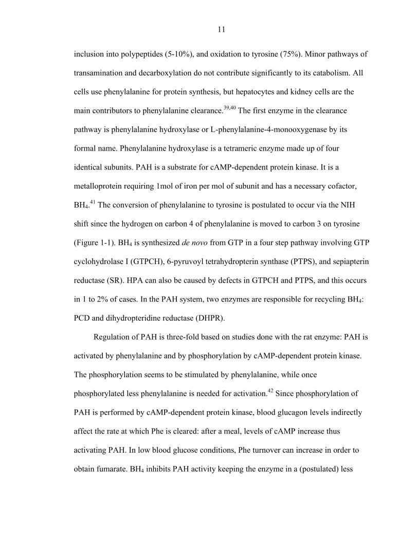
inclusion into polypeptides (5-10%), and oxidation to tyrosine (75%). Minor pathways of
transamination and decarboxylation do not contribute significantly to its catabolism. All
cells use phenylalanine for protein synthesis, but hepatocytes and kidney cells are the
main contributors to phenylalanine clearance.39,40 The first enzyme in the clearance
pathway is phenylalanine hydroxylase or L-phenylalanine-4-monooxygenase by its
formal name. Phenylalanine hydroxylase is a tetrameric enzyme made up of four
identical subunits. PAH is a substrate for cAMP-dependent protein kinase. It is a
metalloprotein requiring 1mol of iron per mol of subunit and has a necessary cofactor,
BH4.41 The conversion of phenylalanine to tyrosine is postulated to occur via the NIH
shift since the hydrogen on carbon 4 of phenylalanine is moved to carbon 3 on tyrosine
(Figure 1-1). BH4 is synthesized de novo from GTP in a four step pathway involving GTP
cyclohydrolase I (GTPCH), 6-pyruvoyl tetrahydropterin synthase (PTPS), and sepiapterin
reductase (SR). HPA can also be caused by defects in GTPCH and PTPS, and this occurs
in 1 to 2% of cases. In the PAH system, two enzymes are responsible for recycling BH4:
PCD and dihydropteridine reductase (DHPR).
Regulation of PAH is three-fold based on studies done with the rat enzyme: PAH is
activated by phenylalanine and by phosphorylation by cAMP-dependent protein kinase.
The phosphorylation seems to be stimulated by phenylalanine, while once
phosphorylated less phenylalanine is needed for activation.42 Since phosphorylation of
PAH is performed by cAMP-dependent protein kinase, blood glucagon levels indirectly
affect the rate at which Phe is cleared: after a meal, levels of cAMP increase thus
activating PAH. In low blood glucose conditions, Phe turnover can increase in order to
obtain fumarate. BH4 inhibits PAH activity keeping the enzyme in a (postulated) less
12
active conformation; the effect is reversed by Phe. The structure of a PAH dimer has been
elucidated.43 The 452 amino acid monomer is composed of three regions: a regulatory, a
catalytic and a tetramerization domain. The catalytic domain, amino acids 118-427,
contains 13 helices and 9 β strands. The regulatory domain is in the N-terminus while the
tetramerization domain is contained in the C-terminus. The active site is buried in a deep
basket-shaped cleft where the iron atom is bound by H290, H285, Q330 and a water
molecule. Kobe et al. postulated that movement of the N-terminal regulatory domain
about a hinge region, making access to the catalytic site easier, could explain the
regulation by phosphorylation and Phe.43
As of May 2005, 498 disease-causing mutations were recorded in the PAH
database. Sixty-two percent of these mutations are missense mutations.27 In vitro analyses
have been performed to analyze a wide range of these mutations in order to obtain insight
on the genotype-phenotype relationship of PKU and the biochemical mechanism of
disease.44-48 The analyses in mammalian cells have shown that mutations often have a
decrease of immunoreactive protein but no real difference in mRNA amounts. These
“conformational” mutations predispose the protein monomer to incorrect folding or
misassembly of the enzyme, as determined in E. coli expression systems and two-hybrid
analyses, thus leading to increased turnover of the protein.44 In vitro manipulations such
as temperature decreases and increased chaperonin levels can rescue protein amounts,
oligomerization pattern and, for some mutations, activity as well.46 Similar modulating
effects in vivo could explain the discrepancies in phenotypes between patients of identical
genotype.49
13
Animal Models for PKU
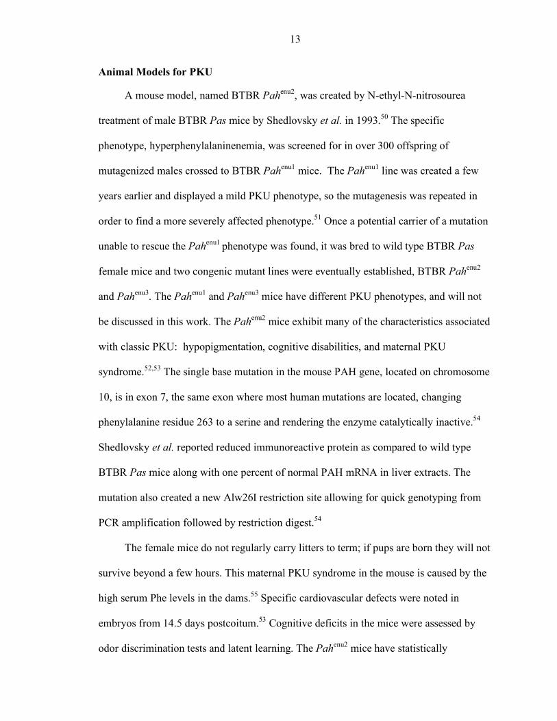
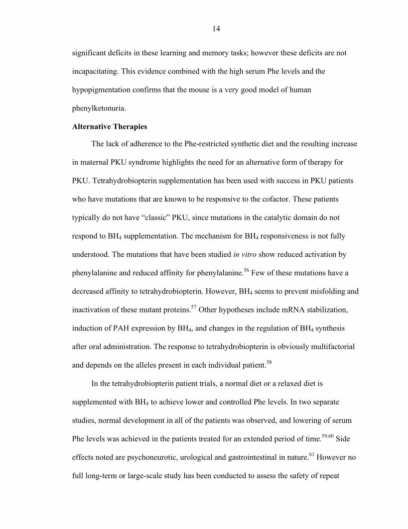
A mouse model, named BTBR Pahenu2, was created by N-ethyl-N-nitrosourea
treatment of male BTBR Pas mice by Shedlovsky et al. in 1993.50 The specific
phenotype, hyperphenylalaninenemia, was screened for in over 300 offspring of
mutagenized males crossed to BTBR Pahenu1 mice. The Pahenu1 line was created a few
years earlier and displayed a mild PKU phenotype, so the mutagenesis was repeated in
order to find a more severely affected phenotype.51 Once a potential carrier of a mutation
unable to rescue the Pahenu1 phenotype was found, it was bred to wild type BTBR Pas
female mice and two congenic mutant lines were eventually established, BTBR Pahenu2
and Pahenu3. The Pahenu1 and Pahenu3 mice have different PKU phenotypes, and will not
be discussed in this work. The Pahenu2 mice exhibit many of the characteristics associated
with classic PKU: hypopigmentation, cognitive disabilities, and maternal PKU
syndrome.52,53 The single base mutation in the mouse PAH gene, located on chromosome
10, is in exon 7, the same exon where most human mutations are located, changing
phenylalanine residue 263 to a serine and rendering the enzyme catalytically inactive.54
Shedlovsky et al. reported reduced immunoreactive protein as compared to wild type
BTBR Pas mice along with one percent of normal PAH mRNA in liver extracts. The
mutation also created a new Alw26I restriction site allowing for quick genotyping from
PCR amplification followed by restriction digest.54
The female mice do not regularly carry litters to term; if pups are born they will not
survive beyond a few hours. This maternal PKU syndrome in the mouse is caused by the
high serum Phe levels in the dams.55 Specific cardiovascular defects were noted in
embryos from 14.5 days postcoitum.53 Cognitive deficits in the mice were assessed by
odor discrimination tests and latent learning. The Pahenu2 mice have statistically
14
significant deficits in these learning and memory tasks; however these deficits are not
incapacitating. This evidence combined with the high serum Phe levels and the
hypopigmentation confirms that the mouse is a very good model of human
phenylketonuria.
Alternative Therapies
The lack of adherence to the Phe-restricted synthetic diet and the resulting increase
in maternal PKU syndrome highlights the need for an alternative form of therapy for
PKU. Tetrahydrobiopterin supplementation has been used with success in PKU patients
who have mutations that are known to be responsive to the cofactor. These patients
typically do not have “classic” PKU, since mutations in the catalytic domain do not
respond to BH4 supplementation. The mechanism for BH4 responsiveness is not fully
understood. The mutations that have been studied in vitro show reduced activation by
phenylalanine and reduced affinity for phenylalanine.56 Few of these mutations have a
decreased affinity to tetrahydrobiopterin. However, BH4 seems to prevent misfolding and
inactivation of these mutant proteins.57 Other hypotheses include mRNA stabilization,
induction of PAH expression by BH4, and changes in the regulation of BH4 synthesis
after oral administration. The response to tetrahydrobiopterin is obviously multifactorial
and depends on the alleles present in each individual patient.58
In the tetrahydrobiopterin patient trials, a normal diet or a relaxed diet is
supplemented with BH4 to achieve lower and controlled Phe levels. In two separate
studies, normal development in all of the patients was observed, and lowering of serum
Phe levels was achieved in the patients treated for an extended period of time.59,60 Side
effects noted are psychoneurotic, urological and gastrointestinal in nature.61 However no
full long-term or large-scale study has been conducted to assess the safety of repeat
15
administration of BH4. While BH4 supplementation can be three to four times more
expensive than the Phe-restricted diet, it could help prevent the effects of maternal PKU
syndrome by stabilizing Phe levels and preventing concentrations from peaking during
the day. Phenylalanine variation during pregnancy was found to have a negative effect on
head circumference at birth by the Collaborative Study.22
Enzyme replacement therapy could be an attractive alternative to treat all PKU
patients. Most of the work with enzyme replacement therapy has been done with the
enzyme Phenylalanine Ammonia-Lyase (PAL) since it does not require a cofactor for
activity.62 Oral delivery of enteric gelatin-coated PAL capsules was shown to be
successful and reduced Phe levels by 22% in PKU patients. While promising it may not
be enough for classic phenotypes and more work is being done to protect the activity of
the protein from the acidic environment of the stomach and optimize it for the intestinal
environment. PEGylation of PAL was also tested in mice: the enzyme has a longer half-
life, but after multiple injections it is quickly cleared from circulation. Enzyme
replacement therapy with PAH has also been explored, but the requirement for co-
injection of BH4 does not make it as attractive a therapy as PAL.
Gene therapy for PKU would be an ideal form of treatment to improve the quality
of life of PKU and HPA patients and to prevent maternal PKU syndrome. Skin, muscle
and bone marrow have been explored as possible targets for gene therapy, but the
availability of the cofactor has limited success in these approaches.63-66 With recombinant
adenovirus, two groups achieved lowering of serum Phe in the BTBR Pahenu2 mice using
the Rous Sarcoma virus LTR and CAG promoters respectively.67,68 However, both
groups reported antibodies raised against adenovirus and complete reversal of treatment
16
after two weeks. All of these experiments were introducing functional human PAH, as
opposed to mouse PAH.
Adeno-associated virus (AAV) has also been successfully used to treat HPA in the
mouse model. Using recombinant AAV (rAAV) serotype 5 carrying the mouse PAH
gene, long-term correction of mice, 40 weeks, was achieved in males, but not in
females.69 One third less vector was needed in the males than in the females to achieve a
similar Phe clearance during the first 6 weeks, at which point the female’s serum Phe
levels returned to their hyperphenylalaninemic state. The minimum effective dose in
males in this study was 3x10¹³ vector genomes of rAAV5. With rAAV2, the human PAH
gene was delivered to mice with a WPRE element included in the cassette.70 Again,
female mice did not respond to the same dose that was found effective in males, 2x10¹²
vector genomes. This dose was effective up to 25 weeks, at which point an increase in
serum Phe levels was noted. According to the authors, this was due to a loss in vector
DNA amounts as determined by semiquantitative PCR. All of these studies have shown
that it is possible to treat hyperphenylalaninemia in the mice by gene therapy, but more
work is required to achieve true long-term correction in the males, and the same response
in female mice.
Gene Therapy Vectors Based on Adeno-Associated Virus
Somatic gene therapy for the correction of inherited genetic disorders is the desired
hallmark of future individualized medicine. Viral vectors for such delivery have been
studied for many years.71 Genome size, immunogenicity, length of gene expression and
integration capabilities are factors that can affect the choice of a viral vector. Adeno-
associated virus has many attractive qualities for human use: it is nonpathogenic, it can
infect dividing and non-dividing cells, it does not have to contain any viral coding
17
sequences, and it can mediate long-term gene expression in animal models.72-74 There are
over 50 AAV serotypes known; each one may have slightly different cell tropism
offering the possibility of enhanced transduction for different targeted organs.75
Unfortunately the genome size of AAV is its main limitation since many genes are longer
than the 4.68kb packaging limit. Reports on a small percentage of integration into active
chromatin regions and the possibility of increased tumorigenesis have darkened the
prospects of this gene therapy vector.76 Nonetheless, rAAV serotype 2 is currently in use
in clinical trials, and still remains a vector of choice for the development of gene therapy
for inherited disorders.
Adeno-Associated Virus Biology
Adeno-associated virus is part of the family Parvoviridae and is classified as a
dependovirus in the Parvovirinae subfamily. Dependoviruses require the presence of
helper viruses, such as Adeno virus or Herpes virus, to establish a productive infection. In
the absence of such a helper virus, a latent infection can be maintained by integration of
the virus DNA into the genome. In humans, the main AAV integration site is 19q13.3
where the genome usually integrates in tandem repeats.77 This can be rescued by
subsequent infection with the helper virus. The virus has not been associated with disease
in humans.
The AAV genome is single-stranded DNA and is 4679 bases in length for AAV
serotype 2.71 It is flanked on both sides by 145-nucleotide inverted terminal repeats (ITR)
composed of three palindromic sequences with only seven bases remaining unpaired
when folded. The ITRs are the only elements required in cis for encapsidation. The
genome encodes nonstructural proteins (Rep78, Rep 68, Rep 52 and Rep 40) and capsid
proteins (VP1, VP2, and VP3). These proteins are expressed from three polymerase II
18
promoters, p5, p19 and p40, from alternatively spliced mRNA. The Rep proteins are
required for DNA replication, establishment of latent infections, site-specific integration
into chromosome 19, and encapsidation of the genome. The Cap proteins combine 60
subunits into T=1 icosahedral symmetry with VP2 as the major structural component of
the small virion.
AAV serotype 2 binds to the ubiquitously expressed cell surface heparin sulfate
proteoglycan (HSPG). It requires fibroblast growth factor receptor type 1 and the integrin
αVβ5 for entry into the cell.78,79 Uptake occurs through standard endocytosis from
clathrin-coated pits, and the capsid is removed in the nucleus.80 The virus genome can be
found in the nucleus two hours after infection. Receptors used by the other AAV
serotypes include sialic acid and PDGFR, giving each one a different preferred cell type.
Current Trends and Applications of rAAV
Recombinant AAV virus can be made in the lab without the use of helper viruses.
Recombinant virus production is accomplished by providing only the necessary proteins
required for DNA replication and encapsidation on a plasmid that is independent of the
recombinant AAV plasmid. Both plasmids are co-transfected into cells, and rAAV virus
can be purified free of helper virus. This method is efficient and produces low particle-to-
infectivity ratios. The ITRs are the only wild type virus sequence left on the recombinant
virus. Thus it is incapable of replicating once it has entered the cell. The viral genome is
slowly converted from single stranded DNA to double stranded DNA, delaying the onset
of expression in the cell. The genome is maintained in the nucleus as a linear or circular
high molecular weight concatemer.81
Many animal models have been treated with rAAV vectors to correct a variety of
inherited disorders in a variety of tissues. Tissues successfully targeted by direct in vivo
19
methods include liver, muscle, heart, brain, lung, eye and kidney.74,82-87 Hemophilia B has
been treated by liver-directed gene therapy both in a mouse model and in a canine model
demonstrating the safety and the long-term expression mediated by rAAV2.73,88 These
studies also showed a dose response correlating increased factor IX circulating levels
with higher rAAV doses.
Clinical trials with AAV serotype 2 are underway for a number of diseases across
the US. Two trials, one for Cystic Fibrosis and one for Hemophilia B, have published a
number of updates. Delivery of rAAV2 to the lungs has not resulted in any adverse
effects to date, but it does not seem that the virus is transducing the lung epithelial cells
very efficiently.75 Results in the Hemophilia B trials have been a little more encouraging.
While delivery of rAAV2 containing the Factor IX gene to the muscle was well tolerated,
only a mild effect on Factor IX concentrations, 1% of normal, has so far been achieved.
When the liver was the target in a partner study, 5 to 12% of normal Factor IX levels
have been observed in the circulation of one patient for 5 weeks, but then dropped to
2.7%. AAV was detected in the semen of one patient and this seems to have been cleared
after 3 months.75 The results of these trials have not yet led to the cures hoped for, but
they have shown that rAAV2 delivery to humans is relatively well-tolerated and can
achieve modest therapeutic effects.
RNA and DNA as Therapeutic Agents
Antisense oligonucleotides can be used to target specific messenger RNAs to
inhibit translation or to induce cleavage and degradation. Antisense RNA
oligonucleotides, ribozymes and short interfering RNAs have been studied over the years
for their potential uses as therapeutic compounds in cancers and dominant diseases.
20
While these different molecules have distinct advantages, we will focus on ribozymes
because this methodology seems best suited for the particular problems seen in PKU.
RNA Interference
The process of RNA interference was discovered in the worm Caenorhabditis
elegans.89 When double stranded RNA (dsRNA) is introduced, sequence-specific post-
transcriptional gene silencing occurs. The enzyme DICER, an RNase III, processes long
dsRNA molecules by cleaving the dsRNA into 22-nucleotide short interfering RNAs
(siRNAs). This duplex is unwound and binds to its target RNA via RISC, RNA-induced
silencing complex. If the siRNA sequence perfectly matches its target, cleavage occurs
approximately at 10 nucleotides from the 5’ end of the target sequence. It is now known
that the general mechanism of dsRNA response is conserved in most eukaryotes, thus the
recent developments in siRNA technology for use in mammalian cells.
The currently most popular approach for expression of siRNA uses a PolIII
promoter to express a hairpin that encodes for both the sense and the antisense RNA
sequence. This is then delivered directly to the cells by transfection or cloned into a viral
vector for easy delivery into animal models. Much work has been done on determining
markers for functional siRNA design. One of the requirements for good siRNAs is the
need for 2 nucleotide 3’ overhangs.90 Internal general requirements include low GC
content, three or more A/U base pairs at the 3’ end of the sense strand, and lack of
internal repeats.91,92 The presence of A/U base pairs at that end confirms previous results
obtained by Schwarz which suggested that the strand that is included into RISC has the
least tightly bound 5’ end, thus preferentially selecting the antisense strand.93
RNA interference has been used extensively for functional gene studies in cell
cultures, and is being studied for targeting cancer genes, viral infections, and genetic
21
disorders.94-96 Safety of siRNA use in humans is currently being assessed in a clinical
trial where an siRNA targeting VEGF is being tested to help prevent age-related macular
degeneration.97 However, more study is necessary since siRNAs have been implicated in
chromatin architecture in several organisms, and the role and mechanism of siRNAs has
not yet been fully elucidated in mammalian cells.98 MicroRNAs (miRNAs) are made
from precursor miRNAs in mammalian cells and have been associated with
developmental gene regulation.99 MicroRNAs are also 21 to 23 nucleotides when
processed from their longer precursors. They often function as translational repressors
and do not contain an exact match to their targets raising concerns about possible side
effects of introduced siRNAs. The inexact match of miRNAs to their targets also implies
that to create a cDNA that is resistant to a designed siRNA will require extensive
modifications. This requirement is the main reason why we focused on hammerhead
ribozymes for our study. Nonetheless, the large amount of research being done with
siRNAs should soon uncover the best and safest way to use them in gene-function studies
and as therapeutic agents.
Ribozymes
Ribozymes are RNA molecules capable of catalyzing chemical reactions without
protein assistance. Hairpin ribozymes, RNaseP, Group I and II introns can catalyze such
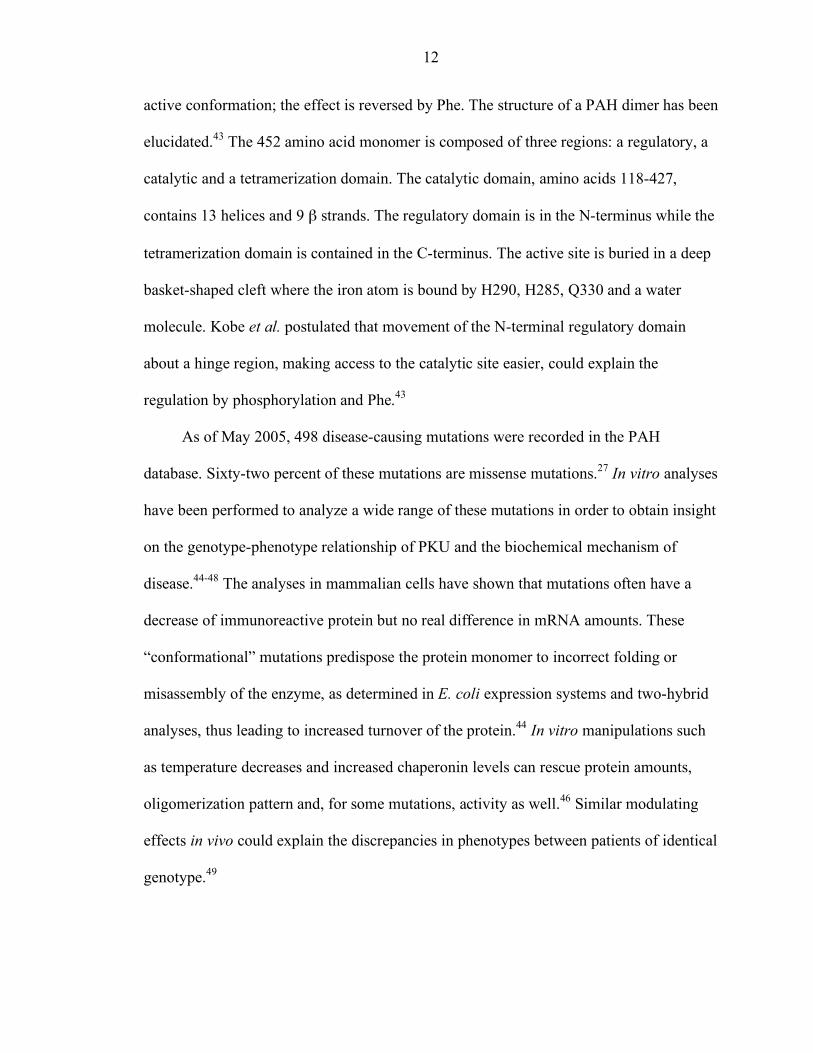
reactions as ribonucleotide transesterification and hydrolysis.100 Hammerhead ribozymes
were discovered in plant satellite virus RNAs and mediate rolling circle replication. They
self-cleave the RNA in an in-line transesterification reaction.101 Since the sequence
requirements of the reaction have been discovered, they have been engineered to catalyze
the same reaction in trans for a specific chosen target.102 The target sequence of a
ribozyme contains “NUX’, N is any nucleotide and X is any nucleotide but G. The
22
ribozyme will cleave the mRNA after X. The actual rate of cleavage is significantly
affected by the sequence of NUX with GUC and AUC having higher activities.103 The
typical lab hammerhead ribozyme is 33 to 35 nucleotides long, depending on the length
of the hybridizing arms, and its core structure includes stems I and III (the hybridizing
arms) and stem II, a hairpin structure used for maintaining stability of the required
folding for catalysis (Figure 1-2). Once bound to its target, the cleavage reaction takes
place and the RNA products are subsequently degraded.
Since they do not require proteins for catalysis, the main issue with ribozyme-use
in therapeutics is choosing the right delivery method. If used as stabilized RNA
molecules direct or general injections must be tested to ascertain co-localization with its
target RNA while keeping in mind the half-life of the ribozyme. Delivery with a viral
vector can obviate the previous issue as long as the virus will infect the correct cell type.
The proper promoter must be chosen so that the ribozyme can exit the nucleus to reach
the mRNA target without being processed or degraded itself. Ribozymes only need 12
nucleotides for target recognition, and the cleavage target rules are not as restricted as
initially thought.104 Nonetheless, specificity of the cleavage reaction has been
demonstrated.105 Many groups have successfully used ribozymes as anti-viral treatments,
anti-cancer treatments, and disease treatments for disorders such as Alzheimer’s and
retinis pigmentosa.106,107
A hammerhead ribozyme, Angiozyme, is currently in Phase II clinical trials for
treatment of advanced colorectal cancers in combination with chemotherapy.108 The
ribozyme targets VEGFR-1 and is a chemically modified molecule injected
subcutaneously on a daily basis. Patients who had detectable VEGFR-1 levels prior to
23
therapy which declined post therapy have been found to have better clinical outcomes
than patients whose levels did not change. Another stabilized hammerhead ribozyme is in
a clinical trial for Hepatitis C Virus.109 Clinical trials are also underway to target HIV
infection with lentivirus-introduced hammerhead and hairpin ribozymes.110 Ribozymes
are a useful tool for specific mRNA degradation and offer many advantages in their
simplicity.
The research presented in this thesis includes careful analysis of the BTBR Pahenu2
mouse model, in vitro experiments defining mutant and normal PAH protein interactions,
and various rAAV-derived gene therapy experimental results for the treatment of
phenylketonuria. General physiological observations of the BTBR Pahenu2 mouse model
along with careful molecular analysis of PAH in the mice are reported in order to provide
further insight into the disease status in the model. From the observations made in mouse
livers, the interaction between normal and mutant protein subunits of PAH was further
evaluated in cell culture and determined to lead to dominant-negative interference. The
development of a hammerhead ribozyme directed against mouse PAH, to prevent the
dominant-negative interference, is detailed along with its evaluation in cell culture and in
vivo. Finally, the construction of a novel vector carrying both the mouse PAH gene and
the ribozyme, expressed from a modified tRNAVal promoter, is described along with
initial in vivo results in male mice. The findings reported in this research clearly show
that gene therapy for PKU is possible, but when treating patients with missense
mutations, the prevention of an interaction between normal and mutant protein subunits
may be necessary in order for the gene therapy to be successful at lower doses of rAAV.
Moreover, the data show the need for careful evaluation of mouse models, of both

24
missense and null mutants, when evaluating the possible treatment of genetic diseases by
gene therapy and the clinical relevance to human patients.
25
Figure 1-1 Phenylalanine conversion to tyrosine. The enzyme PAH converts Phe to Tyr
using BH4 and oxygen. BH4 is recycled via a two-step pathway which utilizes NADH.
26
Figure 1-2 Hammerhead ribozyme structure. The ribozyme is aligned to its 12-nucleotide
target. The cleavage reaction takes place after the X. Stems 1 and III represent the hybridizing arms when bound to the target RNA; stem II is a hairpin structure which stabilizes the ribozyme structure.
27
CHAPTER 2 MATERIALS AND METHODS
The research presented in this dissertation required the use of many experimental
methods. They are described in this chapter in detail to allow other researchers to use the
experiments presented. Any specific modifications to the methods are explained where
relevant in the following chapters.
In Vitro Ribozyme Analysis
For these experiments, all enzymes were from Promega (Madison, WI) unless
otherwise indicated. Radioactive nucleotides were ordered from Amersham Biosciences
(Piscataway, NJ). The water used in all of the experiments was deionized, distilled,
purified through ion-exchange chromatography and autoclaved. All gels for ribozyme
analysis were 8 M urea, acrylamide sequencing gels run in 1 X TBE buffer and
prewarmed to approximately 45°C prior to loading the samples, which were heat
denatured at 85°C for 3 minutes followed by chilling on ice for 3 minutes. The gels were
fixed in 40% methanol, 10% acetic acid and 3% glycerol for 30 to 45 minutes and
subsequently dried at 80°C under vacuum. All of these protocols were described by Fritz
et al.111
Deprotection of RNA Oligos
The chosen ribozyme sequences and 12-nucleotide targets were ordered from
Dharmacon, Inc (Lafayette, CO). The oligos were resuspended in 100 µl of water. Using
the provided deprotection TEMED buffer, 20 µl of each oligo was diluted to 100 µl with
the buffer and incubated at 60°C for 30 minutes. The reaction was stopped by drying in a
28
SpeedVac (Savant) for 30 minutes. Assuming 99% efficiency in synthesis, the oligos
were resuspended at 200 pmole/µl for the ribozymes, and at 300 pmole/µl for the targets.
Working dilutions were prepared: 2 pmole/µl of ribozyme, and 10 pmole/µl of targets.
Samples were stored at -70°C until further use.
Target End-Labelling
The targets were end-labeled with γ-[32P]-ATP to allow for detection of the intact
and cleaved targets on a polyacrylamide sequencing-grade gel. Twenty picomoles of the
target were added to 10 µl of 1 X polynucleotide kinase buffer containing 1µl of RNasin
Ribonuclease Inhibitor, 1 µl of 0.1 M DTT, 1 µl of polynucleotide kinase (5 to 10 units),
and 4 µCi of γ-[32P]-ATP. After incubation at 37°C for 30 minutes, sixty-five microliters
of water were added, and the labeled target was extracted with
phenol:chloroform:isoamyl alcohol. This was purified on a Sephadex G-50 column (USA
Scientific, Ocala, FL). The labeled target can be stored at -20°C.
Time Course of Cleavage Reactions
The purpose of the time course experiment is to test the efficiency of the ribozymes
against the short 12-nucleotide target. Excess target is mixed 10:1 with the ribozyme in a
single large reaction from which timed samples are taken and subsequently run on an 8%
gel. First, two picomoles of the ribozyme working dilution was mixed with 88 µl of water
and 13 µl of 400 mM Tris-HCl. This was incubated at 65°C for 2 minutes and left at
room temperature for 10 minutes to allow for proper folding of the ribozyme. Thirteen
microliters of a 1:10 dilution of RNasin in 0.1 M DTT was added with 13 µl of 200 mM
MgCl2 for a 20 mM MgCl2 final concentration. This concentration is used for all first
time course experiments with new ribozymes. The reaction was then incubated at 37°C
29
for 10 minutes. Prepared tubes each with 20 µl of RNA formamide buffer (90%
formamide, super pure grade, 50 mM EDTA pH 8.0, 0.05% bromophenol blue, 0.05%
xylene cyanol) were placed on ice at this time and were labeled for the desired time
points, typically 0, 0.5, 1, 2, 4, 8, 16, 32, 64, and 128 minutes. Two microliters of
unlabeled target from the working dilution or 20 pmoles, and 2 µl of end-labeled target
were added in that order to the reaction. Immediately after adding the hot target, 10 µl of
the reaction was removed and added to the prepared tube labeled 0. This was repeated for
each time point. The samples can be stored at -20°C.
Six microliters of each timed sample was electrophoresed on an 8% gel at 40 mV
until the bromophenol blue was approximately 2/3 down. Once fixed and dried, it was
exposed to a phosphorescent screen overnight, and scanned with a PhoshorImager
(Molecular Dynamics, Sunnyvale CA) for analysis. The percent of cleaved target at each
time point was calculated from the intensity of the product band over the total intensity of
the product and the intact target.
In Vitro Transcription
A linearized and purified pGEM-T-mPAH plasmid was used to create full-length
mPAH transcripts with T7 RNA Polymerase. The reaction was set up in 20 µl as follows:
4 µl of 5 X polymerase buffer, 2 µl 100 mM DTT, 1 µl RNasin (40 units), 1 µl of a
solution of 20 mM each ATP, CTP, and GTP, 1 µl 4 mM UTP, 2 µl linearized pGEM-T-
mPAH (100 ng), 2 µl or 20 µCi of [α-32P]-UTP, and 6 µl of water. One microliter of T7
RNA polymerase or 20 units was added last. The reaction was incubated at 37°C for 2
hours. Forty microliters of water was added to the reaction and was extracted with 100 µl
of phenol:chloroform:isoamyl alcohol. The aqueous layer was then purified on a G-50
30
column, and 1 µl of the eluate was checked in a scintillation counter to calculate the
concentration of the labeled transcript.
Full-Length Transcript Cleavage Reaction
The full-length transcript was incubated with the ribozyme at 37°C to determine if
the cleavage site is accessible when the entire mRNA sequence is present. The reaction
was set up with the desired ratio of ribozyme to target and magnesium concentration. For
ribozyme I209, the experiment was set up in 30 µl as follows: 3 µl 400 mM Tris, 2 µl or
6 picomoles of ribozyme, 10 µl or 1.5 picomoles of labeled transcript, 3 µl of 200 mM
MgCl2, 3 µl of a 1:10 dilution of RNasin in 0.1 M DTT and 9 µl of water. Samples were
taken at time 0, 1 and 2 hours. A 5% acrylamide gel was needed to separate the
anticipated cleavage products of 862 and 660 bases. The gel was not fixed but dried and
exposed to a phosphorescent screen overnight.
Multiple Turnover Kinetic Analysis
The kinetic properties of the ribozyme are calculated with the Michaelis-Menten
equation from a series of duplicate cleavage reactions set up with increasing ratios of
target to ribozyme. By increasing the ratio from 1:40 to 1:1000, saturation of the
ribozyme is achieved thus the cleavage reaction becomes the rate-limiting step in the
experiment. The series of duplicate reaction is shown in Table 2-1. Each reaction was set
up as in the time course of cleavage reaction by adding the items in order and incubating
at 65°C for 2 minutes and 10 minutes at room temperature after the ribozyme addition.
The RNasin and the magnesium chloride were then added and the tubes were placed at
37°C for a minimum of 10 minutes. The 30pmole/µl target mixture contained 15 µl of
end-labeled target, 15 µl of 300 pmole/µl target stock and 120 µl of water. A 3 pmole/µl
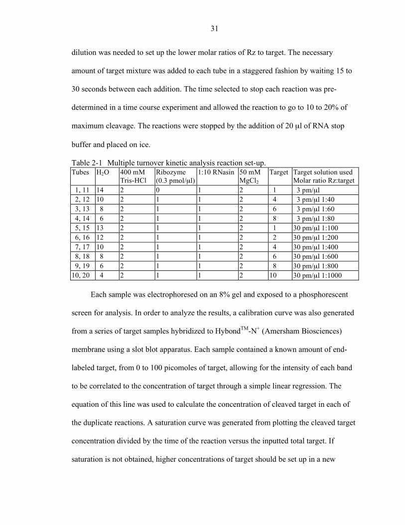
31
dilution was needed to set up the lower molar ratios of Rz to target. The necessary
amount of target mixture was added to each tube in a staggered fashion by waiting 15 to
30 seconds between each addition. The time selected to stop each reaction was pre-
determined in a time course experiment and allowed the reaction to go to 10 to 20% of
maximum cleavage. The reactions were stopped by the addition of 20 µl of RNA stop
buffer and placed on ice.
Table 2-1 Multiple turnover kinetic analysis reaction set-up. Tubes H2O 400 mM
Tris-HCl Ribozyme (0.3 pmol/µl)
1:10 RNasin 50 mM MgCl2
Target Target solution used Molar ratio Rz:target
1, 11 14 2 0 1 2 1 3 pm/µl 2, 12 10 2 1 1 2 4 3 pm/µl 1:40 3, 13 8 2 1 1 2 6 3 pm/µl 1:60 4, 14 6 2 1 1 2 8 3 pm/µl 1:80 5, 15 13 2 1 1 2 1 30 pm/µl 1:100 6, 16 12 2 1 1 2 2 30 pm/µl 1:200 7, 17 10 2 1 1 2 4 30 pm/µl 1:400 8, 18 8 2 1 1 2 6 30 pm/µl 1:600 9, 19 6 2 1 1 2 8 30 pm/µl 1:800 10, 20 4 2 1 1 2 10 30 pm/µl 1:1000
Each sample was electrophoresed on an 8% gel and exposed to a phosphorescent
screen for analysis. In order to analyze the results, a calibration curve was also generated
from a series of target samples hybridized to HybondTM-N+ (Amersham Biosciences)
membrane using a slot blot apparatus. Each sample contained a known amount of end-
labeled target, from 0 to 100 picomoles of target, allowing for the intensity of each band
to be correlated to the concentration of target through a simple linear regression. The
equation of this line was used to calculate the concentration of cleaved target in each of
the duplicate reactions. A saturation curve was generated from plotting the cleaved target
concentration divided by the time of the reaction versus the inputted total target. If
saturation is not obtained, higher concentrations of target should be set up in a new
32
experiment. A Lineweaver-Burk plot was created by graphing 1/v versus 1/S: v is the
velocity or cleaved target (nM/minute), and S is the total input substrate concentration
(nM). The equation of the line was used to determine Vmax, Km and kcat. Vmax is the
absolute value of 1/Y when X is 0; Km is the absolute value of 1/X when Y is 0; kcat is
Vmax divided by the concentration of ribozyme used in the experiment, 15 nM.
Molecular Cloning Protocols
Molecular cloning protocols used in this study followed manufacturer’s
recommendations for each enzyme or component used. Enzymes were from New
England BioLabs (Beverly, MA) unless otherwise indicated. Other general protocols
were adapted from Molecular Cloning: A Laboratory Manual.112
Cloning of Ribozyme Vectors
Ribozyme I209 and its two null derivatives were cloned into the vector p21-newhp
which is based on pTR-UF12. The restriction sites at the cloning site are Hind III and Spe
I and the ribozymes were ordered as DNA oligos including the correct restriction site
bases from Sigma Genosys (The Woodlands, TX). The oligos were gel purified, annealed
and ligated into the digested p21-newhp vector. Sure Cells (Stratagene, La Jolla, CA)
were used for transformations to ensure ITR retention. Clones of each ribozyme were
sequenced and selected based on ITR retention by Sma I digests. The RzI209 plasmid
was renamed CB-RzI209 (Figure 5-6).
The ribozyme I209 vector used for packaging for animal experiments was modified
from the original CB-RzI209 vector. The PYF 441 enhancer, the HSV thymidine kinase
promoter and the neomycin cassette were removed from the vector by Sal I digestion and
religation because the neomycin cassette contains cryptic splice sites which could
33
interference with gene expression if integrated into the genome.113 This plasmid was
named CB-RzI209-(-Neo).
Construction of CB-mPAH-F263S
The plasmid vector CB-mPAH was created from p43-hAAT developed by Loiler
and Flotte at the University of Florida. Directed mutagenesis of mPAH was achieved
using synthetic DNA oligonucleotides as PCR primers. The 5’ primer contained the
desired base changes to change phenylalanine 263 to a serine and started at a unique
restriction site, Xho I, to allow quick cloning of the mutagenized cassette (Table 2-2).
The PCR reaction was set up using EasyStartTM PCR Mix-in-a-tube (Molecular
BioProducts, San Diego, CA). The 132 base pair PCR product was gel purified and
ligated into pGEM-T (Promega). After bacterial transformations into XL1-Blue MRF’
cells (Stratagene) and sequencing of the obtained clones, the fragment was cut from
pGEM-T with Xho I and Hind III, gel purified and ligated into pGEM-T-mPAH. The
mutated gene, mPAH-F263S, was moved from pGEM-T to the CB backbone released
from an EcoR I and Not I digest. Bacterial transformations into Sure cells was followed
by sequencing of clones obtained. Large DNA preparations were performed with
Qiagen’s Plasmid Giga Kit.
Construction of CB-mPAH-Hd
The same strategy used for the construction of CB-mPAH-F263S was used to
change the CB-mPAH plasmid to a resistant sequence to the RzI209. PCR mutagenesis
was utilized to introduce silent base changes around the sequence for Isoleucine 209. The
5’ primer again contained the necessary base changes, and started at a unique restriction
site, Stu I (Table 2-3). After gel purification of the 322 base pair product, it was ligated
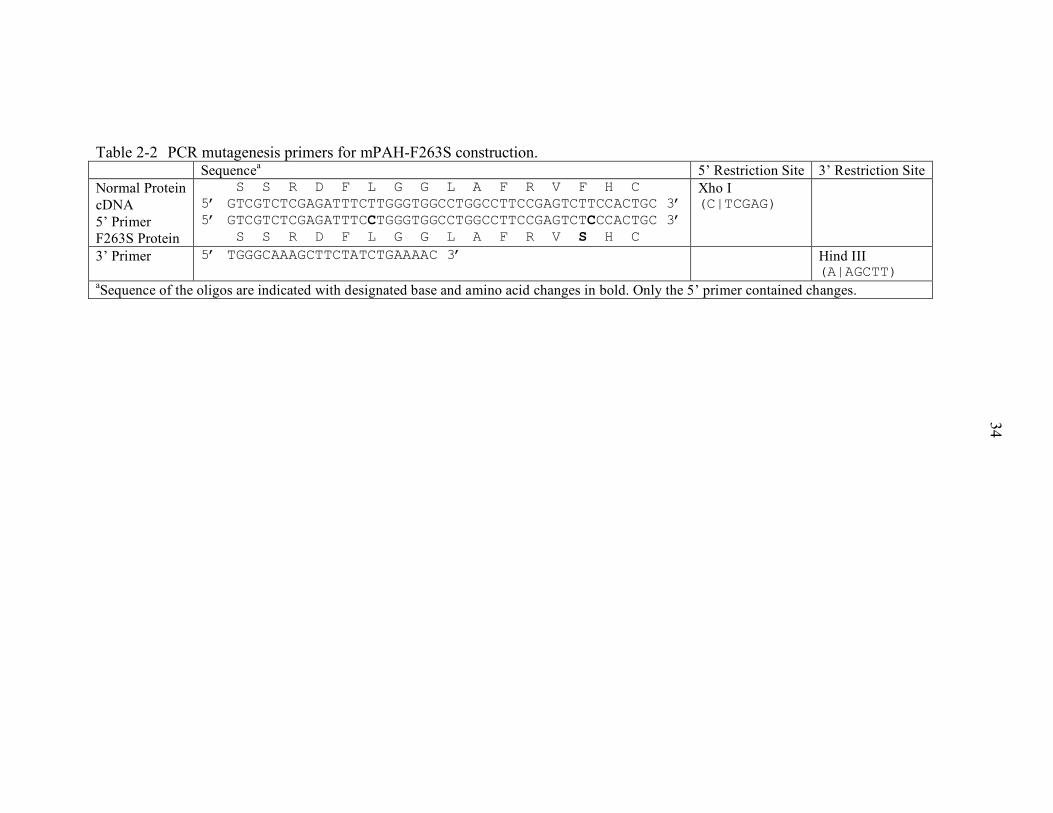
34
Table 2-2 PCR mutagenesis primers for mPAH-F263S construction. Sequencea 5’ Restriction Site 3’ Restriction Site Normal Protein cDNA 5’ Primer F263S Protein
5’ GSCGSTTRGADATFTCLTGGGTGGGLGGAGGFTCRGAVCCFTCHACC 5’ GTCGTCTCGAGATTTCTTGGGTGGCCTGGCCTTCCGAGTCTTCCACTGC 3’ 5’ GTCGTCTCGAGATTTCCTGGGTGGCCTGGCCTTCCGAGTCTCCCACTGC 3’ 5’ GSCGSTTRGADATFTCLTGGGTGGGLGGAGGFTCRGAVCCSTCHACC
Xho I (C|TCGAG)
3’ Primer 5’ TGGGCAAAGCTTCTATCTGAAAAC 3’ Hind III (A|AGCTT)
aSequence of the oligos are indicated with designated base and amino acid changes in bold. Only the 5’ primer contained changes.
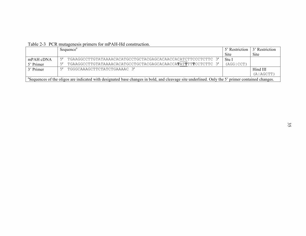
35
Table 2-3 PCR mutagenesis primers for mPAH-Hd construction. Sequencea 5’ Restriction
Site 3’ Restriction Site
mPAH cDNA 5’ Primer
5’ TGAAGGCCTTGTATAAAACACATGCCTGCTACGAGCACAACCACATCTTCCCTCTTC 3’ 5’ TGAAGGCCTTGTATAAAACACATGCCTGCTACGAGCACAACCATATTTTTCCTCTTC 3’
Stu I (AGG|CCT)
3’ Primer 5’ TGGGCAAAGCTTCTATCTGAAAAC 3’ Hind III (A|AGCTT)
aSequences of the oligos are indicated with designated base changes in bold, and cleavage site underlined. Only the 5’ primer contained changes.
36
into pGEM-T, moved to pGEM-T-mPAH and the entire mutagenized gene, mPAH-Hd,
cloned into the CB backbone to create CB-mPAH-Hd. The mPAH-Hd cassette was also
cloned into the CB-WPRE backbone released from an EcoR I and Not I digest.
Sequencing of pGEM-T with the PCR insert and the final vector constructs was done,
and the CB-mPAH-Hd and CB-mPAH-Hd-WPRE plasmids were selected based on ITR
retention. XLI Blue MRF’ cells were used for all pGEM-T transformations, and Sure
cells were used for all plasmids containing ITR sequences.
Construction of tRNA-RzI209
The tRNA-RzI209 cassettewas designed in order to conserve necessary folding
properties for the tRNA sequence and to allow the ribozyme’s hybridizing arms to reach
their target sequence. From the necessary sequence a multi-step strategy was devised to
construct the 200 base pair cassette. Two restriction sites each about one-third from the
ends of the cassette were selected for cloning the cassette in three sets of oligos. The
plasmid pGEM-3Zf(+) (Promega) was modified with a new elongated multiple cloning
site to allow for the sequential cloning of the tRNA cassette (Table 2-4). The modified
pGEM-3Zf(+) was renamed pGEM-3Zf(+)-MCS2. Each oligo set for the tRNA cassette
was ordered from Sigma Genosys and gel purified prior to annealing and ligation into
pGEM-3Zf(+)-MCS2 (Table 2-5). LyoComp GT116 cells (InvivoGen, San Diego, CA)
were used for each cloning step, and blue white screening was utilized to help select
properly ligated clones. A positive digest screen was used if possible at each cloning step.
Once the full cassette was cloned into pGEM-3Zf(+), it was sequenced prior to cloning
into the CB-mPAH-Hd plasmid. The cassette was introduced after the
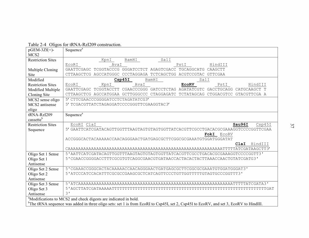
37
Table 2-4 Oligos for tRNA-RzI209 construction. pGEM-3Zf(+)-MCS2
Sequencea
Restriction Sites Multiple Cloning Site
KpnI BamHI SalI EcoRI AvaI PstI HindIII GAATTCGAGC TCGGTACCCG GGGATCCTCT AGAGTCGACC TGCAGGCATG CAAGCTT CTTAAGCTCG AGCCATGGGC CCCTAGGAGA TCTCAGCTGG ACGTCCGTAC GTTCGAA
Modified Restriction Sites Modified Multiple Cloning Site
Csp45I BamHI SalI EcoRI KpnI AvaI EcoRV PstI HindIII GAATTCGAGC TCGGTACCTT CGAACCCGGG GATCCTCTAG AGATATCGTC GACCTGCAGG CATGCAAGCT T CTTAAGCTCG AGCCATGGAA GCTTGGGCCC CTAGGAGATC TCTATAGCAG CTGGACGTCC GTACGTTCGA A
MCS2 sense oligo MCS2 antisense oligo
5’CTTCGAACCCGGGGATCCTCTAGATATCG3’ 5’TCGACGTTATCTAGAGGATCCCCGGGTTCGAAGGTAC3’
tRNA-RzI209 cassetteb
Sequencea
Restriction Sites Sequence
EcoRI ClaI Sau96I Csp45I 5’GAATTCATCGATACAGTTGGTTTAAGTAGTGTAGTGGTTATCACGTTCGCCTGACACGCGAAAGGTCCCCGGTTCGAA FokI EcoRV ACCGGGCACTACAAAAACCAACAGGGAACTGATGAGCGCTTCGGCGCGAAATGTGGATGGGATAT ClaI HindIII CAAAAAAAAAAAAAAAAAAAAAAAAAAAAAAAAAAAAAAAAAAAAAAAAAAAAAAAAAAAATTTTTATCGATAAGCTT3’
Oligo Set 1 Sense Oligo Set 1 Antisense
5'AATTCATCGATACAGTTGGTTTAAGTAGTGTAGTGGTTATCACGTTCGCCTGACACGCGAAAGGTCCCCGGTT3' 5'CGAACCGGGGACCTTTCGCGTGTCAGGCGAACGTGATAACCACTACACTACTTAAACCAACTGTATCGATG3'
Oligo Set 2 Sense Oligo Set 2 Antisense
5'CGAAACCGGGCACTACAAAAACCAACAGGGAACTGATGAGCGCTTCGGCGCGAAATGTGGATGGGAT3' 5'ATCCCATCCACATTTCGCGCCGAAGCGCTCATCAGTTCCCTGTTGGTTTTTGTAGTGCCCGGTTT3'
Oligo Set 3 Sense Oligo Set 3 Antisense
5'ATCAAAAAAAAAAAAAAAAAAAAAAAAAAAAAAAAAAAAAAAAAAAAAAAAAAAAAAAAAAAATTTTTATCGATA3' 5'AGCTTATCGATAAAAATTTTTTTTTTTTTTTTTTTTTTTTTTTTTTTTTTTTTTTTTTTTTTTTTTTTTTTTTTTTGAT3'
aModifications to MCS2 and check digests are indicated in bold. bThe tRNA sequence was added in three oligo sets: set 1 is from EcoRI to Csp45I, set 2, Csp45I to EcoRV, and set 3, EcoRV to HindIII.
38
SV40 polyA sequence at the Cla I site and screened for proper orientation by restriction
digest. This clone was also sequenced.
Cell Culture Protocols
HEK-293 and all 293T cells were cultured in Dubelco’s Modified Eagle Media
containing 25 mM glucose, 4 mM L-glutamine, and 0.04 mM phenol red. These were
passaged on a weekly basis with 0.25% trypsin and used for transient cell transfections of
various plasmid DNA.
Transient Cell Transfection with CaPO4
Transient cell transfections of HEK-293 cells were done in 6-well plates (9.6 cm2
per well) with 80% confluent cells passaged 24 hours earlier. The desired DNA amounts
were mixed in water and 300 µl of 2.5 M CaCl2 was added to a final volume of 3.0 ml.
This solution was added dropwise to 3.0 ml of 2 X HBSS (Hank’s buffered salt solution)
while vortexing lightly. After a 15-minute incubation, it was separated into four wells of
the 6-well plate and added directly to the complete media. Four hours later the media was
changed to fresh complete media. Cells were harvested after 48 to 72 hours by removing
the media, washing with PBS and scraping in 1.0 ml of PBS per well. Of four wells, 3
were collected together in PBS, and one was collected using 1.0 ml of TRIzol Reagent
instead of PBS. The 3.0 ml of cells were centrifuged at 1000 rpm and resuspended in 0.5
ml of homogenization buffer: 1.5 ml 1 M KCl, 8.5 ml H2O, and 0.5 µl of β-
mercaptoethanol. The cells were homogenized in a glass homogenizer and this was rinsed
with 0.5 ml of buffer and added to the homogenate saved into a fresh tube. The lysate
was obtained by centrifugation at 14,000 rpm at 4°C.
39
Transient Transfections using Superfect
293T cells were obtained from Dr Chang’s laboratory. His lab constructed a stably
integrated 293T cell line via a lentivirus system containing pTYF-mPAH and named
293T-TYF-mPAH. The lentivirus plasmid was constructed using our lab’s mPAH
cassette from CB-mPAH. These cells were used for siRNA transfections using SuperFect
(Qiagen). Cells were passed into a 6-well plate 24 hours prior to transcription at a
concentration 4x10⁵ cells per well. Seventy-five microliters of DMEM was mixed with
3.5 µg of DNA at a minimum 1 µg/µl concentration. After vortexing, 7 µl of SuperFect
was added to the center of the DMEM/DNA solution, mixed up and down five times, and
incubated at room temperature for 7 to 12 minutes. The mixture was then added dropwise
to one well of cells containing 0.6 ml fresh complete media. After 6 hours the media was
changed to 2 ml of complete media. Cells were harvested 48 hours after transfection with
1.0 ml TRIzol Reagent and stored at -20°C until RNA extractions.
Phenylalanine Hydroxylase Activity Assay
The PAH activity assay is based on the reduction of NADH to NAD in the
recycling pathway of BH4 (Figure 1-1). Cell transfection or tissue samples are set up
twice: one reaction without phenylalanine and one with phenylalanine thus the assay
measures the phenylalanine-linked reduction of NADH at 340 nm. Four samples are run
in one assay with the 8-sample holder of the Genesys 5 UV-Vis spectrophotometer
(Thermo Electron Corporation, Cambridge, UK). The reactions were set up in 1ml total
volume with 100 µl of cell lysate or 20 µl of tissue homogenate. Each sample included
the following: 0.10 M potassium phosphate buffer, pH 6.8, 0.25 U catalase, 0.10 mU
DHPR, 0.04 mM 6-Methyl-5,6,7,8-tetrahydropterine (6-MPH4), 0.2 mM NADH, and 1
mM Phe if included. Both NADH and 6-MPH4 were added last and the reactions’
40
decrease in absorbance at 340 nm were recorded every five minutes for 30 minutes. After
calculating the protein concentration of the samples, the Phe-linked reduction of NADH
was calculated as specific activity in units/mg of total protein.
Protein Concentration Assay
The protein concentration assay is a modified short-protocol Lowry assay.
Standards were set up with 10 X BSA (New England BioLabs) ranging from 0 mg to 100
mg of protein. Typically 10 µl of tissue homogenate and 20 µl of cell lysate were diluted
to 100 µl with water for analysis. Six hundred microliters of copper reagent (0.6 mM
Na2CuEDTA, 0.2 M Na2CO3, 0.1 M NaOH) were added to the protein and incubated at
room temperature for 10 minutes. Sixty microliters of a 1:1 dilution of Folin &
Ciocalteu’s phenol reagent (Sigma) in water were added and allowed to incubate for 30 to
45 minutes at room temperature. Each reaction and standard is set up in duplicate, and the
entire 760 µl was read at 500 nm. From the average of the duplicate standards, a linear
regression was calculated to get an equation of the line used to determine the protein
concentration of the experimental samples.
Western Blotting
The mouse PAH antibody was raised against a 142 amino acid N-terminal peptide
in rabbit. The antisera was used at a 1:1000 dilution in TBS-T with 5% milk. Tissue
homogenate or cell lysate were electrophoresed on 12% acrylamide Tris-HCl gels with a
discontinuous buffer system containing SDS. The protein was transferred to
nitrocellulose membranes, blocked overnight, and incubated with two primary antibodies:
the mouse PAH antisera and a rabbit polyclonal antibody to GAPDH (Abcam,
Cambridge, UK) as a loading control. The secondary antibody was horseradish
peroxidase-linked and developed against rabbit IgG in donkey (Amersham). Detection of
41
samples was done with ECLTM Western Blotting Detection Reagents (Amersham), and
the blots were exposed to Kodak XAR film for 0.5 to 4 minutes. Laser densitometry was
used to quantify the intensity of the bands.
Northern Blotting
Tissue samples saved in RNAlater (Ambion) were processed with TRIzol
Reagent. The tissues, approximately 100 mg, were homogenized with an electric tissue
homogenizer in 1.5 ml of TRIzol, and the manufacturer’s protocol was followed for RNA
extraction. Samples were resuspended in an appropriate volume of nuclease-free water
and stored at -70°C. Levels of ribosomal RNA were adjusted, and electrophoresed on 2.2
M formaldehyde, 1 X Mops electrophoresis buffer and 1% agarose gels. The RNA was
transferred overnight in 10 X SSC by upward capillary transfer to a neutral nylon
membrane, HybondTM-N (Amersham). After UV irradiation, the membrane was stained
briefly with ethidium bromide for RNA visualization. Probe labeling was accomplished
with the RediprimeTM II system (Amersham). Hybridization to the mPAH and human
copper zinc Super Oxide Dismutase (CuZnSOD) cDNA probes was done in Church
buffer overnight at 65°C. After washing three times in 0.2 X SSC, the membranes were
exposed to Kodak XAR film at –70°C, and using various timed exposures, the levels of
PAH mRNA in the different tissues were compared using laser densitometry.
Recombinant Adeno-Associated Virus Packaging
All packaging of vector DNA was done by the University of Florida Vector Core
Laboratory. The only serotype used for these experiments was rAAV type 2, matching
the type 2 ITR sequences present in our vector plasmids. Large preparations of plasmid
DNA was done in our laboratory using Qiagen’s Plasmid Giga Kit. After testing the
42
DNA preparation by restriction digest and cell transfection for the PAH activity assay,
the entire yield was given to the Vector Core Laboratory. Our vector DNA was then co-
transfected into HEK-293 cells with the pDG plasmid which contains AAV’s rep and cap
genes along with the required adenovirus genes.114 After 48 hours a cell pellet was
obtained, freeze-thawed, and the lysate was separated on an iodixonal step-gradient. The
virus was purified on a heparin affinity column, and after concentration the virus was
titered by quantitative competitive PCR and infectious center assay.
Animal Procedures
Growth Rate Analysis
Selected litters were closely followed from birth for 56 days. Pups from the
heterozygote dam to Pahenu2 male matings were tattooed on their paws within the first 6
days after birth in order to specifically follow each pup’s growth rate and later sort
according to genotype. India ink was injected with a 36-gauge needle into the paws of the
pups after chilling them briefly on ice. A sequential pattern for each pup was used: no
tattoo, left front, right front, left rear, right rear, left front and left rear, etc. until all
double combinations were exhausted if necessary. Pups were weighed every 3 to 4 days
for 36 days, then every week until day 56. The data was sorted by genotype and then by
sex within each genotype. Weights were averaged, standard deviations were calculated
and all data sets were analyzed by student’s t-tests and ANOVA.
Blood Collection
Animals were bled without anesthesia in a rotating tail injector (Braintree
Scientific, Braintree, MA). Blood samples, approximately 90 to 110 µl, were collected
into heparinized capillary tubes: the tails of the mice were cut with veterinary scissors
and the tail was massaged towards the cut to collect the blood. The entire procedure,
43
including weighing the mouse if necessary takes approximately five to six minutes per
mouse. The content of the capillary tube was transferred into a 200 µL tube. This was
spun down at 10,000 g and the serum was collected into a second tube. Both blood and
serum samples were stored at -20°C until further use. Mice were usually bled in late
afternoon on a weekly basis, but can be bled more often if necessary.
Microplate Serum Phenylalanine Assay
The phenylalanine assay is a modification of the assay developed in 1962 by
McCaman.115 Only 7.5 µl of serum is needed for triplicate readings. Each sample was
TCA precipitated, with 11.2 µl of 0.3 N TCA and chilled on ice for 10 minutes or stored
at -20°C until the assay was set up. Using a PCR plate for 96 samples, 64 µl of cocktail
was dispensed into each well. The cocktail contains 4.40 ml 0.3 M succinate, pH 5.8,
1.76 ml of 30 mM ninhydrin, and 0.880 ml 5 mM L-leucyl-L-alanine. Standards were
prepared from 10 mM phenylalanine in 0.3 N TCA: 0 mM, 0.05 mM, 0.10 mM, 0.25
mM, 0.50 mM, 0.75 mM, 1.0 mM and 1.25 mM. Each standard and sample was run in
triplicate. Four microliters of each standard was added to the appropriate wells. The
precipitated serum samples were spun down at 13,000 rpm or 16,000 g for 10 minutes in
a microcentrifuge. Four microliters of each sample is added to the wells, and the plate
was capped prior to placing in a thermocycler to incubate the samples at 60°C for 2
hours.
Two hundred microliters of copper reagent is added to each well of a 96-well 0.5
ml black fluorimeter plate (Nunc, Denmark). The copper reagent is composed of 1.6 g
Na2CO3, 0.065 g potassium sodium tartrate, 0.060 g CuSO4·5H2O each dissolved in about
300 ml H2O and added together to bring up to 1 liter. One hundred microliters was added
44
to the incubated samples in the PCR plate and then the entire volume was moved into the
fluorimeter plate. The PCR plate was washed with another 100 µl of copper reagent and
added to the fluorimeter plate as well. The plate was read twice on an FLx800
Multidetection Microplate Reader (BioTek, Winooski VT). Both readings were averaged
to calculate the phenylalanine concentration in each sample.
Food Consumption Measurement
Food consumption was calculated as an average per day per gram of mouse. Mice
were kept in standard housing, two, three or four to a cage separated by sex and
genotype. The mice and the food were weighed once a week when the mice were
changed to new cages. The first week, only the food in the new cage was weighed by
moving it to a weigh boat. At the second and subsequent time points, the leftover food
was weighed to calculate the food consumption, and the new food placed with the mice
was weighed as well. This was repeated for two to four weeks, and performed at
consistent times to allow correct per day calculations.
Portal Vein Injections
All mice selected for gene therapy experiments were bled 2 to 3 times prior to the
portal vein injections in order to obtain a baseline serum phenylalanine concentration. At
the time of surgery, all mice were typically between 10 to 14 weeks old. All instruments
and solutions were sterilized by autoclaving prior to surgery. The mice were weighed and
injected subcutaneously with Baytril and Buprenex diluted 1 to 10 separately with
injection-saline solution. General anesthesia was accomplished with isoflurane. After
cleaning the ventral surface, a midline abdominal cut was made and the portal vein was
exposed for injection with or without moving intestines outside of the body cavity. Using
a 29-gauge needle, the injection was made into the portal vein in an approximate volume
45
of 0.3 cc. After bleeding was stopped with cotton tipped applicators, 0.5 cc to 1 cc of pre-
warmed LRS solution was added into the body cavity to prevent adhesions. The
abdominal muscle wall was sewn with 4.0 silk and the outer skin was stapled closed.
Mice were placed in clean warm cages and monitored for 15 to 30 minutes before being
taken back into the colony. Sterile peanut butter was given to the mice on a piece of
normal chow to monitor their recovery on the next day. Cottage cheese or phenylalanine-
free chocolate can also be given to increase their appetite if necessary. Once a day for the
following week the mice were checked to make sure the wounds were clean and no
staples were lost. The staples were removed a week after surgery at which time the mice
are bled for the first time point.
Phenylalanine Loading
A phenylalanine solution was prepared in 0.9% saline and filtered sterilized
through a sterile syringe and a 0.22 µm filter. The pH of the solution was 7.4. The
concentration of 26.4 mg/ml was determined by spectrophotometric analysis. On the day
of the study, each animal was bled (0 hour), weighed and then injected subcutaneously
with 0.8 mg L-Phe per gram of body weight. The volumes injected thus ranged from 1.0
to 1.5 ml per mouse. Each blood sample taken was approximately 30 µl. Timed blood
samples were obtained at 1.5, 3, 6, 12, and 24 hours. Serum was separated by
centrifugation and the Phe concentrations determined with the normal fluorimetric assay
and repeated three times.
Sacrifice and Tissue Collection
Gene therapy-treated animals were sacrificed with an overdose of sodium
pentobarbital followed by cervical dislocation or brain perfusion with 4%
paraformaldehyde. The following tissues were collected (prior to perfusion): muscle,
46
kidney, spleen, lung, liver and testes, and quick frozen on dry ice and stored at -70°C.
Instruments used for dissection were cleaned between each tissue with Decon* Eliminase*
Decontaminant (East Sussex, UK) to prevent DNA cross-contamination. Approximately
100 mg of liver was saved in 2 ml of RNAlater (Ambion, Austin, TX) and placed on ice
then moved to -20°C. All other tissues were fixed in 10% formalin for general
pathological examination. Liver sections saved in tissue cassettes were sent to the
Pathology Core for paraffin embedding and sectioning. These were processed by
dehydration with graded ethanol solutions, cleared with xylene and embedded in paraffin.
Five-micron sections mounted on glass slides were hydrated in graded alcohol solutions,
stained with hematoxylin and counterstained with eosin. These were dehydrated once
again to place a coverslip on the slide using Permount.
Normal animals were sacrificed by CO2 asphyxiation followed by cervical
dislocation. Necessary tissue samples were removed and saved by quick freezing, in
RNAlater or as tissue homogenates. Approximately 100 mg of tissue was homogenized
in 2 ml homogenization buffer in a glass homogenizer. This was centrifuged at 16,000 g
for 10 minutes at 4°C. The supernatant was saved at -70°C or used immediately for PAH
activity assays.
RNase Protection Assays
RNA samples were extracted with TRIzol Reagent (Invitrogen) and protocol.
Probes for the RNase Protection Assay (RPA) were designed, PCR amplified from CB-
mPAH and cloned into pGEM-T. The clones selected for transcription reactions were
checked to be in the correct orientation for T7 RNA polymerase transcription, digested
with NotI, and purified by phenol:chloroform extraction. Transcription of the probes was
47
done using Ambion’s MaxiScript® In vitro Transcription Kit. Probes were gel purified
and eluted overnight. Fifteen micrograms of RNA from the experimental samples were
set up with CB-mPAH sense probes, antisense probes as negative controls, and Actin
probes as loading controls. Yeast RNA, 2.5 µg, was included with all probes with or
without RNAse addition as RNase efficiency controls. The assay was done using
Ambion’s RPA IIITM Kit and protocol. The samples were run on 5% acrylamide, 8 M
urea, 1 X TBE gels and exposed to film at -70°C for various times to detect protected
RNA fragments. Laser densitometry was done to calculate relative levels of protected
RNA.
Southern Blotting
DNA was extracted from tissue samples in tail buffer: 50 mM NaCl, 25 mM
EDTA, 50 mM Tris pH 8.0, with 50 µl of 10% SDS, and 50 µl of 10 mg/ml proteinase K.
One hundred milligrams of tissue was incubated in 500 µl of buffer overnight at 55°C, 2
µl of 10 mg/ml RNase A solution was added in the morning for 1 hour incubation at
55°C. Aqueous layers were subsequently extracted with the following: 500 µl phenol for
1 hour at room temperature, 500 µl phenol:chloroform for 30 minutes at room
temperature, and 500 µl chloroform for 2 minutes at room temperature. The last aqueous
layer was ethanol precipitated and resuspended in an appropriate volume of TE pH 8.0.
DNA sample quality was checked by incubating at 37°C for 1 hour in 1 X NEB
restriction buffer 3 and 1 X BSA, then electrophoresed on 1% agarose gels against
unincubated samples.
Twenty micrograms of DNA was digested overnight. The digested samples were
run on 0.8% agarose 1 X TAE gels at 5 V/cm for 5 hours. DNA was transferred by
48
downward capillary transfer in 10 X SSPE onto BioRad Zeta-Probe GT membrane. The
membrane was UV crosslinked and baked for 30 minutes at 80°C. Church buffer with
added sheared salmon sperm DNA was used for hybridization at 65°C. Pre-hybridization
was done for 4 hours. Fifty nanograms of gel purified digested DNA fragments were
labeled with Rediprime II at room temperature. The probes were purified on G-50
columns and half of the probes were used in 10ml of hybridization buffer for a 12x 14cm
membrane. Washes were done at 65°C for a total of 3 hours. Membranes were exposed
for 48 hours to Kodak BioMax-MS film in a TransScreen HE cassette at -70°C.
Subsequent exposures were done as necessary. Laser densitometry was used to measure
band intensities.
RNA Interference Protocols
Generation of siRNA Cassettes
DNA oligos for the three designed siRNAs were ordered from Sigma-Genosys. The
oligos were gel purified and diluted to 10 µM based on concentrations obtained from
optical density readings. Ambion’s SilencerTM Express siRNA Expression Cassette Kit
was used for generating each cassette containing the human U6 promoter. The provided
GAPDH and negative cassette controls were also made. The PCR reactions were done in
a Mastercycler (Eppendorf) using Platinum Taq (Invitrogen) according to provided
protocols with 50°C for the annealing temperature. Concentrations of the final product
were determined by optical density.
Reverse Transcriptase Reaction and Polymerase Chain Reaction
RNA samples were extracted using TRIzol Reagent and protocol. Five hundred
nanograms of RNA were used for the reverse transcription reaction followed by
49
polymerase chain reaction (RT-PCR). All reactions were set up in 200 µl tubes and all
incubation steps were done in the Mastercycler to ensure even incubations. The lid
temperature of the Mastercycler was kept at 105°C throughout the experiment. All RNA
samples were from cell transfections, thus were first treated with RQ1 RNase-Free
DNase (Promega) for 30 minutes at 37°C. EDTA was added to the samples and incubated
at 65°C for 10 minutes for inactiviation of the DNase. The reverse transcription reaction
was then set up using Promega’s AccessQuickTM RT-PCR System. This includes a mix
that contains the necessary components for reverse transcription and PCR reactions. The
reverse transcriptase was added last. A one hour incubation at 48°C for reverse
transcription was done followed by 35 cycles of 94°C 0.5 minute, 55°C 1 minute, 72°C 5
minutes, and one last 5 minute incubation at 72°C. Half of the reactions were then run on
15% polyacrylamide 1 X TBE gels with an appropriate ladder. The gels were stained
with ethidium bromide for visualization on an Eagle EyeTM Imaging System (Stratagene).
Relative intensites of the bands was calculated using the software UN-SCAN-IT gel
Automated Digitizing System Version 5.1 (Silk Scientific Corporation, Orem, UT).
50
CHAPTER 3 ANIMAL MODEL ANALYSIS
The most useful animal model for phenylketonuria is the BTBR Pahenu2 mouse. A
single nucleotide mutation in the PAH locus created the missense mutation F263S.54 This
mutation renders phenylalanine hydroxylase completely inactive. The mice have
“classic” PKU: hyperphenylalaninemia, cognitive deficits, maternal PKU syndrome, and
hypopigmentation (Figure 3-1). Various studies of the mouse model have been done (see
the Animal model section of Chapter 1), but some physiological parameters that we have
observed with our colony have not been previously described. This chapter describes
some general observations about the mouse model, and experiments performed to further
understand this model of classic phenylketonuria.
General Sex Dimorphism in BTBR Pahenu2 Mice
Growth Curve Analysis
A number of papers have been published about the BTBR Pahenu2 model, but none
mentions any differences between the Pahenu2 male and female mice. Early on in our
colony it was observed that the female mice were smaller and more fragile than their
male counterparts. We decided to quantitate this observed difference from birth to
adulthood. Three litters from male -/- to female +/- matings were followed from birth
until post-natal day 72. All pups were tattooed with India Ink in different patterns on
their paws between day 3 and day 6 to follow each pup’s weight specifically, since
genotype of the pups cannot be ascertained visually immediately after birth. Coat color
identification between days 15-20 was used to assess genotype in these crosses which
51
was later confirmed by serum Phe levels obtained at weaning. Five heterozygous males,
seven heterozygous females, five Pahenu2 males and four Pahenu2 females were thus
followed from birth. Weights pre-determined in our inventory were added to the averages
of these mice for the adult average values to obtain more accurate numbers. Three wild
type litters were also followed from birth, but without tattooing of the pups. Eleven wild
type males and seventeen wild type females were thus included in these calculations.
Again values obtained from our inventory were also added into the adult weight averages.
The results in Figure 3-2 show that Pahenu2 mice are smaller than both heterozygote
and wild type mice. Using unpaired t-test analysis (Table 3-1) from day 0 to day 56, wild
type and heterozygous mice do not show any significant trends in the differences in their
growth. While some points show significant differences, they can be explained by litter
sizes: the wild type litters had each three to four more pups than the followed
heterozygous litters thus being somewhat smaller at some of the early time points.
Comparing males and females separately explains the significant p values towards the
end of the 56 days. While males are not significantly different in their growth rates, the
wild type females remain somewhat smaller than the heterozygous females, a lasting
effect of the litter size differences that does not continue once the animals reach
adulthood. Adult females, both wild type and heterozygote, are generally smaller than
their male counterparts as expected.
The Pahenu2 mice compared to heterozygous mice are significantly smaller starting
at day 6 with a p value below 0.05. The p value comes below 0.001 at day 12 until day 49
when it comes back to just below 0.05. Again looking at the males alone, the difference
in growth rates is only significant until day 49 when the PKU males reach weights that
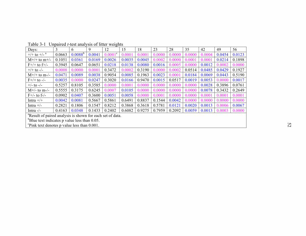
52
Table 3-1 Unpaired t-test analysis of litter weights Days: 3 6 9 12 15 18 23 28 35 42 49 56 +/+ to +/- a 0.0663 0.0088b 0.0041 0.0001c 0.0001 0.0001 0.0000 0.0000 0.0000 0.0004 0.0454 0.0123 M+/+ to m+/- 0.1051 0.0361 0.0169 0.0026 0.0035 0.0045 0.0002 0.0000 0.0001 0.0001 0.0214 0.1898 F+/+ to f+/- 0.3945 0.0647 0.0651 0.0218 0.0138 0.0080 0.0016 0.0005 0.0000 0.0012 0.0002 0.0000 +/+ to -/- 0.0008 0.0000 0.0001 0.3472 0.0002 0.3190 0.0000 0.0002 0.0514 0.0485 0.0429 0.1927 M+/+ to m-/- 0.0471 0.0089 0.0038 0.9054 0.0085 0.1963 0.0023 0.0001 0.0184 0.0069 0.0443 0.5190 F+/+ to -/- 0.0035 0.0000 0.0247 0.3020 0.0166 0.9470 0.0015 0.0517 0.0019 0.0053 0.0000 0.0017 +/- to -/- 0.5257 0.8105 0.3585 0.0000 0.0001 0.0000 0.0000 0.0000 0.0000 0.0028 0.3896 0.0761 M+/- to m-/- 0.5555 0.3175 0.6245 0.0007 0.0105 0.0000 0.0000 0.0000 0.0000 0.0078 0.3432 0.2649 F+/- to f-/- 0.0902 0.0407 0.3600 0.0051 0.0058 0.0000 0.0001 0.0000 0.0000 0.0001 0.0001 0.0001 Intra +/+ 0.0042 0.0081 0.5667 0.5861 0.6491 0.8837 0.1544 0.0042 0.0000 0.0000 0.0000 0.0000 Intra +/- 0.2821 0.1806 0.1547 0.8212 0.3868 0.3618 0.5781 0.0121 0.0020 0.0013 0.0006 0.0067 Intra -/- 0.4163 0.0348 0.1433 0.2402 0.6082 0.9275 0.7959 0.2092 0.0059 0.0013 0.0003 0.0000 aResult of paired analysis is shown for each set of data. bBlue text indicates p value less than 0.05. cPink text denotes p value less than 0.001.
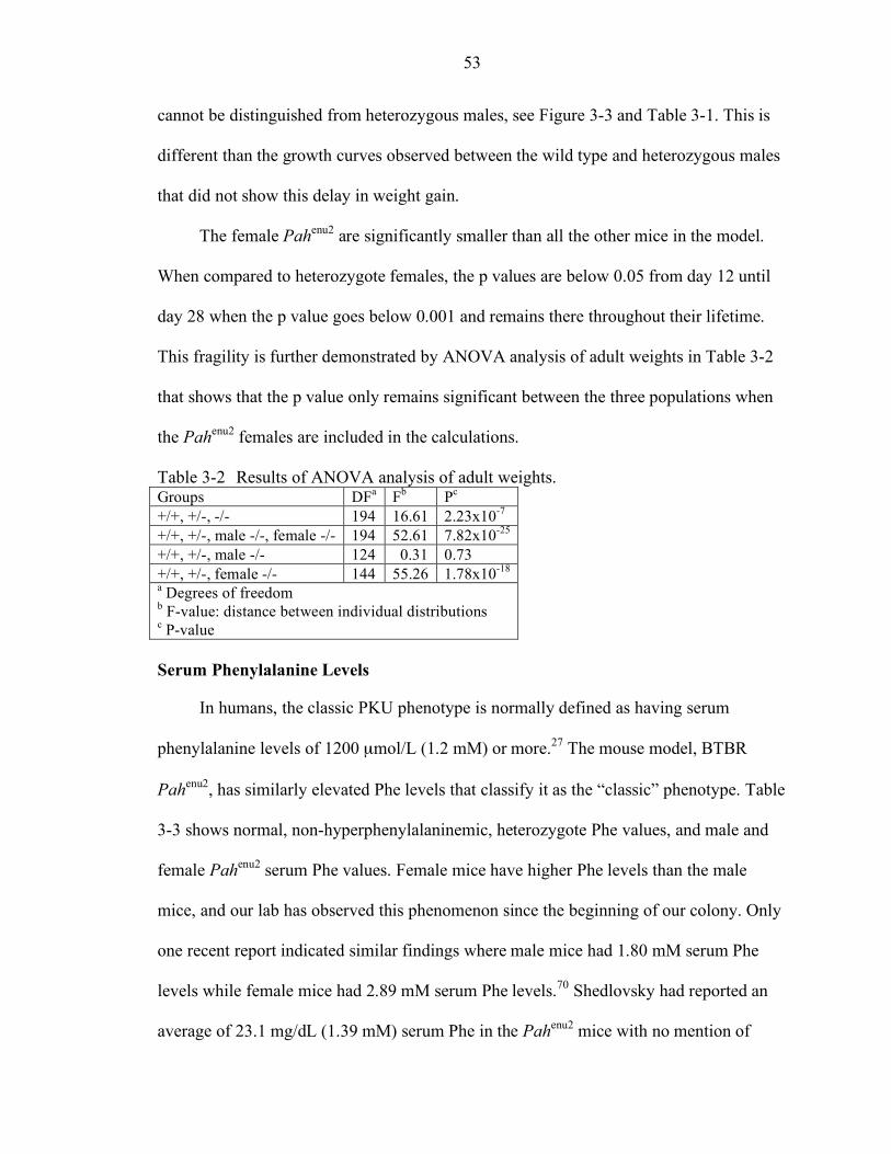
53
cannot be distinguished from heterozygous males, see Figure 3-3 and Table 3-1. This is
different than the growth curves observed between the wild type and heterozygous males
that did not show this delay in weight gain.
The female Pahenu2 are significantly smaller than all the other mice in the model.
When compared to heterozygote females, the p values are below 0.05 from day 12 until
day 28 when the p value goes below 0.001 and remains there throughout their lifetime.
This fragility is further demonstrated by ANOVA analysis of adult weights in Table 3-2
that shows that the p value only remains significant between the three populations when
the Pahenu2 females are included in the calculations.
Table 3-2 Results of ANOVA analysis of adult weights. Groups DFa Fb Pc
+/+, +/-, -/- 194 16.61 2.23x10⁻⁷ +/+, +/-, male -/-, female -/- 194 52.61 7.82x10⁻²⁵ +/+, +/-, male -/- 124 0.31 0.73 +/+, +/-, female -/- 144 55.26 1.78x10⁻¹⁸ a Degrees of freedom b F-value: distance between individual distributions c P-value Serum Phenylalanine Levels
In humans, the classic PKU phenotype is normally defined as having serum
phenylalanine levels of 1200 µmol/L (1.2 mM) or more.27 The mouse model, BTBR
Pahenu2, has similarly elevated Phe levels that classify it as the “classic” phenotype. Table
3-3 shows normal, non-hyperphenylalaninemic, heterozygote Phe values, and male and
female Pahenu2 serum Phe values. Female mice have higher Phe levels than the male
mice, and our lab has observed this phenomenon since the beginning of our colony. Only
one recent report indicated similar findings where male mice had 1.80 mM serum Phe
levels while female mice had 2.89 mM serum Phe levels.70 Shedlovsky had reported an
average of 23.1 mg/dL (1.39 mM) serum Phe in the Pahenu2 mice with no mention of
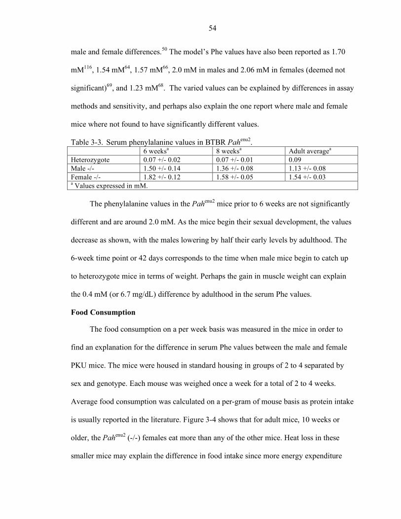
54
male and female differences.50 The model’s Phe values have also been reported as 1.70
mM116, 1.54 mM64, 1.57 mM66, 2.0 mM in males and 2.06 mM in females (deemed not
significant)69, and 1.23 mM68. The varied values can be explained by differences in assay
methods and sensitivity, and perhaps also explain the one report where male and female
mice where not found to have significantly different values.
Table 3-3. Serum phenylalanine values in BTBR Pahenu2. 6 weeksa 8 weeksa Adult averagea
Heterozygote 0.07 +/- 0.02 0.07 +/- 0.01 0.09 Male -/- 1.50 +/- 0.14 1.36 +/- 0.08 1.13 +/- 0.08 Female -/- 1.82 +/- 0.12 1.58 +/- 0.05 1.54 +/- 0.03 a Values expressed in mM.
The phenylalanine values in the Pahenu2 mice prior to 6 weeks are not significantly
different and are around 2.0 mM. As the mice begin their sexual development, the values
decrease as shown, with the males lowering by half their early levels by adulthood. The
6-week time point or 42 days corresponds to the time when male mice begin to catch up
to heterozygote mice in terms of weight. Perhaps the gain in muscle weight can explain
the 0.4 mM (or 6.7 mg/dL) difference by adulthood in the serum Phe values.
Food Consumption
The food consumption on a per week basis was measured in the mice in order to
find an explanation for the difference in serum Phe values between the male and female
PKU mice. The mice were housed in standard housing in groups of 2 to 4 separated by
sex and genotype. Each mouse was weighed once a week for a total of 2 to 4 weeks.
Average food consumption was calculated on a per-gram of mouse basis as protein intake
is usually reported in the literature. Figure 3-4 shows that for adult mice, 10 weeks or
older, the Pahenu2 (-/-) females eat more than any of the other mice. Heat loss in these
smaller mice may explain the difference in food intake since more energy expenditure
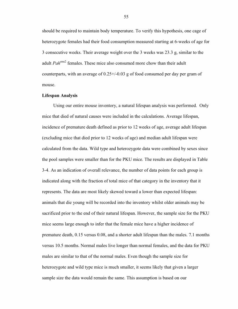
55
should be required to maintain body temperature. To verify this hypothesis, one cage of
heterozygote females had their food consumption measured starting at 6-weeks of age for
3 consecutive weeks. Their average weight over the 3 weeks was 23.3 g, similar to the
adult Pahenu2 females. These mice also consumed more chow than their adult
counterparts, with an average of 0.25+/-0.03 g of food consumed per day per gram of
mouse.
Lifespan Analysis
Using our entire mouse inventory, a natural lifespan analysis was performed. Only
mice that died of natural causes were included in the calculations. Average lifespan,
incidence of premature death defined as prior to 12 weeks of age, average adult lifespan
(excluding mice that died prior to 12 weeks of age) and median adult lifespan were
calculated from the data. Wild type and heterozygote data were combined by sexes since
the pool samples were smaller than for the PKU mice. The results are displayed in Table
3-4. As an indication of overall relevance, the number of data points for each group is
indicated along with the fraction of total mice of that category in the inventory that it
represents. The data are most likely skewed toward a lower than expected lifespan:
animals that die young will be recorded into the inventory whilst older animals may be
sacrificed prior to the end of their natural lifespan. However, the sample size for the PKU
mice seems large enough to infer that the female mice have a higher incidence of
premature death, 0.15 versus 0.08, and a shorter adult lifespan than the males. 7.1 months
versus 10.5 months. Normal males live longer than normal females, and the data for PKU
males are similar to that of the normal males. Even though the sample size for
heterozygote and wild type mice is much smaller, it seems likely that given a larger
sample size the data would remain the same. This assumption is based on our
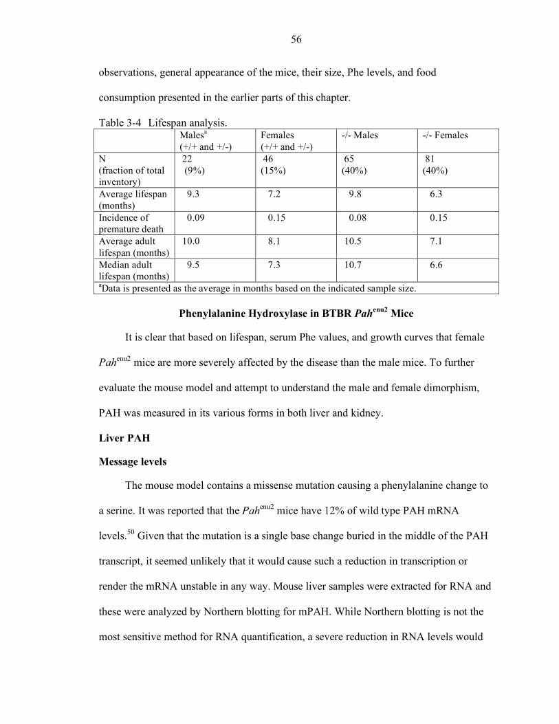
56
observations, general appearance of the mice, their size, Phe levels, and food
consumption presented in the earlier parts of this chapter.
Table 3-4 Lifespan analysis. Malesa
(+/+ and +/-) Females (+/+ and +/-)
-/- Males -/- Females
N (fraction of total inventory)
22 (9%)
46 (15%)
65 (40%)
81 (40%)
Average lifespan (months)
9.3 7.2 9.8 6.3
Incidence of premature death
0.09 0.15 0.08 0.15
Average adult lifespan (months)
10.0 8.1 10.5 7.1
Median adult lifespan (months)
9.5 7.3 10.7 6.6
aData is presented as the average in months based on the indicated sample size.
Phenylalanine Hydroxylase in BTBR Pahenu2 Mice
It is clear that based on lifespan, serum Phe values, and growth curves that female
Pahenu2 mice are more severely affected by the disease than the male mice. To further
evaluate the mouse model and attempt to understand the male and female dimorphism,
PAH was measured in its various forms in both liver and kidney.
Liver PAH
Message levels
The mouse model contains a missense mutation causing a phenylalanine change to
a serine. It was reported that the Pahenu2 mice have 12% of wild type PAH mRNA
levels.50 Given that the mutation is a single base change buried in the middle of the PAH
transcript, it seemed unlikely that it would cause such a reduction in transcription or
render the mRNA unstable in any way. Mouse liver samples were extracted for RNA and
these were analyzed by Northern blotting for mPAH. While Northern blotting is not the
most sensitive method for RNA quantification, a severe reduction in RNA levels would
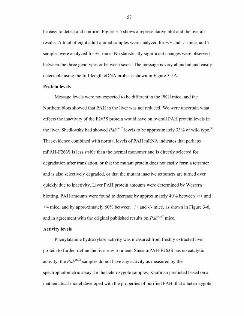
57
be easy to detect and confirm. Figure 3-5 shows a representative blot and the overall
results. A total of eight adult animal samples were analyzed for +/+ and -/- mice, and 7
samples were analyzed for +/- mice. No statistically significant changes were observed
between the three genotypes or between sexes. The message is very abundant and easily
detectable using the full-length cDNA probe as shown in Figure 3-5A.
Protein levels
Message levels were not expected to be different in the PKU mice, and the
Northern blots showed that PAH in the liver was not reduced. We were uncertain what
effects the inactivity of the F263S protein would have on overall PAH protein levels in
the liver. Shedlovsky had showed Pahenu2 levels to be approximately 33% of wild type.50
That evidence combined with normal levels of PAH mRNA indicates that perhaps
mPAH-F263S is less stable than the normal monomer and is directly selected for
degradation after translation, or that the mutant protein does not easily form a tetramer
and is also selectively degraded, or that the mutant inactive tetramers are turned over
quickly due to inactivity. Liver PAH protein amounts were determined by Western
blotting. PAH amounts were found to decrease by approximately 40% between +/+ and
+/- mice, and by approximately 60% between +/+ and -/- mice, as shown in Figure 3-6,
and in agreement with the original published results on Pahenu2 mice.
Activity levels
Phenylalanine hydroxylase activity was measured from freshly extracted liver
protein to further define the liver environment. Since mPAH-F263S has no catalytic
activity, the Pahenu2 samples do not have any activity as measured by the
spectrophotometric assay. In the heterozygote samples, Kaufman predicted based on a
mathematical model developed with the properties of purified PAH, that a heterozygote
58
patient might be expected to have 40% of normal activity based on reported ratios of
serum Phe values in obligate heterozygote versus normal patients. Sample biopsies from
these patients have been reported to be around 30% of normal activity.117 We have
calculated an average of 42% activity in heterozygote mice, agreeing with Kaufman’s
prediction. Table 3-5 contains the result of the PAH assays with the liver samples.
Table 3-5 PAH activity in liver samples. +/+ +/- -/- Activity averagea 1.00 0.42 0.02 Standard deviation 0.15 0.10 0.07 N 9 8 13 aResults are given as averages +/- standard deviation relative to wild type activity levels.
Protein levels in heterozygote mice were approximately 62% of wild type, while
activity has been found to be only 42% of wild type. The Pahenu2 mice have 41% of
normal protein amounts with, as expected, 0% normal activity. This suggests that the
mPAH-F263S monomers are turned over quickly in the liver possibly due to instability or
improper folding. In heterozygotes, with one copy of the mutated gene, the mice show
16% of protein above the assumed 50% normal protein. If the 16% ‘extra’ protein is
assumed to be the mPAH-F263S protein amount, one could assume that Pahenu2 mice
should only have 32% protein. However, 41% was actually observed. Moreover, the 42%
of normal activity levels consistently observed in the +/- mice suggests that an interaction
between normal and mutant monomer is taking place thus reducing the total potential
activity levels and perhaps the amounts of normal monomers. The antibody is incapable
of distinguishing between normal and mutant monomer, thus we are unable to determine
what relative amounts of each protein is present in the heterozygote mice.
59
Kidney PAH
Similar experiments were conducted with kidney samples. Results are shown in
Figure 3-7. Unlike liver PAH, protein amounts are equal between each genotype. Activity
levels decrease by 50% from wild type mice to heterozygote mice. The regulation of
turnover and /or the stability of the protein seem to be different between the two organs,
which is consistent with the state of activation observed with purified kidney rat PAH.38
The rat kidney enzyme was not responsive to pre-incubation with phenylalanine unlike
liver PAH, and it was found to have higher in vitro activity with BH4. Since the
promoters are the same, it is presumed that post-translational modifications are different
in each organ leading to the differences observed in stability of the protein.
It is interesting that the liver and the kidney do not seem to regulate PAH in the
same manner, and perhaps the kidney could be a good target for gene therapy.
Colocalization of the cofactor BH4 and the AAV vector would have to be achieved, thus
requiring that the cell types where PAH and BH4 are present in the kidney be susceptible
to rAAV infection. Studies are currently underway to determine the localization of PAH
in the mouse kidney.
Discussion
The mouse model for phenylketonuria, BTBR Pahenu2, was examined in detail for
this study. BTBR Pahenu2 mice have a single base mutation in the PAH gene leading to
the missense mutation F263S in exon 7.54 The mutated enzyme is catalytically inactive,
and the mice have classic PKU: hyperphenylalaninemia, hypopigmentation, cognitive
deficits and maternal PKU syndrome. We found that female BTBR Pahenu2 mice are
significantly smaller than the male Pahenu2 mice and have a shorter lifespan and a higher
incidence of premature death than any of the other mice. Serum phenylalanine levels in
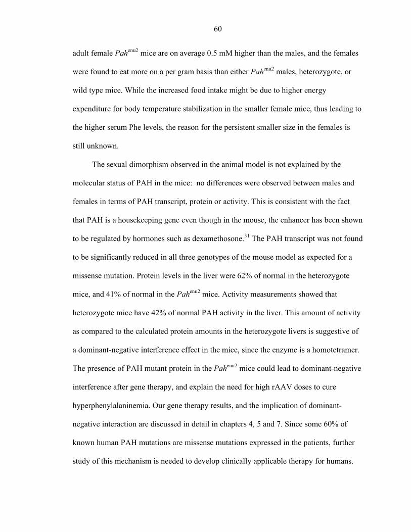
60
adult female Pahenu2 mice are on average 0.5 mM higher than the males, and the females
were found to eat more on a per gram basis than either Pahenu2 males, heterozygote, or
wild type mice. While the increased food intake might be due to higher energy
expenditure for body temperature stabilization in the smaller female mice, thus leading to
the higher serum Phe levels, the reason for the persistent smaller size in the females is
still unknown.
The sexual dimorphism observed in the animal model is not explained by the
molecular status of PAH in the mice: no differences were observed between males and
females in terms of PAH transcript, protein or activity. This is consistent with the fact
that PAH is a housekeeping gene even though in the mouse, the enhancer has been shown
to be regulated by hormones such as dexamethosone.31 The PAH transcript was not found
to be significantly reduced in all three genotypes of the mouse model as expected for a
missense mutation. Protein levels in the liver were 62% of normal in the heterozygote
mice, and 41% of normal in the Pahenu2 mice. Activity measurements showed that
heterozygote mice have 42% of normal PAH activity in the liver. This amount of activity
as compared to the calculated protein amounts in the heterozygote livers is suggestive of
a dominant-negative interference effect in the mice, since the enzyme is a homotetramer.
The presence of PAH mutant protein in the Pahenu2 mice could lead to dominant-negative
interference after gene therapy, and explain the need for high rAAV doses to cure
hyperphenylalaninemia. Our gene therapy results, and the implication of dominant-
negative interaction are discussed in detail in chapters 4, 5 and 7. Since some 60% of
known human PAH mutations are missense mutations expressed in the patients, further
study of this mechanism is needed to develop clinically applicable therapy for humans.
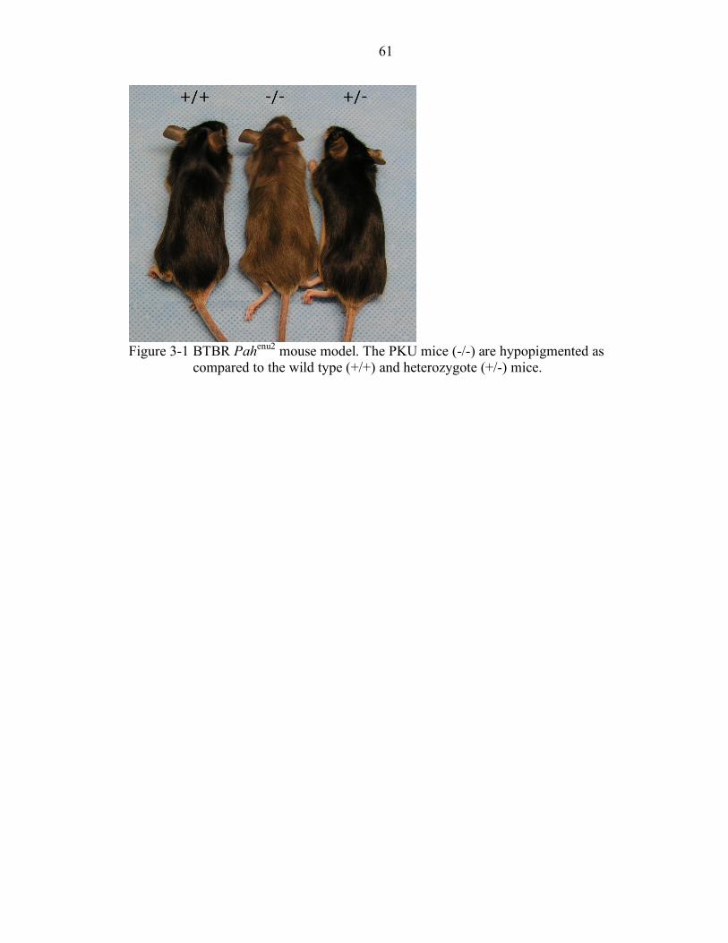
61
Figure 3-1 BTBR Pahenu2 mouse model. The PKU mice (-/-) are hypopigmented as
compared to the wild type (+/+) and heterozygote (+/-) mice.
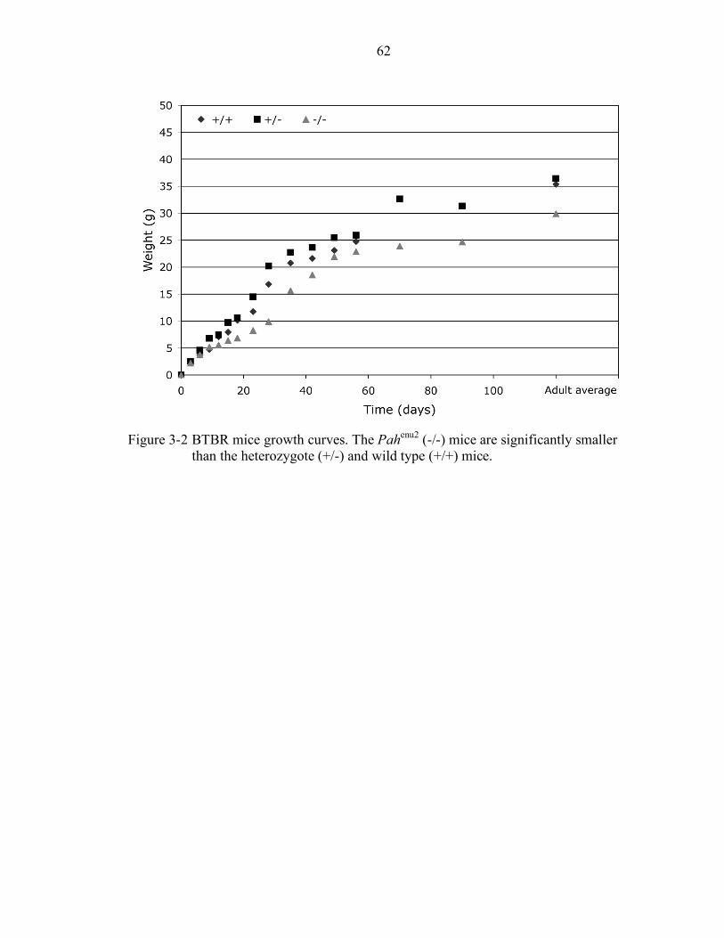
62
Figure 3-2 BTBR mice growth curves. The Pahenu2 (-/-) mice are significantly smaller
than the heterozygote (+/-) and wild type (+/+) mice.
63
Figure 3-3 Male and female weight differences. While female Pahenu2 (-/- Females)
remain significantly smaller than all the other mice, the male Pahenu2 (-/- Males) catch up to the heterozygote (+/-) mice around day 40.
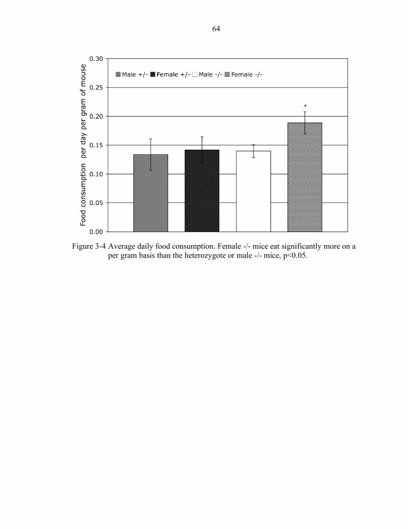
64
Figure 3-4 Average daily food consumption. Female -/- mice eat significantly more on a
per gram basis than the heterozygote or male -/- mice, p<0.05.
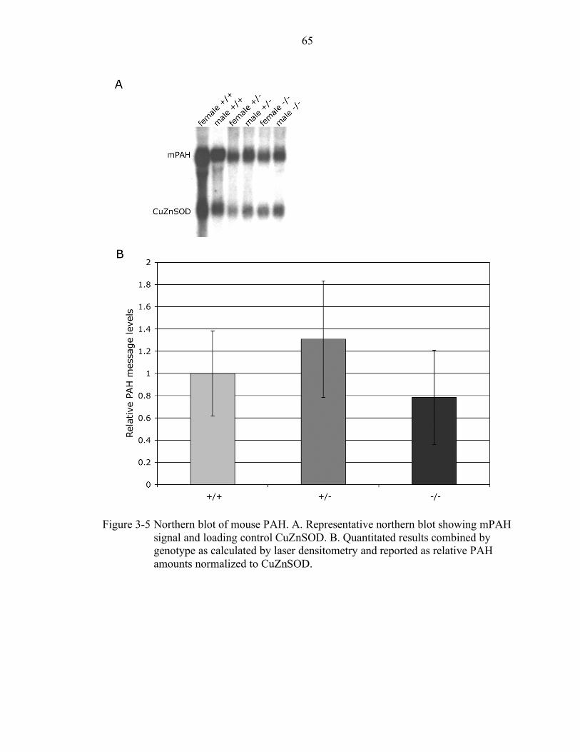
65
Figure 3-5 Northern blot of mouse PAH. A. Representative northern blot showing mPAH
signal and loading control CuZnSOD. B. Quantitated results combined by genotype as calculated by laser densitometry and reported as relative PAH amounts normalized to CuZnSOD.
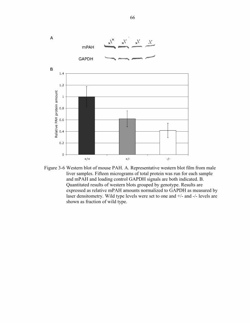
66
Figure 3-6 Western blot of mouse PAH. A. Representative western blot film from male
liver samples. Fifteen micrograms of total protein was run for each sample and mPAH and loading control GAPDH signals are both indicated. B. Quantitated results of western blots grouped by genotype. Results are expressed as relative mPAH amounts normalized to GAPDH as measured by laser densitometry. Wild type levels were set to one and +/- and -/- levels are shown as fraction of wild type.
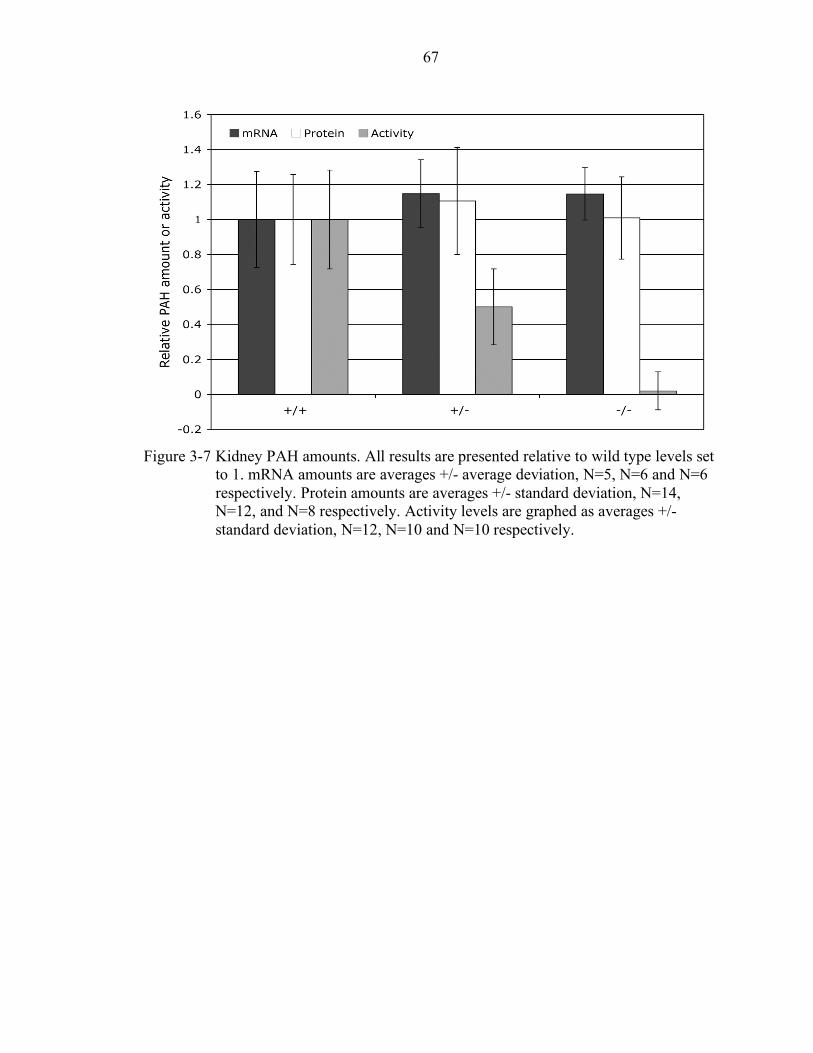
67
Figure 3-7 Kidney PAH amounts. All results are presented relative to wild type levels set
to 1. mRNA amounts are averages +/- average deviation, N=5, N=6 and N=6 respectively. Protein amounts are averages +/- standard deviation, N=14, N=12, and N=8 respectively. Activity levels are graphed as averages +/- standard deviation, N=12, N=10 and N=10 respectively.
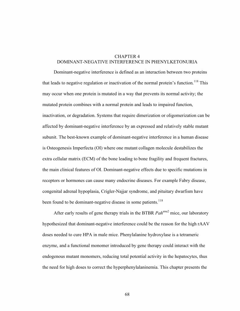
68
CHAPTER 4 DOMINANT-NEGATIVE INTERFERENCE IN PHENYLKETONURIA
Dominant-negative interference is defined as an interaction between two proteins
that leads to negative regulation or inactivation of the normal protein’s function.118 This
may occur when one protein is mutated in a way that prevents its normal activity; the
mutated protein combines with a normal protein and leads to impaired function,
inactivation, or degradation. Systems that require dimerization or oligomerization can be
affected by dominant-negative interference by an expressed and relatively stable mutant
subunit. The best-known example of dominant-negative interference in a human disease
is Osteogenesis Imperfecta (OI) where one mutant collagen molecule destabilizes the
extra cellular matrix (ECM) of the bone leading to bone fragility and frequent fractures,
the main clinical features of OI. Dominant-negative effects due to specific mutations in
receptors or hormones can cause many endocrine diseases. For example Fabry disease,
congenital adrenal hypoplasia, Crigler-Najjar syndrome, and pituitary dwarfism have
been found to be dominant-negative disease in some patients.118
After early results of gene therapy trials in the BTBR Pahenu2 mice, our laboratory
hypothesized that dominant-negative interference could be the reason for the high rAAV
doses needed to cure HPA in male mice. Phenylalanine hydroxylase is a tetrameric
enzyme, and a functional monomer introduced by gene therapy could interact with the
endogenous mutant monomers, reducing total potential activity in the hepatocytes, thus
the need for high doses to correct the hyperphenylalaninemia. This chapter presents the
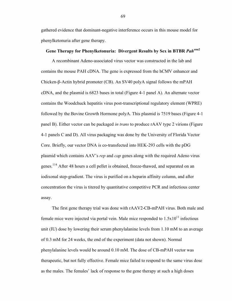
69
gathered evidence that dominant-negative interference occurs in this mouse model for
phenylketonuria after gene therapy.
Gene Therapy for Phenylketonuria: Divergent Results by Sex in BTBR Pahenu2
A recombinant Adeno-associated virus vector was constructed in the lab and
contains the mouse PAH cDNA. The gene is expressed from the hCMV enhancer and
Chicken-β-Actin hybrid promoter (CB). An SV40 polyA signal follows the mPAH
cDNA, and the plasmid is 6823 bases in total (Figure 4-1 panel A). An alternate vector
contains the Woodchuck hepatitis virus post-transcriptional regulatory element (WPRE)
followed by the Bovine Growth Hormone polyA. This plasmid is 7519 bases (Figure 4-1
panel B). Either vector can be packaged in trans to produce rAAV type 2 virions (Figure
4-1 panels C and D). All virus packaging was done by the University of Florida Vector
Core. Briefly, our vector DNA is co-transfected into HEK-293 cells with the pDG
plasmid which contains AAV’s rep and cap genes along with the required Adeno virus
genes.114 After 48 hours a cell pellet is obtained, freeze-thawed, and separated on an
iodixonal step-gradient. The virus is purified on a heparin affinity column, and after
concentration the virus is titered by quantitative competitive PCR and infectious center
assay.
The first gene therapy trial was done with rAAV2-CB-mPAH virus. Both male and
female mice were injected via portal vein. Male mice responded to 1.5x10¹¹ infectious
unit (IU) dose by lowering their serum phenylalanine levels from 1.10 mM to an average
of 0.3 mM for 24 weeks, the end of the experiment (data not shown). Normal
phenylalanine levels would be around 0.10 mM. The dose of CB-mPAH vector was
therapeutic, but not fully effective. Female mice failed to respond to the same virus dose
as the males. The females’ lack of response to the gene therapy at such a high doses
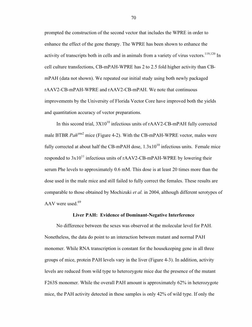
70
prompted the construction of the second vector that includes the WPRE in order to
enhance the effect of the gene therapy. The WPRE has been shown to enhance the
activity of transcripts both in cells and in animals from a variety of virus vectors.119,120 In
cell culture transfections, CB-mPAH-WPRE has 2 to 2.5 fold higher activity than CB-
mPAH (data not shown). We repeated our initial study using both newly packaged
rAAV2-CB-mPAH-WPRE and rAAV2-CB-mPAH. We note that continuous
improvements by the University of Florida Vector Core have improved both the yields
and quantitation accuracy of vector preparations.
In this second trial, 3X10¹⁰ infectious units of rAAV2-CB-mPAH fully corrected
male BTBR Pahenu2 mice (Figure 4-2). With the CB-mPAH-WPRE vector, males were
fully corrected at about half the CB-mPAH dose, 1.3x10¹⁰ infectious units. Female mice
responded to 3x10¹¹ infectious units of rAAV2-CB-mPAH-WPRE by lowering their
serum Phe levels to approximately 0.6 mM. This dose is at least 20 times more than the
dose used in the male mice and still failed to fully correct the females. These results are
comparable to those obtained by Mochizuki et al. in 2004, although different serotypes of
AAV were used.69
Liver PAH: Evidence of Dominant-Negative Interference
No difference between the sexes was observed at the molecular level for PAH.
Nonetheless, the data do point to an interaction between mutant and normal PAH
monomer. While RNA transcription is constant for the housekeeping gene in all three
groups of mice, protein PAH levels vary in the liver (Figure 4-3). In addition, activity
levels are reduced from wild type to heterozygote mice due the presence of the mutant
F263S monomer. While the overall PAH amount is approximately 62% in heterozygote
mice, the PAH activity detected in these samples is only 42% of wild type. If only the
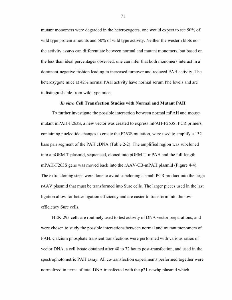
71
mutant monomers were degraded in the heterozygotes, one would expect to see 50% of
wild type protein amounts and 50% of wild type activity. Neither the western blots nor
the activity assays can differentiate between normal and mutant monomers, but based on
the less than ideal percentages observed, one can infer that both monomers interact in a
dominant-negative fashion leading to increased turnover and reduced PAH activity. The
heterozygote mice at 42% normal PAH activity have normal serum Phe levels and are
indistinguishable from wild type mice.
In vitro Cell Transfection Studies with Normal and Mutant PAH
To further investigate the possible interaction between normal mPAH and mouse
mutant mPAH-F263S, a new vector was created to express mPAH-F263S. PCR primers,
containing nucleotide changes to create the F263S mutation, were used to amplify a 132
base pair segment of the PAH cDNA (Table 2-2). The amplified region was subcloned
into a pGEM-T plasmid, sequenced, cloned into pGEM-T-mPAH and the full-length
mPAH-F263S gene was moved back into the rAAV-CB-mPAH plasmid (Figure 4-4).
The extra cloning steps were done to avoid subcloning a small PCR product into the large
rAAV plasmid that must be transformed into Sure cells. The larger pieces used in the last
ligation allow for better ligation efficiency and are easier to transform into the low-
efficiency Sure cells.
HEK-293 cells are routinely used to test activity of DNA vector preparations, and
were chosen to study the possible interactions between normal and mutant monomers of
PAH. Calcium phosphate transient transfections were performed with various ratios of
vector DNA, a cell lysate obtained after 48 to 72 hours post-transfection, and used in the
spectrophotometric PAH assay. All co-transfection experiments performed together were
normalized in terms of total DNA transfected with the p21-newhp plasmid which
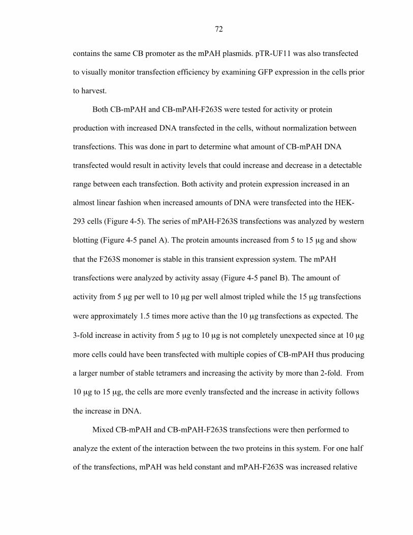
72
contains the same CB promoter as the mPAH plasmids. pTR-UF11 was also transfected
to visually monitor transfection efficiency by examining GFP expression in the cells prior
to harvest.
Both CB-mPAH and CB-mPAH-F263S were tested for activity or protein
production with increased DNA transfected in the cells, without normalization between
transfections. This was done in part to determine what amount of CB-mPAH DNA
transfected would result in activity levels that could increase and decrease in a detectable
range between each transfection. Both activity and protein expression increased in an
almost linear fashion when increased amounts of DNA were transfected into the HEK-
293 cells (Figure 4-5). The series of mPAH-F263S transfections was analyzed by western
blotting (Figure 4-5 panel A). The protein amounts increased from 5 to 15 µg and show
that the F263S monomer is stable in this transient expression system. The mPAH
transfections were analyzed by activity assay (Figure 4-5 panel B). The amount of
activity from 5 µg per well to 10 µg per well almost tripled while the 15 µg transfections
were approximately 1.5 times more active than the 10 µg transfections as expected. The
3-fold increase in activity from 5 µg to 10 µg is not completely unexpected since at 10 µg
more cells could have been transfected with multiple copies of CB-mPAH thus producing
a larger number of stable tetramers and increasing the activity by more than 2-fold. From
10 µg to 15 µg, the cells are more evenly transfected and the increase in activity follows
the increase in DNA.
Mixed CB-mPAH and CB-mPAH-F263S transfections were then performed to
analyze the extent of the interaction between the two proteins in this system. For one half
of the transfections, mPAH was held constant and mPAH-F263S was increased relative
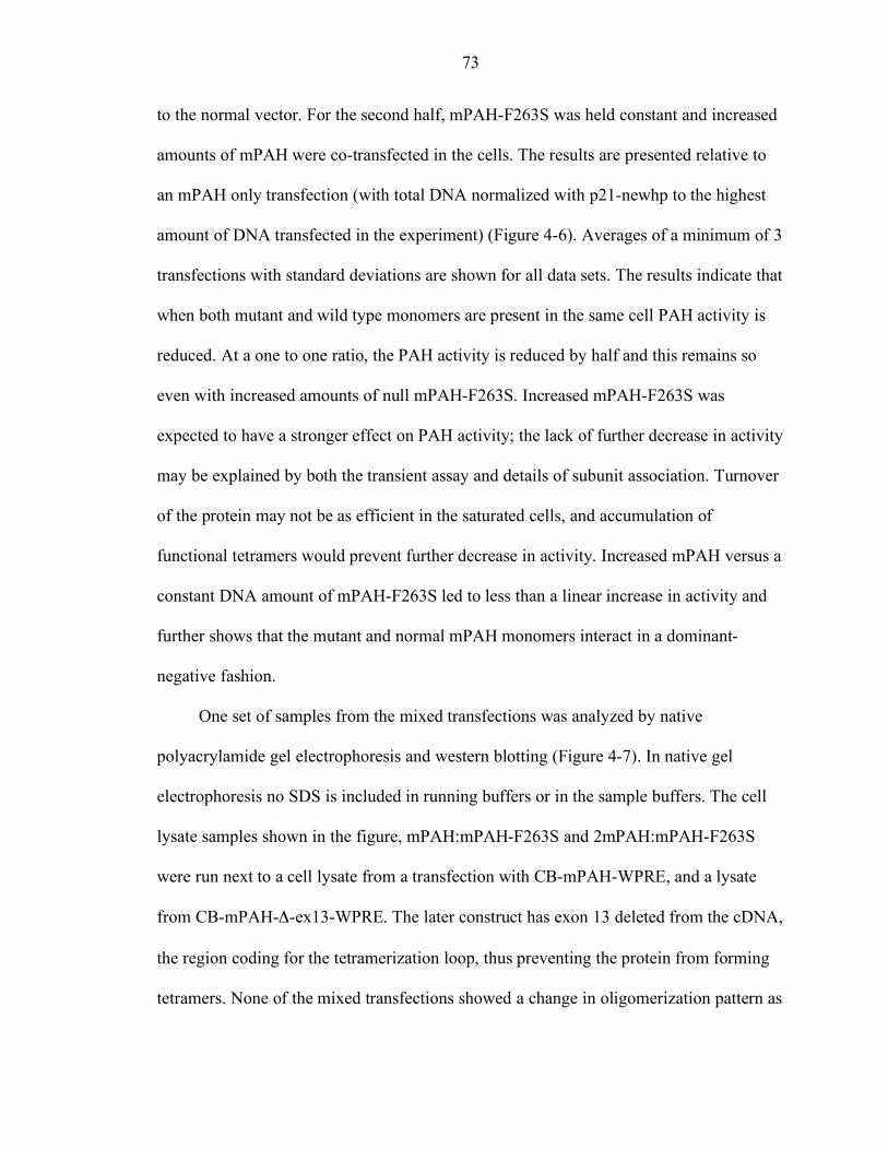
73
to the normal vector. For the second half, mPAH-F263S was held constant and increased
amounts of mPAH were co-transfected in the cells. The results are presented relative to
an mPAH only transfection (with total DNA normalized with p21-newhp to the highest
amount of DNA transfected in the experiment) (Figure 4-6). Averages of a minimum of 3
transfections with standard deviations are shown for all data sets. The results indicate that
when both mutant and wild type monomers are present in the same cell PAH activity is
reduced. At a one to one ratio, the PAH activity is reduced by half and this remains so
even with increased amounts of null mPAH-F263S. Increased mPAH-F263S was
expected to have a stronger effect on PAH activity; the lack of further decrease in activity
may be explained by both the transient assay and details of subunit association. Turnover
of the protein may not be as efficient in the saturated cells, and accumulation of
functional tetramers would prevent further decrease in activity. Increased mPAH versus a
constant DNA amount of mPAH-F263S led to less than a linear increase in activity and
further shows that the mutant and normal mPAH monomers interact in a dominant-
negative fashion.
One set of samples from the mixed transfections was analyzed by native
polyacrylamide gel electrophoresis and western blotting (Figure 4-7). In native gel
electrophoresis no SDS is included in running buffers or in the sample buffers. The cell
lysate samples shown in the figure, mPAH:mPAH-F263S and 2mPAH:mPAH-F263S
were run next to a cell lysate from a transfection with CB-mPAH-WPRE, and a lysate
from CB-mPAH-Δ-ex13-WPRE. The later construct has exon 13 deleted from the cDNA,
the region coding for the tetramerization loop, thus preventing the protein from forming
tetramers. None of the mixed transfections showed a change in oligomerization pattern as
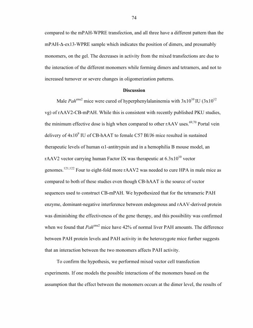
74
compared to the mPAH-WPRE transfection, and all three have a different pattern than the
mPAH-Δ-ex13-WPRE sample which indicates the position of dimers, and presumably
monomers, on the gel. The decreases in activity from the mixed transfections are due to
the interaction of the different monomers while forming dimers and tetramers, and not to
increased turnover or severe changes in oligomerization patterns.
Discussion
Male Pahenu2 mice were cured of hyperphenylalaninemia with 3x10¹⁰ IU (3x10¹²
vg) of rAAV2-CB-mPAH. While this is consistent with recently published PKU studies,
the minimum effective dose is high when compared to other rAAV uses.69,70 Portal vein
delivery of 4x10⁹ IU of CB-hAAT to female C57 Bl/J6 mice resulted in sustained
therapeutic levels of human α1-antitrypsin and in a hemophilia B mouse model, an
rAAV2 vector carrying human Factor IX was therapeutic at 6.3x10¹⁰ vector
genomes.121,122 Four to eight-fold more rAAV2 was needed to cure HPA in male mice as
compared to both of these studies even though CB-hAAT is the source of vector
sequences used to construct CB-mPAH. We hypothesized that for the tetrameric PAH
enzyme, dominant-negative interference between endogenous and rAAV-derived protein
was diminishing the effectiveness of the gene therapy, and this possibility was confirmed
when we found that Pahenu2 mice have 42% of normal liver PAH amounts. The difference
between PAH protein levels and PAH activity in the heterozygote mice further suggests
that an interaction between the two monomers affects PAH activity.
To confirm the hypothesis, we performed mixed vector cell transfection
experiments. If one models the possible interactions of the monomers based on the
assumption that the effect between the monomers occurs at the dimer level, the results of
75
the mixed transfection experiments agree with the statistical predictions. For example, at
a 1:1 ratio of CB-mPAH and CB-mPAH-F263S ¼ of dimers would be fully wild type, ½
would be mixed, and ¼ would be mutant. If the assumption is made that a mixed dimer
might have between zero and full activity, the observation of 62% of normal activity
suggests that mixed dimers are active at about 75% of wild type dimer activity. At the 1:2
transfection ratio, only 11% of dimers are wild type, and 44% are mixed. This predicts
PAH activity should be about 45% of full activity, close to the observed 52%. At the
higher ratio of 1:3, the results fell outside of the predicted PAH activity range (probably
due to saturation of the cell culture system or turnover of missense protein), but
importantly, were still lower than normal. The cell culture model data confirmed our
hypothesis that interaction between normal and mutated monomers leads to reduced PAH
activity thus increasing the rAAV doses needed to reduce HPA in male mice.
Female mice have serum Phe levels 1.5 times higher than males. Other studies have
suggested rAAV2 DNA is retained at about 7-fold higher levels in males than in females,
although this may depend on vector dose.123 Based on these numbers, ten to fourteen-fold
more vector may be needed to cure Pahenu2 females. However, the females were not
corrected at a dose 20-fold higher than in the males, suggesting that additional
mechanisms may cause the observed difference in the therapy response. Multiple studies
are in progress to attempt to elucidate the male and female dimorphism in the response to
gene therapy. While this will be key towards curing maternal PKU syndrome in the mice,
and potentially humans, this study concentrated on reducing overall needed vector doses
to cure HPA by targeting the dominant-negative interference.
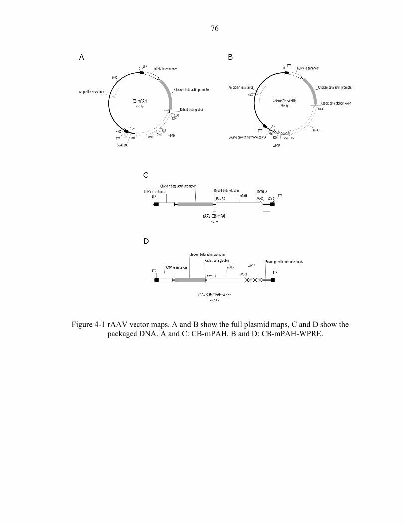
76
Figure 4-1 rAAV vector maps. A and B show the full plasmid maps, C and D show the packaged DNA. A and C: CB-mPAH. B and D: CB-mPAH-WPRE.
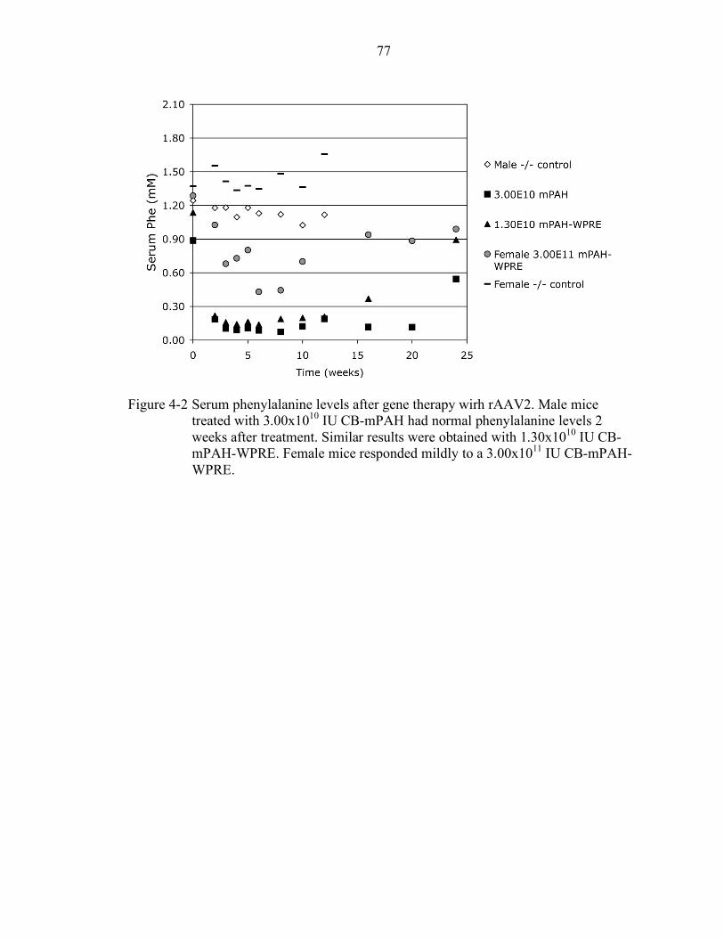
77
Figure 4-2 Serum phenylalanine levels after gene therapy wirh rAAV2. Male mice
treated with 3.00x10¹⁰ IU CB-mPAH had normal phenylalanine levels 2 weeks after treatment. Similar results were obtained with 1.30x10¹⁰ IU CB-mPAH-WPRE. Female mice responded mildly to a 3.00x10¹¹ IU CB-mPAH-WPRE.
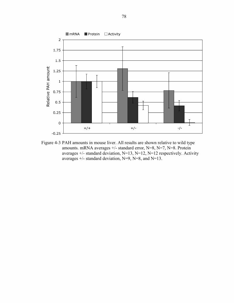
78
Figure 4-3 PAH amounts in mouse liver. All results are shown relative to wild type
amounts. mRNA averages +/- standard error, N=8, N=7, N=8. Protein averages +/- standard deviation, N=13, N=12, N=12 respectively. Activity averages +/- standard deviation, N=9, N=8, and N=13.
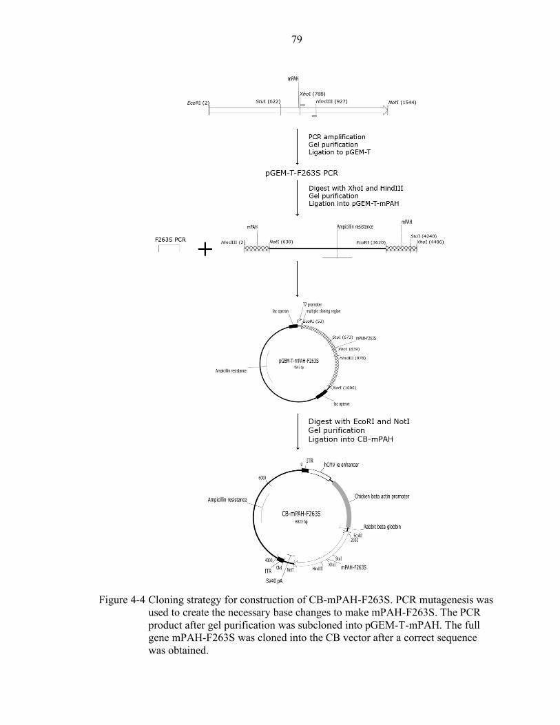
79
Figure 4-4 Cloning strategy for construction of CB-mPAH-F263S. PCR mutagenesis was
used to create the necessary base changes to make mPAH-F263S. The PCR product after gel purification was subcloned into pGEM-T-mPAH. The full gene mPAH-F263S was cloned into the CB vector after a correct sequence was obtained.
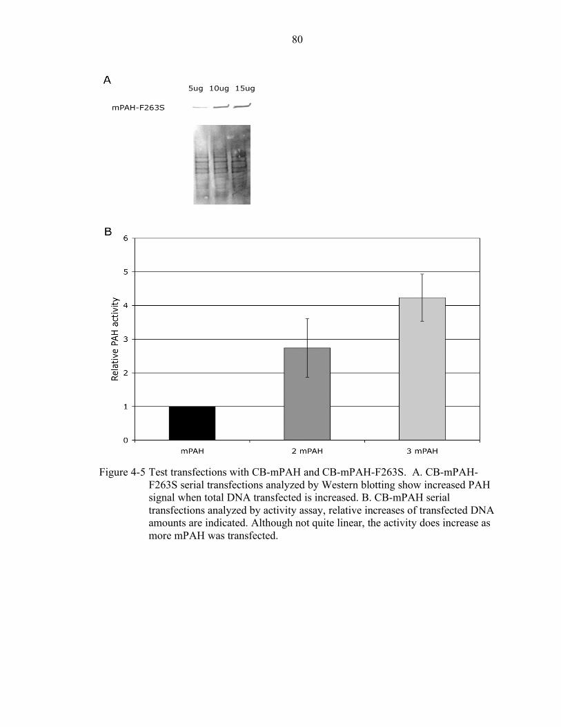
80
Figure 4-5 Test transfections with CB-mPAH and CB-mPAH-F263S. A. CB-mPAH-
F263S serial transfections analyzed by Western blotting show increased PAH signal when total DNA transfected is increased. B. CB-mPAH serial transfections analyzed by activity assay, relative increases of transfected DNA amounts are indicated. Although not quite linear, the activity does increase as more mPAH was transfected.
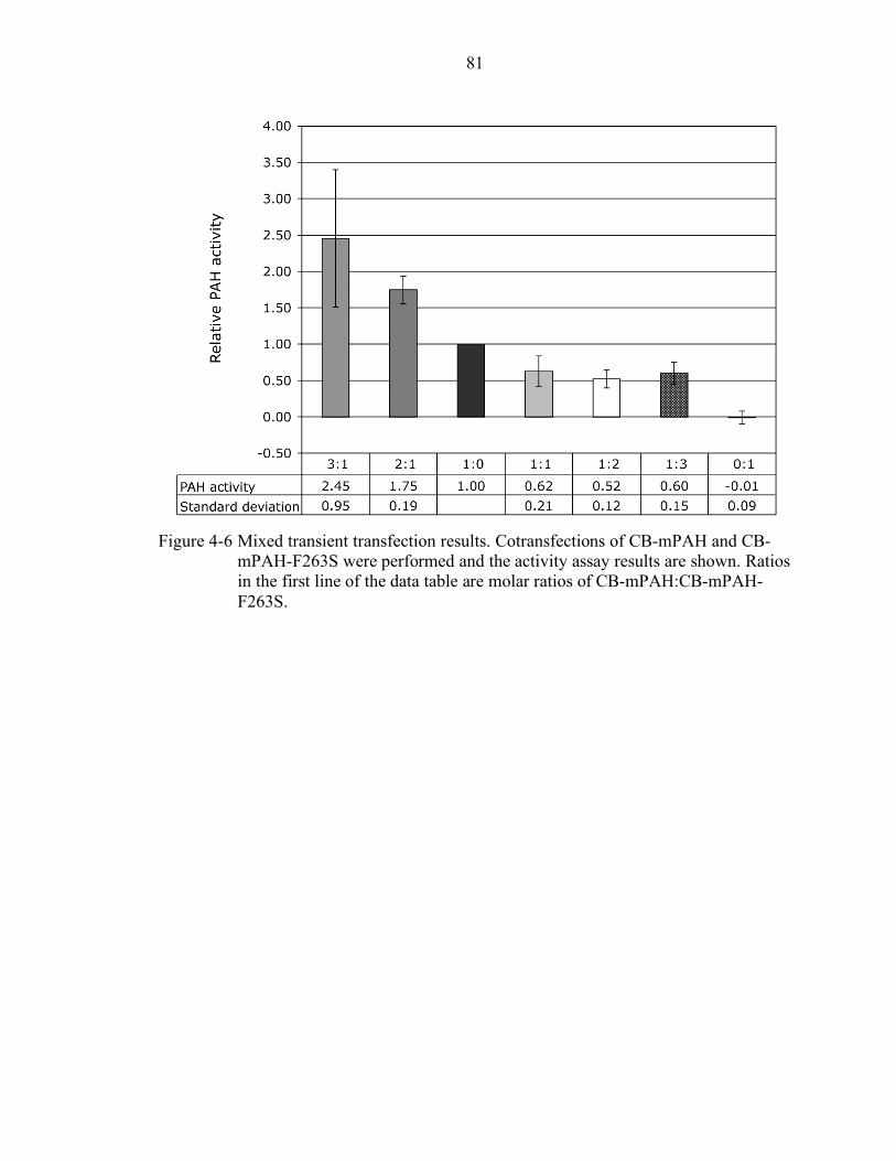
81
Figure 4-6 Mixed transient transfection results. Cotransfections of CB-mPAH and CB-
mPAH-F263S were performed and the activity assay results are shown. Ratios in the first line of the data table are molar ratios of CB-mPAH:CB-mPAH-F263S.
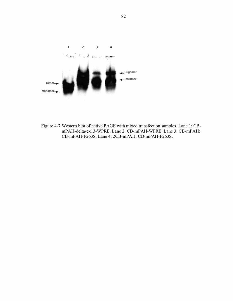
82
Figure 4-7 Western blot of native PAGE with mixed transfection samples. Lane 1: CB-
mPAH-delta-ex13-WPRE. Lane 2: CB-mPAH-WPRE. Lane 3: CB-mPAH: CB-mPAH-F263S. Lane 4: 2CB-mPAH: CB-mPAH-F263S.
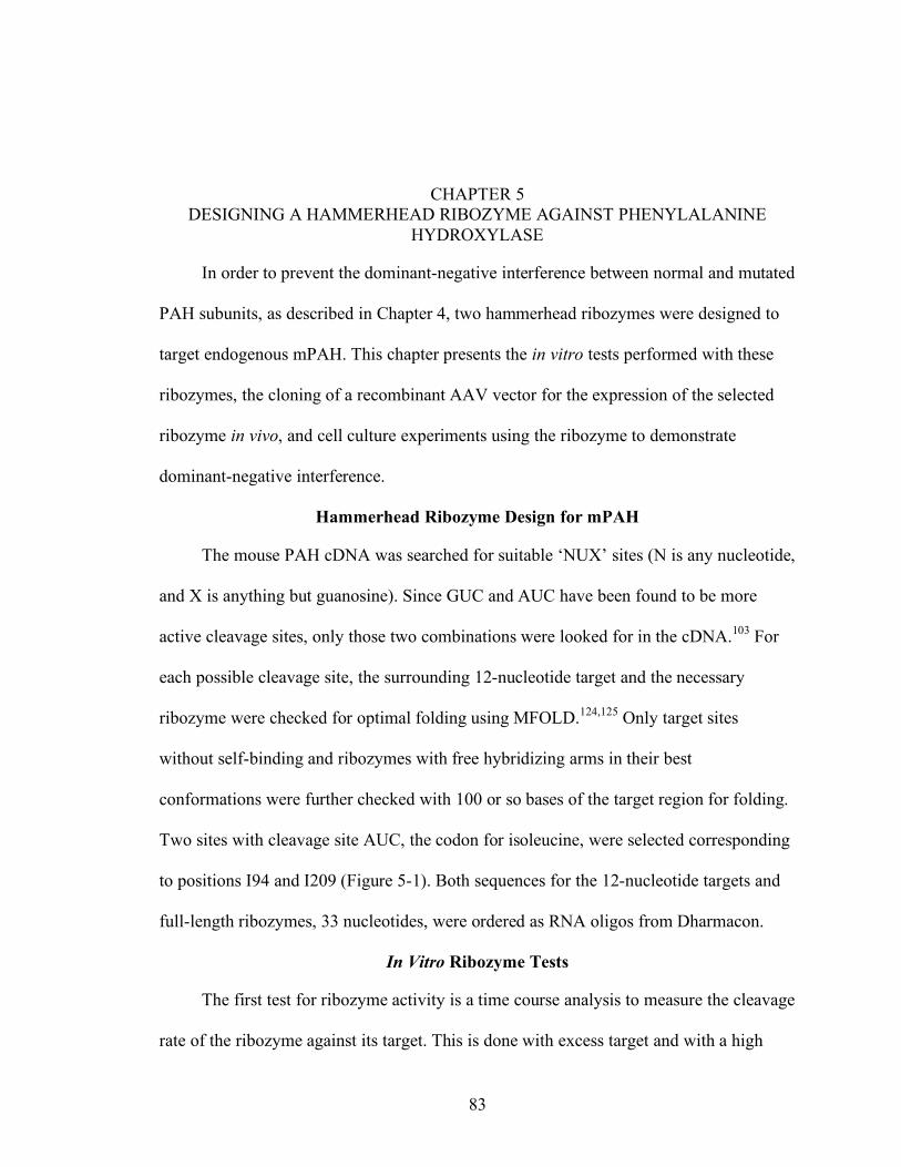
83
CHAPTER 5 DESIGNING A HAMMERHEAD RIBOZYME AGAINST PHENYLALANINE
HYDROXYLASE
In order to prevent the dominant-negative interference between normal and mutated
PAH subunits, as described in Chapter 4, two hammerhead ribozymes were designed to
target endogenous mPAH. This chapter presents the in vitro tests performed with these
ribozymes, the cloning of a recombinant AAV vector for the expression of the selected
ribozyme in vivo, and cell culture experiments using the ribozyme to demonstrate
dominant-negative interference.
Hammerhead Ribozyme Design for mPAH
The mouse PAH cDNA was searched for suitable ‘NUX’ sites (N is any nucleotide,
and X is anything but guanosine). Since GUC and AUC have been found to be more
active cleavage sites, only those two combinations were looked for in the cDNA.103 For
each possible cleavage site, the surrounding 12-nucleotide target and the necessary
ribozyme were checked for optimal folding using MFOLD.124,125 Only target sites
without self-binding and ribozymes with free hybridizing arms in their best
conformations were further checked with 100 or so bases of the target region for folding.
Two sites with cleavage site AUC, the codon for isoleucine, were selected corresponding
to positions I94 and I209 (Figure 5-1). Both sequences for the 12-nucleotide targets and
full-length ribozymes, 33 nucleotides, were ordered as RNA oligos from Dharmacon.
In Vitro Ribozyme Tests
The first test for ribozyme activity is a time course analysis to measure the cleavage
rate of the ribozyme against its target. This is done with excess target and with a high
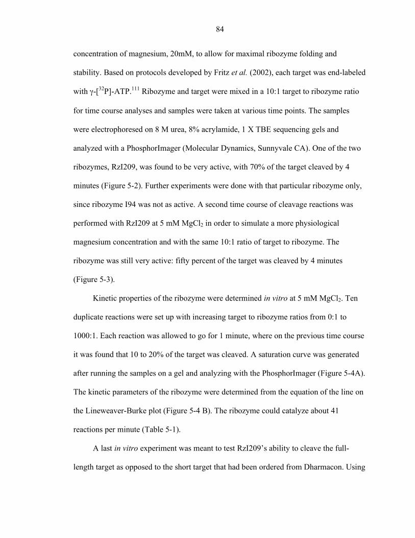
84
concentration of magnesium, 20mM, to allow for maximal ribozyme folding and
stability. Based on protocols developed by Fritz et al. (2002), each target was end-labeled
with γ-[32P]-ATP.111 Ribozyme and target were mixed in a 10:1 target to ribozyme ratio
for time course analyses and samples were taken at various time points. The samples
were electrophoresed on 8 M urea, 8% acrylamide, 1 X TBE sequencing gels and
analyzed with a PhosphorImager (Molecular Dynamics, Sunnyvale CA). One of the two
ribozymes, RzI209, was found to be very active, with 70% of the target cleaved by 4
minutes (Figure 5-2). Further experiments were done with that particular ribozyme only,
since ribozyme I94 was not as active. A second time course of cleavage reactions was
performed with RzI209 at 5 mM MgCl2 in order to simulate a more physiological
magnesium concentration and with the same 10:1 ratio of target to ribozyme. The
ribozyme was still very active: fifty percent of the target was cleaved by 4 minutes
(Figure 5-3).
Kinetic properties of the ribozyme were determined in vitro at 5 mM MgCl2. Ten
duplicate reactions were set up with increasing target to ribozyme ratios from 0:1 to
1000:1. Each reaction was allowed to go for 1 minute, where on the previous time course
it was found that 10 to 20% of the target was cleaved. A saturation curve was generated
after running the samples on a gel and analyzing with the PhosphorImager (Figure 5-4A).
The kinetic parameters of the ribozyme were determined from the equation of the line on
the Lineweaver-Burke plot (Figure 5-4 B). The ribozyme could catalyze about 41
reactions per minute (Table 5-1).
A last in vitro experiment was meant to test RzI209’s ability to cleave the full-
length target as opposed to the short target that had been ordered from Dharmacon. Using

85
the pGEM-T-mPAH construct, an RNA, corresponding to the full-length mRNA, was
transcribed with T7 polymerase. The transcribed product was incubated at 20 mM MgCl2
with excess ribozyme at 37°C. Samples taken at one and two hours were run on a 5%
PAGE 8 M Urea gel, and both cleavage products, 842 and 660 bases, were detected on
the gel (Figure 5-5). Ribozyme I209 is capable of cleaving full-length PAH mRNA in an
in vitro reaction.
Table 5-1 Ribozyme I209 kinetic properties. Vmax 625.00 nM/min Km 12104.38 nM kcat 41.67 min-1
Cloning RzI209 into p21-nhp and Designing a Ribozyme-Resistant mPAH
The chosen rAAV vector for expressing the ribozyme, p21-newhp is based on pTR-
UF12. It contains the hybrid CMV enhancer and chicken β-actin promoter that is closely
related to the hybrid promoter contained in the CB-mPAH plasmids. The hCMV
enhancer in p21-newhp is 381 nucleotides long of which 361 nucleotides from the 3’ end
are identical to the 3’ end of the hCMV enhancer on CB-mPAH which is 535 nucleotides
in total. The p21-newhp plasmid is designed to express the ribozyme from the CB
promoter and follows the hammerhead ribozyme with a hybrid hairpin ribozyme to
evenly cleave the 3’ ends of the transcripts to allow the ribozyme maximum ability to
reach its target mRNA.125 The ribozyme is followed by an SV40 polyA, 187 nucleotides
that is identical to the 5’ end of the SV40 polyA in CB-mPAH which is 222 nucleotides
long. The ribozyme vector also includes a neomycin cassette driven by the PYF 441
enhancer and an HSV thymidine kinase promoter followed by a bovine growth hormone
polyA (BGHpA) signal that is identical to the BGHpA on the CB-mPAH-WPRE
plasmid, both 208 nucleotides long.
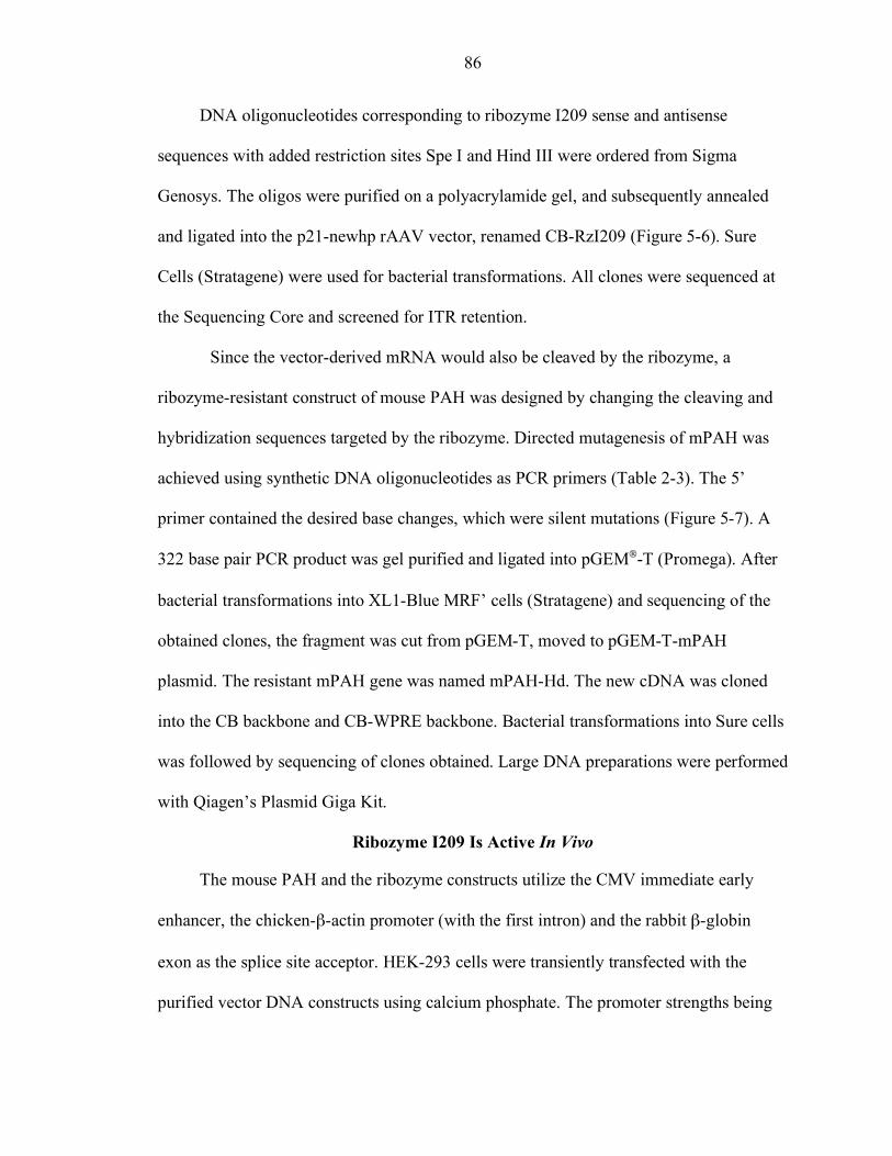
86
DNA oligonucleotides corresponding to ribozyme I209 sense and antisense
sequences with added restriction sites Spe I and Hind III were ordered from Sigma
Genosys. The oligos were purified on a polyacrylamide gel, and subsequently annealed
and ligated into the p21-newhp rAAV vector, renamed CB-RzI209 (Figure 5-6). Sure
Cells (Stratagene) were used for bacterial transformations. All clones were sequenced at
the Sequencing Core and screened for ITR retention.
Since the vector-derived mRNA would also be cleaved by the ribozyme, a
ribozyme-resistant construct of mouse PAH was designed by changing the cleaving and
hybridization sequences targeted by the ribozyme. Directed mutagenesis of mPAH was
achieved using synthetic DNA oligonucleotides as PCR primers (Table 2-3). The 5’
primer contained the desired base changes, which were silent mutations (Figure 5-7). A
322 base pair PCR product was gel purified and ligated into pGEM-T (Promega). After
bacterial transformations into XL1-Blue MRF’ cells (Stratagene) and sequencing of the
obtained clones, the fragment was cut from pGEM-T, moved to pGEM-T-mPAH
plasmid. The resistant mPAH gene was named mPAH-Hd. The new cDNA was cloned
into the CB backbone and CB-WPRE backbone. Bacterial transformations into Sure cells
was followed by sequencing of clones obtained. Large DNA preparations were performed
with Qiagen’s Plasmid Giga Kit.
Ribozyme I209 Is Active In Vivo
The mouse PAH and the ribozyme constructs utilize the CMV immediate early
enhancer, the chicken-β-actin promoter (with the first intron) and the rabbit β-globin
exon as the splice site acceptor. HEK-293 cells were transiently transfected with the
purified vector DNA constructs using calcium phosphate. The promoter strengths being
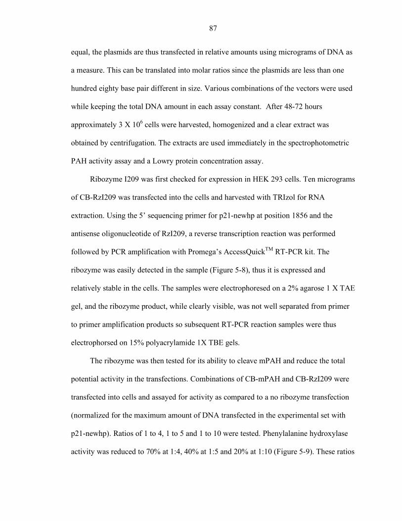
87
equal, the plasmids are thus transfected in relative amounts using micrograms of DNA as
a measure. This can be translated into molar ratios since the plasmids are less than one
hundred eighty base pair different in size. Various combinations of the vectors were used
while keeping the total DNA amount in each assay constant. After 48-72 hours
approximately 3 X 10⁶ cells were harvested, homogenized and a clear extract was
obtained by centrifugation. The extracts are used immediately in the spectrophotometric
PAH activity assay and a Lowry protein concentration assay.
Ribozyme I209 was first checked for expression in HEK 293 cells. Ten micrograms
of CB-RzI209 was transfected into the cells and harvested with TRIzol for RNA
extraction. Using the 5’ sequencing primer for p21-newhp at position 1856 and the
antisense oligonucleotide of RzI209, a reverse transcription reaction was performed
followed by PCR amplification with Promega’s AccessQuickTM RT-PCR kit. The
ribozyme was easily detected in the sample (Figure 5-8), thus it is expressed and
relatively stable in the cells. The samples were electrophoresed on a 2% agarose 1 X TAE
gel, and the ribozyme product, while clearly visible, was not well separated from primer
to primer amplification products so subsequent RT-PCR reaction samples were thus
electrophorsed on 15% polyacrylamide 1X TBE gels.
The ribozyme was then tested for its ability to cleave mPAH and reduce the total
potential activity in the transfections. Combinations of CB-mPAH and CB-RzI209 were
transfected into cells and assayed for activity as compared to a no ribozyme transfection
(normalized for the maximum amount of DNA transfected in the experimental set with
p21-newhp). Ratios of 1 to 4, 1 to 5 and 1 to 10 were tested. Phenylalanine hydroxylase
activity was reduced to 70% at 1:4, 40% at 1:5 and 20% at 1:10 (Figure 5-9). These ratios
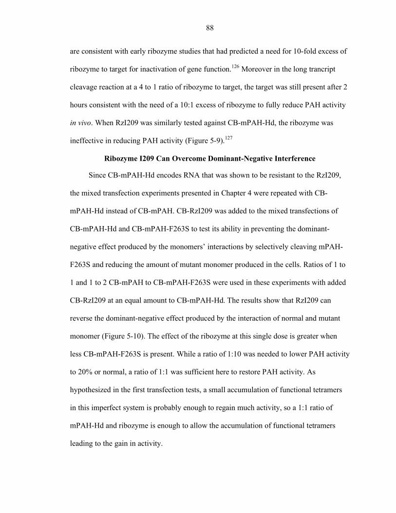
88
are consistent with early ribozyme studies that had predicted a need for 10-fold excess of
ribozyme to target for inactivation of gene function.126 Moreover in the long trancript
cleavage reaction at a 4 to 1 ratio of ribozyme to target, the target was still present after 2
hours consistent with the need of a 10:1 excess of ribozyme to fully reduce PAH activity
in vivo. When RzI209 was similarly tested against CB-mPAH-Hd, the ribozyme was
ineffective in reducing PAH activity (Figure 5-9).127
Ribozyme I209 Can Overcome Dominant-Negative Interference
Since CB-mPAH-Hd encodes RNA that was shown to be resistant to the RzI209,
the mixed transfection experiments presented in Chapter 4 were repeated with CB-
mPAH-Hd instead of CB-mPAH. CB-RzI209 was added to the mixed transfections of
CB-mPAH-Hd and CB-mPAH-F263S to test its ability in preventing the dominant-
negative effect produced by the monomers’ interactions by selectively cleaving mPAH-
F263S and reducing the amount of mutant monomer produced in the cells. Ratios of 1 to
1 and 1 to 2 CB-mPAH to CB-mPAH-F263S were used in these experiments with added
CB-RzI209 at an equal amount to CB-mPAH-Hd. The results show that RzI209 can
reverse the dominant-negative effect produced by the interaction of normal and mutant
monomer (Figure 5-10). The effect of the ribozyme at this single dose is greater when
less CB-mPAH-F263S is present. While a ratio of 1:10 was needed to lower PAH activity
to 20% or normal, a ratio of 1:1 was sufficient here to restore PAH activity. As
hypothesized in the first transfection tests, a small accumulation of functional tetramers
in this imperfect system is probably enough to regain much activity, so a 1:1 ratio of
mPAH-Hd and ribozyme is enough to allow the accumulation of functional tetramers
leading to the gain in activity.
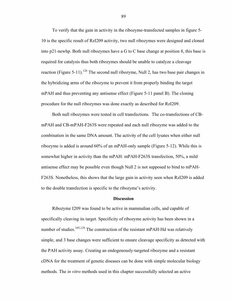
89
To verify that the gain in activity in the ribozyme-transfected samples in figure 5-
10 is the specific result of RzI209 activity, two null ribozymes were designed and cloned
into p21-newhp. Both null ribozymes have a G to C base change at position 8, this base is
required for catalysis thus both ribozymes should be unable to catalyze a cleavage
reaction (Figure 5-11).126 The second null ribozyme, Null 2, has two base pair changes in
the hybridizing arms of the ribozyme to prevent it from properly binding the target
mPAH and thus preventing any antisense effect (Figure 5-11 panel B). The cloning
procedure for the null ribozymes was done exactly as described for RzI209.
Both null ribozymes were tested in cell transfections. The co-transfections of CB-
mPAH and CB-mPAH-F263S were repeated and each null ribozyme was added to the
combination in the same DNA amount. The activity of the cell lysates when either null
ribozyme is added is around 60% of an mPAH-only sample (Figure 5-12). While this is
somewhat higher in activity than the mPAH: mPAH-F263S transfection, 50%, a mild
antisense effect may be possible even though Null 2 is not supposed to bind to mPAH-
F263S. Nonetheless, this shows that the large gain in activity seen when RzI209 is added
to the double transfection is specific to the ribozyme’s activity.
Discussion
Ribozyme I209 was found to be active in mammalian cells, and capable of
specifically cleaving its target. Specificity of ribozyme activity has been shown in a
number of studies.105,128 The construction of the resistant mPAH-Hd was relatively
simple, and 3 base changes were sufficient to ensure cleavage specificity as detected with
the PAH activity assay. Creating an endogenously-targeted ribozyme and a resistant
cDNA for the treatment of genetic diseases can be done with simple molecular biology
methods. The in vitro methods used in this chapter successfully selected an active
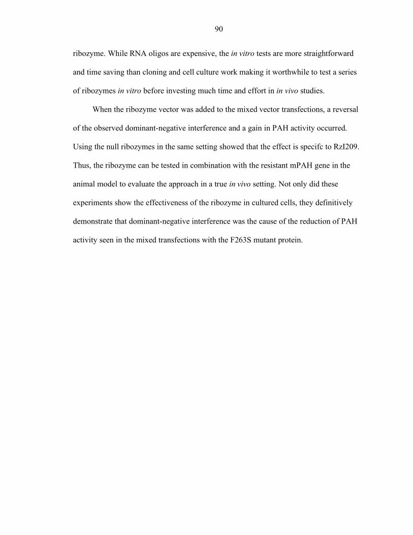
90
ribozyme. While RNA oligos are expensive, the in vitro tests are more straightforward
and time saving than cloning and cell culture work making it worthwhile to test a series
of ribozymes in vitro before investing much time and effort in in vivo studies.
When the ribozyme vector was added to the mixed vector transfections, a reversal
of the observed dominant-negative interference and a gain in PAH activity occurred.
Using the null ribozymes in the same setting showed that the effect is specifc to RzI209.
Thus, the ribozyme can be tested in combination with the resistant mPAH gene in the
animal model to evaluate the approach in a true in vivo setting. Not only did these
experiments show the effectiveness of the ribozyme in cultured cells, they definitively
demonstrate that dominant-negative interference was the cause of the reduction of PAH
activity seen in the mixed transfections with the F263S mutant protein.
91
Figure 5-1 Mouse PAH ribozyme designs. Ribozymes are aligned to their targets with
cleavage target site in red (A and B). Optimal folding conformations of the ribozymes as determined by MFOLD, GC bonds are indicated in red, AU in blue (C and D). A,C Ribozyme I94. B,D Ribozyme I209.
92
Figure 5-2 Time Course analyses with ribozymes at 20mM MgCl2. Time is indicated in
minutes. A. Ribozyme I94 failed to effectively cleave its target. B. Ribozyme I209 cleaved 43% of its target by 1 minute.
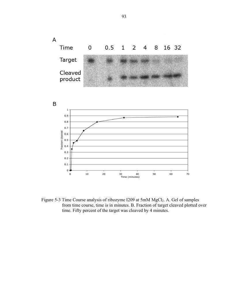
93
Figure 5-3 Time Course analysis of ribozyme I209 at 5mM MgCl2. A. Gel of samples
from time course, time is in minutes. B. Fraction of target cleaved plotted over time. Fifty percent of the target was cleaved by 4 minutes.

94
Figure 5-4 Ribozyme I209 kinetic analysis. A. Saturation curve. B. Lineweaver Burke
Plot generated from A.
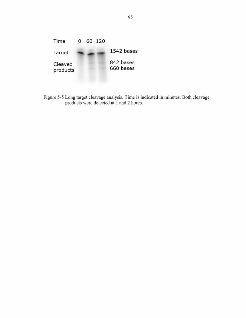
95
Figure 5-5 Long target cleavage analysis. Time is indicated in minutes. Both cleavage
products were detected at 1 and 2 hours.
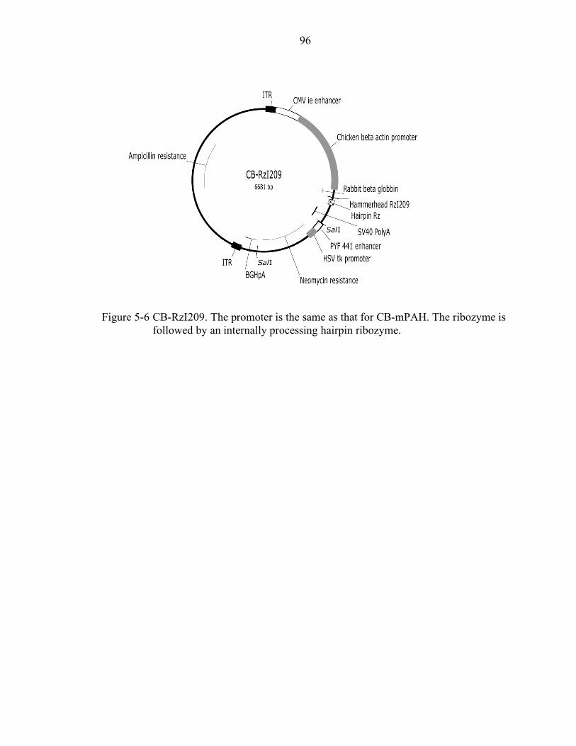
96
Figure 5-6 CB-RzI209. The promoter is the same as that for CB-mPAH. The ribozyme is
followed by an internally processing hairpin ribozyme.

97
Figure 5-7 Cloning strategy for the construction of a ribozyme-resistant mPAH clone.
PCR mutagenesis was performed to selectively change bases in order to render mPAH resistant to RzI209. After gel purification, the PCR product was subcloned into pGEM-T-mPAH. The full length mutagenized cDNA, mPAH-Hd, was cloned into the CB vector.
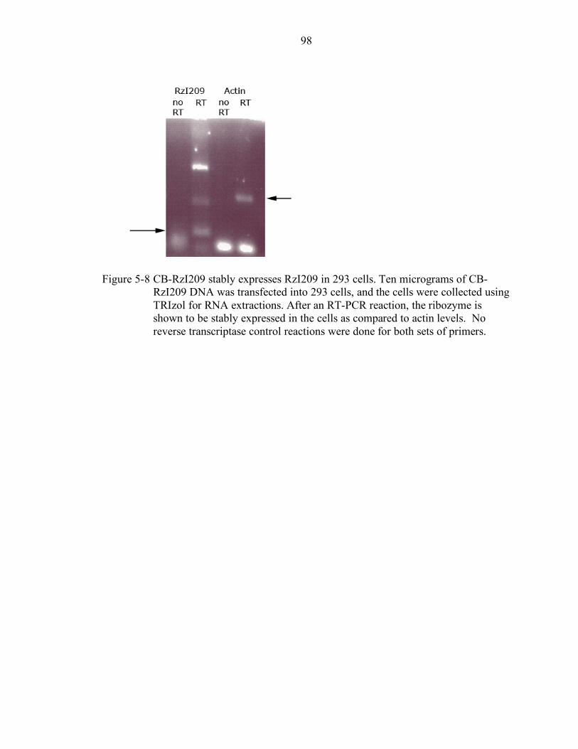
98
Figure 5-8 CB-RzI209 stably expresses RzI209 in 293 cells. Ten micrograms of CB-
RzI209 DNA was transfected into 293 cells, and the cells were collected using TRIzol for RNA extractions. After an RT-PCR reaction, the ribozyme is shown to be stably expressed in the cells as compared to actin levels. No reverse transcriptase control reactions were done for both sets of primers.
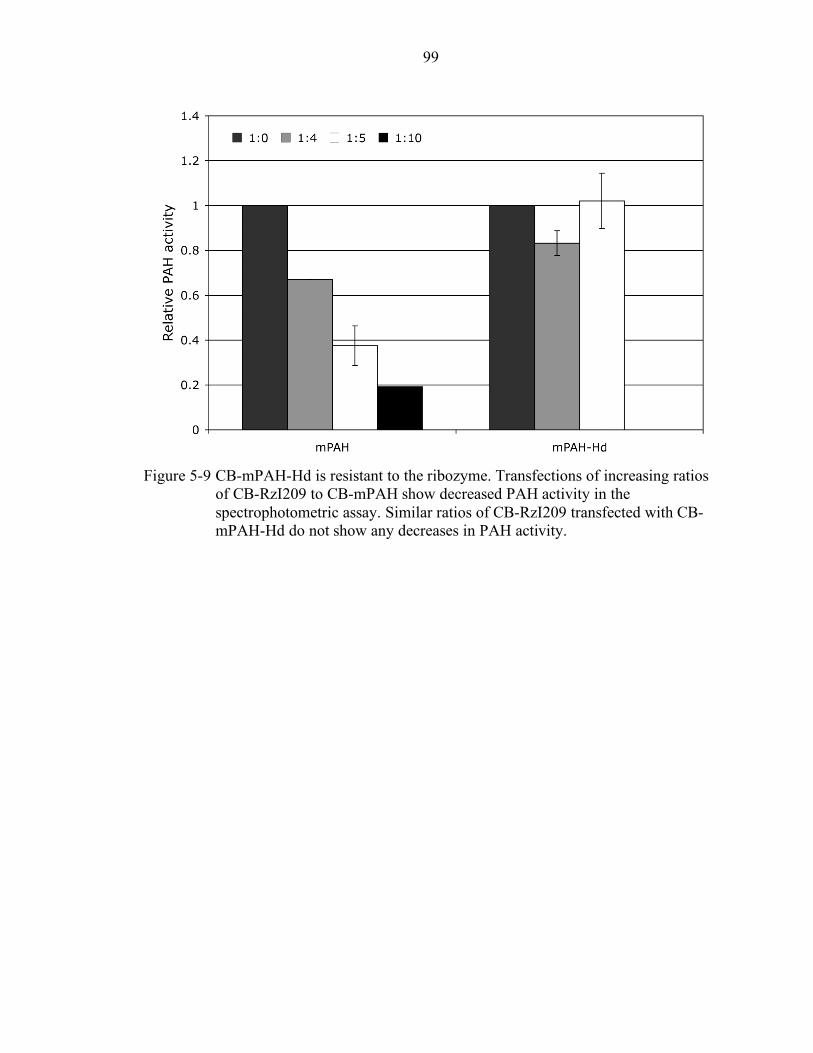
99
Figure 5-9 CB-mPAH-Hd is resistant to the ribozyme. Transfections of increasing ratios
of CB-RzI209 to CB-mPAH show decreased PAH activity in the spectrophotometric assay. Similar ratios of CB-RzI209 transfected with CB-mPAH-Hd do not show any decreases in PAH activity.
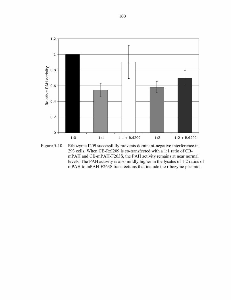
100
Figure 5-10 Ribozyme I209 successfully prevents dominant-negative interference in
293 cells. When CB-RzI209 is co-transfected with a 1:1 ratio of CB-mPAH and CB-mPAH-F263S, the PAH activity remains at near normal levels. The PAH activity is also mildly higher in the lysates of 1:2 ratios of mPAH to mPAH-F263S transfections that include the ribozyme plasmid.
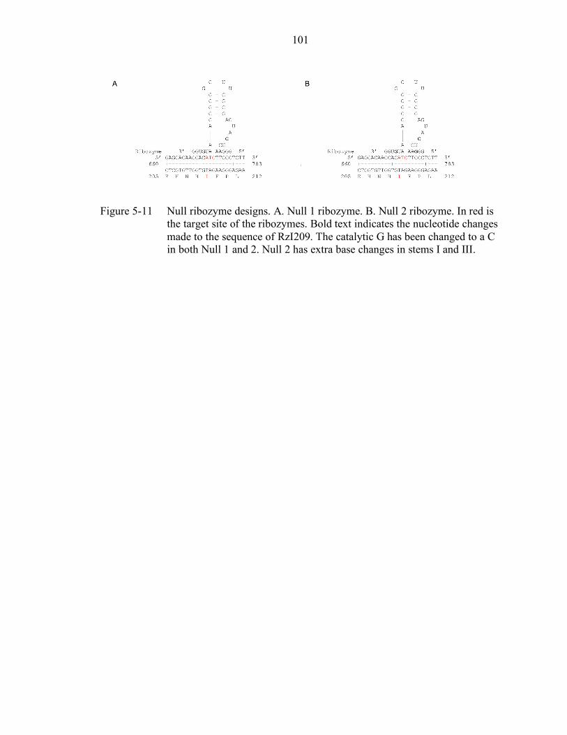
101
Figure 5-11 Null ribozyme designs. A. Null 1 ribozyme. B. Null 2 ribozyme. In red is
the target site of the ribozymes. Bold text indicates the nucleotide changes made to the sequence of RzI209. The catalytic G has been changed to a C in both Null 1 and 2. Null 2 has extra base changes in stems I and III.
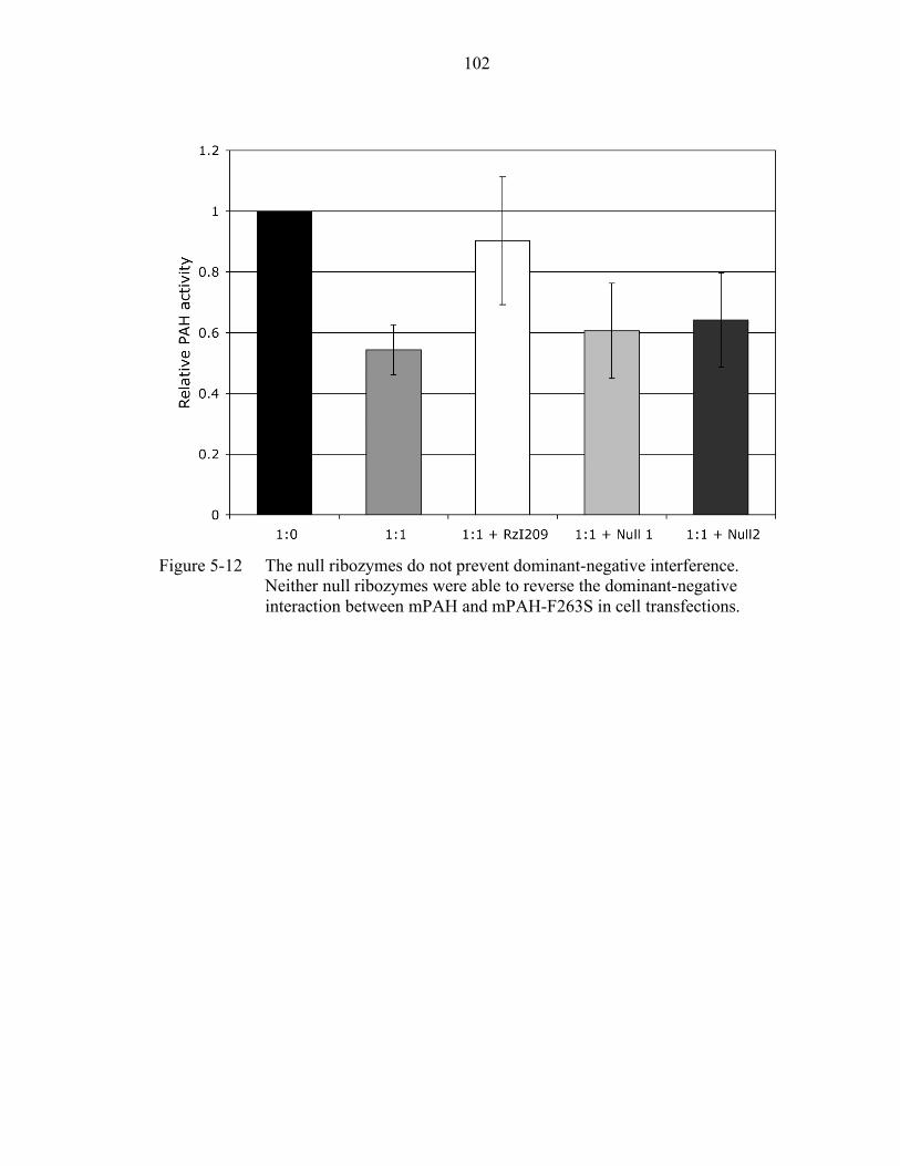
102
Figure 5-12 The null ribozymes do not prevent dominant-negative interference.
Neither null ribozymes were able to reverse the dominant-negative interaction between mPAH and mPAH-F263S in cell transfections.
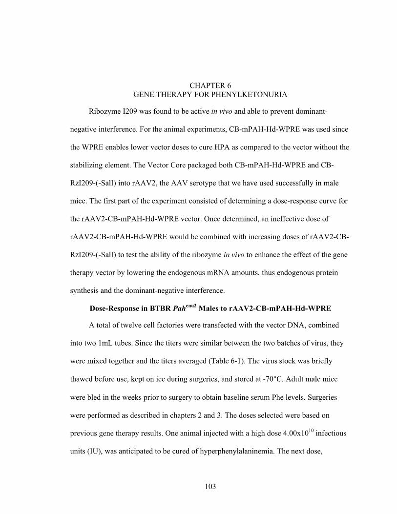
103
CHAPTER 6 GENE THERAPY FOR PHENYLKETONURIA
Ribozyme I209 was found to be active in vivo and able to prevent dominant-
negative interference. For the animal experiments, CB-mPAH-Hd-WPRE was used since
the WPRE enables lower vector doses to cure HPA as compared to the vector without the
stabilizing element. The Vector Core packaged both CB-mPAH-Hd-WPRE and CB-
RzI209-(-SalI) into rAAV2, the AAV serotype that we have used successfully in male
mice. The first part of the experiment consisted of determining a dose-response curve for
the rAAV2-CB-mPAH-Hd-WPRE vector. Once determined, an ineffective dose of
rAAV2-CB-mPAH-Hd-WPRE would be combined with increasing doses of rAAV2-CB-
RzI209-(-SalI) to test the ability of the ribozyme in vivo to enhance the effect of the gene
therapy vector by lowering the endogenous mRNA amounts, thus endogenous protein
synthesis and the dominant-negative interference.
Dose-Response in BTBR Pahenu2 Males to rAAV2-CB-mPAH-Hd-WPRE
A total of twelve cell factories were transfected with the vector DNA, combined
into two 1mL tubes. Since the titers were similar between the two batches of virus, they
were mixed together and the titers averaged (Table 6-1). The virus stock was briefly
thawed before use, kept on ice during surgeries, and stored at -70°C. Adult male mice
were bled in the weeks prior to surgery to obtain baseline serum Phe levels. Surgeries
were performed as described in chapters 2 and 3. The doses selected were based on
previous gene therapy results. One animal injected with a high dose 4.00x10¹⁰ infectious
units (IU), was anticipated to be cured of hyperphenylalaninemia. The next dose,
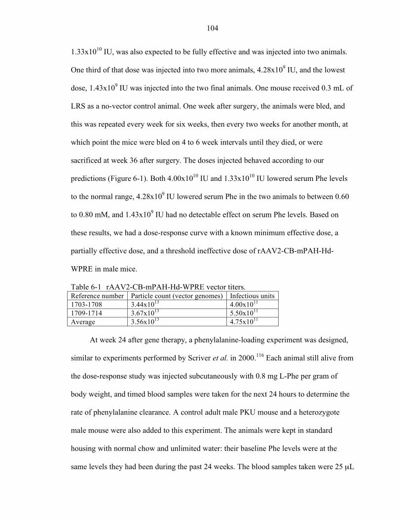
104
1.33x10¹⁰ IU, was also expected to be fully effective and was injected into two animals.
One third of that dose was injected into two more animals, 4.28x10⁹ IU, and the lowest
dose, 1.43x10⁹ IU was injected into the two final animals. One mouse received 0.3 mL of
LRS as a no-vector control animal. One week after surgery, the animals were bled, and
this was repeated every week for six weeks, then every two weeks for another month, at
which point the mice were bled on 4 to 6 week intervals until they died, or were
sacrificed at week 36 after surgery. The doses injected behaved according to our
predictions (Figure 6-1). Both 4.00x10¹⁰ IU and 1.33x10¹⁰ IU lowered serum Phe levels
to the normal range, 4.28x10⁹ IU lowered serum Phe in the two animals to between 0.60
to 0.80 mM, and 1.43x10⁹ IU had no detectable effect on serum Phe levels. Based on
these results, we had a dose-response curve with a known minimum effective dose, a
partially effective dose, and a threshold ineffective dose of rAAV2-CB-mPAH-Hd-
WPRE in male mice.
Table 6-1 rAAV2-CB-mPAH-Hd-WPRE vector titers. Reference number Particle count (vector genomes) Infectious units 1703-1708 3.44x10¹³ 4.00x10¹¹
1709-1714 3.67x10¹³ 5.50x10¹¹
Average 3.56x10¹³ 4.75x10¹¹
At week 24 after gene therapy, a phenylalanine-loading experiment was designed,
similar to experiments performed by Scriver et al. in 2000.116 Each animal still alive from
the dose-response study was injected subcutaneously with 0.8 mg L-Phe per gram of
body weight, and timed blood samples were taken for the next 24 hours to determine the
rate of phenylalanine clearance. A control adult male PKU mouse and a heterozygote
male mouse were also added to this experiment. The animals were kept in standard
housing with normal chow and unlimited water: their baseline Phe levels were at the
same levels they had been during the past 24 weeks. The blood samples taken were 25 µL
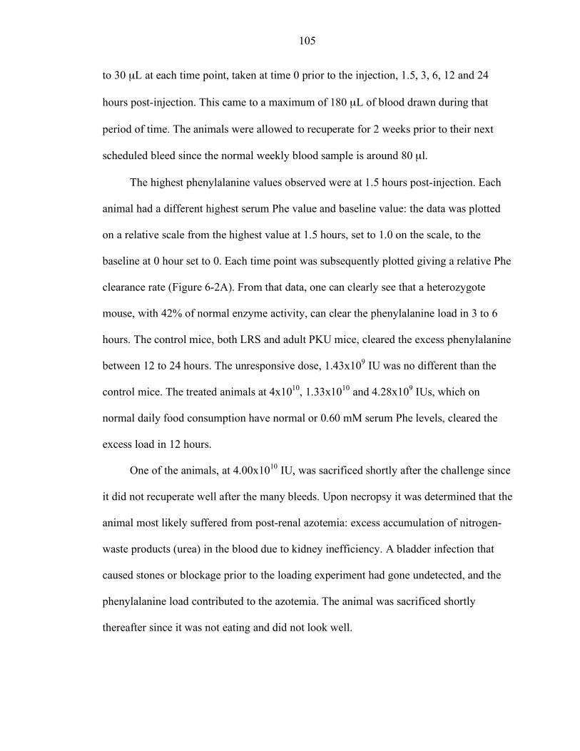
105
to 30 µL at each time point, taken at time 0 prior to the injection, 1.5, 3, 6, 12 and 24
hours post-injection. This came to a maximum of 180 µL of blood drawn during that
period of time. The animals were allowed to recuperate for 2 weeks prior to their next
scheduled bleed since the normal weekly blood sample is around 80 µl.
The highest phenylalanine values observed were at 1.5 hours post-injection. Each
animal had a different highest serum Phe value and baseline value: the data was plotted
on a relative scale from the highest value at 1.5 hours, set to 1.0 on the scale, to the
baseline at 0 hour set to 0. Each time point was subsequently plotted giving a relative Phe
clearance rate (Figure 6-2A). From that data, one can clearly see that a heterozygote
mouse, with 42% of normal enzyme activity, can clear the phenylalanine load in 3 to 6
hours. The control mice, both LRS and adult PKU mice, cleared the excess phenylalanine
between 12 to 24 hours. The unresponsive dose, 1.43x10⁹ IU was no different than the
control mice. The treated animals at 4x10¹⁰, 1.33x10¹⁰ and 4.28x10⁹ IUs, which on
normal daily food consumption have normal or 0.60 mM serum Phe levels, cleared the
excess load in 12 hours.
One of the animals, at 4.00x10¹⁰ IU, was sacrificed shortly after the challenge since
it did not recuperate well after the many bleeds. Upon necropsy it was determined that the
animal most likely suffered from post-renal azotemia: excess accumulation of nitrogen-
waste products (urea) in the blood due to kidney inefficiency. A bladder infection that
caused stones or blockage prior to the loading experiment had gone undetected, and the
phenylalanine load contributed to the azotemia. The animal was sacrificed shortly
thereafter since it was not eating and did not look well.

106
Mice are bled in the late afternoon, at the end of their 12-hour inactivity period. For
this phenylalanine challenge, we started the experiment in the morning since many bleeds
were required following the load. When the samples were assayed, we noticed that in the
treated animals, the time 0 bleed was higher than their normal afternoon values (Figure 6-
2B). This was not at the time further investigated since insufficient animals were left in
the experiment. With a different group of gene-therapy-treated animals, we have
performed timed bleeds: one blood sample was obtained in the morning, and one at the
normal time, in late afternoon (Table 6-2). On average, all treated animals, no matter their
level of response to the treatment, had higher Phe levels in the morning by approximately
0.27 mM. The LRS animal also had higher levels in the morning by 0.33 mM, equivalent
to untreated Pahenu2 males with 0.34 mM difference. Neither heterozygote nor wild type
males have significant differences in their serum Phe levels between morning and
afternoon bleeds. The gene therapy animals’ PAH activity do not clear daily
phenylalanine load immediately, but require time, perhaps up to 12 hours, to do so.
Heterozygote animals with 42% of normal activity have stable phenylalanine levels
throughout the day.
Table 6-2 Serum phenylalanine levels for timed bleeds in male Pahenu2 mice. Average AM Phe Average PM Phe Difference Gene therapy treated mice
DDa DDa 0.27
LRS control 1.53 1.20 0.33 Pahenu2 males 1.23 0.89 0.34 +/- males 0.07 0.05 0.02 +/+ males 0.13 0.12 0.01 aDose –dependent: varies for each gene therapy dose.
The following samples were saved from all animals at the time of sacrifice: liver,
kidney, lung, testes, muscle and spleen for DNA extraction; liver for RNA; and the whole
body for necropsy and histologic analysis. The liver samples saved in RNAlaterTM were
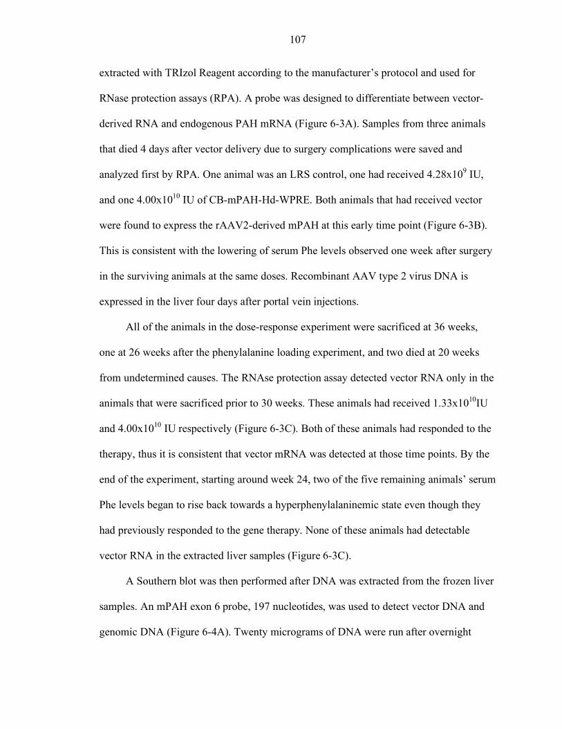
107
extracted with TRIzol Reagent according to the manufacturer’s protocol and used for
RNase protection assays (RPA). A probe was designed to differentiate between vector-
derived RNA and endogenous PAH mRNA (Figure 6-3A). Samples from three animals
that died 4 days after vector delivery due to surgery complications were saved and
analyzed first by RPA. One animal was an LRS control, one had received 4.28x10⁹ IU,
and one 4.00x10¹⁰ IU of CB-mPAH-Hd-WPRE. Both animals that had received vector
were found to express the rAAV2-derived mPAH at this early time point (Figure 6-3B).
This is consistent with the lowering of serum Phe levels observed one week after surgery
in the surviving animals at the same doses. Recombinant AAV type 2 virus DNA is
expressed in the liver four days after portal vein injections.
All of the animals in the dose-response experiment were sacrificed at 36 weeks,
one at 26 weeks after the phenylalanine loading experiment, and two died at 20 weeks
from undetermined causes. The RNAse protection assay detected vector RNA only in the
animals that were sacrificed prior to 30 weeks. These animals had received 1.33x10¹⁰IU
and 4.00x10¹⁰ IU respectively (Figure 6-3C). Both of these animals had responded to the
therapy, thus it is consistent that vector mRNA was detected at those time points. By the
end of the experiment, starting around week 24, two of the five remaining animals’ serum
Phe levels began to rise back towards a hyperphenylalaninemic state even though they
had previously responded to the gene therapy. None of these animals had detectable
vector RNA in the extracted liver samples (Figure 6-3C).
A Southern blot was then performed after DNA was extracted from the frozen liver
samples. An mPAH exon 6 probe, 197 nucleotides, was used to detect vector DNA and
genomic DNA (Figure 6-4A). Twenty micrograms of DNA were run after overnight
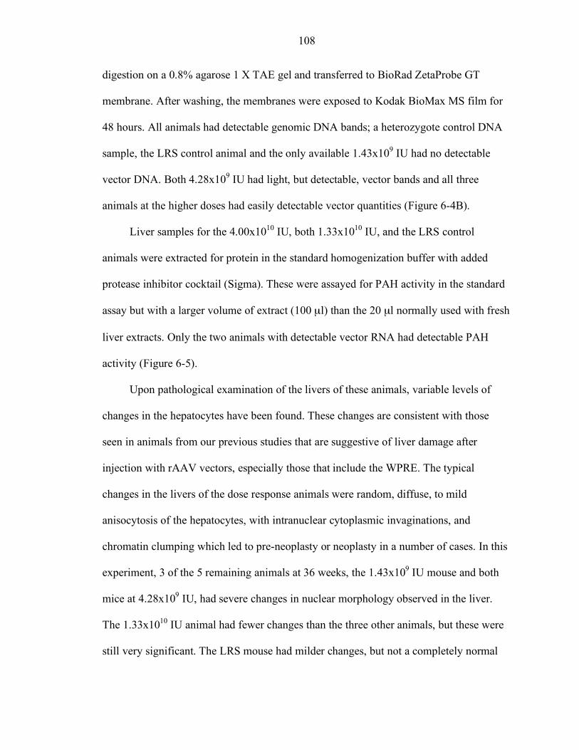
108
digestion on a 0.8% agarose 1 X TAE gel and transferred to BioRad ZetaProbe GT
membrane. After washing, the membranes were exposed to Kodak BioMax MS film for
48 hours. All animals had detectable genomic DNA bands; a heterozygote control DNA
sample, the LRS control animal and the only available 1.43x10⁹ IU had no detectable
vector DNA. Both 4.28x10⁹ IU had light, but detectable, vector bands and all three
animals at the higher doses had easily detectable vector quantities (Figure 6-4B).
Liver samples for the 4.00x10¹⁰ IU, both 1.33x10¹⁰ IU, and the LRS control
animals were extracted for protein in the standard homogenization buffer with added
protease inhibitor cocktail (Sigma). These were assayed for PAH activity in the standard
assay but with a larger volume of extract (100 µl) than the 20 µl normally used with fresh
liver extracts. Only the two animals with detectable vector RNA had detectable PAH
activity (Figure 6-5).
Upon pathological examination of the livers of these animals, variable levels of
changes in the hepatocytes have been found. These changes are consistent with those
seen in animals from our previous studies that are suggestive of liver damage after
injection with rAAV vectors, especially those that include the WPRE. The typical
changes in the livers of the dose response animals were random, diffuse, to mild
anisocytosis of the hepatocytes, with intranuclear cytoplasmic invaginations, and
chromatin clumping which led to pre-neoplasty or neoplasty in a number of cases. In this
experiment, 3 of the 5 remaining animals at 36 weeks, the 1.43x10⁹ IU mouse and both
mice at 4.28x10⁹ IU, had severe changes in nuclear morphology observed in the liver.
The 1.33x10¹⁰ IU animal had fewer changes than the three other animals, but these were
still very significant. The LRS mouse had milder changes, but not a completely normal
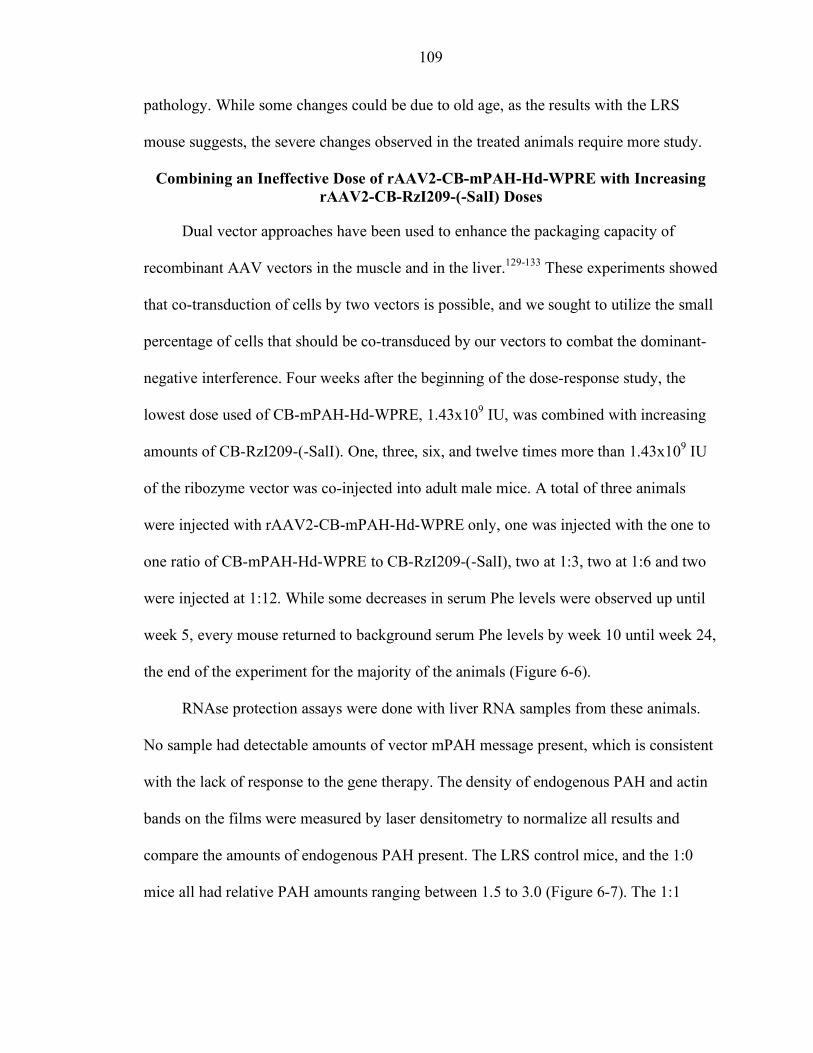
109
pathology. While some changes could be due to old age, as the results with the LRS
mouse suggests, the severe changes observed in the treated animals require more study.
Combining an Ineffective Dose of rAAV2-CB-mPAH-Hd-WPRE with Increasing rAAV2-CB-RzI209-(-SalI) Doses
Dual vector approaches have been used to enhance the packaging capacity of
recombinant AAV vectors in the muscle and in the liver.129-133 These experiments showed
that co-transduction of cells by two vectors is possible, and we sought to utilize the small
percentage of cells that should be co-transduced by our vectors to combat the dominant-
negative interference. Four weeks after the beginning of the dose-response study, the
lowest dose used of CB-mPAH-Hd-WPRE, 1.43x10⁹ IU, was combined with increasing
amounts of CB-RzI209-(-SalI). One, three, six, and twelve times more than 1.43x10⁹ IU
of the ribozyme vector was co-injected into adult male mice. A total of three animals
were injected with rAAV2-CB-mPAH-Hd-WPRE only, one was injected with the one to
one ratio of CB-mPAH-Hd-WPRE to CB-RzI209-(-SalI), two at 1:3, two at 1:6 and two
were injected at 1:12. While some decreases in serum Phe levels were observed up until
week 5, every mouse returned to background serum Phe levels by week 10 until week 24,
the end of the experiment for the majority of the animals (Figure 6-6).
RNAse protection assays were done with liver RNA samples from these animals.
No sample had detectable amounts of vector mPAH message present, which is consistent
with the lack of response to the gene therapy. The density of endogenous PAH and actin
bands on the films were measured by laser densitometry to normalize all results and
compare the amounts of endogenous PAH present. The LRS control mice, and the 1:0
mice all had relative PAH amounts ranging between 1.5 to 3.0 (Figure 6-7). The 1:1
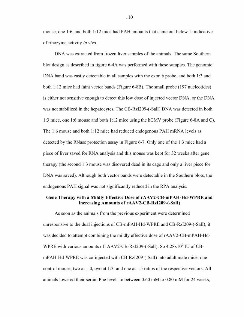
110
mouse, one 1:6, and both 1:12 mice had PAH amounts that came out below 1, indicative
of ribozyme activity in vivo.
DNA was extracted from frozen liver samples of the animals. The same Southern
blot design as described in figure 6-4A was performed with these samples. The genomic
DNA band was easily detectable in all samples with the exon 6 probe, and both 1:3 and
both 1:12 mice had faint vector bands (Figure 6-8B). The small probe (197 nucleotides)
is either not sensitive enough to detect this low dose of injected vector DNA, or the DNA
was not stabilized in the hepatocytes. The CB-RzI209-(-SalI) DNA was detected in both
1:3 mice, one 1:6 mouse and both 1:12 mice using the hCMV probe (Figure 6-8A and C).
The 1:6 mouse and both 1:12 mice had reduced endogenous PAH mRNA levels as
detected by the RNase protection assay in Figure 6-7. Only one of the 1:3 mice had a
piece of liver saved for RNA analysis and this mouse was kept for 32 weeks after gene
therapy (the second 1:3 mouse was disovered dead in its cage and only a liver piece for
DNA was saved). Although both vector bands were detectable in the Southern blots, the
endogenous PAH signal was not significantly reduced in the RPA analysis.
Gene Therapy with a Mildly Effective Dose of rAAV2-CB-mPAH-Hd-WPRE and Increasing Amounts of rAAV2-CB-RzI209-(-SalI)
As soon as the animals from the previous experiment were determined
unresponsive to the dual injections of CB-mPAH-Hd-WPRE and CB-RzI209-(-SalI), it
was decided to attempt combining the mildly effective dose of rAAV2-CB-mPAH-Hd-
WPRE with various amounts of rAAV2-CB-RzI209-(-SalI). So 4.28x10⁹ IU of CB-
mPAH-Hd-WPRE was co-injected with CB-RzI209-(-SalI) into adult male mice: one
control mouse, two at 1:0, two at 1:3, and one at 1:5 ratios of the respective vectors. All
animals lowered their serum Phe levels to between 0.60 mM to 0.80 mM for 24 weeks,
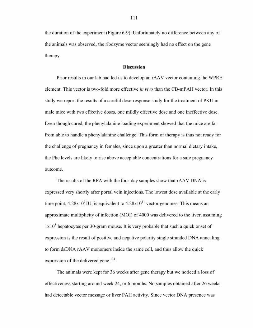
111
the duration of the experiment (Figure 6-9). Unfortunately no difference between any of
the animals was observed, the ribozyme vector seemingly had no effect on the gene
therapy.
Discussion
Prior results in our lab had led us to develop an rAAV vector containing the WPRE
element. This vector is two-fold more effective in vivo than the CB-mPAH vector. In this
study we report the results of a careful dose-response study for the treatment of PKU in
male mice with two effective doses, one mildly effective dose and one ineffective dose.
Even though cured, the phenylalanine loading experiment showed that the mice are far
from able to handle a phenylalanine challenge. This form of therapy is thus not ready for
the challenge of pregnancy in females, since upon a greater than normal dietary intake,
the Phe levels are likely to rise above acceptable concentrations for a safe pregnancy
outcome.
The results of the RPA with the four-day samples show that rAAV DNA is
expressed very shortly after portal vein injections. The lowest dose available at the early
time point, 4.28x10⁹ IU, is equivalent to 4.28x10¹¹ vector genomes. This means an
approximate multiplicity of infection (MOI) of 4000 was delivered to the liver, assuming
1x10⁸ hepatocytes per 30-gram mouse. It is very probable that such a quick onset of
expression is the result of positive and negative polarity single stranded DNA annealing
to form dsDNA rAAV monomers inside the same cell, and thus allow the quick
expression of the delivered gene.134
The animals were kept for 36 weeks after gene therapy but we noticed a loss of
effectiveness starting around week 24, or 6 months. No samples obtained after 26 weeks
had detectable vector message or liver PAH activity. Since vector DNA presence was
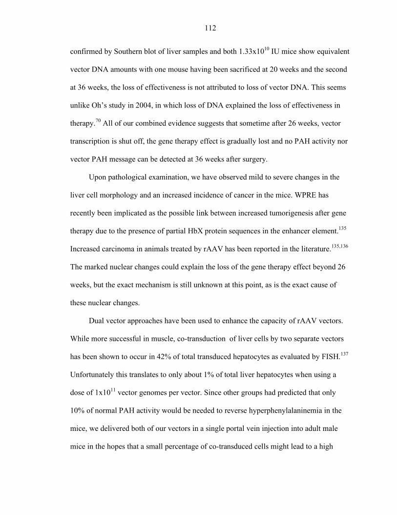
112
confirmed by Southern blot of liver samples and both 1.33x10¹⁰ IU mice show equivalent
vector DNA amounts with one mouse having been sacrificed at 20 weeks and the second
at 36 weeks, the loss of effectiveness is not attributed to loss of vector DNA. This seems
unlike Oh’s study in 2004, in which loss of DNA explained the loss of effectiveness in
therapy.70 All of our combined evidence suggests that sometime after 26 weeks, vector
transcription is shut off, the gene therapy effect is gradually lost and no PAH activity nor
vector PAH message can be detected at 36 weeks after surgery.
Upon pathological examination, we have observed mild to severe changes in the
liver cell morphology and an increased incidence of cancer in the mice. WPRE has
recently been implicated as the possible link between increased tumorigenesis after gene
therapy due to the presence of partial HbX protein sequences in the enhancer element.135
Increased carcinoma in animals treated by rAAV has been reported in the literature.135,136
The marked nuclear changes could explain the loss of the gene therapy effect beyond 26
weeks, but the exact mechanism is still unknown at this point, as is the exact cause of
these nuclear changes.
Dual vector approaches have been used to enhance the capacity of rAAV vectors.
While more successful in muscle, co-transduction of liver cells by two separate vectors
has been shown to occur in 42% of total transduced hepatocytes as evaluated by FISH.137
Unfortunately this translates to only about 1% of total liver hepatocytes when using a
dose of 1x10¹¹ vector genomes per vector. Since other groups had predicted that only
10% of normal PAH activity would be needed to reverse hyperphenylalaninemia in the
mice, we delivered both of our vectors in a single portal vein injection into adult male
mice in the hopes that a small percentage of co-transduced cells might lead to a high
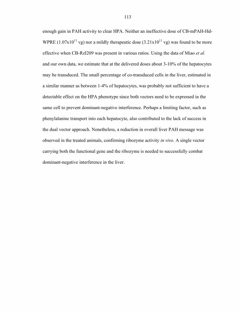
113
enough gain in PAH activity to clear HPA. Neither an ineffective dose of CB-mPAH-Hd-
WPRE (1.07x10¹¹ vg) nor a mildly therapeutic dose (3.21x10¹¹ vg) was found to be more
effective when CB-RzI209 was present in various ratios. Using the data of Miao et al.
and our own data, we estimate that at the delivered doses about 3-10% of the hepatocytes
may be transduced. The small percentage of co-transduced cells in the liver, estimated in
a similar manner as between 1-4% of hepatocytes, was probably not sufficient to have a
detectable effect on the HPA phenotype since both vectors need to be expressed in the
same cell to prevent dominant-negative interference. Perhaps a limiting factor, such as
phenylalanine transport into each hepatocyte, also contributed to the lack of success in
the dual vector approach. Nonetheless, a reduction in overall liver PAH message was
observed in the treated animals, confirming ribozyme activity in vivo. A single vector
carrying both the functional gene and the ribozyme is needed to successfully combat
dominant-negative interference in the liver.
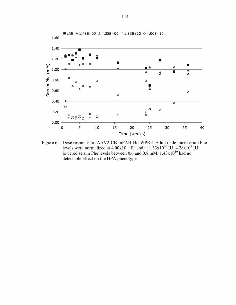
114
Figure 6-1 Dose response to rAAV2-CB-mPAH-Hd-WPRE. Adult male mice serum Phe
levels were normalized at 4.00x10¹⁰ IU and at 1.33x10¹⁰ IU. 4.28x10⁹ IU lowered serum Phe levels between 0.6 and 0.8 mM. 1.43x10¹⁰ had no detectable effect on the HPA phenotype.
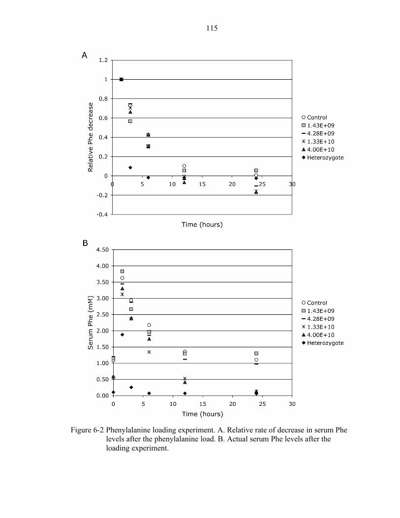
115
Figure 6-2 Phenylalanine loading experiment. A. Relative rate of decrease in serum Phe
levels after the phenylalanine load. B. Actual serum Phe levels after the loading experiment.
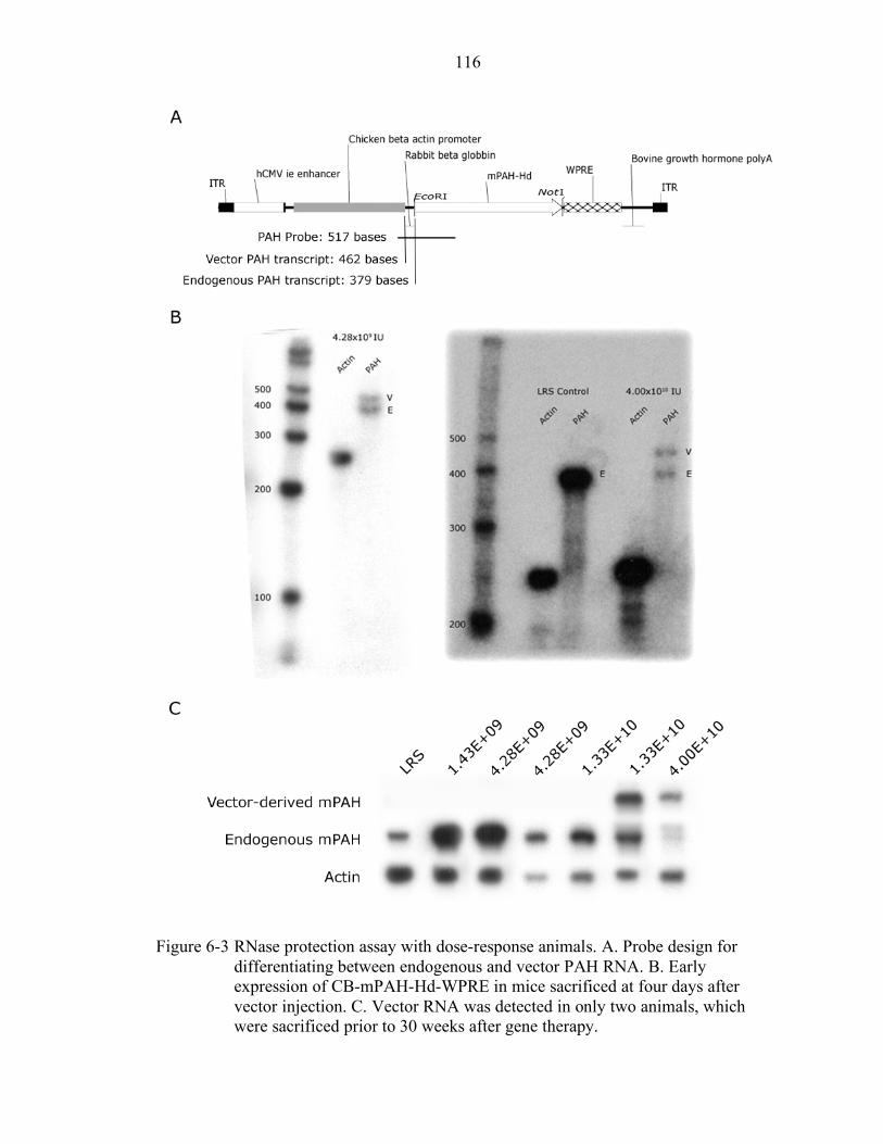
116
Figure 6-3 RNase protection assay with dose-response animals. A. Probe design for
differentiating between endogenous and vector PAH RNA. B. Early expression of CB-mPAH-Hd-WPRE in mice sacrificed at four days after vector injection. C. Vector RNA was detected in only two animals, which were sacrificed prior to 30 weeks after gene therapy.
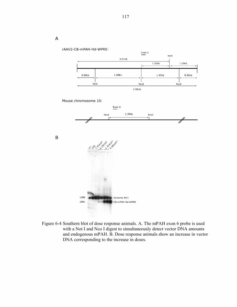
117
Figure 6-4 Southern blot of dose response animals. A. The mPAH exon 6 probe is used
with a Not I and Nco I digest to simultaneously detect vector DNA amounts and endogenous mPAH. B. Dose response animals show an increase in vector DNA corresponding to the increase in doses.
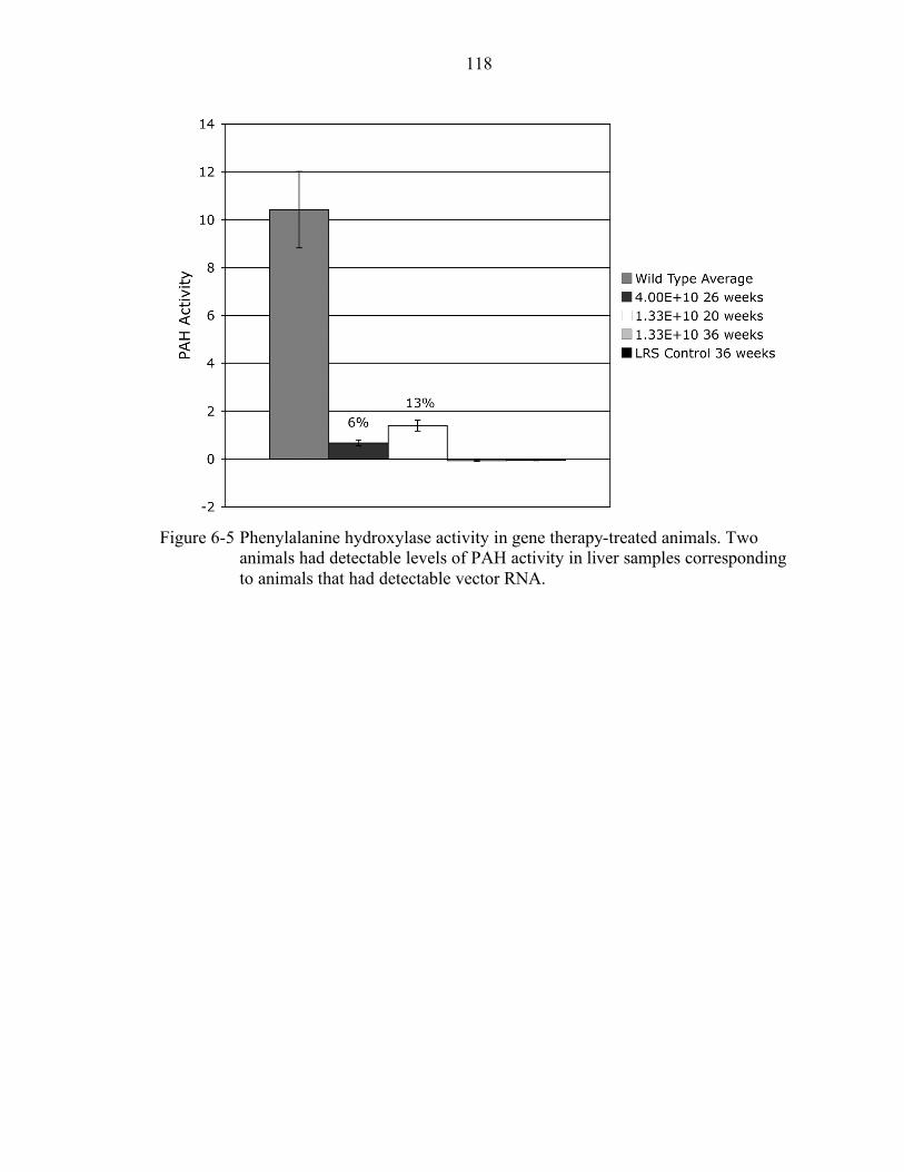
118
Figure 6-5 Phenylalanine hydroxylase activity in gene therapy-treated animals. Two
animals had detectable levels of PAH activity in liver samples corresponding to animals that had detectable vector RNA.
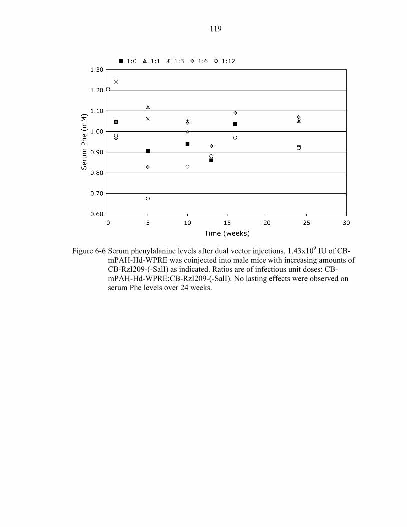
119
Figure 6-6 Serum phenylalanine levels after dual vector injections. 1.43x10⁹ IU of CB-
mPAH-Hd-WPRE was coinjected into male mice with increasing amounts of CB-RzI209-(-SalI) as indicated. Ratios are of infectious unit doses: CB-mPAH-Hd-WPRE:CB-RzI209-(-SalI). No lasting effects were observed on serum Phe levels over 24 weeks.
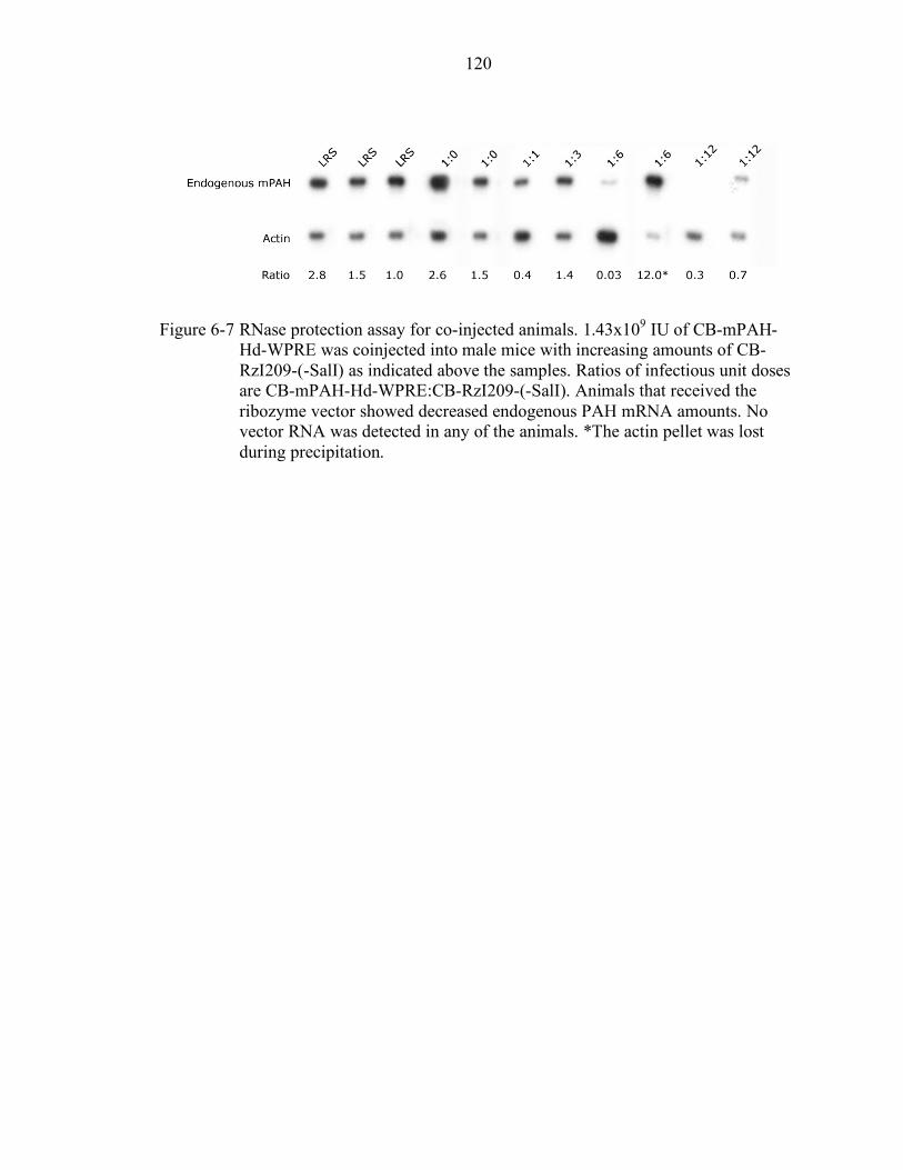
120
Figure 6-7 RNase protection assay for co-injected animals. 1.43x10⁹ IU of CB-mPAH-
Hd-WPRE was coinjected into male mice with increasing amounts of CB-RzI209-(-SalI) as indicated above the samples. Ratios of infectious unit doses are CB-mPAH-Hd-WPRE:CB-RzI209-(-SalI). Animals that received the ribozyme vector showed decreased endogenous PAH mRNA amounts. No vector RNA was detected in any of the animals. *The actin pellet was lost during precipitation.
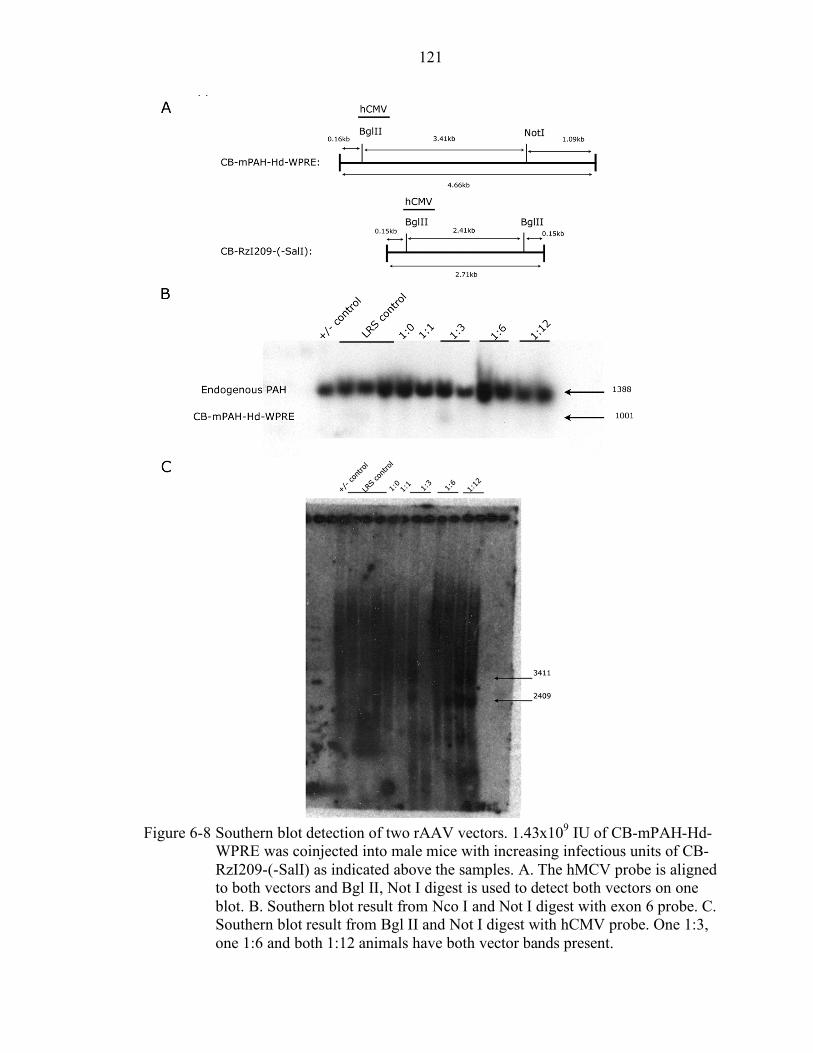
121
Figure 6-8 Southern blot detection of two rAAV vectors. 1.43x10⁹ IU of CB-mPAH-Hd-
WPRE was coinjected into male mice with increasing infectious units of CB-RzI209-(-SalI) as indicated above the samples. A. The hMCV probe is aligned to both vectors and Bgl II, Not I digest is used to detect both vectors on one blot. B. Southern blot result from Nco I and Not I digest with exon 6 probe. C. Southern blot result from Bgl II and Not I digest with hCMV probe. One 1:3, one 1:6 and both 1:12 animals have both vector bands present.
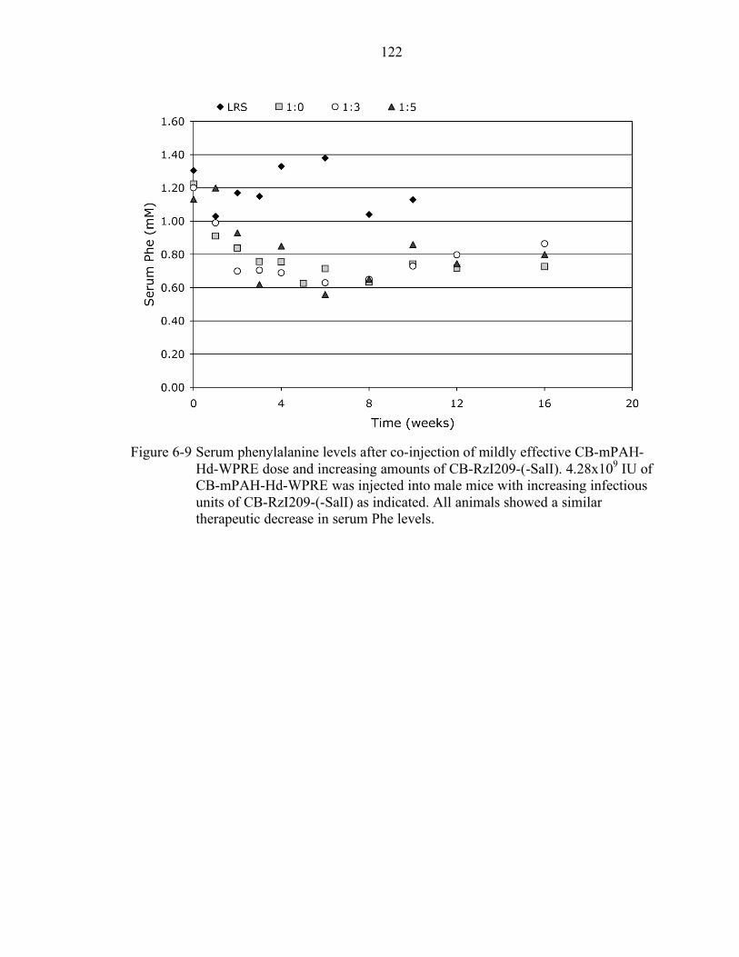
122
Figure 6-9 Serum phenylalanine levels after co-injection of mildly effective CB-mPAH-
Hd-WPRE dose and increasing amounts of CB-RzI209-(-SalI). 4.28x10⁹ IU of CB-mPAH-Hd-WPRE was injected into male mice with increasing infectious units of CB-RzI209-(-SalI) as indicated. All animals showed a similar therapeutic decrease in serum Phe levels.
123
CHAPTER 7 DEVELOPMENT OF A SINGLE VECTOR CARRYING THE MOUSE PAH GENE
AND RIBOZYME I209
The current ribozyme construct in the p21-newhp vector is designed to prevent
early degradation and to promote export from the nucleus based on the splice site
acceptor in the β-globin exon just upstream of the ribozyme sequence. To construct a
single rAAV vector carrying both the mPAH gene and the ribozyme, the choice of a new
promoter for the ribozyme was necessary because of AAV genome size packaging limits.
The promoter for the ribozyme would have to offer stability to prevent early ribozyme
degration and allow for export of the ribozyme from the nucleus so that it and the
endogenous PAH mRNA could be in the same cell compartment. Experiments by
Kuwabara et al. have demonstrated the feasibility of expressing ribozymes just
downstream of a modified tRNAVal.138-141 They have shown by in situ hybridization and
northern blots of cytoplasmic and nuclear fractions that the tRNA-driven ribozymes are
exported to the cytoplasm after direct injection into the nuclei of HeLa cells and after
expression from transiently transfected plasmid DNA.142 Since the tRNA has both the A
and B box motifs intact, it still serves as its own promoter using RNA polymerase III.
Kawasaki and Taira reported in 2002 that adding a short polyA tail 3’ of a ribozyme
construct coupled the activity of a helicase to the ribozyme thus allowing cleavage of any
target site regardless of secondary structure.143 Based on this evidence we opted to
include a 60 adenosine tail 3’ of the ribozyme construct to enhance target accessibility
and overall stability of the ribozyme. Another element studied by the same group,
constitutive transport element (CTE), can also interact with RNA helicases but has a
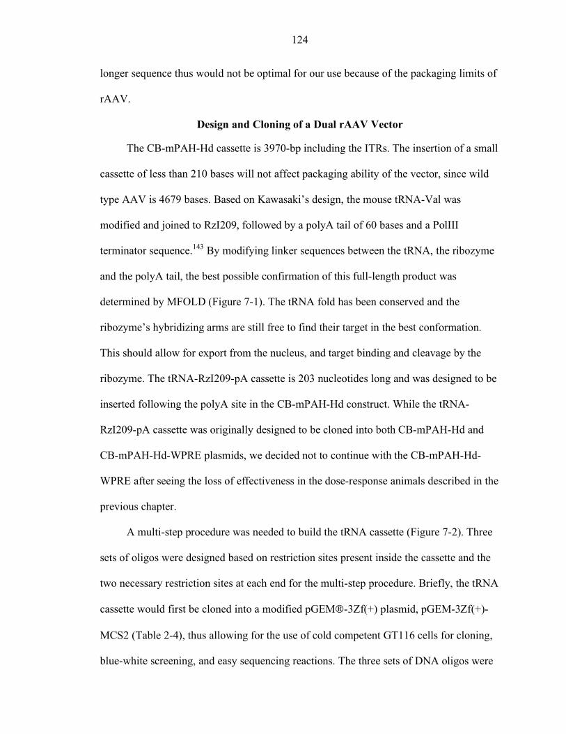
124
longer sequence thus would not be optimal for our use because of the packaging limits of
rAAV.
Design and Cloning of a Dual rAAV Vector
The CB-mPAH-Hd cassette is 3970-bp including the ITRs. The insertion of a small
cassette of less than 210 bases will not affect packaging ability of the vector, since wild
type AAV is 4679 bases. Based on Kawasaki’s design, the mouse tRNA-Val was
modified and joined to RzI209, followed by a polyA tail of 60 bases and a PolIII
terminator sequence.143 By modifying linker sequences between the tRNA, the ribozyme
and the polyA tail, the best possible confirmation of this full-length product was
determined by MFOLD (Figure 7-1). The tRNA fold has been conserved and the
ribozyme’s hybridizing arms are still free to find their target in the best conformation.
This should allow for export from the nucleus, and target binding and cleavage by the
ribozyme. The tRNA-RzI209-pA cassette is 203 nucleotides long and was designed to be
inserted following the polyA site in the CB-mPAH-Hd construct. While the tRNA-
RzI209-pA cassette was originally designed to be cloned into both CB-mPAH-Hd and
CB-mPAH-Hd-WPRE plasmids, we decided not to continue with the CB-mPAH-Hd-
WPRE after seeing the loss of effectiveness in the dose-response animals described in the
previous chapter.
A multi-step procedure was needed to build the tRNA cassette (Figure 7-2). Three
sets of oligos were designed based on restriction sites present inside the cassette and the
two necessary restriction sites at each end for the multi-step procedure. Briefly, the tRNA
cassette would first be cloned into a modified pGEM-3Zf(+) plasmid, pGEM-3Zf(+)-
MCS2 (Table 2-4), thus allowing for the use of cold competent GT116 cells for cloning,
blue-white screening, and easy sequencing reactions. The three sets of DNA oligos were
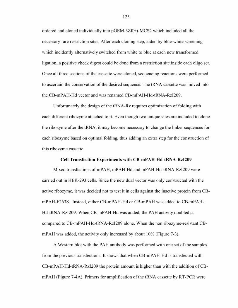
125
ordered and cloned individually into pGEM-3Zf(+)-MCS2 which included all the
necessary rare restriction sites. After each cloning step, aided by blue-white screening
which incidently alternatively switched from white to blue at each new transformed
ligation, a positive check digest could be done from a restriction site inside each oligo set.
Once all three sections of the cassette were cloned, sequencing reactions were performed
to ascertain the conservation of the desired sequence. The tRNA cassette was moved into
the CB-mPAH-Hd vector and was renamed CB-mPAH-Hd-tRNA-RzI209.
Unfortunately the design of the tRNA-Rz requires optimization of folding with
each different ribozyme attached to it. Even though two unique sites are included to clone
the ribozyme after the tRNA, it may become necessary to change the linker sequences for
each ribozyme based on optimal folding, thus adding an extra step for the construction of
this ribozyme cassette.
Cell Transfection Experiments with CB-mPAH-Hd-tRNA-RzI209
Mixed transfections of mPAH, mPAH-Hd and mPAH-Hd-tRNA-RzI209 were
carried out in HEK-293 cells. Since the new dual vector was only constructed with the
active ribozyme, it was decided not to test it in cells against the inactive protein from CB-
mPAH-F263S. Instead, either CB-mPAH-Hd or CB-mPAH was added to CB-mPAH-
Hd-tRNA-RzI209. When CB-mPAH-Hd was added, the PAH activity doubled as
compared to CB-mPAH-Hd-tRNA-RzI209 alone. When the non ribozyme-resistant CB-
mPAH was added, the activity only increased by about 10% (Figure 7-3).
A Western blot with the PAH antibody was performed with one set of the samples
from the previous transfections. It shows that when CB-mPAH-Hd is transfected with
CB-mPAH-Hd-tRNA-RzI209 the protein amount is higher than with the addition of CB-
mPAH (Figure 7-4A). Primers for amplification of the tRNA cassette by RT-PCR were
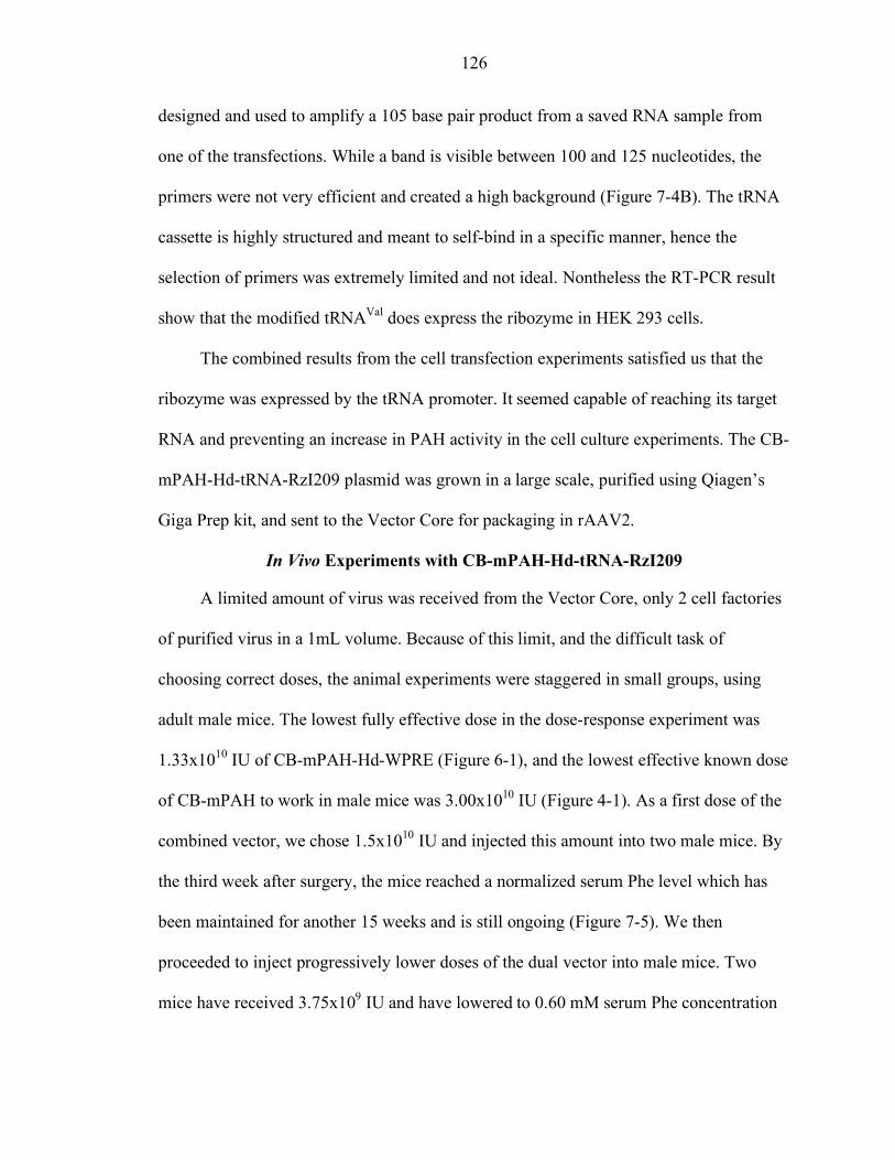
126
designed and used to amplify a 105 base pair product from a saved RNA sample from
one of the transfections. While a band is visible between 100 and 125 nucleotides, the
primers were not very efficient and created a high background (Figure 7-4B). The tRNA
cassette is highly structured and meant to self-bind in a specific manner, hence the
selection of primers was extremely limited and not ideal. Nontheless the RT-PCR result
show that the modified tRNAVal does express the ribozyme in HEK 293 cells.
The combined results from the cell transfection experiments satisfied us that the
ribozyme was expressed by the tRNA promoter. It seemed capable of reaching its target
RNA and preventing an increase in PAH activity in the cell culture experiments. The CB-
mPAH-Hd-tRNA-RzI209 plasmid was grown in a large scale, purified using Qiagen’s
Giga Prep kit, and sent to the Vector Core for packaging in rAAV2.
In Vivo Experiments with CB-mPAH-Hd-tRNA-RzI209
A limited amount of virus was received from the Vector Core, only 2 cell factories
of purified virus in a 1mL volume. Because of this limit, and the difficult task of
choosing correct doses, the animal experiments were staggered in small groups, using
adult male mice. The lowest fully effective dose in the dose-response experiment was
1.33x10¹⁰ IU of CB-mPAH-Hd-WPRE (Figure 6-1), and the lowest effective known dose
of CB-mPAH to work in male mice was 3.00x10¹⁰ IU (Figure 4-1). As a first dose of the
combined vector, we chose 1.5x10¹⁰ IU and injected this amount into two male mice. By
the third week after surgery, the mice reached a normalized serum Phe level which has
been maintained for another 15 weeks and is still ongoing (Figure 7-5). We then
proceeded to inject progressively lower doses of the dual vector into male mice. Two
mice have received 3.75x10⁹ IU and have lowered to 0.60 mM serum Phe concentration
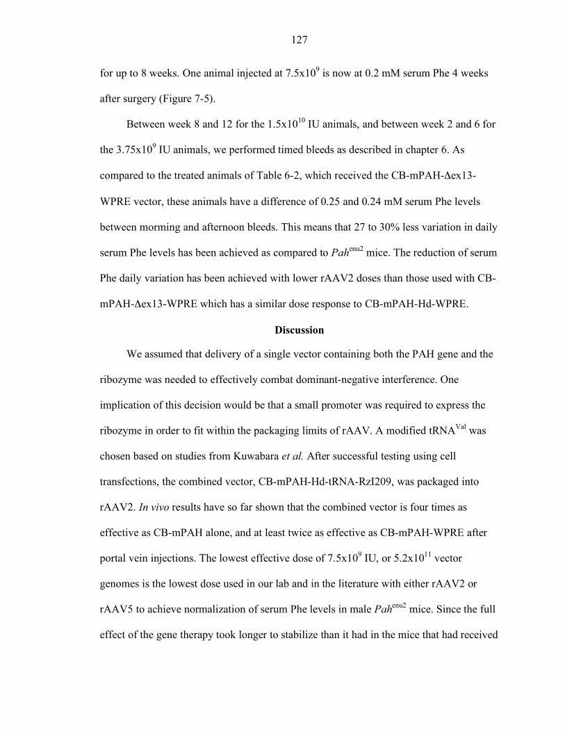
127
for up to 8 weeks. One animal injected at 7.5x10⁹ is now at 0.2 mM serum Phe 4 weeks
after surgery (Figure 7-5).
Between week 8 and 12 for the 1.5x10¹⁰ IU animals, and between week 2 and 6 for
the 3.75x10⁹ IU animals, we performed timed bleeds as described in chapter 6. As
compared to the treated animals of Table 6-2, which received the CB-mPAH-Δex13-
WPRE vector, these animals have a difference of 0.25 and 0.24 mM serum Phe levels
between morming and afternoon bleeds. This means that 27 to 30% less variation in daily
serum Phe levels has been achieved as compared to Pahenu2 mice. The reduction of serum
Phe daily variation has been achieved with lower rAAV2 doses than those used with CB-
mPAH-Δex13-WPRE which has a similar dose response to CB-mPAH-Hd-WPRE.
Discussion
We assumed that delivery of a single vector containing both the PAH gene and the
ribozyme was needed to effectively combat dominant-negative interference. One
implication of this decision would be that a small promoter was required to express the
ribozyme in order to fit within the packaging limits of rAAV. A modified tRNAVal was
chosen based on studies from Kuwabara et al. After successful testing using cell
transfections, the combined vector, CB-mPAH-Hd-tRNA-RzI209, was packaged into
rAAV2. In vivo results have so far shown that the combined vector is four times as
effective as CB-mPAH alone, and at least twice as effective as CB-mPAH-WPRE after
portal vein injections. The lowest effective dose of 7.5x10⁹ IU, or 5.2x10¹¹ vector
genomes is the lowest dose used in our lab and in the literature with either rAAV2 or
rAAV5 to achieve normalization of serum Phe levels in male Pahenu2 mice. Since the full
effect of the gene therapy took longer to stabilize than it had in the mice that had received
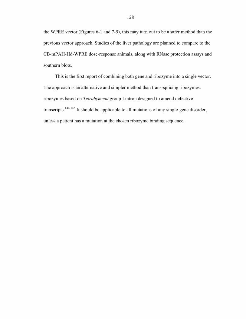
128
the WPRE vector (Figures 6-1 and 7-5), this may turn out to be a safer method than the
previous vector approach. Studies of the liver pathology are planned to compare to the
CB-mPAH-Hd-WPRE dose-response animals, along with RNase protection assays and
southern blots.
This is the first report of combining both gene and ribozyme into a single vector.
The approach is an alternative and simpler method than trans-splicing ribozymes:
ribozymes based on Tetrahymena group I intron designed to amend defective
transcripts.144,145 It should be applicable to all mutations of any single-gene disorder,
unless a patient has a mutation at the chosen ribozyme binding sequence.
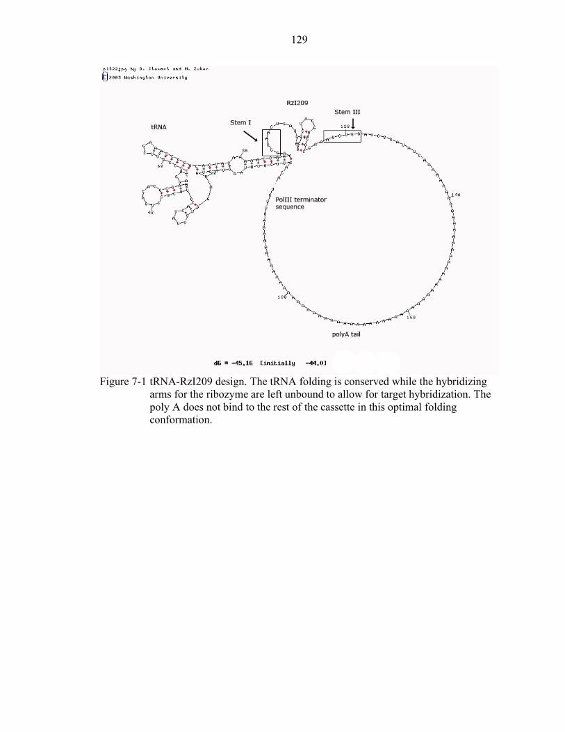
129
Figure 7-1 tRNA-RzI209 design. The tRNA folding is conserved while the hybridizing
arms for the ribozyme are left unbound to allow for target hybridization. The poly A does not bind to the rest of the cassette in this optimal folding conformation.
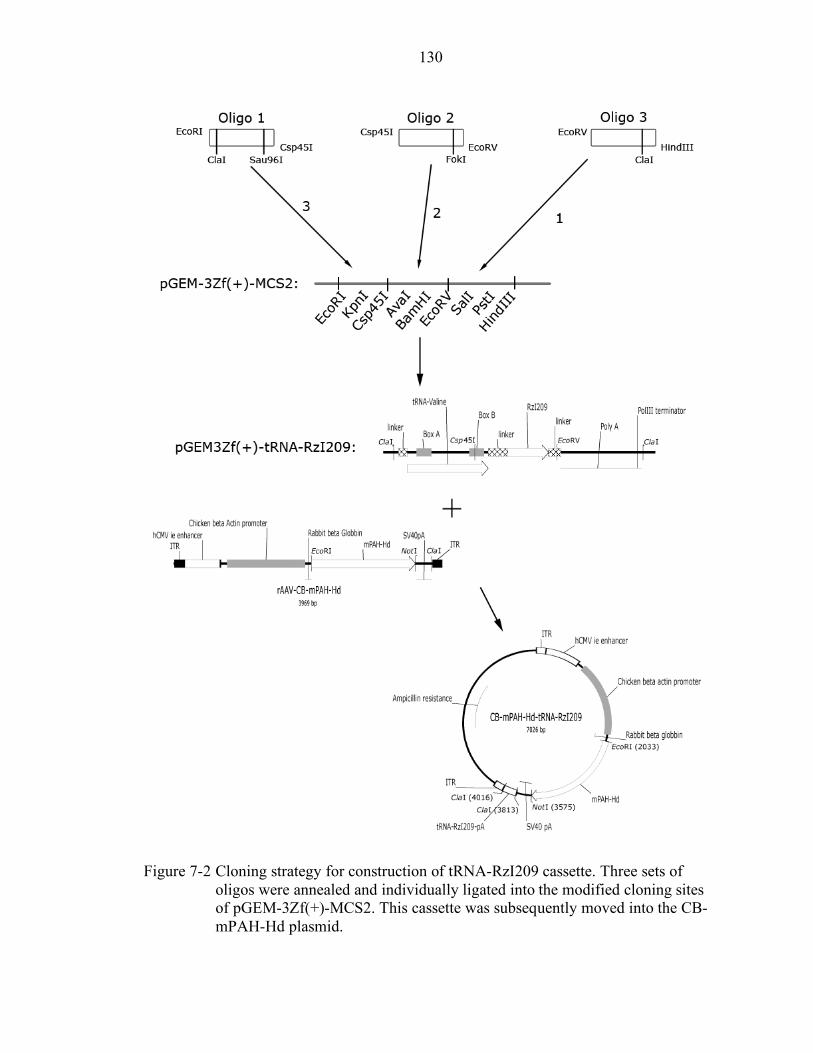
130
Figure 7-2 Cloning strategy for construction of tRNA-RzI209 cassette. Three sets of
oligos were annealed and individually ligated into the modified cloning sites of pGEM-3Zf(+)-MCS2. This cassette was subsequently moved into the CB-mPAH-Hd plasmid.
131
Figure 7-3 Results of transient cell transfections with CB-mPAH-Hd-tRNA-RzI209.
Addition of an equal amount of CB-mPAH-Hd to CB-mPAH-Hd-tRNA-RzI209 doubles the PAH activity while addition of CB-mPAH does not result in a significant increase in PAH activity.
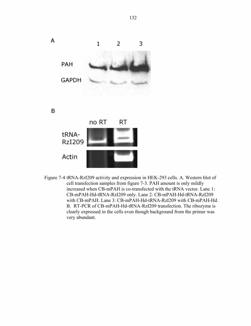
132
Figure 7-4 tRNA-RzI209 activity and expression in HEK-293 cells. A. Western blot of
cell transfection samples from figure 7-3. PAH amount is only mildly increased when CB-mPAH is co-transfected with the tRNA vector. Lane 1: CB-mPAH-Hd-tRNA-RzI209 only. Lane 2: CB-mPAH-Hd-tRNA-RzI209 with CB-mPAH. Lane 3: CB-mPAH-Hd-tRNA-RzI209 with CB-mPAH-Hd. B. RT-PCR of CB-mPAH-Hd-tRNA-RzI209 transfection. The ribozyme is clearly expressed in the cells even though background from the primer was very abundant.
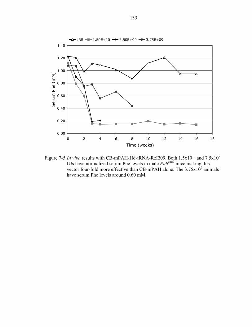
133
Figure 7-5 In vivo results with CB-mPAH-Hd-tRNA-RzI209. Both 1.5x10¹⁰ and 7.5x10⁹
IUs have normalized serum Phe levels in male Pahenu2 mice making this vector four-fold more effective than CB-mPAH alone. The 3.75x10⁹ animals have serum Phe levels around 0.60 mM.
134
CHAPTER 8 DEVELOPMENT OF SHORT INTERFERING RNAS FOR MURINE PAH
RNA interference is the process of post-transcriptional silencing induced by double
stranded RNA. Short interfering RNAs are duplexes of 20 to 22 nucleotides and are
processed by RISC to target a messenger RNA for cleavage and degradation. As an
alternative approach to hammerhead ribozymes to prevent dominant-negative
interference, short interfering RNAs were designed to target mPAH. Since siRNAs have
many different selection rules, and need to be tested in vivo for true determination of
activity, Ambion’s Silencer kit was chosen to test three selected siRNAs against
mPAH.91,92 The kit creates the siRNA cassette by PCR amplification and is designed to
include a chosen promoter, in these experiments, human U6. The method skips lengthy
cloning steps while still expressing the siRNA in the cells as opposed to transfecting
RNA oligos which have a finite half-life.146
Short Interfering RNA Site Selection
Potential siRNA sites were selected from the PAH cDNA sequence in the rAAV-
CB-mPAH vector. Only the first 500 nucleotides of the cDNA were screened for the best
possible target sites. There are a number of papers published that can help one select
possible good RNA interference sites, but none of the rules are foolproof, and this is
often emphasized by the authors.
To select siRNA target sites three available tools on the Internet were used:
Ambion’s website, Promega’s website, and a trial version of SVMRNAi version 5.0.
Each website or program came up with different lists of best siRNA positions; comparing
135
all three lists, target sites that were within 20 bases of each other were further examined
using BLAST searches. If the designed siRNA was found to correspond to another gene
besides PAH in the first 100 results by BLAST search, it was rejected as a possible
design. The list of possible target sites was reduced and finally three target sites were
selected for testing and the PCR primers necessary for the Silencer kit were ordered from
Sigma-Genosys (Table 8-1).
siRNA Cell Culture Tests
The three siRNAs were made according to Ambion’s specifications. To determine
what concentration of siRNA cassette was needed to test in cell cultures, the siGAPDH
and siNegative control for human cells included in Ambion’s kit were amplified
alongside the mPAH siRNAs. Three concentrations were tested with the siGAPDH and
siNegative in HEK-293 cells by standard calcium phosphate transfection: 100 ng/well,
200 ng/well, and 300 ng/well of a six-well plate. After two days, the cells were harvested
for RNA extraction with TRIzol, and RT-PCR reactions were set up to detect the
amounts of GAPDH and Actin in each transfection. Based on the normalized GAPDH
results, 300 ng/well was selected as a reasonable concentration to use in HEK-293 cells
(Figure 8-1).
The three siRNAs for mPAH were then tested versus co-transfected CB-mPAH plasmid,
300 ng siRNA and 2 µg CB-mPAH. Each transfection was done in one well of a 6-well
plate with approximately 8x10⁵ cells per well and harvested after 48 hours for RNA with
TRIzol. The results were analyzed by RT-PCR for mPAH amount and GAPDH was used
as an internal control in these experiments. The best siRNA was si333, knocking down
PAH to approximately 50% (Figure 8-2).
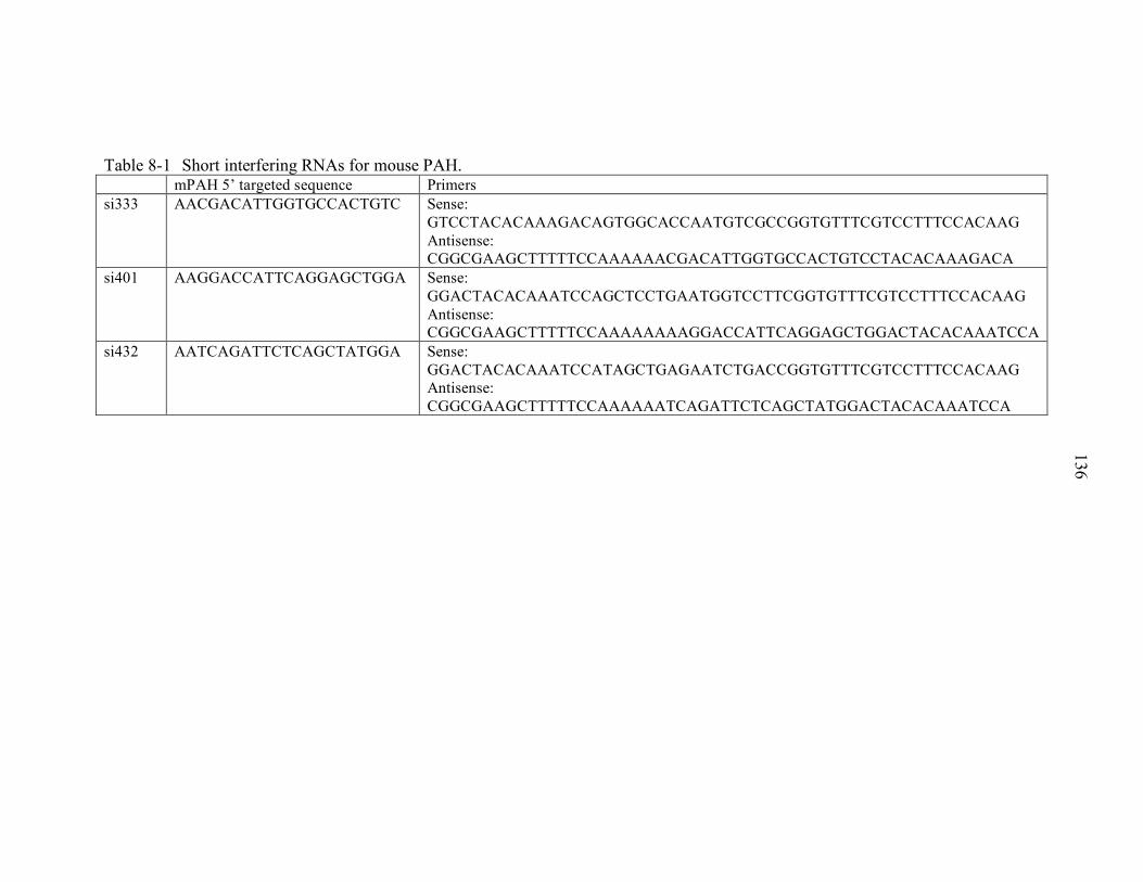
136
Table 8-1 Short interfering RNAs for mouse PAH. mPAH 5’ targeted sequence Primers si333 AACGACATTGGTGCCACTGTC Sense:
GTCCTACACAAAGACAGTGGCACCAATGTCGCCGGTGTTTCGTCCTTTCCACAAG Antisense: CGGCGAAGCTTTTTCCAAAAAACGACATTGGTGCCACTGTCCTACACAAAGACA
si401 AAGGACCATTCAGGAGCTGGA Sense: GGACTACACAAATCCAGCTCCTGAATGGTCCTTCGGTGTTTCGTCCTTTCCACAAG Antisense: CGGCGAAGCTTTTTCCAAAAAAAAGGACCATTCAGGAGCTGGACTACACAAATCCA
si432 AATCAGATTCTCAGCTATGGA Sense: GGACTACACAAATCCATAGCTGAGAATCTGACCGGTGTTTCGTCCTTTCCACAAG Antisense: CGGCGAAGCTTTTTCCAAAAAATCAGATTCTCAGCTATGGACTACACAAATCCA
137
Using a different cell line, 293T, the transfections with the siRNAs and CB-mPAH
were repeated. The transfections into this cell line were done using Qiagen’s Superfect
transfection reagent as described in Chapter 2. The same amounts of DNA were used and
the results were also analyzed by RT-PCR in the same manner. The results were similar,
but si402 led to a better reduction of mPAH than si333 in these cells (Figure 8-2). The
reductions were not quite as great as those achieved in 293 cells. One last transfection
experiment was performed with a newly acquired cell line, 293T-TYF-mPAH created by
Dr Chang’s laboratory from our CB-mPAH vector. The cells constitutively express the
mPAH gene and were transfected with 300 ng of each siRNA, and 2 µg of empty
“carrier” plasmid pGEM-3ZF(+)-MCS2 using the superfect protocol. The cells were
harvested for RNA after 48 hours with TRIzol. The results of the RT-PCR show that
si333 can reduce the expression of mPAH by 40% while the other two siRNAs did not
efficiently reduce the mPAH message (Figure 8-2).
Discussion
Based on all the combined results, si333 seems to be the best choice of siRNA from
all three designs tested. The siRNA that performed the worst in all three cell lines, si432,
followed only three of the selection rules suggested in Reynolds et al., but had been
chosen because of its lower GC content.91 Both si333 and si401 followed four suggested
rules: si333 has 52.4% GC content, 2 G/C at the 5’ end of the sense strand, 3 A/U at the
5’ end of the antisense strand and does not contain a G/C stretch; si401 has 52.4% GC
content, a U at position 10, 2 G/C at the 5’ end of the sense strand and does not contain a
G/C stretch. It is interesting that si333 broke some of the negative selection rules for
specific bases at certain positions but yet performed the best overall, reemphasizing the
need for in vivo testing after algorithm-based design.
138
Further tests at various concentrations could be done to see if more knockdown of
mPAH mRNA can be achieved with si333. For use in the mouse model in gene therapy
experiments, another promoter, active in murine cells, will have to be tested to confirm
the activity of the siRNA. tRNA promoters have been used to express short interfering
RNAs and would be a good choice for the expression of si333.147,148 The si333 could be
cloned into the Csp45I and ClaI sites to replace RzI209 in the current tRNA cassette in
the pGEM3Zf(+) plasmid for separate target to siRNA testing. If active, a resistant
mPAH vector will have to be constructed by extensive PCR mutagenesis and tested as
well prior to any animal experiments.
Our CB-mPAH vector includes approximately 50 nucleotides of the 5’ UTR of
PAH and about 400 nucleotides of the 3’ UTR. An alternate siRNA design could be
targeted against either region. If the siRNA was found to be active in cell culture tests,
removing that region from our construct would render the vector completely resistant to
the siRNA, avoiding the mutagenesis required with the current siRNAs.

139
Figure 8-1 Cell culture siRNA working concentration determination. Three amounts of
siRNAs, 100 ng, 200 ng and 300 ng, were tested by CaPO4 transfection of 4x10⁵ HEK-293 cells plated in 6-well plates in 2 ml of media. RNA was extracted for RT-PCR reactions. Density of obtained GAPDH and Actin bands were evaluated with UN-SCAN-IT gel and normalized GAPDH amounts are graphed above.
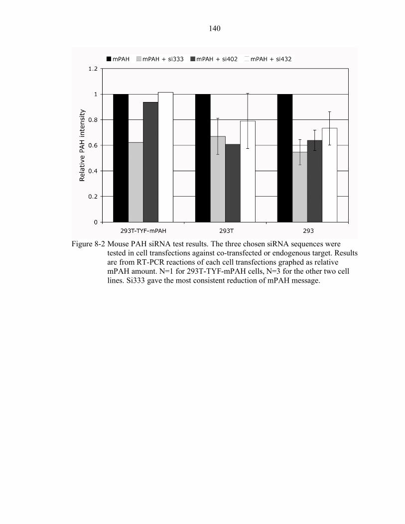
140
Figure 8-2 Mouse PAH siRNA test results. The three chosen siRNA sequences were
tested in cell transfections against co-transfected or endogenous target. Results are from RT-PCR reactions of each cell transfections graphed as relative mPAH amount. N=1 for 293T-TYF-mPAH cells, N=3 for the other two cell lines. Si333 gave the most consistent reduction of mPAH message.
141
CHAPTER 9 SUMMARY, CONCLUSION AND FUTURE DIRECTIONS
General Significance
The earliest studies for developing gene therapy for PKU were done in 1986 by
Ledley et al.149 While a few groups have successfully treated the mouse model BTBR
Pahenu2 with either adenovirus or recombinant AAV type 2 and type 5 (see Chapter 1),
the results have been disappointing at best. High rAAV doses are needed to treat male
mice, and female mice require even higher vector doses to reach therapeutic serum Phe
levels. Moreover, loss of effectiveness has been reported after 25 weeks.70 This study
reports the lowest therapeutic rAAV type 2 dose in males while using a novel vector that
carries both a hammerhead ribozyme and a resistant gene to replace the endogenous
mutant mRNA. The results and their implications are summarized below along with
planned future studies for the treatment of maternal PKU syndrome.
Summary and Conclusion
The mouse model for phenylketonuria, BTBR Pahenu2, was examined in detail for
this study. While the sexual dimorphism observed in the mice are not explained at the
molecular level, the protein and activity levels seen in the heterozygote mice support the
possibility of dominant-negative interference. Systems that involve oligomerization are
known to be susceptible to dominant-negative interference when mutant proteins are
expressed and relatively stable. Dominant-negative interference is the cause of quite a
few endocrine diseases and genetic diseases, which a classic example being Osteogenesis
Imperfecta. The cell transfection experiments with CB-mPAH and CB-mPAH-F263S
142
confirmed that the PAH monomers interact leading to reduced activity in cell lysates.
Therefore, gene therapy for most phenylketonuria patients will require both gene
replacement and a method to prevent the dominant-negative interference.
Hammerhead ribozymes were chosen as the means to degrade endogenous PAH
mRNA. Their small size and allele specificity were ideal for development of a dual gene
replacement and antisense gene therapy approach. Based on the results of the separate
vector experiments, there seems to be a minimum number of cells required that express
functional PAH activity in order to clear a significant amount of serum phenylalanine.
While the doses have been reduced further to 7.5x10⁹ IU with the combined ribozyme
and gene vector, it is still unknown at this point if the number of cells transduced
represents the minimum amount of needed cells for Phe clearance or if the ribozyme
activity is not sufficient to completely prevent the dominant-negative interference.
The loss of effectiveness observed in the dose-response experiment after 24 weeks
was unanticipated since other studies have shown therapeutic effects from similar vectors
for up to 52 weeks.121 The lack of vector message and PAH activity in the animals that
had returned to high serum Phe levels was expected. However the presence of the vector
DNA in those same animals, and the nuclear morphological changes have very serious
implications for the field of rAAV gene therapy. We are closely monitoring the animals
that received the combined gene-ribozyme vector, which does not contain the WPRE, and
this may add evidence about the source for the morphological changes: rAAV DNA,
protein overexpression, protein-protein interaction or the WPRE element. It is unclear, at
the high doses needed to treat HPA, how many hepatocytes have been transduced and
how many copies of the vector genome are present per cell. It may be unhealthy for cells
143
to have high vector copy numbers with highly expressed PAH protein that is not secreted,
unlike hAAT. The presence of an unstable missense protein complicates the situation.
The WPRE, which allows increased translation by stabilizing the mRNA, could thus be
more destructive overtime and outweigh the benefits it provides early on by allowing
lower vector doses. Furthermore, the dual gene-ribozyme vector shows that simple
overexpression was not the most effective approach in lowering needed vector doses.
While more study is needed to understand the increased tumorigenesis and morphological
changes, it is clear that careful evaluation of both a mouse model and the human disease
is needed before a successful, safe gene therapy approach for humans can be designed.
Future Directions
Reducing overall amounts of needed vector will be necessary prior to clinical trials
both for safety issues and cost of vector production. While the current result have reduced
vector doses, the use of newer serotypes of rAAV, namely rAAV8, will probably lead to
an even further reduction of needed doses to cure HPA both in males and in females.
Recombinant AAV8 has been reported to be 10 to 100 times more effective in liver than
AAV2, 5 or 7.150 If 10 times more effective, 5.2x10¹⁰ vector genomes could be used to
treat male mice, near the doses tested to treat human patients in current various clinical
trials.75,151
The CB-mPAH-Hd-tRNA-RzI209 vector is only 4172 nucleotides long. Adding a
second ribozyme with its own tRNA promoter, approximately 200 bases into the
construct could further enhance the effect against the dominant-negative interference and
allow for further reduction of needed vector doses. Multimeric ribozymes expressed from
single promoters have been used in various studies by repeating the same ribozyme
sequence in a series engineered for self-cleavage in between each repeat.152,153 Using the
144
same ribozyme cloned twice could be easily tested within a short time and would avoid
developing a second different ribozyme for mPAH. As an alternative and a comparison to
hammerhead ribozymes, the designed siRNAs could be cloned with an appropriate
promoter and tested to confirm their activity. A resistant mPAH vector would be
constructed by extensive PCR mutagenesis to prevent translation inhibition and cleavage
by the siRNA. Alternatively an siRNA could be designed against the UTRs present in the
vector which could be removed to avoid mutagenesis of the vector construct.
The results of this study, while extremely successful in reducing rAAV2 doses
needed to cure HPA in male mice, have not been repeated in female mice. The doses
needed to cure females are difficult to predict with this new vector. Twenty fold higher
doses of the rAAV2-CB-mPAH-WPRE vector have previously lowered female serum
Phe levels to a therapeutic range. If similar amounts are required with this vector,
1.5x10¹² IU will be needed to cure the females. A dose-response study with the dual
vector is planned in females to assess the effect of the ribozyme in vivo to combat
dominant-negative interference in female livers.
Prevention of maternal PKU syndrome remains the focus of gene therapy studies
for PKU. Untreated Pahenu2 females do not normally carry litters to term. Following gene
therapy with normalization of serum Phe levels in females, we plan on mating them to
both Pahenu2 and heterozygote males. If litters are obtained and the pups survive beyond a
few hours, both dams and pups will be closely monitored to assess the extent of reversal
of maternal PKU syndrome. General growth parameters can be compared to normal
Pahenu2 and heterozygote growth curves obtained in this study.
145
Liver-directed gene therapy for PKU has been the focus of all published studies to
date. Our data obtained from the mice shows that the kidney might be an interesting
target for gene therapy as well. The enzyme seems to be differently regulated in the
kidney as compared to the liver. Protein amounts between all three genotypes were not
reduced in the kidney while activity levels were at 50% of normal in heterozygotes as
would be predicted for 1 copy of the normal gene. Serotypes of rAAV need to be tested
for transduction of kidney cells and co-localized to endogenous BH4 since it is required
for PAH activity. It is unknown at this point if rAAV-derived PAH protein would cause
dominant-negative interference in the kidney, but heterozygote PAH activity levels seem
to indicate otherwise. Nonetheless, the combined vector would prevent the possibility of
dominant-negative interference by reducing endogenous PAH protein production. If a
serotype of rAAV can be found to transduce the proper cell type, perhaps treatment of
one or both kidneys could be more effective in clearing serum Phe levels than the liver
since the kidney has been predicted to be able to clear 50% of serum Phe levels.
Dual Gene Replacement and Antisense Technology Approaches for the Treatment of Genetic Diseases
The combined gene replacement and antisense molecule approach used in this
study will be especially useful for the treatment of autosomal dominant diseases. For
example, in Osteogenesis Imperfecta expressed mutant collagen type 1α1 and 1α2
proteins lead to very severe clinical phenotypes.154 The incorporation of the mutant 1α1
or 1α2 collagen protein into the triple helix of type I collagen destabilizes the collagen
molecules and the extracellular matrix leading to bone fragility. Targeting the
endogenous mutant collagen transcript with a ribozyme would prevent its association in
the triple helix while the vector-derived normal gene would be included in the helix and
146
would improve the strength of the ECM. Using the endogenous promoter for collagen
will be necessary for treatment of this disease since dosage imbalances of COL1A1 can
also cause disease.
Autosomal recessive diseases could also benefit from a dual gene replacement and
antisense molecule approach in gene therapy. Lesch-Nyhan syndrome and Kelley-
Seegmiller syndrome (gout) result from the deficiency of the enzyme hypoxanthine
guanine phosphoribosyl transferase (HPRT). In both syndromes there is a residual
amount of activity below 10% of normal. HPRT is also a tetrameric enzyme thus the
possibility of causing dominant-negative interference after gene therapy could be avoided
by using a ribozyme against the endogenous transcript. In cystic fibrosis, 40% of known
CFTR mutations are missense mutations. Some mutations are degraded in the ER due to
the instability or improper folding, and the wild type protein is thought to be inefficiently
processed as well. When treating patients by gene therapy with expressed mutations it
might be advantageous to the vector-derived protein to prevent translation of the
endogenous CFTR mRNA by using antisense technology. If only 25% of translated
CFTR is fully processed perhaps a greater effect from the gene therapy could be observed
by only allowing translation, thus attempted processing, of the normal protein.
This study shows that gene therapy for phenylketonuria is possible and can lead to
hyperphenylalaninemia reversal on a normal diet. By determining the reason for the
needed high vector doses in previous studies, we were able to devise a successful
approach to prevent dominant-negative interference in the treated animals thus reducing
rAAV2 doses. The mouse model for PKU, BTBR Pahenu2, expresses a missense protein.
Many animal models for genetic diseases are knock-out mice that do not produce any
147
mutant protein. While useful for preliminary studies, treatment of these mice cannot
translate directly to human clinical trials. As the PAH database shows, relatively few
mutations actually lead to nonsense or absent proteins due to complete degradation or
lack of expression. Careful assessment of normal and mutant protein interactions need to
be performed to develop appropriate gene therapy approaches in genetic diseases.
148
GLOSSARY
AAV Adeno-associated virus
ANOVA Analysis of variance
AP2 Activator protein 2
BGHpA Bovine growth hormone poly A
BH4 Tetrahydrobiopterin
BTBR Black and tan, tufted mice
CAG Hybrid promoter containing the hCMV enhancer and a modified chicken β-actin promoter and first intron
CB Same hybrid promoter as CAG
C/EBP CCAAT/ enhancer binding protein
CFTR Cystic fibrosis transmembrane conductance regulator
CRE cAMP response element
CSF Cerobrospinal fluid
COL1A1 Collagen type 1, alpha 1 gene
COL1A2 Collagen type 1, alpha 2 gene
CuZnSOD Copper zinc supper oxide dismutase rat probe
DCoH Dimerization cofactor to HNF1
DICER Endoribonuclease Dicer
DHPR Dihydropteridine reductase
DMEM Dubelcco’s modification of Eagle’s medium
149
ECM Extracellular matrix
ER Endoplasmic reticulum
FISH Fluorescent in situ hybridization
GAPDH Glyceraldehyde phosphate dehydrogenase
GRE Glucocorticoid response element
GTPCH GTP cyclohydrolase I
HBSS Hank’s buffered salt solution
hCMV Human cytomegalovirus immediate early enhancer
HEK-293 Human embryonic kidney transformed cell line
HIV Human immunodeficiency virus
HNF1 Hepatocellular nuclear factor 1
HPA Hyperphenylalaninemia
HPRT Hypoxanthine-guanine phosphoribosyl transferase
HSPG Heparin sulfate proteoglycan
IgG Immunoglobulin class G
IQ Intelligence quotient
ITR Inverted terminal repeat
IU Infectious units
LRS Lactated ringer’s injection solution
LTR Long terminal repeat
6-MPH4 6-Methyl-5,6,7,8-tetrahydropterine
NIH shift National Institute of Health shift: intramolecular hydrogen migration
NMDA N-methyl-D-aspartate
150
OI Osteogenesis imperfecta
PAGE Polyacrylamide gel electrophoresis
PAH Phenylalanine hydroxylase protein
Pahenu2 Murine mutagenized line with F263S amino acid change in both Pah genes
p21-newhp pTR-UF12-derived vector with “new hairpin”
PAL Phenylalanine ammonia lyase
PKU Phenylketonuria
PCD Pterin-4-α-carbinolamine dehydratase
PTPS 6-pyruvoyl tetrahydropterin synthase
RISC RNA-induced silencing complex
RPA RNase protection assay
RT-PCR Reverse transcription and polymerase chain reaction
Rz Ribozyme
siRNA Short interfering RNA
SR Sepiapterin reductase
SV40pA Simian virus 40 polyA
UTR Untranslated region
VEGFR-1 Vascular endothelial growth factor receptor 1
vg Vector genomes
WPRE Woodchuck hepatitis virus post-transcriptional element
151
LIST OF REFERENCES
1. Folling, I. The discovery of phenylketonuria. Acta Paediatr Suppl 407, 4-10 (1994).
2. Penrose, LS. Phenylketonuria--a problem in eugenics. Ann Hum Genet 62 (Pt 3), 193-202 (1998).
3. Koch, R & Cruz, Fdl. Historical aspects and overview of research on phenylketonuria. Mental Retardation and Developmental Disabilities Research Reviews 5, 101-103 (1999).
4. Guthrie, R & Susi, A. A Simple Phenylalanine Method For Detecting Phenylketonuria In Large Populations Of Newborn Infants. Pediatrics 32, 338-43 (1963).
5. Glushakov, AV, Dennis, DM, Sumners, C, Seubert, CN & Martynyuk, AE. L-phenylalanine selectively depresses currents at glutamatergic excitatory synapses. J Neurosci Res 72, 116-24 (2003).
6. Glushakov, AV, Glushakova, O, Varshney, M, Bajpai, LK, Sumners, C, Laipis, PJ, Embury, JE, Baker, SP, Otero, DH, Dennis, DM, Seubert, CN & Martynyuk, AE. Long-term changes in glutamatergic synaptic transmission in phenylketonuria. Brain 128, 300-7 (2005).
7. Bedin, M, Estrella, CH, Ponzi, D, Duarte, DV, Dutra-Filho, CS, Wyse, AT, Wajner, M & Wannmacher, CM. Reduced Na(+), K(+)-ATPase activity in erythrocyte membranes from patients with phenylketonuria. Pediatr Res 50, 56-60 (2001).
8. Joseph, B & Dyer, CA. Relationship between myelin production and dopamine synthesis in the PKU mouse brain. J Neurochem 86, 615-26 (2003).
9. Costabeber, E, Kessler, A, Severo Dutra-Filho, C, de Souza Wyse, AT, Wajner, M & Wannmacher, CM. Hyperphenylalaninemia reduces creatine kinase activity in the cerebral cortex of rats. Int J Dev Neurosci 21, 111-6 (2003).
10. Hommes, FA. The effect of hyperphenylalaninaemia on the muscarinic acetylcholine receptor in the HPH-5 mouse brain. J Inherit Metab Dis 16, 962-74 (1993).
152
11. Kaufman, S. Tetrahydrobiopterin: basic biochemistry and role in human disease, viii, 420 (Johns Hopkins University Press, Baltimore, 1997).
12. Acosta, PB, Yannicelli, S, Singh, R, Mofidi, S, Steiner, R, DeVincentis, E, Jurecki, E, Bernstein, L, Gleason, S, Chetty, M & Rouse, B. Nutrient intakes and physical growth of children with phenylketonuria undergoing nutrition therapy. J Am Diet Assoc 103, 1167-73 (2003).
13. Smith, I, Beasley, MG & Ades, AE. Effect on intelligence of relaxing the low phenylalanine diet in phenylketonuria. Arch Dis Child 66, 311-6 (1991).
14. Smith, I, Lobascher, ME, Stevenson, JE, Wolff, OH, Schmidt, H, Grubel-Kaiser, S & Bickel, H. Effect of stopping low-phenylalanine diet on intellectual progress of children with phenylketonuria. Br Med J 2, 723-6 (1978).
15. Leuzzi, V, Pansini, M, Sechi, E, Chiarotti, F, Carducci, C, Levi, G & Antonozzi, I. Executive function impairment in early-treated PKU subjects with normal mental development. J Inherit Metab Dis 27, 115-25 (2004).
16. American Academy of Pediatrics: Maternal phenylketonuria. Pediatrics 107, 427-8 (2001).
17. Levy, HL. Historical background for the maternal PKU syndrome. Pediatrics 112, 1516-8 (2003).
18. Lenke, RR & Levy, HL. Maternal phenylketonuria and hyperphenylalaninemia. An international survey of the outcome of untreated and treated pregnancies. N Engl J Med 303, 1202-8 (1980).
19. Blau, N & Scriver, CR. New approaches to treat PKU: how far are we? Mol Genet Metab 81, 1-2 (2004).
20. Paul, D. A double-edged sword. Nature 405, 515 (2000).
21. Koch, R, Hanley, W, Levy, H, Matalon, R, Rouse, B, Trefz, F, Guttler, F, Azen, C, Friedman, E, Platt, L & de la Cruz, F. Maternal phenylketonuria: an international study. Mol Genet Metab 71, 233-9 (2000).
22. Koch, R, Hanley, W, Levy, H, Matalon, K, Matalon, R, Rouse, B, Trefz, F, Guttler, F, Azen, C, Platt, L, Waisbren, S, Widaman, K, Ning, J, Friedman, EG & de la Cruz, F. The Maternal Phenylketonuria International Study: 1984-2002. Pediatrics 112, 1523-9 (2003).
23. Rouse, B & Azen, C. Effect of high maternal blood phenylalanine on offspring congenital anomalies and developmental outcome at ages 4 and 6 years: the importance of strict dietary control preconception and throughout pregnancy. J Pediatr 144, 235-9 (2004).
153
24. Hanley, WB, Azen, C, Koch, R, Michals-Matalon, K, Matalon, R, Rouse, B, Trefz, F, Waisbren, S & de la Cruz, F. Maternal Phenylketonuria Collaborative Study (MPKUCS)--the 'outliers'. J Inherit Metab Dis 27, 711-23 (2004).
25. National Institutes of Health Consensus Development Conference Statement: phenylketonuria: screening and management, October 16-18, 2000. Pediatrics 108, 972-82 (2001).
26. Scriver, CR, Hurtubise, M, Konecki, D, Phommarinh, M, Prevost, L, Erlandsen, H, Stevens, R, Waters, PJ, Ryan, S, McDonald, D & Sarkissian, C. PAHdb 2003: what a locus-specific knowledgebase can do. Hum Mutat 21, 333-44 (2003).
27. Scriver, CR & Kaufman, S. Hyperphenylalaninemia: Phenylalanine Hydroxylase Deficiency. in The metabolic and molecular bases of inherited disease, Vol. II (eds. Charles R. Scriver, MDCM, Arthur L. Beaudet, MD, William S. Sly, MD & David Valle, MD) (McGraw-Hill, New York, 2001).
28. Kwok, SC, Ledley, FD, DiLella, AG, Robson, KJ & Woo, SL. Nucleotide sequence of a full-length complementary DNA clone and amino acid sequence of human phenylalanine hydroxylase. Biochemistry 24, 556-61 (1985).
29. Ledley, FD, Grenett, HE, DiLella, AG, Kwok, SC & Woo, SL. Gene transfer and expression of human phenylalanine hydroxylase. Science 228, 77-9 (1985).
30. DiLella, AG, Kwok, SC, Ledley, FD, Marvit, J & Woo, SL. Molecular structure and polymorphic map of the human phenylalanine hydroxylase gene. Biochemistry 25, 743-9 (1986).
31. Faust, DM, Catherin, AM, Barbaux, S, Belkadi, L, Imaizumi-Scherrer, T & Weiss, MC. The activity of the highly inducible mouse phenylalanine hydroxylase gene promoter is dependent upon a tissue-specific, hormone-inducible enhancer. Mol Cell Biol 16, 3125-37 (1996).
32. Konecki, DS, Wang, Y, Trefz, FK, Lichter-Konecki, U & Woo, SL. Structural characterization of the 5' regions of the human phenylalanine hydroxylase gene. Biochemistry 31, 8363-8 (1992).
33. Lei, XD & Kaufman, S. Identification of hepatic nuclear factor 1 binding sites in the 5' flanking region of the human phenylalanine hydroxylase gene: implication of a dual function of phenylalanine hydroxylase stimulator in the phenylalanine hydroxylation system. Proc Natl Acad Sci U S A 95, 1500-4 (1998).
34. Dahl, HH & Mercer, JF. Isolation and sequence of a cDNA clone which contains the complete coding region of rat phenylalanine hydroxylase. Structural homology with tyrosine hydroxylase, glucocorticoid regulation, and use of alternate polyadenylation sites. J Biol Chem 261, 4148-53 (1986).
154
35. Wang, Y, DeMayo, JL, Hahn, TM, Finegold, MJ, Konecki, DS, Lichter-Konecki, U & Woo, SL. Tissue- and development-specific expression of the human phenylalanine hydroxylase/chloramphenicol acetyltransferase fusion gene in transgenic mice. J Biol Chem 267, 15105-10 (1992).
36. Tourian, A, Goddard, J & Puck, TT. Phenylalanine hydroxylase activity in mammalian cells. J Cell Physiol 73, 159-70 (1969).
37. Ayling, JE, Pirson, WD, al-Janabi, JM & Helfand, GD. Kidney phenylalanine hydroxylase from man and rat. Comparison with the liver enzyme. Biochemistry 13, 78-85 (1974).
38. Rao, DN & Kaufman, S. Purification and state of activation of rat kidney phenylalanine hydroxylase. J Biol Chem 261, 8866-76 (1986).
39. Moller, N, Meek, S, Bigelow, M, Andrews, J & Nair, KS. The kidney is an important site for in vivo phenylalanine-to-tyrosine conversion in adult humans: A metabolic role of the kidney. Proc Natl Acad Sci U S A 97, 1242-6 (2000).
40. Lichter-Konecki, U, Hipke, CM & Konecki, DS. Human phenylalanine hydroxylase gene expression in kidney and other nonhepatic tissues. Mol Genet Metab 67, 308-16 (1999).
41. Scriver, CR & Clow, CL. Phenylketonuria and other phenylalanine hydroxylation mutants in man. Annu Rev Genet 14, 179-202 (1980).
42. Doskeland, AP, Martinez, A, Knappskog, PM & Flatmark, T. Phosphorylation of recombinant human phenylalanine hydroxylase: effect on catalytic activity, substrate activation and protection against non-specific cleavage of the fusion protein by restriction protease. Biochem J 313 (Pt 2), 409-14 (1996).
43. Kobe, B, Jennings, IG, House, CM, Michell, BJ, Goodwill, KE, Santarsiero, BD, Stevens, RC, Cotton, RG & Kemp, BE. Structural basis of autoregulation of phenylalanine hydroxylase. Nat Struct Biol 6, 442-8 (1999).
44. Waters, PJ. How PAH gene mutations cause hyper-phenylalaninemia and why mechanism matters: insights from in vitro expression. Hum Mutat 21, 357-69 (2003).
45. Waters, PJ, Scriver, CR & Parniak, MA. Homomeric and heteromeric interactions between wild-type and mutant phenylalanine hydroxylase subunits: evaluation of two-hybrid approaches for functional analysis of mutations causing hyperphenylalaninemia. Mol Genet Metab 73, 230-8 (2001).
46. Pey, AL, Desviat, LR, Gamez, A, Ugarte, M & Perez, B. Phenylketonuria: genotype-phenotype correlations based on expression analysis of structural and functional mutations in PAH. Hum Mutat 21, 370-8 (2003).
155
47. Mirisola, MG, Cali, F, Gloria, A, Schinocca, P, D'Amato, M, Cassara, G, Leo, GD, Palillo, L, Meli, C & Romano, V. PAH gene mutations in the Sicilian population: association with minihaplotypes and expression analysis. Mol Genet Metab 74, 353-61 (2001).
48. Gamez, A, Perez, B, Ugarte, M & Desviat, LR. Expression analysis of phenylketonuria mutations. Effect on folding and stability of the phenylalanine hydroxylase protein. J Biol Chem 275, 29737-42 (2000).
49. Kayaalp, E, Treacy, E, Waters, PJ, Byck, S, Nowacki, P & Scriver, CR. Human phenylalanine hydroxylase mutations and hyperphenylalaninemia phenotypes: a metanalysis of genotype-phenotype correlations. Am J Hum Genet 61, 1309-17 (1997).
50. Shedlovsky, A, McDonald, JD, Symula, D & Dove, WF. Mouse models of human phenylketonuria. Genetics 134, 1205-10 (1993).
51. McDonald, JD, Bode, VC, Dove, WF & Shedlovsky, A. Pahhph-5: a mouse mutant deficient in phenylalanine hydroxylase. Proc Natl Acad Sci U S A 87, 1965-7 (1990).
52. Zagreda, L, Goodman, J, Druin, DP, McDonald, D & Diamond, A. Cognitive deficits in a genetic mouse model of the most common biochemical cause of human mental retardation. J Neurosci 19, 6175-82 (1999).
53. McDonald, JD, Dyer, CA, Gailis, L & Kirby, ML. Cardiovascular defects among the progeny of mouse phenylketonuria females. Pediatr Res 42, 103-7 (1997).
54. McDonald, JD & Charlton, CK. Characterization of mutations at the mouse phenylalanine hydroxylase locus. Genomics 39, 402-5 (1997).
55. Cho, S & McDonald, JD. Effect of maternal blood phenylalanine level on mouse maternal phenylketonuria offspring. Mol Genet Metab 74, 420-5 (2001).
56. Pey, AL, Perez, B, Desviat, LR, Martinez, MA, Aguado, C, Erlandsen, H, Gamez, A, Stevens, RC, Thorolfsson, M, Ugarte, M & Martinez, A. Mechanisms underlying responsiveness to tetrahydrobiopterin in mild phenylketonuria mutations. Hum Mutat 24, 388-99 (2004).
57. Erlandsen, H, Pey, AL, Gamez, A, Perez, B, Desviat, LR, Aguado, C, Koch, R, Surendran, S, Tyring, S, Matalon, R, Scriver, CR, Ugarte, M, Martinez, A & Stevens, RC. Correction of kinetic and stability defects by tetrahydrobiopterin in phenylketonuria patients with certain phenylalanine hydroxylase mutations. Proc Natl Acad Sci U S A 101, 16903-8 (2004).
156
58. Desviat, LR, Perez, B, Belanger-Quintana, A, Castro, M, Aguado, C, Sanchez, A, Garcia, MJ, Martinez-Pardo, M & Ugarte, M. Tetrahydrobiopterin responsiveness: results of the BH4 loading test in 31 Spanish PKU patients and correlation with their genotype. Mol Genet Metab 83, 157-62 (2004).
59. Trefz, FK & Blau, N. Potential role of tetrahydrobiopterin in the treatment of maternal phenylketonuria. Pediatrics 112, 1566-9 (2003).
60. Shintaku, H, Kure, S, Ohura, T, Okano, Y, Ohwada, M, Sugiyama, N, Sakura, N, Yoshida, I, Yoshino, M, Matsubara, Y, Suzuki, K, Aoki, K & Kitagawa, T. Long-term treatment and diagnosis of tetrahydrobiopterin-responsive hyperphenylalaninemia with a mutant phenylalanine hydroxylase gene. Pediatr Res 55, 425-30 (2004).
61. Blau, N & Erlandsen, H. The metabolic and molecular bases of tetrahydrobiopterin-responsive phenylalanine hydroxylase deficiency. Mol Genet Metab 82, 101-11 (2004).
62. Kim, W, Erlandsen, H, Surendran, S, Stevens, RC, Gamez, A, Michols-Matalon, K, Tyring, SK & Matalon, R. Trends in enzyme therapy for phenylketonuria. Mol Ther 10, 220-4 (2004).
63. Christensen, R, Guttler, F & Jensen, TG. Comparison of epidermal keratinocytes and dermal fibroblasts as potential target cells for somatic gene therapy of phenylketonuria. Mol Genet Metab 76, 313-8 (2002).
64. Harding, CO, Wild, K, Chang, D, Messing, A & Wolff, JA. Metabolic engineering as therapy for inborn errors of metabolism--development of mice with phenylalanine hydroxylase expression in muscle. Gene Ther 5, 677-83 (1998).
65. Lin, CM, Tan, Y, Lee, YM, Chang, CC & Hsiao, KJ. Expression of human phenylalanine hydroxylase activity in T lymphocytes of classical phenylketonuria children by retroviral-mediated gene transfer. J Inherit Metab Dis 20, 742-54 (1997).
66. Harding, CO, Neff, M, Jones, K, Wild, K & Wolff, JA. Expression of phenylalanine hydroxylase (PAH) in erythrogenic bone marrow does not correct hyperphenylalaninemia in Pah(enu2) mice. J Gene Med 5, 984-93 (2003).
67. Fang, B, Eisensmith, RC, Li, XH, Finegold, MJ, Shedlovsky, A, Dove, W & Woo, SL. Gene therapy for phenylketonuria: phenotypic correction in a genetically deficient mouse model by adenovirus-mediated hepatic gene transfer. Gene Ther 1, 247-54 (1994).
157
68. Nagasaki, Y, Matsubara, Y, Takano, H, Fujii, K, Senoo, M, Akanuma, J, Takahashi, K, Kure, S, Hara, M, Kanegae, Y, Saito, I & Narisawa, K. Reversal of hypopigmentation in phenylketonuria mice by adenovirus-mediated gene transfer. Pediatr Res 45, 465-73 (1999).
69. Mochizuki, S, Mizukami, H, Ogura, T, Kure, S, Ichinohe, A, Kojima, K, Matsubara, Y, Kobayahi, E, Okada, T, Hoshika, A, Ozawa, K & Kume, A. Long-term correction of hyperphenylalaninemia by AAV-mediated gene transfer leads to behavioral recovery in phenylketonuria mice. Gene Ther 11, 1081-6 (2004).
70. Oh, HJ, Park, ES, Kang, S, Jo, I & Jung, SC. Long-term enzymatic and phenotypic correction in the phenylketonuria mouse model by adeno-associated virus vector-mediated gene transfer. Pediatr Res 56, 278-84 (2004).
71. Muzyczka, N & Berns, K. Parvoviridae: The viruses and their replication. in Fundamental virology (eds. Knipe, D, Howley, P & Griffin, D) 1089-1121 (Lippincott Williams & Wilkins, Philadelphia, 2001).
72. Song, S, Embury, J, Laipis, P, Berns, K, Crawford, J & Flotte, T. Stable therapeutic serum levels of human alpha-1 antitrypsin (AAT) after portal vein injection of recombinant adeno-associated virus (rAAV) vectors. Gene Ther 8, 1299-306 (2001).
73. Herzog, R, Yang, E, Couto, L, Hagstrom, J, Elwell, D, Fields, P, Burton, M, Bellinger, D, Read, M, Brinkhous, K, Podsakoff, G, Nichols, T, Kurtzman, G & High, K. Long-term correction of canine hemophilia B by gene transfer of blood coagulation factor IX mediated by adeno-associated viral vector. Nat Med 5, 56-63 (1999).
74. Kaspar, B, Roth, D, Lai, N, Drumm, J, Erickson, D, McKirnan, M & Hammond, H. Myocardial gene transfer and long-term expression following intracoronary delivery of adeno-associated virus. J Gene Med 7, 316-24 (2005).
75. Hildinger, M & Auricchio, A. Advances in AAV-mediated gene transfer for the treatment of inherited disorders. Eur J Hum Genet 12, 263-71 (2004).
76. Nakai, H, Montini, E, Fuess, S, Storm, T, Grompe, M & Kay, M. AAV serotype 2 vectors preferentially integrate into active genes in mice. Nat Genet 34, 297-302 (2003).
77. Yang, C, Xiao, X, Zhu, X, Ansardi, D, Epstein, N, Frey, M, Matera, A & Samulski, R. Cellular recombination pathways and viral terminal repeat hairpin structures are sufficient for adeno-associated virus integration in vivo and in vitro. J Virol 71, 9231-47 (1997).
78. Qing, K, Mah, C, Hansen, J, Zhou, S, Dwarki, V & Srivastava, A. Human fibroblast growth factor receptor 1 is a co-receptor for infection by adeno-associated virus 2. Nat Med 5, 71-7 (1999).
158
79. Summerford, C, Bartlett, J & Samulski, R. AlphaVbeta5 integrin: a co-receptor for adeno-associated virus type 2 infection. Nat Med 5, 78-82 (1999).
80. Thomas, CE, Storm, TA, Huang, Z & Kay, MA. Rapid uncoating of vector genomes is the key to efficient liver transduction with pseudotyped adeno-associated virus vectors. J Virol 78, 3110-22 (2004).
81. Nakai, H, Yant, S, Storm, T, Fuess, S, Meuse, L & Kay, M. Extrachromosomal recombinant adeno-associated virus vector genomes are primarily responsible for stable liver transduction in vivo. J Virol 75, 6969-76 (2001).
82. Ohashi, K, Nakai, H, Couto, L & Kay, M. Modified infusion procedures affect recombinant adeno-associated virus vector type 2 transduction in the liver. Hum Gene Ther 16, 299-306 (2005).
83. Liu, M, Yue, Y, Harper, S, Grange, R, Chamberlain, J & Duan, D. Adeno-associated virus-mediated microdystrophin expression protects young mdx muscle from contraction-induced injury. Mol Ther 11, 245-56 (2005).
84. Passini, M, Macauley, S, Huff, M, Taksir, T, Bu, J, Wu, I, Piepenhagen, P, Dodge, J, Shihabuddin, L, O'Riordan, C, Schuchman, E & Stewart, G. AAV vector-mediated correction of brain pathology in a mouse model of Niemann-Pick A disease. Mol Ther 11, 754-62 (2005).
85. Virella-Lowell, I, Zusman, B, Foust, K, Loiler, S, Conlon, T, Song, S, Chesnut, K, Ferkol, T & Flotte, T. Enhancing rAAV vector expression in the lung. J Gene Med (2005).
86. Hennig, A, Ogilvie, J, Ohlemiller, K, Timmers, A, Hauswirth, W & Sands, M. AAV-mediated intravitreal gene therapy reduces lysosomal storage in the retinal pigmented epithelium and improves retinal function in adult MPS VII mice. Mol Ther 10, 106-16 (2004).
87. Takeda, S, Takahashi, M, Mizukami, H, Kobayashi, E, Takeuchi, K, Hakamata, Y, Kaneko, T, Yamamoto, H, Ito, C, Ozawa, K, Ishibashi, K, Matsuzaki, T, Takata, K, Asano, Y & Kusano, E. Successful gene transfer using adeno-associated virus vectors into the kidney: comparison among adeno-associated virus serotype 1-5 vectors in vitro and in vivo. Nephron Exp Nephrol 96, e119-26 (2004).
88. Snyder, R, Miao, C, Meuse, L, Tubb, J, Donahue, B, Lin, H, Stafford, D, Patel, S, Thompson, A, Nichols, T, Read, M, Bellinger, D, Brinkhous, K & Kay, M. Correction of hemophilia B in canine and murine models using recombinant adeno-associated viral vectors. Nat Med 5, 64-70 (1999).
89. Fire, A, Xu, S, Montgomery, MK, Kostas, SA, Driver, SE & Mello, CC. Potent and specific genetic interference by double-stranded RNA in Caenorhabditis elegans. Nature 391, 806-11 (1998).
159
90. McManus, MT & Sharp, PA. Gene silencing in mammals by small interfering RNAs. Nat Rev Genet 3, 737-47 (2002).
91. Reynolds, A, Leake, D, Boese, Q, Scaringe, S, Marshall, WS & Khvorova, A. Rational siRNA design for RNA interference. Nat Biotechnol 22, 326-30 (2004).
92. Ui-Tei, K, Naito, Y, Takahashi, F, Haraguchi, T, Ohki-Hamazaki, H, Juni, A, Ueda, R & Saigo, K. Guidelines for the selection of highly effective siRNA sequences for mammalian and chick RNA interference. Nucleic Acids Res 32, 936-48 (2004).
93. Schwarz, DS, Hutvagner, G, Du, T, Xu, Z, Aronin, N & Zamore, PD. Asymmetry in the assembly of the RNAi enzyme complex. Cell 115, 199-208 (2003).
94. Song, RX, Zhang, Z, Mor, G & Santen, RJ. Down-regulation of Bcl-2 enhances estrogen apoptotic action in long-term estradiol-depleted ER+ breast cancer cells. Apoptosis 10, 667-78 (2005).
95. Xia, H, Mao, Q, Eliason, SL, Harper, SQ, Martins, IH, Orr, HT, Paulson, HL, Yang, L, Kotin, RM & Davidson, BL. RNAi suppresses polyglutamine-induced neurodegeneration in a model of spinocerebellar ataxia. Nat Med 10, 816-20 (2004).
96. Zhang, W, Yang, H, Kong, X, Mohapatra, S, San Juan-Vergara, H, Hellermann, G, Behera, S, Singam, R, Lockey, RF & Mohapatra, SS. Inhibition of respiratory syncytial virus infection with intranasal siRNA nanoparticles targeting the viral NS1 gene. Nat Med 11, 56-62 (2005).
97. Hede, K. Blocking cancer with RNA interference moves toward the clinic. J Natl Cancer Inst 97, 626-8 (2005).
98. Dillin, A. The specifics of small interfering RNA specificity. Proc Natl Acad Sci U S A 100, 6289-91 (2003).
99. Tang, G. siRNA and miRNA: an insight into RISCs. Trends in Biochemical Sciences 30, 106 (2005).
100. Doudna, JA & Cech, TR. The chemical repertoire of natural ribozymes. Nature 418, 222-8 (2002).
101. Scott, WG, Murray, JB, Arnold, JR, Stoddard, BL & Klug, A. Capturing the structure of a catalytic RNA intermediate: the hammerhead ribozyme. Science 274, 2065-9 (1996).
102. Ruffner, DE, Stormo, GD & Uhlenbeck, OC. Sequence requirements of the hammerhead RNA self-cleavage reaction. Biochemistry 29, 10695-702 (1990).
160
103. Shimayama, T, Nishikawa, S & Taira, K. Generality of the NUX rule: kinetic analysis of the results of systematic mutations in the trinucleotide at the cleavage site of hammerhead ribozymes. Biochemistry 34, 3649-54 (1995).
104. Kore, AR, Vaish, NK, Kutzke, U & Eckstein, F. Sequence specificity of the hammerhead ribozyme revisited; the NHH rule. Nucleic Acids Res 26, 4116-20 (1998).
105. Shaw, LC, Skold, A, Wong, F, Petters, R, Hauswirth, WW & Lewin, AS. An allele-specific hammerhead ribozyme gene therapy for a porcine model of autosomal dominant retinitis pigmentosa. Mol Vis 7, 6-13 (2001).
106. Puerta-Fernandez, E, Romero-Lopez, C, Barroso-delJesus, A & Berzal-Herranz, A. Ribozymes: recent advances in the development of RNA tools. FEMS Microbiol Rev 27, 75-97 (2003).
107. Castanotto, D, Li, JR, Michienzi, A, Langlois, MA, Lee, NS, Puymirat, J & Rossi, JJ. Intracellular ribozyme applications. Biochem Soc Trans 30, 1140-5 (2002).
108. Bergsland, EK. Update on clinical trials targeting vascular endothelial growth factor in cancer. Am J Health Syst Pharm 61, S12-20 (2004).
109. Usman, N & Blatt, LM. Nuclease-resistant synthetic ribozymes: developing a new class of therapeutics. J Clin Invest 106, 1197-202 (2000).
110. Sullenger, BA & Gilboa, E. Emerging clinical applications of RNA. Nature 418, 252-8 (2002).
111. Fritz, JJ, White, DA, Lewin, AS & Hauswirth, WW. Designing and characterizing hammerhead ribozymes for use in AAV vector-mediated retinal gene therapies. Methods Enzymol 346, 358-77 (2002).
112. Sambrook, J, Russell, DW & Maniatis, T. Molecular cloning: a laboratory manual, 3 v. (Cold Spring Harbor Laboratory Press, Cold Spring Harbor, N.Y., 2001).
113. Meyers, EN, Lewandoski, M & Martin, GR. An Fgf8 mutant allelic series generated by Cre- and Flp-mediated recombination. Nat Genet 18, 136-41 (1998).
114. Zolotukhin, S, Byrne, B, Mason, E, Zolotukhin, I, Potter, M, Chesnut, K, Summerford, C, Samulski, R & Muzyczka, N. Recombinant adeno-associated virus purification using novel methods improves infectious titer and yield. Gene Ther 6, 973-85 (1999).
115. McCaman, M. Fluorimetric method for the determination of phenylalanine in serum. Journal of Laboratory and Clinical Medicine (1962).
161
116. Sarkissian, CN, Boulais, DM, McDonald, JD & Scriver, CR. A heteroallelic mutant mouse model: A new orthologue for human hyperphenylalaninemia. Mol Genet Metab 69, 188-94 (2000).
117. Kaufman, S. A model of human phenylalanine metabolism in normal subjects and in phenylketonuric patients. Proc Natl Acad Sci U S A 96, 3160-4 (1999).
118. Phillips, JA, 3rd. Dominant-negative diabetes insipidus and other endocrinopathies. J Clin Invest 112, 1641-3 (2003).
119. Paterna, JC & Bueler, H. Recombinant adeno-associated virus vector design and gene expression in the mammalian brain. Methods 28, 208-18 (2002).
120. Zufferey, R, Donello, JE, Trono, D & Hope, TJ. Woodchuck hepatitis virus posttranscriptional regulatory element enhances expression of transgenes delivered by retroviral vectors. J Virol 73, 2886-92 (1999).
121. Song, S, Embury, J, Laipis, PJ, Berns, KI, Crawford, JM & Flotte, TR. Stable therapeutic serum levels of human alpha-1 antitrypsin (AAT) after portal vein injection of recombinant adeno-associated virus (rAAV) vectors. Gene Ther 8, 1299-306 (2001).
122. Snyder, RO, Miao, C, Meuse, L, Tubb, J, Donahue, BA, Lin, HF, Stafford, DW, Patel, S, Thompson, AR, Nichols, T, Read, MS, Bellinger, DA, Brinkhous, KM & Kay, MA. Correction of hemophilia B in canine and murine models using recombinant adeno-associated viral vectors. Nat Med 5, 64-70 (1999).
123. Davidoff, A, Ng, C, Zhou, J, Spence, Y & Nathwani, A. Sex significantly influences transduction of murine liver by recombinant adeno-associated viral vectors through an androgen-dependent pathway. Blood 102, 480-8 (2003).
124. Zuker, M. Mfold web server for nucleic acid folding and hybridization prediction. Nucleic Acids Res 31, 3406-15 (2003).
125. Qi, X, Lewin, AS, Hauswirth, WW & Guy, J. Optic neuropathy induced by reductions in mitochondrial superoxide dismutase. Invest Ophthalmol Vis Sci 44, 1088-96 (2003).
126. Lieber, A & Strauss, M. Selection of efficient cleavage sites in target RNAs by using a ribozyme expression library. Mol Cell Biol 15, 540-51 (1995).
127. Charron, CE, Lewin, AS & Laipis, PJ. Exploring the Use of Ribozyme with Gene Therapy for Phenylketonuria. Molecular Therapy 7, S396 (2003).
128. Toudjarska, I, Kilpatrick, MW, Niu, J, Wenstrup, RJ & Tsipouras, P. Delivery of a hammerhead ribozyme specifically downregulates mutant type I collagen mRNA in a murine model of osteogenesis imperfecta. Antisense Nucleic Acid Drug Dev 11, 341-6 (2001).
162
129. Nakai, H, Storm, T & Kay, M. Increasing the size of rAAV-mediated expression cassettes in vivo by intermolecular joining of two complementary vectors. Nat Biotechnol 18, 527-32 (2000).
130. Duan, D, Yue, Y & Engelhardt, J. Expanding AAV packaging capacity with trans-splicing or overlapping vectors: a quantitative comparison. Mol Ther 4, 383-91 (2001).
131. Duan, D, Yue, Y, Yan, Z & Engelhardt, J. A new dual-vector approach to enhance recombinant adeno-associated virus-mediated gene expression through intermolecular cis activation. Nat Med 6, 595-8 (2000).
132. Yan, Z, Zhang, Y, Duan, D & Engelhardt, J. Trans-splicing vectors expand the utility of adeno-associated virus for gene therapy. Proc Natl Acad Sci USA 97, 6716-21 (2000).
133. Mah, C, Sarkar, R, Zolotukhin, I, Schleissing, M, Xiao, X, Kazazian, HH & Byrne, BJ. Dual vectors expressing murine factor VIII result in sustained correction of hemophilia A mice. Hum Gene Ther 14, 143-52 (2003).
134. Nakai, H, Storm, T & Kay, M. Recruitment of single-stranded recombinant adeno-associated virus vector genomes and intermolecular recombination are responsible for stable transduction of liver in vivo. J Virol 74, 9451-63 (2000).
135. Kingsman, SM, Mitrophanous, K & Olsen, JC. Potential oncogene activity of the woodchuck hepatitis post-transcriptional regulatory element (WPRE). Gene Ther (2004).
136. Donsante, A, Vogler, C, Muzyczka, N, Crawford, JM, Barker, J, Flotte, T, Campbell-Thompson, M, Daly, T & Sands, MS. Observed incidence of tumorigenesis in long-term rodent studies of rAAV vectors. Gene Ther 8, 1343-6 (2001).
137. Miao, C, Nakai, H, Thompson, A, Storm, T, Chiu, W, Snyder, R & Kay, M. Nonrandom transduction of recombinant adeno-associated virus vectors in mouse hepatocytes in vivo: cell cycling does not influence hepatocyte transduction. J Virol 74, 3793-803 (2000).
138. Kuwabara, T, Warashina, M, Koseki, S, Sano, M, Ohkawa, J, Nakayama, K & Taira, K. Significantly higher activity of a cytoplasmic hammerhead ribozyme than a corresponding nuclear counterpart: engineered tRNAs with an extended 3' end can be exported efficiently and specifically to the cytoplasm in mammalian cells. Nucleic Acids Res 29, 2780-8 (2001).
139. Kuwabara, T, Warashina, M, Nakayama, A, Ohkawa, J & Taira, K. tRNAVal-heterodimeric maxizymes with high potential as geneinactivating agents: simultaneous cleavage at two sites in HIV-1 Tat mRNA in cultured cells. Proc Natl Acad Sci U S A 96, 1886-91 (1999).
163
140. Warashina, M, Kuwabara, T, Kato, Y, Sano, M & Taira, K. RNA-protein hybrid ribozymes that efficiently cleave any mRNA independently of the structure of the target RNA. Proc Natl Acad Sci U S A 98, 5572-7 (2001).
141. Kato, Y, Kuwabara, T, Warashina, M, Toda, H & Taira, K. Relationships between the activities in vitro and in vivo of various kinds of ribozyme and their intracellular localization in mammalian cells. J Biol Chem 276, 15378-85 (2001).
142. Kuwabara, T, Warashina, M, Sano, M, Tang, H, Wong-Staal, F, Munekata, E & Taira, K. Recognition of engineered tRNAs with an extended 3' end by Exportin-t (Xpo-t) and transport of tRNA-attached ribozymes to the cytoplasm in somatic cells. Biomacromolecules 2, 1229-42 (2001).
143. Kawasaki, H & Taira, K. Identification of genes by hybrid ribozymes that couple cleavage activity with the unwinding activity of an endogenous RNA helicase. EMBO Rep 3, 443-50 (2002).
144. Byun, J, Lan, N, Long, M & Sullenger, BA. Efficient and specific repair of sickle beta-globin RNA by trans-splicing ribozymes. Rna 9, 1254-63 (2003).
145. Shin, K-S, Sullenger, BA & Lee, S-W. Ribozyme-Mediated Induction of Apoptosis in Human Cancer Cells by Targeted Repair of Mutant p53 RNA. Molecular Therapy 10, 365 (2004).
146. Castanotto, D, Li, H & Rossi, JJ. Functional siRNA expression from transfected PCR products. Rna 8, 1454-60 (2002).
147. Boden, D, Pusch, O, Lee, F, Tucker, L, Shank, PR & Ramratnam, B. Promoter choice affects the potency of HIV-1 specific RNA interference. Nucleic Acids Res 31, 5033-8 (2003).
148. Kawasaki, H & Taira, K. Short hairpin type of dsRNAs that are controlled by tRNA(Val) promoter significantly induce RNAi-mediated gene silencing in the cytoplasm of human cells. Nucleic Acids Res 31, 700-7 (2003).
149. Ledley, FD, Grenett, HE, McGinnis-Shelnutt, M & Woo, SLC. Retroviral-Mediated Gene Transfer of Human Phenylalanine Hydroxylase into NIH 3T3 and Hepatoma Cells. PNAS 83, 409-413 (1986).
150. Sarkar, R, Tetreault, R, Gao, G, Wang, L, Bell, P, Chandler, R, Wilson, J & Kazazian, HJ. Total correction of hemophilia A mice with canine FVIII using an AAV 8 serotype. Blood 103, 1253-60 (2004).
151. Nathwani, AC, Davidoff, AM & Linch, DC. A review of gene therapy for haematological disorders. Br J Haematol 128, 3-17 (2005).
164
152. Smicun, Y, Kilpatrick, MW, Florer, J, Toudjarska, I, Wu, G, Wenstrup, RJ & Tsipouras, P. Enhanced intracellular availability and survival of hammerhead ribozymes increases target ablation in a cellular model of osteogenesis imperfecta. Gene Ther 10, 2005-12 (2003).
153. Bai, J, Rossi, J & Akkina, R. Multivalent anti-CCR ribozymes for stem cell-based HIV type 1 gene therapy. AIDS Res Hum Retroviruses 17, 385-99 (2001).
154. Cole, WG. Advances in osteogenesis imperfecta. Clin Orthop Relat Res, 6-16 (2002).
165
BIOGRAPHICAL SKETCH
Catherine Elisabeth Charron was born April 30, 1979, in Montreal, Canada.
Educated in French schools during her youth, she attended a five-month English course in
fifth grade where she was immersed in the language for the first time. She continued her
English education through advanced classes during 6th, 7th, and 8th grade. At the age of
fourteen, she and her family moved to Fort Lauderdale, Florida, where she attended
Cardinal Gibbons High School. While this was her first experience in an all-English
school, she graduated third in her class in 1997. She entered the University of Florida in
the fall of 1997 and obtained a Bachelor of Science degree in chemistry in December
2000. During that time she joined the laboratory of Dr. Jon Stewart in the Chemistry
Department and was a recipient of the University of Florida Scholars program award. In
the fall of 2001, she began her doctoral education at the University of Florida, College of
Medicine Interdisciplinary Program in Biomedical Sciences. She joined the laboratory of
Dr. Philip Laipis in 2002 and began to work on developing gene therapy approaches for
the treatment of phenylketonuria. After obtaining her doctoral degree in genetics,
Catherine plans to pursue a career in academic research in the field of gene therapy for
inherited genetic diseases.
Recommended