Gastrulation – Chick, Fish, Mammal
Developmental Biology – Biology 4361
November 15, 2005
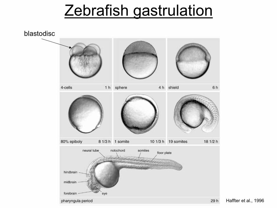
Haffter et al., 1996
Zebrafish gastrulation blastodisc
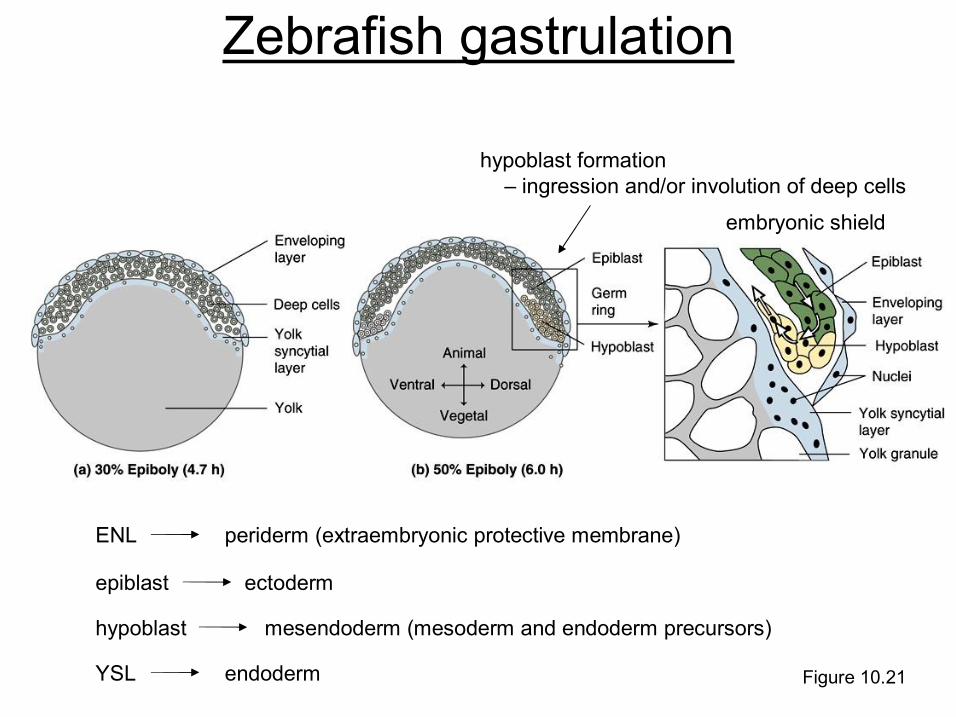
Figure 10.21
Zebrafish gastrulation
hypoblast formation – ingression and/or involution of deep cells
embryonic shield
ENL periderm (extraembryonic protective membrane)
epiblast ectoderm
hypoblast mesendoderm (mesoderm and endoderm precursors)
YSL endoderm
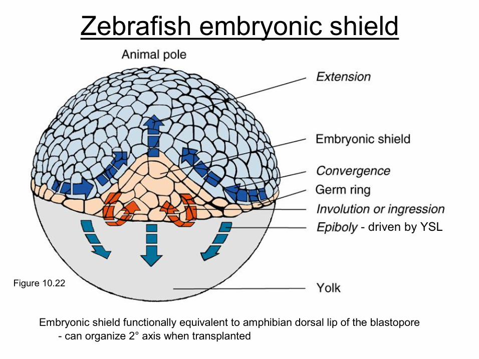
Figure 10.22
Zebrafish embryonic shield
driven by YSL
Embryonic shield functionally equivalent to amphibian dorsal lip of the blastopore can organize 2° axis when transplanted
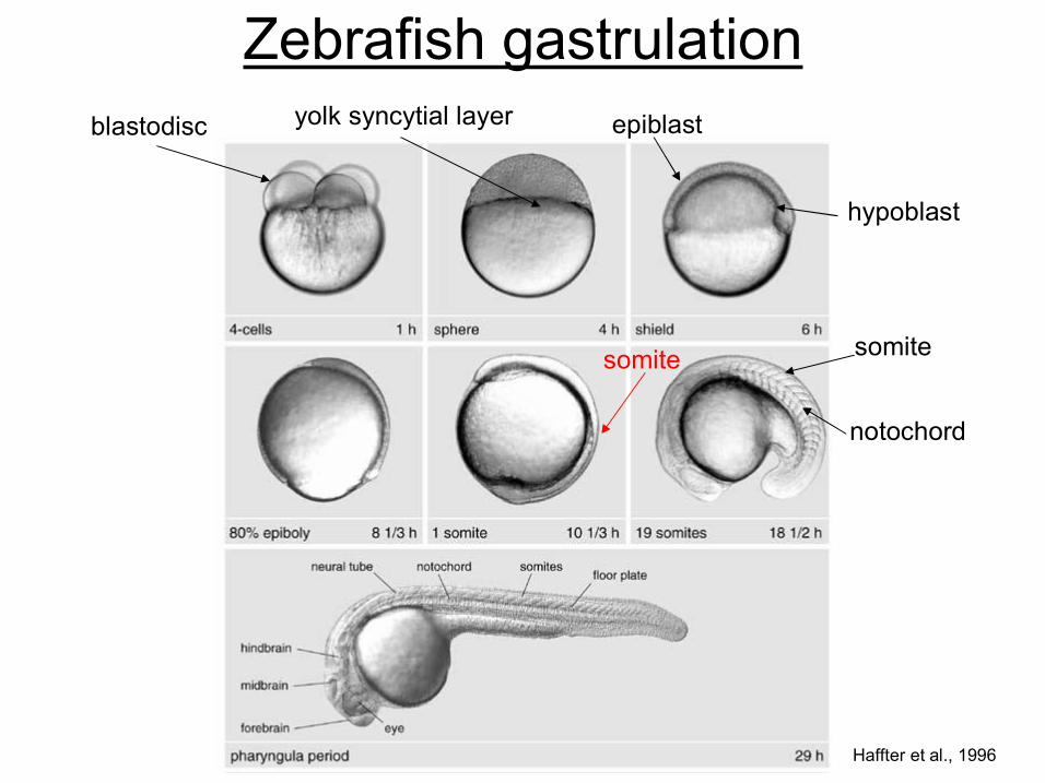
Haffter et al., 1996
Zebrafish gastrulation blastodisc yolk syncytial layer epiblast
hypoblast
somite
notochord
somite
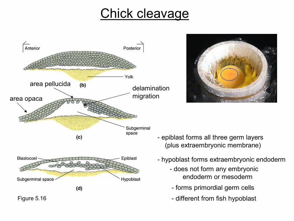
Figure 5.16
epiblast forms all three germ layers (plus extraembryonic membrane)
hypoblast forms extraembryonic endoderm
Chick cleavage
delamination migration
area pellucida
area opaca
different from fish hypoblast forms primordial germ cells
does not form any embryonic endoderm or mesoderm
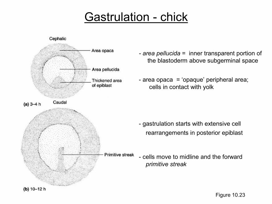
Figure 10.23
Gastrulation chick
area pellucida = inner transparent portion of the blastoderm above subgerminal space
area opaca = ‘opaque’ peripheral area; cells in contact with yolk
gastrulation starts with extensive cell rearrangements in posterior epiblast
cells move to midline and the forward primitive streak
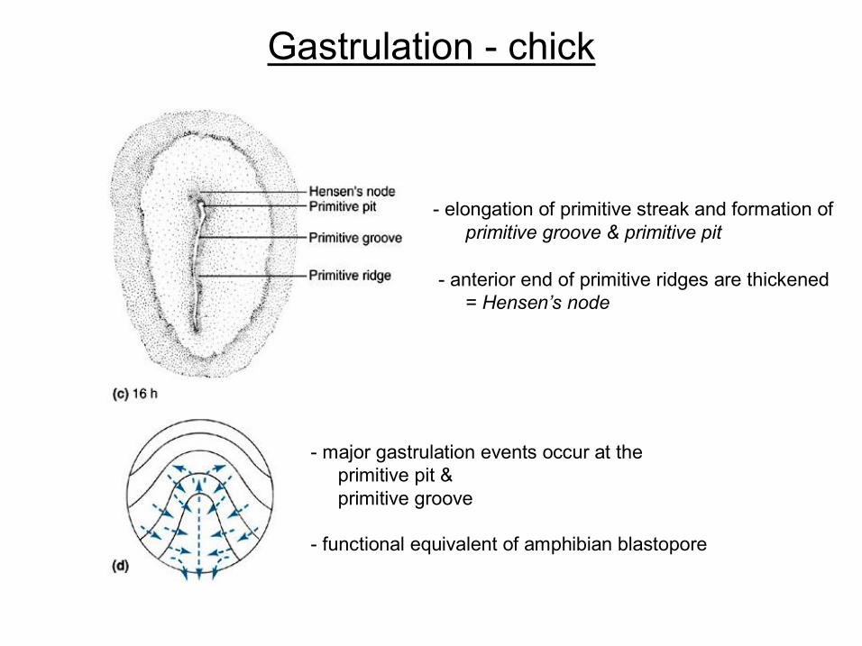
Gastrulation chick
major gastrulation events occur at the primitive pit & primitive groove
elongation of primitive streak and formation of primitive groove & primitive pit
anterior end of primitive ridges are thickened = Hensen’s node
functional equivalent of amphibian blastopore
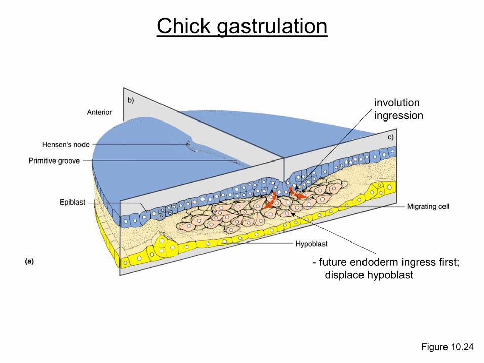
Figure 10.24
involution ingression
future endoderm ingress first; displace hypoblast
Chick gastrulation
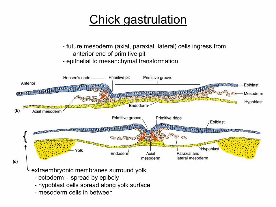
Chick gastrulation
future mesoderm (axial, paraxial, lateral) cells ingress from anterior end of primitive pit
epithelial to mesenchymal transformation
extraembryonic membranes surround yolk ectoderm – spread by epiboly hypoblast cells spread along yolk surface mesoderm cells in between
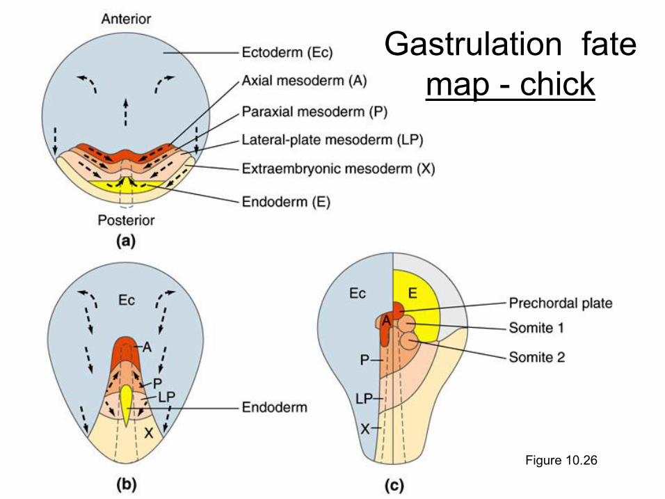
Figure 10.26
Gastrulation fate map chick
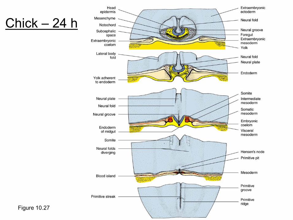
Figure 10.27
Chick – 24 h
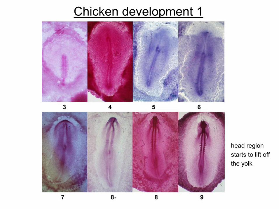
Chicken development 1
head region starts to lift off the yolk
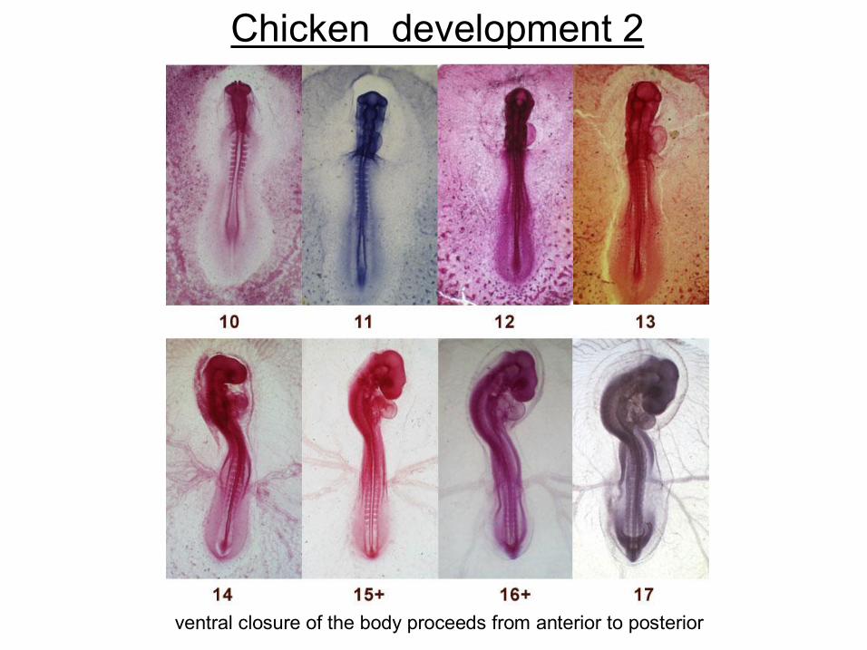
Chicken development 2
ventral closure of the body proceeds from anterior to posterior
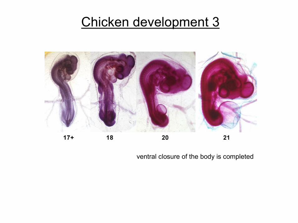
Chicken development 3
ventral closure of the body is completed
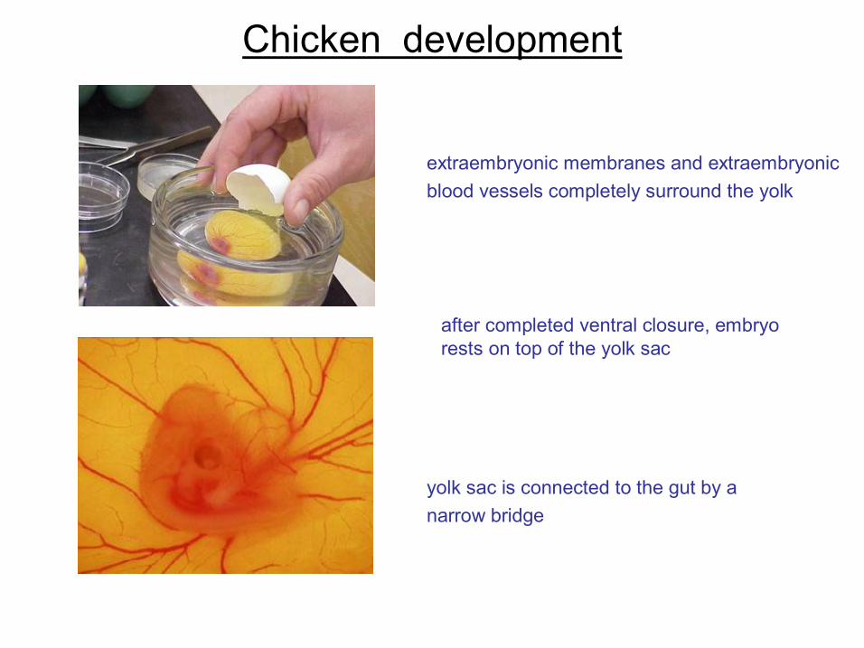
Chicken development
extraembryonic membranes and extraembryonic blood vessels completely surround the yolk
yolk sac is connected to the gut by a narrow bridge
after completed ventral closure, embryo rests on top of the yolk sac
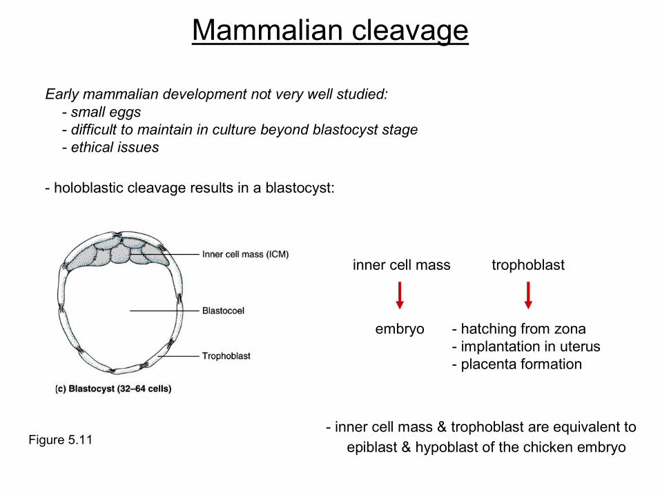
Early mammalian development not very well studied: small eggs difficult to maintain in culture beyond blastocyst stage ethical issues
Figure 5.11 inner cell mass & trophoblast are equivalent to
epiblast & hypoblast of the chicken embryo
Mammalian cleavage
holoblastic cleavage results in a blastocyst:
hatching from zona implantation in uterus placenta formation
trophoblast
embryo
inner cell mass
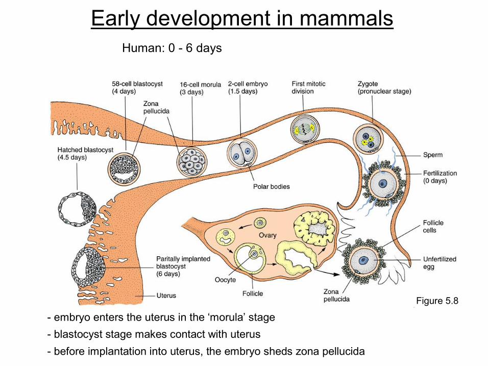
Early development in mammals
Figure 5.8
embryo enters the uterus in the ‘morula’ stage blastocyst stage makes contact with uterus before implantation into uterus, the embryo sheds zona pellucida
Human: 0 6 days
Gilbert SF, Developmental Biology, 6 th ed, Sinauer, 2000
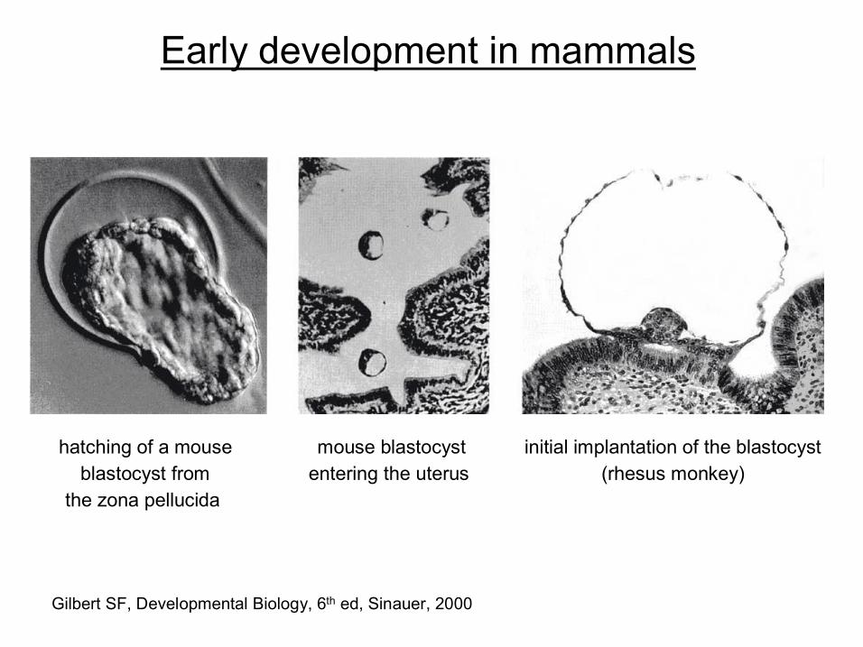
hatching of a mouse blastocyst from
the zona pellucida
Early development in mammals
mouse blastocyst entering the uterus
initial implantation of the blastocyst (rhesus monkey)
Early development in mammals
Gilbert SF, Developmental Biology, 7 th ed, Sinauer, 2003
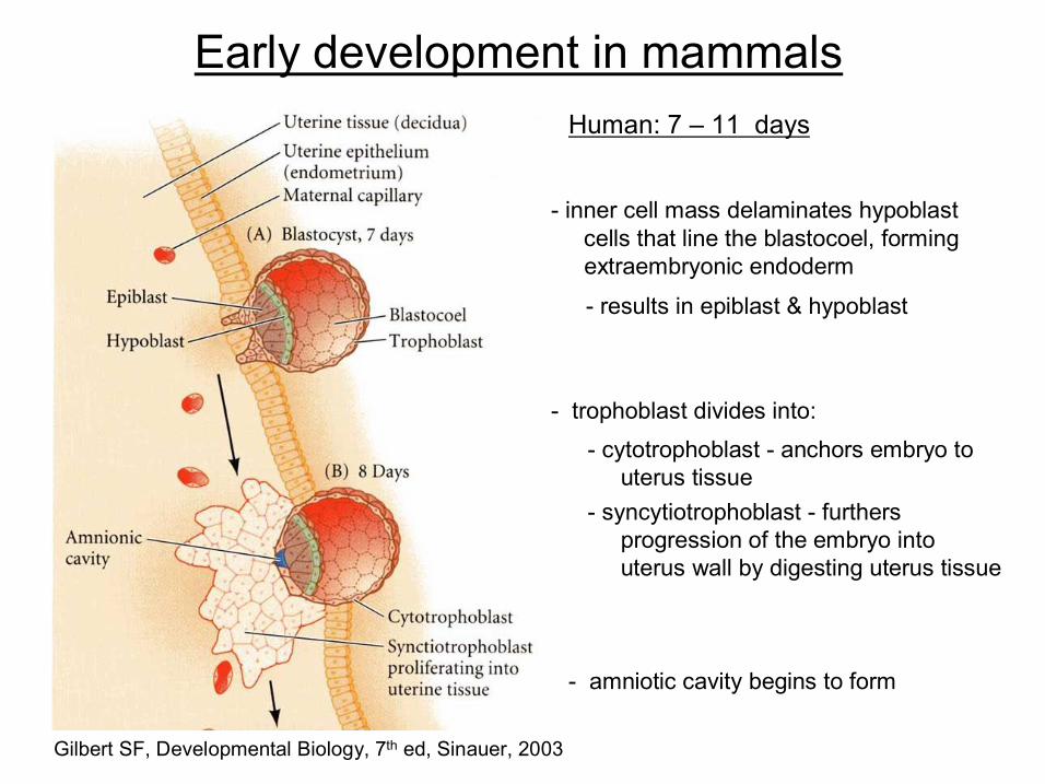
amniotic cavity begins to form
results in epiblast & hypoblast
inner cell mass delaminates hypoblast cells that line the blastocoel, forming extraembryonic endoderm
trophoblast divides into:
syncytiotrophoblast furthers progression of the embryo into uterus wall by digesting uterus tissue
cytotrophoblast anchors embryo to uterus tissue
Human: 7 – 11 days
Early development in mammals
Gilbert SF, Developmental Biology, 7 th ed, Sinauer, 2003
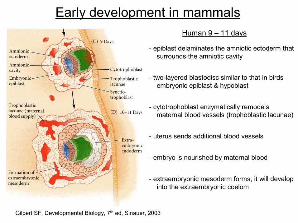
extraembryonic mesoderm forms; it will develop into the extraembryonic coelom
embryo is nourished by maternal blood
uterus sends additional blood vessels
cytotrophoblast enzymatically remodels maternal blood vessels (trophoblastic lacunae)
twolayered blastodisc similar to that in birds embryonic epiblast & hypoblast
epiblast delaminates the amniotic ectoderm that surrounds the amniotic cavity
Human 9 – 11 days
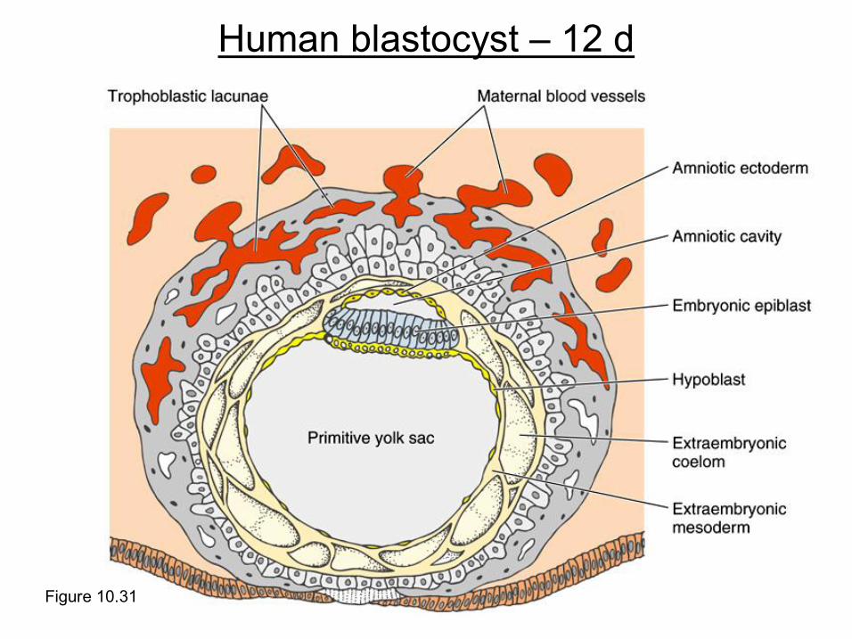
Figure 10.31
Human blastocyst – 12 d
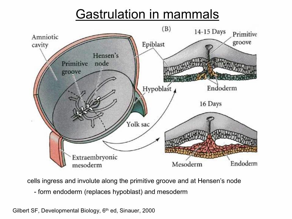
cells ingress and involute along the primitive groove and at Hensen’s node
Gilbert SF, Developmental Biology, 6 th ed, Sinauer, 2000
Gastrulation in mammals
form endoderm (replaces hypoblast) and mesoderm
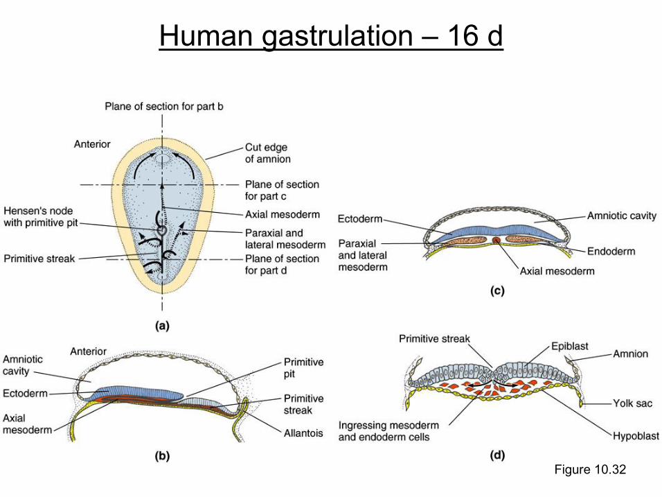
Figure 10.32
Human gastrulation – 16 d
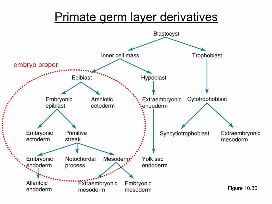
Figure 10.30
Primate germ layer derivatives
embryo proper
Recommended