PARADIGM SHIFTS IN PERSPECTIVE
Gastric Acid-Dependent Diseases: A Twentieth-CenturyRevolution
George Sachs • Jai Moo Shin • Keith Munson •
David R. Scott
Published online: 23 May 2014
� Springer Science+Business Media New York (Outside the USA) 2014
Introduction
Until recently, peptic ulcer disease (PUD) has been a major
scourge to humanity, associated with high incidence of
morbidity and mortality, the latter due primarily to foregut
perforation or hemorrhage. Advancements in the under-
standing of the pathophysiology and treatment of PUD
have included the discovery of gastric HCl secretion by
Prout [1], and the realization that PUD only occurred in the
presence of gastric acid [2], leading to the pronouncement
‘‘no acid, no ulcer.’’ Apart from a strict bland diet [3], only
surgery, ranging from partial or total gastrectomy to
vagotomy and to selective or highly selective vagotomy
successfully reduced gastric acid secretion [4, 5]. In the last
quarter of the twentieth century, three major advances took
place, completely altering the treatment of gastric acid-
related diseases: (1) the development of histamine2 recep-
tor antagonists (H2RA) as a result of rational drug design;
(2) the development of proton pump inhibitors (PPIs), in
part serendipitous but also followed by rational drug
design; and (3) recognition that infection by Helicobacter
pylori (Hp) is a major causative factor in peptic ulcer
disease and even of gastric cancer [6, 7]. In the last two
decades, PUD has been replaced by gastro-esophageal
reflux disease (GERD) as the major reason for physician
consultation due to foregut-related symptoms.
The Regulation of Acid Secretion
Although gastric acid secretion was discovered in the
1700s, its regulation has only been deduced relatively
recently. At the time of the introduction of H2 RAs, gastrin,
discovered by Edkins [8] and isolated by Gregory and
Tracy, was thought by many to be the major direct stim-
ulant of acid secretion [9]. Nevertheless, the first-genera-
tion H2RAs, cimetidine or burimamide completely
inhibited gastrin-stimulated acid secretion in rats [10],
indicating that the major secretory action of gastrin was
mediated by histamine. Subsequent characterization of the
enterochromaffin-like (ECL) cell revealed that gastrin-
stimulated histamine release from this master regulator of
acid secretion since gastrin stimulation of acid secretion by
isolated rabbit gastric glands was completely inhibited by
H2RAs with a 1 nM affinity [11]. Therefore, the lower
affinity (10 nM) gastrin receptor expressed in parietal cells
is likely involved with trophic effects such as regulation of
differentiation or growth rather than with stimulation of
acid secretion. ECL cells are also stimulated by pituitary
adenylate cyclase-activating peptide (PACAP) but not by
acetylcholine, the vagal mediator of acid secretion, which
depends on its binding to muscarinic receptor subtype 3 on
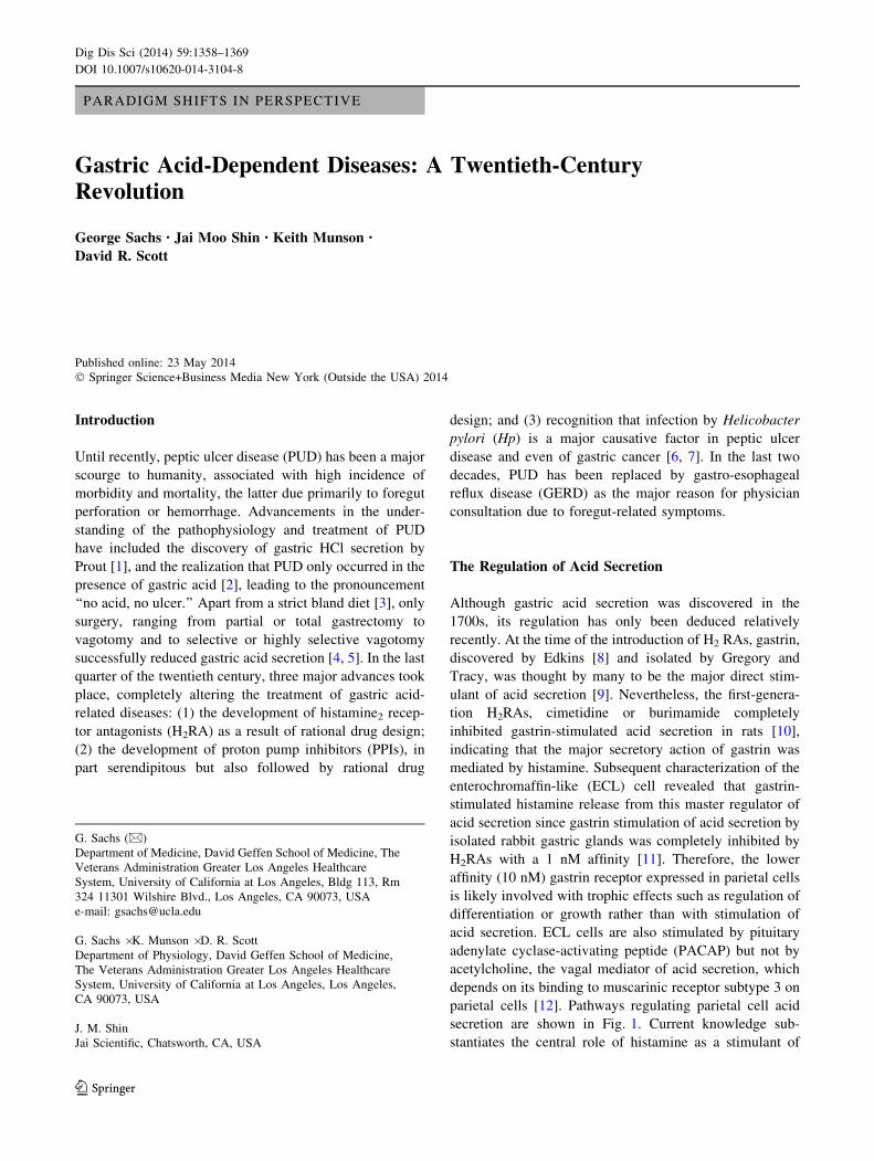
parietal cells [12]. Pathways regulating parietal cell acid
secretion are shown in Fig. 1. Current knowledge sub-
stantiates the central role of histamine as a stimulant of
G. Sachs (&)
Department of Medicine, David Geffen School of Medicine, The
Veterans Administration Greater Los Angeles Healthcare
System, University of California at Los Angeles, Bldg 113, Rm
324 11301 Wilshire Blvd., Los Angeles, CA 90073, USA
e-mail: [email protected]
G. Sachs � K. Munson � D. R. Scott
Department of Physiology, David Geffen School of Medicine,
The Veterans Administration Greater Los Angeles Healthcare
System, University of California at Los Angeles, Los Angeles,
CA 90073, USA
J. M. Shin
Jai Scientific, Chatsworth, CA, USA
123
Dig Dis Sci (2014) 59:1358–1369
DOI 10.1007/s10620-014-3104-8
gastric acid secretion, paving the way for the discovery of
H2RA as a means of down regulating secretion.
Histamine and Histamine Receptor Antagonists
Of the four histamine receptor subtypes, H1 through H4, the
first of these to be targeted was the H1 receptor which is
responsible for rhinitis and other allergic syndromes. The
first successful small ligand receptor antagonist class ever
developed was the H1RA [13] based on the structure of
histamine, a known pro-inflammatory ligand and also
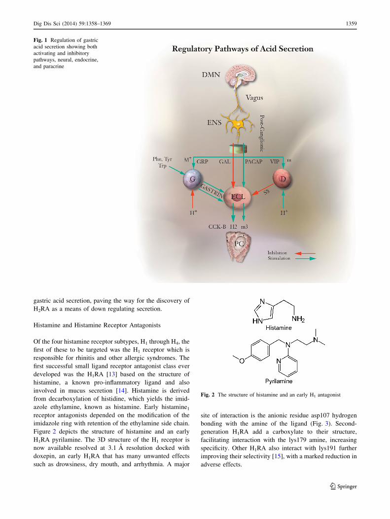
involved in mucus secretion [14]. Histamine is derived
from decarboxylation of histidine, which yields the imid-
azole ethylamine, known as histamine. Early histamine1
receptor antagonists depended on the modification of the
imidazole ring with retention of the ethylamine side chain.
Figure 2 depicts the structure of histamine and an early
H1RA pyrilamine. The 3D structure of the H1 receptor is
now available resolved at 3.1 A resolution docked with
doxepin, an early H1RA that has many unwanted effects
such as drowsiness, dry mouth, and arrhythmia. A major
site of interaction is the anionic residue asp107 hydrogen
bonding with the amine of the ligand (Fig. 3). Second-
generation H1RA add a carboxylate to their structure,
facilitating interaction with the lys179 amine, increasing
specificity. Other H1RA also interact with lys191 further
improving their selectivity [15], with a marked reduction in
adverse effects.
Fig. 1 Regulation of gastric
acid secretion showing both
activating and inhibitory
pathways, neural, endocrine,
and paracrine
Fig. 2 The structure of histamine and an early H1 antagonist
Dig Dis Sci (2014) 59:1358–1369 1359
123

Ash and Schild clearly differentiated the histamine
receptors expressed in the circulatory system from those
expressed in stomach or uterus [16] concluding that there
were at least two classes of histamine receptors. James
Black moved from Imperial Chemical Industries (ICI)
where he had developed the first b-adrenergic inhibitor,
propranolol, to Smith Kline & French (SK&F) in Welwyn
(a stately home used during World War II to test miniature
submarines since there was a very deep pond on the pre-
mises) recruited by Bill Duncan also from Glasgow. Black
et al. realized that the secret to developing H2RAs was not
to modify the imidazole ring of histamine as had been done
for the H1RA, but to modify the side chain. I was privi-
leged, as the gastrointestinal consultant for SK&F in
Philadelphia, to be sent to Welwyn to review Jimmy
Black’s progress. Within a seven-year project cycle,
6 years had passed for the H2 receptor project without
success! I was most impressed with the team including
Mike Parsons, Robin Ganellin and Graham Durant and
submitted a positive report to Peter Ridley and Bryce
Douglas the then head of research at SK&F, a Glaswegian
like Jimmy. A crucial tool developed by Mike Parsons was
the measurement of acid secretion in the rat rather than
measuring the degree of ulceration. The next year, since I
was aware of the H2 antagonistic action of burimamide, I
was able inhibit histamine-stimulated adenylate cyclase
and submit an even more positive report [17], which
eventuated in the introduction of cimetidine (Tagamet�) as
the first anti-secretory medication indicated for the treat-
ment of peptic ulcer disease [10, 18] (Fig. 4). Cimetidine
was rapidly followed by the second-generation H2RAs
ranitidine, famotidine and nizatidine with somewhat dif-
ferent structures and differing duration of action, of which
Fig. 4 The structure of
cimetidine and two other H2
antagonists. Reprinted with
permission from Macmillan
Publishers Ltd: Modlin and
Sachs [60]
Fig. 5 The effect of once a day, evening administration of ranitidine on
intragastric pH showing good night time effect on day 1 with 50 % loss of
effect by day 7 through day 28 and little effect during the day requiring
BID dosing for more effective control of acid secretion compared to the
four times per day recommended for cimetidine. Reprinted with
permission from Macmillan Publishers Ltd: Modlin and Sachs [60]
Fig. 3 The 3D structure of the H1 receptor with doxepin docked,
showing interaction with Asp107 H? bonding with the amine group
of the antagonist
1360 Dig Dis Sci (2014) 59:1358–1369
123

the most effective is famotidine [19]. The discovery of
H2RAs not only revolutionized therapy of acid-related
diseases, but also vastly improved the understanding of the
control of acid secretion, as discussed above.
As experience increased with the use of H2RAs, certain
drawbacks to their use became apparent. Firstly, their
action was short lived, requiring multiple daily doses.
Secondly, all patients exhibited tolerance whereby after
one week of treatment, the response was reduced by
*50 % [20] (Fig. 5). Hence, although relatively effective
at accelerating the healing rate of duodenal ulcer, they were
less effective in the treatment of gastric ulcers. Further-
more, the response in patients suffering from gastro-
esophageal reflux disease (GERD) was mostly inadequate
[21]. A different means of inhibition of acid secretion was
required. Fortunately, by that time, the gastric H?, K?
ATPase, the final step of acid secretion that cannot be
bypassed, had been discovered. Ganser and Forte showed
the presence of a K?-stimulated ATPase in frog gastric
microsomes [22] and Peter Scholes working in Peter
Mitchell’s laboratory showed that dog microsomes alka-
linized the medium in the presence of K? in the medium
upon addition of MgATP [23].
Reprising our discovery of a K?-stimulated ATPase in
hog gastric vesicles in 1968, we demonstrated details of the
H? for K? ATPase reaction mechanism showing conclu-
sively that it was an electroneutral H? for K? exchange P2-
type ATPase lacking a K? conductance in resting enzyme
[24]. This conclusion was based on the lack of effect of
lipid permeable ions on either ATP-dependent H? or Rb?
transport and the absence of changes in a membrane
potential sensitive dye during H? for K? exchange in the
absence of the K? ionophore, valinomycin (24). A later
paper used membrane potential dyes such as diethylox-
adicarbocyanine or oxonol dyes to confirm electroneutral-
ity [25].
Mechanism of Acid Secretion: The Gastric H?, K?
ATPase
HCl secretion by the gastric parietal cell depends on acti-
vation of the gastric H?, K? ATPase, termed the proton
pump. This enzyme is found uniquely in gastric parietal
cells and in renal collecting ducts. It is an electroneutral H?
for K? exchange P2-type (phosphorylating) ATPase with
ten membrane-spanning segments and a b subunit with one
transmembrane segment and six or seven glycosylation
sites. The catalytic cycle is shown in Fig. 6.
At neutral pH, 2 H? are exchanged for 2 K? per
hydrolysis of 1 ATP, but as the luminal pH falls, the
exchange stoichiometry is 1 H? per 1 K? per 1 ATP. This
stoichiometry change is explained by the pKa of one of the
hydronium binding sites which remains protonated at
luminal pH \ 3.0 [26]. Structurally, it is a heterodimer of an
a and b subunit like the Na?, K? ATPase which may exist
as a dimeric oligomer, i.e., a2b2 as indicated by stoichi-
ometry of labeling with inhibitors or phosphorylation [27].
Although its 3D structure has not been solved, that of two
homologous pumps has been [27, 28]; providing valuable
clues about the structure of the H?, K? ATPase by homology
modeling, providing in turn a plausible explanation for the
mechanism of acid inhibition by PPIs. The cytoplasmic
domain has three loops, the N or nucleotide binding domain,
the A or activation domain, and P the phosphorylation domain
(Fig. 7). The key to transport of H? from the cytosol and
absorption of K? from the lumen are conformational changes
induced by phosphorylation of Asp386 by MgATP, which
Fig. 7 A homology model of the arrangement of the two subunits of
the gastric H, K ATPase in the E2 form where the N and A domains
have moved inward to the P domain opening a luminal vestibule
allowing K and inhibitor access. The ion binding and glycosylation
sites of the beta subunit are also shown
Fig. 6 The catalytic cycle of the gastric H?, K? ATPase. Binding of
ATP and hydronium ion results in phosphorylation of the alpha
subunit to result in the E1-P form which then occludes the ion and
spontaneously converts to the E2-P form, releasing hydronium ion to
the lumen. K? then binds resulting in dephosphorylation and
formation of the K? occluded form which then releases K? to the
cytoplasm reforming E1
Dig Dis Sci (2014) 59:1358–1369 1361
123
changes the direction of net ion flux from cyto-
plasm ? lumen to lumen ? cytoplasm due to conforma-
tional changes in the cytoplasmic domain wherein the N and A
domains move close to the P domain with closure of the
cytoplasmic access pathway. There is then a spontaneous
opening of a luminal vestibule allowing ion access from the
luminal side. The E2 form is illustrated in Fig. 7, showing the
placement of the ß subunit and six glycosylation sites. The ß
subunit of the sodium pump has only three glycosylation sites
which has likely impeded efforts to crystallize the protein.
Activation of the Pump
In the resting parietal cell, most of the ATPase is seques-
tered in cytoplasmic tubulovesicles, and upon stimulation,
it is trafficked to the apical microvilli of the secretory
canaliculus [28]. Along with the pump, there is trafficking
of the K? channel complex KCNQ1 and KCNE2 as well as
a Cl- channel. The channel trafficking is distinct from the
trafficking of the ATPase [29, 30].
Proton Pump Inhibitors (PPIs)
This novel acid therapeutic class was initially discovered
serendipitously by the Swedish company, AB Hassle dur-
ing a secondary screen of antiviral compound candidates
for anti-ulcer activity in a rat ulcer model. A compound,
pyridine 2-acetamide, inhibited gastric acid secretion, even
after conversion to the more stable pyridine 2-thioaceta-
mide. Since the imidazole ring was considered essential for
the anti-secretory activity of H2RA, Hassle added an
imidazole ring to pyridine 2-thioacetamide based on advice
from their gastrointestinal consultant Lars Olbe. Since
imidazole was not available, benzimidazole was added to
produce pyridine 2-methylthiobenzimidazole. With an
added sulfoxide to increase stability, timoprazole, the
precursor of all PPIs, resulted. Since timoprazole, unlike
H2RAs, inhibited acid secretion independently of stimulus,
it was considered a new class of antisecretory compound.
My laboratory presented data showing that a polyclonal
antibody against the gastric H?, K? ATPase reacted with
extracts of the stomach, thyroid, and thymus. We therefore
hypothesized that the compound inhibited the gastric H?,
K? ATPase, the final step of acid secretion. Timoprazole
did not inhibit purified H?, K? ATPase when net acid
secretion was ‘‘short-circuited’’ by the H? for K? exchange
ionophore, nigericin. Since timoprazole was acid unstable,
we tested timoprazole against the H?, K? ATPase using
conditions that promoted H? for K? exchange though the
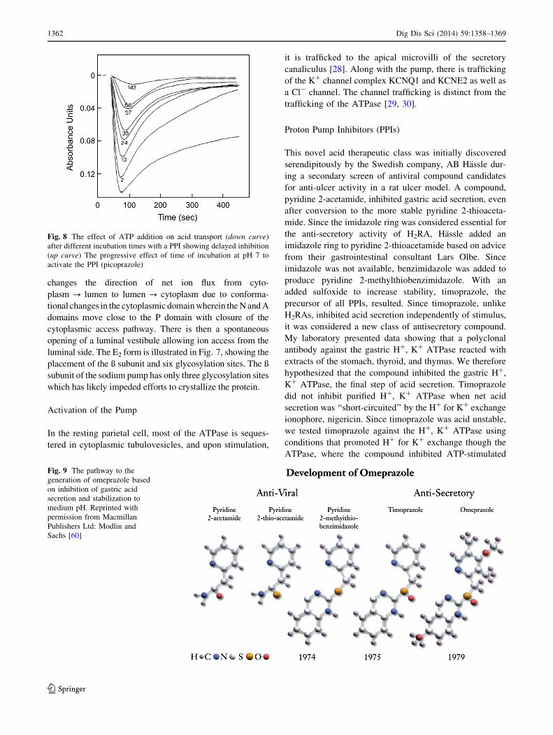
ATPase, where the compound inhibited ATP-stimulated
Fig. 8 The effect of ATP addition on acid transport (down curve)
after different incubation times with a PPI showing delayed inhibition
(up curve) The progressive effect of time of incubation at pH 7 to
activate the PPI (picoprazole)
Fig. 9 The pathway to the
generation of omeprazole based
on inhibition of gastric acid
secretion and stabilization to
medium pH. Reprinted with
permission from Macmillan
Publishers Ltd: Modlin and
Sachs [60]
1362 Dig Dis Sci (2014) 59:1358–1369
123

H? secretion (Fig. 8) [31]. This and experiments like this
indicated that timoprazole was an acid-activated prodrug,
serving as the basis for omeprazole, as shown in Fig. 9,
with substitution on both the pyridine and benzimidazole
rings.
Since they are acid activated, PPIs are usually enteric
coated in order to impair gastric drug release. Since there is
significant activation of all PPIs at neutral pH, IV formu-
lation of the PPIs pantoprazole and esomeprazole requires
that the compound must be dissolved in buffer with
pH [ 9.0 in order to delay activation immediately prior to
administration [32].
During the development of omeprazole, serious con-
cerns were raised by the management of Hassle as to its
value, in particular since ranitidine then dominated the
antisecretory market. With my [GS] clinical background,
my knowledge of its molecular mechanism and potent
antisecretory properties, I was convinced that omeprazole
would become the market leader in its class, with world-
wide sales exceeding US $1 billion annually, which grossly
underestimated its peak sales revenue of US $6 billion,
with the overall revenue of all drugs in its class exceeding
US $20 billion annually. Every 3 months, I attended a
meeting of the ‘‘PPI team’’ in a basement room at Hassle,
which helped me maintain close collaborative ties between
my laboratory and that of Hassle. The team of about 12
scientists was given total freedom by Anders Vedin, the
research head at Hassle, to understand every key aspect of
omeprazole and to bring it to market with no interference
from oversight committees. Many of the team members
spent time in my laboratory including Herbert Helander,
Thomas Berglindh, Bjorn Wallmark, Pia Lorentzon, and
Karen Gedda. Also, at the time, Byk-Gulden in Konstanz,
Germany, allied with SK&F, started PPI development,
culminating in the development of pantoprazole (Proto-
nix�) aided by the translational studies of David Keeling
and Alex Simon on loan to my laboratory. Also very
important at the time was the Astra Hassle marketing
manager, Ian Talmage, who was a remarkably talented in
the art of product branding. All of these talented individ-
uals have remained lifelong friends.
As phase III trials were underway, rats, but not mice or
dogs, developed ECL cell carcinoid tumors, halting the
clinical PPI trials, which the involved scientists were
convinced was an effect of hypergastrinemia and not pri-
marily drug dependent, supported by data generated by a
Hassle team led by Enar Carlsson, which was sufficient to
re-start the clinical trials. A key experiment used to support
the hypergastrinemia hypothesis was that rats treated with
high-dose H2RA also developed ECL tumors [33]. In
contrast to rats, the ECL cell is terminally differentiated in
humans, explaining why ECL tumors have not been
described in humans receiving long-term, high-dose PPI
therapy. Nevertheless, experimental findings prompted the
competition to claim carcinogenicity for omeprazole [34].
Since the clinical introduction of omeprazole, several
other drugs have entered the market which are also acid-
activated prodrugs, sharing similar advantages and disad-
vantages with omeprazole (Fig. 10). Esomeprazole (Nexi-
um�) is the S-enantiomer of omeprazole which at 40 mg
per day compared to 20 mg per day of omeprazole shows a
slight benefit in acid control. The most recent entry to the
market is the D-enantiomer of lansoprazole (Dexilant�)
which is formulated be released immediately and after a
*4-h delay. All of the PPIs in clinical use are activated
prodrugs forming a thiophilic reactive group that binds
covalently to one or more cysteines on the gastric H?, K?
ATPase irreversibly inhibiting the enzyme.
Fig. 10 The currently marketed
PPIs
Dig Dis Sci (2014) 59:1358–1369 1363
123

The mechanism of activation of the PPIs is a remarkable
series of chemical steps as shown in Fig. 11, elucidated by
Arne Brandstrom and Per Lindberg at Hassle and refined
by Jai Moo Shin in my laboratory [35, 36]. There is still
disagreement as to whether the active compound in vivo is
the sulfenic acid or the sulfenamide which was the form
isolated at Hassle. Please see [37] for a comprehensive
review of this area. The mechanism is shown as a general
structural form (Fig. 11). The top of Fig. 11 shows the
protonation of the pyridine ring with a pKa between 4.0
(omeprazole, lansoprazole, pantoprazole) and 5.0 (rabep-
razole), accumulating the protonated form only in the
actively secreting parietal cell, since this is the only space
with a pH \ 4.0. Below that is shown the protonation of
the benzimidazole ring with a pKa of\2.0 and in brackets
is shown the mechanism of activation where the C2 of the
protonated benzimidazole ring reacts with the unprotonated
fraction of the pyridine moiety rearranging to a permanent
thioreactive, cationic, tetracyclic sulfenamide that binds
covalently to one or more luminally accessible cysteines of
the a subunit of the gastric ATPase. In aqueous solution,
the sulfenic acid dehydrates to form the sulfenamide. The
cationic sulfenic acid or sulfenamide remains trapped in the
parietal cell canaliculus. In the particular case of pantop-
razole, cysteine 813 and cysteine 822 become covalently
linked (Fig. 12). With other PPIs different cysteines are
linked but cysteine 813 is derivatized by all PPIs and must
be considered as the central target for this class of drug. It
is easy to visualize that binding of a PPI covalently in this
region will fix the pump in the E2 form and inhibits cycling
back to the E1 form.
Thus, since PPIs require ongoing acid secretion in order
to be activated, they are administered 30–60 min before
breakfast so that peak blood concentrations coincide with
maximal H?, K? ATPase activity. In spite of the need for
Fig. 11 The pathway of pH activation of a benzimidazole-based PPI.
Protonation of the pyridine (pka 4.0) allows selective accumulation of
the drug in the active parietal cell and then at highly acidic pH of the
secreting cell, protonation of the benzimidazole (pka * 1–2) results
in rearrangement, forming the thio-active drug
Fig. 12 The binding sites cys813 and cys822 of pantoprazole on the
luminal face of the alpha subunit
1364 Dig Dis Sci (2014) 59:1358–1369
123
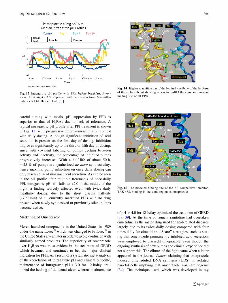
careful timing with meals, pH suppression by PPIs is
superior to that of H2RAs due to lack of tolerance. A
typical intragastric pH profile after PPI treatment is shown
in Fig. 13, with progressive improvement in acid control
with daily dosing. Although significant inhibition of acid
secretion is present on the first day of dosing, inhibition
improves significantly up to the third or fifth day of dosing,
since with covalent labeling of pumps cycling between
activity and inactivity, the percentage of inhibited pumps
progressively increases. With a half-life of about 50 h,
*25 % of pumps are synthesized de novo synthesis/day,
hence maximal pump inhibition on once daily dosing can
only reach 75 % of maximal acid secretion. As can be seen
in the pH profile after multiple treatments of once-daily
PPI, intragastric pH still falls to \2.0 in the middle of the
night, a finding scarcely affected even with twice daily
mealtime dosing, due to the short plasma half-life
(*90 min) of all currently marketed PPIs with no drug
present when newly synthesized or previously silent pumps
become active.
Marketing of Omeprazole
Merck launched omeprazole in the United States in 1989
under the name Losec� which was changed to Prilosec� in
the United States a year later in order to avoid confusion with
similarly named products. The superiority of omeprazole
over H2RAs was most evident in the treatment of GERD
which became, and continues to be, the major clinical
indication for PPIs. As a result of a systematic meta-analysis
of the correlation of intragastric pH and clinical outcome,
maintenance of intragastric pH [ 3.0 for 12 h/day opti-
mized the healing of duodenal ulcer, whereas maintenance
of pH [ 4.0 for 18 h/day optimized the treatment of GERD
[38, 39]. At the time of launch, ranitidine had overtaken
cimetidine as the major drug used for acid-related diseases
largely due to its twice daily dosing compared with four
times daily for cimetidine. ‘‘Scare’’ strategies, such as stat-
ing that omeprazole permanently inhibited acid secretion,
were employed to discredit omeprazole, even though the
ongoing synthesis of new pumps and clinical experience did
not support this. The climax of the fight came when a letter
appeared in the journal Lancet claiming that omeprazole
induced unscheduled DNA synthesis (UDS) in isolated
parietal cells implying that omeprazole was carcinogenic
[34]. The technique used, which was developed in my
Fig. 13 Intragastric pH profile with PPIs before breakfast. Arrow
show pH at night \2.0. Reprinted with permission from Macmillan
Publishers Ltd: Harder et al. [61]
Fig. 14 Higher magnification of the luminal vestibule of the E2 form
of the alpha subunit showing access to cys813 the common covalent
binding site of all PPIs
Fig. 15 The modeled binding site of the K? competitive inhibitor,
TAK-438, binding in the same region as omeprazole
Dig Dis Sci (2014) 59:1358–1369 1365
123

laboratory [40, 41], did not discriminate between surface
cells and oxyntic cells. Surface cells are derived from con-
stantly dividing stem cells which continuously
physiologically incorporate the DNA precursor thymidine,
which is not indicative of UDS. Moreover, the Lancet arti-
cle’s author published half of the dose–response curve,
whereas the complete curve was bell-shaped and clearly
could not represent UDS. Several subsequently published
studies did not support the UDS hypothesis, removing any
lingering doubts regarding the superiority of PPIs over
H2RAs for treatment of GERD [42].
Current Situation of Clinical Use of PPIs
The PPI class of drugs, though still the most widely used in
the acid-related disease market, has been attributed with
certain drawbacks over time. For example, 20 % of GERD
patients continue to have reflux symptoms despite maximal
PPI therapy, due to the presence of poorly suppressed
nocturnal acid secretion. A novel sulfonamide prodrug of
omeprazole AGN904, with delayed absorption is capable
of maintaining intragastric pH [ 5.0 for 24 h/day, prom-
ises to be effective for the recalcitrant 20 % of GERD
patients with prominent nocturnal secretion [43], and also
for the eradication of Hp which become persistent and
Fig. 16 The outcomes of infection by Helicobacter pylori. Reprinted
with permission from Macmillan Publishers Ltd: Sachs and Scott [62]
a b c
Fig. 17 Periplasmic buffering by Helicobacter pylori and its regu-
lation. a Urea crosses the outer membrane (OM) and then the inner
membrane (IM) through the pH-gated urea channel, UreI, at an
external pH \ 6.0. Cytoplasmic urease forms 2NH3 ? H2CO3 and
the latter is converted to CO2 by cytoplasmic b-carbonic anhydrase.
These gases cross the IM, and the CO2 is converted to HCO3- by the
membrane bound a-carbonic anhydrase thus maintaining periplasmic
pH at *6.1, the effective pKa of the CO2/HCO3- couple. Exiting
NH3 neutralizes the H? produced by carbonic anhydrase as well as
entering H? and can also exit the OM to neutralize the medium and
thus allows elevation of periplasmic pH to higher than medium. b The
role of the pH responsive two component system, TCS, FlgS encoded
by HP0244. Activation results in recruitment of urease proteins to
UreI and the immediate access of urea and outward transport of CO2,
NH3 and NH4? through UreI increases the rate of periplasmic
buffering. c A model representing the role of regulation by the TCS
ArsRS (encoded by HP0165/HP0166). At neutral pH, ArsS is inactive
and ArsR is not phosphorylated. This ArsR binds to the promoter of
the sRNA that targets the ureB part of the ureAB mRNA (ureB-
sRNA) and consequent truncation of ureAB mRNA with a decline in
urease activity. This reflects a likely adaptation to neutral pH. At
acidic pH, ArsS is activated with ArsR phosphorylation and this
results in upregulation of ureAB mRNA and consequent increase in
acid-protective urease activity
1366 Dig Dis Sci (2014) 59:1358–1369
123

hence untreatable at intragastric pH \ 3.0 [44] contributing
to the [30 % failure of standard triple therapy in acid
hypersecretors. Some adverse effects of PPIs include
hypocalcaemia in elderly patients undergoing chronic
therapy [45]. A very small number of patients may develop
hypomagnesaemia with serious effects on the central ner-
vous, cardiac, and musculoskeletal systems [46], possibly
due to a mutation in the duodenal Mg transporters
enhancing their susceptibility to activated PPIs in the
duodenum [47]. On the whole, however, given their rela-
tively minor adverse effects discovered only after millions
of patient-years of experience, and their unceasing and
potent effectiveness, which has revolutionized the treat-
ment of acid-peptic foregut disease, these drugs have a
remarkably low risk/benefit.
Reversible Inhibitors of the Gastric H?, K? ATPase
With generation of the E2P form, a luminal vestibule is
formed by changes in orientation of the transmembrane
domains (illustrated in Fig. 14), emphasizing the access
pathway for inhibitors such PPIs and the K? competitive
inhibitors of the pump. In the early 1980s, my laboratory
was exploring the role of Ca2? in stimulus-secretion cou-
pling in the parietal cell, testing Ca2? channel blockers
such as trifluoperazine, verapamil, and 8-(N,N-diethyl-
amino)octyl-3,4,5-trimethoxybenzoate on acid secretion in
rabbit gastric glands. The Ki for acid secretion is at least
ten times higher than the Ki for Ca2? channel blockade,
due to direct competitive inhibition of the ATPase with K?
[48]. Based on this study, we demonstrated that an exper-
imental antisecretory drug SCH28080, originally synthe-
sized as an omeprazole mimetic, was in fact a K?
competitive proton pump inhibitor [49]. Due to the huge
PPI market, intense pharmaceutical interest has been
directed at the discovery of a potent, safe, and long-lasting
antisecretory drugs to compete with the PPIs, as illustrated
in the Fig. 15, which eventuated in the synthesis of pyr-
rolo-pyridines such as TAK-438 [50, 51]. The success of
this new class depends on very slow dissociation from the
pump with binding to the same vestibule as omeprazole.
This compound, currently under development in the Far
East, if successful will have the advantage of immediate,
meal independent inhibition of acid secretion and no need
for enteric coating.
Helicobacter Pylori
In 1983, Warren and Marshall provided evidence that
infection with Hp contributes substantially to duodenal
ulcer recurrence [7] (Fig. 16). This discovery revolution-
ized the concepts of pathogenesis and the treatment of
PUD. When the relationship between Hp infection and
gastric adenocarcinoma was found, it seemed clear that
such infection should be actively treated. Nevertheless,
there is still controversy as to whether the bacterium is a
pathogen or a commensal [52], hence the concept of test
and treat is not universally accepted [53]. The relationship
of Hp infection to PUD and to gastric cancer created a
paradigm shift in treatment of PUD [54], suggesting
strongly that prophylactic eradication is justified. It is
universally accepted that eradication is needed for symp-
tomatic disease.
There are two major areas of current research, the host
response and the mechanisms behind the ability of the
organism to colonize the human stomach. In my laboratory,
we have been mostly concerned with discovering the
means whereby only Hp is able to colonize the normal
human stomach. Hp is a neutralophile, meaning that it
grows best at neutral pH and does not grow at pH \ 5.0 or
[8.2. The key property exhibited by this organism is its
ability to buffer its periplasm to near neutral in acidic
environments, mimicking a neutralophilic environment
(Fig. 17). The transcriptome of Hp recovered from the
gerbil stomach is consistent with a \pH 4.0 habitat [55].
Eradication of Hp
In the 1990s, triple therapy using two antibiotics such as
amoxicillin with either clarithromycin or metronidazole
and an antisecretory drug such as an H2RA or PPI was an
effective means of Hp eradication, with *90 % efficacy.
In the twenty-first century, resistance to the latter two
antibiotics reduced efficacy to\70 %, requiring a different
regimen [53]. To improve eradication rates, which is of
particular importance given the billions of infected indi-
viduals worldwide, either novel antibiotic independent
targets are required or a modification of antibiotic therapy
is required. One novel method is to add bismuth subcitrate
to the triple therapy, termed quadruple therapy, or to add
other antibiotics, although these approaches only modestly
improve eradication rates [56]. From the analysis of the
gastric biology of the organism, several druggable targets
have been discovered, such as the external facing proteins
UreI, HP0165, or FrbP4 or ExbD or NixA [57, 58]. Inter-
ference with these molecular targets will require develop-
ment of novel agents present in the stomach for a sufficient
time to facilitate Hp killing.
Another approach derives from an understanding of the
need for acid suppression coupled with antibiotics in
eradication regimens. As noted above, at pH 3.0 the
organism ceases to grow, developing a persistent pheno-
type. Potent acid suppression, achieving pH [ 5.0, 24 h/
day, abolishes this persistence [44]. Although current PPIs
fall short of this goal, the use of a more effective PPIs such
Dig Dis Sci (2014) 59:1358–1369 1367
123
as the prodrug of omeprazole, AGN904, or the aforemen-
tioned long-acting K? competitive inhibitor, TAK-438,
may overcome this problem. Since resistance to amoxi-
cillin is very rare, treatment with omeprazole and amoxi-
cillin in slow omeprazole metabolizers which suppresses
acid secretion to near this level resulted in excellent
eradication [59], supporting this strategy. Hence, this dual
therapy could successfully replace current triple therapy.
Summary
In the last quarter of the twentieth century, the treatment
of PUD radically changed from the former mainstays of
diet and surgery to the development of H2 receptor
antagonists that were the first effective medical means of
PUD treatment. Nevertheless, a relatively weak response
for heartburn or gastroesophageal reflux disease and
tolerance development prompted the search for more
effective treatments. The discovery of the gastric H?, K?
ATPase, the final step of acid secretion, termed the
proton pump, followed by the development of the proton
pump inhibitors forever altered PUD and GERD treat-
ment. The discovery of the causative role of infection by
Hp now make its eradication the standard-of-care for
patients with gastric symptoms. All clinically useful Hp
eradication regimens include a PPI, presumably to reduce
the persistent state of the organism in the stomach by
elevating intragastric pH. The future may lie in appli-
cation of more effective means of eradication and more
effective inhibitors of the gastric proton pump.
References
1. Prout W. On the nature of the acid and saline matters usually
existing in stomachs of animals. Philos Trans R Soc Lond.
1824;114:45–49.
2. Schwartz C. Beitrage zur Pathologie und chirugischen des pen-
etrierenden Magenheschwuren. Mitt Grenzengeb Med Chir
(Jena). 1900;5:821–848.
3. Sippy BW. Landmark article May 15, 1915: gastric and duodenal
ulcer. Medical cure by an efficient removal of gastric juice corrosion.
By Bertram W. Sippy. J Am Med Assoc. 1983;250:2192–2197.
4. Horsley GW, Barnes WC. Twenty-five years’ experience with
Billroth I gastric resection. Ann Surg. 1957;145:758–766 (dis-
cussion, 766–759).
5. McLeod RS, Cohen Z. Highly selective vagotomy and truncal
vagotomy and pyloroplasty for duodenal ulcer: a clinical review.
Can J Surg. 1979;22:113–120.
6. Herrera V, Parsonnet J. Helicobacter pylori and gastric adeno-
carcinoma. Clin Microbiol Infect Off Publ Euro Soc Clin
Microbiol Infect Dis. 2009;15:971–976.
7. Marshall BJ, Warren JR. Unidentified curved bacilli in the
stomach of patients with gastritis and peptic ulceration. Lancet.
1984;1:1311–1315.
8. Edkins JS. The chemical mechanism of gastric secretion. J
Physiol. 1906;34:133–144.
9. Gregory RA, Tracy HJ, Harris JI, et al. Minigastrin; corrected
structure and synthesis. Hoppe-Seyler’s Zeitschrift fur Physio-
logische Chemie.. 1979;360:73–80.
10. Black JW, Duncan WA, Durant CJ, Ganellin CR, Parsons EM.
Definition and antagonism of histamine H 2-receptors. Nature.
1972;236:385–390.
11. Sachs G, Zeng N, Prinz C. Physiology of isolated gastric endo-
crine cells. Annu Rev Physiol. 1997;59:243–256.
12. Wilkes JM, Kajimura M, Scott DR, Hersey SJ, Sachs G. Mus-
carinic responses of gastric parietal cells. J Membr Biol.
1991;122:97–110.
13. Bovet D. Introduction to antihistamine agents and antergan
derivative. Ann N Y Acad Sci. 1950;50:1089–1126.
14. Beaven MA. Histamine: its role in physiological and pathological
processes. Monogr Allergy. 1978;13:1–113.
15. Shimamura T, Shiroishi M, Weyand S, et al. Structure of the
human histamine H1 receptor complex with doxepin. Nature.
2011;475:65–70.
16. Ash AS, Schild HO. Receptors mediating some actions of his-
tamine. Br J Pharmacol Chemother.. 1966;27:427–439.
17. Sung CP, Jenkins BC, Burns LR, et al. Adenyl and guanyl cyclase
in rabbit gastric mucosa. Am J Physiol. 1973;225:1359–1363.
18. Brimblecombe RW, Duncan WA, Durant GJ, Ganellin CR, Parsons
ME, Black JW. The pharmacology of cimetidine, a new histamine
H2-receptor antagonist. Br J Pharmacol. 1975;53:435P–436P.
19. Schunack W. What are the differences between the H2-receptor
antagonists? Aliment Pharmacol Ther. 1987;1:493S–503S.
20. Nwokolo CU, Prewett EJ, Sawyerr AM, Hudson M, Lim S,
Pounder RE. Tolerance during 5 months of dosing with raniti-
dine, 150 mg nightly: a placebo-controlled, double-blind study.
Gastroenterology. 1991;101:948–953.
21. Hotz J. [Pathophysiology of esophageal motility] Zeitschrift fur
Gastroenterologie. 1990;28:52–55 (discussion 70–51).
22. Ganser AL, Forte JG. K?-stimulated ATPase in purified micro-
somes of bullfrog oxyntic cells. Biochim Biophys Acta.
1973;307:169–180.
23. Lee J, Simpson G, Scholes P. An ATPase from dog gastric
mucosa: changes of outer pH in suspensions of membrane vesi-
cles accompanying ATP hydrolysis. Biochem Biophys Res
Commun. 1974;60:825–832.
24. Sachs G, Chang HH, Rabon E, Schackman R, Lewin M, Sacco-
mani G. A nonelectrogenic H? pump in plasma membranes of
hog stomach. J Biol Chem. 1976;251:7690–7698.
25. Rabon E, Chang H, Sachs G. Quantitation of hydrogen ion and
potential gradients in gastric plasma membrane vesicles. Bio-
chemistry. 1978;17:3345–3353.
26. Rabon EC, McFall TL, Sachs G. The gastric [H, K]ATPase:H?/
ATP stoichiometry. J Biol Chem. 1982;257:6296–6299.
27. Shin JM, Sachs G. Dimerization of the gastric H?, K(?)-ATP-
ase. J Biol Chem. 1996;271:1904–1908.
28. Forte JG, Zhu L. Apical recycling of the gastric parietal cell H,
K-ATPase. Ann Rev Physiol. 2010;72:273–296.
29. Lambrecht NW, Yakubov I, Scott D, Sachs G. Identification of
the K efflux channel coupled to the gastric H-K-ATPase during
acid secretion. Physiol Genomics. 2005;21:81–91.
30. Nguyen N, Kozer-Gorevich N, Gliddon BL, et al. Independent
trafficking of the KCNQ1 K? channel and H?-K?-ATPase in
gastric parietal cells from mice American journal of physiology.
Gastrointest Liver Physiol. 2013;304:G157–G166.
31. Wallmark B, Sachs G, Mardh S, Fellenius E. Inhibition of gastric
(H??K?)-ATPase by the substituted benzimidazole, picopraz-
ole. Biochim Biophys Acta. 1983;728:31–38.
32. Armstrong D, Bair D, James C, Tanser L, Escobedo S, Nevin K.
Oral esomeprazole vs. intravenous pantoprazole: a comparison of
1368 Dig Dis Sci (2014) 59:1358–1369
123
the effect on intragastric pH in healthy subjects. Aliment Phar-
macol Ther. 2003;18:705–711.
33. Ekman L, Hansson E, Havu N, Carlsson E, Lundberg C. Toxi-
cological studies on omeprazole. Scand J Gastroenterol Suppl.
1985;108:53–69.
34. Burlinson B, Morriss SH, Gatehouse DG, Tweats DJ. Genotoxi-
city studies of gastric acid inhibiting drugs. Lancet. 1990;335:
419–420.
35. Lindberg P, Nordberg P, Alminger T, Brandstrom A, Wallmark
B. The mechanism of action of the gastric acid secretion inhibitor
omeprazole. J Med Chem. 1986;29:1327–1329.
36. Sachs G, Shin JM, Besancon M, Prinz C. The continuing
development of gastric acid pump inhibitors. Aliment Pharmacol
Ther. 1993;7:4–12 (discussion 29–31).
37. Sachs G, Shin JM, Briving C, Wallmark B, Hersey S. The
pharmacology of the gastric acid pump: the H?, K?ATPase.
Annu Rev Pharmacol Toxicol. 1995;35:277–305.
38. Burget DW, Chiverton SG, Hunt RH. Is there an optimal degree
of acid suppression for healing of duodenal ulcers? A model of
the relationship between ulcer healing and acid suppression.
Gastroenterology. 1990;99:345–351.
39. Hunt RH. The relationship between the control of pH and healing
and symptom relief in gastro-oesophageal reflux disease. Aliment
Pharmacol Ther. 1995;9:3–7.
40. Blum AL, Shah GT, Wiebelhaus VD, et al. Pronase method for
isolation of viable cells from Necturus gastric mucosa. Gastro-
enterology. 1971;61:189–200.
41. Scott D, Reuben M, Zampighi G, Sachs G. Cell isolation and
genotoxicity assessment in gastric mucosa. Dig Dis Sci.
1990;35:1217–1225.
42. Fryklund J, Falknas AK, Helander HF. Omeprazole does not
cause unscheduled DNA synthesis in rabbit parietal cells in vitro.
Scand J Gastroenterol. 1992;27:521–528.
43. Hunt RH, Armstrong D, Yaghoobi M, et al. Predictable pro-
longed suppression of gastric acidity with a novel proton pump
inhibitor, AGN 201904-Z. Aliment Pharmacol Ther. 2008;28:
187–199.
44. Marcus EA, Inatomi N, Nagami GT, Sachs G, Scott DR. The
effects of varying acidity on Helicobacter pylori growth and the
bactericidal efficacy of ampicillin. Aliment Pharmacol Ther.
2012;36:972–979.
45. Subbiah V, Tayek JA. Tetany secondary to the use of a proton-
pump inhibitor. Ann Intern Med. 2002;137:219.
46. Tamura T, Sakaeda T, Kadoyama K, Okuno Y. Omeprazole- and
esomeprazole-associated hypomagnesaemia: data mining of the
public version of the FDA adverse event reporting system. Int J
Med Sci. 2012;9:322–326.
47. Lameris AL, Hess MW, van Kruijsbergen I, Hoenderop JG, Bin-
dels RJ. Omeprazole enhances the colonic expression of the
Mg(2?) transporter TRPM6. Pflugers Arch. 2013;465:1613–1620.
48. Im WB, Blakeman DP, Mendlein J, Sachs G. Inhibition of
(H?K?)-ATPase and H? accumulation in hog gastric mem-
branes by trifluoperazine, verapamil and 8-(N,
N-diethylamino)octyl-3,4,5-trimethoxybenzoate. Biochim Bio-
phys Acta. 1984;770:65–72.
49. Wallmark B, Briving C, Fryklund J, et al. Inhibition of gastric
H?, K?-ATPase and acid secretion by SCH 28080, a substituted
pyridyl(1,2a)imidazole. J Biol Chem. 1987;262:2077–2084.
50. Hori Y, Imanishi A, Matsukawa J, et al. 1-[5-(2-Fluorophenyl)-1-
(pyridin-3-ylsulfonyl)-1H-pyrrol-3-yl]-N-methylmethanamin e
monofumarate (TAK-438), a novel and potent potassium-com-
petitive acid blocker for the treatment of acid-related diseases. J
Pharmacol Exp Ther. 2010;335:231–238.
51. Shin JM, Inatomi N, Munson K, et al. Characterization of a novel
potassium-competitive acid blocker of the gastric H, K-ATPase,
1-[5-(2-fluorophenyl)-1-(pyridin-3-ylsulfonyl)-1H-pyrrol-3-yl]-
N-methylmethanamin e monofumarate (TAK-438). J Pharmacol
Exp Ther. 2011;339:412–420.
52. Chen Y, Blaser MJ. Helicobacter pylori colonization is inversely
associated with childhood asthma. J Infect Dis. 2008;198:
553–560.
53. Malfertheiner P, Megraud F, O’Morain CA et al. Management of
Helicobacter pylori infection—the maastricht IV/florence con-
sensus report. Gut. 2012;61:646–664.
54. Moss SF. The rediscovery of H. pylori bacteria in the gastric
mucosa by Robin Warren, and implications of this finding for
human biology and disease. Dig Dis Sci. 2013;58:3072–3078.
55. Scott DR, Marcus EA, Wen Y, Oh J, Sachs G. Gene expression
in vivo shows that Helicobacter pylori colonizes an acidic niche
on the gastric surface. Proc Natl Acad Sci USA. 2007;104:
7235–7240.
56. Malfertheiner P, Bazzoli F, Delchier JC, et al. Helicobacter
pylori eradication with a capsule containing bismuth subcitrate
potassium, metronidazole, and tetracycline given with omepra-
zole versus clarithromycin-based triple therapy: a randomised,
open-label, non-inferiority, phase 3 trial. Lancet. 2011;377:
905–913.
57. Ang S, Lee CZ, Peck K, et al. Acid-induced gene expression in
Helicobacter pylori: study in genomic scale by microarray. Infect
Immun. 2001;69:1679–1686.
58. Wen Y, Marcus EA, Matrubutham U, Gleeson MA, Scott DR,
Sachs G. Acid-adaptive genes of Helicobacter pylori. Infect Im-
mun. 2003;71:5921–5939.
59. Furuta T, Shirai N, Ohashi K, Ishizaki T. Therapeutic impact of
CYP2C19 pharmacogenetics on proton pump inhibitor-based
eradication therapy for Helicobacter pylori. Methods Find Exp
Clin Pharmacol. 2003;25:131–143.
60. Modlin IM, Sachs G. Acid related diseases: biology and treat-
ment. Lippincott: Williams and Wilkins; 2004.
61. Harder H, Teyssen S, Stephan F, et al. Effect of 7-day therapy
with different doses of the proton pump inhibitor lansoprazole on
the intragastric pH in healthy human subjects. Scand J Gastro-
enterol. 1999;34:551–561.
62. Sachs G, Scott DR. Helicobacter pylori: eradication or preser-
vation. F1000 Med Rep 2012;4:7.
Dig Dis Sci (2014) 59:1358–1369 1369
123
Recommended