5/13/2018 Gambar histo mata - slidepdf.com
http://slidepdf.com/reader/full/gambar-histo-mata 1/3
Gambar histo mata
EYE
Stained with haematoxylin and eosin
1 - cornea
2 - iris3 - posterior chamber of the eye
4 - lens5 - vitreous body
6 - ciliary body7 - retina
8 - choroid
9 - sclera
10 - canal of Sclemm
11 - growth area of the lens
12 - anterior epithelium of the lens
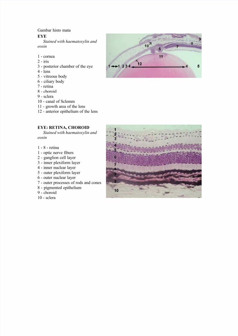
EYE: RETINA, CHOROID
Stained with haematoxylin and eosin
1 - 8 - retina
1 - optic nerve fibers2 - ganglion cell layer
3 - inner plexiform layer 4 - inner nuclear layer
5 - outer plexiform layer
6 - outer nuclear layer 7 - outer processes of rods and cones
8 - pigmented epithelium
9 - choroid
10 - sclera

5/13/2018 Gambar histo mata - slidepdf.com
http://slidepdf.com/reader/full/gambar-histo-mata 2/3
EYE: CORNEA
Stained with haematoxylin and eosin
1 - anterior epithelium (stratified
squamous epithelium)
2 - anterior basement (Bowman's)membrane
3 - substantia propria
4 - posterior basement (Descemet's)
membrane
5 - posterior epithelium (simple
squamous or endothelium)
EYE: CORNEA Stained with haematoxylin and
eosin anterior epithelium (stratifiedsquamous epithelium)
2 - anterior basement (Bowman's)membrane
3 - substantia propria4 - basal layer of the epithelium
5 - intermediate layer of theepithelium
6 - superficial layer of the epithelium
EYE: IRIS
Stained with haematoxylin and eosin
1 - anterior epithelium
2 - anterior terminal layer 3 - vascular layer
4 - posterior terminal layer 5 - pigmented layer
6 - blood vessel7 - dilator pupillae musle
8 - constrictor pupillae musle

5/13/2018 Gambar histo mata - slidepdf.com
http://slidepdf.com/reader/full/gambar-histo-mata 3/3
Recommended