EFFECT OF FEEDING GOSSYPOL FROM COTTONSEED MEALAND VITAMIN E TO CATTLE
By
JUAN VELASQUEZ-PEREIRA
A DISSERTATION PRESENTED TO THE GRADUATE SCHOOLOF THE UNIVERSITY OF FLORIDA IN PARTIAL FULFILLMENT
OF THE REQUIREMENTS FOR THE DEGREE OFDOCTOR OF PHILOSOPHY
UNIVERSITY OF FLORIDA
1996

To my parents
Jose Velasquez Delgadillo
Olga Pereira de Velasquez
To my brothers, sisters and their families
Jose Luis
Marco Antonio
Aurora Maria
Rodrigo Eugenio
Carlos Alfonso
Francisco Javier
Alvaro Fernando
Olga Natalia
Diego Vicente
To my wife and my unborn child
To my country

ACKNOWLEDGMENTS
The author is especially grateful to his parents Dr. and Mrs. Jose and Olga
Velasquez, brothers and sisters Jose Luis, Marco, Aurora, Rodrigo, Carlos, Francisco,
Alvaro, Olga, and Diego Velasquez Pereira for their love, moral and financial support.
He is grateful with his wife Maria del Pilar Soler Velasquez and Dr. Jose Soler, Mrs.
Carmen Garcia Soler, and their family for their love and moral support during his studies.
Special appreciation is due his advisor and chairman of his committee. Dr. Lee R.
McDowell, and to Dr. Joseph H. Conrad, Dr. Frank G. Martin, Dr. Carlos Risco, Dr.
Peter Chenoweth, Dr. David Prichard, and Dr. Charles Staples, members of his
supervisory committee, for their guidance throughout all stages of his graduate work.
Special thanks go to Mrs. Nancy Wilkinson at the Animal Nutrition Laboratory
for her friendship, guidance, assistance and "patience" with the laboratory analyses.
He is deeply grateftil to Leonel and Suyapa Espinoza and their children, Jeronimo
and Dania Velez, Juan C. Ruiz and his parents, Rey Acosta and his family, and Dr. Jose
Paz and his family for their friendship and moral support during his studies. ^ '
This project would not have been possible without the generous financial and
technical support from F. Hoffmann-LaRoche Ltd. representatives including Dr. Martin
Frigg (Basle, Switzerland) and Dr. Scot Williams (Nutley, New Jersey).
He is grateful to Dr. David Kinard and the National Cottonseed Products
Association, and Mr. Tom Wedegaertner and Cotton Incorporated for their technical and
financial support for his Ph. D. program.
Special thanks go to Professor Don Wakeman, Lorraine McDowell, Paul Dixon,
Jerry Wasdin, the late Linda Kneuper and the members of the staff of the Beef Research
Unit and Santa Fe Research Unit. Without their skilled work this research would not
have been possible.
iv

TABLE OF CONTENTS
ACKNOWLEDGMENTSiii
LIST OF TABLES
LIST OF FIGURES x
ABSTRACT xv
CHAPTERS
1 INTRODUCTION1
2 LITERATURE REVIEW 4
Gossypol Biosynthesis 4Chemical Structure 5
Metabolism of Gossypol 7The Toxicity of Gossypol \q
Young Ruminants \\
Ruminants ^2Diagnosis and Recommendations 20
3 FEEDING COTTONSEED MEAL AND VITAMIN E TO BEEFHEIFERS
22
Introduction 22Materials and Methods 23Results and Discussion 3qImplications
V

4 LONG TERM EFFECTS OF FEEDING COTTONSEED MEAL ANDVITAMIN E TO DAIRY CALVES 60
Introduction 50Materials and Methods 61
Results and Discussion 67Implications 93
5 LONG TERM EFFECTS OF FEEDING COTTONSEED MEAL ANDVITAMIN E TO DAIRY BULLS. I. HEMATOLOGICAL ANDTISSUE PARAMETERS 95
Introduction 95Materials and Methods 96Results and Discussion 102Implications 124
6 LONG TERM EFFECTS OF FEEDING COTTONSEED MEAL ANDVITAMIN E TO DAIRY BULLS. II. REPRODUCTIVEPARAMETERS 126
Introduction 126Materials and Methods 127Results and Discussion I33Implications 151
7 SUMMARY AND CONCLUSIONS 152
LIST OF REFERENCES 158
BIOGRAPHICAL SKETCH 169
vi

LIST OF TABLES
Table page
3-1. Composition of dietary supplements 25
3-2. Effect of cottonseed meal (CSM) and vitamin E on weight gain and average
daily gain (ADG) of beef heifers 31
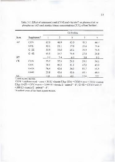
3-3. Effect of cottonseed meal (CSM) and vitamin E on plasma alkaline
phosphatase (AP) and creatine kinase (CK) concentrations (lU/L) of beefheifers 33
3-4. Effect of cottonseed meal (CSM) and vitamin E on plasma retinol palmitate
(RETP), retinol (RET), total retinoids, and p-carotene (p-C) concentrations
(lag/mL) of beef heifers 3g
3-5. Effect of cottonseed meal (CSM) and vitamin E on blood hemoglobin andhematocrit of beef heifers 44
3-6. Effect of cottonseed meal (CSM) and vitamin E on a-tocopherol (a-T), retinol
(RET), retinol palmitate (RETP), and p-carotene (p-C) concentrations in tissue
of beef heifers (|ig/g of wet tissue) 52
3-7. Effect of cottonseed meal (CSM) and vitamin E on micromineral concentrationof liver, heart, and muscle of beef heifers (mg/kg dry matter) 54
4- 1 . Composition of calf diets 53
4-2. Effect of cottonseed meal (CSM) and vitamin E on weight gain and averagedaily gain (ADG) of dairy calves 68
4-3. Average intake of feed (kg • animal ' • d ') and free gossypol (FG)
(g • animal"' • d"') of dairy calves in phase 1 69
4-4. Effect of cottonseed meal (CSM) and vitamin E on blood hemoglobinconcentration (g/dL) of dairy calves 74
vii

4-5. Effect of cottonseed meal (CSM) and vitamin E on blood hematocrit (%) ofdairy calves 75
4-6. Effect of cottonseed meal (CSM) and vitamin E on plasma (+)-, (-)-, and total
(T)-gossypol concentration of dairy calves (|ig/mL) 79
4-7. Feed (kg animal"' • d"' and as %BW) and calculated free gossypol
(g • animal ' • d"') and vitamin E (lU • animal ' • d"') intake of dairy
calves in phase 2 gO
4-8. Effect of cottonseed meal (CSM) and vitamin E on plasma alkaline
phosphatase of dairy calves (lU/L) 85
4-9. Tissue concentrations of a-tocopherol (a-T) and P-carotene (P-C), plasmaa -tocopherol, blood hemoglobin (hemo), hematocrit (hema), and erythrocyte
osmotic fragility (EOF), and free gossypol (FG) intake of dead calves 87
4-10. Effect of cottonseed meal (CSM) and vitamin E on tissue (+)-, (-)-, and total
(T)- gossypol of dead calves gg
4-11. Heart and liver histopathology of dead calves 91
4- 12. Effect of cottonseed meal (CSM) and vitamin E on health of dairy calves 94
5- 1. Composition of dietary supplements 97
5-2. Effect of cottonseed meal (CSM) and vitamin E on weight gain and averagedaily gain (ADG) of dairy bulls 103
5-3. Effect of cottonseed meal (CSM) and vitamin E on blood hemoglobin andhematocrit, and plasma alkaline phosphatase (AP) and creatine kinase (CK)of dairy bulls 207
5-4. Effect of cottonseed meal (CSM) and vitamin E on plasma gossypolconcentration of dairy bulls (ng/mL) 1 13
5-5. Effect of cottonseed meal (CSM) and vitamin E on a-tocopherol (a-T),retinol palmitate (RETP), and p-carotene (P-C) concentrations in tissue ofdairy bulls (^g/g of fresh tissue)
1 1
7
5-6. Effect of cottonseed meal (CSM) and vitamin E on micromineralconcentration of liver, heart, and testis of dairy bulls (mg/kg dry matter) ... 1 1
9
viii
6-1. Composition of dietary supplements 128
6-2. Effect of cottonseed meal (CSM) and vitamin E on semen characteristics of
dairy bulls 134
6-3. Effect of cottonseed meal (CSM) and vitamin E on sperm production of dairy
bulls 142
6-4. Effect of cottonseed meal (CSM) and vitamin E on testicular characteristics
of dairy bulls1 44
6-5. Effect of cottonseed meal (CSM) and vitamin E on sex drive performance ofdairy bulls during two libido tests 147
6-6. Effect of cottonseed meal (CSM) and vitamin E on testosterone concentration
in plasma of bulls after a GnRH challenge 1 49
.'i \
ix

LIST OF FIGURES
figure page
1.Tautomer forms of gossypol. (a) aldehyde, (b) hemiacetal, and (c) phenolic
quinoid 5
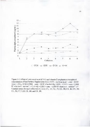
1. Effect of cottonseed meal (CSM) and vitamin E on plasma a -tocopherol
concentration of beef heifers. Supplements were: CON = soybean meal +com + 30 lU vitamin E/kg; GOS = CSM + com + 30 lU vitamin E/kg;
G+2E = CSM + com + 2,000 lU vitamin E • animal ' • d"'; G+4E = CSM +com + 4,000 lU vitamin E animal ' • d '. Standard errors for each collection
(C) were: CI, .16; C2, .71; C3, .90; C4, .82; C5, .99; C6, .90; C7, 1.20;
C8, .86; and C9, .80 35
2. Effect of cottonseed meal (CSM) and vitamin E on erythrocyte osmoticfragility in .65% saline. Supplements were: CON = soybean meal + corn +30 lU vitamin E/kg; GOS = CSM + com + 30 lU vitamin E/kg; G+2E = CSM+ com + 2,000 lU vitamin E • animal ' • d '; G+4E = CSM + com + 4,000 lUvitamin E • animal"' • d"'. Standard errors for each collection (C) were:CI, .35; C2, .92; C3, 3.32; C4, 4.60; C5, 3.10; C6, 2.50; C7, .80; C8, .93;
and C9, 2.60
3. Effect of cottonseed meal (CSM) and vitamin E on erythrocyte osmoticfragility in .55% saline. Supplements were: CON = soybean meal + com +30 lU vitamin E/kg; GOS = CSM + com + 30 lU vitamin E/kg; G+2E =CSM + com + 2,000 lU vitamin E • animal ' • d '; G+4E = CSM + com +4,000 lU vitamin E • animal ' • d '. Standard errors for each collection (C)were: CI, 5.0; C2, 4.1; C3, 10.0; C4, 6.8; C5, 2.1; C6, 2.9; C7, 3.5; C8, 4.2;and C9, 5.3
4. Effect of cottonseed meal (CSM) and vitamin E on plasma (-)-gossypol.
Supplements were: CON = soybean meal + com + 30 lU vitamin E/kg;GOS = CSM + com + 30 lU vitamin E/kg; G+2E = CSM + com + 2,000 lUvitamin E • animal ' • d '; G+4E = CSM + com + 4,000 lU vitamin E -
animal ' • d '. Standard errors for each collection (C) were: CI, .03; C3 .30:

5. Effect of cottonseed meal (CSM) and vitamin E on plasma (+)-gossypol.
Supplements were: CON = soybean meal + com + 30 lU vitamin E/kg;
GOS = CSM + com + 30 lU vitamin E/kg; G+2E = CSM + com + 2,000 lUvitamin E • animal ' • d '; G+4E = CSM + com + 4,000 lU vitamin E •
animal"' • d"'. Standard errors for each collection (C) were: CI, .03; C3, .30;
C5, .20;andC9, .30
6. Effect of cottonseed meal (CSM) and vitamin E on plasma total gossypol.
Supplements were: CON = soybean meal + com + 30 lU vitamin E/kg;
GOS = CSM + com + 30 lU vitamin E/kg; G+2E = CSM + com + 2,000 lUvitamin E • animal ' d '; G+4E = CSM + com + 4,000 lU vitamin E •
animal ' d"'. Standard errors for each collection (C) were: CI, .07; C3, .63;
C5, .50; and C9, .73.
7. Effect of cottonseed meal (CSM) and vitamin E on liver (L), muscle (M), andheart (H), (+)-, and (-)-gossypol concentrations. Supplements were: CON =soybean meal + com + 30 lU vitamin E/kg; GOS = CSM + com + 30 lUvitamin E/kg; G+2E = CSM + com + 2,000 lU vitamin E • animal ' • d ';
G+4E = CSM + com + 4,000 lU vitamin E • animal ' • d '. Standard errors ofCON, GOS, G+2E and G+4E respectively were: L+: 1.2, 7.8, 7.3, and 7.3;
L-: 3.0, 3.4, 3.1, and 3.1; H+: .5 for all; H-: 3.9, 4.2, 3.9, and 3.9; M+: .1
for all; and M-: .3 for all
8. Effect of cottonseed meal (CSM) and vitamin E on in vitro stimulated lipid
peroxidation of liver homogenates. Supplements were: CON = soybean meal+ com + 30 lU vitamin E/kg; GOS = CSM + com + 30 lU vitamin E/kg;G+2E = CSM + com + 2,000 lU vitamin E • animal ' d '; G+4E = CSM +com + 4,000 lU vitamin E • animal ' • d '. Standard errors for each time (T)were: TO, .14; T50, .13; Tl 00, .51; and T200, 1.27
9. Effect of cottonseed meal (CSM) and vitamin E on in vitro stimulated lipid
peroxidation of heart homogenates. Supplements were: CON = soybean meal+ com + 30 lU vitamin E/kg; GOS = CSM + com + 30 lU vitamin E/kg;G+2E = CSM + com + 2,000 lU vitamin E • animal ' d '; G+4E = CSM +com + 4,000 lU vitamin E • animal ' • d '. Standard errors for each time (T)were: TO, .3; T50, .3; TlOO, .3; and T200, .8
3-10. Effect of cottonseed meal (CSM) and vitamin E on skinfold thickness after
PHA injection. Supplements were: CON = soybean meal + com + 30 lU
vitamin E/kg; GOS = CSM + com + 30 lU vitamin E/kg; G+2E = CSM +
com + 2,000 lU vitamin E • animal ' • d"'; G+4E = CSM + com + 4,000 lU
vitamin E • animal"' • d"'. Standard errors for each time (T) were: T12, .4;
T24, .4; and T48, .3 58
4-1 . Effect of cottonseed meal (CSM) and vitamin E on plasma a -tocopherol
concentration of dairy calves in phase 1 group 1 . Diets were: CON = soybean
meal + 30 lU vitamin E/kg; GOS = CSM + 30 lU vitamin E/kg; G+2E =
CSM + 2,000 lU vitamin E • animal ' d '; G+4E = CSM + 4,000 lU vitamin
E • animal"' d"'. Standard errors for each collection (C) were: CI, .2; C2, .4;
C3, .4; C4, .4; C5, .6; C6, .5; and C7, .4 71
4-2. Effect of cottonseed meal (CSM) and vitamin E on plasma a -tocopherol
concentration of dairy calves in phase 1 group 2. Diets were: CON =
soybean meal + 30 lU vitamin E/kg; GOS - CSM + 30 lU vitamin E/kg;
G+2E = CSM + 2,000 lU vitamin E • animal"' • d"'; G+4E = CSM + 4,000 lU
vitamin E • animal"' • d"'. Standard errors for each collection (C) were: CI, .2;
C2, .7; C3, .2; C4, .3; and C5, .5 72
4-3. Effect of cottonseed meal (CSM) and vitamin E on erythrocyte osmotic
fragility (.65% saline) of dairy calves in phase 1 group 1. Diets were: CON =
soybean meal + 30 lU vitamin E/kg; GOS = CSM + 30 lU vitamin E/kg;
G+2E = CSM + 2,000 lU vitamin E • animal"' • d '; G+4E = CSM + 4,000 lUvitamin E • animal"' • d"'. Standard errors for each collection (C) were:
CI, 1.7; C2, 1.7; C3, 1.7; C4, 4.3; C5, 4.8; C6, 4.4; and C7, 4.2 76
4-4. Effect of cottonseed meal (CSM) and vitamin E on erythrocyte osmotic
fragility (.65% saline) of dairy calves in phase 1 group 2. Diets were:
CON - soybean meal + 30 lU vitamin E/kg; GOS = CSM + 30 lU vitamin
E/kg; G+2E = CSM + 2,000 lU vitamin E • animal"' • d"'; G+4E = CSM +
4,000 lU vitamin E • animal"' • d"'. Standard errors for each collection (C)
were: CI, 2.2; C2, 3.3; C3, 2.6; C4, 6.1; and C5, 6.7 77
4-5. Effect of cottonseed meal (CSM) and vitamin E on erythrocyte osmotic
fragility (.65% saline) of dairy calves in phase 2. Diets were: CON =
soybean meal + 30 lU vitamin E/kg; GOS = CSM + 30 lU vitamin E/kg;
G+2E = CSM + 2,000 lU vitamin E animal ' • d "; G+4E = CSM + 4,000 lUvitamin E • animal"' • d"'. Standard errors for each collection (C) were:
CI, 3.8; C2, 2.8; C3, 2.4; and C4, 2.3 83
4-6. Effect of cottonseed meal (CSM) and vitamin E on plasma a -tocopherol
concentration of dairy calves in phase 2. Diets were: CON = soybean meal
+ 30 lU vitamin E/kg; GOS = CSM + 30 lU vitamin E/kg; G+2E = CSM + -
2,000 lU vitamin E animal ' d '; G+4E = CSM + 4,000 lU vitamin E •,
animal ' • d '. Standard errors for each collection (C) were: CI, .4; C2, .4;
C3. .3:andC4. .4 86
5-1. Effect of cottonseed meal (CSM) and vitamin E on erythrocyte osmotic
fragility in .65% saline. Supplements were: CON = soybean meal + com +
30 lU vitamin E/kg; GOS = CSM + com + 30 lU vitamin E/kg; G+4E =
CSM + com + 4,000 lU vitamin E • animal"' d"'. Standard errors for each
collection (C) were: CI, 2.5; C2, 2.1; C3, 2.4; C4, 3.1; C5, 2.0; C6, 1.3;
C7,4.5;C8, 1.4;andC9, 1.5 105
5-2. Effect of cottonseed meal (CSM) and vitamin E on erythrocyte osmotic
fragility in .55% saline. Supplements were: CON = soybean meal + com +
30 lU vitamin E/kg; GOS = CSM + com + 30 lU vitamin E/kg; G+4E =
CSM + com + 4,000 lU vitamin E animal"' • d '. Standard errors for each
collection (C) were: CI, 4.3; C2, 4.1; C3, 5.0; C4, 5.7; C5, 4.7; C6, 4.3;
C7, 4.4; C8, 4.6; and C9, 4.2 106
5-3. Effect of cottonseed meal (CSM) and vitamin E on plasma a-tocopherol
concentration of dairy bulls. Supplements were: CON = soybean meal +
com + 30 lU vitamin E/kg; GOS = CSM + com + 30 lU vitamin E/kg;
G+4E = CSM + com + 4,000 lU vitamin E animal"' • d"'. Standard errors
for each collection (C) were: CI, .3; C2, .2; C3, .3; C4, .2; C5, .3; C6, .4;
C7, .4; C8, .3; C9, .4; and CIO, .4 110
5-4. Effect of cottonseed meal (CSM) and vitamin E on liver (L), heart (H), and
testis (T) (+)-, and (-)-gossypol concentrations. Supplements were: CON =
soybean meal + com + 30 lU vitamin E/kg; GOS = CSM + com + 30 lUvitamin E/kg; G+4E = CSM + com + 4,000 lU vitamin E • animal ' d"';
Standard errors for each tissue and isomer were: L+, 7.8; L-, 5.3; H+, .7;
H-, 3.5;T+, .6; and T-, 1.11 114
5-5. Effect of cottonseed meal (CSM) and vitamin E on in vitro stimulated lipid
peroxidation of liver homogenates. Supplements were: CON = soybean
meal + com + 30 lU vitamin E/kg; GOS = CSM + com + 30 lU vitamin
E/kg; G+4E = CSM + com + 4,000 lU vitamin E • animal"' • d"'. Standard
errors for each time (T) were: TO, .15; T50, .36; TlOO, .81; and T200, .80. ... 121
xiii
5-6. Effect of cottonseed meal (CSM) and vitamin E on in vitro stimulated lipid
peroxidation of heart homogenates. Supplements were: CON = soybean
meal + com + 30 lU vitamin E/kg; GOS = CSM + com + 30 lU vitamin
E/kg; G+4E = CSM + com + 4,000 lU vitamin E • animal'' • d '. Standard
errors for each time (T) were: TO, .1 1; T50, .23; TlOO, 1.05; and
T200, .80 122
5-7. Effect of cottonseed meal (CSM) and vitamin E on in vitro stimulated lipid
peroxidation of testis homogenates. Supplements were: CON = soybean
meal + com + 30 lU vitamin E/kg; GOS = CSM + com + 30 lU vitamin
E/kg; G+4E = CSM + com + 4,000 lU vitamin E • animal ' • d '. Standard
errors for each time (T) were: TO, .65; T50, 3.75; TlOO, 4.10; and
T200.4.10 123
6- 1 . Electron micrographs of bull sperm a) supplemented with cottonseed meal
(GOS) revealing gaps in the mitochondria helix of the midpiece
(magnification 19,430), and b) supplemented with soybean meal (CON)
(magnification 18,750) 138
6-2. Electron micrograph of the testicle of a bull supplemented with cottonseed
meal (GOS) revealing extensive vacuolization of mitochondria and loss of
cristae. Magnification 18,200 139
6-3. Effect of cottonseed meal (CSM) and vitamin E on the percentage of bulls
that reached puberty at each semen collection. Supplements were: CON =
soybean meal + com + 30 lU vitamin E/kg; GOS = CSM + com + 30 lU
vitamin E/kg; G+4E = CSM + com + 4,000 lU vitamin E • animal ' • d ' 146
6-4 Effect of cottonseed meal (CSM) and vitamin E on plasma testosterone
concentration after a GnRH challenge. Supplements were: CON = soybean
meal + com + 30 lU vitamin E/kg; GOS = CSM + com + 30 lU vitamin
E/kg; G+4E = CSM + com + 4,000 lU vitamin E • animal"' • d '. Standard
errors for each time (T) were: TO, .15; T30, .64; T60, .73; T120, .92;
T180, .99; T240, 1.08; and T300, 1.00 150
xiv

Abstract of Dissertation Presented to the Graduate School
of the University of Florida in Partial Fulfillment of the
Requirements for the Degree of Doctor of Philosophy
EFFECT OF FEEDING GOSSYPOL FROM COTTONSEED MEAL ANDVITAMIN E TO CATTLE
By
Juan Velasquez-Pereira
December 1996
Chairperson: Lee R. McDowell
Major Department: Animal Science
A series of experiments were conducted to evaluate the value of supplemental
vitamin E on the physiological response to free gossypol (FG) from cottonseed meal
(CSM) fed to ruminants and preruminants. Experiments 1 (yearling heifers) and 2
(newborn bull calves from 2 wk to 6 mo) used the following dietary treatments: CON:
soybean meal-based diet; GOS: CSM-based diet; G+2E: CSM-based diet + 2,000 lU
vitamin E • animal ' • d '; G+4E: CSM-based diet + 4,000 lU vitamin E • animal ' • d '.
Experiment 3 (bulls from 6 to 16 mo) lacked treatment G+2E. Erythrocyte osmotic
fi-agility (EOF) was increased (P < .05) in animals fed CSM; however, vitamin E
supplementation lowered EOF in experiment 1 . Vitamin E supplementation did not
influence plasma or tissue gossypol concentration. Cottonseed meal seemed to aid in the
absorption and deposition of a-T in animals fed a normal level of vitamin E. In heart.
XV

neck muscle, and testis (-)-gossypol was higher (P < .05) than (+)-gossypol, while the
reverse was true for liver. In vitro lipid peroxidation of tissue indicates that gossypol acts
as an antioxidant in lipid peroxidation systems and its role as antioxidant may be dose or
tissue dependant. In calves, hemoglobin and hematocrit were decreased (P < .05) in GOS
calves, and vitamin E supplementation counteracted (P < .05) this effect. Ten calves died,
6 from the GOS and 2 each from the G+2E and G+4E treatments. Necropsy findings
were compatible with gossypol toxicity. Histopathological examination revealed
centrilobular necrosis in the liver and atrophy and vacuolation of cardiocytes. In bulls,
percentage motility, normal, and live sperm, and daily sperm production were lower (P <
.1), while percentage of primary abnormalities, and abnormal midpieces were increased
(P < .05) in the GOS bulls. There was a trend of gossypol to decrease and vitamin E to
improve libido score. The results of the GOS libido test may indicate lack of sexual
maturity which agrees with sperm production data.
In experiment 1, gossypol (4 g FG/d for 1 12 d) did not have any adverse effect on
animal performance, nor did it cause any sign of gossypol toxicity. In experiment 2,
feeding GSM at a level to provide 400 mg FG/kg of feed DM caused death of some
calves with gossypol related toxicity signs. Vitamin E supplementation increased
performance and may have conferred some protection against gossypol toxicity. In
experiment 3, feeding gossypol to Holstein bulls negatively affected some reproductive
measurements; however, vitamin E supplementation improved these conditions.
Gossypol did not decrease plasma or tissue a -tocopherol in any of the three experiments.
XV i

CHAPTER 1
INTRODUCTION
By-products of the cotton fiber and cottonseed oil industry such as whole
cottonseed (WCS) and cottonseed meal (CSM) are an important source of economical
protein and energy in livestock rations. Approximately six million tons of cottonseed are
produced per year in the United States (NCPA, 1990). Gossypol [(2,2'-binaphthalene)-
8,8'-dicarboxaldehyde-l,r,6,6',7,7'-hexahydroxy-5,5'-diisopropyl-3 ,3 -dimethyl] is a
yellow polyphenolic pigment found in cottonseed. Gossypol found in cottonseed is
referred to as free gossypol (FG), which is the form that is toxic to livestock. Bound
gossypol (BG) is found in by-products of the cottonseed oil industry along with FG.
Bound gossypol has not been found to be a major toxicant for livestock, but its role in
animal toxicity is not yet fiilly understood (Jones, 1991). Free gossypol is transformed to
BG as the cottonseed is treated with heat and pressure in the process of extracting the oil.
Cottonseed meal, or any other by-product of the cottonseed oil industry may be less toxic
to livestock as their concentration ofFG is lower, but Calhoun and Holmberg (1991)
suggested that BG may be released during the digestive process and have a physiological
effect in the animal.
In the past several years, FG in ruminant diets has increased due to an increased
consumption ofWCS compared with CSM. Lusby et al (1991) reported that WCS is
1

being fed to ruminants more than any other by-product of the cottonseed oil industry (e.g.
2.1 million tons ofWCS vs 1.5 million tons of CSM). In Florida, the WCS used in dairy
rations has increased in the past several years, and the daily consumption ofFG may be
above 1 5 g per cow. . ,..
Although ruminants seem to have a large capacity to detoxify gossypol, toxicity
resulting from consumption of this compound has been observed. Gossypol has been
found to affect liver function, erythrocyte oxygen carrying or releasing capacity,
respiration rate, feed intake and productive and reproductive capacity (Lindsey et al.,
1980; Calhoun et al, 1990; Gray et al., 1990). It is also believed that gossypol may
compromise the immune response of ruminants (Holmberg et al., 1988; Hudson et al.,
1988). Gossypol intake by mature ruminants may overwhelm nominal detoxification
capacity and become absorbed at potentially toxic levels (Randel et al., 1992). Lindsey et
al. (1980) suggested that these toxicity signs can be intensified when ruminants are under
physiological, nutritional, and/or environmental stress.
Preruminants are especially susceptible to gossypol toxicosis. It seems that the
undeveloped young ruminant rumen is unable to detoxify free gossypol (Morgan et al.,
1988). Calhoun et al. (1990) found that osmotic fragility of erythrocytes was increased
when gossypol was fed to lambs. Holmberg et al. (1988) reported that high dietary
concentrations of gossypol for calves caused death with lesions compatible with gossypol
toxicity. Risco et al. (1992) found that feeding 800 mg FG/kg to calves resulted in the
death of four animals as a result of circulatory failure associated with gossypol toxicity.
Gossypol has been found to have an antifertility effect. In humans, which are
very sensitive to gossypol; it can provoke damage to the reproductive tract that often is
irreversible. In China, dosage of 60 to 70 mg per day of gossypol for 35 to 42 days has
been reported to produce sterility in human males, as well as resulting in some side
effects (Qian and Wang, 1984). The concentration of gossypol in meat for human
consumption has been very low in muscles of lambs fed high amounts of gossypol,
although significant amounts were found in liver and kidney. The amount of gossypol
that can be found in meat from livestock fed cottonseed products has not been yet clearly
established.
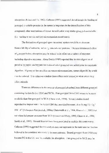
Gossypol can interact directly with biological membranes by promoting the
formation of highly-reactive oxygen-containing free radicals (Janero and Burghardt,
1988). Free radicals provoke oxidative injury and compromise the antioxidant system of
living organisms (Bender et al., 1988). These authors found that systemic levels of alpha-
tocopherol, ascorbate and glutathione peroxidase, and other antioxidants were reduced by
feeding rats high amounts of gossypol. In dairy cattle, Lane and Stuart (1990) found that
feeding gossypol decreased plasma alpha-tocopherol concentrations, indicating a possible
relationship between gossypol toxicity and vitamin E.
There are few data showing the effect of supplemental vitamin E in counteracting
gossypol toxicity. Thus, at the University of Florida, a series of experiments were
undertaken to evaluate the value of supplemental vitamin E on the physiological response
to CSM (gossypol) fed to ruminants and young ruminants prior to rumen development.

CHAPTER 2
LITERATURE REVIEW
Gossypol is a naturally occurring polyphenolic substance present in various parts
of plants belonging to the Malvaceae family. Gossypol content of the plant varies
depending on environmental factors and plant species. It is probably the most important
antiquality compound in cottonseed and its by-products and tends to limit their utilization
when fed to livestock (especially nonruminants and preruminants).
This chapter addresses some aspects of biosynthesis, chemistry, metabolism, and
toxicity of gossypol with reference to ruminants and preruminants.
Gossypol Biosynthesis
The biosynthesis of gossypol occurs in various parts of the cotton and other plants
that belong to the Malvaceae family. In cotton root homogenates, Heinstein et al. (1962)
reported that according to the labeling pattern and incorporation of '''C-labeled acetate the
biosynthesis of gossypol may occur via the isoprenoid pathway. This pathway of
biosynthesis was later confirmed when Heinstein et al. (1970) observed that six
molecules of mevalonate-2-"'C were incorporated stereospecifically into one molecule of
gossypol.
4

5
Chemical Structure
Gossypol is a yellow polyphenolic pigment that has a molecular weight of 518.54
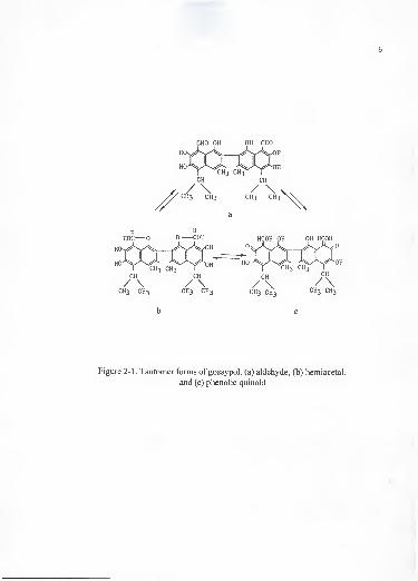
and a molecular formula of C30 H30 Og. Gossypol occurs in three tautomeric forms:
aldehyde, hemiacetal and phenolic quinoid tautomers (Figure 2-1). It is a highly reactive
substance due to the two phenolic hydroxyl and the aldehyde groups that allow gossypol
to react with a variety of compounds (Abou-Donia, 1989). It is a cherical molecule
because of steric hindrance about the intemaphthy bond (Cass et al., 1991). When
gossypol is isolated from plants, it is a mixture of (+)-, and (-)-gossypol isomers. There
are differences in the amount of each isomer isolated from different plants. In Thespesia
polpunea the isolation of gossypol yields an excess of (+)-gossypol, while Gossypium
barhadense yields more (-)-gossypol (Cass et al., 1991). The stereospecificity of
gossypol is in part involved in the toxic effect seen with this compound. The (-)-gossypol
isomer appears to be responsible for the antifertility effect observed in rats (Wang et al.,
1992).
Gossypol in whole cottonseed (WCS) is found in the pigment glands which
appear as small black dots on the cut surface of the seed (Jones, 1991). Its concentration
in the seed is related to environmental conditions as well as agricultural practices. Three
terms are commonly used to describe gossypol: free (FG), bound (BG), and total (TG)
gossypol.
CHO OH OH CHO
CH3 CH3 CH3 CH3 CH3 CH3 CH3 CH3
b c
Figure 2-1 . Tautomer forms of gossypol. (a) aldehyde, (b) hemiacetal,
and (c) phenolic quinoid

Bound gossypol is used to describe the amount of the substance that binds to
proteins and other compounds during WCS oil extraction process. Different procedures
of extracting the oil from WCS yield different amounts ofBG and FG. The direct solvent
extraction of cottonseed oil yields the highest FG content in the cottonseed meal (CSM)
while the screw press process yields the lowest. All gossypol found in the undisrupted
seed is in the free form, which plays an important role in the toxicity of the compound.
Total gossypol is equal to the sum ofFG and BG and, therefore, is not affected by the
processing of the seed, but differences in varieties and environmental factors affecting the
growth of the cotton plant.
Metabolism of Gossypol
Gossypol is absorbed through the intestine as well as the epithelial lining of the
stomach with the small intestine being the main site (Abou-Donia et al., 1970). Before
reaching the site of absorption gossypol undergoes some changes. Bressani et al. (1964)
reported greater FG excretion than intake in dogs, suggesting that some bound gossypol
may have been released during its passage throughout the gastrointestinal tract (GIT). In
ruminants, although these changes have not been quantified, higher amounts of gossypol
are suspected to react not only in the stomach and intestine but in the rumen. In lambs,
Calhoun and Wang (1995) found that absorption of gossypol from cottonseed meats was
greater when it was fed to bypass the rumen than when rumination was allowed. In the
rumen, gossypol may react with soluble proteins and, therefore, reduce its later
absorption (Reiser and Fu, 1962). Calhoun (1995) suggested that although the binding of
gossypol to soluble proteins in the rumen is important in the detoxification of this
compound, other mechanisms of rumen detoxification may render gossypol unavailable
(i.e. binding to metals and (or) microorganism membranes).
The first action of gossypol upon the animal system would be to decrease
bioavailability of nutrients, including minerals and proteins. The next detrimental effect
of gossypol before absorption may be related to its effect on a variety of enzymes
including digestive enzymes. Abou-Donia (1989) reported that in vitro digestion of
proteins by pepsin and trypsin was reduced when gossypol was added prior to enzymatic
digestion. If gossypol has an effect on rumen microorganisms, rumen digestibility could
also be reduced. It is unknown whether these effects exist and(or) to what extent they
affect animals.
There are differences in the amount of gossypol absorbed from different gossypol
containing feedstuffs (i.e. CSM and WCS). Free gossypol from CSM seems to be more
available than free gossypol in WCS (Chase et al., 1994). Several studies found
reproductive impairment in bulls fed CSM that provided between 14 to 16 mg FG • kg"'
BW • d ' (Velasquez-Pereira et al., 1996a; Chenoweth et al., 1994; Risco et al., 1993) but
not when fed greater amounts from WCS (Cusack and Perry, 1995; Chase et al., 1994;
Smith et al., 1991). Several theories have been postulated to explain this controversy.
Calhoun (1995) suggested that FG analysis may not represent in the ruminant the fraction
believed to be correlated with toxicity in nonruminants. Bound gossypol from CSM may
become FG in the GIT and be available for absorption. Free gossypol in WCS may be

9
released at a slower rate and may become permanently bound and not available for
absorption in the small intestine.
After absorption, most gossypol is excreted via bile in rats, suggesting a biliary
circulation of gossypol between the liver and GIT. Qian and Wang (1984) suggested that
gossypol fits the criteria for a type of compound excreted via the bile, such as compounds
of high molecular weight containing anionic groups and two or more aromatic rings.
Abou-Donia ( 1 989) suggested the following pathway for the metabolism of gossypol:
gossypol is absorbed from the gastrointestinal tract, mainly the small intestine; it enters
the liver via the hepatic artery or through the lymph into the hepatic sinusoid and is taken
up by Kupffer cells. In the liver, gossypol is metabolized, conjugated, and excreted with
the bile into the duodenum. Some of the gossypol excreted is reabsorbed, completing an
enterohepatic cycle that may be repeated several times which involves a gradual excretion
via feces. The mechanism of excretion seems to require an active secretory process that
can be saturated.
Gossypol isomers have been found to have the following pattern of accumulation
in ruminant tissues (Kim et al., 1996; Velasquez-Pereira et al., 1996b): in the liver total
gossypol is higher than any other tissue, but (+)-gossypol concentration is greater than
(-)-gossypol. The contrary is found in muscle and heart, where (-)-gossypol is greater
than (+)-gossypol. Total gossypol accumulation pattern is liver>heart>muscle. The fact
that gossypol is accumulated in the liver may be related to the tissue susceptibility to
gossypol toxicity or to the fact that the liver is the main route of its metabolism. Also,
gossypol is a lipophilic compound, therefore, it tends to accumulate in tissues or cell
10
fractions containing high amounts of Hpid membrane e.g. mitochondria, lysosomal and
microsomal fractions (Baran and Ismailov, 1993).
Gossypol has been reported to be cardiotoxic, causing degeneration of cardiac
muscle. The (-)-gossypol has been shown to be the optically active form that induces
fertility impairment in male animals (Wang et al., 1987). Accumulation of this isomer in
liver and heart tissues is similar, which may be related to the toxicity signs shown during
gossypol toxicosis.
Lower accumulation of (-)-gossypol in the liver relative to (+)-gossypol may be
related to greater affinity of (-)-gossypol for plasma proteins (Wu and Reidenberg, 1 990).
The liver is perfused by a fluid similar to plasma which has greater protein concentration
than the interstitial fluid found in heart and testis (Joseph et al., 1986). Therefore, (-)-
gossypol could be bound to proteins at the time this protein rich fluid perfuses the liver
and be redirected to other tissues where it can interact with cellular components that had
greater affinity to bind (-)-gossypol than plasma protein. The (+)-gossypol may not be
accumulated in tissues other than liver, where it seems to be eliminated, due to its low
specific interaction with cellular components (Wang et al., 1992).
The Toxicity of Gossypol
There are several important factors that play a role in gossypol toxicity
development, including the type of animal (ruminant vs. nonruminants); ruminants have
greater capacity to detoxify gossypol before absorption than nonruminants. Among

11
ruminants, stage of rumen development would affect gossypol toxicity with young
ruminants with undeveloped rumens behaving like nonruminant animals.
Composition of the diet (i. e. protein and mineral concentrations) is also
important. The effect of gossypol is both time and dose dependant. It seems that a
quantity of gossypol must be accumulated to a certain concentration in order to exert its
effect and cause clinical signs of gossypol toxicity or death.
Young Ruminants
Young ruminants with undeveloped rumens are more susceptible to gossypol
toxicity than older animals due to lack of a detoxification mechanism that occurs in the
rumen of adult animals (Reiser and Fu, 1962). Calhoun and Wang (1995) demonstrated
that the rumen plays an important role in detoxifying gossypol. These researchers fed 20
or 30 mg FG • kg"' BW • d"' to two groups of animals. The first group consisted of
weaned lambs fed a diet containing cottonseed meats as the FG source, while in the
second group the cottonseed meats were mixed with the milk replacer in order to bypass
the rumen. The weaned lambs exhibited no signs of gossypol poisoning whereas all
milk-fed lambs died due to gossypol poisoning. Risco et al. (1992) conducted an
experiment to define at what concentration FG is safe in young calves. Diets containing
0, 100, 200, 400, or 800 mg FG/kg of diet DM were fed to Holstein calves from 1 to 120
d of age. Clinical evidence of disease was limited to calves fed 400 or 800 mg FG/kg.
Authors concluded that a ration containing up to 200 mg FG/kg is safe, 400 mg FG/kg
approaches toxicity and 800 mg FG/kg causes death. Also, death losses from gossypol

12
toxicosis in calves has been reported in the following range (mg/kg of diet DM): 4000
total gossypol (Hudson et al., 1988), from 240 to 380 FG (Holmberg et al., 1988), and
from 100 to 220 FG (Zelski et al., 1995).
Postmortem and histological findings of fatally intoxicated calves included
widespread edema and congestion, particularly in the lungs and body cavities; straw-
colored efflision noted in the abdominal and thoracic cavities; flabby and dilatated heart;
and enlarged and congested liver with centrilobular degeneration (Risco et al., 1992). The
clinical signs of gossypol toxicity are compatible with heart failure, although a direct
effect of gossypol on the liver cannot be ruled out. Blood components and blood
chemistry characteristics also may be affected by gossypol. Risco et al. (1992) reported
that changes in erythrocyte characteristics such as hemoglobin concentration and
hematocrit are not conclusive in young ruminants fed gossypol indicating normal
hematopoietic response is maintained, thus the degree of anemia caused by gossypol is
mild. Furthermore, chemistry panels designed to monitor serum proteins, energy status,
serum electrolytes, renal function, liver function, and necrosis generally are not usefiil in
demonstrating impending gossypol toxicity when used as a group monitoring test.
Ruminants
Reiser and Fu (1962) reported that binding of gossypol to soluble proteins within
the rumen may confer ruminants the ability to withstand gossypol intakes greater than
nonruminants. Furthermore, Calhoun (1995) suggested that ruminal bacteria also could
play a role in gossypol detoxification.
13
Although ruminants seem to have a large capacity to detoxify gossypol, toxicity
resulting from consumption of this compound has been observed. Gossypol has been
found to affect liver functions, erythrocyte oxygen carrying or releasing capacity,
respiration rate, feed intake and production and reproduction capacity (Calhoun et al.,
1990; Gray et al., 1990; Lindsey et al., 1980). Gossypol also may compromise the
immune response of ruminants (Holmberg et al., 1988; Hudson et al., 1988). These
findings indicate that gossypol intake by mature ruminants may overwhelm ruminal
detoxification and become absorbed at concentrations potentially toxic (Randel et al.,
1992).
Lindsey et al. (1980) reported that these toxicity signs can be intensified when
ruminants are under physiological, nutritional, and/or environmental stress. Some of the
clinical signs observed by Lindsey et al. (1980) in mature dairy cattle fed 6.6 and 42.7 g
FG • animal"' d ' were reduced milk production and respiratory stress. Physiological
changes included reduced hemoglobin concentration and increased erythrocyte osmotic
fragility (EOF). Smalley and Bicknell (1982) reported that intake of 2.7 to 4.5 kg of
ammoniated WCS by dairy cows resulted in 10% mortality rate. Calhoun et al. (1995b)
reported that three dairy cows fed ammoniated WCS died of causes related to gossypol
toxicity. Although most of the experiments reporting negative effects of gossypol on
ruminants had used large amounts of cottonseed products, some special conditions such
as treatment of the seed which makes gossypol more available have to be monitored very
carefully.
Lane and Stuart (1990) in a field study reported that gossypol intake (up to 42 g
FG/d) by mature lactating dairy cattle reduced plasma a-tocopherol (a-T) concentration,
and suggested that the antioxidant system of animals fed gossypol may be compromised.
In this study no statistical data were shown and gossypol analysis may not represent the
real values. Willard et al. (1995) found that by d84, cows given 4 g FG • animal"' • d"'
that showed elevated EOF, had lower plasma a-T concentration than control cows that
showed low EOF. No supplemental vitamin E was given in the previous experiment. In
that experiment, four animals with the highest EOF were chosen from 1 7 receiving 4 g
FG • animal ' • d"' in a group fed experiment to determine plasma a-T concentration. It
may have been possible that these animals consumed more supplement (high EOF) with
low vitamin E content, and consumed less forage which would have been an excellent
source of vitamin E. In a more controlled study, Mena et al. (1996) reported that
gossypol from CSM, WCS or both (900 to 1 800 mg TG/kg of diet DM) sources did not
reduce concentrations of antioxidant vitamins, and that in fact it increased plasma a-T
concentrations.
Erythrocyte fragility is a very sensitive indicator of systemic gossypol status,
since it appears shortly after gossypol consumption has started. It has been reported to
increase in several studies with gossypol-fed ruminants (Velasquez-Pereira et al., 1996c;
Risco et al., 1993). The mechanism by which gossypol affects erythrocytes is not well
understood. From in vitro experiments, the effect of gossypol on cells has been explained
by the ability of gossypol to interact with proteins, especially membrane bound, and by
15
binding and modifying the properties of the lipid bilayer matrix (Reyes et al., 1984).
These researchers found that gossypol binds strongly to bilayers of different lipid
compositions and induces an electrical conductance that is accompanied by an increase in
proton permeability. Furthermore, gossypol caused an increase in diffusion in lipid
membranes in the presence of NaCl, which could explain the increase in EOF.
de Peyster et al. (1986) found an increase in membrane permeability due to
gossypol treatment, and suggested that degeneration of cell membranes at all levels of
organization may exist with acute prolonged exposure to gossypol in vivo. More
evidence of the effect of gossypol on membranes was reported by Tanphaichitr et al.
(1995) who found that gossypol at low concentrations intercalates in the phospholipid
bilayer with the hydrophobic domains of the toxicant interacting with the phospholipid
hydrocarbon chain, while its hydrophilic groups would be exposed to the aqueous phase.
The intercalation of gossypol into a lipid bilayer, such as the erythrocyte membrane,
would alter its fluidity and therefore may explain increased erythrocyte fragility.
Gossypol has been found to have an antifertility effect. Gossypol added to in
vitro culture negatively affected embryo development (Zirkle et al., 1988), and in culture
luteal cells decreased progesterone synthesis (Gu et al., 1990). In vivo experiments have
demonstrated that feeding cows and heifers 20 mg FG • kg'' BW • d"' did not affect
reproductive performance (Willard et al, 1995; Gray et al., 1993).
In the male ruminant, gossypol seems to exert a unique and selective effect on the
reproductive system. A reduction in sperm production and motility are the most common
effects of gossypol toxicity.

A particular damage termed segmental aplasia of the mitochondrial sheath of the
sperm midpiece has been reported in rats (Oko and Hrudka, 1982) and also in male
ruminant sperm (Chenoweth et al., 1994).
Risco et al. (1993) found a lower percentage of normal sperm in the semen of
bulls fed 16.6 mg FG • kg"' BW • d"' from CSM. Defects of sperm midpieces accounted
for most of the abnormalities. These researchers observed this effect after 9 wk of
feeding the gossypol-containing diet. Chase et al. (1994) did not find differences in sperm
abnormalities when bulls were fed approximately 6 mg FG • kg"' BW • d"' from CSM or
60 mg FG • kg"' BW • d"' from WCS. Collectively, Risco et al. (1993) and Chase et al.
(1994) demonstrated in ruminants that gossypol effect is both dose and time dependent,
and that source of gossypol influences the development of toxicity.
The detrimental effect of gossypol on male bovine reproduction seems to appear
when greater concentrations than those used by Chase et al. (1994) are fed and in the
form of CSM, since feeding high gossypol diets in the form ofWCS have yielded
inconsistent results (Cusack and Perry, 1995; Smith et al., 1991; Arshami and Ruttle,
1988). Cusack and Perry (1995) reported that mature bulls fed 20.4 to 50.8 mg FG • kg"'
BW • d"' from WCS for 8 mo did not show any sign of reproductive impairment.
Similarly, Smith et al. (1991) reported that feeding high concentrations of gossypol (64 to
75 mg FG • kg-' BW • d"') in the form ofWCS for 4 mo did not affect sperm
characteristics or cause testicular degeneration in bulls. Contrary to these two
experiments, Arshami and Ruttle (1988) found that WCS, CSM and cottonseed hulls fed
to bulls caused testicular histological changes, indicating a detrimental effect on the

17
spermatogenic tissue and associated cells. Several reasons have been proposed to explain
these inconsistent results. 1) Cusack and Perry (1995) suggested that the feeding ofWCS
with gossypol chelating agents such as Ca, Na, protein, and trace minerals may have
affected the availability of gossypol in their study and that of Smith et al. (1991). 2)
Different concentrations of total gossypol and isomers are found in different species of
cotton and even within the same species grown in different envirormients. 3) Calhoun
(1995) suggested that treatment of the WCS (i.e. ammoniation) caused a different pattern
of gossypol absorption. Therefore, differences in feeding practices and source of feed
may account for different effects among experiments.
Sperm abnormalities, especially those associated with the midpiece, are increased
due to gossypol ingestion. Segmental aplasia of the mitochondrial sheath of the sperm
midpiece is a very characteristic defect caused by gossypol in nonruminants. Chenoweth
et al. (1994) reported that sperm defects such as missing segments of the mitochondria
helix, and irregular outlines and fractures in the sperm midpiece were caused by feeding
bulls CSM (16.6 mg FG kg ' BW • d ' ). Sperm motility also may be affected by
gossypol feeding and (or) be associated with increase midpiece abnormalities.
Chenoweth et al. (1994) found that feeding of relatively high concentration ofFG to bulls
did not affect adversely percentage motility at the same time that sperm abnormalities
were increased (9 wk); however, some differences in sperm motility were apparent
between the control and the gossypol-treated animals at the end of the experiment.
Reduction in sperm motility has been reported in several animal species treated with
gossypol (Randel et al, 1992; Wang et al., 1988).
18
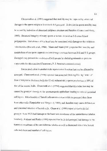
Chenoweth et al. (1994) suggested that motility may be impaired by structural
damage to the sperm midpiece in animals fed gossypol. Reduction in sperm motility may
be caused by induction of abnormal midpiece structure and function (Cusack and Perry,
1995). Structural integrity ofmany sperm structures depend on S-S cross linked
polypeptides. Disturbance of this bond may be associated with damage to the midpiece
mitochondria (Baccetti et al., 1986). Mann and Mann (1981) reported that motility and
metabolism of the sperm depends on interchange reactions between S-H and S-S groups.
Gossypol may prevent the oxidation of S-H groups by chelating minerals or proteins
responsible for this reaction (Chenoweth, P. J. Personal communication).
Sperm production is another male reproductive function that can be affected by
gossypol. Chenoweth et al. (1994) reported that gossypol (16.6 mg FG • kg"' BW • d '
from CSM) fed to Brahman bulls for 12 wk reduced daily sperm production to 50% of
that of the control bulls. Chenoweth et al. (1994) suggested that this reduction may be
caused by gossypol damage to the spermatogenic epithelium leading to reduced germinal
cell layers. Mitochondria of Sertoli cells are more susceptible to gossypol than those
from other cells (Tanpaichitr and Fitzgerald, 1989), and therefore may cause disfunction
and abnormal structure of Sertoli cells. Chase et al. (1990) reported that bulls fed
gossypol from WCS had damage to the basement membrane of the seminiferous tubules.
Similarly, Arshami and Ruttle (1988) reported that bulls fed gossypol had damage to the
basement membrane of the seminiferous tubules as well as decreased size of the Sertoli
cells and decreased number of cell layers.
19
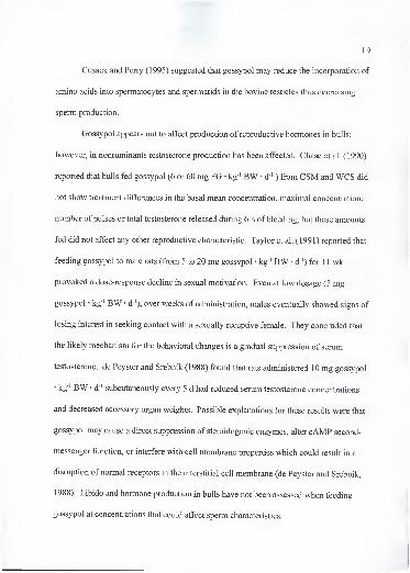
Cusack and Perry (1995) suggested that gossypol may reduce the incorporation of
amino acids into spermatocytes and spermatids in the bovine testicles thus decreasing
sperm production.
Gossypol appears not to affect production of reproductive hormones in bulls;
however, in nonruminants testosterone production has been affected. Chase et al. (1990)
reported that bulls fed gossypol (6 or 60 mg FG • kg ' BW • d ' ) from CSM and WCS did
not show treatment differences in the basal mean concentration, maximal concentration,
number of pulses or total testosterone released during 6 h of bleeding, but these amounts
fed did not affect any other reproductive characteristic. Taylor et al. (1991) reported that
feeding gossypol to male rats (from 5 to 20 mg gossypol • kg"' BW • d ') for 1 1 wk
provoked a dose-response decline in sexual motivation. Even at low dosage (5 mg
gossypol • kg"' BW • d"'), over weeks of administration, males eventually showed signs of
losing interest in seeking contact with a sexually receptive female. They concluded that
the likely mechanism for the behavioral changes is a gradual suppression of serum
testosterone, de Peyster and Srebnik (1988) found that rats administered 10 mg gossypol
kg"' BW • d"' subcutaneously every 5 d had reduced serum testosterone concentrations
and decreased accessory organ weights. Possible explanations for these results were that
gossypol may cause a direct suppression of steroidogenic enzymes, alter cAMP second-
messenger function, or interfere with cell membrane properties which could result in a
disruption of normal receptors in the interstitial cell membrane (de Peyster and Srebnik,
1988). Libido and hormone production in bulls have not been assessed when feeding
gossypol at concentrations that could affect sperm characteristics.

20
Gossypol has been found to inhibit the generation of free radicals in some studies
(Janero and Burghardt, 1988) and to stimulate it in others (de Peyster et al., 1984).
Recently, Barhoumi and Burghardt (1996) reported that gossypol promoted the formation
of reactive oxygen species and the depletion of glutathione in rat hepatocytes. Bender et
al. (1988) reported a reduction in antioxidants in testes of rats fed gossypol. Damage due
to lipid peroxidation in human sperm has been associated with loss of sperm motility,
inactivation of enzymes, and loss of membrane integrity (Aitken and Fisher, 1994).
Gossypol appears to affect late spermatogenesis when spermatids discard the
majority of their cytoplasm and thus they have lower cytoplasmic defensive enzymes
(Aitken and Fisher, 1994). Therefore the effect of gossypol on semen characteristics may
in part be related to increased free radical production and(or) inhibition of enzymes
related to the antioxidant defense system. * i-», V \ j
^
Diagnosis and Recommendations '' * r '
'
When suspecting or investigating a gossypol problem, the following factors
should be considered (Risco and Velasquez, 1994): determine if cottonseed products are
being fed; analyze sources of gossypol individually by an official gossypol testing
laboratory; from these results calculate the amount of free gossypol in feeds and compare
them to the recommended concentrations suggested by Calhoun and Holmberg (1991).
There must be a clinical history which is suggestive of gossypol toxicosis.

21
Among other characteristics are involvement of muhiple animals, sudden death
syndrome, chronic respiratory problems that are unresponsive to antibiotics, and(or)
history of infertility. Necropsy findings are compatible with cardiovascular and
respiratory failure, and histopathology examination of liver reveals centrolobular hepatic
congestion and necrosis related to hepatic anoxia.
Calhoun and Holmberg (1991) recommended that adult cows should be fed no
more than 600 mg FG/kg of diet DM. Consumption ofFG at this concentration should
not adversely affect reproduction in females (Gray et al., 1993). In male ruminant used
for breeding, the maximum intake allowed is 200 mg FG/kg of diet DM and for young
ruminants, 100 mg FG/kg (Calhoun and Holmberg, 1991).

CHAPTER 3
FEEDING COTTONSEED MEAL AND VITAMIN E TO BEEF HEIFERS
Introduction
Gossypol is a yellow polyphenolic pigment found in the glands of roots, stems,
and seeds of plants from the malvaceae family. This pigment is found in the cottonseed
and its by-products, limiting its use in animal diets especially for nonruminant species. In
ruminants, gossypol seems to have little effect when whole cottonseeds (WCS) and/or
by-products are fed within safe limits (Calhoun and Holmberg, 1991 ; Rogers and Poore,
1995). There are three forms in which gossypol content of a product is expressed: free
gossypol (FG) or the natural form which exists in the seed; bound gossypol (BG), and
total gossypol (TG). Cottonseed meal (GSM) contains variable amounts ofFG and BG,
depending on the process used to extract the oil from the seed (Jones, 1991). Although
toxicity effects are expected when high concentrations ofFG are fed, ruminants have the
capacity to detoxify large amounts by binding it to soluble proteins in the rumen (Reiser
and Fu, 1962). Gossypol has been found to affect liver functions, erythrocyte oxygen
carrying or releasing capacity, respiration rate, feed intake, and production and
reproduction efficiency (Lindsey et al., 1980; Gray et al., 1990).
22

Gossypol effect on animals seems, at least to some extent, to be related to the
formation of free radicals or to a decreased concentration of antioxidants such as
a-tocopherol (a-T), and P-carotene (P-C). In a field study. Lane and Stuart (1990)
reported a negative relationship between FG intake and serum retinol and tocopherol
concentration of dairy cattle fed WCS at a concentration to provide from 1 0 to 4 1 . 1 g FG
• animal ' • d"'. In a more controlled study, Willard et al. (1995) found that cows receiving
4 g FG • animal ' • d"' had lower (P < .05) serum a-T and P-C concentrations than cows
fed a control diet with no gossypol. Bender et al. (1988) found that a-T, ascorbate and
glutathione peroxidase, and other antioxidants were reduced by feeding rats high
concentrations of gossypol. An in vitro experiment indicated that gossypol generated the
superoxide radical in the presence of liver microsome and NADPH (de Peyster et al.,
1984).
The effect of feeding high amoimts of a-T on the gossypol status of heifers fed
CSM has not been investigated. Therefore the objective of diis experiment was to
evaluate the effect of high concentrations of supplemental vitamin E on gossypol status
and physiological response of beef heifers fed CSM.
Materials and Methods
Animals, diets and management. Thirty-two yearling Limousin and Angus
crossbred heifers, daughters oftwo different sires and averaging 3 1 5 kg BW, were used
in a 1 12-d experiment.

24
Animals were assigned randomly to one of four dietary supplements. Supplement
one (CON) was based on soybean meal (SBM), com, and adequate vitamin E (30 lU/kg
of diet DM). Supplement two (GOS) was based on CSM, com, and adequate vitamin E
(30 lU/kg of diet DM). Supplement three (G+2E) contained CSM, com, and 2,000 lU
vitamin E • animal"' • d"'. Supplement four (G+4E) contained CSM, com, and 4,000 lU
vitamin E • animal"' • d"'. Treatments based on CSM provided 4 g FG • animal"' • d"'.
Supplements (Table 3-1) were formulated to provide equal amounts of CP and TDN, and
to meet NRC (1984) nutritional requirements. Heifers were fed the supplement daily via
Calan gates and had free access to water and poor-quality mature bermuda grass
{Cynodon dactylori) hay with an a-T concentration of 9.1 lU vitamin E/kg. The protocol
for all heifer procedures had been approved by the University Animal Use Committee.
Blood and tissue sampling and analyses. Blood was collected every 2 wk via
jugular venipuncture with an 1 8-gauge needle into heparinized vacutainer blood
collection tubes for a total of 9 collections. Blood was analyzed for erythrocyte osmotic
fragility (EOF), hemoglobin, and hematocrit. Erythrocyte osmotic fragility, measured as
percentage hemolysis, was evaluated in .65 and .55% buffered saline solution as
described by Risco et al. (1993). Hemoglobin was determined using a colorimetric
procedure (Sigma Chemical Co., St. Louis, MO). Hematocrit was determined from blood
using a micro hematocrit centrifuge (lEC MB Centrifuge, Needham Heights, MA).
Blood was centrifuged for 25 min at 700 x g, and plasma was removed and stored at -
20 °C until analyzed for a-T (all collections), retinol (RET), retinol palmitate (RETP),

25
Table 3-1. Composition of dietary supplements^.
Supplement''
Item CON GOS G+2E G+4E
Offered (kg/df 4.2 4.5 4.5 4.5
DM (%) 88.0 88.0 88.0 88.0
CSM (Vof 0.0 75.0 75.0 75.0
SBM (Vof 65.0 .0 .0 .0
Com (%f 33.0 23.0 23.0 23.0
Limestone 1.0 1.0 1.0 1.0
Trace minerals (%)"^ 1.0 1.0 1.0 1.0
CP (%)" 37 ±2 36 ± 4 36 ±2 37 ±2Vitamin E {lUfkgf 47 ±9 44 ±10 : 488 ±54 871 ± 138
Vitamin A (lU/kg)" 10600 6293 6293 6293
(+)-gossypol (VoY .00 .35 .36 , .36
(-)-gossypol (%y .00 .86 .88 .87
Free gossypol {%y .00 .10 .10 .10
Total gossypol {%y .00 1.10 1.13 1.14
FG intake'' .00 4.5 4.5 4.5
Ca {%y 0.6 ± .06 0.6 ± .04 0.7 ± .03 0.7 ± .02
K (%)" 1.3 ±.14 1.2 ± .10 1.2 ±.09 1.2 ± .08
Mg (%)" .2 ± .01 .5 ± .01 .5± .01 .5 ± .01
P {%Y .6 ±.13 1.1 ± .07 1.1 ±.06 1.1 ± .06
Cu (mg/kg)'' 12.5 ± 1.67 13.5 ±3.26 16.1 ±6.35 14.9 ±2.89
Zn (mg/kgY 45 ±27 64 ±32 66 ±29 68 ±34Mn (mg/kg)'' 44 ± 10 39 ±7 38 ±5 42 ±8Fe (mg/kg)" 149 ± 108 102 ± 18 107 ±27 116 ±30
Se (mg/kg)" .28 ± .06 .33 ± .06 .30 ±.04 .34 ± .02
"Values are means of eight mixes ± standard deviation of the mean.
''CON = soybean meal (SBM) + com + 30 lU vitamin E/kg; GOS = cottonseed meal (CSM) + com + 30 lUvitamin E/kg; G+2E = CSM + com + 2,000 lU vitamin E • animal ' • d '; G+4E = CSM + com + 4,000 lUvitamin E • animal ' • d"'.
' As fed basis.
DM basis.
' As fed basis. A composite sample from all mixing dates. An lU/kg = retinol acetate (^g/kg) * 2.91
^As fed basis. Composite sample. HPLC procedure (Calhoun et al. (1995a), and Kim and Calhoun
(1995)). Texas A&M.*As fed basis. Composited sample. AOCS procedure. Texas A&M.•As fed basis. FG = free gossypol. FG intake (g • animal ' • d"') = Amount offered (g/d) * % FG.
26
P-C, alkaline phosphatase (AP), creatine kinase (CK) (every other collection), (+)-, (-)-,
and total gossypol (collections 1, 3, 5, and 9). At the end of the experiment, animals were
slaughtered and portions of liver, heart, and neck muscle (Stemo mandibularis) were
collected and frozen at -20°C until analyzed for a-T, P-C, (+)-, (-)-, and total gossypol,
Fe, Cu, and Zn. Liver samples were analyzed also for RET, RETP, and Se. Samples of
supplements were collected and analyzed for a-T, RETP, Ca, K, Mg, Mn, P, CP, Fe, Zn,
Cu, and Se.
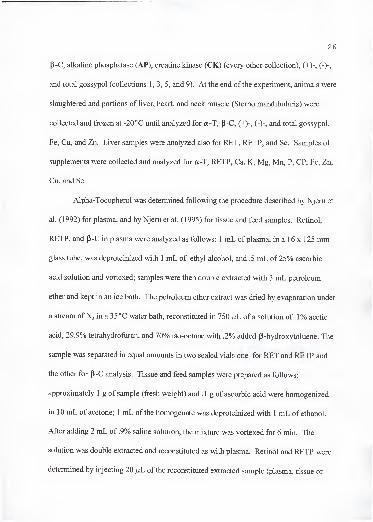
Alpha-Tocopherol was determined following the procedure described by Njeru et
al. (1992) for plasma, and by Njeru et al. (1995) for tissue and feed samples. Retinol,
RETP, and P-C in plasma were analyzed as follows: 1 mL of plasma, in a 16 x 125 mm
glass tube, was deproteinized with 1 mL of ethyl alcohol, and .5 mL of25% ascorbic
acid solution and vortexed; samples were then double extracted with 3 mL petroleum
ether and kept in an ice bath. The petroleum ether extract was dried by evaporation under
a stream ofNj in a 35 °C water bath, reconstituted in 750 /jL of a solution of .1% acetic
acid, 29.9% tetrahydrofuran, and 70% iso-octane with .2% added P-hydroxytoluene. The
sample was separated in equal amounts in two sealed vials one for RET and RETP and
the other for p-C analysis. Tissue and feed samples were prepared as follows: .
approximately 1 g of sample (fresh weight) and .1 g of ascorbic acid were homogenized .
in 10 mL of acetone; 1 mL of the homogenate was deproteinized with 1 mL of ethanol.
After adding 2 mL of .9% saline solution, the mixture was vortexed for 6 min. The
solution was double extracted and reconstituted as with plasma. Retinol and RETP were
determined by injecting 20 fxL of the reconstituted extracted sample (plasma, tissue or

27
feed) into the HPLC system. The HPLC system consisted of a Perkin Elmer 550 terminal
(Perkin-Elmer, Analytical Instruments, Norwalk, CT), an ISS-100 auto sampler (Perkin-
Elmer), a Series 4 Liquid chromatograph pump (Perkin-Elmer), and a Lichrosorb Si 60
5fxm, 4 mm ID x 250 mm column (Hibar Fertigsaule RT pre-packed column RT 250-4 E,
Merck, Darmstadt, Germany). The mobile phase consisted of 70% iso-octane, 29.9%
tetrahydrofuran, and .1% acetic acid. The UV detector was an ABI Analytical
Spectroflow 757 set at a wavelength of 325 nm and sensitivity of .005. Data were
collected by a LCI- 100 Laboratory Computing Integrator (Perkin-Elmer). Flow rate was
1 mL/min. The retention time ofRET was 6.1 min and for RETP 2.47 min. Standards
consisted of 10 ng ofRET (Sigma Chemical Co., St. Louis, MO) and 10 ng ofRETP
(Sigma Chemical Co., St. Louis, MO). Analysis for P-C was similar except that the
mobile phase was 90% iso-octane, 9.9% tetrahydrofuran, and .1% acetic acid; the
wavelength was set at 450 nm, and the retention time was 3.05 min. Standards consisted
of 100 ng P-C (Sigma Chemical Co., St. Louis, MO).
Cell mediated immune response was measured on d 50 by determining the
animals ability to respond to phytohemagglutinin-P (PHA; Sigma Chemical Co., St.
Louis, MO). Phytohemagglutinin-P was prepared in physiological buffered saline to a
concentration of 150 ^g/.l mL. The hair on the right side of the neck was clipped, the
skin-fold thickness was then measured using constant tension skin-fold calipers. A . 1 mL
ofPHA solution was injected intradermally at the clipped site and skin-fold thickness was
measured at 6, 12, 24, and 48 h after injection.

28
Stimulated lipid peroxidation was performed in heart and liver samples according
to a modification of the procedure of Kornbrust and Mavis (1980). Briefly,
approximately 1 g of fresh tissue was homogenized in 9 mL of 1.15% KCl solution. An
aliquot (100 ^L) was incubated in a water bath at 37 °C for the specified length of time
(0, 50, 100, and 200 min) with 500 |iL of 8 mM Tris-malate buffer, 200 of 5 mM
FeS04-7H20 and 200 of 2 mM ascorbic acid solutions. Peroxidation was terminated
by rapid addition of a 2 mL solution containing thiobarbituric acid (.375%) and 15%
trichloroacetic acid in .25 N HCl, and boiling in a water bath for 15 min. Then samples
were centrifuged for 15 min at 700 x g. The amount of colored product was measured
spectrophotometrically at 535 nm. Lipid peroxidation was expressed in terms of nmol
malondialdehyde (MDA)/mg of protein. Janero and Burghardt (1989) suggested that this
test cannot be used as anything else other than an empirical indicator of membrane
oxidative injury, therefore care should be taken in the interpretation of these results.
Enzymes were determined using kits (CK:Sigma Diagnostic Procedure No. 520;
AP: Sigma Diagnostic Procedure No. 104). Minerals were determined according to the
procedures described by Rojas et al. (1995) and Whetter and Ullrey (1978). Samples of
plasma and tissue for gossypol analyses were shipped in dry ice to Texas A&M
University where they were analyzed by HPLC according to the procedures described by
Calhoun et al. (1995a), and Kim and Calhoun (1995).
Statistical analvses. Plasma, lipid peroxidation, and weight data were analyzed by
repeated measures analysis of variance in a completely randomized design using GLM
procedureof SAS (1988). The Greenhouse-Geiser Epsilon was used to determine

significant levels for the F-test. The model included the effect of sire, treatment and the
interaction between sire and treatment. Sire was considered a random effect, hence the
interaction between sire and treatment was used as the error term for the treatment effect.
When the sire effect probability value was less than .2, PROC MIXED (SAS, 1996) was
used to calculate the correct standard errors of the least square means for a mixed model.
Tissue concentrations of (+)-, (-)-, and total gossypol were analyzed using PROC
MIXED of SAS (1996), with a nested mixed model where a 3 x 3 factorial design (three
tissues X three isomers) was nested within each animal, and animals were nested within a
4x2 factorial design (four treatments x two sires (random)). The nine measurements
(three gossypol values for each of three tissues) per animal were assumed to be equally
correlated with each other. Analyses of variables containing single or calculated
observations where performed by ANOVA (SAS, 1988). These variables were tissue
concentrations of a-T, p-C, RET, RETP, and minerals. The effect of treatment was
tested using the interaction between sire and treatment. When the overall treatment effect
was significant {P < .05) or tended to significance (P < .1), separation of means was done
using the Duncan multiple range test for all the variables except gossypol tissue
concentration for which the Tukey test (PROC MIXED) was used. Pooled standard
errors were reported when all treatments had the same number of replicates, otherwise
individual values for each least square mean were given.

Results and Discussion
30
Animal performance and blood parameters. Although the intake ofCSM was
high (3.4 kg • animal"' • d"'), calculated FG intake was approximately 600 mg FG • kg ' of
diet, (based on an average intake of 7.5 kg of DM/d, NRC, 1984) during the experimental
period. This amount ofFG intake was on the upper limit of the safe guidelines given by
Rogers and Poore (1995) and Calhoun and Holmberg (1991). In order to achieve an
intake of 4 g FG • animal ' • d"', supplements had a high CP concentration which was not
representative of normal diets. This high concentration of dietary CP could have affected
the absorption and metabolism of gossypol by supplying a greater number of free
8 -amino groups with which gossypol may have combined in the digestive tract and then
excreted and (or) by facilitating the metabolism and detoxification of the absorbed
gossypol (Abou-Donia, 1976). On average, supplements supplied the targeted amount of
vitamin E (Table 3-1).
All supplements produced an increase (P < .01) in body weight over time (Table
3-2). Average daily gain did not differ (P > . 1) among heifers fed the four supplements
throughout the experiment. Likewise, weight of beef heifers supplemented with 0, .5,
2.5, 5, 10, and 20 g FG • animal"' • d"', did not differ in a study conducted by Gray et al.
(1993). Calk et al. (1992) reported that vitamin E or lysine supplementation to lambs fed
a basal diet containing 20% CSM with . 1 84% FG, increased ADG over those not
receiving vitamin E. Supplements had been fed ad libitum, therefore an increase in ADG
in lambs fed vitamin E could have been due to increased intake. Also a decrease in ADG

31
Table 3-2. Effect of cottonseed meal (CSM) and vitamin E on weight gain and average
daily gain (ADG) of beef heifers".
Item
Supplement''
CON GOS G+2E G+4E SE^
Initial weight (kg) 317 299 314 328 13
Final weight (kg) 407 387 401 415 13
1 12 d gain (kg) 89 87 88 87 5
1 12 d ADG (kg) .79 .78 .78 .78 .04
''Least square means.
''CON = soybean meal + com + 30 lU vitamin E/kg; GOS = CSM + com + 30 lU vitamin
E/kg; G+2E = CSM + com + 2,000 lU vitamin E • animal ' • d"'; G+4E = CSM + com +4,000 lU vitamin E • animal ' d ".
"Standard error of the least square means.
32
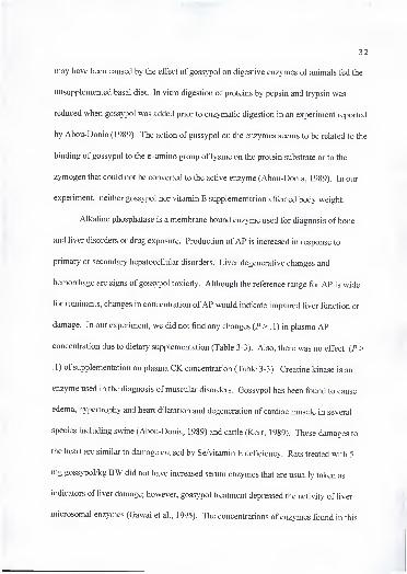
may have been caused by the effect of gossypol on digestive enzymes of animals fed the
unsupplemented basal diet. In vitro digestion of proteins by pepsin and trypsin was
reduced when gossypol was added prior to enzymatic digestion in an experiment reported
by Abou-Donia (1989). The action of gossypol on the enzymes seems to be related to the
binding of gossypol to the e -amino group of lysine on the protein substrate or to the
zymogen that could not be converted to the active enzyme (Abou-Donia, 1989). In our
experiment, neither gossypol nor vitamin E supplementation affected body weight.
Alkaline phosphatase is a membrane bound enzyme used for diagnosis of bone
and liver disorders or drug exposure. Production ofAP is increased in response to
primary or secondary hepatocellular disorders. Liver degenerative changes and
hemorrhage are signs of gossypol toxicity. Although the reference range for AP is wide
for ruminants, changes in concentration ofAP would indicate impaired liver function or
damage. In our experiment, we did not find any changes (P > . 1) in plasma AP
concentration due to dietary supplementation (Table 3-3). Also, there was no effect (P >
.1) of supplementation on plasma CK concentration (Table 3-3). Creatine kinase is an
enzyme used in the diagnosis of muscular disorders. Gossypol has been found to cause
edema, hypertrophy and heart dilatation and degeneration of cardiac muscle in several
species including swine (Abou-Donia, 1989) and cattle (Kerr, 1989). These damages to
the heart are similar to damage caused by Se/vitamin E deficiency. Rats treated with 5
mg gossypol/kg BW did not have increased serum enzymes that are usually taken as
indicators of liver damage; however, gossypol treatment depressed the activity of liver
microsomal enzymes (Gawai et al., 1995). The concentrations of enzymes found in this
33
Table 3-3. Effect of cottonseed meal (CSM) and vitamin E on plasma alkaline
phosphatase (AP) and creatine kinase concentrations (lU/L) of beef heifers\
Collection
Item Supplement^ 1 3 5 7 9
AP CON 65.9 46.9 43.0 41.3 44.7
GOS 45.1 21.1 17.6 21.6 18.4
G+2E 55.9 33.0 26.2 25.9 26.8
G+4E 41.0 26.7 26.4 27.8 24.8
SE' 7.7 7.4 6.9 5.8 9.1
CK CON 29.2 32.9 29.5 39.1 34.2
GOS 38.1 49.3 41.5 47.2 47.0
G+2E 26.4 43.6 36.0 81.7 55.8
G+4E 25.8 45.6 45.6 60.3 48.0
SE 4.0 13.5 4.5 17.0 7.2
^Least square means.
''CON = soybean meal + com + 30 lU vitamin E/kg; GOS = CSM + com + 30 lU vitamin'
E/kg; G+2E = CSM + com + 2,000 lU vitamin E • animal ' • d '; G+4E = CSM + com +4,000 lU vitamin E • animal ' • d ". -~ ,
•"Standard error of the least square means. J
'

34
experiment, do not suggest liver or muscle damage by feeding gossypol at the
concentration provided in this experiment. Blood chemistry has been demonstrated not to
be useful for monitoring gossypol toxicosis (Risco et al., 1992) and changes in enzyme
concentrations may be indicative of liver failure only during terminal stages of the
disease in calves.
Plasma a-T concentration (Figure 3-1) showed a time x treatment effect {P < .01).
Animals supplemented with high amounts of vitamin E (G+2E and G+4E) had greater (P
< .05) plasma a-T concentrations than animals supplemented with a more typical amount
(CON and GOS) from collection 2 to the end of the experiment (Figure 3-1). Heifers fed
CON or GOS supplements had equal amounts of supplemental vitamin E (30 lU/kg) and
similar analyzed concentrations (Table 3-1). Therefore, at the estimated dietary vitamin
E requirement for beef cattle, FG intake of 4 g/d did not decrease plasma a-T
concentration. Although not statistically different, plasma a-T concentration appeared to
be greater in animals in GOS than CON treatments. Animals in G+2E and G+4E did not
show differences (P >.l) in plasma a-T concentration, but were greater (P < .05) than
the low vitamin E supplemented treatments. Vitamin E absorption from the
gastrointestinal tract is aided by the presence of dietary fats; therefore, higher fat content
ofCSM compared to SBM (150 vs 103 g) based supplement (NRC, 1984) could have
increased vitamin E absorption. Since there was not a treatment with high vitamin E and
no gossypol included in this experiment, it is not possible to speculate on the effect of
gossypol on plasma a-T concentration at high levels of vitamin E supplementation.
However, the fact that no differences were found between the two high concentrations
35
Figure 3-1. Effect of cottonseed meal (CSM) and vitamin E on plasma a -tocopherol
concentration of beef heifers. Supplements were: CON = soybean meal + com + 30 lUvitamin E/kg; GOS = CSM + com + 30 lU vitamin E/kg; G+2E = CSM + com + 2,000
lU vitamin E • animal ' • d '; G+4E = CSM + com + 4,000 lU vitamin E • animal ' • d"'.
Standard errors for each collection (C) were: CI, .16; C2, .71; C3, .90; C4, .82; C5, .99;
C6, .90; CI, 1.20; C8, .86; and C9, .80.
(G+2E and G+4E), may indicate a reduced absorption of vitamin E either by gossypol
effect or most likely saturation of vitamin E absorption. At high doses of vitamin E
supplementation, the efficiency of vitamin E absorption decreased in rats (Traber et al.,
1 986). Furthermore, it has been reported that animals receiving excess dietary vitamin E
absorbed only 1 to 5% of the dose (Hoffmann-La Roche Inc., 1994). Hydrolysis of
vitamin E esters also could be reduced when feeding high concentrations of the vitamin.
Absorption of tocopherols depends on various factors such as: a) pancreatic enzymes b)
bile acids c) pH level of intestinal contents d) intestinal motility, and e) other food
components, in particular fatty acids (Hidiroglou et al., 1992). A healthy liver is
necessary for absorption and metabolism of vitamin E. The liver is targeted during
gossypol toxicity, causing hemorrhage and degenerative changes.
Contrary to our results. Lane and Stuart (1990) reported that serum a-T and P-C
concentrations were reduced in dairy cows fed high concentrations of gossypol
(approximately 40 g/d). In this study, however, no statistical data were showoi and
gossypol analysis may not represent the real values. In a more controlled study. Mena et
al. (1996) reported that gossypol from CSM, WCS or both (900 to 1800 mg total
gossypol/kg) sources did not reduce plasma concentrations of antioxidant vitamins, and
that in fact it increased plasma a -tocopherol concentrations.' >
Erythrocyte fragility is a very sensitive indicator of systemic gossypol status,
since it appears shortly after gossypol consumption has started. Willard et al. (1995)
found that by d 84, cows given 4 g FG • animal ' • d ' that showed elevated EOF had
lower {P < .05) plasma a-T concentrations than cows not fed gossypol. No supplemental

37
vitamin E was given in this experiment. Four animals with the highest EOF were chosen
from 17 receiving 4 g FG • animal"' • d ' in a group-fed experiment to determine plasma
a-T concentration. It may have been possible that these animals consumed more
supplement (high EOF) with low vitamin E content, and consumed less forage which
would have been an excellent source of vitamin E.
In the present study, supplementation treatments did not affect (P >.l) plasma
concentrations of RETP, RET, total retinoids, and p-C (Table 3-4). Analysis of a
composite feed sample from each dietary supplement indicated a higher concentration of
vitamin A in CON than in the other treatments (Table 3-1) even though all treatments had
20,000 lU vitamin A • animal"' • d ' added to the supplements. This was not reflected in
greater plasma RET, RETP or total retinoids concentrations which agree with data
reported by Eicher et al. (1994), who reported that plasma concentrations ofRET and
RETP did not reflect supplementation of vitamin A at normal doses. Similarly
Westendorf et al. (1990) did not find increased plasma vitamin A concentration in steers
fed normal concentrations of the vitamin. In a study with cottonseed products, plasma
P-C concentration of cows supplemented with 4 g FG • animal ' • d ' and high EOF was
lower than cows receiving 0 g FG • animal ' • d ', by d 84 after initiation of
supplementation (Willard et al., 1995). These authors related this change to seasonal
availability of p-C in forages rather than any effect of gossypol on plasma p-C
concentration.
In this study, EOF at .65 or .55% (Figure 3-2 and 3-3, respectively) saline
solution, revealed a time x treatment effect (P < .01). Cottonseed meal supplementation

38
Table 3-4. Effect of cottonseed meal (CSM) and vitamin E on plasma retinol palmitate
(RETP), retinol (RET), total retinoids, and P -carotene (P-C) concentrations (/^g/mL) of
beef heifers".
Collection
Item Supplement'' 1 3 5 7 9
POM .IS 1 A.10 .10
GOS 26 15 18 It lf>
G+2E .23 14 17tit 18 19
G+4E .22 14 17 21
.04 .02 .01 .01 .02
RET CON .62 53 52 AQ
GOS .53 .55 45 43 41
G+2E .58 .48 .43 41 41
G+4E .64 .49 43 42
.Uo .02 .03 .03 .02
Total CON .84 .77 .70 .73 . .69
Retinoids GOS .84 .79 .70 \71 -G+2E .86 .70 .67 .68 .7Q
G+4E .88 .71 .68 .72 .76
SE .05 .02 .03 .03 .03
p-c CON 1.15 1.29 1.56 1.61
GOS 1.37 1.46 1.60 1.69
G+2E 1.50 1.45 1.77 1.78
G+4E 1.40 1.52 1.79 1.82
SE .20 .07 .04 .05"Least square means.
''CON = soybean meal + com + 30 lU vitamin E/kg; GOS = CSM + com + 30 lU vitaminE/kg; G+2E = CSM + com + 2,000 lU vitamin E • animal " • d '; G+4E = CSM + com +4,000 lU vitamin E • animal ' • d '.
''Standard error of the least square means.
Missing samples in collection three.

39
•"Si-
Collection
A- CON GOS ^ G+2E hd- G+4E
Figure 3-2. Effect of cottonseed meal (CSM) and vitamin E on erythrocyte osmotic
fragility in .65% saline. Supplements were: CON = soybean meal + com + 30 lU vitamin
E/kg; GOS = CSM + com + 30 lU vitamin E/kg; G+2E = CSM + com + 2,000 lU
vitamin E • animal ' • d '; G+4E = CSM + com + 4,000 lU vitamin E • animal ' • d '.
Standard errors for each collection (C) were: CI, .35; C2, .92; C3, 3.32; C4, 4.60; C5,
3.10; C6, 2.50; C7, .80; C8, .93; and C9, 2.60.
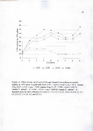
40
1 2 3 4 5 6 7 8
Collection
CON GOS ^ G+2E -a- G+4E
Figure 3-3. Effect of cottonseed meal (CSM) and vitamin E on erythrocyte osmotic
fragility in .55% saline. Supplements were: CON = soybean meal + com + 30 lU vitamin
E/kg; GOS = CSM + com + 30 lU vitamin E/kg; G+2E = CSM + com + 2,000 lUvitamin E • animal ' • d '; G+4E = CSM + com + 4,000 lU vitamin E • animal ' • d '.
Standard errors for each collection (C) were: CI, 5.0; C2, 4.1; C3, 10.0; C4, 6.8; C5, 2.1;
C6, 2.9; C7, 3.5; C8, 4.2; and C9, 5.3. . , I

41
(GOS) tended to increase EOF (/*<.!) over CON in collection 4. However, in vitamin E
and gossypol supplemented animals (G+2E and G+4E) EOF did not differ (P > .1) from
CON animals. On collections 5, 6 and 7, CON animals had lower (P < .05) EOF than the
rest of the treatments, but the animals supplemented with 4,000 lU vitamin E • animal"' •
d"' tended to have lower (P < A) EOF than animals consuming gossypol with 30 lU
vitamin E/kg (GOS). By the end of the experiment (collections 8 and 9), CON animals
had lower (P < .05) EOF than the other treatments, and vitamin E supplementation no
longer had any effect on EOF. A similar trend was observed with EOF measured at .55%
saline. From these results, it appeared that vitamin E at high doses would slow down the
increase in EOF for animals consuming gossypol. The EOF increase in animals
consuming a gossypol containing feedstuff is an indicator of gossypol intake, but its use
as a diagnostic tool for gossypol toxicity is questionable. In this study, the effect of
gossypol on EOF was not accompanied by any other clinical sign of gossypol toxicity.
It is difficuh to explain these results since the mechanism of gossypol effect on
the erythrocyte is not well understood. From in vitro experiments, the effect of gossypol
on cells has been explained by the ability of gossypol to interact with proteins, especially
membrane bound, and by binding and modifying the properties of the lipid bilayer matrix
(Reyes et al., 1984). These researchers found that gossypol binds strongly to bilayers of
different lipid compositions and induces an electrical conductance that is accompanied by
an increase in proton permeability. Furthermore, they showed that gossypol caused an
increase in diffusion in lipid membranes in the presence of NaCl, which could explain the
increase in EOF found in our experiment, de Peyster et al. (1986) found an increase in

membrane permeability due to gossypol treatment, and suggested that degeneration of
cell membranes at all levels of organization may exist with acute prolonged exposure to
gossypol in vivo. More evidence on the effect of gossypol on membranes was reported
by Tanphaichitr et al. (1995) who found that gossypol at low concentrations intercalates
in the phospholipid bilayer with the hydrophobic domains of the toxicant interacting with
the phospholipid hydrocarbon chain, while its hydrophilic groups would be exposed to
the aqueous phase. The intercalation of gossypol into a lipid bilayer, such as the
erythrocyte membrane, would alter its fluidity and therefore may explain increased
erythrocyte fragility, de Peyster et al. (1986) found that the effect of gossypol in lipid
membranes is dependent on the lipid composition, with membranes composed of
cholesterol showing very little gossypol effect. This finding led them to hypothesize that
gossypol and cholesterol may occupy the same domains in the membrane, leaving little
opportunity for insertion of gossypol into the cholesterol rich domains, or that gossypol
may bind either more efficiently or exclusively to the fatty acid and(or) other non-
cholesterol lipid components of membranes than it does to cholesterol. Alpha-tocopherol
as well as cholesterol and gossypol are located in the lipid membranes of the cell,
therefore, it may be possible that vitamin E had reduced the effect of gossypol on EOF by
occupying the same domain in the membrane and thus reducing the intercalation of
gossypol into the lipid membrane of the erythrocyte. However, at the end of the
experiment this effect was not seen because gossypol effect is both time and dose
dependant, with multiple oral doses gradually increasing the amount of the toxicant

43
retained in the body. Gossypol effect on the EOF has been also reported in several
studies with cattle (Willard et al., 1995; Risco et al., 1993; Lindsey et al., 1980).
Treatment tended to have an effect (P < .1) on hemoglobin concentration (Table
3-5). In collections 7 and 9, heifers fed GOS and G+2E had lower (P < .05) blood
hemoglobin concentrations than CON fed heifers. Blood hematocrit did not change {P >
.1) during the experimental period (Table 3-5). All hematocrit values were within the
normal range for cattle, although hemoglobin values were greater than the suggested
normal range for this class of animals possibly due to hemolysis. Despite this,
comparison may be valid since all samples were treated similarly. Lindsey et al. (1980)
reported lower blood hemoglobin {P <.07) concentration in dairy cows fed 3.5 and 24.2 g
FG • animal"' • d '. Similar results were found in dairy calves (Risco et al., 1992). Our
results agree with those of Risco et al. (1993) who found no clinical or hematological
evidence of decreased survival of red blood cells.
Gossypol enantiomers have been found to have different toxicological and ;-
pharmacokinetic effects. Calhoun et al. (1995b) suggested that plasma gossypol reflects
the availability of gossypol in the diet and the proportion of isomers in the source being
fed. Furthermore, they suggested that plasma gossypol concentration plateau at about 4
to 6 wk and remains fairly constant until the diet is changed. Plasma (-)-, (+)- and total
gossypol concentrations as affected by dietary supplement are presented in Figure 3-4,
3-5 and 3-6 respectively. Plasma (-)-, (+)-, and total gossypol concentrations in heifers of
GOS, G+2E, and G+4E treatments increased {P < .05) from the initial sampling to the

On
Cl
U
CL,
D!Z1
S
o ^ (N>/S iri \o
in 00 o— — (N
r-; ^ O
O 00
O00
^ O
\0 u-i
IT) in
o ou ocIS
S ^
ON
00
(N vo
K ON
00 ON
ON ON
u-i O ^
00 <Nvd vd
+ +
C/3
o oo vd
o in m 00^ ON vo^ m CO
rn vo
ON <n
ON vd
00 00o6 vd
ON 0000 t-^
—; vo00 u-i
o o
o
ONCO
m (N 00 00ON vd Tf vdm cn CO CI
vo On
00 00vd K
00 r<^
VO 00
ON ^vd 00
rn Onin
+ +
vo
vo
r<-i in ^ O \o
oo
+
oo+
u
w(N+O
c
2 -o>
S 6
+
ooo
bb +Hoo+
O
OO
om+
o
+
+
1 ili> c Sera "
g W2 O
^ II sZ -t;
CO O >u v_;
J U D
to SCO
1) CO
+-» CO
I—I U T3
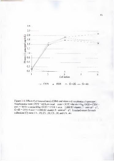
Collection
^ CON A GOS ^ G+2E -a- G+4E
Figure 3-4. Effect of cottonseed meal (CSM) and vitamin E on plasma (-)-gossypol.
Supplements were: CON = soybean meal + com + 30 lU vitamin E/kg; GOS = CSM +com + 30 lU vitamin E/kg; G+2E = CSM + com + 2,000 lU vitamin E • animal ' • d"';
G+4E = CSM + corn + 4,000 lU vitamin E • animal ' • d"'. Standard errors for eachcollection (C) were: CI, .03; C3, .30; C5, .30; and C9, .40.

1 3 5 7 9Collection
^ CON GOS ^ G+2E G+4E
Figure 3-5. Effect of cottonseed meal (CSM) and vitamin E on plasma (+)-gossypol.
Supplements were: CON = soybean meal + corn + 30 lU vitamin E/kg; GOS = CSM +com + 30 lU vitamin E/kg; G+2E = CSM + com + 2,000 lU vitamin E • animal ' • d ';
G+4E = CSM + com + 4,000 lU vitamin E • animal ' d '. Standard errors for eachcollection (C) were: CI, .03; C3, .30; C5, .20; and C9, .30.
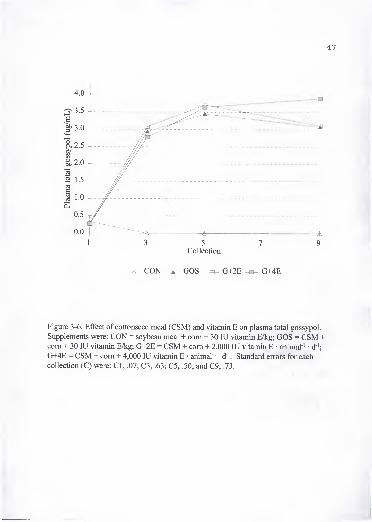
47
4.0 -
Collection
^ CON GOS s- G+2E^ G+4E
Figure 3-6. Effect of cottonseed meal (CSM) and vitamin E on plasma total gossypol.
Supplements were: CON = soybean meal + com + 30 lU vitamin E/kg; GOS = CSM +com + 30 lU vitamin E/kg; G+2E = CSM + com + 2,000 lU vitamin E • animal ' • d"';
G+4E = CSM + com + 4,000 lU vitamin E • animal ' • d '. Standard errors for each
collection (C) were: CI, .07; C3, .63; C5, .50; and C9, .73.
48
end of the experiment, and were similar (P> A) among the three treatments receiving
CSM. Heifers in the CON treatment had lower {P < .05) gossypol concentrations than
the other three treatments after the first collection. Plasma total gossypol concentration
was lower than the safer upper limit of 5 |Lig/mL proposed by Calhoun et al. (1995b).
Lower total gossypol plasma concentrations were reported by Lindsey et al. (1980) when
cows were fed 3.5 or 24.2 g FG • animal ' • d '. Also, Lindsey et al. (1980) did not find
differences in plasma gossypol concentrations between the two levels ofFG intake,
raising a concern as to whether FG in feed is a reliable indicator of bioavailability.
Calhoun et al. (1995b) reported that dairy cows consuming from 27.2 to 33.8 g TG •
animal"' • d ' from WCS had average plasma gossypol concentrations of 3.0 |ig/mL with a
range of 1.2 to 5.8 |jg/mL.
The (-)-gossypol isomer has been found to disappear at a faster rate fi-om the body
than (+)-gossypol following a single oral dose in humans and dogs (Wu et al., 1986). •
,
This could either be due to a faster rate of elimination or that (-)-gossypol is more likely
to be metabolized in vivo than (+)-gossypol, therefore explaining its higher biological
activity (Matlin et al., 1985). Daily dosage of gossypol was eliminated five times slower
than a single oral dose in rats and mice, which indicates that multiple dosage increased
retention of gossypol (Abou-Donia et al., 1988). This agrees with our results where
plasma gossypol concentration increased with time at the same dosage.
Tissue measurements. Tissue concentrations of (+)-, (-)-, and total gossypol
shown in Figure 3-7. No differences (P > . 1) were found among heifers fed the GOS,
G+2E, and G+4E treatments in tissue gossypol concentrations, but they were greater

Figure 3-7. Effect of cottonseed meal (CSM) and vitamin E on liver (L), muscle (M), andheart (H), (+)-, and (-)-gossypol concentrations. Supplements were: CON = soybean meal+ com + 30 lU vitamin E/kg; GOS = CSM + com + 30 lU vitamin E/kg; G+2E = CSM +com + 2,000 lU vitamin E • animal ' • d '; G+4E = CSM + com + 4,000 lU vitamin E •
animal ' • d '. Standard errors of CON, GOS, G+2E and G+4E respectively were: L+: 1.2,
7.8, 7.3, and 7.3; L-: 3.0, 3.4, 3.1, and 3.1; H+: .5 for all; H-: 3.9, 4.2, 3.9, and 3.9; M+: .1
for all; and M-: .3 for all.

50
(P < .01) than CON heifers. Liver (+)-gossypol was greater (P < .01) than (-)-gossypol
concentrations in the heifers fed CSM (GOS, G+2E, and G+4E), while heart and muscle
had greater (P < .05) (-)-gossypol than (+)-gossypol in the same treatments. The order of
(+)-, and total gossypol accumulation in tissue was liver > heart > muscle (P < .05), and
for (-)-gossypol liver and heart > muscle (P < .01). Supplemental vitamin E had no effect
on total gossypol and the accumulation of its isomer in tissue.
Similar results where (+)-gossypol concentration was greater in liver while (-)-
gossypol was greater in heart and muscle, were reported in lambs fed 20 to 30 mg FG •
kg-' BW • d ' (Kim et al., 1996). In contrast to these results, the proportion of gossypol
isomers in liver were similar in pigs fed a wide range of gossypol-containing diets (Knabe
et al., 1995). In rats, similar deposition patterns were found by Jensen et al. (1982). The
major excretory organ for gossypol appears to be the liver in several species including
swine (Abou-Donia and Dieckert, 1974) and rats (Abou-Donia et al., 1970). Abou-Donia
(1989) suggested a pathway of metabolism of gossypol. Gossypol is absorbed from the
gastrointestinal tract, mainly the small intestine. It enters the liver via the hepatic artery or
through the lymph into the hepatic sinusoid and is taken up by Kupffer cells. In the liver,
gossypol is metabolized, conjugated, and excreted with the bile into the duodenum.
Some of the excreted gossypol is reabsorbed, completing an enterohepatic cycle that may
be repeated several times leading to a gradual excretion via feces. The mechanism of
excretion seems to require an active secretory process that can be saturated. The fact that
gossypol is accumulated in the liver may be related to the tissue susceptibility to gossypol
toxicity or to the fact that the liver is the main route of gossypol metabolism.
Also, gossypol is a lipophilic compound, therefore it tends to accumulate in tissues or cell
fractions containing high amounts of lipid membranes e.g. mitochondria, lysosomal and
microsomal fractions (Baran and Ismailov, 1993). Gossypol has been reported to be
cardiotoxic, causing degeneration of cardiac muscle. Our results suggest that since the
heart is the second highest gossypol accumulator of the tissues studied, gossypol has the
potential for causing cardiac aberrations. The (-)-gossypol has been shown to be the
optically active form that induces fertility impairment in male animals (Wang et al.,
1987). Accumulation of this isomer in liver and heart tissues was similar, which may be
related to the toxicity signs shown during gossypol toxicosis. Gossypol isomers have
been shown to differ in their pharmacokinetic in dogs and humans (Wu et al., 1986), and
rats (Chen etal., 1987).
Liver RET and liver, heart, and muscle P-C concentrations did not differ (P > .1)
among treatments (Table 3-6). However, liver RETP concentrations increased (P < .05)
in the G+2E and G+4E treatments. It has been suggested that vitamin E protects retinyl
esters in the storage globules of the liver cells, preventing a rapid breakdown of retinyl
ester storage in the liver (Sondergaard, 1972). Olson (1991) suggested that retinyl ester
hydrolase, an enzyme involved in the release of vitamin A from the liver, is regulated by
vitamin E status, activated in vitamin E deficiency and inhibited in vitamin E sufficiency.
Vitamin E supplementation (G+2E and G+4E) increased (P < .05) a-T concentration in
liver, heart and muscle (Table 3-6). Feeding gossypol did not have an effect on a-T
concentration in these tissues (CON vs GOS; P > .1) at a normal level of vitamin E
supplementation (30 lU vitamin E/ kg). There was no difference between the two high

Table 3-6. Effect of cottonseed meal (CSM) and vitamin E on a-tocopherol (a-T), retinol
(RET), retinol palmitate (RETP), and p -carotene (P-C) concentrations in tissue of beef
heifers (\ig/g of wet tissue)^
Supplement''
Tissue Item CON GOS G+2E G+4E SE^
Liver a-T 5.V 7.V 19.9" 22.4" .8
RET 7.4 7.5 8.9 11.4 1.9
RETP 39.0^ 48.7^f 65.3'^'= 74.7" 12.0
P-C 15.4 16.9 15.5 16.3 .9
Heart a-T 5.8^ 7.6^ 21.6" 23.1" 1.2
P-C 5.8 5.3 5.5 5.4 .3
Muscle a-T 2.6' 3.3^ 8.1" 8.3" .6
P-C 4.5 4.5 4.6 4.8 .4
"Least square means.
''CON = soybean meal + com + 30 lU vitamin E/kg; GOS = CSM + com + 30 lU vitamin
E/kg; G+2E = CSM + com + 2,000 lU vitamin E • animal ' • d"'; G+4E = CSM + com +4,000 lU vitamin E • animal ' • d '.
''Standard error of the least square means.
"^•^Least square means with different superscripts in the same row differ P < .05.

concentrations of vitamin E supplementation on tissue a-T concentration. These results
were similar to those found for plasma a-T concentration.
Trace minerals (i.e. Fe, Cu, Se, and Zn) have been implicated in gossypol
metabolism. Heart failure, seen in gossypol toxicosis, is also a clinical sign of Se
deficiency. Iron is chelated by gossypol reducing iron's bioavailability. Skutches et al.
(1973) suggested that gossypol chelated Fe in the liver of pigs, and reduced Fe
availability for hemoglobin synthesis. Gossypol also chelates Cu and Zn (de Peyster and
Wang, 1993). Yu et al. (1981) reported that gossypol-treated rats had reduced Zn
concentration in testis, and suggested that the chelation ofZn by gossypol may be the
cause of its antispermatogenic activity. In our experiment, Cu, Fe, Se, and Zn
concentrations in the experimental diets were within normal concentrations (Table 3-1),
and feeding CSM did not affect (P > .1) concentrations of Cu, Fe, and Zn in heart, muscle
and liver, and Se in liver (Table 3-7).
Gossypol has been demonstrated to inhibit free radical damage to lipid'
membranes in some studies (Janero and Burghardt, 1988) and to stimulate it in others (de
Peyster et al., 1984). Bender et al. (1988) suggested that oxidative injury caused by the
generation of reactive oxygen species may be the cause of gossypol toxicity. In this
study, they found a decrease in the concentration of antioxidants, including a-T, in testis
of rats fed gossypol acetic acid. Our data on in vitro stimulated lipid peroxidation in liver
homogenate (Figure 3-8) indicated that from 50 min of incubation onward, liver from
heifers fed CON supplement had greater {P < .05) MDA production than the other
heifers. Feeding CSM (GOS) did not increase (P > .1) lipid peroxidation above the CSM
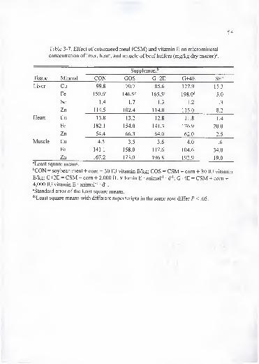
54
Table 3-7. Effect of cottonseed meal (CSM) and vitamin E on micromineral
concentration of liver, heart, and muscle of beef heifers (mg/kg dry matter)\
Supplement''
1 issue ivimerai LiUS Lr+4b
T i\if^rJ_/l VCl QQ Hyy.o 7n 7/u. / oj.O 1 07 Q1 J.J
Fe \50.6' 146.9« 165.5^ 198.0"* 5.0
Se 1.4 1.7 1.3 1.2 .3
Zn 114.5 102.4 114.0 115.0 8.2
Heart Cu 13.8 13.2 12.8 11.8 1.4
Fe 182.1 154.0 141.3 176.9 20.0
Zn 54.4 66.3 64.0 62.0 2.5
Muscle Cu 4.5 3.5 3.6 4.0 .6
Fe 141.1 158.0 117.6 104.6 • 34.0
Zn 167.2 173.0 196.8 192.9 19.0
"Least square means.
''CON = soybean meal + com + 30 lU vitamin E/kg; GOS = CSM + com + 30 lU vitamin
E/kg; G+2E = CSM + com + 2,000 lU vitamin E • animal"' • d "; G+4E = CSM + com +4,000 lU vitamin E • animal"' d '.
"Standard error of the least square means.
''^Least square means with different superscripts in the same row differ P < .05.
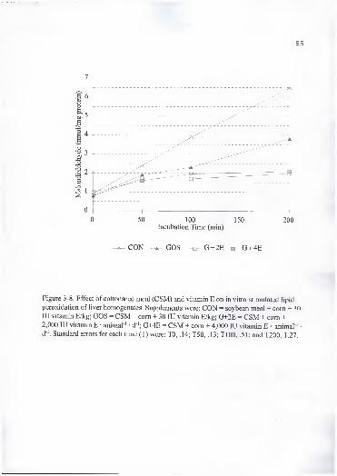
Figure 3-8. Effect of cottonseed meal (CSM) and vitamin E on in vitro stimulated lipid
peroxidation of liver homogenates. Supplements were: CON = soybean meal + com + 30lU vitamin E/kg; GOS = CSM + com + 30 lU vitamin E/kg; G+2E = CSM + com +2,000 lU vitamin E • animal ' • d '; G+4E = CSM + com + 4,000 lU vitamin E • animal '
d '. Standard errors for each time (T) were; TO, .14; T50, .13; TlOO, .51; and T200, 1.27.

56
and vitamin E supplemented animals (G+2E and G+4E). In vitro stimulated lipid
peroxidation of heart tissue did not differ (P> A) among treatments (Figure 3-9) possibly
because of the resistance of this tissue to in vitro peroxidation (Kombrust and Mavis,
1980). Sheriff et al. (1986) found similar results on the ability of gossypol to reduce
lipid peroxidation in human spermatozoa and erythrocytes. Janero and Burghardt ( 1 988)
found that gossypol effectively prevented O2" dependant Fe-promoted peroxidation of
myocardial membrane phospholipids and could attenuate the kinetics and extent of
peroxidation. Furthermore, they suggested that the gossypol antioxidant effect is through
the scavenging of lipid radical intermediates. Contrary to our results, de Peyster et al.
(1984) found that gossypol when incubated with rat liver microsomes or human sperm,
promotes the formation of O2", causing oxidative injury. A possible explanation for the
difference in results is that a metabolic product of gossypol (such as redox-cycling
quinone) may be responsible for the ability of gossypol to generate oxygen radicals and
cause the side effects associated with gossypol treatment (Janero and Burghardt, 1988; de
Peyster etal., 1984).
Skinfold response to PHA intradermal injection on d 50 did not differ (P > .1)
among treatments (Figure 3-10). Neither CSM alone nor with vitamin E affected the cell
mediated immune response.

0 50 100 150 200Incubation Time (min)
^ CON A GOS ^ G+2E^ G+4E
Figure 3-9. Effect of cottonseed meal (CSM) and vitamin E on in vitro stimulated lipid
peroxidation of heart homogenates. Supplements were: CON = soybean meal + com + 30
lU vitamin E/kg; GOS = CSM + com + 30 lU vitamin E/kg; G+2E = CSM + com +2,000 lU vitamin E • animal ' d '; G+4E = CSM + com + 4,000 lU vitamin E • animal-^ •
d '. Standard errors for each time (T) were: TO, .3; T50, .3; TlOO, .3; and T200, .8.
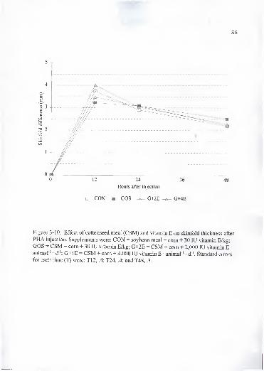
58
5 -r
Hours after injection
^ CON -m- GOS G+2E G+4E
Figure 3-10. Effect of cottonseed meal (CSM) and vitamin E on skinfold thickness after
PHA injection. Supplements were: CON = soybean meal + com + 30 lU vitamin E/kg;
GOS = CSM + corn + 30 lU vitamin E/kg; G+2E = CSM + com + 2,000 lU vitamin E •
animal ' • d '; G+4E = CSM + corn + 4,000 lU vitamin E • animal ' • d '. Standard errors
for each time (T) were: T12, .4; T24, .4; and T48, .3.

Implications
Free gossypol fed at 4 g • animal'' • d"' at a low concentration of vitamin E
supplementation (30 mg/kg of feed DM) did not decrease concentrations of antioxidant
vitamins, including a-T, vitamin A and P-C or have any detrimental effect on growth
performance of beef heifers.

CHAPTER 4
LONG TERM EFFECTS OF FEEDING COTTONSEED MEAL AND VITAMIN E TODAIRY CALVES.
Introduction
Gossypol is a natural pigment found in the cotton plant (Gossypium spp.) and
other plants belonging to the Malvaceae family. This pigment has been recognized to be
toxic to nonruminant species and if fed at high enough concentrations also to ruminants,
thus limiting the use of cottonseed products as feed for livestock.
Preruminants are especially susceptible to gossypol toxicosis. The undeveloped
rumen appears unable to detoxify gossypol (Morgan et al., 1988). Soluble protein
binding with gossypol has been proposed as a mechanism by which gossypol is
detoxified in the rumen (Reiser and Fu, 1962). It is not known precisely at what age the
rumen matures or becomes fimctional in calves. Therefore, young ruminants with
undeveloped rumens appear to function as nonruminant animals, and gossypol cannot be
detoxified effectively (Risco et al., 1992).
Toxicity signs and death of calves have been reported when feedstuffs containing
gossypol were fed (Holmberg et al., 1988; Risco et al., 1992; Zelski et al., 1995). Risco
et al. (1992) suggested that diets for dairy calves that contain up to 200 mg free gossypol
(FG)/kg of diet DM are safe, 400 mg FG/kg ofDM approaches toxicity, and 800 mg
60
61
FG/kg ofDM results in death losses. In a more recent study (Zelski et al., 1995) calves
fed from 100 to 220 mg FG/kg of feed resulted in more than 40% mortality. The authors
also reported that gossypol toxicity signs ranged from preacute death during an acute
phase, to poor growth during a chronic phase. There is a limited capacity for gossypol
detoxification in preruminants and calves are susceptible to gossypol toxicity at
concentrations comparable to those affecting nonruminant animals (i.e. swine).
Some gossypol toxicity signs have been related to a decrease in antioxidant
concentrations and(or) an increase in the formation of reactive oxygen species. Janero
and Burghardt (1988) suggested that gossypol can interact with biological membranes by
promoting the formation of highly-reactive oxygen-containing free radicals. This and
other studies (Willard et al., 1995; Lane and Stuart, 1990; Bender et al., 1988;) suggest
that antioxidants play a key role in the metabolism of gossypol. Therefore, the objectives
of this experiment were to evaluate the effect of feeding a diet containing 400 mg FG/kg
of feed DM to dairy calves from 2 wk to 6 mo of age, and to evaluate the value of
supplemental vitamin E on the effect of gossypol on preruminant animals.
Materials and Methods
This experiment was divided into two phases. Phase 1 describes animals
individually fed and phase 2 describes those which were group fed.
Animals, diets and management. In phase 1 of this experiment, two groups of
male Holstein calves were purchased from Florida dairies. The first group (group 1)

62
Started the experiment when calves were approximately 2 wk of age (August, 1993),
while the second group (group 2) started at approximately 4 wk of age (September,
1993). Phase 1 ended in October for both groups. The duration of this phase of the
experiment was 82 and 50 d for groups 1 and 2, respectively. During the first two weeks
of the experiment, approximately 30% of calves from group 1 died from causes
determined to be unrelated to treatments (heat stress), therefore their data were not
included in this experiment. Calves were fed 4 L/d of a commercial milk replacer (Ultra
Suckle 2000® from Mama Pro Corporation) containing .454 kg of replacer in 4 L of
water. Calves had free access to the following treatments (Table 4-1): 1) CON: a ration
base on soybean meal (SBM) + 30 lU vitamin E/kg of feed; 2) GOS: a ration based on
cottonseed meal (CSM) + 30 lU vitamin E/kg of feed; 3) G+2E a ration based on CSM +
2,000 lU vitamin E • calf • d '; and 4) G+4E a ration based on CSM + 4,000 lU vitamin
E • calf d '. Vitamin E in the G+2E and G+4E treatments was fed with the milk
replacer. The number of animals per treatment in phase 1 were as follows: in group 1,
CON had 8 calves, GOS had 1 1, G+2E had 6 and G+4E had 8 calves; in group 2, CON
had 5, GOS had 4, G+2E had 5, and G+4E had 5 calves. Animals were individually
weaned when they were consuming 1% of their body weight in dry feed. After weaning
and while in phase 1, animals were fed individually the starter rations twice a day, and
vitamin E (G+2E and G+4E) was fed in the morning with the dry feed, mixing it with
half of the previous day intake.
In phase 2, calves from the two groups were moved to 16 pens (4 pens per
treatment) and the amount of supplemental vitamin E changed. Calves fed G+2E

63
Table 4-1 . Composition of calf diets.
Treatment'
Item CON GOS G+2E G+4E
DM (%) 91.00 91.00 91.00 91.00
CSM (%f .00 30.00 30.00 30.00
SBM (%)" 22.00 .00 .00 .00
Com (%)'' 19.00 22.00 22.00 22.00
Alfalfa meal (%)" 14.00 3.00 3.00 3.00
Oats (%)" 18.00 18.00 18.00 18.00
Beet pulp (%)'' 22.00 22.00 22.00 22.00
Molasses (%)'' 3.00 3.00 3.00 3.00
Mmerals (%)'' 2.00 2.00 2.00 2.00
CP (Vof 22.00 22.00 22.00 22.00XT'. ' 1 ^ yTT T /I \ /J
Vitamm E (lU/kg)'^ 41.00 45.00 45.00 45.00
Vitamm E (lU/kgf 41.00 45.00 525.00 1291.00
(+)-gossypol (%y .00 .15 .15 .15
(-)-gossypol (%)<" .00 .35 .35 J5Free gossypol .01 .04 .04 mTotal gossypol (%)« .02 .50 .50 .50
Ca (%)^ .52 .50 .50 .50
K (%)^ 1.10 .80 .80 .81
Mg (%y .21 .30 .34 .34
P (yo)*^ .51 .60 .60 .60
Cu (mg/kg)'= 16.50 14.50 14.50 14.50
Zn (mg/kg)'= 44.50 44.30 44.33 44.33
Mn (mg/kg)'= 62.14 48.32 48.33 48.33
Fe (mg/kg)*^ 252.83 178.23 178.24 178.24
Se (mg/kg)"^ .23 .21 .21 .21^CON = soybean meal (SBM) + 30 lU vitamin E/kg; GOS = cottonseed meal (CSM) + 30 lU vitamin E/kg;G+2E = CSM + 2,000 lU vitamin E- animal '
• d '; G+4E = CSM + 4,000 lU vitamin E • animal ' • d '.
•"As fed basis.
DM basis .
'' As fed basis. Phase 1
.
' As fed basis. Phase 2.
^Composite sample. HPLC procedure (Calhoun et al. (1995a), and Kim and Calhoun (1995)). Texas A&M.^Composited sample. AOCS procedure. Texas A&M.
received 700 lU vitamin E/ kg of feed and those fed G+4E received 1 ,400 lU vitamin E/
kg of feed. The number of animals per treatment were as follow: 13 (CON), 14 (GOS),
1 1 (G+2E), and 1 1 (G+4E). AH diets were isocaloric and isonitrogenous and met the
requirements of other nutrients for this class of animal (NRC, 1989). Diets based on
CSM were formulated to provide 400 mg FG/kg of diet DM to ensure a mild level of
toxicity, and to test the effect of vitamin E supplementation. Fresh feed and clean water
were offered ad libitum. Sick calves were treated with antibiotics in the case of
respiratory problems and oral fluid electrolyte replacement therapy for calves with
diarrhea (under the supervision of a veterinarian).
Calves were weighed every two weeks in both phases, and individual (phase 1)
and group (phase 2) feed intake data were recorded daily. Average daily feed intake was
calculated by 14-d periods so as to match collection days. In phase 1, there were 6
periods and 7 blood and weight collections for group 1 , and 4 periods and 5 collections
for group 2. In phase 2 there were 3 periods and 4 weight and blood collections. The
protocol for all calf procedures had been approved by the University Animal Use
Committee.
Blood and tissue sampling and analyses. Blood samples were collected every 2
wk via jugular venipuncture with an 18-gauge needle into heparinized vacutainer blood
collection tubes. Blood was analyzed for erythrocyte osmotic fragility (EOF),
hemoglobin, and hematocrit. Erythrocyte osmotic fragility, measured as percentage
hemolysis, was evaluated in .65% buffered saline solution as described by Risco et al.
(1993). Hemoglobin was determined using a colorimetric procedure (Sigma Chemical

65
Co., St. Louis, MO). Hematocrit was determined from blood using a micro hematocrit
centrifuge (lEC MB Centrifuge, Needham Heights, MA). Blood was centrifuged for 25
min at 700 x g. Plasma was removed and stored at -20 °C until analyzed for a-T (all
collections), alkaline phosphatase (AP) (phase 2), (+)-, and (-)-gossypol isomers (at
approximately the beginning, middle and end of phase 1).
Complete necropsies were performed on dead calves. This consisted of a
description of significant findings and collection of heart and liver tissues for
histopathology and laboratory analyses. Tissues were frozen at -20 °C until analyzed for
a-T, p -carotene (p-C), (+)-, (-)-, and total gossypol concentration. Samples of diets were
collected and analyzed for concentration of a-T, Ca, K, Mg, Mn, P, CP, Fe, Zn, Cu, and
Se.
Alpha-Tocopherol was analyzed following the procedure described by Njeru et al.
(1992) for plasma, and by Njeru et al. (1995) for tissue and feed samples. Tissue p-C
was prepared as follow: approximately 1 g of sample (fresh weight) and .1 g of ascorbic
acid were homogenized in 10 ml of acetone; 1 ml of the homogenate was deproteinized
with 1 mL of ethanol. After adding 2 ml of .9% saline, the mixture was vortexed for 6
min. The solution was double extracted with 3 mL petroleum ether, and kept in an ice
bath. The petroleum ether extract was dried by evaporation under a stream of in a
35 °C water bath, reconstituted in 750 fj.L of .1% acetic acid, 29.9% tetrahydrofuran, and
70% iso-octane with .2% added p -hydroxy toluene. Beta-carotene was determined by
injecting 20 /nL of the reconstituted extracted sample into the HPLC system. The HPLC
systems consisted of a Perkin Elmer 550 terminal (Perkin-Elmer, Analytical Instruments,

Norwalk, CT.), an ISS-100 auto sampler (Perkin-Elmer), a Series 4 Liquid
chromatograph pump (Perkin-Elmer), and a Lichrosorb Si 60 5/j.m, 4 mm ID x 250 mm
column (Hibar Fertigsaule RT pre-packed column RT 250-4 E, Merck, Darmstadt,
Germany). The mobile phase consisted of 90% iso-octane, 9.9% tetrahydrofiiran, and
.1% acetic acid. The UV detector was an ABI Analytical Spectroflow 757 set at a
wavelength of 450 nm and sensitivity of .005. Data were collected by a LCI-lOO
Laboratory Computing Integrator (Perkin-Elmer). Flow rate was 1 mL/min and the
retention time was 3.05 min. Standards consisted of 100 ng P-C (Sigma Chemical Co.,
St. Louis, MO).
Alkaline phosphatase was determined using a kit (Sigma Diagnostic Procedure
No. 104). Minerals were determined according to the procedures described by Rojas et
al. (1995) and Whetter and Ullrey (1978). Samples ofplasma and tissue for gossypol
analyses were shipped in dry ice to Texas A&M where they were analyzed by HPLC
according to the procedures described by Calhoun et al. (1995a) and Kim and Calhoun
(1995).
Statistical analyses. In phase 1, group 1 and 2 data were analyzed separately.
Continuous data such as body weight, feed intake, hematocrit, hemoglobin, and EOF,
plasma concentration of (-)-, (+)-, and total gossypol, and a-T were analyzed by repeated
measures analysis of variance in a completely randomized design using GLM procedure
ofSAS (SAS, 1988).
In phase 2, calves were group fed in 16 pens (4 pens/treatment) therefore pen
(random) effect was included in the model. Continuous data such as body weight, feed
67
intake, hematocrit, hemoglobin, and EOF, plasma concentrations of AP, and a-T were
analyzed by repeated measures analysis of variance in a completely randomized design
using GLM procedure of SAS (SAS, 1988). The Greenhouse-Geiser Epsilon was used to
determine significant levels for the F-test. Treatment and pen within treatment effects
were tested using animal within pen and treatment effect. Pen within treatment effect
was removed from the model when it was not significant (P > .2). When the pen within
treatment effect was kept in the model PROC MIXED (SAS, 1996) was used to calculate
the correct standard errors of the least square means for a mixed model. There were two
sets of animals with an age difference of 2 to 4 wk, therefore age was used as a covariate.
The time x treatment interaction was tested using time x pen within treatment interaction.
When the overall treatment effect was significant (P < .05) or tended to be significant (P
< .1) separation of means was done using Duncan muhiple range test. Pooled standard
errors were reported when all treatments had the same number of replicates, otherwise
individual values for each least square mean were given.
Results and Discussion
Phase 1 In group 1, calves receiving the high vitamin E treatment (G+4E) had
greater (P < .05) total weight gain and ADG than CON animals (Table 4-2). No
differences in calf performance were observed in group 2. In group 1, average daily feed
intake followed the same trend as body weight gain (Table 4-3). Calves fed large
amounts of vitamin E (G+4E) had greater (P < .05) feed intake in periods 4, 5, and 6 than
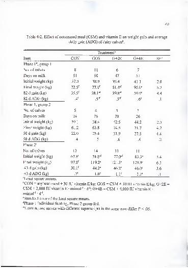
68
Table 4-2. Effect of cottonseed meal (CSM) and vitamin E on weight gain and average
daily gain (ADG) of dairy calves\
Treatment"'
Item CON GOS G+2E G+4E SE'
Phase 1'', group 1
No. of calves 8 11 6 7
Days on milk 51 50 47 51
Initial weight (kg) 37.0 38.9 39.4 41.1 2.0
Final weight (kg) 72.5^ 77.1^ 81.4'=^ 95.0-= 5.7
82 d gain (kg) 35.5^ 38.pf 39.8^f 50.9'= 4.4
82 d ADG (kg) .4^ .6' .1
Phase 1 , group 2
No. of calves 5 4 5 5
Days on milk 24 26 20 26
Initial weight (kg) 39.1 38.4 42.5 44.2 2.0
Final weight (kg) 61.2 63.8 74.5 71.7 4.7
50 d gain (kg) 22.0 25.4 31.9 27.5 4.4
50 d ADG (kg) .4 .5 .6 .6 .2
Phase 2"^
No. of calves 13 14 11 11
Initial weight (kg) 67.8^ 74.8^f 77.0'^ 83.3' 3.4
Final weight (kg) 97.8^ 119.2^ 121.5'= 129.9' 6.3
43 d gain (kg) 30.1^ 44.2^ 46.2'= 46.0' 3.6
43 d ADG (kg) 1.0^ \.V l.P .1
^Least square means.
''CON - soybean meal + 30 lU vitamin E/kg; GOS = CSM + 30 lU vitamin E/kg; G+2E =
CSM + 2,000 lU vitamin E • animal ' • d '; G+4E = CSM + 4,000 lU vitamin E •
animal ' d"'.
'^Standard error of the least square means.
''Phase 1 individual feeding; Phase 2 group fed.
'•^Least square means with different superscripts in the same row differ P<.05.

69
Table 4-3. Average intake of feed (kg • animal"' • d"') and free gossypol (FG) (g • animal
• d"') of dairy calves in phase P.
Treatment''
Intake Period CON GOS G+2E G+4E SE"
Group 1"
Feed 1 .05 .04 .02 .05 .01
2 .26 .24 .22 .23 .05
3 .55 .53 .62 .68 .13
4 .91^ l.OS^f 1.33"'' 1.59" .20
5 1.35^ 1.85^f 2.06" 2.49" .23
6 1.98^ 2.50^ 2.52'" 3.09" .19
FG 1 .00'" .02" .01"^ .02" .00
2 .00^ .lO'' .09" .09" 02
3 .00'' .2P .25" .27" .05
4 .00'^ .43" .53" .64" .08
5 .008 .74' .82*"" 1.00" .08
6 .00« 1.00*^ 1.07*^ 1 23" 07
Group 2''
Feed 1 .23^ .20^ .40" .22^ .05
2 .51^ .45^ .85" .55^ .09
3 1.14f 1.21''" 1.92" 1.48" .15
4 1.66^ 1.96f 2.73" 2.24*"" .18
FG 1 .008 .08^ .16" .09^ .01
2 .008 .18f .34" .22^ .03
3 .008 .48'' .77" .59^ .04
4 .008 .79' 1.09" .90^ .06
'Least square means.
''CON = soybean meal + 30 lU vitamin E/kg; GOS = CSM + 30 lU vitamin E/kg; G+2E =
CSM + 2,000 lU vitamin E • animal ' • d"'; G+4E = CSM + 4,000 lU vitamin E • animal"' •
d"'.
"Standard error of the least square means.
""Individual feeding.
"'^8Least square means with different superscripts in the same row differ Z' < .05.
CON animals, and greater (P < .05) than calves on diets G+2E and GOS in period 6. In
lambs, vitamin E supplementation (85 lU/kg of diet DM)) had been reported to improve
weight gain and feed intake when fed a basal diet containing 20% CSM (Calk et al.,
1992). Richer et al. (1992) reported that calves fed supplemental vitamin E had greater
ADO than control animals, and this difference occurred after 6 wk of feeding. Risco et
al. (1992) did not find an effect of feeding gossypol (100 to 800 mg FG/kg of diet DM)
on feed intake and body weight gain of calves in the first 83 d of age. Differences in
ADG and weight gain could be explained by differences in feed intake. Results similar to
the present experiment were found by Reddy et al. (1985) in which vitamin E
supplementation tended to increase performance and intake of calves. Gossypol appeared
not to have a negative effect on feed intake or gain of calves during phase 1.
Plasma a-T concentration of calves in groups 1 and 2 are presented in Figures 4-1
and 4-2, respectively. Calves supplemented with vitamin E (G+2E and G+4E) had
greater (P < .05) plasma a-T concentrations than calves fed CON and GOS diets from
collection 4 through collection 7 in group 1 and fi-om collection 3 to 5 in group 2. Both
groups of calves on CON and GOS treatments had similar plasma a-T concentrations
during all collections. Therefore gossypol intake (Table 4-3) by calves fed 30 lU vitamin
E/kg of diet DM did not decrease plasma concentrations of a-T. The lack of statistical
difference between the two high concentrations of vitamin E supplementation could be
the result of reduced vitamin E absorption at high levels of supplementation as shown
with laboratory animals (Traber et al., 1986). Pancreatic and bile secretions are essential
for adequate absorption and transport of vitamin E and pancreatic enzyme activity does

71
6.0 -r
1 2 3 4 5 6Collection
A- CON GOS H3- G+2E -n- G+4E
Figure 4-1.Effect of cottonseed meal (CSM) and vitamin E on plasma a -tocopherol
concentration of dairy calves in phase 1 group 1. Diets were: CON = soybean meal + 30lU vitamin E/kg; GOS = CSM + 30 lU vitamin E/kg; G+2E = CSM + 2,000 lU vitamin E• animal ' • d '; G+4E = CSM + 4,000 lU vitamin E • animal ' • d '. Standard errors for
each collection (C) were: CI, .2; C2, .4 C3, .4; C4, .4; C5, .6; C6, .5; and C7, .4.

72
Figure 4-2. Effect of cottonseed meal (CSM) and vitamin E on plasma a-tocopherol
concentration of dairy calves in phase 1 group 2. Diets were: CON = soybean meal + 30lU vitamin E/kg; GOS = CSM + 30 lU vitamin E/kg; G+2E = CSM + 2,000 lU vitamin Eanimal ' • d"'; G+4E = CSM + 4,000 lU vitamin E • animal ' • d '. Standard errors for
each collection (C) were: CI, .2; C2, .7; C3, .2; C4, .3; and C5, .5.

73
not peak until calves are about 1 mo old (Reddy et al., 1987). This could explain the lack
of differences in plasma a-T concentration observed in the early periods.
Blood hemoglobin concentration (Table 4-4) and hematocrit (Table 4-5) were not
affected by treatment in any of the two groups during phase 1 . Risco et al. (1992)
reported that overall changes in hematology and serum chemistry were modest and
insufficient to be used for diagnosis of gossypol toxicity in calves fed from 0 to 800 mg
FG/kg.
Erythrocyte osmotic fragility increased as feed intake increased, and was greater
{P < .05) in calves receiving CSM than those receiving the CON diet in the last two
collections in group 1 (Figure 4-3). In group 2, EOF did not differ (P >. 1) among
treatments (Figure 4-4). The effect of gossypol on EOF has been found to be dose and
time dependent. The differences between groups 1 and 2 on EOF may have been the
resuh of not only time of exposure but intake ofFG (Table 4-3). It appeared that vitamin
E supplementation at high doses (G+2E and G+4E) did not have an effect on reducing
EOF increase as observed in heifers (Velasquez-Pereira et al, 1996c). Reyes et al. (1984)
found that gossypol binds to lipid bilayers and induces an electrical conductance that is
accompanied by an increase in proton permeability. Gossypol may have altered the
permeability of the erythrocyte membrane and caused it to lyse when exposed to a .65%
saline solution, de Peyster et al. (1986) suggested that alteration of cell membrane
permeability may occur in vivo with prolonged exposure to gossypol. Furthermore,
Morgan et al. (1988) speculated that increased membrane permeability may be
responsible for myocardial injury detected in lambs fed gossypol acetic acid.

Table 4-4. Effect of cottonseed meal (CSM) and vitamin E on blood hemoglobin
concentration (g/dL) of dairy calves^.
Treatment''
Phase'^ Collection CON GOS G+2E G+4E SE*"
Phase 1 ,group 1 1 13.6
111 1.6
1 /\ o10.8
1 ^ o13.8 1.1
2 13.4111^11.9
1 1
1 1.2 12.91 r\1.0
3 13.2 11.4 12.3 13.4 .9
41/1114.1 10.6 10.9
1 AVIA .7
51 o12.8 10.4 10.8
1 1 rv1 1.0 .6
61 1
12.1 10.3 10.911 c1 1.5 .7
7 11.5 10.8 11.7 11.2 .5
Phase 1 ,group 2 1 11.7 13.8 12.8 12.5 1.1
2 10.6 12.7 11.8 11.2 1.0
3 11.2 12.3 11.4 11.3 .8
4 12.0 12.0 10.9 10.7 .7
5 12.3 11.5 11.2 10.6 1.1
Phase 2 1 11.8 10.5 11.5 10.9 .5
2 MS)'' 10.5^ W.T^ 11.3*^^ .3
3 12.P 10.5^ 11.3^f 11.6^ .4
4 12.2^f 11.2^ 12.2^f 12.6"= .4
"Least square means.
''CON = soybean meal + 30 lU vitamin E/kg; GOS = CSM + 30 lU vitamin E/kg; G+2E =
CSM + 2,000 lU vitamin E • animal ' • d '; G+4E = CSM + 4,000 lU vitamin E •
animal"' • d"'.
''Standard error of the least square means.
''Phase 1 individual feeding; Phase 2 group fed.
"'^Least square means with different superscripts in the same row differ P < .05.
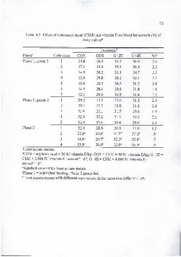
75
Table 4-5. Effect of cottonseed meal (CSM) and vitamin E on blood hematocrit (%) of
dairy calves^.
Treatment''
Phase'' Collection CON GOS G+2E G+4E SE'=
1 34.8 30.4 31.7 36.0 2.4
2 37.1 32.4 33.1 36.4 2.2
3 34.9 30.2 31.5 34.7 2.3
4 35.6 29.8 30.2 32.1 2.1
5 35.8 28.7 30.5 31.7 1.8
6 34.5 28.4 30.6 31.8 1.8
7 32.1 29.6 30.9 31.0 1.2
1 29.2 37.5 32.0 31.5 2.4
2 29.1 35.5 31.0 31.6 2.4
3 31.6 35.1 31.7 29.8 1.9
4 32.4 33.2 31.2 30.5 2.1
5 32.9 32.6 30.6 29.6 2.2
1 32.4 28.9 30.3 31.0 1.1
2 33.6^ 29.8^ 31.7^'' 33.2^ .9
3 34.0"= 29.7^ 32.3"' 33.8^ .7
4 33.9^ 30.9^ 33.9^ 35.3^ .9
Phase 1 ,group 1
Phase 1 ,group 2
Phase 2
^Least square means.
''CON = soybean meal + 30 lU vitamin E/kg; GOS = CSM + 30 lU vitamin E/kg; G+2E =
CSM + 2,000 lU vitamin E • animal ' • d '; G+4E = CSM + 4,000 lU vitamin E •
animal"' • d"'.
'^Standard error of the least square means.
''Phase 1 = individual feeding; Phase 2 group fed.
'''^Least square means with different superscripts in the same row differ P < .05.
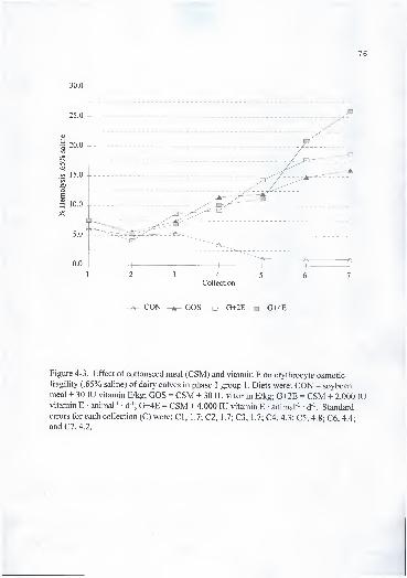
76
Figure 4-3. Effect of cottonseed meal (CSM) and vitamin E on erythrocyte osmoticfragility (.65% saline) of dairy calves in phase 1 group 1. Diets were: CON = soybeanmeal + 30 lU vitamin E/kg; GOS = CSM + 30 lU vitamin E/kg; G+2E = CSM + 2,000 lUvitamin E • animal ' • d '; G+4E = CSM + 4,000 lU vitamin E animal ' • d '. Standard
errors for each collection (C) were: CI, 1.7; C2, 1.7; C3, 1.7; C4, 4.3; C5, 4.8; C6, 4.4;
and C7, 4.2.

Figure 4-4. Effect of cottonseed meal (CSM) and vitamin E on erythrocyte osmoticfragility (.65% saline) of dairy calves in phase 1 group 2. Diets were: CON = soybeanmeal + 30 lU vitamin E/kg; GOS = CSM + 30 lU vitamin E/kg; G+2E = CSM + 2,000 lUvitamin E • animal ' • d '; G+4E = CSM + 4,000 lU vitamin E • animal ' d '. Standard
errors for each collection (C) were: CI, 2.2; C2, 3.3; C3, 2.6; C4, 6.1; and C5, 6.7.

78
Erythrocyte osmotic fragility analysis is an in vitro procedure that has not been
found to be related with any gossypol toxicity signs.
Plasma gossypol concentrations are shown in Table 4-6. In group 1 , (+)-, (-)-, and
total-gossypol were greater (P < .05) in CSM-fed calves than CON in collections 5 and 7.
G+2E calves had lower plasma gossypol concentrations than G+4E and GOS treatments.
In group 2, plasma concentrations of total gossypol and its isomers were greater in CSM
fed calves than CON in the last collection only. Calhoun et al. (1995b) suggested that
plasma gossypol reflects the availability of gossypol in the diet and the proportion of
isomers in the source being fed. Furthermore, they suggested 5 \ig/mL of total gossypol
in plasma as a safe upper level. In this study, plasma gossypol concentrations were below
this critical value. This range was proposed for adult dairy cattle and may not indicate
the safe upper level for young ruminants. The rumen plays a key role in the metabolism
of gossypol, and preruminants or ruminants fed gossypol in a manner which by-passes
the rumen absorb FG similarly to nonruminants (Calhoun, 1995).
Phase 2 . Phase 2 of this experiment included data on animals group fed in 1
6
pens (4 pens per treatment). Final weight, total gain and ADG (Table 4-2) were greater
(P < .05) for calves receiving CSM diets than the CON treatment. Calves fed G+4E
consistently had greater (P < .05) feed intake (Table 4-7) than CON calves but similar to
calves fed GOS and G+2E diets. There was not differences (P > .1) in feed intake
calculated as percentage of body weight. Calculated FG intake did not differ (P > .1)
among animals receiving CSM. Calculated vitamin E intake was similar (P > .1)
between calves fed the CON and GOS, but lower (P < .05) than calves fed the
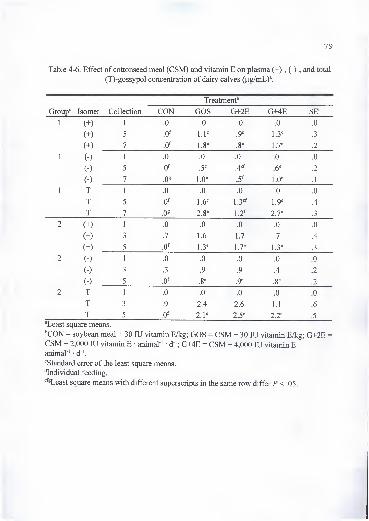
79
Table 4-6. Effect of cottonseed meal (CSM) and vitamin E on plasma (+)-, (-)-, and total
(T)-gossypol concentration of dairy calves {\ig/mLf.
Treatment''
Lrroup Isomer Collection (j+zb
11
1.U .u .u .U n
Of 1 1^ Qe1 .J
1.J
7 nf.u 1 fie
1 .6fie.8 1 7e
.z
11
11 0 0 n .U
n
K ) -J .J4ef
.D .z
( \\-) 7
/ 1 .ucf.J 1 He 1
. 1
11 T1 1 .u .u
A.U
T J 1 6«1 .J 1 Q«
1 .7 .'t
17/
O oe 1 Tfl.Z 1.1 .3
2 (+) 1 .0 .0 .0 .0 .0
(+) 3 .7 1.6 1.7 .7 .4
(+) 5 .0^ 1.3^ 1.7^ 1.3^ .3
2 (-) 1 .0 .0 .0 .0 .0
(-) 3 .3 .9 .9 .4 .2
(-) 5 .0^ .8^ .9-= .8"= .2
T 1 .0 .0 .0 .0 .0
T 3 .9 2.4 2.6 1.1 .6
T 5 2.P 2.5^ 2.2*= .5
"Least square means.
''CON = soybean meal + 30 lU vitamin E/kg; GOS = CSM + 30 lU vitamin E/kg; G+2E =
CSM + 2,000 lU vitamin E • animal ' • d '; G+4E = CSM + 4,000 lU vitamin E •
animal'' • d"'.
"Standard error of the least square means.
''Individual feeding.
^*^^Least square means with different superscripts in the same row differ P < .05.
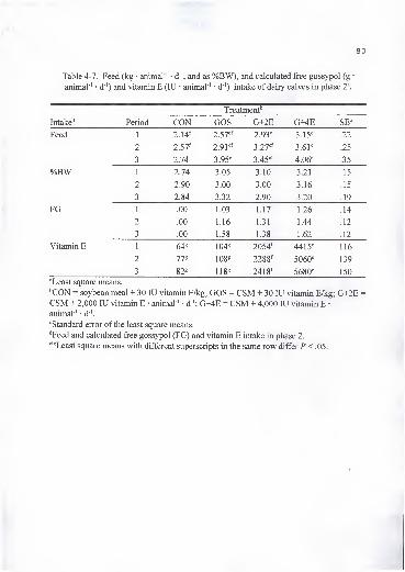
80
Table 4-7. Feed (kg • ammal"' d ", and as %BW), and calculated free gossypol (g•
animal"' • d"') and vitamin E (lU • animal"' • d"') intake of dairy calves in phase 2^.
Treatment'
intaice Period HJJN KJ+ZtL
Feed 11 9 14f
Z. It 9 ^7efZ.J /
9 Q'X^L.yj J. 1 J 99.zz
Z.J /9 01Z.7 1 X 97ef
J.Z / J.O 1 9<N.ZJ
*>
3 l.l'V 3.95^ 3.45" 4.06^ .35
%BW 1 2.74 3.05 3.10 3.21 .15
2 2.90 3.00 3.00 3.16 .15
3 2.84 3.32 2.90 3.20 .19
FG 1 .00 1.03 1.17 1.26 .14
2 .00 1.16 1.31 1.44 .12
3 .00 1.58 1.38 1.62 .12
Vitamin E 164S 104*^ 2054^ 4415^ 116
2 77« 1088 2288^ 5060^ 139
3 82^ 1188 24 18^ 5680^ 150
"Least square means.
''CON = soybean meal + 30 lU vitamin E/kg; GOS = CSM + 30 lU vitamin E/kg; G+2E =
CSM + 2,000 lU vitamin E • animal"' • d"'; G+4E = CSM + 4,000 lU vitamin E •
animal ' • d"'.
'^Standard error of the least square means.
''Feed and calculated free gossypol (FG) and vitamin E intake in phase 2.
^*^8Least square means with different superscripts in the same row differ P < .05.
t
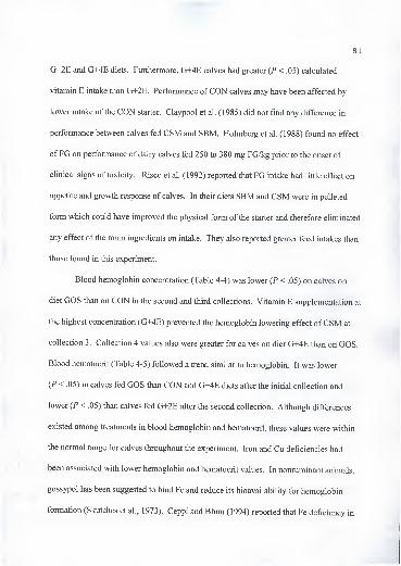
81
G+2E and G+4E diets. Furthermore, G+4E calves had greater (P < .05) calculated
vitamin E intake than G+2E. Performance ofCON calves may have been affected by
lower intake of the CON starter. Claypool et al. (1985) did not find any difference in
performance between calves fed CSM and SBM. Holmberg et al. (1988) found no effect
ofFG on performance of dairy calves fed 250 to 380 mg FG/kg prior to the onset of
clinical signs of toxicity. Risco et al. (1992) reported that FG intake had little effect on
appetite and growth response of calves. In their diets SBM and CSM were in pelleted
form which could have improved the physical form of the starter and therefore eliminated
any effect of the main ingredients on intake. They also reported greater feed intakes than
those found in this experiment.
Blood hemoglobin concentration (Table 4-4) was lower (P < .05) on calves on
diet GOS than on CON in the second and third collections. Vitamin E supplementation at
the highest concentration (G+4E) prevented the hemoglobin lowering effect ofCSM at
collection 3. Collection 4 values also were greater for calves on diet G+4E than on GOS.
Blood hematocrit (Table 4-5) followed a trend similar to hemoglobin. It was lower
(P < .05) in calves fed GOS than CON and G+4E diets after the initial collection and
lower (P < .05) than calves fed G+2E after the second collection. Although differences
existed among treatments in blood hemoglobin and hematocrit, these values were within
the normal range for calves throughout the experiment. Iron and Cu deficiencies had
been associated with lower hemoglobin and hematocrit values. In nonruminant animals,
gossypol has been suggested to bind Fe and reduce its bioavailability for hemoglobin
formation (Skutches et al., 1973). Ceppi and Blum (1994) reported that Fe deficiency in

82
calves decreased hemoglobin concentration and hematocrit percentage. However, Fe
deficiency does not cause an effect on EOF similar to gossypol (Lindsay et al., 1980).
They also suggested that gossypol effect on decreasing hemoglobin was independent of
gossypol-induced Fe deficiency. Gossypol has been found to inhibit several enzymes
including glucose-6-phosphate dehydrogenase, the rate limiting enzyme in the hexose-
monophosphate pathway (Bender et al., 1988). Inhibition of this enzyme can cause a
decrease in NADPH which is necessary to reduce glutathione, an important component of
the cell antioxidant system. Enzyme inhibition in this pathway or excess oxidants may
cause reduced hemoglobin and hematocrit values (Duncan et al., 1994). Therefore, the
effect of vitamin E found in this experiment may be related to the antioxidant role of
vitamin E.
Erythrocyte osmotic fragility at .65% saline solution as affected by treatment is
presented in Figure 4-5. Animals receiving CSM-based starters had greater (P < .05)
EOF in all collections than the CON calves. Feeding vitamin E did not reduce EOF.
Calhoun et al. (1992) reported that the positive association between malondialdehyde and
EOF indicated lipid peroxidation may be a factor in altering erythrocyte membrane
fragility. Furthermore, they suggested the peroxidation effect to be secondary to changes
in the structural integrity of the erythrocyte membrane. Results from this experiment
indicated that gossypol effect on erythrocyte fragility may be related to alteration of
membrane integrity through mechanisms not related to decreased antioxidant levels.
Alkaline phosphatase is a membrane bound enzyme used for diagnosis of bone
and liver disorders or drug exposure. Production ofAP is increased in response to
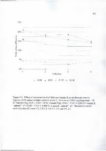
83
Figure 4-5. Effect of cottonseed meal (CSM) and vitamin E on erythrocyte osmotic
fragility (.65% saline) of dairy calves in phase 2. Diets were: CON = soybean meal + 30lU vitamin E/kg; GOS = CSM + 30 lU vitamin E/kg; G+2E = CSM + 2,000 lU vitamin E• animal ' • d '; G+4E = CSM + 4,000 lU vitamin E • animal"' • d '. Standard errors for
each collection (C) were: CI, 3.8; C2, 2.8; C3, 2.4; and C4, 2.3.

primary or secondary hepatocellular disorders. Liver degenerative changes and
hemorrhages are signs of gossypol toxicity. Although the reference range for AP is wide
for ruminants, changes in concentrations ofAP would indicate impaired liver function or
damage. In our experiment, we did not find any changes (P > .1) in plasma AP
concentration due to treatment (Table 4-8).
Plasma a-T concentrations (Figure 4-6) were similar iP> A) between calves fed
CON and GOS diets, while lower (P < .05) than calves on G+2E and G+4E diets. Calves
fed G+2E diet had lower (P < .05) plasma a-T concentrations than those fed G+4E in all
collections except collection 2. Ranking of calculated vitamin E intake (Table 4-7) by
diet was CON = GOS < G+2E < G+4E (P < .05). CON and GOS diets supplied equal
amounts of supplemental vitamin E. Therefore, at this level of supplementation feeding
CSM (400 mg FG/kg of diet DM) did not decrease plasma a-T concentration. Since
there was no treatment of high vitamin E and no CSM included in this experiment, it is
not possible to speculate on the effect of gossypol on plasma a-T concentration at high
levels of vitamin E supplementation. Gossypol seemed not to be related to lowering
plasma a-T concentrations as reported by Lane and Stuart (1990) and Willard et al.
(1995).
Mortality. During the experimental period, 10 calves died by causes suspected to
be treatment related. Six of these animals belonged to the GOS treatment and 2 each to
G+2E and G+4E with no deaths in the CON treatment (Tables 4-9 and 4-10). Intake of
FG by these animals at the time of death was fi-om .8 to 2.2 g/d.
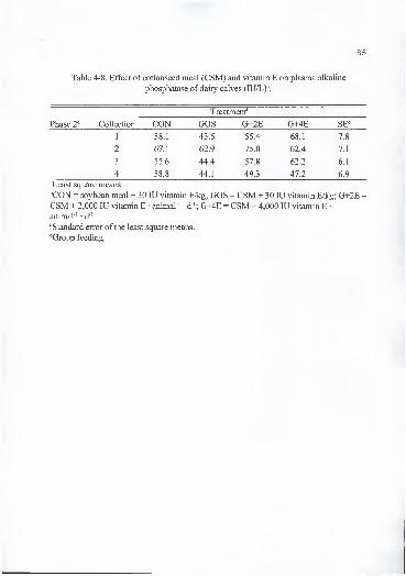
85
Table 4-8. Effect of cottonseed meal (CSM) and vitamin E on plasma alkaline
phosphatase of dairy calves (lU/L)".
Treatment''
Phase 1^ Collection CON GOS G+2E G+4E SE^
1 58.1 43.5 55.4 68.1 7.8
2 67.1 62.9 75.0 62.4 7.1
3 55.6 44.4 57.8 62.2 6.1
4 58.8 44.1 49.3 47.2 6.9
"Least square means.
''CON = soybean meal + 30 lU vitamin E/kg; GOS = CSM + 30 lU vitamin E/kg; G+2E =
CSM + 2,000 lU vitamin E animal ' d"'; G+4E = CSM + 4,000 lU vitamin E •
animal ' • d"'.
"Standard error of the least square means.
''Group feeding.

86
^ CON GOS G+2E G+4E
Figure 4-6. Effect of cottonseed meal (CSM) and vitamin E on plasma a -tocopherol
concentration of dairy calves in phase 2. Diets were: CON = soybean meal + 30 lUvitamin E/kg; GOS = CSM + 30 lU vitamin E/kg; G+2E = CSM + 2,000 lU vitamin E •
animal ' • d '; G+4E = CSM + 4,000 lU vitamin E • animal ' • d '. Standard errors for eachcollection (C) were: CI, .4; C2, .4 C3, .3; and C4, .4.
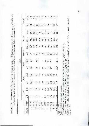
87
B
-aoo5
s03
(U>
otin
tl-
OPJ
oa
Ha
uCO.
HI
a
uCO.
8
UCO.
I
CO
OO
OO
o
o
in
o o in o(N (N inO
CT)
(N ^^ ^ (N
OO
in r<-) OO
in
ON
m ON
OOin
pin
00
ON
NO
ON<N
00 r-;
^ 00
in
COOO
OO
o o
m in (N
o o o o in
00
OnON
GOOo
in in Pin 00m r-H
00 (N rn^ <N
o m NOin in
in in p in pNO(N (N
inm (N
00 rn in
<^r—
H
in 00 in m o(N <N NO
in r~; 1
i> in1
1
11.1(N00
1
j
17.0
NO inin iri
p m mNO NO in
On r<-i ON in00 NO in
p ONOn Om
GOS
G+2 G+2 G+4 G+4
NOOO m NOm
NO
NO omNO
c
CO
inNO
>
I—
<
ooo
+
t/3
+
s
IXi
o. w
w • gCp ^ .ti
CO >
£2 0'='S |J-i (N
ffi ^ +
^ > u
W
i E O
^ t5 ^ §a 'i^ >-•
«^ >->
o
s
3
c'-4-»
oJJ
'oo--»
C0o—o-tCO
Om_ +
^ 2 V-5 GO T3O •
C3
GO 2
9 I
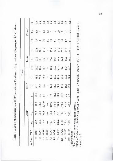
C3
sCO
t/3
1)
>
+
+
as
m asrnr<-i
o1 m CM m ro
ON
^ 00 u-i 1 r--
(N ^ (N r~; (NO 00 '^j- rn
r- O o00 1
o 00
1
°° ON 0000 On rn 00
(N (N (N IT) m (N
o o O ON IT) iri <r~i
M3 as1
<N od On vd OCM CN (N
rn oo1—
H
r<-i
iri O od
(N 00 o00 in
lO ooi> r-^
CN) --^ V-l
r-- O Tf;
o vdm r--
C/3 C/3 t/5 C/3
O O O Oo o o a
IT) (N r~-
00 ONONo 00
<Nas ON ON
m oo K
m 00On r-'in r<-i
on 00O OO O(N NO
OO
ONOn
mo
in
00 ONin Nd
OO in
m W PJ W+ + + +o o O Om NO NO o
rnNO
a — +
to^

Hematocrit and hemoglobin values were within normal range during the last
blood collection, and EOF (.65%) varied between 1.3 to 37.8%. Calculated FG intake per
kg BW/d before death was more than 10 mg FG kg ' BW • d '. Calhoun and Wang
(1995) reported that weaned lambs fed up to 30 mg FG • kg ' BW • d ' exhibit no sign of
gossypol poisoning while lambs fed 20 to 30 mg FG • kg ' BW • d"' mixed with the milk
replacer in order to by-pass the rumen died after 21 d of feeding. In another report, Kim
et al. (1996) reported similar results to the previous study when lambs were fed at 20 to
30 mg FG • kg"' BW • d"'. They also reported greater tissue gossypol concentrations of
fatally intoxicated lambs than those reported in this study (Table 4-10). In agreement
with the data of Kim et al. (1996), proportions of tissue gossypol enantiomers
accumulation were similar, with (-)-gossypol accumulating more than (+)-gossypol in
heart, and the reverse for liver. At this age, calves accumulated gossypol in testis which
may have subsequently affected reproductive parameters at puberty.
Plasma gossypol concentrations were below 5 |ig/mL in all animals except calf
33. The concentration of 5 [ig/mL has been reported as a safe upper level for adult dairy
cattle (Calhoun, 1995). Concentrations ofplasma and tissue a-T seemed to agree with
the treatment diets. Liver Cu concentrations ranged from 3 1 1 .7 to 865.7 mg/kg dry tissue
which were considered to be high for this class of animal (Netherlands Committee on
Mineral Nutrition, 1 973). de Peyster and Wang (1 993) reported that gossypol may
chelate Cu and Zn. Gossypol could have chelated Cu and Fe in the liver and reduced
their availability. Lower hemoglobin concentration found in calves of the GOS group
could have been the result of lower Cu and Fe availability or inhibition of enzymes

related with the antioxidant system; however, the increase in hemoglobin concentration
due to vitamin E supplementation supports the hypothesis of decreased antioxidants other
than a-T.
Bloat was determined to be the cause of death in calves 17 and 5 with no
significant finding upon necropsy except that calf 5 had a large thymic mass. Calf 5 had
the lowest concentration of heart and liver gossypol of all dead animals (Table 4-10).
Histopathological examination of heart and liver of these calves revealed a normal
condition for calf 17 and a mild lipoid vacuolation of cytoplasm of centrilobular
hepatocyte in calf 5 (Table 4-11).
Calves 6, 42, 486, and 33 had substantial accumulation of straw-colored fluid in
the abdominal and thoracic cavities. The heart of calf 42 had small areas ofhemorrhage
on the cardiac and pericardial surface. It was also slightly enlarged and the right
ventricular surface appeared congested. There were small petechial hemorrhage on the
tracheal mucosa surface. Similar findings were noted in calf 486 with pericardial fluid
noted and hemorrhage in the trachea. The only significant findings on the gross necropsy
of calf 3 was severe ascites.
Histopathological findings of heart and liver of the 10 dead calves are shown in
Table 4-11. The heart microscopic findings ranged from insignificant to severe atrophy
and fragmentation of cardiocytes, and vacuolation of cytoplasm. The liver examination
revealed fi-om insignificant damage to severe centrilobular necrosis.
In general, histological changes in the heart and liver were equivocal in most dead
animals, and had no consistent pattern with gossypol accumulation in tissue. However,

3'
91
Table 4-11 . Heart and liver histopathology of dead calves".
D AN TRT 0 Organ findings S
119 3 GOS H mild autolysis with several posmortem rods
L marked autolysis
124 5 GOS H eosinophilic and swollen cardiocytes with loss of +striations; pyknotic nuclei
L mild lipoid vacuolation of cytoplasm of centrilobular +hepatocytes
108 12 GOS H moderate vacuolation of sarcoplasm of cardiocytes -H-
L normal
95 17 GOS H normal •
L normal
91 42 GOS H myofiber atrophy and eosinophilia of cardiocytes; ++pyknosis of nuclei
L moderate centrilobular necrosis of hepatocytes ++
so GOS H moderate mterfascicular edema thinning of myofibers +
I moderate centrilobular and midzonal necrosis -H-
84 33 G+2E H atrophy of cardiocytes;moderate interfascicular edema; +++vacuolation of cytoplasm and nuclei
L severe centrilobular and midzonal necrosis of +++hepatocytes; vacuolation of cytoplasm of peripheral
hepatocytes
83 636 G+2E H normal
L mild centrilobular vacuolation of hepatocytes ;;•+'
65 6 G+4E H normal
L normal
119 630 G+4E H atrophy and fragmentation of cardiocytes; interfascicular +++edema; vacuolation of cytoplasm
L sever centrilobular and midzonal necrosis of hepatocytes +++"D = days on diet; AN = animal number; TRT = treatment: GOS = cottonseed meal (CSM) + 30 lUvitamin E/kg; G+2E = CSM + 2,000 lU vitamin E animal ' d"'; G+4E = CSM + 4,000 lU vitamin Eanimal' • d'; O = organ: H = heart, L = liver; S = degree of pathology: + mild, ++ moderate, +++ marked.
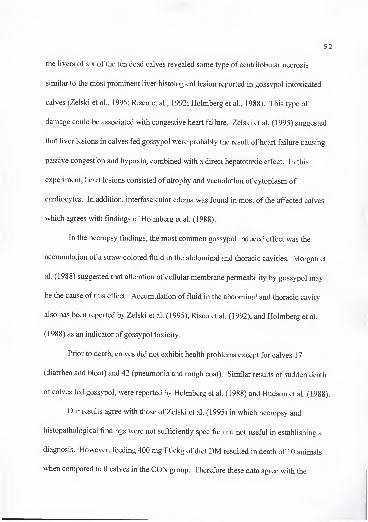
92
the livers of six of the ten dead calves revealed some type of centrilobular necrosis
similar to the most prominent liver histological lesion reported in gossypol intoxicated
calves (Zelski et al., 1995; Risco et al., 1992; Holmberg et al., 1988). This type of
damage could be associated with congestive heart failure. Zelski et al. (1995) suggested
that liver lesions in calves fed gossypol were probably the result of heart failure causing
passive congestion and hypoxia, combined with a direct hepatotoxic effect. In this
experiment, heart lesions consisted of atrophy and vacuolation of cytoplasm of
cardiocytes. In addition, interfascicular edema was found in most of the affected calves
which agrees with findings of Holmberg et al. (1988).
In the necropsy findings, the most common gossypol induced effect was the
accumulation of a straw-colored fluid in the abdominal and thoracic cavities. Morgan et
al. (1988) suggested that alteration of cellular membrane permeability by gossypol may
be the cause of this effect. Accumulation of fluid in the abdominal and thoracic cavity
also has been reported by Zelski et al. (1995), Risco et al. (1992), and Holmberg et al.
(1 988) as an indicator of gossypol toxicity.
Prior to death, calves did not exhibit health problems except for calves 1
7
(diarrhea and bloat) and 42 (pneumonia and rough coat). Similar results of sudden death
of calves fed gossypol, were reported by Holmberg et al. (1988) and Hudson et al. (1988).
Our results agree with those of Zelski et al. (1995) in which necropsy and
histopathological findings were not sufficiently specific and not usefiil in establishing a
diagnosis. However, feeding 400 mg FG/kg of diet DM resulted in death of 10 animals
when compared to 0 calves in the CON group. Therefore these data agree with the

93
hypothesis postulated by Risco et al. (1992) who reported that 400 mg FG/kg resulted in
mild toxicity in calves. Supplementation of high vitamin E did not result in necropsy or
histopathological findings different from those found in calves supplemented with normal
concentrations. Lindsey et al. (1980) suggested that the effect of gossypol may be more
pronounced if animals are exposed to some kind of stress. The role of vitamin E on
disease resistance has long been recognized, therefore vitamin E could have increased
resistance of these calves to diseases and therefore reduced the stress level (Table 4-12).
However, a direct interaction between vitamin E and gossypol can not be ruled out.
Implications
Cottonseed meal fed at a concentration to provide 400 mg FG/kg of diet DM was
associated with death in 10 calves. Necropsy findings were similar to those reported for
preruminants fatally intoxicated with gossypol. Vitamin E supplementation at high
concentrations increased feed intake and weight gain and may have conferred calves
some protection against gossypol toxicity.

94
Table 4-12. Effect of cottonseed meal (CSM) and vitamin E on health of dairy calves.
Treatment'
Item CON GOS G+2E G+4E
Days at risk'' 648 891 567 536
Respiratory treatmenf" .017 .026 .018 .017
Diarrhea treatment'' .015 .015 .023 .007
Total treated days'^ .032 .041 .041 .024
^CON = soybean meal + 30 lU vitamin E/kg; GOS = CSM + 30 lU vitamin E/kg; G+2E =
CSM + 2,000 lU vitamin E • animal ' • d '; G+4E = CSM + 4,000 lU vitamin E •
animal'" • d"'.
''Sum of days each calf was alive in each treatment.
'Values for respiratory, diarrhea, and total treated days are number of treated days divided
by days at risk for each treatment.
CHAPTER 5
LONG TERM EFFECTS OF FEEDING COTTONSEED MEAL AND VITAMIN E TODAIRY BULLS. I. HEMATOLOGICAL AND TISSUE PARAMETERS
Introduction
By-products of the cotton fiber and cottonseed oil industry such as whole
cottonseed (WCS) and cottonseed meal (GSM) are an important source of economical
protein in livestock rations. Gossypol [(2, 2'-binaphthalene) -8, 8'-dicarboxaldehyde-l,
r,6, 6', 7, 7'-hexahydroxy -5, 5'-diisopropyl -3, 3'-dimethyl] is a yellow polyphenolic
pigment found in cottonseeds. Gossypol found in cottonseeds is referred to as free
gossypol (FG), which is the form toxic to livestock. Bound gossypol (BG) has not been
found to be a major toxicant for livestock, but its role in animal toxicity is not yet fiilly
understood (Jones, 1991). Free gossypol is transformed to BG as the WCS are treated
with heat and pressure in the process of oil extraction. Cottonseed meal or any other by-
product of the cottonseed oil industry may be less toxic to livestock as their concentration
ofFG is lowered. However, Calhoun and Holmberg (1991) suggested that BG may be
released during the digestive process and have a physiological effect on the animal.
Janero and Burghardt (1988) suggested that gossypol can interact with biological
membranes by promoting the formation of highly-reactive oxygen-containing free
radicals. This and other studies (Willard et al, 1995; Lane and Stuart, 1990; Bender et
95
96
al., 1988) have suggested that antioxidants play a key role in the metabolism of gossypol.
Therefore, the objective of this phase of the experiment was to evaluate the effect of long
term feeding ofCSM fed to provide 14 mg FG • kg ' BW • d ' and the use of vitamin E to
counteract the negative effect of gossypol on hematological and tissue parameters in dairy
bulls from 6 to 1 5 mo of age.
Materials and Methods
Animals, diets and management. In a previous phase of the experiment (Chapter
4), the effect of feeding CSM (400 mg FG/kg of diet DM) and supplemental vitamin E on
calf performance from 2 wk of age to six mo was tested using four diets based on 1)
soybean meal (SBM) + 30 lU vitamin E/kg, 2) CSM + 30 lU vitamin E/kg, 3) CSM
supplemented with 700 lU vitamin E/kg of diet DM, and 4) CSM supplemented with
1400 lU vitamin E/kg of diet DM. A second phase of this experiment reported herein
utilized eight bulls for each of the following dietary treatments (Table 5-1): 1) CON a
supplement based on SBM + com + 30 lU vitamin E/kg, 2) GOS a supplement based on
CSM + com + 30 lU vitamin E/kg, and 3) G+4E a supplement based on CSM + com +
4,000 lU vitamin E • bull ' • d '. Supplements GOS and G+4E were formulated to supply
14 mg FG • kg BW • d"'. All supplements were isocaloric and isonitrogenous, and
satisfied animal requirements for all other nutrients (NRC, 1989). Animals were selected
to represent their previous diets. The bulls were housed in 12 pens, two animals per pen
and four pens per treatment, from 6 to 15 mo of age. The diets consisted of the treatment
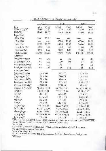
Table 5-1. Composition of dietary supplements''
97
£m QQS rT+4R
Item Tnitia] Final Initial Final Initial Final
Offered (ko^/d)'^ 2 60 5 60 2 70 6 00 7 70 6 no
DM (%) 88.00 88.00 88 00 88 00\J\J .\J\J KJ KJ .\j\J 88 00O O ,\J\J 88 00OO. v/v/
Ineredient''
SBM (%) 59.0 71.0
CSM (%) 6>1 00 80 00\J 1 t\J\J \j\J ,\J\J (\1 00\J 1 .\J\J RO 00
Com (%) 38.00 27.50 30.00 18.50 30.00 18.50
Limestone (9/n\ 1 001 .\J\J 50 1 00 ^01 .\J\J 1 on ^0
Minerals (%) 2.00 1.00 2 00 1 00 2.00 1.00
Vit.E (lU/kg) 30.00 30.00 30 00 30 00 1481.50 666.60
Analyses
C+VeossvDol .00 .00 33 40 .32 .40
f-VeossvDol (^/oY .00 .00 79 98 .77 .97
Free gossypol {%Y .00 .00 08 1
1
.08 .11
Total gossypol (%)'' .00 .00 1 06 116 1.06 1.14
Average values^
f+^-gOSSVDOl (%) .00 ± .00 32 ± 03 .32 ± .03
f-V20SSVt)0l (Vo) .00 ± .00 79 ± 08 .79 ± .08
Free eossvnol .00± .00 08 ± 01 .08 ± .01
Total eossvnol (Va\ .00 ± .00 1 02 ± 07 1.03 ±.07Vitamin A (lU/kg)*' 2076.61 2759 10 2759.10
Vitamin E nU/ke')''T X ^(h4Xxxxxx ' ' 1 X v_y / xvc^ / 38.84 ± 11.55 44 77 4- n 11/ _J -I- 1 J , 1 1 741.42 ± 182.90
CP {%Y 38.59 ±4.31 37.09 ±2.43Ca .47 ± .17 .48 ±.13 .42 ± .12
K 1.41 ±.44 1.42±.10 1.42 ± .36
Mg .22 ± .06 .57 ±.06 .55 ± 10
P .71 ±.08 1.12±.05 1.11 ±.08Cu (mg/kg)s 16.67 ±7.65 15.97 ±2.60 14.86 ±3.17Zn (mg/kg)s 69.33 ±28.39 73.29 ±11.77 70.52 ±13.85Mn (mg/kg)s 52.61 ±26.95 36.22 ±13.51 31.05 ± 13.55
Fe (mg/kg)8 182.59± 91.51 161.71 ±76.54 139.03 ±40.00Se (mg/kg)s .21 ± .11 .20 ± .04 .17± 04
'CON = soybean meal (SBM) + com + 30 lU vitamin E/kg; GOS = cottonseed meal (CSM) + com + 30 lUvitamin E/kg; G+4E = CSM + com + 4,000 lU vitamin E • animal ' • d '.
''As fed basis.
" As fed. HPLC procedure (Calhoun et al. (1995a), and Kim and Calhoun (1995)). Texas A&M.''As fed. AOACS procedure. Texas A&M.'Mean of 8 mixing ± SD.
^ As fed. A composited sample from all mixing dates. An lU/kg = Retinol acetate (ug/kg) * 2 9
1
8DM basis.

supplement and a free choice low quality hay (vitamin E concentration of <9.0 lU/kg).
Supplements were recalculated every month in order to provide 14 mg FG • kg ' BW •
d '. The protocol for bull procedures had been approved by the University Animal Use
Committee.
Blood and tissue sampling and analyses. Blood and plasma samples were ;
collected every 28 d (n = 10) via jugular venipuncture with an 18-gauge needle into
heparinized vacutainer blood collection tubes. Blood was analyzed for erythrocyte
osmotic fragility (EOF), hemoglobin, and hematocrit. Erythrocyte osmotic fragility,
measured as percentage hemolysis, was evaluated in .65 and .55% buffered saline
solution as described by Risco et al. (1993). Hemoglobin was determined using a
colorimetric procedure (Sigma Chemical Co., St. Louis, MO). Hematocrit was
determined from blood using a micro hematocrit centrifiige (lEC MB Centriftige,
Needham Heights, MA). Blood was centrifuged for 25 min at 700 x g. Plasma was
removed and stored at -20°C until analyzed for a -tocopherol (a-T), alkaline phosphatase
(AP), and creatine kinase (CK) (all collections), (+)-, and (-)-gossypol isomers
(collections 1, 6 and 10). At the end of the experiment animals were slaughtered and
portions of liver, heart, and testis were collected and frozen at -20°C until analyzed for
a-T, p -carotene (P-C), (+)-, (-)-, and total gossypol, Fe, Cu, and Zn. Liver samples
were also analyzed for retinol palmitate (RETP) and Se. Samples of supplements were
collected and analyzed for a-T, RETP, Ca, K, Mg, Mn, P, CP, Fe, Zn, Cu, and Se.
Alpha-tocopherol was analyzed following the procedure described by Njeru et al.
(1992) for plasma, and by Njeru et al. (1995) for tissue and feed samples. Tissue and

99
feed RETP, and P-C were prepared as follows: approximately 1 g of sample (fresh
weight) and . 1 g of ascorbic acid were homogenized in 1 0 ml of acetone; 1 ml of the
homogenate was deproteinized with 1 mL of ethanol. After adding 2 ml of .9% saline,
the mixture was vortexed for 6 min. Samples were double extracted with 3 mL petroleum
ether and kept in an ice bath. The petroleum ether extract was dried by evaporation under
a stream of in a 35 °C water bath, reconstituted in 750 /^L of .1% acetic acid, 29.9%
tetrahydrofuran, and 70% iso-octane with .2% added P -hydroxy toluene. The sample was
separated in equal amounts in two sealed vials one for RETP and the other vial for P-C
analysis. Retinol palmitate was determined by injecting 20 yuL of the reconstituted
extracted sample (tissue or feed) into the HPLC system. The HPLC systems consisted of
a Perkin Elmer 550 terminal (Perkin-Elmer, Analytical Instruments, Norwalk, CT.), an
ISS-100 auto sampler (Perkin-Elmer), a Series 4 Liquid chromatograph pump (Perkin-
Elmer), and a Lichrosorb Si 60 Sptm, 4 mm ID x 250 mm column (Hibar Fertigsaule RT
pre-packed column RT 250-4 E, Merck, Darmstadt, Germany). The mobile phase
consisted of 70% iso-octane, 29.9% tetrahydrofuran, and .1% acetic acid. The UV
detector was an ABI Analytical Spectroflow 757 set at a wavelength of 325 nm and
sensitivity of .005. Data were collected by a LCI-lOO Laboratory Computing Integrator
(Perkin-Elmer). Flow rate was 1 mL/min. The retention time ofRETP was 2.47 min.
Standards consisted of 10 ng of RETP (Sigma Chemical Co., St. Louis, MO). Analysis
of P-C was similar except that the mobile phase was 90% iso-octane, 9.9%
tetrahydrofuran, and .1% acetic acid; the wavelength was set at 450 nm, and the retention

; \ 100
time was 3.05 min. Standards consisted of 100 ng P-C (Sigma Chemical Co., St. Louis,
MO).
Stimulated lipid peroxidation was performed in heart, testis, and liver samples
according to a modification of the procedure of Kornbrust and Mavis (1980). Briefly,
approximately 1 g of fresh tissue was homogenized in 9 mL of 1 . 1 5% KCl. An aliquot
(100 |xL) was incubated in a water bath at 37 °C for the specified length of time (0, 50,
100, and 200 min) with 500 ^L of 8 mM Tris-malate buffer, 200 of 5 mM
FeS04-7H20 and 200 f^L of 2 mM ascorbic acid solutions. Peroxidation was terminated
by rapid addition of a 2 mL solution containing thiobarbituric acid (.375%) and 15%
trichloroacetic acid in .25 N HCl, and boiling in a water bath for 15 min. Samples were
then centrifliged for 1 5 min at 700 x g. The amount of colored product was measured
spectrophotometrically at 535 nm. Lipid peroxidation was expressed in terms of nmol
malondialdehyde (MDA)/mg of protein. Janero and Burghardt (1989) suggested that this
test can not be used as anything else other than an empirical indicator of membrane
oxidative injury, therefore care should be taken in the interpretation of these results.
Enzymes were determined using kits (CK:Sigma Diagnostic Procedure No. 520;
AP: Sigma Diagnostic Procedure No. 104). Minerals were determined according to the
procedures described by Rojas et al. (1995) and Whetter and Ullrey (1978). Samples of
plasma and tissue for gossypol analyses were shipped in dry ice to Texas A&M
University where they were analyzed by HPLC according to the procedures described by
Calhoun et al. (1995a), and Kim and Calhoun (1995).
101
Statistical analyses . Continuous data such as hematocrit, hemoglobin, EOF, lipid
peroxidation, plasma CK, AP, (-)-, (+)-, and total gossypol, and a-T were analyzed by
repeated measures analysis of variance in a completely randomized design using GLM
procedure of SAS (SAS, 1988). The Greenhouse-Geiser Epsilon was used to determine
significant levels of the F-test. Analyses of variables containing single or calculated
observations where performed by ANOVA (SAS, 1988). Treatment and pen within
treatment effects were tested using bull within pen and treatment effect. Pen within
treatment effect was removed from the model when it was not significant (P > .2). When
the pen within treatment effect was kept in the model PROC MIXED (SAS, 1996) was
used to calculate the correct standard errors of the least square means for a mixed model.
There were two sets of animals with an age difference of approximately 4 wk, therefore
age was used as a covariate. Tissue (+)-, (-)-, and total gossypol were analyzed with a
nested mixed analysis of covariace model by the PROC MIXED (SAS, 1996). where a 3
X 3 factorial design (three tissues x three isomers) occurs within each animal, where
animals (a random effect) were nested within treatments. There were nine measurements
within each animal (three gossypol values per each of three tissues) and they were
assumed to be equally correlated. When the overall treatment effect was significant {P <
.05) or tended to be significant (Z' < .1), separation of means was done using Duncan
multiple range test for all variables except gossypol tissue concentration for which the
Tukey test was used (available in PROC MIXED). Pooled standard errors were reported
when all treatments had the same number of replicates, otherwise individual values for
each least square mean were given.

102
Results and Discussion
Free gossypol intake in the animals on GOS and G+4E treatments, calculated
from the supplement analyses, ranged from 12.7 to 17 mg FG • kg " BW • d ' (from 2.2 to
6.5 g FG • animal"' • d"' ) during the 10 mo period. On average, supplements had the
targeted amount of vitamin E specified in the experimental protocol. Proportions of
various components of the supplements are shown in Table 5-1.
Animal performance and blood parameters. Animal ADG, total gain, initial and
final weights were not affected by treatment (Table 5-2). Vitamin E has been found to
increase ADG in lambs fed a diet containing 20% GSM (Calk et al., 1992). Here the
increase in ADG could have been due to a vitamin E effect on intake. Chase et al. (1 994)
found that bulls fed gossypol containing products (16 g FG • bull ' • d ') tended to have
reduced (P < . 1) body weight gains; however, this was attributed to a high fat content of
the supplements rather than a direct effect of gossypol on performance. Also, a decrease
in weight gain may have been caused by the effect of gossypol on digestive enzymes of
animals fed CSM or WCS. In vitro digestion of proteins by pepsin and trypsin was
reduced when gossypol was added prior to enzymatic digestion (Abou-Donia, 1 989). The
action of gossypol on the enzymes seems to be related to the binding of gossypol with the
e -amino group of lysine on the protein substrate or to the zymogen that could not be
converted to the active enzyme (Abou-Donia, 1989). Supplements used in the present

103
Table 5-2. Effect of cottonseed meal (CSM) and vitamin E on weight gain and average
daily gain (ADG) of dairy bulls^
Supplement*"
Item CON GOS G+4E SE^
Initial weight (kg) 163.4 164.3 178.6 4.3
Final weight (kg) 387.8 371.9 402.1 11.9
Weight gain (kg) 224.5 207.6 223.5 10.0
ADG (kg) 1.0 .9 1.0 .1
^Least square means.
''CON = soybean meal + com + 30 lU vitamin E/kg; GOS = CSM + com + 30 lU vitamin
E/kg; G+4E = CSM + com + 4,000 lU vitamin E • animal"' • d"'.
•^Standard error of the least square means.

104
experiment had a high CP concentration which could have increased gossypol fecal
excretion and decreased tissue deposition (Abou-Donia, 1976).
Erythrocyte osmotic fragility, as measured in .65 and .55% dilution (Figures 5-1
and 5-2), was greater (P < .001) at all collection periods for bulls fed GOS and G+4E
supplements than for the CON. Feeding 4,000 lU vitamin E • h"' • d"' did not reduce the
increase in EOF caused by gossypol as reported by Velasquez-Pereira et al. (1996c),
possibly because the animals in GOS and G+4E had been previously fed 400 mg/kg FG
from week 2 to 6 mo of age. Risco et al. (1993) found that Brahman bulls fed 8.2 g FG
or 16.6 mg FG • kg ' BW • d"' exhibited elevated EOF compared with controls. These
researchers also reported that gossypol caused erythrocyte acanthocytosis in three out of
eight bulls fed gossypol, which has been reported in conditions affecting the erythrocyte
membrane (Risco et al., 1993). Gray et al. (1993) reported increased EOF in heifers and'
cows consuming 10 or 20 g ofFG • animal ' • d '. Calk et al. (1992) reported that feeding
lysine or vitamin E did not protect against the effect of gossypol on EOF in lambs.
Gossypol has been found to intercalate into phospholipid bilayers, affecting the structure
and dynamics of membranes (Tanphaichitr et al., 1995). This property of gossypol may
affect the permeability of the erythrocyte membrane to anions causing the increase in
EOF. Although increased EOF has been reported as a clinical indicator of gossypol
intoxication in cattle (Calhoun et al., 1990; Lindsey et al., 1980), there are no reports
relating this laboratory procedure with clinical or reproductive signs of toxicity.
Feeding gossypol with and without supplemental vitamin E to bulls did not affect
hemoglobin concentration (Table 5-3), which was within the normal range throughout the

Figure 5-1. Effect of cottonseed meal (CSM) and vitamin E on erythrocyte osmotic
fragility in .65% saline. Supplements were: CON = soybean meal + com + 30 lU vitamin
E/kg; GOS = CSM + com + 30 lU vitamin E/kg; G+4E = CSM + com + 4,000 lUvitamin E • animal"' • d"'. Standard errors for each collection (C) were: CI, 2.5; C2, 2.1;
C3, 2.4; C4, 3.1; C5, 2.0; C6, 1.3; C7, 4.5; C8, 1.4; and C9, 1.5.
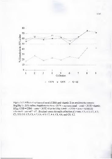
106
5 6Collection
CON GOS G+4E
Figure 5-2. Effect of cottonseed meal (CSM) and vitamin E on erythrocyte osmotic
fragility in .55% saline. Supplements were: CON = soybean meal + com + 30 lU vitamin
E/kg; GOS = CSM + com + 30 lU vitamin E/kg; G+4E = CSM + com + 4,000 lUvitamin E • animal ' d"'. Standard errors for each collection (C) were: CI, 4.3; C2, 4.1;
C3, 5.0; C4, 5.7; C5, 4.7; C6, 4.3; C7, 4.4; C8, 4.6; and C9, 4.2. r

107
o
U
s
o-oin
u u o in -1 m CNOD
Os_
in CNm CN 00 o OS CN CN m CNro
-o If
SOuo00 CN OS 00 sq 00 00
OS 00 COCN
O CTs CN CN CN
aMD
-o
'I-
-DOS OS oo 00 o CN sq OS o
00r<S
CNOm CN so CN rs CN CN CN rn
r<-i
o(N
Ur-
uin in 00 OS so CN
00sq
if
pOSCN
o(N (N
CN(N
o SO in CN SO in
VO
T3OS
U uoOS in so OS o r~; ro o
O<N
OSCN
OS as rn inm
ini> u
CN SO CN 00 so t>-osq
u
in(N
00CN
CN O CN CN inr<-) m CN
>ouin OS in OS r<-) OS
-o urn
O so(N
00CN
CN 00 r<S CN CN SOcn m m
OJO a> o CN 00 OS OS OS in ro OS pCO SO
CN roOS od rn CN CN
f—
^
so rnm
<NOS
w uOS OS r-- t~- OS m so
uOS
uO 00
>nO CN CN od OS OS SO rn
r'l
rsiuOS
uCN 00 OS o SO
T3OS
u VT3OS CN
m C?s
CNrn o o rn l>
m
OhDC/2
CON GOS G+4ECON GOS G+4E
CON GOS G+4ECON GOS G+4E
<
I00 o 00
.a
"3)
os
I6
cCO
lil
a
B
>Dooo
+Eou+
U
+abo^^Wc
"i
'>
Dom
oo+
UII
OOah
o —V Va, a.
o -a
S c
I iO O<J <J
S .s
o. cx
>
o
1)
C O
« T3
(1> <UO. D.3 3(/I «i
ti »-•
c a
> ?to to
E S^
to (33 3O- CT-to to
O to
u 55
to to

108
study. Bulls on the CON treatment had greater (P < .05) hematocrit percentage than
those on the GOS treatment in collections 1, 2, 4, and 5, and tended to be greater (P < .1)
for collections 7 and 10 (Table 5-3). There was no difference between the G+4E and
GOS or CON treatments in any of the collections except collection 2. Hemoglobin
concentration and hematocrit percentage have been reported to be lower in animals
receiving gossypol. Lindsey et al. (1980) reported that feeding 24.2 g ofFG from solvent
extracted CSM to lactating dairy cows decreased hemoglobin concentration and
hematocrit percentage. They also reported that the major physiological effects of
gossypol in lactating dairy cows were aherations in normal erythrocyte structure,
metabolism, and (or) function that affect hemoglobin, EOF, and hematocrit.
Furthermore, although Fe and Cu deficiencies can cause a decrease in hemoglobin and
hematocrit values, in this experiment Fe and Cu in the supplements (Table 5-1) met the
NRC (1989) recommendations and their concentrations in liver were within normal
ranges. Iron deficiency has been found to decrease hemoglobin concentration and
hematocrit percentage in calves (Ceppi and Blum, 1994), but not to cause an effect on
EOF similar to gossypol (Lindsey et al., 1980).
Alkaline phosphatase is a membrane bound enzyme used in the diagnosis of bone
and liver disorders and drug exposure. Production ofAP is increased in response to
primary or secondary hepatocellular disorders. Liver degenerative changes and
hemorrhage are signs of gossypol toxicity. Although the AP reference range for
ruminants is wide, changes in concentrations ofAP would indicate impaired liver
function or damage. In our experiment, plasma AP concentration (Table 5-3) decreased

109
{P < .05) in bulls fed the GOS and G+4E supplements. Gawai et al. (1995) reported that
gossypol depressed liver microsomal enzymes of rats given 5 mg of gossypol
intraperitoneally for 5 d. As AP is a membrane bound microsomal enzyme that is
produced rather than secreted when hepatic injury occurs, it is possible that gossypol may
have decreased the production of this enzyme. Further investigation is needed to
determine if gossypol inhibits the production ofAP and if concentrations of this enzyme
could be used in the diagnosis of gossypol toxicity. Lindsey et al. (1980) found that
feeding mature lactating dairy cows 251 to 273 mg of total gossypol (TG) • kg ' BW • d"'
from WCS and CSM for 14 wk did not cause liver damage.
Treatment did not affect {P> A) plasma CK concentration (Table 5-3). Creatine
kinase is an enzyme used in the diagnosis of muscular disorders. Gossypol has been
found to cause edema, hypertrophy and dilatation of heart and cardiac muscle
degeneration in several species including swine (Abou-Donia, 1989) and cattle (Kerr,
1989). This heart damage is similar to damage caused by Se/vitamin E deficiency.
Based on these enzyme concentrations, there was no evidence of muscle damage by
feeding gossypol at concentrations provided in this experiment.
Animals fed supplement G+4E had greater (P < .05) plasma a-T concentrations
than animals fed CON and GOS supplements at all collections (Figure 5-3). Although
CON and GOS supplements had the same added vitamin E (30 lU/kg of feed), animals
fed GOS had greater (P < .05) plasma a-T concentrations than animals receiving CON at
all collections except 5 and 6. At this low concentration of supplemental vitamin E,

110
10 -r
123456789 10
Collection
^ CON ^ GOS ^ G+4E
Figure 5-3. Effect of cottonseed meal (CSM) and vitamin E on plasma a-tocopherol
concentration of dairy bulls. Supplements were: CON = soybean meal + com + 30 lUvitamin E/kg; GOS = CSM + com + 30 lU vitamin E/kg; G+4E = CSM + com + 4,000
lU vitamin E • animal ' • d"'. Standard errors for each collection (C) were: CI, .3; C2, .2;
C3, .3; C4, .2; C5, .3; C6, .4; C7, .4; C8, .3; C9, .4; and CIO, .4.

Ill
gossypol did not decrease plasma a-T concentration. The increase in a-T concentrations
in animals fed GOS could be due to a greater a-T content ofCSM based supplements
than in SBM based supplements, or to an effect ofCSM on absorption of a-T. Since
there was no treatment with high vitamin E and no gossypol included in this experiment,
it is not possible to speculate on the effect of gossypol on plasma a-T concentration at
high levels of vitamin E supplementation. Contrary to our results, Lane and Stuart (1990)
reported that serum a-T and P-C were reduced in dairy cows fed high concentrations of
gossypol (approximately 40 g). In this study no statistical data were shown and gossypol
analysis may not represent the real values. In a more controlled study, Mena et al. (1996)
reported that gossypol from CSM, WCS or both (900 to 1800 mg total gossypol/kg)
sources did not reduce concentrations of antioxidant vitamins, and that in fact it increased
plasma a -tocopherol concentrations.
Willard et al. (1995) found that by d 84 cows given 4 g FG • animal"' • d ' with
elevated EOF had lower {P < .05) plasma a-T concentrations than cows not
supplemented. No supplemental vitamin E was given in the previous experiment. From
that experiment, four animals with the highest EOF were chosen from 1 7 animals
receiving 4 g FG • animal"' • d"' in a group fed experiment to determine plasma a-T
concentration. It is possible that the animals consuming more supplement (high EOF)
with low vitamin E content may have consumed less forage which is an excellent source
of vitamin E.
Gossypol enantiomers have been found to have different toxicological and
pharmacokinetic effects. Calhoun et al. (1995b) suggested that plasma gossypol

112
concentration reflects dietary gossypol availability and the proportion of isomers in the
source being fed. Furthermore these authors suggested that a plateau was reached after 4
to 6 wk of consumption and remained fairly constant until the diet was changed. Plasma
(-)-, (+)- and total gossypol concentrations as affected by dietary supplement are
presented in Table 5-4. Plasma (-)-, (+)-, and total gossypol concentrations in bulls fed
GOS and G+4E supplements were greater (P < .05) than bulls fed CON from the initial
sampling to the end of the experiment and were similar (P > . 1) among the treatments
receiving CSM. Plasma total gossypol concentration was lower than the safer upper limit
of 5 i^g/mL proposed by Calhoun et al. (1995b). Lower plasma total gossypol
concentrations were reported by Lindsey et al. (1980) when cows were fed 3.5 or 24.2 g
FG • animal ' • d"'. Also Lindsey et al. (1980) did not find differences in plasma gossypol
concentrations between cows fed the two levels ofFG intake raising a concern of using
FG in feed as a measurement of bioavailability. Calhoun et al. (1995b) reported that
dairy cows consuming from 27.2 to 33.8 g TG • animal ' • d"' from WCS had average
plasma gossypol concentrations of 3.0 |ag/mL with a range of 1.2 to 5.8 |ig/mL.
Tissue parameters . Tissue concentrations of (+)-, (-)-, and total gossypol are
shown in Figure 5-4. No differences (P > . 1) were found in tissue gossypol
concentrations between bulls fed GOS and G+4E supplements and they were greater (P <
.05) than bulls fed the CON supplement. Liver (+)-gossypol was greater (P < .01) than
(-)-gossypol concentration in bulls receiving CSM (GOS and G+4E). While heart and .
testis had greater (-)-gossypol (P < .05) than (+)-gossypol on bulls on the same
treatments. The order of (+)-gossypol accumulation in tissue was liver > heart and testis

113
Table 5-4. Effect of cottonseed meal (CSM) and vitamin E on plasma gossypol
concentration of dairy bulls (lag/mL)"
Supplement''
llCIIl
(-) gossypol 1 .OO'^ 1.22" 1.14" .08
6 .00"= 1.40" 1.35" .06
10 .00"= 1.53" 1.69" .11
(+) gossypol 1 .00^ 1.47" 1.28" .09
6 .00'= 1.68" 1.57" .06 " '
10 .00' 1.85" 1.77" .12
Total- 1 .00^ 2.69" 2.42" .17
gossypol 6 .00« 3.08" 2.91" .12
10 .OO" 3.37" 3.46" .22
"Least square means.
''CON = soybean meal + com + 30 lU vitamin E/kg; GOS - CSM + com + 30 lU vitamin
E/kg; G+4E = CSM + com + 4,000 lU vitamin E • animal ' • d '.
•^Standard error of the least square means.
"^Least square means with different superscripts in the same row differ P < .05.

114
Figure 5-4. Effect of cottonseed meal (CSM) and vitamin E on liver (L), heart (H), andtestis (T) (+)-, and (-)-gossypol concentrations. Supplements were: CON = soybean meal+ com + 30 lU vitamin E/kg; GOS = CSM + com + 30 lU vitamin E/kg; G+4E = CSM +corn + 4,000 lU vitamin E • animal"' • d"'; Standard errors for each tissue and isomerwere: L+, 7.8; L-, 5.3; H+, .7; H-, 3.5; T+, .6; and T-, 1.11.

115
(P < .05), and for (-)-, and total gossypol liver > heart > testis (P < .01). Supplementation
of vitamin E did not have an effect on (-)-, (+)-, or total gossypol accumulation in tissue.
The fact that gossypol damaged spermatogenic cells in cattle (Chenoweth et al.,
1 994) and that the testis accumulated less gossypol than the vital organs (i.e. liver and
heart), strongly suggests a specific vulnerability and sensitivity of testicular cells to the
action of gossypol.
The (-)-gossypol has been shown to be the optically active form that induces
fertility impairment in male animals (Wang et al., 1987). The lower accumulation of (-)-
gossypol in the liver relative to (+)-gossypol may be related to a greater (-)-gossypol
affinity for plasma proteins (Wu and Reidenberg, 1990). The liver is perfused by a fluid
similar to plasma which has greater protein concentration than the interstitial fluid found
in heart and testis (Joseph et al., 1986). Therefore, (-)-gossypol could be bound to
proteins at the time this protein rich fluid perfuses the liver and be redirected to other
tissues where it can interact with cellular components that had greater affinity to bind (-)-
gossypol than plasma protein. The (+)-gossypol may not be accumulated in tissues other
than liver where it seems to be eliminated due to its low specific interaction with cellular
components (Wang et al., 1992).
Similar results where (+)-gossypol concentration was greater in liver while (-)-
gossypol was greater in heart and muscle were reported in lambs fed 20 to 30 mg FG •
kg ' BW- d ' (Kim et al., 1996). In contrast to these results, the proportion of gossypol
isomers in liver has been shown to be similar in pigs fed a wide range of gossypol
containing diets (Knabe et al., 1995). In rats, similar deposition patterns and a gradual

116
accumulation of gossypol over time were found by Jensen et al. (1982). The major
excretory organ for gossypol appears to be the liver in several species including swine
(Abou-Donia and Dieckert, 1974) and rats (Abou-Donia et al., 1970). Abou-Donia
(1989) suggested a pathway for the metabolism of gossypol. Gossypol is absorbed from
the gastrointestinal tract, mainly the small intestine. It enters the liver via the hepatic
artery or through the lymph into the hepatic sinusoid and is taken up by Kupffer cells. In
the liver, gossypol is metabolized, conjugated, and excreted with the bile into the
duodenum. Some of the gossypol excreted is reabsorbed completing an enterohepatic
cycle that may be repeated several times leading to a gradual excretion via feces. The
mechanism of excretion seems to require an active secretory process that can be
saturated. Gossypol accumulation in liver may be related to its key role in the
metabolism of the toxicant.
Liver, heart, and testis concentration of p-C did not differ {P> A) among f
treatments (Table 5-5). Feeding CSM did not affect tissue deposition of p-C and RETP.
However, liver RETP concentration increased (P < .05) as vitamin E supplementation
increased (Table 5-5). It has been suggested that vitamin E protects retinyl esters in the
storage globules of the liver cells, preventing a rapid breakdown of retinyl esters stored in
liver (Sondergaard, 1972). Olson (1991) suggested that retinyl ester hydrolase, an
enzyme involved in the release of vitamin A from the liver, is regulated by vitamin E
status, activated in vitamin E deficiency and inhibited in vitamin E sufficiency. Vitamin
E supplementation (G+4E) increased (P < .05) a-T concentration in liver, heart, and
testis (Table 5-5). Greater (P < .05) a-T concentrations in heart and testis were found in

117
Table 5-5. Effect of cottonseed meal (CSM) and vitamin E on a-tocopherol (a-T),
retinol palmitate (RETP), and P -carotene (P-C) concentrations in tissue of dairy bulls
{[ig/g of fresh tissue)^
Supplement''
Tissue Item CON GOS G+4E SE'^
Liver a-T 3 .84= 6.10= 22.58" 1.00
RETP 1.63' 1.96= 3.70" .56
P-C 11.51 11.28 11.67 .64
Heart a-T 6.78^ 9.10= 26.03" .57
P-C 4.13 4.31 4.56 .20
Testis a-T 4.76^ 6.85= 16.4" .59
P-C 2.97 3.11 3.11 .22
"Least square means.
"CON = soybean meal + com + 30 lU vitamin E/kg; GOS = CSM + com + 30 lU vitamin
E/kg; G+4E = CSM + com + 4,000 lU vitamin E • animal ' d '.
"Standard error of the least square means.
"='^Least square means with different superscripts in the same row differ P < .05.
118
bulls fed GOS vs CON supplements at a normal concentration of vitamin E
supplementation (30 lU/kg). It has been suggested that a decrease in a-T due to chemical
toxicity is found only when the mechanism of cytotoxicity involves lipid peroxidation.
However, not all oxidative chemical toxicity involves a-T depletion and lipid _ . •
peroxidation (Anonymous, 1989).
Some trace minerals (e.g. Fe, Cu, Se, and Zn) have been implicated in gossypol
metabolism. Heart failure, seen in gossypol toxicosis, is also one of the clinical signs of
Se deficiency. Iron appears to be chelated by gossypol, reducing its bioavailability.
Gossypol also chelates Cu and Zn (de Peyster and Wang, 1993). In our experiment, no
differences (P > . 1 ) were found in heart and testis Cu, Fe, and Zn concentrations among
treatments (Table 5-6). However, liver concentrations of Fe and Se were greater (P <
.05) in bulls fed the GOS than the CON supplements. Accumulation of Fe in liver of
animals receiving G+4E was not different (P > .1) from bulls fed either the CON or GOS-
based diets. Liver Cu was greater (P < .05) in the G+4E than CON animals, but similar
to GOS, however, it was within the normal range of 200 to 600 |ig/g of dry tissue.
Contrary to our results, Yu et al. (1981) indicated that gossypol reduced Zn concentration
in testis of rats treated with gossypol, and suggested that gossypol chelates Zn and this
process may be related to gossypol antispermatogenic effects. Skutches et al. (1973)
suggested that gossypol chelates Fe in the liver of pigs, and reduces Fe availability for
hemoglobin synthesis. Abou-Donia (1989) indicated that the initial effect of gossypol in
swine may be to chelate Fe, thereby interfering with its utilization.

119
Table 5-6. Effect of cottonseed meal (CSM) and vitamin E on micromineral
concentration of liver, heart, and testis of dairy bulls (mg/kg dry matter)*
Supplement''
Tissue Mineral CON GOS G+4E SE'
Liver Cu 241.8' 353.3'*' 450.1'' 47.6
Fe 120.9' 215.3" 181.8"' 22.0
Se .5' 1.1'' .1
Zn 109.6 139.4 111.1 11.3
Heart Cu 15.2 23.9 14.7 7.0
Fe 188.9 182.0 161.2 21.8
Zn 61.0 92.9 66.3 12.6
Testis Cu 6.9 12.4 9.2 2.6
Fe 75.1 73.8 74.9 3.5
Zn 71.4 73.1 76.1 2.5
"Least square means.
''CON = soybean meal + com + 30 lU vitamin E/kg; GOS = CSM + com + 30 lU vitamin
E/kg; G+4E = CSM + com + 4,000 lU vitamin E • animal ' • d"'.
•^Standard error of the least square means.
'''Least square means with different superscripts in the same row differ P < .05.
120
In our experiment, although Fe was accumulated in the Uver, there was no
indication of Fe being unavailable for the synthesis of hemoglobin possibly due to greater
than requirement dietary concentrations. Selenium may be involved in the metabolism of
gossypol since feeding CSM to bulls increased its accumulation in the liver.
Gossypol has been reported to inhibit free radical damage to lipid membranes in
some studies (Janero and Burghardt, 1988) and to stimulate it in others (de Peyster et al.,
1984). Bender et al. (1988) suggested that oxidative injury caused by the generation of
reactive oxygen species may be the cause of gossypol toxicity. In the previous study,
they found a decrease in the concentration of antioxidants in testis of rats fed gossypol
acetic acid. Our data on in vitro stimulated lipid peroxidation in liver homogenates
(Figure 5-5) indicated that peroxidation of lipids in GOS and G+4E supplemented bulls
was lower {P < .05) than those supplemented with CON at all times. Heart homogenate
lipid peroxidation (Figure 5-6) was lower {P < .05) in G+4E than CON supplemented
bulls at 50, 1 00 and 200 min, and than GOS at 1 00 and 200 min of incubation. Bulls
supplemented with GOS had lower {P < .05) heart lipid peroxidation than CON at 50 and
100 min of incubation. In the testis (Figure 5-7), G+4E supplemented bulls had lower {P
< .05) lipid peroxidation than CON at all times (except 0) and lower (P < .05) than GOS
fed animals at 100 and 200 min of incubation. Consumption ofCSM (gossypol) did not
increase in vitro lipid peroxidation above those in the CON treatment in any of the tissues
tested. The difference in the degree of protection against lipid peroxidation of the GOS
treatment may have been due to different patterns of accumulation of gossypol
(liver>heart>testis). Sheriff et al. (1986) found similar results on the ability of gossypol
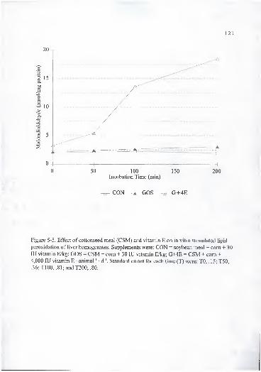
121
Figure 5-5. Effect of cottonseed meal (CSM) and vitamin E on in vitro stimulated lipid
peroxidation of liver homogenates. Supplements were: CON = soybean meal + com + 30
lU vitamin E/kg; GOS = CSM + com + 30 lU vitamin E/kg; G+4E = CSM + com +
4,000 lU vitamin E • animal ' • d"'. Standard errors for each time (T) were: TO, .15; T50,
.36;T100, .81;andT200, .80.
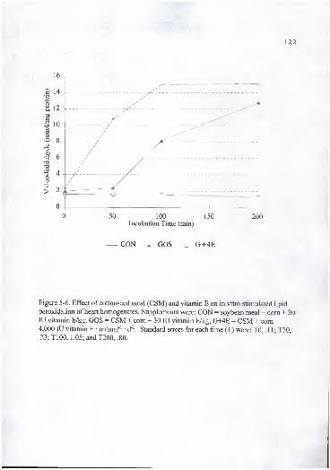
122
0 50 100 150 200Incubation Time (min)
^ CON ^ COS G+4E
Figure 5-6. Effect of cottonseed meal (CSM) and vitamin E on in vitro stimulated lipid
peroxidation of heart homogenates. Supplements were: CON = soybean meal + com + 30
lU vitamin E/kg; GOS = CSM + com + 30 lU vitamin E/kg; G+4E = CSM + com +4,000 lU vitamin E • animal"' d '. Standard errors for each time (T) were: TO, .1 1; T50,
.23;T100, 1.05; and T200, .80.

123
Figure 5-7. Effect of cottonseed meal (CSM) and vitamin E on in vitro stimulated lipid
peroxidation of testis homogenates. Supplements were: CON = soybean meal + corn + 30lU vitamin E/kg; GOS = CSM + com + 30 lU vitamin E/kg; G+4E = CSM + com +4,000 lU vitamin E • animal"' • d '. Standard errors for each time (T) were: TO, .65; T50,
3.75; TlOO, 4.10; and T200, 4.10.

124
to reduce lipid peroxidation in human spermatozoa and erythrocytes. Janero and
Burghardt (1988) found that gossypol effectively prevents Oj" dependant Fe-promoted
peroxidation of myocardial membrane phospholipids and could attenuate the kinetics and
extent of peroxidation. Furthermore, they suggested that the gossypol antioxidant effect
is through scavenging lipid radical intermediates and not by removing Fe from the system
through gossypol chelation, de Peyster et al. (1984) found that gossypol when incubated
with rat liver microsome and human sperm, promotes the formation of Oj' thus causing
oxidative injury. A possible explanation given to explain these conflicting results is that
a metabolic product of gossypol (such as redox-cycling quinone) may be responsible for
the ability of gossypol to generate oxygen radicals and cause the side effects associated
with gossypol treatment (Janero and Burghardt, 1988; de Peyster et al., 1984). Also,
gossypol may promote the formation of reactive oxygen species especially the production
ofhydrogen peroxide (de Peyster et al., 1984) that could lead to a partial depletion of
glutathione as seen in culture rat hepatocytes treated with gossypol (Barhoumi and •
Burghardt, 1996). Depletion of glutathione may not be accompanied with reduction in a-
T concentrations (Anonymous, 1989). " « . ^
Implications
Cottonseed meal fed to dairy bulls at a concentration provided in this experiment
(14 mg FG • kg-' BW • d ') was not detrimental to growth of animals. Tissue gossypol
concentration was not affected by vitamin E supplementation nor did gossypol

consumption decrease vitamin E concentration in plasma and (or) tissue. In the testis,
concentration of (-) gossypol, the isomer related to antifertility in males, was found in
greater concentration than (+)-gossypol.

CHAPTER 6
LONG TERM EFFECTS OF FEEDING COTTONSEED MEAL AND VITAMIN E TODAIRY BULLS. II. REPRODUCTIVE PARAMETERS
Introduction
Although whole cottonseed (WCS), and cottonseed meal (CSM) are important
sources of protein for ruminants, they contain the toxic polyphenolic pigment gossypol.
Gossypol has been found to adversely affect liver fiinction, erythrocyte oxygen carrying
or releasing capacity, respiration rate, feed intake and production and reproductive
capacity (Lindsey et al., 1980; Calhoun et al., 1990; Gray et al., 1990). Although
ruminants seem to have a large capacity to detoxify gossypol, toxicity resulting from
consumption of this compound has been observed. Gossypol intake by mature ruminants
may overwhelm ruminal detoxification and become absorbed at potentially toxic
concentrations (Randel et al., 1992).
In ruminants, gossypol has adversely affected sperm production associated with
a damage to the spermatogenic epithelium, leading to reduced germinal cell layers
(Arshami and Ruttle, 1988; Chase et al., 1989). Chase et al. (1989) reported delay in age
to puberty in Brahman bulls fed 60 mg free gossypol (FG) • kg"' BW • d"', but not when
fed 6 mg FG • kg ' BW • d '. After 5 wk of feeding gossypol the percentage of normal
spermatozoa was lower in bulls fed 8.2 g FG than in control bulls (Risco et al., 1993).
126

127
Bender et al. (1988) found that several antioxidants were reduced by feeding rats
high concentrations of gossypol. In dairy cattle, Lane and Stuart (1990) found that
feeding high amounts of gossypol decreased plasma a -tocopherol (a-T) concentrations
indicating a possible relationship between gossypol toxicity and vitamin E.
The present study was designed to evaluate the long term feeding of cottonseed
meal and the use of vitamin E to counteract potential gossypol effects on reproductive
development of dairy bulls.
Materials and Methods
Animals, diets and management. In a previous phase of the experiment (Chapter
4), the effect of feeding CSM (400 mg FG/kg of diet DM) and supplemental vitamin E on
calf performance from 2 wk to six mo of age was tested using four diets based on 1
)
soybean meal (SBM) + 30 lU vitamin E/kg, 2) CSM + 30 lU vitamin E/kg, 3) CSM
supplemented with 700 lU vitamin E/kg of diet DM, and 4) CSM supplemented with
1400 lU vitamin E/kg of diet DM. A second phase of this experiment reported herein
utilized eight bulls for each of the following dietary treatments (Table 6-1): 1) CON a
supplement based on SBM + com + 30 lU vitamin E/kg, 2) GOS a supplement based on
CSM + com + 30 lU vitamin E/kg, and 3) G+4E a supplement based on CSM + com +
4,000 lU vitamin E • bull ' • d"'. Supplements GOS and G+4E were formulated to supply
14 mg FG • kg BW • d"'. All supplements were isocaloric and isonitrogenous, and
satisfied animal requirements for all other nutrients (NRC, 1989). Animals were selected

Table 6- 1 . Composition of dietary supplements'*
£QN QQS rT+4F,
Item Initial Final Initial Final Initial Final
Offered (kg/d)" 2.60 5.60 2.70 6.00 2.70 6.00
DM (%) 88.00 88.00 88.00 88.00 88.00 88.00
Ingredient''
SBM (%) 59.0 71.0 — — — —CSM (%) — — 67.00 80.00 67.00 80.00
Com (%) 38.00 27.50 30.00 18.50 30.00 18.50
Limestone (%) 1.00 .50 1.00 .50 1.00 .50
Minerals (%) 2.00 1.00 2.00 1.00 2.00 1.00
Vit.E (lU/kg) 30.00 30.00 30.00 30.00 1481.50 666.60
Analyses
(+)-gossypol (%)'^ .00 .00 .33 .40 .32 .40
(-)-gossypol {%T .00 .00 .79 .98 .77 .97
Free gossypol (%)'' .00 .00 .08 .11 .08 .11
Total gossypol (%y .00 .00 1.06 1.16 1.06 1.14
Average values*
(+)-gossypol (%) .00 ± .00 .32 ±.03 .32 ± .03
(-)-gossypol (%) .00 ± .00 .79 ±.08 .79 ± .08
Free gossypol (%) .00 ± .00 .08 ±.01 .08 ±.01
Total gossypol (%) .00± .00 1.02 ±.07 1.03 ±.07
Vitamin A (lU/kg)'' 2076.61 2759.10 2759.10
Vitamin E (lU/kg)'' 38.84± 11.55 44.73 ± 13.11 741.42 ± 182.90
CP 38.59 ±4.31 37.89 ±2.03 37.09 ± 2.43
Ca .47 ± .17 .48 ±.13 .42 ± .12
K 1.41 ±.44 1.42 ±.10 1.42 ±.36
Mg .22 ± .06 .57 ±.06 .55 ± .10
P .71 ±.08 1.12 ±.05 1.11 ± .08
Cu (mg/kg)g 16.67 ±7.65 15.97 ±2.60 14.86±3.17
Zn (mg/kg)s 69.33 ±28.39 73.29 ± 11.77 70.52 ± 13.85
Mn (mg/kg)^ 52.61 ±26.95 36.22 ± 13.51 31.05 ±13.55Fe (mg/kg)s 182.59±91.51 161.71 ±76.54 139.03 ±40.00Se (mg/kg)s .21 ± .11 .20 ± .04 .17± .04
"CON = soybean meal (SBM) + com + 30 lU vitamin E/kg; COS = cottonseed meal (CSM) + com + 30 lUvitamin E/kg; G+4E = CSM + com + 4,000 lU vitamin E • animal ' • d '.
""As fed basis.
" As fed. HPLC procedure (Calhoun et al. ( 1 995a), and Kim and Calhoun ( 1 995)). Texas A&M.•As fed. AOACS procedure. Texas A&M."Mean of 8 mixing ± SD.
^ As fed. A composited sample from all mixing dates. An lU/kg = Retinol acetate (ng/kg) * 2.91
*DM basis.
129
to represent their previous diets. The bulls were housed in 12 pens, two animals per pen
and four pens per treatment, from 6 to 15 mo of age. The diets consisted of the treatment
supplement and a free choice low quality hay (vitamin E concenfration of <9.0 lU/kg).
Supplements were recalculated every month in order to provide 14 mg FG • kg'' BW • d '.
The protocol for bull procedures had been approved by the University Animal Use
Committee.
Testicular measurements and semen evaluation . Begirming at approximately 1
2
mo of age and continuing at weekly intervals for nine wk, a breeding soundness
evaluation (BSE) program was carried out on each bull as described by Chenoweth and
Ball, 1980). Semen was analyzed for sperm concentration and motility. Age at puberty
was determined when bulls provided an ejaculate containing 50 million sperm with at
least 10% showing progressive motility (Wolf et al., 1965). Once sperm production was
established, semen was assessed for morphological variation using counts of 200
spermatozoa stained with eosin-nigrosin and wet preparations examined with phase
contrast microscopy (Chenoweth et al., 1994). Evaluation consisted of percentage live
and normal spermatozoa, and percentage of spermatozoa with primary (proximal droplet,
abnormal acrosome, coiled tail, abnormal head, and abnormal midpiece), and secondary
(distal cytoplasmic droplet, kinked tail, and detached head) abnormalities. A small
amount of semen was added to buffered isotonic formal saline and this preparation was
examined for midpiece abnormalities using differential-phase (DIC) microscopy
employing a Zeiss Axioskop microscope (lOOOx). In the final semen collection, an
aliquot of semen was collected from four bulls per treatment, centrifuged and the seminal
• . : : 130
plasma separated and stored for later gossypol analysis following the procedure of
Calhoun etal. (1995a). '1
Before the end of the experiment each bull was administered 100 ^g ofGnRH
intramuscularly. Blood samples were collected 30 min prior to, just before and at 30, 60,
120, 180, 240, and 300 min after GnRH challenge. Plasma was separated by
centrifiigation and stored at- 20 °C for later analysis of testosterone (T) by
radioimmunoassay (Total testosterone Coat-A-Count® kit fi-om Diagnostic Product
Corporation). The intra-assay and inter-assay coefficients of variation were 6.5 and 9.2%
respectively, the total binding was 38% and the non specific binding was 1%. Basal
concentration of T was calculated as the average of - 30 and 0 min concentrations,
maximal concentration was the highest concentration measured after GnRH challenge, .
and the area under the curve was calculated from 0 to 300 min using the trapezoidal rule
(Murdoch and Dunn, 1982).
Libido test . Prior to the beginning and at the end of the BSE, bulls were evaluated
for sex-drive according to the technique described by Chenoweth (1983). Briefly, two
nonpregnant females of similar size to the bulls were placed in service crates located
approximately 5 m apart in a pen. Two bulls were admitted to the pen and observed for
10 min. This test was repeated the following day with, in most cases, different cohorts
and restrained females from the previous day. The following sexually-related activities
were recorded and timed: investigation, flehmen, licking, chin resting, incomplete mount,
complete mount, service and sexual inactivity. The following variables were estimated:
number of minutes during the test when sexual inactivity occurred (NDX), time to first

131
mount (TOM) and service (TOS), number ofmounts (NOM), and services (NOS). Bulls
were assigned a score for libido (LSC) based on sexual behavior and serving capacity,
where 0 represented no sexual interest and 10 represented two services followed by
renewed interest. The score then increased by 1 for each subsequent service (Chenoweth
etal, 1996).
At the end of the experiment, animals were sacrificed, and the following variables
measured: left and right testicular, and epididymal weight, left and right testicular
circumference, length, width, and depth. Paired testicular volume (V) was calculated as
the sum of the volume of the right and the left testicle using the following equation: V =
Tir^h, where r = (width + depth)/4 and h = length (Fields et al., 1979). Mid-parenchymal
portions of the right testicle were excised for sperm production estimation,
histopathological and electron microscopy (EM) examination.
The portions of testicle for histopathology were fixed in Bouins fixative,
dehydrated in alcohol, cleared in xylene and embedded in paraffin. Sections of 5 ^
thickness were cut and stained with haematoxylin-eosine and Fe haematoxylin.
The portions for EM were fixed in cold 2% glutaraldehyde in .2 M sodium
cacodylate buffer (pH 7.2-7.4) at the slaughter house. At the laboratory testicular tissues
were cut in pieces of 1 to 2 mm in size and left in the fixative for an additional hour.
They were then washed in buffer (10 min x 6), postfixed in 1% osmium tetroxide for 1 h,
washed for 30 min (3x10 min), dehydrated through an ethanol-acetone series (25, 50,
75, 100% ethanol, 100% Acetone), and embedded in Spurr's resin. Semen samples from
the last collection (5 mL) fi-om each bull were added to tubes containing 1 mL of fixative

132
as above to stabilize the tissue. In the laboratory the samples were spun down and the
pellets resuspended in fixative for 1 h. Pellets were washed in buffer, postfixed in 1%
osmium tetroxide (1 h), washed, dehydrated and embedded as were the testes. Thin
sections were cut on a RMC MT-6000 XL ultramicrotome, stained with 5% uranyl
acetate and lead citrate, then viewed on a Zeiss 10 transmission electron microscope.
Estimates of sperm production were made by counting elongated spermatids and
sperm in homogenized tissue as described by Chenoweth et al. (1994). Daily sperm
production per g ofparenchyma (DSPG) and daily sperm production (DSP) were
estimated using an hematocytometer and the following formula (Chenoweth et al., 1994):
DSPG = AX(B+Y)/5.32Y and DSP = DSPG (.99Z) where A = hematocytometer
constant, X = hematocytometer count, B = dilution factor, Y = parenchyma sample
weight, 5.32 = time divisor for Bos taurus (Amann et al., 1974) and Z = testis
parenchyma weight.
Statistical analvsis. Data were analyzed by least squares analysis of variance
using the GLM procedure of SAS (1988). Testicular and semen characteristics were
analyzed as a completely randomized design. Treatment and pen within treatment effects
were tested using bull within pen and treatment effect. Pen within treatment effect was
removed from the model when it was not significant (P > .2). When the pen within
treatment effect was kept in the model PROC MIXED (SAS, 1996) was used to calculate
the correct standard errors of the least square means for a mixed model. There were two
sets of animals with an age difference of 4 wk, therefore age was used as covariate. Due
to variability in the numbers of collections per animal, the weight statement of the GLM
133
procedure of SAS was used for semen characteristics. When the overall treatment effect
was significant (P < .05) or tended to be significant (P < A), separation of means was
done using Duncan multiple range test. Pooled standard errors were reported when all
treatments had the same number of replicates, otherwise individual values for each least
square mean were given.
Age at puberty was analyzed on the arcsin conversion of the proportion of animals
that reached puberty at each collection by treatment. A regression analysis was
performed and the 95% confidence intervals of the slope and intercept were compared.
Resuhs and Discussion
Calculated FG intake per animal in this trial ranged from 2.3 g at the beginning of
the experiment to 5.3 g at the end. Free gossypol concentrations of approximately 14 mg
• kg"' BW • d"' have been found to cause some reproductive problems in the male bovine
(Chase et al., 1994; Risco et al., 1993). Data on performance, blood, and tissue responses
were discussed in Chapter 5.
Testicular measurements and semen evaluation . Sperm morphology was
evaluated using classifications previously described (Chenoweth et al., 1994; Chenoweth
et al., 1993). Average percentage of normal and live sperm were lower (P < .05) in bulls
supplemented with GOS than in bulls fed G+4E and CON (Table 6-2). Risco et al.
(1993) found lower percentage of normal sperm in the semen of bulls fed 16.6 mg FG
kg"' BW • d"' and defects of sperm midpieces accounted for most of the abnormalities.
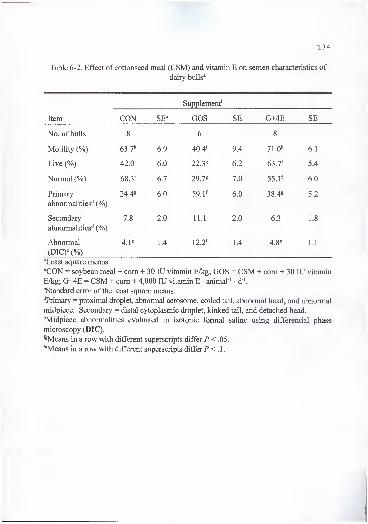
134
Table 6-2. Effect of cottonseed meal (CSM) and vitamin E on semen characteristics of
dairy bulls*
Supplement''
Item CON SE^ GOS SE G+4E SE
No. of bulls 8 6 8
Motility (%) 63.7" 6.9 40.4' 9.4 71.0" 6.1
Live (%) 42.0^ 6.0 22.38 6.2 63.7^ 5.4
Normal (%) 68.3^ 6.7 29.78 7.0 55.V 6.0
Primary
abnormalities'' (%)
24.48 6.0 59.1^ 6.0 38.4^ 5.2
Secondary
abnormalities'' (%)
7.8 2.0 11.1 2.0 6.3 1.8
Abnormal
(DIC)'^ (%)
4.18 1.4 12.2^ 1.4 4.88 1.1
^Least square means.
''CON = soybean meal + com + 30 lU vitamin E/kg; GOS = CSM + com + 30 lU vitamin
E/kg; G+4E = CSM + com + 4,000 lU vitamin E • animal ' • d"'.
^Standard error of the least square means.
""Primary = proximal droplet, abnormal acrosome, coiled tail, abnormal head, and abnormal
midpiece. Secondary = distal cytoplasmic droplet, kinked tail, and detached head.
^Midpiece abnormalities evaluated in isotonic formal saline using differential phase
microscopy (DIG).
'^Means in a row with different superscripts differ P < .05.
""Means in a row with different superscripts differ P < A.
Chase et al. (1994) did not find differences in sperm abnormalities when bulls
were fed approximately 6 mg FG • kg ' BW • d ' from CSM or 60 mg FG • kg ' BW • d '
from WCS. Sperm morphology is affected only when greater gossypol concentrations
than those used by Chase et al. (1994) are fed and in the form of CSM, since feeding high
gossypol diets in the form ofWCS has yielded inconsistent resuhs (Cusack and Perry,
1995; Smith et al., 1991; Arshami and Ruttle, 1988). Cusack and Perry (1995) reported
that bulls fed 20.4 to 50.8 mg FG • kg ' BW • d ' from WCS did not show any sign of
reproductive impairment. Similarly Smith et al. (1991) reported that feeding high
concentrations of gossypol (64 to 75 FG • kg"' BW • d"') from WCS did not affect sperm
characteristics or cause testicular degeneration. In contrast with these two experiments,
Arshami and Ruttle (1988) found that WCS, CSM and cottonseed hulls fed to bulls
caused testicular histological changes, indicating a detrimental effect on the
spermatogenic tissue and associated cells. Several mechanisms have been proposed to
explain these inconsistent results. 1) Cusack and Perry (1995) suggested that the feeding
ofWCS with gossypol chelating agents such as Ca, Na, protein, and trace minerals may
have affected the availability of gossypol in his and Smith et al. (1991) studies. 2)
Different concentrations of total gossypol and isomers are found in different species of
cotton and even within the same species grown in different environments, and 3) Calhoun
(1995) suggested that treatment of the WCS (i.e. ammoniation) can modify the pattern of
gossypol absorption. Therefore, differences in feeding practices and source of feed may
account for different effects among experiments.

136
The reduction of morphologically normal sperm was accompanied by a reduction
(/'<.!) on the percentage of motile sperm from bulls fed the GOS supplement (Table 6-
2). Feeding vitamin E (G+4E) counteracted the negative effect ofCSM on the percentage
of normal, live, and motile sperm. Chenoweth et al. (1994) found that the feeding of
relatively high concentrations ofFG to bulls did not adversely affect percentage of live
sperm despite a significant reduction in the number of morphologically normal sperm.
Although the concentration ofFG fed by Chenoweth et al. (1994) was similar to the
concentration fed in this experiment, the duration of the trial (10 wk vs 16 mo) may have
resulted in a more pronounced effect of gossypol on sperm, since they also observed
some differences in sperm motility between the control and the gossypol-treated bulls at
the end of the experiment. Reduction in sperm motility has been reported in several
animal species treated with gossypol (Randel et al., 1992; Wang et al., 1988). Chenoweth
et al. (1994) suggested that motility may be impaired by structural damage to the sperm
midpiece in animals treated with gossypol.
Primary abnormalities were found to be greater (P < .05) in bulls fed the GOS
supplement than in any other treatment, while secondary abnormalities were not affected
(P > .1) by treatment (Table 6-2). Primary abnormalities have not been affected in bulls
fed CSM or WCS in previous studies (Chase et al, 1994; Jimenez et al., 1989). All of the
primary defects considered in this experiment are found in the classification of Blom
(1973) as major morphological defects which have been correlated with impaired fertility.
Such sperm defects also have been associated with disturbed spermatogenesis (Barth and
Oko, 1989).

137
Percentage of abnormal midpieces, as evaluated with differential phase
microscopy was greater (P < .05) in GOS fed bulls than in the other two treatments
(Table 6-2). Although supplementation affected midpiece abnormalities, the percentage
of abnormal midpieces was lower than those found by Chenoweth et al. (1994). In this
study, the defect in the midpiece (segmental aplasia) was not consistent as reported by
Chenoweth et al. (1994) in Brahman bulls or by Oko and Hrudka (1982) in gossypol-
treated rats. However, electron microscopy from a bull sperm in the GOS treatment in
this study revealed gaps in the mitochondrial helix of the midpiece (Figure 6-1) as
reported in rats (Oko and Hrudka, 1982). This sperm defect has been associated with
malformations of the mitochondrial sheath which are induced in late spermatogenesis
(Chenoweth et al., 1994). These gaps in the mitochondria helix seem to be caused
specifically by gossypol and they are observed from late-stage spermatids through
epididymal spermatozoa. Also, Randel et al. (1992) suggested that this defect may be
responsible for the observed sperm motility reduction in animals treated with gossypol.
Electron microscopy of the testis revealed that extensive mitochondrial damage had
occurred in the testis of bulls supplemented with GOS (Figure 6-2), presented as a loss of
cristae and extensive vacuolization. Similar damage has been seen in tumor cells cultured
with (-)-gossypol (Benz et al., 1990) and in the testis of gossypol treated rats (20 mg FG •
kg " BW • d ') (Oko and Hrudka, 1982). Histological examination of the testes revealed
that 6 of 8 bulls supplemented with GOS had irregular and degenerative germ cell layers
that also showed vacuolation and reduced spermatid numbers. Vitamin E supplemented

1 um
Figure 6-1. Electron micrographs of bull sperm a) supplemented with cottonseed meal(GOS) revealing gaps in the mitochondria helix of the midpiece (magnification 19,430),
and b) supplemented with soybean meal (CON) (magnification 18,750).
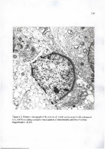
Figure 6-2. Electron micrograph of the testicle of a bull supplemented with cottonseed
meal (GOS) revealing extensive vacuolization of mitochondria and loss of cristae.
Magnification 18,200.

' ~ 140
bulls (G+4E) had less histological damage, with only one animal presenting vacuolization
and reduced spermatid numbers.
Several hypotheses have been proposed to explain the detrimental effect of
gossypol on male reproduction. 1) Gossypol inhibited the mitochondrial respiratory
chain in testicles (Reyes et al., 1988) which could impair sperm energy metabolism; 2)
Gossypol binds to proteins and amino acids, therefore potentially affecting synthesis of
proteins and their incorporation into spermatocytes and spermatids. Among others,
histidine has been found to be involved in the metabolism of gossypol (Javed and Waqar,
1 995); 3) Gossypol can alter the properties of lipid membranes which can modify protein
function and structure (Cuellar and Ramirez, 1993); 4) Structural integrity of many sperm
structures depend on S-S cross linked polypeptides. Disturbance of this bond may be
associated with damage to the midpiece mitochondria (Baccetti et al., 1986). Mann and
Mann (1981) reported that motility and metabolism of the sperm depends on interchange
reactions between S-H and S-S groups. Gossypol may prevent the oxidation of S-H
groups by chelating minerals or proteins responsible for this reaction (Chenoweth, P. J.
Personal communication); and 5) gossypol has been found to inhibit the generation of
free radicals in some studies (Janero and Burghardt, 1988) and to stimulate it in others (de
Peyster et al., 1984). Recently, Barhoumi and Burghardt (1996) reported that gossypol
promoted the formation of reactive oxygen species and the depletion of glutathione in rat
hepatocytes. Bender et al. (1988) reported a reduction in antioxidants in the testes of rats
fed gossypol. Damage due to lipid peroxidation in human sperm has been associated

141
with loss of sperm motility, inactivation of enzymes, and loss of membrane integrity
(Aitken and Fisher, 1994).
Daily sperm production per g ofparenchyma (10^/g testicular parenchyma) was
reduced (P < .05) in animals supplemented with GOS (Table 6-3). The same trend was
observed for DSP. Vitamin E supplementation (G+4E) counteracted (P < .05) the
reduction in sperm production caused by gossypol in CSM. In bulls, gossypol caused
damage to the spermatogenic epithelium which reduced the number of germinal cell
layers without reducing tubule diameter (Chase et al., 1990). Chenoweth et al. (1994)
found lower DSPG and DSP in bulls fed gossypol than in bulls receiving a diet without
gossypol. The depression of spermatogenesis is a known effect of gossypol on male
reproduction. Randel et al. (1992) suggested that a decrease in sperm production in
ruminants fed gossypol could be caused by damage to germ cells contained within the
germinal epithelium. Damage to Sertoli cells also may be involved with decreased
spermatogenesis.
In this experiment (Chapter 5, Table 5-6), gossypol did not affect testicular
concentrations of Cu, Fe, and Zn which were fed within required concentrations.
However, it does not imply that they were not chelated by gossypol and therefore
unavailable for normal metabolism.
In this study, feeding vitamin E to bulls receiving 14 mg FG • kg ' BW • d"'
increased the percentage of normal sperm, percentage motility, DSPG, and DSP and
decreased the percentage of primary and midpiece (DIC) abnormalities. Vitamin E may
have reduced the negative effect of gossypol on these parameters by reducing the damage

142
Table 6-3. Effect of cottonseed meal (CSM) and vitamin E on sperm production of dairy
bulls^
Supplement''
Item CON GOS G+4E SE
DSPG" (xlOVg) 14.6^ 10.28 17.6f1.0
DSP« (xlO')3.2f 2.28 4.1^ .3
^Least square means.
''CON = soybean meal + com + 30 lU vitamin E/kg; GOS = CSM + com + 30 lU vitamin
E/kg; G+4E = CSM + com + 4,000 lU vitamin E • animal"' • d"'.
"Standard error of the least square means. •
''Daily sperm production per g of parenchyma. "
•^Daily sperm production.
•^^Means in a row with different superscripts differ P < .05.

143
of reactive species of oxygen or by sparing the effect of enzymes involved in the
antioxidant system of the testicle. The effect of vitamin E in protecting the sperm
membrane against lipid peroxidation may be more important in late spermiogenesis when
sperm discard most of their cytoplasm leading to lowered concentrations of cytoplasmic
defensive enzymes (Aitken and Fisher, 1994). The effect of gossypol on the
spermatogenic epithelium may not be related to lipid peroxidation since in vitro testicular
lipid peroxidation was lower in animals fed CSM than CON, and feeding gossypol did
not reduce a-T concentration in testicles (Chapter 5). However, depletion of glutathione
as well as direct structural damage to the mitochondrial membrane could be caused by
gossypol. We could not explain the fact that only bulls in the GOS treatment in this
experiment had detectable concentrations of gossypol in seminal plasma (from .23 to
4.32 n-g/mL) but this may be related to the observed beneficial effect of vitamin E
supplementation on reproductive parameters of bulls fed gossypol-containing feedstuffs.
Table 6-4 shows the effect of the treatments on testicular characteristics of bulls.
Paired testicular and epididymal weights, paired testicular volume and scrotal
circumference were not affected by supplementation (/*>.!). However, animals
supplemented with G+4E showed the highest and those fed GOS the lowest testicular
weights and volumes. Chenoweth et al. (1994) found a similar trend where gossypol fed
bulls had the lowest testicular volumes and weights, but failed to find statistical
differences. These results are consistent with studies which did not find any effect of
gossypol on scrotal circumference and testicular weights (Chase et al., 1994; Chenoweth
et al., 1994; Jimenez et al., 1989). The apparent damage of gossypol to spermatogenic

144
Table 6-4. Effect of cottonseed meal (CSM) and vitamin E on testicular characteristics of
dairy bulls"
Supplement''
Item •' CON GOS G+4E SE'^
Paired testicle weight (g) 494.9 490.0 563.5 37.0
Paired testicle volume (cm^) 706.8 657.4 747.8 45.0
Paired epididymal weight (g) 48.3 41.8 50.9 3.1
Scrotal circunference (cm) 32.4 31.2 31.4 .8
"Least square means.
''CON = soybean meal + com + 30 lU vitamin E/kg; GOS = CSM + com + 30 lU vitamin
E/kg; G+4E = CSM + com + 4,000 lU vitamin E • animal ' d ".
''Standard error of the least square means.

145
epithelium has not been associated with reduction of scrotal circumference in the bovine.
This could be the result of a reduction in layers of the spermatogenic epithelium without
corresponding changes in tubule diameter (Chenoweth et al., 1994).
Percentage of animals that reached puberty (production of 50 X 10* sperm with at
least 10% motility) is shown in Figure 6-3. During the experimental period, four bulls
supplemented with GOS did not produce enough semen to be classified as pubertal.
Vitamin E feeding decreased (P < .05) and CSM feeding without supplemental vitamin E
increased (P < .05) age at sexual maturity in this study.
Results of the libido tests are presented in Table 6-5. Bulls fed GOS exhibited
more sexual inactivity {P < .05) at the first test than bulls in other treatments. Vitamin E
supplementation to bulls receiving gossypol (G+4E) improved NOM in the first test and
TOS in the second test. There was a trend of gossypol (GOS) to decrease and vitamin E
(G+4E) to improve LSC. The lower results of GOS group first test may indicate a
relative lack of sexual maturity which agrees with sperm production data. At time of first
test (12 mo of age), none of GOS, two of CON, and six of G+4E bulls had reached
puberty as per experimental protocol (ejaculate with 50 x 10* sperm and at least 10%
motility). The effect of vitamin E supplementation on some measurements of sexual
behavior may be related to its antioxidant properties and its accumulation in tissues with
high steroidogenic activity and thus high reactive oxygen species production such as
tesficular tissue. Taylor et al. (1991) reported that feeding gossypol to male rats (from 5
to 20 mg gossypol • kg'' BW • d"') for 1 1 wks provoked a dose-response decline in sexual
motivation. Even at a low dosage (5 mg gossypol • kg"' BW • d"'), males eventually
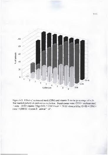
146
Figure 6-3. Effect of cottonseed meal (CSM) and vitamin E on the percentage of bulls
that reached puberty at each semen collection. Supplements were: CON = soybean meal
+ com + 30 lU vitamin E/kg; GOS = CSM + com + 30 lU vitamin E/kg; G+4E = CSM +com + 4,000 lU vitamin E • animal"' • d"'.

147
Table 6-5. Effect of cottonseed meal (CSM) and vitamin E on sex drive performance of
dairy bulls during two libido tests"
Supplement''
Item'^ Test CON SE*^ GOS SE G+4E SE
LSC 1 7.9 1.2 6.8 1.0 9.1 1.1
2 10.4 .7 8.9 .5 10.0 .6
NOM 1 5.7^^ 1.2 3.2^ 1.1 6.3" 1.2
2 9.4 1.3 9.5 .9 7.9 1.1
NOS 1 1.4' ;5•
1.4 1.9 .5
2 2.4 .4 1.7 .4 2.3 .4
TOM 1 156.1 60.0 224.5 57.0 135.0 57.0
(sec)2 38.0" 7.0 29.0"*' 5.0 21.
0^5.0
TOS 1 222.8 94.2 231.6 69.2 205.7 69.2
(sec)2 154.4"^ 71.4 212.8" 52.4 69.2^ 59.8
NDX 1 1.2" .8 3.9^ .7 1.7"^ .8
(min)2 .1 .2 .1 .1 .2 .2
^Least square means.
''CON = soybean meal + com + 30 lU vitamin E/kg; GOS = CSM + com + 30 lU vitamin
E/kg; G+4E = CSM + com + 4,000 lU vitamin E • animal ' • d '.
"LSC = libido score; NOM = number of mounts; NOS = number of services; TOM = time
to first mount; TOS = time to first service; NDX= sexual inaction time (min). Ten minutes
allowed per test.
''Standard error of the least square means.
"'^Means in a row with different superscripts differ P< A.
^''Means in a row with different superscripts differ P < .05.

148
showed signs of losing interest in sexually receptive females over weeks of
administration. Authors suggested that the likely mechanism for the behavioral changes
is a gradual suppression of serum testosterone, de Peyster and Srebnik (1988) found that
rats administered 10 mg gossypol • kg"' BW • d"' subcutaneously every 5 d had reduced
serum testosterone concentrations and decreased accessory organ weights. Possible
explanations for these results were that gossypol may cause a direct suppression of
steroidogenic enzymes, alteration ofcAMP second-messenger function, or interference
with cell membrane properties which could result in a disruption ofnormal receptors in
the interstitial cell membrane (de Peyster and Srebnik, 1988).
Chase et al. (1994) reported that bulls fed gossypol (6 or 60 mg FG • kg ' BW •
d ') from CSM and WCS did not show treatment differences in the basal mean or
maximal, number of pulses or total T released during 6 h of blood sampling. Likewise, in
this experiment, neither the maximal GnRH-induced concentration of T nor the area
under the GnRH-induced T curve differed (/*>.!) among treatments (Table 6-6 and
Figure 6-4). There was a trend for gossypol to reduce and vitamin E to increase T
production after GnRH challenge. Lin et al. (1980) reported that rat Leydig cells cultured
with 10-' and lO"'' M of gossypol had reduced LH stimulated production of T. In canine
testicular interstitial cells, Mushtaq et al. ( 1 996) reported that gossypol inhibited hCG-
stimulated T production and suggested that gossypol may interfere with hCG binding to
its receptor and(or) subsequent signal transduction or hCG-dependent enzymatic activity.
Vitamin E effect on the metabolism of gossypol has not yet been investigated.
However, vitamin E seemed to confer protection against the detrimental effects of
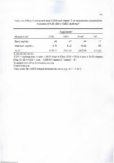
149
Table 6-6. Effect of cottonseed meal (CSM) and vitamin E on testosterone concentration
in plasma of bulls after a GnRH challenge^
Supplement''
Measurement CON GOS G+4E SE*^
Basal (ng/mL) .64 .47 .64 .15
Maximal'' (ng/mL) 9.74 8.42 10.60 .90
AUC^ 2176.77 1933.14 2457.64 213.50
^Least square means.
''CON = soybean meal + com + 30 lU vitamin E/kg; GOS = CSM + com + 30 lU vitamin
E/kg; G+4E = CSM + com + 4,000 lU vitamin E animal ' d"'.
"^Standard error of the least square means.
''GnRH induced.
^Area under the GnRH induced testosterone curve (ng • mL"' • min ')
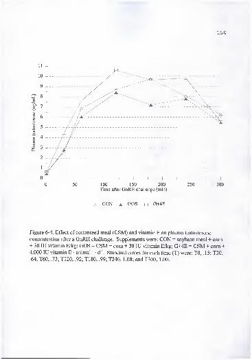
150
11 -1-
0 . 50 . 100 150 200 250 300Time after GnRH challenge (min)
^ CON A GOS ^ G+4E
Figure 6-4. Effect of cottonseed meal (CSM) and vitamin E on plasma testosterone
concentration after a GnRH challenge. Supplements were: CON = soybean meal + com+ 30 lU vitamin E/kg; GOS = CSM + com + 30 lU vitamin E/kg; G+4E = CSM + com +
4,000 lU vitamin E • animal"' • d '. Standard errors for each time (T) were: TO, .15; T30,
.64; T60, .73; T120, .92; T180, .99; T240, 1.08; and T300, 1.00.
151
gossypol on sperm production and quality. The possible mechanism involved may be
related to protection against reactive oxygen species produced by gossypol, or by sparing
the effect of other antioxidants affected directly by gossypol (i.e. GSH), or by direct
physical interaction of gossypol or its metabolites with a-T in lipid membranes. The
latter supposition is supported by the fact that no gossypol was present in seminal plasma
of G+4E animals.
Implications
Cottonseed meal fed to Holstein bulls at a concentration to provide 14 mg FG •
kg"' BW • d"' from 6 mo to 16 mo of age resulted in increased sperm abnormalities,
decreased sperm production, and adversely affected some aspects of sexual behavior.
Vitamin E feeding at a concentration of 4,000 lU • h"' • d'' reversed the negative effects of
gossypol on reproductive measurements.

CHAPTER 7
SUMMARY AND CONCLUSIONS
By-products of the cotton fiber and cottonseed oil industry such as whole
cottonseed and cottonseed meal (CSM) are important sources of economical protein and
energy in livestock rations. Gossypol [(2,2'-binaphthalene)-8,8'-dicarboxaldehyde-
l,r6,6',7,7'-hexahydroxy-5,5'-diisopropyl-3,3'-dimethyl] is a yellow polyphenolic
pigment found in cottonseed. This pigment is recognized to be toxic to nonruminants and
at high enough concentrations, to ruminants, limiting the use ofwhole cottonseed and its
by-products as feed to livestock.
There are few data showing the effect of supplemental vitamin E in counteracting
gossypol toxicity. At the University of Florida, a series of experiments were undertaken
to evaluate the value of supplemental vitamin E on the physiological response to CSM
(gossypol) fed to ruminants and young ruminants prior to rumen development.
Experiment 1 was conducted for 1 12 d with yearling beef heifers to evaluate the
effects of feeding CSM with different concentrations of vitamin E on hematological and
tissue components. Thirty-two yearling beef heifers (n=8) were assigned randomly to four
dietary treatments: 1) CON a supplement based on soybean meal (SBM) + corn + 30 lU
vitamin E/kg; 2) GOS a supplement based on CSM + com + 30 lU vitamin E/kg; 3)
G+2E a supplement based on CSM + com + 2,000 lU vitamin E animal ' • d '; and 4)
152
153
G+4E a supplement based on CSM + com + 4,000 lU vitamin E animal'' • d '.
Cottonseed meal was fed to provide 4 g of free gossypol (FG) animal"' • d"'. Blood
samples were collected at the start of the experiment and every two wk thereafter up to 16
wk. There was a time x treatment interaction (P < .01) for plasma a -tocopherol (a-T)
concentration; however, feeding gossypol did not reduce plasma a-T. Weight gain,
retinol palmitate, retinol, p -carotene (P-C), hemoglobin and hematocrit were not
affected by treatment. Erythrocyte osmotic fragility (EOF) was increased (P < .05) in
gossypol fed animals; however, vitamin E supplementation lowered EOF. Heifers fed
supplements GOS, G+2E, and G+4E had greater (P < .01) (-), (+), and total gossypol than
CON from collection 2 (after two wk of feeding the supplements) up to the end of the
experiment. There was a treatment effect (F < .05) on vitamin E and gossypol
concentrations in different tissues, while not significant {P > .05) for trace minerals (Cu,
Zn, Fe, and Se). Vitamin E concentration in tissue increased with increased dietary
supplementation. In heart and neck muscle (-)-gossypol was greater (P < .05) than (+)-
gossypol, while the reverse was true for liver. Gossypol decreased in vitro lipid
peroxidation of liver homogenates and did not have any effect on vitamin concentration
of tissue. Gossypol pattern of disposition in tissue was liver > heart > muscle.
In experiment 2, newborn male Holstein calves were used to test the effect of 400
mg FG/kg of feed DM and to determine if vitamin E supplementation could counteract
gossypol toxicity. The experiment was divided in two phases: 1) individual feeding, and
2) group feeding. The treatments were as follow: CON: SBM based starter; GOS: CSM
based starter; G+2E: CSM based starter + 2,000 lU vitamin E • calf • d '; and G-I-4E:

154
CSM based starter + 4,000 lU vitamin E • calf • d '. Vitamin E (G+4E) improved (P <
.05) weight gain and feed intake over CON in most periods. Plasma gossypol
concentrations were greater (P < .05) in calves receiving CSM than in CON calves;
however, there was no difference among those calves which received CSM. Hemoglobin
and hematocrit were decreased (P < .05) in GOS fed calves, whereas vitamin E
supplementation counteracted (P < .05) this effect. Plasma a-T concentration was not
affected (P > . 1 ) by gossypol intake and followed the supplementation pattern. During
the experimental period, 10 calves died, 6 from the GOS and 2 each from the G+2E and
G+4E treatments. Here, necropsy findings from some calves were compatible with
gossypol toxicity. Histopathological examination revealed centrilobular necrosis in the
liver and atrophy and vacuolation of cardiocytes.'
Experiment 3 was conducted to determine the effect of long term feeding ofCSM
on hematological, tissue and reproductive parameters of Holstein bulls. Twenty four
Holstein bulls, approximately 6 mo of age were placed on the following dietary
treatments: 1) CON a supplement based on SBM + com + 30 lU vitamin E/kg , 2) GOS a
supplement based on CSM + com + 30 lU vitamin E/kg, and 3) G+4E a supplement
based on CSM + com + 4,000 lU vitamin E • bull"' • d"'. Cottonseed meal based diets
were formulated to supply 14 mg FG • kg"' BW • d"'. These animals had been in
experiment 2 which evaluated the effect of feeding the same type of diets but from one
wk to 6 mo of age. Blood and weight were collected every mo for a total of 10
collections. Animal ADG, total gain, and final weights were not affected by treatment.
Erythrocyte osmotic fragility was greater (P < .01) in bulls fed GOS and G+4E compared
155
to the CON treatment. Feeding 4,000 lU vitamin E • bull"' • d"' did not reduce the
increase in EOF caused by gossypol as in experiment 1 . The CON supplemented bulls
had greater (P < .05) hematocrit percentage than the bulls fed GOS in collections 1, 2, 4,
and 5, and tended to be greater (P < .1) in collections 7 and 10. Animals receiving G+4E
had greater (P < .05) plasma a-T concentration than animals in the CON and GOS
treatments in all collections, and animals in GOS treatment had greater (P < .05) plasma
a-T concentration than animals in CON in all collections except 5 and 6. Plasma (-)-,
(+)-, and total gossypol concentrations in bulls fed GOS and G+4E were greater (P < .05)
than CON fed bulls from the initial sampling to the end of the experiment, and were
similar (P > .1) among treatments receiving CSM. Percentage motility, normal, and live
sperm, and daily sperm production were lower (P < . 1) in bulls fed GOS than in the other
two treatments. Percentage of primary abnormalities and abnormal midpieces were
greater (P < .05) in the GOS supplemented bulls than in the other two treatments. At 12
and 16 mo of age bulls were given two assessments for sex-drive traits. Bulls receiving
gossypol (GOS) exhibited less sexual activity (P < .05) at the first test than bulls in other
treatments. Vitamin E supplementation to bulls receiving gossypol improved the number
of mounts in the first test and time to first service in the second test. There was a trend
for gossypol to decrease and vitamin E to improve libido score. The results of GOS first
test may indicate a lack of sexual maturity of bulls fed the GOS supplement which agrees
with sperm production data. At time of first test (12 mo of age) none of GOS, two of
CON, and six of G+4E bulls had reached puberty as per experimental protocol. Vitamin
E supplementation did not affect plasma or tissue gossypol concentration. Cottonseed

meal seemed to aid in the absorption and deposition of a-T in animals fed a normal
concentration of vitamin E. Gossypol pattern of disposition in tissue was liver > heart >
testis. In vitro lipid peroxidation of tissue indicates that gossypol acts as an antioxidant in
lipid peroxidation systems and its role as an antioxidant may be dose or tissue dependant.
Vitamin E supplementation reduced lipid peroxidation in heart and testis below the level
of the other two treatments. Feeding CSM to dairy bulls did not affect growth, nor did it
affect vitamin E status. Gossypol negatively affected some reproductive measurements
of Holstein bulls; however vitamin E supplementation counteracted the detrimental effect
of gossypol.
In conclusion, in experiment 1, gossypol fed at 4 g FG • animal"' • d"' at low
concentration of vitamin E supplementation (30 mg/kg of feed) did not decrease
concentrations of antioxidant vitamins, including a-T, vitamin A and P-C or have any
detrimental effect on performance of beef heifers. In experiment 2, CSM fed at a
concentration to provide 400 mg FG/kg of diet DM resulted in death of 10 calves with
necropsy findings similar to those reported for fatally intoxicated preruminants. Vitamin
E supplementation at high concentrations increased feed intake and weight gain and may
have conferred some protection upon calves against gossypol toxicity. In experiment 3,
CSM fed to dairy bulls at a concentration provided in this experiment (14 mg FG • kg"'
BW • d"') was not detrimental to growth of animals. Tissue gossypol concentration was
not affected by vitamin E supplementation, nor did gossypol consumption decreased
vitamin E concentration in plasma and (or) tissue. In the testis, concentration of (-)
gossypol, the isomer related to antifertility in males, was found in greater concentration

157
than (+)-gossypol. Gossypol increased sperm abnormalities, decreased sperm production,
and adversely affected some aspects of sexual behavior. Vitamin E feeding at a
concentration of 4,000 lU • h"' • d"' reversed the negative effect of gossypol on selected
reproductive measurements.

LIST OF REFERENCES
Abou-Donia, M. B. 1976. Physiological effect and metabolism of gossypol. Residue Rev.
61:125-160.
Abou-Donia, M. B. 1989. Gossypol. In: P. R. Cheeke (Ed.) Toxicants of Plant Origin.
Vol. IV: Phenolics. pp. 1-22. CRC Press, Boca Raton, FL.
Abou-Donia, M. B., and J. W. Dieckert. 1974. Urinary and bilary excretion of '""C-
gossypol in swine. J. Nutr. 104:754-760.
Abou-Donia, M. B., C. M. Lyman, and J. W. Dieckert. 1970. Metabolic fate of gossypol:
the metabolism of '''C-gossypol in rats. Lipids 5:938-946.
Abou-Donia, M. B., M. A. Othman, and P. Obih. 1988. Interspecies comparison of
pharmacokinetic profile and bioavailability of (±)-gossypol in male Fisher-344
rats and male B6C3F mice. Toxicology 55:37-51.
Aitken, J., and H. Fisher. 1994. Reactive oxygen species generation and humanspermatozoa: the balance of benefit and risk. BioEssays 16:259-267.
Amann, R. P., J. F. Kavanaugh, L. C. Griel, and J. K. Voglmayr. 1974. Spermproduction of Holstein bulls determined from testicular spermatid reserves, after
cannulation of rete testis or vas deferens, and by daily ejaculation. J. Dairy Sci.
74:93-104.
Anonymous. 1989. Protein thiols and alpha-tocopherol in chemical cytotoxicity. Nutr.
Rev. 47:150-151.
Arshami, J., and J.L. Ruttle. 1988. Effects of diets containing gossypol on spermatogenic
tissue of young bulls. Theriogenology. 30:507-516.
Baccetti, B. E., A. Bigliardi, T. Burrini, T, Renieri, and G. Selmi. 1986. The action of
gossypol on rat germinal cells. Gam. Res. 13:1-17.
Baran, N. 1., and A. I. Ismailov. 1993. Biological activity of gossypol and its derivatives.
Chem. Nat. Compounds. 29:334-349.
158
159
Barhoumi, R., and R. C. Burghardt. 1996. Kinetic analysis of the chronology of patulin-
and gossypol-induced cytotoxicity in vitro. Fundam. Appl. Toxicol. 30:290-297.
Barth, A. D., and Oko, R. 1989. Abnormal Morphology of Bovine Spermatozoa. Iowa
State University Press, Ames, lA.
Bender, H. S., S. Z. Derolf, and H. P. Misra. 1988. Effects of gossypol on the antioxidant
defense system of the rat testis. Arch. Androl. 21 :59-70.
Benz, C. C, M. A. Keniry, J. M. Ford, A. J. Townsend, F. W. Cox, S. Palayoor, S. A.
Matlin, W. N. Halt, and K. H. Cowan. 1990. Biochemical correlates of the
antitumor and antimitochondrial properties of gossypol enantiomers. Mol.
Pharmacol. 37:840-847.
Blom, E. 1973. The ultrastructure of some characteristics sperm defects and a proposal
for a new classification of the bull spermiogram. Nord. Vet. Med. 25:383-391
.
Bressani, R. A., L. G. Elias, and J. E. Braham. 1964. Studies in dogs on the absorption of
gossypol from cottonseed flour containing vegetable mixture. J. Nutr. 83:209-213.
Calhoun, M. C. 1995. Understanding and managing gossypol in cattle diets. In: Proc.
Southwest Nutr. Manag. Conf pp. 17-26. Ahwatukee, AZ.
Calhoun, M. C, J. R. Clark, J. Simoni, J. E. Huston, S. W. Kuhlmann, and C. B. Calk.
1992. Effects of gossypol on osmotic fragility of erythrocytes and plasma
hemoglobin and malondialdehyde levels of lambs. J. Anim. Sci. 70(Suppl.
l):32(Abstr.).
Calhoun, M., and C. Holmberg. 1991. Safe use of cotton by-products as feed ingredients
for ruminants: a review. In: L. A. Jones, D. H. Kinard, and J. S. Mills (Eds.)
Cattle Research with Gossypol Containing Feeds, pp. 97-129. National
Cottonseed Products Association. Memphis, TN.
Calhoun, M. C, J. E. Huston, S. W. Kuhlmann, B. C. Baldwin, Jr., B. S. Engdahl, and K.
W. Bales. 1990. Free gossypol intake and erythrocyte fragility of lambs fed
cottonseed meal processed by different methods. J. Anim. Sci. 68(Suppl. 1):53
(Abstr.).
Calhoun, M. C, K. S. Kelton, S. W. Kuhlmann, E. McDonald, and H. L. Kim. 1995a.
Total, (+)- and (-)-gossypol in serum of holstein cows. J. Anim. Sci. 73(Suppl.
1):33 (Abstr.).
160
Calhoun, M. C, S. W. Kuhlmann, and B. C. Baldwin. 1995b. Assessing the gossypol
status of cattle fed cotton feed products. In: Proceedings from the Pacific
Northwest Nutrition Conference, pp. 1-14. Seattle, WA.
Calhoun, M. C, and P. J. Wang. 1995. A procedure for assessing gossypol availability.
Symposium on Available Gossypol in Cottonseed Products. Annual Meeting,
Amer. Oil Chem. Soc, San Antonio, TX. May 7-21.
Calk, C. B., S. W. Kuhlmann, M. C. Calhoun, J. E. Huston, B. C. Baldwin, Jr.. and B. S.
Engdahl. 1992. Effect of dietary iron, lysine, and vitamin E on performance and
erythrocyte fragility of lambs fed expander solvent cottonseed meal. J. Anim. Sci.
70(Suppl. 1):33 (Abstr.). . . ^ = » -
Cass, Q. B., E. Tiritan, S. A. Matlin, and E. C. Freire. 1991. Gossypol enantiomer ratios
in cotton seeds. Phytochemistry 30:2655-2657.
Ceppi, A., and J. W. Blum. 1994. Effect of growth hormone on growth performance,
haematology, metabolites and hormones in iron-deficient veal calves. Zentralbl
Veterinarmed B. 41:443-449.
Chase, Jr. C. C, Arshami, J. L., L. Ruttle, R. D. Randel, and C. R. Long. 1990.
Histological characteristics of testes from Brahman bulls fed diets containing
gossypol. J. Anim. Sci. 68(Supp. l):14(Abstr.).
Chase, Jr. C. C, J. Bastidas, L. Ruttle, C. R. Long, and R. D. Randel. 1989.
Reproductive development and function of Brahman bulls fed diets containing
gossypol; growth and sperm parameters near puberty. J. Anim. Sci.
67(Supp.l):368. (Abstr.).
Chase, Jr. C. C, J. Bastidas, L. Ruttle, C. R. Long, and R. D. Randel. 1994. Growth and
reproductive development in Brahman bulls fed diets containing gossypol. J.
Anim. Sci. 72:445-452.
Chen, Q. Q., H. Chen, and H. Lei. 1987. Comparative study on the metabolism of optical
gossypol in rats. J. Ethnopharmacol. 20:31-37.
Chenoweth, P. J. 1983. Examination of bulls for libido and breeding ability. Veterinary
Clinics of North America: Large Animal Practice. 5:59-74.
Chenoweth, P. J., and L. Ball. 1980. Breeding Soundness Examination of Bulls. In: D. A.
Morrow (Ed.) Current Therapy in Theriogenology. Pp. 330-339. W. B. Saunders
Co. Philadelphia, PA.
Chenoweth, P. J., C. C. Chase, R. E. Larsen, M. D. Thatcher, J. F. Bivens, and C. J.
Wilcox. 1996. The assessment of sexual performance in young Bos taurus and
Bos indicus beef bulls. Appl. Anim. Behav. Sci. 48:225-236.
Chenoweth, P. J., F. M. Hopkins, J. C. Spitzer, and J. C. Larsen. 1993. Guidelines for
using bull breeding soundness evaluation form. Theriogenology Handbook B-10.
Chenoweth, P. J., C. A. Risco, R. E. Larsen, J. Velez, N. Shaw, C. C. Chase and T. Tran.
1994. Effects of dietary gossypol on aspects of semen quality, sperm morphology
and sperm production in young brahman bulls. Theriogenology 42:1-13.
Claypool, D. W., C. H. Hoffman, J. E. Oldfield, and H. P. Adams. 1985. Canola meal,
cottonseed, and soybean meals as protein supplements for calves. J. Dairy Sci.
68:67-73.
Cuellar, A., and J. Ramirez. 1993. Further studies on the mechanism of action of
gossypol on mitochondrial membrane. Int. J. Biochem. 25: 1 149-1 1 55.
Cusack, P. M., and V. Perry. 1995. The effect of feeding whole cottonseed on the fertility
of bulls. Aust. Vet. J. 72:463-466.
de Peyster, A. A., P. A. Hyslop, C. E. Kuhn, and R. D. Sanerheber. 1986. Membrane
structural/functional perturbations induced by gossypol. Biochem. Pharm.
35:3293-3300.
de Peyster, A. A. Quintanilla, L. Packer, and M. T. Smith. 1984. Oxygen radical
formation induced by gossypol in rat liver microsomes and human sperm.
Biochem. Biophys. Res. Commun. 118:573-579.
dePeyster, A. A., and H. H. Srebnik. 1988. Reproductive endocrine fimction in gossypol-
treated male rats. Int. J. Fertil. 33:362-371.
de Peyster, A. A., and Y. Y. Wang. 1993. Genetic toxicity studies of gossypol. Mutation
Res. 297:293-312.
Duncan, J. R., K. W. Prasse, and E. A. Mahaffey. 1994. Veterinary Laboratory Medicine.
Clinical Pathology. Iowa State University Press, Ames, IW.
Eicher, S. D., J. L. Morril, F. Blecha, C. G. Chitko-McKown, N. V. Anderson, and J. J.
Higgins. 1994. Leukocyte function of young dairy calves fed milk replacers
supplemented with vitamin A and E. J. Dairy Sci. 77:1399-1407.

t.-t (-
162
Eicher, S. D., J. L. Morrill, F. Blecha, J. J. Higgins, N. V. Anderson, and P. G. Reddy.
1992. Neutrophil and lymphocyte response to supplementation with vitamin Cand E in young calves. J. Dairy Sci. 75:1635-1642.
Fields, M. J., W. C. Bums, and A. C. Wamick. 1979. Age, season and breed effects on
testicular volume and semen traits in young beef bulls. J. Anim. Sci. 48:1299-
1306.
Gawai, K. R., C. Cox, J. Jackson, and R. R. Dalvi. 1995. Changes in the activity of
metabolic and non-metabolic enzymes in rats following co-administration of
gossypol with phenobarbital. Pharmacol. Toxicol. 76:289-291
Gray, M. L., L. W. Greene, and G. L. Williams. 1993. Effect of dietary gossypol
consumption on metabolic homeostasis and reproductive endocrine fimction in
beef heifers and cows. J. Anim. Sci. 71:3052-3059.
Gray, M. L., R. D. Randel, L. W. Greene, and G. L. Williams. 1990. Metabolic
homeostasis and reproductive endocrine fiinction in postpubertal beef heifers fed
varying levels of dietary free gossypol. J. Anim. Sci. 68(Suppl. 1):465 (Abstr.).
Gu, Y., Y. C. Lin, Y. Rikihisa. 1990. Inhibitory effects of gossypol on steroidogenic
pathways in culture bovine luteal cells. Biochem. Biophys. Res. Commun.169:455-461.
Heinstein, P. F., D. L. Herman, S. B. Tove, and F. H. Smith. 1970. Biosynthesis of
gossypol. J. Biol. Chem. 245:4658-4665.
Heinstein, P. F., F. H. Smith, and S. B. Tove. 1962. Biosynthesis of '^C-labeled gossypol.
J. Biol. Chem. 237:2643-2650.
Hidiroglou, N, N. Cave, A. S. Atwal, E. R. Famwoth, and L. R. McDowell. 1992.
Comparative vitamin E requirements and metabolism in livestock. Ann. Rech.
Vet. 23:337-359.
Hoffmann-La Roche Inc. 1994. Vitamin Nutrition for Ruminants. Animal Nutrition andHealth. Hoffmann-La Roche Inc. Nutley, NJ.
Holmberg, C. A., L. D. Weaver, W. M. Guterbock, J. Genes, and P. Montgomery. 1988.
Pathological and toxicological studies of calves fed high concentration cottonseed
meal diet. Vet. Pathol. 25:147-153.
Hudson, L. M., L. A. Kerr, and W. R. Maslin. 1988. Gossypol toxicosis in a herd of beefcalves. J. Am. Vet. Med. Assoc. 192:1303-1305.
163
Janero, D. R., and B. Burghardt. 1988. Protection of rat myocardial phospholipid against
peroxidative injury through superoxide-(xanthine oxidase)-dependent, iron
promoted fenton chemistry by the male contraceptive gossypol. Biochem.
Pharmacol. 37:3335-3342.
Janero, D. R., and B. Burghardt. 1989. Cardiac membrane vitamin E and
malondialdehyde levels in heart muscle of normotensive and spontaneously-
hypertensive rats. Lipids 24:33-38.
Javed, M. H., and M. A. Waqar. 1995. Protective effect of histidine by inhibition of
gossypol on rat liver LDH-5. Biochem. Soc. Trans. 23:626S.
Jensen, D. R., J. N. Tone, R. H. Sorensen, and S. A. Bozek. 1982. Deposition pattern of
the antifertility agent, gossypol, in selected organs of male rats. Toxicology
24:65-72.
Jimenez, D. A., J. E. Chandler, R. W. Adkinson, W. A. Nipper, A. Baham, and A. M.Saxton. 1989. Effect of feeding gossypol in cottonseed meal on growth, semen
quality and spermatogenesis of yearling Holstein bulls. J. Dairy Sci. 72:1866-
1875.
Jones, L. A. 1991. Definition of gossypol and its prevalence in cottonseed products. In: L.
A. Jones, D. H. Kinard, and J. S. Mills (Eds.) Cattle Research with Gossypol
Containing Feeds, pp. 1-18. National Cottonseed Products Association. Memphis,
TN.
Joseph, A. E., S. A. Matlin, and P. Knok. 1986. Cytotoxicity of enantiomers of gossypol.
Br. J. Cancer 54:51 1-513.
Kerr, L. A. 1989. Gossypol toxicosis in cattle. The Compendium Food Anim. 1 1:1 139-
1144.
Kim, H. L., and M. C. Calhoun. 1995. Determination of gossypol in plasma and tissue of
animals. Symposium on Available Gossypol in Cottonseed Products. Annual
Meeting, Amer. Oil Chem. Soc, San Antonio, TX.
Kim, H. L., M. C. Calhoun, and R. D. Stipanovic. 1996. Accumulation of gossypol in
ovine tissue. Comp. Biochem. Physiol. 1 13B:417-420.
Knabe, D. A., P. R. O'Quinn, M. C. Calhoun, K. S. Kelton, and S. W. Kulhmann. 1995
Availability of gossypol in differently processed cottonseed meals for swine. J.
Anim. Sci. 73(Suppl. 1):178 (Abstr.).
164
Kombrust, D. J., and R. D. Mavis. 1980. Relative susceptibility of microsomes from
lung, heart, liver, kidney, brain and testes to lipid peroxidation: correlation with
vitamin E content. Lipids 15:315-322.
Lane, A. G., and R. L. Stuart. 1990. Gossypol intake may affect vitamin status of dairy
cattle. Feedstuffs. July 9, 13.
Lin, Y. C, M. A. Hadley, D. Llingener, and M Dym. 1980. Effect of gossypol in the
reproductive system of male rats. Biol. Reprod. 22(Suppl. l):95(Abstr.)
Lindsey, T. O., G. E. Hawkins, and L. D. Guthrie. 1980. Physiological responses of
lactating cows to gossypol from cottonseed meal rations. J. Dairy Sci. 63:562-573.
Lusby, K., D. Herd, and R. D. Randel. 1991. Recommendation statement on feeding
cottonseed and cottonseed meal to beef cattle in Texas and Oklahoma. In: L. A.
Jones, D. H. Kinard, and J. S. Mills (Ed.) Cattle Research with Gossypol
Containing Feeds, pp. 93-96. National Cottonseed Products Association.
Memphis, TN.
Mann, T., and C. L. Mann. 1981. Male reproductive function and semen. Springer-
Verlag. Heidelberg.
Matlin, S. A., R. Zhou, G. Bialy, R. P. Blye, R. H. Naqui, and M. C. Lindberg. 1985. (-)-
gossypol: an active male antifertility agent. Contraception 31:141-149.
Mena, H., J. T. Huber, J. Santos, J. Aquino, M. Tarazon, and L. Nussio. 1996. The effect
of whole cottonseed and cottonseed meal supplementation on lactating dairy
cows. J. Dairy Sci. 79(Suppl. l):181(Abstr.).
Morgan, S., E. L. Stair, T. Martin, W. C. Edwards, and G. L. Morgan. 1988. Clinical
clinicopathologic, and toxicologic alterations associated with gossypol toxicosis
in feeder lambs. Am. J. Vet. Res. 49:493-499.
Murdoch, W. J., and T. G. Dunn. 1982. Alteration of folicular steroid hormones during
the preovulatory period in the ewe. Biol. Repro. 27:300-307.
Mushtaq, M., S. Kulp, W. Chang, and Y. Lin. 1996. Gossypol inhibits human chorionic
gonadotropin-stimulated testosterone production by cultured canine testicular
interstitial-cells. Res. Comm. Mol. Pathol. Pharmacol. 91:259-272.
National Cottonseed Products Association (NCPA). 1990. The Cotton Processing
Industry Statistical Handbook. National Cottonseed Products ASSN. Menphis,TN.

Netherland Committee on Mineral Nutrition. 1973. Tracing and Treating Mineral
Dissorders in Dairy Cattle. Agriculture Plubishing Documentation, Waginingen,
Netherland.
Njeru, C. A., L. R. McDowell, R. M. Shireman, N. S. Wilkinson, L. X. Rojas, and S. N.
Williams. 1995. Assessment of vitamin E nutritional status in yearling beef
heifers. J. Anim. Sci. 73:1440-1448.
Njeru, C. A., L. R. McDowell, N. S. Wilkinson, S. B. Linda, S. N. Williams, and E. L.
Lentz. 1992. Serum a -tocopherol concentration in sheep after intramuscular
injection of DL-a -tocopherol. J. Anim. Sci. 70:2562-2567.
NRC. 1989. Nutrient Requirements of Dairy Cattle. (6th Rev. Ed.) Natl. Academy Press,
Washington, D. C.
NRC. 1984. Nutrient Requirements of Beef Cattle. (6th Rev. Ed.). National Academy
Press, Washington, D. C.
Oko, R., and F. Hrudka. 1982. Segmental aplasia of the mitochondrial sheath and
sequelae induced by gossypol in rat spermatozoa. Biol. Reprod. 26:183-195.
Olson, J. A. 1991. Vitamin A. In: L. J. Machlin (Ed.) Handbook of Vitamins, pp. 1-57.
Dekker. New York.
Qian, S., and Z. Wang. 1984. Gossypol: a potential antifertility agent for males. Ann.
Rev. Pharmacol. Toxicol. 24:329-360.
Randel, R. D., C. C. Chase, Jr., and S. J. Wyse. 1992. Effects of gossypol and cottonseed
products on reproduction of mammals. J. Anim. Sci. 70:1628-1638.
Reddy, P. G., J. L. Morril, and R. A. Frey. 1987. Vitamin E requirements of dairy calves.
J. Dairy. Sci. 70:123-129.
Reddy, P. G., J. L. Morril, R. A. Frey, M. B. Morril, H. C. Minocha, S. J. Galitzer, and A.
D. Dayton. 1985. Effect of supplemental vitamin E on the performance and
metabolic profiles of dairy calves. J. Dairy Sci. 68:2259-2266.
Reiser , R., and H. C. Fu. 1962. The mechanism of gossypol detoxification by ruminant
animals. J. Nutr. 76:215-218.
Reyes, J., J. Allen, N. Tanphaichitr, A. R. Bellve, and D. J. Benos. 1984. Molecular
mechanisms of gossypol action on lipid membranes. J. Biol. Chem. 259:9607-
9615.

166
Reyes, J. L. Borriero, and D. Benos. 1988. A bioenergetic model of gossypol action:
effects of gossypol on adult rat spermatogenic cells. Am. J. Physiol.
Risco, C. A., P. J. Chenoweth, R. E. Larsen, J. Velez, N. Shaw, T. Tran, and C. C. Chase,
Jr. 1993. The effect of gossypol in cottonseed meal on performance and on
hematological and semen traits in postpubertal brahman bulls. Theriogenology
40:629-642.
Risco, C. A., C. A. Holmberg, and A. Kutches. 1992. Effect of graded concentrations of
gossypol on calf performance: toxicological and pathological considerations. J.
Dairy Sci. 75:2787-2798.
Risco, C. A., and J. Velasquez. 1994. Undestanding gossypol toxicity problems in
feeding cotton by-products to ruminant livestock. International Conference on
Livestock in the Tropics. Gainesville, FL.
Rogers, G. M., and M. H. Poore. 1995. Optimal feeding management of gossypol-
containing diets for beef cattle. Vet. Med. 90:994-1005.
Rojas, L. X., L. R. McDowell, R. J. Cousins, F. G. Martin, N. S. Wilkinson, A. B.
Johnson, and J. B. Velasquez. 1995. Relative bioavailability of two organic and
two inorganic zinc sources fed to sheep. J. Anim. Sci. 73:1202-1207.
SAS. 1988. SAS/STAT® User's Guide (Release 6.03). SAS Inst. Inc., Gary, NC.
SAS. 1996. SAS/STAT® Changes and Enhancements (Release 6.07). SAS Inst. Inc.,
Gary, NC.
Sheriff, D. S., M. E. Fakhri, K. Ghwarsha, and K. B. Elgegan. 1986. Gossypol and lipid
peroxidation. 155:457-458.
Skutches, C. L., D. L. Herman, and F. H. Smith. 1973. Effect of intravenous gossypol
injection on iron utilization in swine. J. Nutr. 1 03 :85 1 -855.
Smalley, S. A., and E. J. Bicknell. 1982. Gossypol toxicity in dairy cattle. Comp. Cont.
Ed. 4:378-385.
Smith, W. A., L. P. Vosloo, C. H. van Niekerk, and F. P. Theron. 1991. Effect of free
gossypol in whole cottonseed on the semen quality of Holstein bulls. S. Afr.
Tydskr. Veek. 21:16-20.
Sondergaard, E. 1972. The influence of vitamin E on the expenditure of vitamin A fromthe liver. Experimentia. 28:773-774.

167
Tanphaichitr, N., and L. M. Fitzgerald. 1989. Incorporation of gossypol and formation of
its protein conjugates in mouse transformed Sertoli (TM4) cells. J. Andrology
10:195-201.
Tanphaichitr, N., M. Namking, S. Tupper, C. Hansen, and P. T. Wong. 1995. Gossypol
effect on the structure and dynamics of phospholipid bilayers. A FT-IR study.
Chem. Phys. Lipids. 75:119-125.
Taylor, G. T., M. G. Griffin, and M. Bardgett. 1991. Search for a male contraceptive: the
effect of gossypol on sexual motivation and epididymal sperm. J. Medicine 22:29-
44.
Traber, M. G., H. J. Kayden, J. B. Green, and M. H. Green. 1986. Absorption of water-
miscible forms of vitamin E in a patient with cholestasis and in thoracic duct-
canulated rats. Am. J. Clin. Nutr. 44:914-923.
Velasquez-Pereira, J., P. J. Chenoweth, L. R. McDowell, C. A. Risco, N. S. Wilkinson,
and S. N. Williams. 1996a. Effect of cottonseed meal and vitamin E on sex- drive
traits of dairy bulls. J. Anim. Sci. 74(Suppl. l):18(Abstr.).
Velasquez-Pereira, J., L. R. McDowell, T. R. Batra, D. Prichard, C. A. Risco, P. J.
Chenoweth, N. S. Wilkinson, and S. N. Williams. 1996b. Feeding cottonseed
meal and supplemental vitamin E to beef heifers. II. Tissue concentrations of
vitamins, minerals and gossypol. J. Anim. Sci. 74(Suppl. l):293(Abstr.).
Velasquez-Pereira, J., L. R. McDowell, T. R. Batra, D. Prichard, N. S. Wilkinson, and S.
N. Williams. 1996c. Feeding cottonseed meal and supplemental vitamin E to beef
heifers. I. Performance and blood parameters. J. Anim. Sci. 74(Suppl.
l):293(Abstr.).
Wang, J., J. Qiu, X. Wu, Z. Zhang, Y. Shao, and R. Cao. 1988. The correlation between
the gossypol contents in blood plasma, rete testis fluid, and cauda epididymal
fluid following chronic treatment with gossypol in rats. J. Androl. 9:397-402.
Wang, J. M., L. Tao, X. L. Wu, L. X. Lin, J. Wu, M. Wang, and G. Y. Zhang. 1992.
Differential binding of (+) and (-) gossypol to plasma protein and their entry into
rat testis. J. Reprod. Fert. 95:277-282.
Wang, N. G., L. Zhou, M. Guan, and H. P. Lei. 1987. Effect of (-)-, and (+)-gossypol onfertility of male rats. J. Ethnopharmacol. 20:21-24.

168
Westendorf, M. L., G. E. Mitchell, Jr., N. Gay, R. E. Tucker, and N. Bradley. 1990.
Plasma vitamin A levels in cattle in response to large doses of vitamin A. Int. J.
Vitam. Nutr. Res. 60:314-319.
Whetter, P. A., and Ullrey, D. E. 1978. Improved fluorimetric method for determining
selenium. J. Assoc. Anal. Chem. 61:927-930.
Willard, S. T., D. A. Neuendorff, A. W. Lewis, and R. D. Randel. 1995. Effects of free
gossypol in the diets of pregnant and postpartum Brahman cows on calf
development and cow performance. J. Anim. Sci. 73:496-507.
Wolf, F. R., J. O. Almquist, and E. B. Hale. 1965. Prepubertal behavior and puberal
characteristics of beef bulls on high nutrient allowance. J. Anim. Sci. 24:761-765.
Wu, D. F., and M. M. Reidenberg. 1990. Stereoselective interaction between gossypol
and rat plasma. Contraception 41 :377-388.
Wu, D., Y. Yu, Z. Tang, and M. Wang. 1986. Pharmacokinetics of (±)-, (+)-, and (-)-
gossypol in humans and dogs. Clin. Pharmacol. Ther. 39:613-618.
Yu, T. H., H. T. Chang, and P. L. Hsieh. 1981 . The in vivo effect of gossypol on Zn, Fe,
and Mn concentrations in rats. Acta Physiol. Sinica. 33:17-23.
Zelski, R. Z., J. T. Rothwell, R. E. Moore, and D. J. Kennedy. 1995. Gossypol toxicity in
preruminant calves. Aust. Vet. J. 72:394-398.
Zirkle, S. M., Y. C. Lin, F. C. Gwasdauskas, and R. S. Canseno. 1988. Effect of
gossypol on bovine embryo development during the preimplantation period.
Theriogenology 30:575-582.

BIOGRAPHICAL SKETCH
Juan Velasquez Pereira was bom on May 23, 1968 in Leon, Nicaragua. He
attended high school at Institute Tecnologico y de Estudios Superiores de Monterrey
(ITESM), Mexico. He graduated from the ITESM with the degree of Ingeniero
Agronomo Zootecnista in December, 1989.
In January 1991, he entered the University of Florida as a graduate student in the
area of animal science. In May 1994, he graduated with the degree of Master in Science.
At present he is a candidate for the degree of Doctor of Philosophy.
He is a member ofGamma Sigma Delta honor society of agriculture and The
Sigma Xi research society.
169

I certify that I have read this study and that in my opinion it conforms to
acceptable standards of scholarly presentation and is fully adequate, in scope and quality,
as a dissertation for the degree of Doctor of Philosoohy.
Lee. R. McDowell, Chair
Professor of Animal Science
I certify that I have read this study and that in my opinion it conforms to
acceptable standards of scholarly presentation and is fully adequate, in scope and quality,
as a dissertation for the degree of Doctor of Philosophy.
David Pricnard
Associate Professor of Animal Science
I certify that I have read this study and that in my opinion it conforms to
acceptable standards of scholarly presentation and is fully adequate, in scope and quality,
as a dissertation for the degree of Doctor of Philosophy.
Peter J.phenoweth
Associate Professor of
Veterinary Medicine
I certify that I have read this study and that in my opinion it conforms to
acceptable standards of scholarly presentation and is fully adequate, in scope and quality,
as a dissertation for the degree of Doctor of Philosophy^
Carlos A. Risco
Associate Professor of
Veterinary Medicine
I certify' that I have read this study and that in my opinion it conforms to
acceptable standards of scholarly presentation and is fully adequate, in scope and quality,
as a dissertation for the degree of Doctor of Philosophy.
Charles R. Staples
Professor of Dairy and
Poultry Science

I certify that 1 have read this study and that in my opinion it conforms to
acceptable standards of scholarly presentation and is fully adequate, in scope and quality,
as a dissertation for the degree of Doctor of Philosophy. .
Frank G. Martin
Professor of Statistics
This dissertation was submitted to the Graduate Faculty of the College of
Agriculture and to the Graduate School and was accepted as partial fulfillment of the
requirements for the degree of Doctor of Philosophy.
December, 1996
Dean, College of Agriculture
Dean, Graduate School
Recommended