Flower Development under Drought Stress: Morphologicaland Transcriptomic Analyses Reveal Acute Responses andLong-Term Acclimation in ArabidopsisC W
Zhao Su,a Xuan Ma,a,b Huihong Guo,a,c Noor Liyana Sukiran,a Bin Guo,a,d Sarah M. Assmann,a and Hong Maa,b,d,1
a Department of Biology and the Huck Institutes of the Life Sciences, Pennsylvania State University, University Park, Pennsylvania16802b Intercollege Graduate Program in Cell and Developmental Biology, Pennsylvania State University, University Park, Pennsylvania16802cCollege of Biological Science and Biotechnology, Beijing Forestry University, Beijing 100083, Chinad State Key Laboratory of Genetic Engineering and Institute of Plant Biology, Institute of Genetics, Center for Evolutionary Biology,School of Life Sciences, Fudan University, Shanghai 200433, China
ORCID IDs: 0000-0003-2205-1346 (Z.S.); 0000-0001-8717-4422 (H.M.).
Drought dramatically affects plant growth and crop yield, but previous studies primarily examined responses to drought duringvegetative development. Here, to study responses to drought during reproductive development, we grew Arabidopsis thaliana plantswith limited water, under conditions that allowed the plants to initiate and complete reproduction. Drought treatment from just afterthe onset of flowering to seed maturation caused an early arrest of floral development and sterility. After acclimation, plants showedreduced fertility that persisted throughout reproductive development. Floral defects included abnormal anther development, lowerpollen viability, reduced filament elongation, ovule abortion, and failure of flowers to open. Drought also caused differential expressionof 4153 genes, including flowering time genes FLOWERING LOCUS T, SUPPRESSOR OF OVEREXPRESSION OF CO1, and LEAFY,genes regulating anther and pistil development, and stress-related transcription factors. Mutant phenotypes of hypersensitivity todrought and fewer differentially expressed genes suggest that DEHYDRATION RESPONSE ELEMENT B1A may have an importantfunction in drought response in flowers. A more severe filament elongation defect under drought inmyb21 plants demonstrated thatappropriate stamen development requires MYB DOMAIN PROTEIN 21 under drought conditions. Our study reveals a regulatorycascade in reproductive responses and acclimation under drought.
INTRODUCTION
Abiotic stresses such as drought, salinity, and high and low tem-peratures negatively affect plant growth and development, causingcellular water deficit, cell membrane injury, loss of enzyme activi-ties, and other defects and resulting in severe reductions of cropyields (Zhu, 2002; Yamaguchi-Shinozaki and Shinozaki, 2006).Drought also enhances the damage caused by other stresses(Farooq et al., 2009). Genes for acclimation to water deficit help tominimize loss of plant productivity during drought. Such geneshave been identified and analyzed in many studies, especiallyduring vegetative development (Kawasaki et al., 2001; Zhu, 2001;Breshears et al., 2005; Schröter et al., 2005; Chaves et al., 2009).For example, one of the plant responses to drought is to increasethe expression levels of genes involved in osmolyte synthesis andmetabolism, thereby enhancing drought tolerance (Kreps et al.,2002; Yancey, 2005).
Plant hormone pathways and many gene families, includingPYRABACTIN RESISTANCE1 (PYR1), PROTEIN PHOSPHATASE2C(PP2C), SNF1-RELATED PROTEIN KINASE (SnRK), DEHYDRATIONRESPONSE ELEMENT B (DREB), NO APICAL MERISTEM/ARABI-DOPSIS TRANSCRIPTION ACTIVATION FACTOR/CUPSHAPEDCOTYLEDON (NAC) and HEAT SHOCK TRANSCRIPTION FACTOR(HSF) families, have important functions in response to differentenvironmental stresses (Zhu, 2002; Ooka et al., 2003; Busch et al.,2005; Park et al., 2009; Klingler et al., 2010). The hormone abscisicacid (ABA) plays key roles in response and acclimation to biotic andabiotic stresses during vegetative development (Fujita et al., 2006;Hirayama and Shinozaki, 2007). ABA is synthesized in response todrought and acts at least in part by binding to ABA receptors ofthe PYR/PYR1-LIKE (PYL)/REGULATORY COMPONENTS OF ABARECEPTORS (RCAR) family in Arabidopsis thaliana (Fujii et al., 2009;Melcher et al., 2009). The ABA-PYR/PYL/RCAR complexes bindto and negatively regulate PP2Cs. The binding of PYR/PYL/RCARto PP2Cs releases and facilitates the phosphorylation of SnRK2s,which then induce downstream responses, including the expressionof transcription factors (Park et al., 2009; Umezawa et al., 2009;Cutler et al., 2010).ABA induces the expression of several genes encoding tran-
scription factors (Shinozaki et al., 2003). Among the encodedproteins are the ABA-RESPONSIVE ELEMENT-BINDING PROTEIN(AREB)/ABA-RESPONSIVE ELEMENT BINDING FACTOR (ABF)proteins, which bind to the ABA-RESPONSIVE ELEMENT (ABRE)cis-acting element regulating RESPONSIVE TO DESSICATION29B
1 Address correspondence to [email protected] or [email protected] author responsible for distribution of materials integral to the findingspresented in this article in accordance with the policy described in theInstructions for Authors (www.plantcell.org) is: Hong Ma ([email protected] or [email protected]).C Some figures in this article are displayed in color online but in black andwhite in the print edition.W Online version contains Web-only data.www.plantcell.org/cgi/doi/10.1105/tpc.113.115428
The Plant Cell, Vol. 25: 3785–3807, October 2013, www.plantcell.org ã 2013 American Society of Plant Biologists. All rights reserved.
(RD29B) expression (Uno et al., 2000). In addition, an ABA-independent pathway is also important for abiotic stress responsesin plant; this pathway includes members of the DEHYDRATION-RESPONSIVE ELEMENT BINDING PROTEIN (DREB) family. Forexample, DREB2, also known as C-repeat binding factor, can ac-tivate via the DEHYDRATION-RESPONSIVE ELEMENT (DRE)/C-REPEAT (CRT) cis-acting element in the RD29A promoter toinduce expression in response to drought, high salinity, or cold (Liuet al., 1998). In addition, DREB1D, DREB1E, and DREB1F can beinduced by osmotic stress such as drought and high salinity (Haakeet al., 2002; Magome et al., 2004). The functions of these genesduring reproductive development are not known.
Flower development requires both early organ identity genesand later genes for organogenesis and tissue formation (Ma,2005). In Arabidopsis, APETALA1 (AP1) and APETALA2 (AP2) arerequired for the A function of the ABC model for organ identity,APETALA3 (AP3) and PISTILLATA are needed for the B function,and AGAMOUS (AG) is essential for the C function (Ma, 2005).Other genes important for anther development include SPL/NZZ(Schiefthaler et al., 1999; Yang et al., 1999; Ito et al., 2004),EXCESS MICROSPOROCYTES1/EXTRA SPOROGENOUS CELLS(EMS1/EXS) (Canales et al., 2002; Zhao et al., 2002), TAPETUMDETERMINANT1 (TPD1) (Yang et al., 2003, 2005), MALESTERILITY1 (MS1) (Wilson et al., 2001), ABORTEDMICROSPORES(AMS) (Sorensen et al., 2003), and DYSFUNCTIONAL TAPETUM1(DYT1) (Zhang et al., 2006). In addition, SHOOT MERISTEMLESS(STM) (Long et al., 1996), SHATTERPROOF (SHP) (Liljegren et al.,2000; Pinyopich et al., 2003),WUSCHEL (WUS) (Mayer et al., 1998;Lohmann et al., 2001), and SEPALLATA (SEP) (Pelaz et al., 2000;Honma and Goto, 2001) are necessary for female development.
Plant vegetative growth is greatly affected by environmentalchanges, including drought, especially during seed germinationand seedling development (Shinozaki and Yamaguchi-Shinozaki,2007). In contrast to the many studies on the effect of droughton vegetative development, there have been few reports aboutthe effect of drought on reproductive growth. Crops such aswheat (Triticum aestivum and rice (Oryza sativa) show partialmale sterility under water deficit, leading to significant reductionof grain production (Sheoran and Saini, 1996; Lalonde et al.,1997; Saini, 1997). Maize (Zea mays) exhibits embryo abortionsoon after pollination in response to drought stress (Andersenet al., 2002), and a transcriptomic analysis of fertilized maizeovaries under water deficit revealed that many events, such asABA signaling and senescence, were activated by drought(Kakumanu et al., 2012). Furthermore, Arabidopsis exhibits fe-male reproductive organ abortion because of salt stress (Parket al., 2004; Sun et al., 2004). Another study uncovered alteredexpression of genes encoding 65 transcription factors in re-sponse to salt stress, including 6 ETHYLENE RESPONSEFACTOR (ERF)/AP2-family members and 11 NAC proteins (Sunet al., 2005). However, these studies did not characterize re-productive development.
To investigate the effect of drought on flower and fruit de-velopment in Arabidopsis, we devised a scheme of controlleddrought stress that allows the plant to survive and reproduce.Plants treated with this scheme, with maintenance of 30 to 35%soil moisture, showed reduced productivity, similar to cropsexperiencing water deficits in the field. Morphological analysis
demonstrated that drought affected several aspects of flowerdevelopment, producing effects that include fewer flowersformed, decreased elongation of filaments, and delayed antherdevelopment and dehiscence. We also used microarray (Affy-metrix ATH1 GeneChip) analysis to examine transcriptomicchanges under drought, identifying thousands of genes that po-tentially mediate drought responses in the flower, including genesencoding transcription factors that likely play crucial regulatoryroles. In particular, we found that DREB1A acts as an earlydrought response regulator in the Arabidopsis flower, and MYBDOMAIN PROTEIN21 (MYB21) is important for filament elonga-tion under drought especially in the drought recovery process.Our analysis revealed distinct phases of responses at both themorphological and transcriptomic levels, supporting a regulatorycascade for reproductive acclimation to drought.
RESULTS
Sustained Water Stress Reveals Severe Effects on Growthand Water Content
To allow Arabidopsis plants to survive and produce flowers andsiliques under constant water deficit, we tested and optimizeda scheme that both presented severe drought stress to theplants and permitted their continued growth and reproduction(see Methods for details). Briefly, wild-type Arabidopsis (Col-0)seeds were sown one per pot, and allowed to grow under well-watered (WW) conditions (80 to 90% soil moisture) for approx-imately 24 d, until they bolted, with approximately eight to ninetrue leaves and a main inflorescence stem (referred to as themain stem hereafter) of 1 cm (Figure 1). At this point (definedas control day 0, C0), half of the plants continued under WW,whereas the other half (drought-treated [DT]) was not wateredfor approximately 80 h, allowing the soil moisture to decrease to35% at treatment day 3 (T3). Soil moisture was then maintainedbetween 30% and 35% by daily addition of the appropriateamount of water as determined by lysimetry (Figure 1A) untiltissues were harvested or seeds became mature. According tothe definition of principal growth stages of Arabidopsis (Boyeset al., 2001), the drought treatment started from growth stage5.90 to growth stage 9.70.We observed that water deficit affected vegetative organ
development (Figure 1; see Supplemental Figure 1 online). Theinflorescences of WW (at C3) and DT (T3) plants had three tofour opened flowers, but the main stem was shorter in DT plantsthan WW plants (Figure 1C). One day later (C4 and T4), the WWand DT plants had similar numbers of open flowers (five to six);however, the DT plants clearly had shorter main stems andbranches than those of the WW plants (see Supplemental Figure1G online). At C5, new rosette leaves formed on the WW plants,but not on the T5 DT plants (Figure 1D). The WW plants con-tinued to generate new flowers, resulting in a total of seven toeight open flowers, whereas the DT inflorescences had feweropen flowers (six to seven; see below for more details). DTplants had more wilted leaves and reduced height than WWplants (Figure 1D). Two days later (T7), leaves of DT plants wereobviously wilted, and the main stem was not upright (Figure 1E).
3786 The Plant Cell
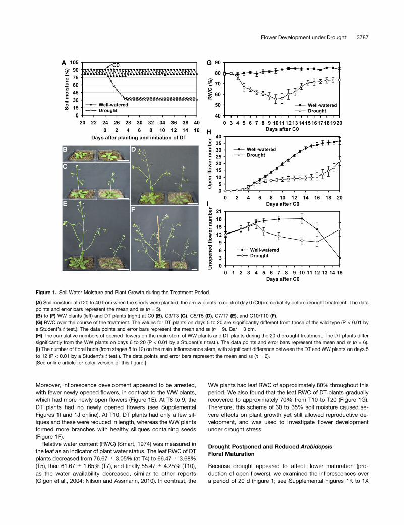
Moreover, inflorescence development appeared to be arrested,with fewer newly opened flowers, in contrast to the WW plants,which had more newly open flowers (Figure 1E). At T8 to 9, theDT plants had no newly opened flowers (see SupplementalFigures 1I and 1J online). At T10, DT plants had only a few sil-iques and these were reduced in length, whereas the WW plantsformed more branches with healthy siliques containing seeds(Figure 1F).
Relative water content (RWC) (Smart, 1974) was measured inthe leaf as an indicator of plant water status. The leaf RWC of DTplants decreased from 76.67 6 3.05% (at T4) to 66.47 6 3.68%(T5), then 61.67 6 1.65% (T7), and finally 55.47 6 4.25% (T10),as the water availability decreased, similar to other reports(Gigon et al., 2004; Nilson and Assmann, 2010). In contrast, the
WW plants had leaf RWC of approximately 80% throughout thisperiod. We also found that the leaf RWC of DT plants graduallyrecovered to approximately 70% from T10 to T20 (Figure 1G).Therefore, this scheme of 30 to 35% soil moisture caused se-vere effects on plant growth yet still allowed reproductive de-velopment, and was used to investigate flower developmentunder drought stress.
Drought Postponed and Reduced ArabidopsisFloral Maturation
Because drought appeared to affect flower maturation (pro-duction of open flowers), we examined the inflorescences overa period of 20 d (Figure 1; see Supplemental Figures 1K to 1X
Figure 1. Soil Water Moisture and Plant Growth during the Treatment Period.
(A) Soil moisture at d 20 to 40 from when the seeds were planted; the arrow points to control day 0 (C0) immediately before drought treatment. The datapoints and error bars represent the mean and SE (n = 5).(B) to (F) WW plants (left) and DT plants (right) at C0 (B), C3/T3 (C), C5/T5 (D), C7/T7 (E), and C10/T10 (F).(G) RWC over the course of the treatment. The values for DT plants on days 5 to 20 are significantly different from those of the wild type (P < 0.01 bya Student’s t test.). The data points and error bars represent the mean and SE (n = 9). Bar = 3 cm.(H) The cumulative numbers of opened flowers on the main stem of WW plants and DT plants during the 20-d drought treatment. The DT plants differsignificantly from the WW plants on days 6 to 20 (P < 0.01 by a Student’s t test.). The data points and error bars represent the mean and SE (n = 6).(I) The number of floral buds (from stages 8 to 12) on the main inflorescence stem, with significant difference between the DT and WW plants on days 5to 12 (P < 0.01 by a Student’s t test.). The data points and error bars represent the mean and SE (n = 6).[See online article for color version of this figure.]
Flower Development under Drought 3787
online) and found that the number of mature flowers formed onthe main stem (the main stem was assayed unless otherwisenoted) in the DT plants was significantly reduced (21.8 6 3.3;n = 6) compared with the WW plants (36.7 6 2.7; n = 6), witha P value of 6.5131026 (Student’s t test). The reduction wasespecially severe from T7 to T13, with zero to one newly openedflower each day (Figure 1H). In contrast, WW plants had steadyproduction of two to four newly opened flowers every day fromC4 to C13 (Figure 1H). The results suggested that the rapidexpansion/elongation of floral organs during floral stages 8 to12 (Smyth et al., 1990) might be particularly dependent on ad-equate water availability. In addition, the number of unopenedfloral buds at stages 8 to 12 increased from C0 to C3/T3 in bothWW and DT plants, respectively (Figure 1I). Although the numberof floral buds remained nearly constant in the WW plants forapproximately10 d, the number dramatically decreased in DTplants (Figure 1I), with only about the half of the normal numberat T10, indicating retardation of new bud formation and growth.The number of floral buds subsequently recovered in the DTplants by T15 (Figure 1I).
In short, in WW Arabidopsis plants, the rate of flower matu-ration increased after flowering and then reached a steady state.However, in DT plants, the oldest buds were able to mature(Figure 1I; see Supplemental Figures 1T to 1V online), but thenthe plants coped by reducing the rate of new bud formation andgrowth at stages 8 to 12, nearly stopping flower maturation forseveral days, before recovering and resuming flower developmentat a reduced steady state rate. These results suggest that Arabi-dopsis is able to respond to reduced water availability by developingand reproducing at a decreased rate, consistent with the reductionand subsequent partial recovery of RWC described in the previoussection.
Flower Development Was First Arrested and Then ShowedPartial Recovery
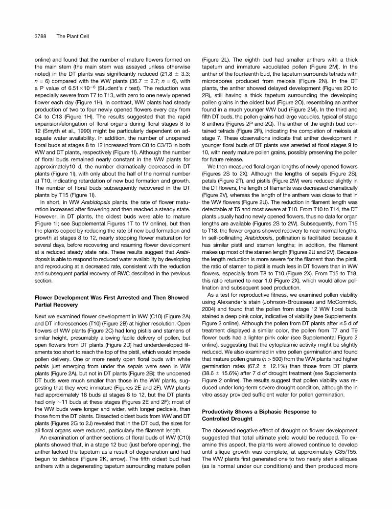
Next we examined flower development in WW (C10) (Figure 2A)and DT inflorescences (T10) (Figure 2B) at higher resolution. Openflowers of WW plants (Figure 2C) had long pistils and stamens ofsimilar height, presumably allowing facile delivery of pollen, butopen flowers from DT plants (Figure 2D) had underdeveloped fil-aments too short to reach the top of the pistil, which would impedepollen delivery. One or more nearly open floral buds with whitepetals just emerging from under the sepals were seen in WWplants (Figure 2A), but not in DT plants (Figure 2B); the unopenedDT buds were much smaller than those in the WW plants, sug-gesting that they were immature (Figures 2E and 2F). WW plantshad approximately 18 buds at stages 8 to 12, but the DT plantshad only ;11 buds at these stages (Figures 2E and 2F); most ofthe WW buds were longer and wider, with longer pedicels, thanthose from the DT plants. Dissected oldest buds from WW and DTplants (Figures 2G to 2J) revealed that in the DT bud, the sizes forall floral organs were reduced, particularly the filament length.
An examination of anther sections of floral buds of WW (C10)plants showed that, in a stage 12 bud (just before opening), theanther lacked the tapetum as a result of degeneration and hadbegun to dehisce (Figure 2K, arrow). The fifth oldest bud hadanthers with a degenerating tapetum surrounding mature pollen
(Figure 2L). The eighth bud had smaller anthers with a thicktapetum and immature vacuolated pollen (Figure 2M). In theanther of the fourteenth bud, the tapetum surrounds tetrads withmicrospores produced from meiosis (Figure 2N). In the DTplants, the anther showed delayed development (Figures 2O to2R), still having a thick tapetum surrounding the developingpollen grains in the oldest bud (Figure 2O), resembling an antherfound in a much younger WW bud (Figure 2M). In the third andfifth DT buds, the pollen grains had large vacuoles, typical of stage8 anthers (Figures 2P and 2Q). The anther of the eighth bud con-tained tetrads (Figure 2R), indicating the completion of meiosis atstage 7. These observations indicate that anther development inyounger floral buds of DT plants was arrested at floral stages 9 to10, with nearly mature pollen grains, possibly preserving the pollenfor future release.We then measured floral organ lengths of newly opened flowers
(Figures 2S to 2X). Although the lengths of sepals (Figure 2S),petals (Figure 2T), and pistils (Figure 2W) were reduced slightly inthe DT flowers, the length of filaments was decreased dramatically(Figure 2V), whereas the length of the anthers was close to that inthe WW flowers (Figure 2U). The reduction in filament length wasdetectable at T5 and most severe at T10. From T10 to T14, the DTplants usually had no newly opened flowers, thus no data for organlengths are available (Figures 2S to 2W). Subsequently, from T15to T18, the flower organs showed recovery to near normal lengths.In self-pollinating Arabidopsis, pollination is facilitated because ithas similar pistil and stamen lengths; in addition, the filamentmakes up most of the stamen length (Figures 2U and 2V). Becausethe length reduction is more severe for the filament than the pistil,the ratio of stamen to pistil is much less in DT flowers than in WWflowers, especially from T8 to T10 (Figure 2X). From T15 to T18,this ratio returned to near 1.0 (Figure 2X), which would allow pol-lination and subsequent seed production.As a test for reproductive fitness, we examined pollen viability
using Alexander’s stain (Johnson-Brousseau and McCormick,2004) and found that the pollen from stage 12 WW floral budsstained a deep pink color, indicative of viability (see SupplementalFigure 2 online). Although the pollen from DT plants after #5 d oftreatment displayed a similar color, the pollen from T7 and T9flower buds had a lighter pink color (see Supplemental Figure 2online), suggesting that the cytoplasmic activity might be slightlyreduced. We also examined in vitro pollen germination and foundthat mature pollen grains (n > 500) from the WW plants had highergermination rates (67.2 6 12.1%) than those from DT plants(38.6 6 15.6%) after 7 d of drought treatment (see SupplementalFigure 2 online). The results suggest that pollen viability was re-duced under long-term severe drought condition, although the invitro assay provided sufficient water for pollen germination.
Productivity Shows a Biphasic Response toControlled Drought
The observed negative effect of drought on flower developmentsuggested that total ultimate yield would be reduced. To ex-amine this aspect, the plants were allowed continue to developuntil silique growth was complete, at approximately C35/T55.The WW plants first generated one to two nearly sterile siliques(as is normal under our conditions) and then produced more
3788 The Plant Cell
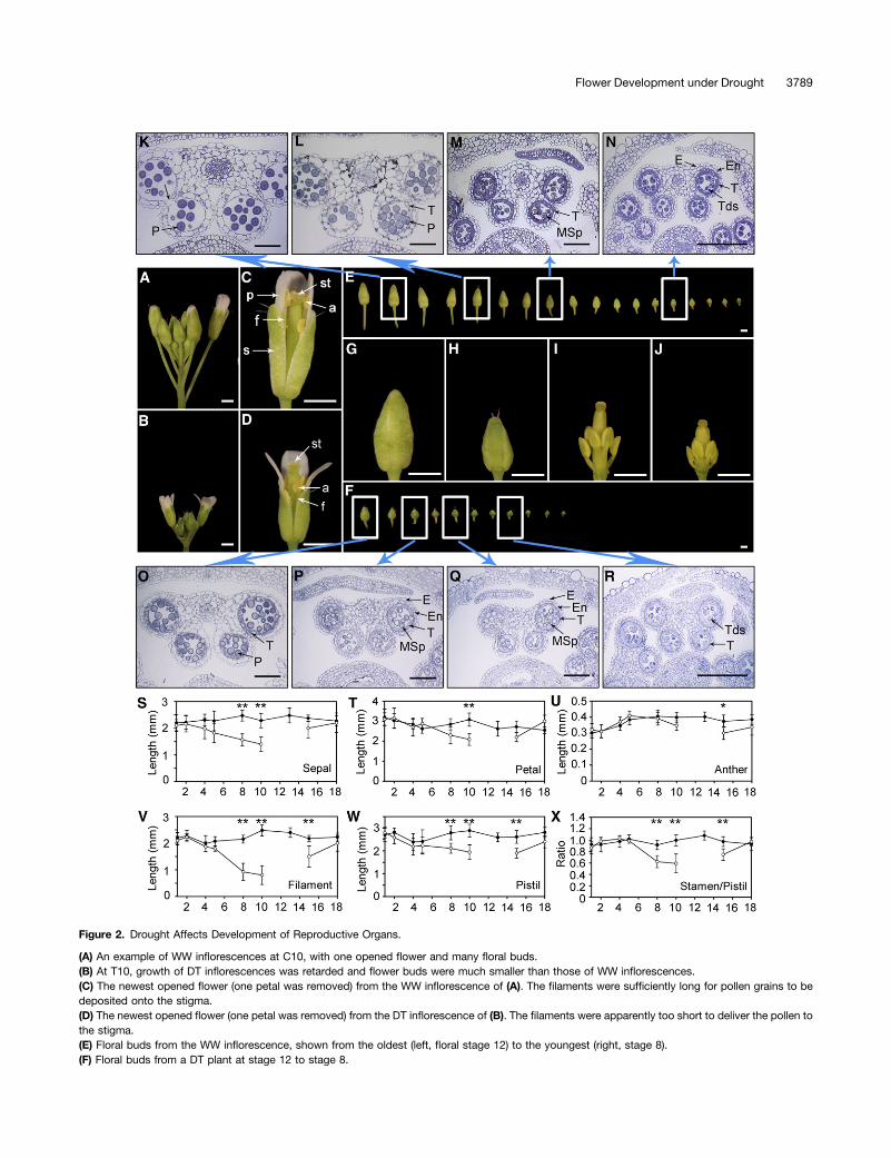
Figure 2. Drought Affects Development of Reproductive Organs.
(A) An example of WW inflorescences at C10, with one opened flower and many floral buds.(B) At T10, growth of DT inflorescences was retarded and flower buds were much smaller than those of WW inflorescences.(C) The newest opened flower (one petal was removed) from the WW inflorescence of (A). The filaments were sufficiently long for pollen grains to bedeposited onto the stigma.(D) The newest opened flower (one petal was removed) from the DT inflorescence of (B). The filaments were apparently too short to deliver the pollen tothe stigma.(E) Floral buds from the WW inflorescence, shown from the oldest (left, floral stage 12) to the youngest (right, stage 8).(F) Floral buds from a DT plant at stage 12 to stage 8.
Flower Development under Drought 3789
than 35 siliques (see Supplemental Figure 3A online), eachcontaining 45 to 50 seeds (Figure 3V). However, the DT plantsexhibited three different silique phenotypes (see SupplementalFigure 3B online). After the same initial one to two sterile sili-ques, the next two to three siliques were fertile but containedfewer seeds; these were from the nearly mature flowers thatwere able to open at the beginning of drought treatment whenthe soil moisture level decreased from 75% to 40% (Figure 1A).The next group of four to six siliques was sterile or nearly sterile,derived from the maturing floral buds when the plants first en-countered severe drought at 30 to 35% soil moisture; thesefloral buds showed the most severe reduction of organ size. Thenext set of ;20 siliques was fertile with reduced seed set of;25seeds, gradually decreasing toward the top of the inflorescence(see Supplemental Figure 3B online). Therefore, the biphasic re-sponse of a short-term arrest (an acute phase) followed by a lon-ger-term recovery at a reduced level (prolonged phase) observedfor floral organ sizes is also manifest in terms of mature seedproduction. It is possible that an acute phase is needed for the DTplants to reprogram reproductive development to enter the pro-longed phase; this prolonged response phase then uses the limitedwater resource to support both flower and silique development,albeit with fewer flowers and seeds per silique.
To illustrate the biphasic response, we examined plants that hadbeen treated for 20 d (T20), when they had produced a number ofsiliques and were still producing flowers (Figure 3A). At this time,the first 10 flowers had passed the time of pollination and exhibiteddifferent degrees of fertility, consistent with what we observed forolder plants as described above. In particular, from the third to theninth silique, the siliques were progressively shorter (Figure 3A). Inthe fourth silique, there were shrunken seeds and white abortedovules (Figures 3B and 3C), suggesting that pollen, ovule, and/orseed development were affected. In the seventh silique, only a fewseeds formed and most ovules appeared aborted (Figures 3D and3E), indicating a greater effect of drought. Other siliques (fifth,sixth, eighth, and ninth) were also sterile or nearly sterile, illus-trating a component of the acute response.
The eleventh flower bud, the first unopened bud on the stem,had anthers with normal appearance and elongated filamentsthat were bent, suggesting that the sepals had not opened andso the internal organs could not extend further (Figures 3F and3G). The sepals of similar buds still adhered to each other 5 d(T25) later, even though they had separated from the base of theflower bud (Figures 3H and 3I). Similar sepal phenotypes were
also observed for the twelfth (Figures 3J to 3M) and the thir-teenth flowers. In the fourteenth flower, the pistil protruded outof the sepals, but the filaments were short (Figures 3N and 3O),presumably resulting in ineffective pollination (Figures 3P and3Q). In the fifteenth flower, the pistil was even more obviouslylonger than the sepals (Figures 3R and 3S), leading to possiblefailure in pollination (Figures 3T and 3U). Strikingly, T20 plantshad recovered and entered the prolonged phase of droughtresponse, with a burst of blossom, illustrated here by the plantin Figure 3A, with 10 mature flowers (numbers 16 to 25) that hadjust opened within 2 d, consistent with the earlier observationthat floral organ sizes recovered to close to normal sizes by T15(Figures 2S to 2X). These and later flowers then led to thesubsequent generation of relatively fertile siliques, as shownin Supplemental Figure 3B online.To test the effect of drought on female reproductive development,
we performed reciprocal crosses between WW and DT plants, withadditional crosses as controls (Figure 3W; see Supplemental Figures3B and 3C online), and determined the silique length and seednumber 2 weeks later. The results indicated that drought also re-duced female fertility in addition to negative effects on pollen de-velopment, although there was likely also an effect of drought on theplants (other than the female reproductive organs).
Overview and Analysis of Gene Expression in Flower underDrought Stress
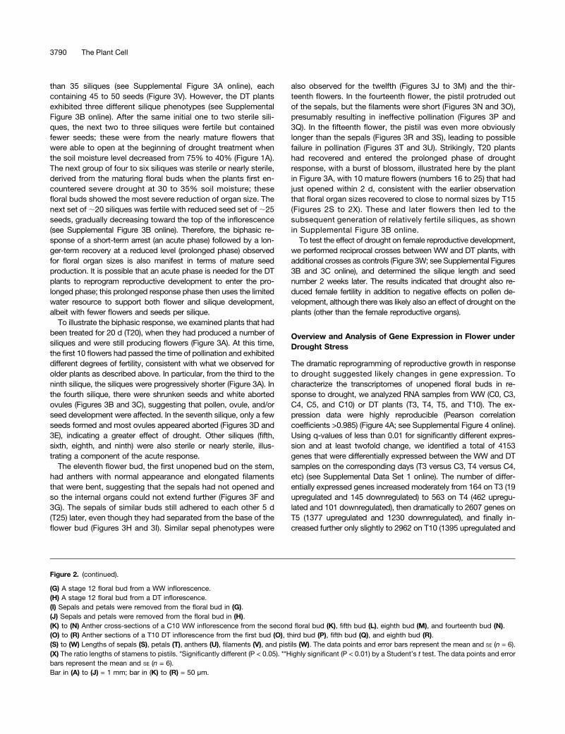
The dramatic reprogramming of reproductive growth in responseto drought suggested likely changes in gene expression. Tocharacterize the transcriptomes of unopened floral buds in re-sponse to drought, we analyzed RNA samples from WW (C0, C3,C4, C5, and C10) or DT plants (T3, T4, T5, and T10). The ex-pression data were highly reproducible (Pearson correlationcoefficients >0.985) (Figure 4A; see Supplemental Figure 4 online).Using q-values of less than 0.01 for significantly different expres-sion and at least twofold change, we identified a total of 4153genes that were differentially expressed between the WW and DTsamples on the corresponding days (T3 versus C3, T4 versus C4,etc) (see Supplemental Data Set 1 online). The number of differ-entially expressed genes increased moderately from 164 on T3 (19upregulated and 145 downregulated) to 563 on T4 (462 upregu-lated and 101 downregulated), then dramatically to 2607 genes onT5 (1377 upregulated and 1230 downregulated), and finally in-creased further only slightly to 2962 on T10 (1395 upregulated and
Figure 2. (continued).
(G) A stage 12 floral bud from a WW inflorescence.(H) A stage 12 floral bud from a DT inflorescence.(I) Sepals and petals were removed from the floral bud in (G).(J) Sepals and petals were removed from the floral bud in (H).(K) to (N) Anther cross-sections of a C10 WW inflorescence from the second floral bud (K), fifth bud (L), eighth bud (M), and fourteenth bud (N).(O) to (R) Anther sections of a T10 DT inflorescence from the first bud (O), third bud (P), fifth bud (Q), and eighth bud (R).(S) to (W) Lengths of sepals (S), petals (T), anthers (U), filaments (V), and pistils (W). The data points and error bars represent the mean and SE (n = 6).(X) The ratio lengths of stamens to pistils. *Significantly different (P < 0.05). **Highly significant (P < 0.01) by a Student’s t test. The data points and errorbars represent the mean and SE (n = 6).Bar in (A) to (J) = 1 mm; bar in (K) to (R) = 50 µm.
3790 The Plant Cell
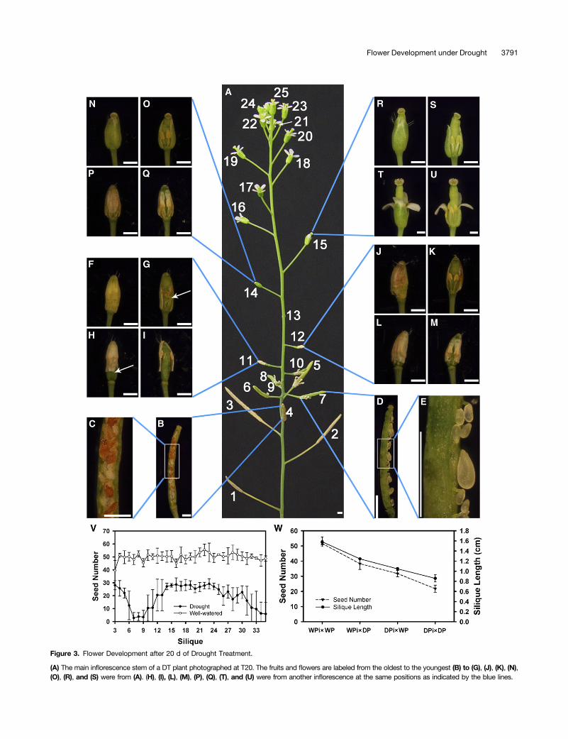
Figure 3. Flower Development after 20 d of Drought Treatment.
(A) The main inflorescence stem of a DT plant photographed at T20. The fruits and flowers are labeled from the oldest to the youngest (B) to (G), (J), (K), (N),(O), (R), and (S) were from (A). (H), (I), (L), (M), (P), (Q), (T), and (U) were from another inflorescence at the same positions as indicated by the blue lines.
Flower Development under Drought 3791
1567 downregulated) (Figure 4B; see Supplemental Table 1 on-line). Among the 19 genes upregulated at T3, the majority (13) alsoincreased on T4, T5, and/or T10. Similarly, the majority (278among 482) of the upregulated genes on T4 were also higher at T5and/or T10 (Figure 4C). Therefore, most of the early induced genestended to remain induced. By contrast, the majority (116 among145) of downregulated genes on T3 were not persistently reducedat T4, T5, or T10 (Figure 4D), suggesting that the decreased ex-pression of these genes was a transient response. Among thegenes that were upregulated (1395) or downregulated (1567) onT10, 786 and 759, respectively, were also increased on T5 (Figures4C and 4D), indicating that many of the transcriptomic changeshad occurred by day 5.
Coexpression Pattern of Drought-Responsive Genesin Flowers
To further examine the floral transcriptomic response to drought,we applied hierarchical clustering to all 4153 genes differentiallyexpressed between WW and DT floral buds, resulting in 10 largeclusters. Using the K-means (K = 10) approach in the hierarchicalclusters, genes were assigned to 1 of 10 clusters, which were thenvisualized with heat map and centroid views (Figures 4E and 4F),revealing general patterns of transcriptomic profiles during thedrought treatment.
The first four clusters (Figures 4Fa to 4Fd) contained geneswith drought-induced significant increase in expression thatpeaked on days 3, 4, 5, and 10, respectively, suggesting thattheir functions might be particularly important in a transientfashion. Clusters 5 and 6 (Figures 4Fe and 4Ff) contained geneswith high levels of expression at two time points, suggesting
more sustained functions, with the genes in cluster 5 actingearlier than those in cluster 6. In contrast, the genes fromclusters 7 to 10 (Figures 4Fg to 4Fj) exhibited various patterns ofdrought-induced decrease in expression. The genes in cluster 7(Figure 4Fg) showed a transient decrease in expression on day 3,whereas those in cluster 8 (Figure 4Fh) had persistent reductionfrom T4 to T10. The expression of genes in clusters 9 (Figure 4Fi)and 10 (Figure 4Fj) showed reduced expression at T5, with furtherreduction on T10 for genes in cluster 10.
Functional Prediction of Different Drought-ResponsiveGene Clusters
To gain further insight into the changing transcriptomic land-scapes in the DT flowers, we performed gene ontology (GO) an-notation enrichment analysis using singular enrichment analysisprovided by agriGO (Du et al., 2010). In both clusters 1 and 2,genes involved in transcriptional regulation were enriched (Figure4G), suggesting that changes in transcriptional regulation wereone of the early responses to drought stress. On the other hand, incluster 4, the genes that function in RNA splicing were enriched,suggesting a potential increase in the level of protein diversityattributable to alternative splicing in the response at day 10. Inaddition, genes for glucosinolate biosynthesis were enriched incluster 1, suggesting a possible function in drought response; thisis consistent with a function of glucosinolates in the regulation byABA of stomatal opening (Zhao et al., 2008) and is possibly relatedto the role of glucosinolates as regulators of biotic response(Hopkins et al., 2009).Among the genes enriched in cluster 5 (e) are genes related to
pollen tube growth, particularly GTP binding signal proteins and
Figure 3. (continued).
(B) The fourth fruit from (A) after dissection.(C) A portion of the fourth fruit with higher magnification.(D) The seventh fruit with fresh ovules and seeds shown.(E) A portion of (D) with higher magnification, showing normal and small ovules.(F) The eleventh bud.(G) The eleventh bud after dissection, showing reduced filament length (arrow).(H) The eleventh bud after maturation, showing the detachment at the base of sepals that were not separated at the top.(I) The dissected bud of (H), showing detached short stamens.(J) The twelfth bud.(K) The dissected twelfth bud with short filaments.(L) The twelfth bud after maturation with sepals detached at the base.(M) The dissected bud from (L).(N) The fourteenth bud.(O) The dissected fourteenth bud.(P) The fourteenth bud after maturation.(Q) The dissected bud from (P).(R) The fifteenth bud.(S) The dissected fifteenth bud.(T) The fifteenth bud after maturation.(U) The bud from (T) with two sepals and a petal removed.(V) The comparison of seed count per silique on the main stem from WW and DT plants. P < 0.01 by a Student’s t test. The data points and error barsrepresent the mean and SE (n = 5).(W) The average seed number and silique length of different crossing plants: WW plants (WPi3WP), WW pistil pollinated with DT pollen (WPi3DP), DTpistil pollinated with WW pollen (DPi3WP), and DT plants (DPi3DP). The data points and error bars represent the mean and SE (n = 3).Bar in (A) to (T) = 1 mm.
3792 The Plant Cell
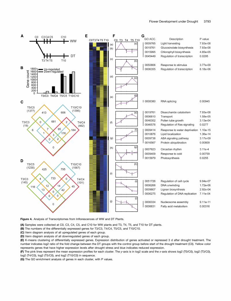
Figure 4. Analysis of Transcriptomes from Inflorescences of WW and DT Plants.
(A) Samples were collected at C0, C3, C4, C5, and C10 for WW plants and T3, T4, T5, and T10 for DT plants.(B) The numbers of the differentially expressed genes for T3/C3, T4/C4, T5/C5, and T10/C10.(C) Venn diagram analysis of all upregulated genes of each group.(D) Venn diagram analysis of all downregulated genes of each group.(E) K-means clustering of differentially expressed genes. Expression distribution of genes activated or repressed 3 d after drought treatment. Thenumber indicates log2 ratio of the fold change between the DT groups with the control group before start of the drought treatment (C0). Yellow colorrepresents genes that have higher expression levels after drought stress and blue indicates reduced expression.(F) The pink lines represent the mean expression profiles for each cluster. The y-axis is in log2 scale and the x-axis shows log2 (T0/C0), log2 (T3/C0),log2 (T4/C0), log2 (T5/C0), and log2 (T10/C0) in sequence.(G) The GO enrichment analysis of genes in each cluster, with P values.
Flower Development under Drought 3793
disaccharide catabolism (Figure 4G). For example, RHO-RELATEDPROTEIN FROM PLANTS 1 (ROP1) and ROP-INTERACTIVE CRIBMOTIF-CONTAINING PROTEIN 6 (RIC6) function in the polarizedgrowth of pollen tubes (Wu et al., 2001; Hwang et al., 2005). Thegenes encoding disaccharide enzymes such as b-galactosidase 7(BGAL-7), BGAL-11, and BGAL-13, which play a role in pollen de-velopment and fertilization (Jakobsen et al., 2005; Wang et al.,2008), also showed elevated expression, suggesting that enhancedactivity of these gene products is needed to promote pollen de-velopment and function under drought stress. Genes related to di-saccharide catabolism (7.93e-08) and transport (1.58e-05) are highlyenriched, which suggests the possible regulation of transport ofsugars and other osmolytes in response to drought. At a later timein cluster 6 (Figure 4Ff), genes responsive to water deprivation andABA-mediated signaling pathways were highly enriched. Amongthem are regulators of ABA signaling, including ABI1, ABI2, ABI5,ABF1, ABF3, and ABF4, and genes encoding PP2CA, SNRK2.3,and SNRK2.6 (OST1) proteins; these genes are known to function inABA-mediated responses to abiotic stresses (Luan, 2003; Cutleret al., 2010; Yoshida et al., 2010). In addition, genes functioningin the ABA-independent pathway, such as those coding forDREB1B, DREB2A, DREB2B, RD20, and RD22 (Bernoux et al.,2008; Matsukura et al., 2010), were enriched (Figure 4G).
Among the downregulated clusters, genes related to pho-tosynthesis and circadian rhythm were enriched in cluster 7(g) (Figure 4G), suggesting that these functions were reducedtransiently as an early drought response. In cluster 9 (i), thegenes for cell cycle regulation and DNA replication were en-riched (Figure 4G), including those coding for cyclins andcyclin-dependent kinases. In cluster 10 (j), genes for nucle-osome subunits were highly enriched (Figure 4G), includingthose for histone H2A, histone H2B, histone H3, and histoneH4. The reduced expression of the genes in clusters 9 and 10suggests that cell proliferation was reduced soon after the
drought treatment, consistent with the observed morpho-logical changes.
Early Induction of ABA and Jasmonic Acid Signaling Genes
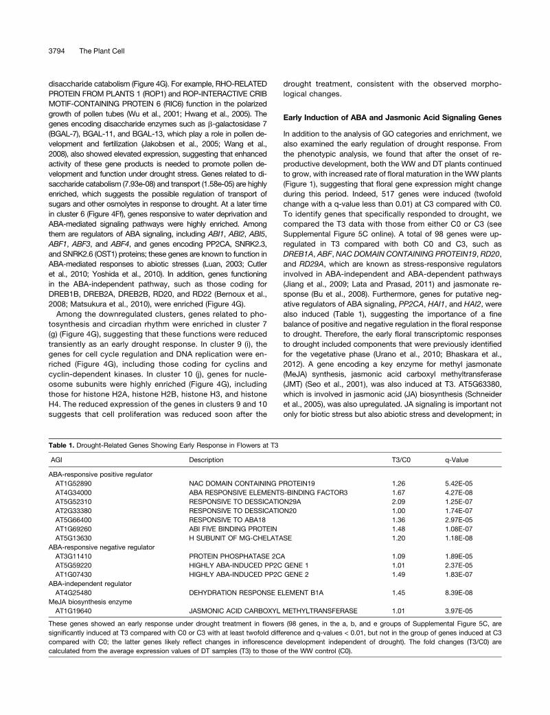
In addition to the analysis of GO categories and enrichment, wealso examined the early regulation of drought response. Fromthe phenotypic analysis, we found that after the onset of re-productive development, both the WW and DT plants continuedto grow, with increased rate of floral maturation in the WW plants(Figure 1), suggesting that floral gene expression might changeduring this period. Indeed, 517 genes were induced (twofoldchange with a q-value less than 0.01) at C3 compared with C0.To identify genes that specifically responded to drought, wecompared the T3 data with those from either C0 or C3 (seeSupplemental Figure 5C online). A total of 98 genes were up-regulated in T3 compared with both C0 and C3, such asDREB1A, ABF, NAC DOMAIN CONTAINING PROTEIN19, RD20,and RD29A, which are known as stress-responsive regulatorsinvolved in ABA-independent and ABA-dependent pathways(Jiang et al., 2009; Lata and Prasad, 2011) and jasmonate re-sponse (Bu et al., 2008). Furthermore, genes for putative neg-ative regulators of ABA signaling, PP2CA, HAI1, and HAI2, werealso induced (Table 1), suggesting the importance of a finebalance of positive and negative regulation in the floral responseto drought. Therefore, the early floral transcriptomic responsesto drought included components that were previously identifiedfor the vegetative phase (Urano et al., 2010; Bhaskara et al.,2012). A gene encoding a key enzyme for methyl jasmonate(MeJA) synthesis, jasmonic acid carboxyl methyltransferase(JMT) (Seo et al., 2001), was also induced at T3. AT5G63380,which is involved in jasmonic acid (JA) biosynthesis (Schneideret al., 2005), was also upregulated. JA signaling is important notonly for biotic stress but also abiotic stress and development; in
Table 1. Drought-Related Genes Showing Early Response in Flowers at T3
AGI Description T3/C0 q-Value
ABA-responsive positive regulatorAT1G52890 NAC DOMAIN CONTAINING PROTEIN19 1.26 5.42E-05AT4G34000 ABA RESPONSIVE ELEMENTS-BINDING FACTOR3 1.67 4.27E-08AT5G52310 RESPONSIVE TO DESSICATION29A 2.09 1.25E-07AT2G33380 RESPONSIVE TO DESSICATION20 1.00 1.74E-07AT5G66400 RESPONSIVE TO ABA18 1.36 2.97E-05AT1G69260 ABI FIVE BINDING PROTEIN 1.48 1.08E-07AT5G13630 H SUBUNIT OF MG-CHELATASE 1.20 1.18E-08
ABA-responsive negative regulatorAT3G11410 PROTEIN PHOSPHATASE 2CA 1.09 1.89E-05AT5G59220 HIGHLY ABA-INDUCED PP2C GENE 1 1.01 2.37E-05AT1G07430 HIGHLY ABA-INDUCED PP2C GENE 2 1.49 1.83E-07
ABA-independent regulatorAT4G25480 DEHYDRATION RESPONSE ELEMENT B1A 1.45 8.39E-08
MeJA biosynthesis enzymeAT1G19640 JASMONIC ACID CARBOXYL METHYLTRANSFERASE 1.01 3.97E-05
These genes showed an early response under drought treatment in flowers (98 genes, in the a, b, and e groups of Supplemental Figure 5C, aresignificantly induced at T3 compared with C0 or C3 with at least twofold difference and q-values < 0.01, but not in the group of genes induced at C3compared with C0; the latter genes likely reflect changes in inflorescence development independent of drought). The fold changes (T3/C0) arecalculated from the average expression values of DT samples (T3) to those of the WW control (C0).
3794 The Plant Cell
addition, the crosstalk between ABA and JA signaling contrib-utes to the balance between growth and defense (Lackmanet al., 2011).
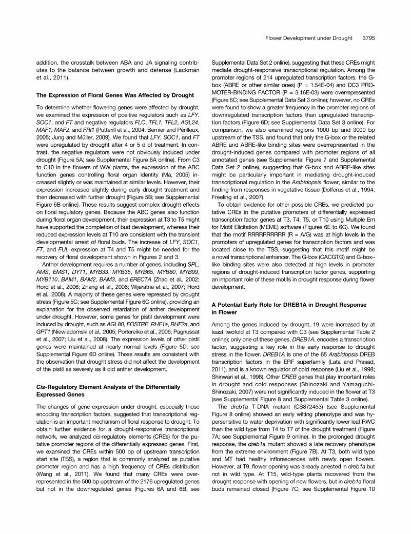
The Expression of Floral Genes Was Affected by Drought
To determine whether flowering genes were affected by drought,we examined the expression of positive regulators such as LFY,SOC1, and FT and negative regulators FLC, TFL1, TFL2, AGL24,MAF1, MAF2, and FRI1 (Putterill et al., 2004; Bernier and Périlleux,2005; Jung and Müller, 2009). We found that LFY, SOC1, and FTwere upregulated by drought after 4 or 5 d of treatment. In con-trast, the negative regulators were not obviously induced underdrought (Figure 5A; see Supplemental Figure 6A online). From C3to C10 in the flowers of WW plants, the expression of the ABCfunction genes controlling floral organ identity (Ma, 2005) in-creased slightly or was maintained at similar levels. However, theirexpression increased slightly during early drought treatment andthen decreased with further drought (Figure 5B; see SupplementalFigure 6B online). These results suggest complex drought effectson floral regulatory genes. Because the ABC genes also functionduring floral organ development, their expression at T3 to T5 mighthave supported the completion of bud development, whereas theirreduced expression levels at T10 are consistent with the transientdevelopmental arrest of floral buds. The increase of LFY, SOC1,FT, and FUL expression at T4 and T5 might be needed for therecovery of floral development shown in Figures 2 and 3.
Anther development requires a number of genes, including SPL,AMS, EMS1, DYT1, MYB33, MYB35, MYB65, MYB80, MYB99,MYB110, BAM1, BAM2, BAM3, and ERECTA (Zhao et al., 2002;Hord et al., 2006; Zhang et al., 2006; Wijeratne et al., 2007; Hordet al., 2008). A majority of these genes were repressed by droughtstress (Figure 5C; see Supplemental Figure 6C online), providing anexplanation for the observed retardation of anther developmentunder drought. However, some genes for pistil development wereinduced by drought, such as AGL80, EOSTRE, RHF1a, RHF2a, andGPT1 (Niewiadomski et al., 2005; Portereiko et al., 2006; Pagnussatet al., 2007; Liu et al., 2008). The expression levels of other pistilgenes were maintained at nearly normal levels (Figure 5D; seeSupplemental Figure 6D online). These results are consistent withthe observation that drought stress did not affect the developmentof the pistil as severely as it did anther development.
Cis-Regulatory Element Analysis of the DifferentiallyExpressed Genes
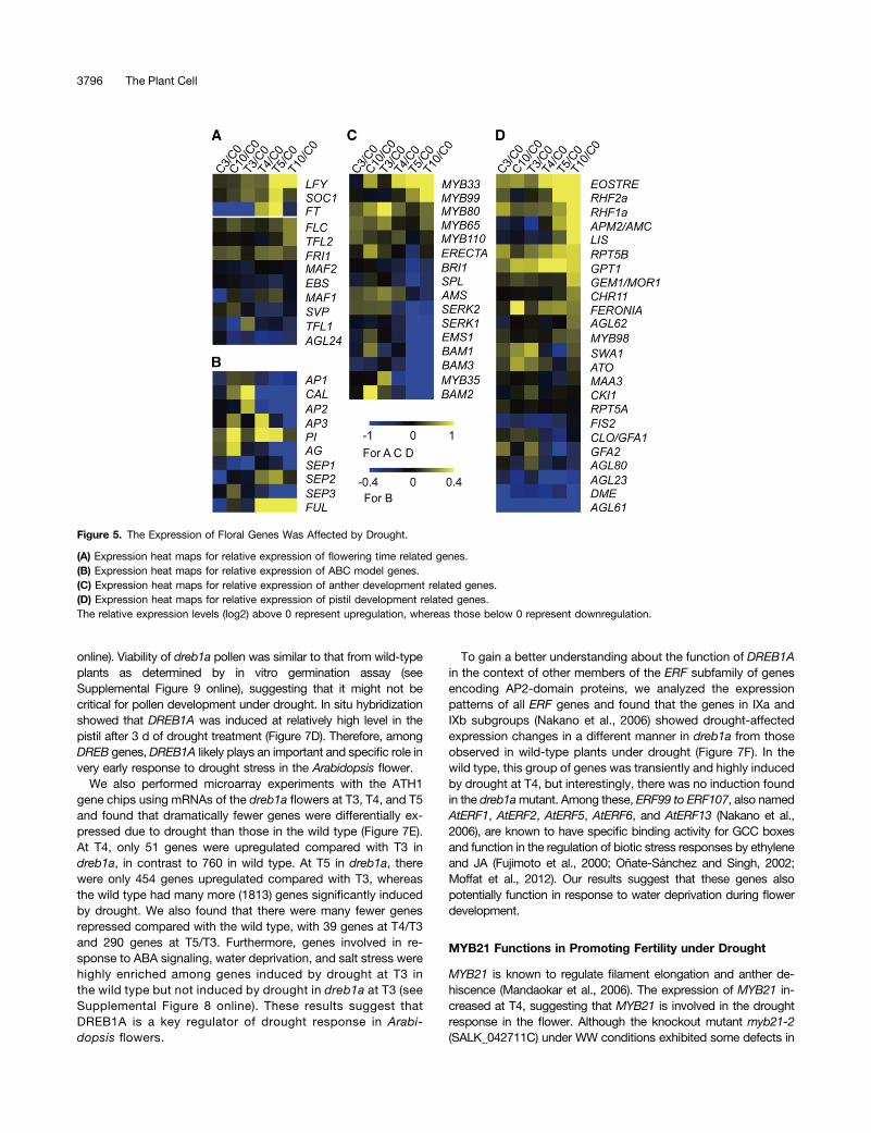
The changes of gene expression under drought, especially thoseencoding transcription factors, suggested that transcriptional reg-ulation is an important mechanism of floral response to drought. Toobtain further evidence for a drought-responsive transcriptionalnetwork, we analyzed cis-regulatory elements (CREs) for the pu-tative promoter regions of the differentially expressed genes. First,we examined the CREs within 500 bp of upstream transcriptionstart site (TSS), a region that is commonly analyzed as putativepromoter region and has a high frequency of CREs distribution(Wang et al., 2011). We found that many CREs were over-represented in the 500 bp upstream of the 2176 upregulated genesbut not in the downregulated genes (Figures 6A and 6B; see
Supplemental Data Set 2 online), suggesting that these CREsmightmediate drought-responsive transcriptional regulation. Among thepromoter regions of 214 upregulated transcription factors, the G-box (ABRE or other similar ones) (P = 1.54E-04) and DC3 PRO-MOTER-BINDING FACTOR (P = 3.16E-03) were overrepresented(Figure 6C; see Supplemental Data Set 3 online); however, no CREswere found to show a greater frequency in the promoter regions ofdownregulated transcription factors than upregulated transcrip-tion factors (Figure 6D; see Supplemental Data Set 3 online). Forcomparison, we also examined regions 1000 bp and 3000 bpupstream of the TSS, and found that only the G-box or the relatedABRE and ABRE-like binding sites were overrepresented in thedrought-induced genes compared with promoter regions of allannotated genes (see Supplemental Figure 7 and SupplementalData Set 2 online), suggesting that G-box and ABRE-like sitesmight be particularly important in mediating drought-inducedtranscriptional regulation in the Arabidopsis flower, similar to thefinding from responses in vegetative tissue (Dolferus et al., 1994;Freeling et al., 2007).To obtain evidence for other possible CREs, we predicted pu-
tative CREs in the putative promoters of differentially expressedtranscription factor genes at T3, T4, T5, or T10 using Multiple Emfor Motif Elicitation (MEME) software (Figures 6E to 6G). We foundthat the motif RRRRRRRRRR (R = A/G) was at high levels in thepromoters of upregulated genes for transcription factors and waslocated close to the TSS, suggesting that this motif might bea novel transcriptional enhancer. The G-box (CACGTG) and G-box–like binding sites were also detected at high levels in promoterregions of drought-induced transcription factor genes, supportingan important role of these motifs in drought response during flowerdevelopment.
A Potential Early Role for DREB1A in Drought Responsein Flower
Among the genes induced by drought, 19 were increased by atleast twofold at T3 compared with C3 (see Supplemental Table 2online); only one of these genes, DREB1A, encodes a transcriptionfactor, suggesting a key role in the early response to droughtstress in the flower. DREB1A is one of the 65 Arabidopsis DREBtranscription factors in the ERF superfamily (Lata and Prasad,2011), and is a known regulator of cold response (Liu et al., 1998;Shinwari et al., 1998). Other DREB genes that play important rolesin drought and cold responses (Shinozaki and Yamaguchi-Shinozaki, 2007) were not significantly induced in the flower at T3(see Supplemental Figure 8 and Supplemental Table 3 online).The dreb1a T-DNA mutant (CS872453) (see Supplemental
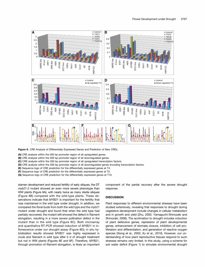
Figure 8 online) showed an early wilting phenotype and was hy-persensitive to water deprivation with significantly lower leaf RWCthan the wild type from T4 to T7 of the drought treatment (Figure7A; see Supplemental Figure 9 online). In the prolonged droughtresponse, the dreb1a mutant showed a late recovery phenotypefrom the extreme environment (Figure 7B). At T3, both wild typeand MT had healthy inflorescences with newly open flowers.However, at T9, flower opening was already arrested in dreb1a butnot in wild type. At T15, wild-type plants recovered from thedrought response with opening of new flowers, but in dreb1a floralbuds remained closed (Figure 7C; see Supplemental Figure 10
Flower Development under Drought 3795
online). Viability of dreb1a pollen was similar to that from wild-typeplants as determined by in vitro germination assay (seeSupplemental Figure 9 online), suggesting that it might not becritical for pollen development under drought. In situ hybridizationshowed that DREB1A was induced at relatively high level in thepistil after 3 d of drought treatment (Figure 7D). Therefore, amongDREB genes, DREB1A likely plays an important and specific role invery early response to drought stress in the Arabidopsis flower.
We also performed microarray experiments with the ATH1gene chips using mRNAs of the dreb1a flowers at T3, T4, and T5and found that dramatically fewer genes were differentially ex-pressed due to drought than those in the wild type (Figure 7E).At T4, only 51 genes were upregulated compared with T3 indreb1a, in contrast to 760 in wild type. At T5 in dreb1a, therewere only 454 genes upregulated compared with T3, whereasthe wild type had many more (1813) genes significantly inducedby drought. We also found that there were many fewer genesrepressed compared with the wild type, with 39 genes at T4/T3and 290 genes at T5/T3. Furthermore, genes involved in re-sponse to ABA signaling, water deprivation, and salt stress werehighly enriched among genes induced by drought at T3 inthe wild type but not induced by drought in dreb1a at T3 (seeSupplemental Figure 8 online). These results suggest thatDREB1A is a key regulator of drought response in Arabi-dopsis flowers.
To gain a better understanding about the function of DREB1Ain the context of other members of the ERF subfamily of genesencoding AP2-domain proteins, we analyzed the expressionpatterns of all ERF genes and found that the genes in IXa andIXb subgroups (Nakano et al., 2006) showed drought-affectedexpression changes in a different manner in dreb1a from thoseobserved in wild-type plants under drought (Figure 7F). In thewild type, this group of genes was transiently and highly inducedby drought at T4, but interestingly, there was no induction foundin the dreb1amutant. Among these, ERF99 to ERF107, also namedAtERF1, AtERF2, AtERF5, AtERF6, and AtERF13 (Nakano et al.,2006), are known to have specific binding activity for GCC boxesand function in the regulation of biotic stress responses by ethyleneand JA (Fujimoto et al., 2000; Oñate-Sánchez and Singh, 2002;Moffat et al., 2012). Our results suggest that these genes alsopotentially function in response to water deprivation during flowerdevelopment.
MYB21 Functions in Promoting Fertility under Drought
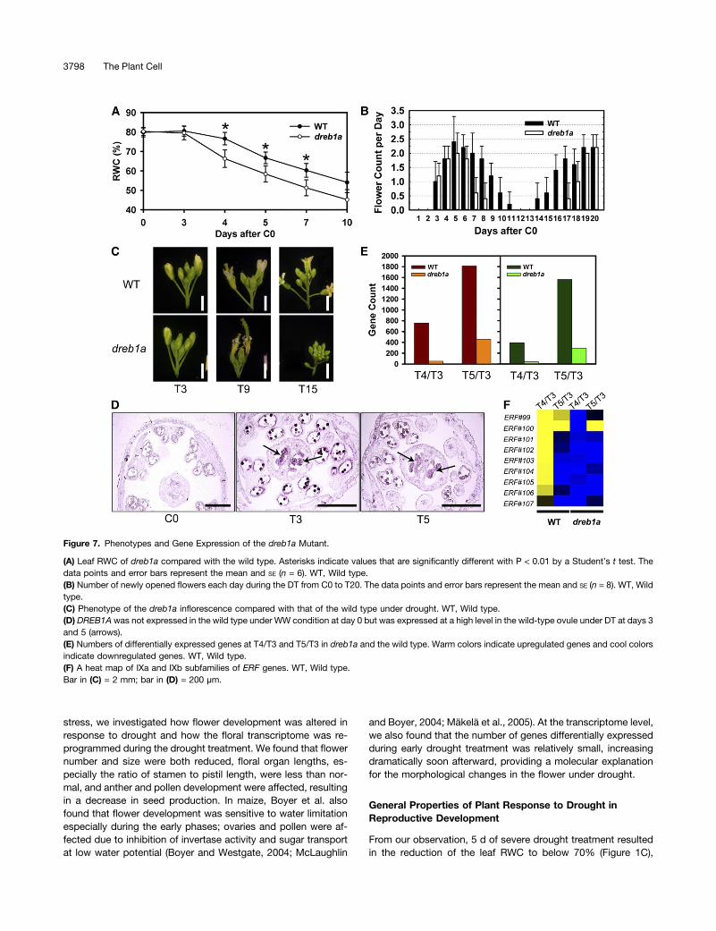
MYB21 is known to regulate filament elongation and anther de-hiscence (Mandaokar et al., 2006). The expression of MYB21 in-creased at T4, suggesting that MYB21 is involved in the droughtresponse in the flower. Although the knockout mutant myb21-2(SALK_042711C) under WW conditions exhibited some defects in
Figure 5. The Expression of Floral Genes Was Affected by Drought.
(A) Expression heat maps for relative expression of flowering time related genes.(B) Expression heat maps for relative expression of ABC model genes.(C) Expression heat maps for relative expression of anther development related genes.(D) Expression heat maps for relative expression of pistil development related genes.The relative expression levels (log2) above 0 represent upregulation, whereas those below 0 represent downregulation.
3796 The Plant Cell
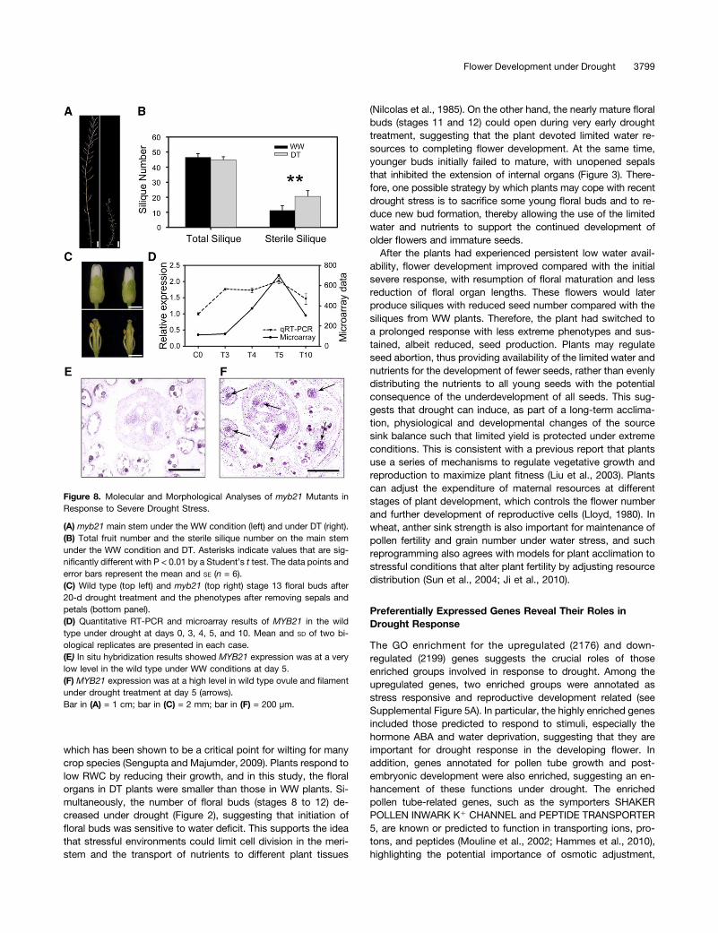
stamen development and reduced fertility of early siliques, the DTmyb21-2 mutant showed an even more severe phenotype thanWW plants (Figure 8A), with nearly twice as many sterile siliques(Figure 8B) compared with the wild-type plants. These ob-servations indicate that MYB21 is important for the fertility thatwas maintained in the wild type under drought. In addition, wecompared the floral buds from both the wild type and the myb21mutant under drought and found that when the wild type hadpartially recovered, the mutant still showed the defect in filamentelongation, resulting in a more severe pollination defect in themutant than in the wild type (Figure 8C). Both microarrayand quantitative RT-PCR showed induction of MYB21 in in-florescence under our drought assay (Figure 8D); in situ hy-bridization results showed MYB21 was highly expressed inovule and filament in wild type after 5 d of drought treatmentbut not in WW plants (Figures 8E and 8F). Therefore, MYB21,through promotion of filament elongation, is likely an important
component of the partial recovery after the severe droughtresponse.
DISCUSSION
Plant responses to different environmental stresses have beenstudied extensively, revealing that responses to drought duringvegetative development include changes in cellular metabolismand in growth and yield (Zhu, 2002; Yamaguchi-Shinozaki andShinozaki, 2006). The acclimation to drought includes inductionof plant defensive genes, repression of plant developmentalgenes, enhancement of stomata closure, inhibition of cell pro-liferation and differentiation, and generation of reactive oxygenspecies (Xiong et al., 2002; Xu et al., 2010). However, our un-derstanding of how plant reproductive tissues respond to suchstresses remains very limited. In this study, using a scheme forsoil water deficit (Figure 1) to simulate environmental drought
Figure 6. CRE Analysis of Differentially Expressed Genes and Prediction of New CREs.
(A) CRE analysis within the 500 bp promoter region of all upregulated genes.(B) CRE analysis within the 500 bp promoter region of all downregulated genes.(C) CRE analysis within the 500 bp promoter region of all upregulated transcription factors.(D) CRE analysis within the 500 bp promoter region of all downregulated genes encoding transcription factors.(E) Sequence logo of CRE prediction for the differentially expressed genes at T4.(F) Sequence logo of CRE prediction for the differentially expressed genes at T5.(G) Sequence logo of CRE prediction for the differentially expressed genes at T10.
Flower Development under Drought 3797
stress, we investigated how flower development was altered inresponse to drought and how the floral transcriptome was re-programmed during the drought treatment. We found that flowernumber and size were both reduced, floral organ lengths, es-pecially the ratio of stamen to pistil length, were less than nor-mal, and anther and pollen development were affected, resultingin a decrease in seed production. In maize, Boyer et al. alsofound that flower development was sensitive to water limitationespecially during the early phases; ovaries and pollen were af-fected due to inhibition of invertase activity and sugar transportat low water potential (Boyer and Westgate, 2004; McLaughlin
and Boyer, 2004; Mäkelä et al., 2005). At the transcriptome level,we also found that the number of genes differentially expressedduring early drought treatment was relatively small, increasingdramatically soon afterward, providing a molecular explanationfor the morphological changes in the flower under drought.
General Properties of Plant Response to Drought inReproductive Development
From our observation, 5 d of severe drought treatment resultedin the reduction of the leaf RWC to below 70% (Figure 1C),
Figure 7. Phenotypes and Gene Expression of the dreb1a Mutant.
(A) Leaf RWC of dreb1a compared with the wild type. Asterisks indicate values that are significantly different with P < 0.01 by a Student’s t test. Thedata points and error bars represent the mean and SE (n = 6). WT, Wild type.(B) Number of newly opened flowers each day during the DT from C0 to T20. The data points and error bars represent the mean and SE (n = 8). WT, Wildtype.(C) Phenotype of the dreb1a inflorescence compared with that of the wild type under drought. WT, Wild type.(D) DREB1A was not expressed in the wild type under WW condition at day 0 but was expressed at a high level in the wild-type ovule under DT at days 3and 5 (arrows).(E) Numbers of differentially expressed genes at T4/T3 and T5/T3 in dreb1a and the wild type. Warm colors indicate upregulated genes and cool colorsindicate downregulated genes. WT, Wild type.(F) A heat map of IXa and IXb subfamilies of ERF genes. WT, Wild type.Bar in (C) = 2 mm; bar in (D) = 200 µm.
3798 The Plant Cell
which has been shown to be a critical point for wilting for manycrop species (Sengupta and Majumder, 2009). Plants respond tolow RWC by reducing their growth, and in this study, the floralorgans in DT plants were smaller than those in WW plants. Si-multaneously, the number of floral buds (stages 8 to 12) de-creased under drought (Figure 2), suggesting that initiation offloral buds was sensitive to water deficit. This supports the ideathat stressful environments could limit cell division in the meri-stem and the transport of nutrients to different plant tissues
(Nilcolas et al., 1985). On the other hand, the nearly mature floralbuds (stages 11 and 12) could open during very early droughttreatment, suggesting that the plant devoted limited water re-sources to completing flower development. At the same time,younger buds initially failed to mature, with unopened sepalsthat inhibited the extension of internal organs (Figure 3). There-fore, one possible strategy by which plants may cope with recentdrought stress is to sacrifice some young floral buds and to re-duce new bud formation, thereby allowing the use of the limitedwater and nutrients to support the continued development ofolder flowers and immature seeds.After the plants had experienced persistent low water avail-
ability, flower development improved compared with the initialsevere response, with resumption of floral maturation and lessreduction of floral organ lengths. These flowers would laterproduce siliques with reduced seed number compared with thesiliques from WW plants. Therefore, the plant had switched toa prolonged response with less extreme phenotypes and sus-tained, albeit reduced, seed production. Plants may regulateseed abortion, thus providing availability of the limited water andnutrients for the development of fewer seeds, rather than evenlydistributing the nutrients to all young seeds with the potentialconsequence of the underdevelopment of all seeds. This sug-gests that drought can induce, as part of a long-term acclima-tion, physiological and developmental changes of the sourcesink balance such that limited yield is protected under extremeconditions. This is consistent with a previous report that plantsuse a series of mechanisms to regulate vegetative growth andreproduction to maximize plant fitness (Liu et al., 2003). Plantscan adjust the expenditure of maternal resources at differentstages of plant development, which controls the flower numberand further development of reproductive cells (Lloyd, 1980). Inwheat, anther sink strength is also important for maintenance ofpollen fertility and grain number under water stress, and suchreprogramming also agrees with models for plant acclimation tostressful conditions that alter plant fertility by adjusting resourcedistribution (Sun et al., 2004; Ji et al., 2010).
Preferentially Expressed Genes Reveal Their Roles inDrought Response
The GO enrichment for the upregulated (2176) and down-regulated (2199) genes suggests the crucial roles of thoseenriched groups involved in response to drought. Among theupregulated genes, two enriched groups were annotated asstress responsive and reproductive development related (seeSupplemental Figure 5A). In particular, the highly enriched genesincluded those predicted to respond to stimuli, especially thehormone ABA and water deprivation, suggesting that they areimportant for drought response in the developing flower. Inaddition, genes annotated for pollen tube growth and post-embryonic development were also enriched, suggesting an en-hancement of these functions under drought. The enrichedpollen tube-related genes, such as the symporters SHAKERPOLLEN INWARK K1 CHANNEL and PEPTIDE TRANSPORTER5, are known or predicted to function in transporting ions, pro-tons, and peptides (Mouline et al., 2002; Hammes et al., 2010),highlighting the potential importance of osmotic adjustment,
Figure 8. Molecular and Morphological Analyses of myb21 Mutants inResponse to Severe Drought Stress.
(A) myb21 main stem under the WW condition (left) and under DT (right).(B) Total fruit number and the sterile silique number on the main stemunder the WW condition and DT. Asterisks indicate values that are sig-nificantly different with P < 0.01 by a Student’s t test. The data points anderror bars represent the mean and SE (n = 6).(C) Wild type (top left) and myb21 (top right) stage 13 floral buds after20-d drought treatment and the phenotypes after removing sepals andpetals (bottom panel).(D) Quantitative RT-PCR and microarray results of MYB21 in the wildtype under drought at days 0, 3, 4, 5, and 10. Mean and SD of two bi-ological replicates are presented in each case.(E) In situ hybridization results showed MYB21 expression was at a verylow level in the wild type under WW conditions at day 5.(F) MYB21 expression was at a high level in wild type ovule and filamentunder drought treatment at day 5 (arrows).Bar in (A) = 1 cm; bar in (C) = 2 mm; bar in (F) = 200 µm.
Flower Development under Drought 3799
signaling transduction and starvation response in the flowerunder drought.
Furthermore, cell wall-modifying enzyme pectinesterases(PMEs) and their inhibitors (PMEIs), such as VGD1 and PMEI2, areimportant for pollen tube growth (Röckel et al., 2008). Pectins areimportant structural determinants of the cell wall in dicots andtheir esterified forms are associated with active cell expansion orgrowth such as pollen tube tip growth. PMEs de-esterify pectinsand mainly exist in the lateral regions of the pollen tube, whereasPMEIs can promote pollen tube tip growth (Röckel et al., 2008).The balance of these functions modulates pollen tube cell wallstability and cell expansion (Bosch et al., 2005; Jiang et al., 2005).The enrichment of these genes among drought-induced genessuggests that elevated levels of such activities are needed tomodulate cell wall integrity and cellular growth when there isgreater osmotic stress and there are limited resources for growth.
Among the downregulated genes (see Supplemental Figure5B), the enriched categories included key regulators of the cellcycle and DNA replication such as cyclins and cyclin-dependentkinases and regulators of microtubule-associated motors. Theseresults are consistent with the finding that drought stress caninhibit the normal cell cycle process, thereby reducing plant celldivision and differentiation (Setter and Flannigan, 2001). Otherenriched groups include biosynthetic genes for JA and gluco-sinolate, which promote defense against disease and herbivoreattack (Adie et al., 2007; Clay et al., 2009), suggesting that theenergy costly defense responses were repressed by drought. JAalso enhances filament elongation (Song et al., 2011); the re-duction of expression of JA biosynthetic genes provides anexplanation for the decreased filament length observed in DTflowers. The expression of genes encoding lipid transfer proteinswas also reduced under drought stress, consistent with previousobservation that lipid transfer protein genes are differentiallyregulated by various abiotic stresses (Wu et al., 2004; Oshinoet al., 2007; Choi et al., 2008).
The Strategy of Reproductive Development under Drought
Our morphological study showed that stamen growth and de-velopment was highly sensitive to drought (Figure 2). From thetranscriptomics results, we also found that many genes impor-tant for anther development showed reduced expression underdrought, providing a link between gene expression and pheno-type (Figure 5). The pistil and the subsequent fruit support ovuledevelopment and seed formation, respectively. In addition, thenumbers of ovules and seeds are small, whereas pollen grainsare numerous and in excess; therefore, partially sacrificing pol-len development could reduce the need for resources while notgreatly decreasing fertility. We found that pistils had higherdrought tolerance than stamens, providing a means for thespecies to survive extreme environments, consistent with theinduction of many pistil-related genes under drought and theirpossible role in the formation of a healthy pistil under stress.These results suggest a possible strategy by which plants pro-tect yield against extreme environments by providing betterprotection of female development than male development.
During the later stages of normal flower development, thefilament elongates dramatically for efficient delivery of the pollen
to the pistil, ensuring subsequent fertilization. We found thatafter 8 d of drought treatment, filament length of DT plants wasobviously shortened compared with WW plants, whereas pistillength was altered to a lesser extent (Figure 2). In addition, an-ther dehiscence was slightly delayed in the DT plants comparedwith WW plants. These findings are consistent with observationsin rice (Liu et al., 2006). The shortened filament and delayedanther dehiscence greatly reduced pollen delivery and pollenavailability to the pistil, thereby decreasing pollination. Sub-sequently, DT flowers exhibited recovery, with resumption oforgan elongation, although to a lesser extent than in WWplants. Arabidopsis MYB family members MYB21, MYB24, andMYB108 are positive regulators of filament elongation and antherdehiscence via the JA and gibberellic acid (GA) signaling path-ways (Mandaokar et al., 2006; Cheng et al., 2009; Mandaokarand Browse, 2009). We found that MYB21 was induced in theflower by drought, suggesting that MYB21 contributes to therecovery in the DT flower. Indeed, the myb21-2 mutant flowersshowed a delay in recovery of filament elongation under droughttreatment compared with wild-type plants. Although MYB21 isan important regulator in JA and GA signaling pathways (Chenget al., 2009), its role in drought response is novel. Because ABAis a key hormone in drought responses, it will be important toinvestigate whether MYB21 serves to integrate different hor-monal pathways, particularly the possible crosstalk betweenJA/GA and ABA in drought response, as hormone crosstalkhas been shown to be important for biotic stresses (Bari andJones, 2009). The induction of the expression of COI1, TIR1,and GID1A/B/C, genes encoding the JA/IAA/GA receptors,under drought suggests their crosstalk and cofunction in responseto drought (Griffiths et al., 2006; Katsir et al., 2008; CalderónVillalobos et al., 2012); however, genes for brassinosteroid sig-naling receptors, BRI1 and BAK1 (Russinova et al., 2004), werenot induced in our study (see Supplemental Figure 11 online). Moreexperimental work is needed to further elucidate their relationshipand crosstalk in response to drought.
Transcriptome Analysis Suggests the Mechanism of PlantResponse to Drought Stress
We investigated floral transcriptomic changes under droughtstress to probe gene activities during flower development af-fected by drought treatment, to obtain clues regarding generegulation in response to drought in the Arabidopsis flower.Among upregulated genes, those encoding transcription factorswere highly enriched, indicating that they play important roles inregulating downstream genes in response to drought. Surpris-ingly, only one transcription factor, DREB1A, was found from theT3/C3 comparison, which revealed 19 genes upregulated by atleast twofold (see Supplemental Table 2 online). This suggeststhat DREB1A might be a key regulator of drought early responsein Arabidopsis flowers. Previous studies showed that DREB1A ismainly involved in cold response (Maruyama et al., 2004; Itoet al., 2006); however, our results showed that DREB1A prob-ably also plays an important role in drought response in flowers.Many drought-responsive genes detected here are key regu-lators in ABA signaling, including DREB1A, ABFs, NAC DOMAINCONTAINING PROTEIN19, RD20, RD29A, and 3 PP2Cs (Table
3800 The Plant Cell
1) (Kasuga et al., 2004; Bu et al., 2008; Xue et al., 2008; Yoshidaet al., 2010).
Flowering time is influenced by environmental factors, includingphotoperiod and temperature, and requires the functions of manygenes (Bernier and Périlleux, 2005). Although a recent study re-ported that FT is induced by drought and accelerates the floraltransition (Riboni et al., 2013), how drought and osmotic stressesaffect flowering was still largely unknown. Our results also indicatedthat FT could be induced after 4 to 5 d of drought treatment. Inaddition, other positive regulators of flowering, such as LFY, SOC1,were induced (Figure 5). At this point, plants have already sensedthe stress signal and begun regulating reproductive growth, suchas acceleration of flower differentiation and seed maturation. Theinduction also could be an indication that these genes have im-portant functions under drought conditions to shorten the life cycleand promote survival under extreme environments, and is consis-tent with the findings that plants often flower earlier when grownunder unfavorable conditions (Verslues and Juenger, 2011). Thepromotion of reproductive growth at the early phase of droughtwould not persist at the late phase because of the continuinglimitation of water. Accordingly, the transcript levels of FT andSOC1 were reduced at day 10 compared with day 5, suggestingthat a prolonged response might decrease the level of positiveflowering regulators, thereby reducing the number of flowers underlong-term severe drought.
Gene Regulatory Network for Plant Development underDrought Stress
Previous studies have demonstrated that phytohormonescoordinate plant growth and development under different
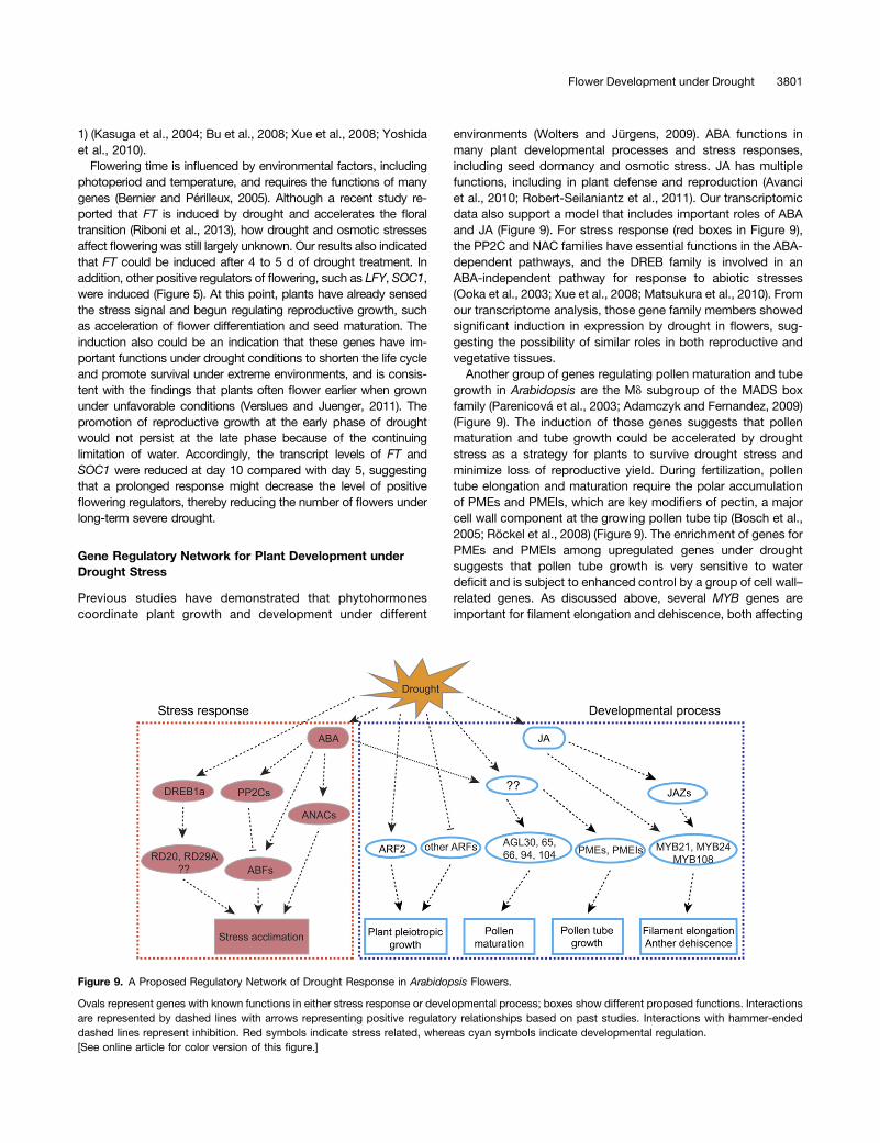
environments (Wolters and Jürgens, 2009). ABA functions inmany plant developmental processes and stress responses,including seed dormancy and osmotic stress. JA has multiplefunctions, including in plant defense and reproduction (Avanciet al., 2010; Robert-Seilaniantz et al., 2011). Our transcriptomicdata also support a model that includes important roles of ABAand JA (Figure 9). For stress response (red boxes in Figure 9),the PP2C and NAC families have essential functions in the ABA-dependent pathways, and the DREB family is involved in anABA-independent pathway for response to abiotic stresses(Ooka et al., 2003; Xue et al., 2008; Matsukura et al., 2010). Fromour transcriptome analysis, those gene family members showedsignificant induction in expression by drought in flowers, sug-gesting the possibility of similar roles in both reproductive andvegetative tissues.Another group of genes regulating pollen maturation and tube
growth in Arabidopsis are the Md subgroup of the MADS boxfamily (Parenicová et al., 2003; Adamczyk and Fernandez, 2009)(Figure 9). The induction of those genes suggests that pollenmaturation and tube growth could be accelerated by droughtstress as a strategy for plants to survive drought stress andminimize loss of reproductive yield. During fertilization, pollentube elongation and maturation require the polar accumulationof PMEs and PMEIs, which are key modifiers of pectin, a majorcell wall component at the growing pollen tube tip (Bosch et al.,2005; Röckel et al., 2008) (Figure 9). The enrichment of genes forPMEs and PMEIs among upregulated genes under droughtsuggests that pollen tube growth is very sensitive to waterdeficit and is subject to enhanced control by a group of cell wall–related genes. As discussed above, several MYB genes areimportant for filament elongation and dehiscence, both affecting
Figure 9. A Proposed Regulatory Network of Drought Response in Arabidopsis Flowers.
Ovals represent genes with known functions in either stress response or developmental process; boxes show different proposed functions. Interactionsare represented by dashed lines with arrows representing positive regulatory relationships based on past studies. Interactions with hammer-endeddashed lines represent inhibition. Red symbols indicate stress related, whereas cyan symbols indicate developmental regulation.[See online article for color version of this figure.]
Flower Development under Drought 3801
pollen delivery (Figure 9), as supported by both transcriptomicdata and mutant phenotypes.
The proposed regulatory network (Figure 9) provides onlya brief outline for the regulation of the two aspects of responseto drought stress in the Arabidopsis flower: physiological re-sponse and developmental acclimation. Much further work isneeded to identify additional components and to demonstratethe interactions among the components. Insights gained fromsuch studies will likely be helpful in efforts to improve crop yieldunder drought and other environmental stresses.
METHODS
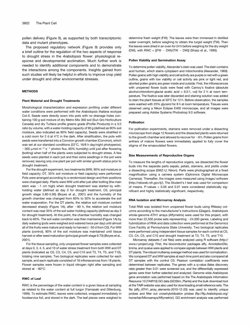
Plant Material and Drought Treatments
Morphological characterization and expression profiling under differentwater conditions were performed with the Arabidopsis thaliana ecotypeCol-0. Seeds were directly sown into pots with no drainage holes con-taining 100 g soil mixture of dry Metro-Mix 360 soil (Sun Gro HorticultureCanada) and dry Turface profile greens grade (Profile Products) in a 3:2ratio by volume, with a water-holding capacity of 90 g (defined as 90% soilmoisture, also indicated as 90% field capacity). Seeds were stratified ina cold room for 2 d at 4°C in the dark. After stratification, the pots withseeds were transferred into a Conviron growth chamber (Conviron), whichwas set at our standard conditions (22°C, 16/8 h day/night photoperiod,;300 mmol m22 s21 photon flux, 60% humidity) until just after flowering(bolting) when half of the plants were subjected to drought stress. A fewseeds were planted in each pot and then extra seedlings in the pot wereremoved, leaving only one plant per pot with similar growth status prior todrought treatment.
For the drought experiment, two treatments (WW, 90% soil moisture orfield capacity; DT, 35% soil moisture or field capacity) were performed.Pots were arranged according to a randomized design and their positionswere changed daily. Plants were WW until after just after bolting (the mainstem was ;1 cm high) when drought treatment was started by with-holding water (defined as day 0 for drought treatment, C0, principalgrowth stage 5.80-5.90) (Boyes et al., 2001) and the humidity of thegrowth chamber was changed from 60% to 30% to accelerate the soilwater evaporation. For the DT plants, the relative soil moisture contentdecreased sharply (Figure 1A); after ;80 h, the relative soil moisturecontent was near 35% of the soil water-holding capacity (defined as day 3for drought treatment). At this point, the chamber humidity was changedback to 60%. The soil water condition was then maintained (Figure 1A) bydaily watering (pots were weighed and watered twice per day) until almostall of the fruits were mature and ready to harvest (;50 d from C0). For WWplants (control), 90% of the soil moisture was maintained until tissueharvest or after seedmaturation (principal growth stage 9.70) (Boyes et al.,2001).
For the tissue sampling, only unopened flower samples were collectedat days 0, 3, 4, 5, and 10 of water stress treatment from both WW and DTplants (indicated as C0, C3, C4, C5, and C10 and T3, T4, T5, and T10),totaling nine samples. Two biological replicates were collected for eachsample, and each replicate consisted of 18 inflorescences from 18 plants.Flower samples were frozen in liquid nitrogen right after sampling andstored at 280°C.
RWC of Leaf
RWC is the percentage of the water content in a given tissue at samplingas related to the water content at full turgor (Yamasaki and Dillenburg,1999). To estimate RWC, leaves were collected, wrapped immediately infoodservice foil, and stored in the dark. The leaf pieces were weighed to
determine fresh weight (FW). The leaves were then immersed in distilledwater overnight, before weighing to obtain the turgid weight (TW). Thenthe leaves were dried in an oven for 24 h before weighing for the dry weight(DW), with RWC = [(FW 2 DW)/(TW 2 DW)] (Sharp et al., 1990).
Pollen Viability and Germination Assay
Todetermine pollen viability, Alexander’s stain was used. The stain containsacid fuchsin, which stains cytoplasm and mitochondria (Alexander, 1969).Pollen grainswith high viability and cell activity are purple or redwith a greenoutline, grains with low viability or cell activity are pink or light red, andaborted pollen grains are green inside and outside. First, the inflorescenceswith unopened flower buds were fixed with Carnoy’s fixative (absolutealcohol:chloroform:glacial acetic acid = 6:3:1, vol) for 2 h at room tem-perature. The fixative was later discarded and staining solution was addedto stain the plant tissues at 50°C for 12 h. Before observation, the sampleswere washed with 25% glycerol for 8 h at room temperature. Tissues wereobserved using a Nikon Eclipse E400 microscope, and all images wereprepared using Adobe Systems Photoshop 9.0 software.
Pollination
For pollination experiments, stamens were removed under a dissectingmicroscope from stage 12 flowers and the dissected plants were returnedto the growth chamber and allowed to recover for;8 h. Pollen grains fromanthers of mature flowers were immediately applied to fully cover thestigma of the emasculated flowers.
Size Measurements of Reproductive Organs
To measure the lengths of reproductive organs, we dissected the flowerbuds into the separate parts sepals, petals, stamens, and pistils undera dissecting scope (SMZ-U; Nikon). Parts were photographed at a fixedmagnification using a camera system (Optronics Digital MicroscopeCameras). Thereafter, the images were measured using ImageJ software(http://rsbweb.nih.gov/ij/). The Student’s t test was used for comparisonof means. P-values < 0.05 and 0.01 were considered statistically sig-nificant and highly statistically significant, respectively.
RNA Isolation and Microarray Analysis
Total RNA was isolated from unopened flower buds using RNeasy col-umns according to the manufacturer’s instructions (Qiagen). Arabidopsiswhole-genome ATH1 arrays (Affymetrix) were used for this project, withmore than 22,500 probe sets representing ;24,000 genes. Labeling andhybridization of RNA and data collection were performed at the GenomicsCore Facility at Pennsylvania State University. Two biological replicateswere performed using independent tissue samples for each control at C0,C3, C4, C5, and C10 and drought treatment at T3, T4, T5, and T10.
Microarray datasets (*.cel files) were analyzed using R software (http://www.r-project.org). First, the bioconductor packages affy, AnnotationDbi,limma, and qvalue were applied to compare signals betweenWWplants andDTplants. The robustmultiarray averagemethodwas used for normalization.We comparedDT andWWsamples at each time point and also compared allDT samples with the control C0. Pearson correlation coefficients weredetermined between replicates. The genes with a q-value (false discoveryrate) greater than 0.01 were screened out, and the differentially expressedgenes were then further selected and analyzed. Genome-wide Arabidopsisgene annotation was performed based on the The Arabidopsis InformationResource 10 (TAIR10) GO data (GOSlim_Plants) and the bulk download toolat the TAIRwebsite was also used for downloading small reference sets. Thefile (affy_ATH1_array_elements-2010-12-20) was used to identify uniqueprobes and filter out crosshybridization probes (ftp://ftp.Arabidopsis.org/home/tair/Microarrays/Affymetrix/). GO enrichment analysis was performed
3802 The Plant Cell
using agriGO, a publicly accessible analysis tool and database (http://bioinfo.cau.edu.cn/agriGO). The Z-score value was used and further converted toa P value to infer statistical significance (Du et al., 2010). Cluster analysis forgene categorization was performed with the TIGR Multi-Experiment Viewer(http://www.tm4.org/mev/). Microarray data were validated by quantitativeRT-PCR of 26 genes. Detailed primer information is listed in SupplementalTable 6 online.
Promoter Motif Analysis
Three types of putative promoter sequence (500 bp, 1 kb, and 3 kbsequences upstream of the start codon) of the genes on the ATH1microarray were downloaded from TAIR. We obtained 101 cis bindingsites from AGRIS (Palaniswamy et al., 2006) that were then analyzed byperl. The number of each binding site was counted. We then comparedthe mean in DT samples against the means of each cis-element site in thewhole genome. The P value was calculated using the Student’s t test andobtained according to occurrences of these CREs in DT gene groups andin all genes. The motifs in drought upregulated genes are more significantthan themotifs in downregulated genes, sowe set 1.0E-10 and 1.0E-05 asthe significance thresholds for the motifs, respectively (Wang et al., 2011).Identification of new conserved motifs was accomplished with MEMEsoftware (version 4.9.0; http://meme.nbcr.net/meme/cgi-bin/meme.cgi),a program designed to identify frequent motifs in biopolymers (Bailey andElkan, 1994).
RNA in Situ Hybridization
Nonradioactive RNA in situ hybridization was performed as previouslydescribed (Hord et al., 2006). Arabidopsis wild-type inflorescences werecollected at days 0, 3, 4, and 5 of drought treatment and fixed in FAA(3.5% formaldehyde, 5% acetic acid, and 50% ethanol alcohol) fixative.The inflorescence samples were vacuumed twice in the fixative each for10 min, with fixation for at least 2 h at room temperature. Samples werethen dehydrated and embedded in paraffin, and 8 µm-thick sections wereprepared andmounted onto slides. After dewaxing of all slides with Histo-Clear, the slides were dehydrated and baked. The antisense and senseRNAs of DREB1A and MYB21 were labeled with digoxigenin by in vitrotranscription of linearized pGEM-T-DREB1A and pGEM-T-MYB21, whichcarry fragments of the DREB1A cDNA amplified with gene-specific primersoMC7398 (59-CTCAGATTATTATTTCCATTTTTAGTA) and oMC7399 (59-GAAACTGAATCAATTTAATTTACACT), and MYB21 cDNA amplified withgene-specific primers oMC7323 (59-CTCGTTGTAGAGGTTTGGGG) andoMC7324 (59-TATGACCCCTTCAACAATTTTAT). Antidigoxigenin antibodiescoupled with alkaline phosphatase and nitro blue tetrazolium were used todetect the hybridization signal.
Accession Numbers
Sequence data from this article can be found in the Arabidopsis GenomeInitiative or GenBank/EMBL databases under accession numbers DREB1A(AT4G25480),MYB21 (AT3G27810),ROP1 (AT3G51300),RIC6 (AT2G20430),ABI1 (AT4G26080), ABI2 (AT5G57050), ABI3 (AT3G24650), ABI4(AT2G40220), ABI5 (AT2G36270), ABF1 (AT1G49720), ABF3 (AT4G34000),ABF4 (AT3G19290), PP2CA (AT3G11410), SNRK2.3 (AT5G66880), SNRK2.6(AT4G33950), DREB1B (AT4G25490), DREB2A (AT5G05410), DREB2B(AT3G11020),RD20 (AT2G33380),RD22 (AT5G25610),RD29A (AT5G52310),CO (AT5G15840),TFL1 (AT5G03840),TFL2 (AT5G17690),MAF2 (AT5G65050),FUL (AT5G60910), ERECTA (AT2G26330),MYB24 (AT5G40350), andMYB108(AT3G06490). Other accession numbers can be found in SupplementalTable 4 online. The wild-type drought project microarray data are availablefromGene Expression Omnibus (GEO) at National Center for BiotechnologyInformation (NCBI) with the series entry GSE40998. Access to these data is
provided at http://www.ncbi.nlm.nih.gov/geo/query/acc.cgi?token=hoxdowwwocaobmandacc=GSE40998. The dreb1a (CS872453) mi-croarray data are available from GEO at NCBI with the series entryGSE43724. Access to these data is provided at http://www.ncbi.nlm.nih.gov/geo/query/acc.cgi?token=fdazviywgskayzuandacc=GSE43724.
Supplemental Data
The following materials are available in the online version of this article.
Supplemental Figure 1. Phenotypic Comparison of WW Plants andDT Plants.
Supplemental Figure 2. Alexander Staining of the Anthers and in VitroGermination of Pollen from WW Plants and DT Plants.
Supplemental Figure 3. Silique Growth Was Affected by DroughtStress.
Supplemental Figure 4. Correlation between Two Biological Repli-cates at Each Time Point.
Supplemental Figure 5. Venn Diagram and Enrichment Analysis ofDifferentially Expressed Genes.
Supplemental Figure 6. Quantitative PCR Verification of MicroarrayData.
Supplemental Figure 7. cis-Regulatory Element Analysis of 1 kb and3 kb Promoters of Differentially Expressed Genes.
Supplemental Figure 8. The dreb1a T-DNA Mutant Information andDREB Genes Expression Pattern under Drought Stress in Flower.
Supplemental Figure 9. Phenotypes of dreb1a Compared with WildType under Early Drought Stress.
Supplemental Figure 10. Phenotypes of dreb1a Compared with WildType under Prolonged Stress.
Supplemental Figure 11. Changes in Expression Patterns of Genesfor IAA, GA, JA, and brassinosteroid Pathways under Drought Stress.
Supplemental Table 1. The Number of Genes with Expression Levelsabove the 50 Threshold (log2 value > 5.64) in Each Dataset and theNumber of Genes Differentially Expressed in Each Dataset Comparedwith Multiple Controls.
Supplemental Table 2. GO Annotation of Genes Differentially Ex-pressed at T3 versus C3.
Supplemental Table 3. The Expression of DREB Genes in ArabidopsisFlower under Drought Stress.
Supplemental Table 4. Primers for Quantitative PCR.
Supplemental Data Set 1. The List of 4153 Genes that WereDifferentially Expressed in Any of the Four Datasets.
Supplemental Data Set 2. Statistical Analysis of the cis-RegulatoryElement within 500 bp, 1 kb, and 3kb Promoter Regions of Differen-tially Expressed Genes.
Supplemental Data Set 3. Statistical Analysis of the cis-RegulatoryElement within 500 bp, 1 kb, and 3kb Promoters Region of Differen-tially Expressed Transcription Factors.
ACKNOWLEDGMENTS
We thank Haikun Zhang and Craig Praul of the Genomics Core Facilityfor help in performing microarray hybridizations. We also thank JiongWang and Yi Hu for help with plant care. This work was supported bya grant from the US Department of Energy (DE-FG02-02ER15332 to
Flower Development under Drought 3803
H.M.) and funds from Rijk Zwaan as well the Biology Department and theHuck Institutes of the Life Sciences at the Pennsylvania State University.
AUTHOR CONTRIBUTIONS
Z.S. and H.M. designed the research. Z.S., X.M., H.G., N.L.S., and B.G.performed different aspects of the research. Z.S., H.M., and S.M.A wrotethe article.
Received June 25, 2013; revised September 30, 2013; accepted October14, 2013; published October 31, 2013.
REFERENCES
Adamczyk, B.J., and Fernandez, D.E. (2009). MIKC* MADS domainheterodimers are required for pollen maturation and tube growth inArabidopsis. Plant Physiol. 149: 1713–1723.
Adie, B.A., Pérez-Pérez, J., Pérez-Pérez, M.M., Godoy, M.,Sánchez-Serrano, J.J., Schmelz, E.A., and Solano, R. (2007).ABA is an essential signal for plant resistance to pathogensaffecting JA biosynthesis and the activation of defenses inArabidopsis. Plant Cell 19: 1665–1681.
Alexander, M.P. (1969). Differential staining of aborted and nonabortedpollen. Stain Technol. 44: 117–122.
Andersen, M.N., Asch, F., Wu, Y., Jensen, C.R., Naested, H., Mogensen,V.O., and Koch, K.E. (2002). Soluble invertase expression is an earlytarget of drought stress during the critical, abortion-sensitive phase ofyoung ovary development in maize. Plant Physiol. 130: 591–604.
Avanci, N.C., Luche, D.D., Goldman, G.H., and Goldman, M.H. (2010).Jasmonates are phytohormones with multiple functions, including plantdefense and reproduction. Genet. Mol. Res. 9: 484–505.
Bailey, T.L., and Elkan, C. (1994). Fitting a mixture model byexpectation maximization to discover motifs in biopolymers. Proc.Int. Conf. Intell. Syst. Mol. Biol. 2: 28–36.
Bari, R., and Jones, J.D. (2009). Role of plant hormones in plantdefence responses. Plant Mol. Biol. 69: 473–488.
Bernier, G., and Périlleux, C. (2005). A physiological overview of thegenetics of flowering time control. Plant Biotechnol. J. 3: 3–16.
Bernoux, M., Timmers, T., Jauneau, A., Brière, C., de Wit, P.J., Marco,Y., and Deslandes, L. (2008). RD19, an Arabidopsis cysteine proteaserequired for RRS1-R-mediated resistance, is relocalized to the nucleus bythe Ralstonia solanacearum PopP2 effector. Plant Cell 20: 2252–2264.
Bhaskara, G.B., Nguyen, T.T., and Verslues, P.E. (2012). Uniquedrought resistance functions of the highly ABA-induced clade Aprotein phosphatase 2Cs. Plant Physiol. 160: 379–395.
Bosch, M., Cheung, A.Y., and Hepler, P.K. (2005). Pectin methylesterase,a regulator of pollen tube growth. Plant Physiol. 138: 1334–1346.
Boyer, J.S., and Westgate, M.E. (2004). Grain yields with limitedwater. J. Exp. Bot. 55: 2385–2394.
Boyes, D.C., Zayed, A.M., Ascenzi, R., McCaskill, A.J., Hoffman, N.E., Davis, K.R., and Görlach, J. (2001). Growth stage-basedphenotypic analysis of Arabidopsis: A model for high throughputfunctional genomics in plants. Plant Cell 13: 1499–1510.
Breshears, D.D., et al. (2005). Regional vegetation die-off in responseto global-change-type drought. Proc. Natl. Acad. Sci. USA 102:15144–15148.
Bu, Q., Jiang, H., Li, C.B., Zhai, Q., Zhang, J., Wu, X., Sun, J., Xie,Q., and Li, C. (2008). Role of the Arabidopsis thaliana NACtranscription factors ANAC019 and ANAC055 in regulating jasmonicacid-signaled defense responses. Cell Res. 18: 756–767.
Busch, W., Wunderlich, M., and Schöffl, F. (2005). Identification ofnovel heat shock factor-dependent genes and biochemicalpathways in Arabidopsis thaliana. Plant J. 41: 1–14.
Calderón Villalobos, L.I., et al. (2012). A combinatorial TIR1/AFB-Aux/IAA co-receptor system for differential sensing of auxin. Nat.Chem. Biol. 8: 477–485.
Canales, C., Bhatt, A.M., Scott, R., and Dickinson, H. (2002). EXS,a putative LRR receptor kinase, regulates male germline cell numberand tapetal identity and promotes seed development in Arabidopsis.Curr. Biol. 12: 1718–1727.
Chaves, M.M., Flexas, J., and Pinheiro, C. (2009). Photosynthesisunder drought and salt stress: Regulation mechanisms from wholeplant to cell. Ann. Bot. (Lond.) 103: 551–560.
Cheng, H., Song, S., Xiao, L., Soo, H.M., Cheng, Z., Xie, D., andPeng, J. (2009). Gibberellin acts through jasmonate to control theexpression of MYB21, MYB24, and MYB57 to promote stamenfilament growth in Arabidopsis. PLoS Genet. 5: e1000440.
Choi, A.M., Lee, S.B., Cho, S.H., Hwang, I., Hur, C.G., and Suh, M.C. (2008). Isolation and characterization of multiple abundant lipidtransfer protein isoforms in developing sesame (Sesamum indicumL.) seeds. Plant Physiol. Biochem. 46: 127–139.
Clay, N.K., Adio, A.M., Denoux, C., Jander, G., and Ausubel, F.M.(2009). Glucosinolate metabolites required for an Arabidopsis innateimmune response. Science 323: 95–101.
Cutler, S.R., Rodriguez, P.L., Finkelstein, R.R., and Abrams, S.R.(2010). Abscisic acid: Emergence of a core signaling network. Annu.Rev. Plant Biol. 61: 651–679.
Dolferus, R., Jacobs, M., Peacock, W.J., and Dennis, E.S. (1994).Differential interactions of promoter elements in stress responses ofthe Arabidopsis Adh gene. Plant Physiol. 105: 1075–1087.
Du, Z., Zhou, X., Ling, Y., Zhang, Z., and Su, Z. (2010). agriGO: A GOanalysis toolkit for the agricultural community. Nucleic Acids Res.38: W64–70.
Farooq, M., Wahid, A., and Kobayashi, N., D., F., and Basra, S.M.A.(2009). Plant drought stress: Effects, mechanisms and management.Sustainable Agric. 29: 185–212.
Freeling, M., Rapaka, L., Lyons, E., Pedersen, B., and Thomas, B.C. (2007). G-boxes, bigfoot genes, and environmental response:Characterization of intragenomic conserved noncoding sequencesin Arabidopsis. Plant Cell 19: 1441–1457.
Fujii, H., Chinnusamy, V., Rodrigues, A., Rubio, S., Antoni, R., Park,S.Y., Cutler, S.R., Sheen, J., Rodriguez, P.L., and Zhu, J.K.(2009). In vitro reconstitution of an abscisic acid signalling pathway.Nature 462: 660–664.
Fujimoto, S.Y., Ohta, M., Usui, A., Shinshi, H., and Ohme-Takagi,M. (2000). Arabidopsis ethylene-responsive element binding factorsact as transcriptional activators or repressors of GCC box-mediatedgene expression. Plant Cell 12: 393–404.
Fujita, M., Fujita, Y., Noutoshi, Y., Takahashi, F., Narusaka, Y.,Yamaguchi-Shinozaki, K., and Shinozaki, K. (2006). Crosstalkbetween abiotic and biotic stress responses: A current view fromthe points of convergence in the stress signaling networks. Curr.Opin. Plant Biol. 9: 436–442.
Gigon, A., Matos, A.R., Laffray, D., Zuily-Fodil, Y., and Pham-Thi, A.T.(2004). Effect of drought stress on lipid metabolism in the leaves ofArabidopsis thaliana (ecotype Columbia). Ann. Bot. (Lond.) 94: 345–351.
Griffiths, J., Murase, K., Rieu, I., Zentella, R., Zhang, Z.L., Powers,S.J., Gong, F., Phillips, A.L., Hedden, P., Sun, T.P., and Thomas,S.G. (2006). Genetic characterization and functional analysis of theGID1 gibberellin receptors in Arabidopsis. Plant Cell 18: 3399–3414.
Haake, V., Cook, D., Riechmann, J.L., Pineda, O., Thomashow, M.F., and Zhang, J.Z. (2002). Transcription factor CBF4 is a regulatorof drought adaptation in Arabidopsis. Plant Physiol. 130: 639–648.
3804 The Plant Cell
Hammes, U.Z., Meier, S., Dietrich, D., Ward, J.M., and Rentsch, D.(2010). Functional properties of the Arabidopsis peptide transportersAtPTR1 and AtPTR5. J. Biol. Chem. 285: 39710–39717.
Hirayama, T., and Shinozaki, K. (2007). Perception and transductionof abscisic acid signals: Keys to the function of the versatile planthormone ABA. Trends Plant Sci. 12: 343–351.
Honma, T., and Goto, K. (2001). Complexes of MADS-box proteins aresufficient to convert leaves into floral organs. Nature 409: 525–529.
Hopkins, R.J., van Dam, N.M., and van Loon, J.J. (2009). Role ofglucosinolates in insect-plant relationships and multitrophic interactions.Annu. Rev. Entomol. 54: 57–83.
Hord, C.L., Chen, C., Deyoung, B.J., Clark, S.E., and Ma, H. (2006).The BAM1/BAM2 receptor-like kinases are important regulators ofArabidopsis early anther development. Plant Cell 18: 1667–1680.
Hord, C.L., Sun, Y.J., Pillitteri, L.J., Torii, K.U., Wang, H., Zhang, S.,and Ma, H. (2008). Regulation of Arabidopsis early antherdevelopment by the mitogen-activated protein kinases, MPK3 andMPK6, and the ERECTA and related receptor-like kinases. Mol.Plant 1: 645–658.
Hwang, J.U., Gu, Y., Lee, Y.J., and Yang, Z. (2005). Oscillatory ROPGTPase activation leads the oscillatory polarized growth of pollentubes. Mol. Biol. Cell 16: 5385–5399.
Ito, T., Wellmer, F., Yu, H., Das, P., Ito, N., Alves-Ferreira, M.,Riechmann, J.L., and Meyerowitz, E.M. (2004). The homeoticprotein AGAMOUS controls microsporogenesis by regulation ofSPOROCYTELESS. Nature 430: 356–360.
Ito, Y., Katsura, K., Maruyama, K., Taji, T., Kobayashi, M., Seki, M.,Shinozaki, K., and Yamaguchi-Shinozaki, K. (2006). Functionalanalysis of rice DREB1/CBF-type transcription factors involved incold-responsive gene expression in transgenic rice. Plant CellPhysiol. 47: 141–153.
Jakobsen, M.K., Poulsen, L.R., Schulz, A., Fleurat-Lessard, P.,Møller, A., Husted, S., Schiøtt, M., Amtmann, A., and Palmgren,M.G. (2005). Pollen development and fertilization in Arabidopsis isdependent on the MALE GAMETOGENESIS IMPAIRED ANTHERSgene encoding a type V P-type ATPase. Genes Dev. 19: 2757–2769.
Ji, X., Shiran, B., Wan, J., Lewis, D.C., Jenkins, C.L., Condon, A.G.,Richards, R.A., and Dolferus, R. (2010). Importance of pre-anthesis anther sink strength for maintenance of grain numberduring reproductive stage water stress in wheat. Plant Cell Environ.33: 926–942.
Jiang, H., Li, H., Bu, Q., and Li, C. (2009). The RHA2a-interacting proteinsANAC019 and ANAC055 may play a dual role in regulating ABAresponse and jasmonate response. Plant Signal. Behav. 4: 464–466.
Jiang, L., Yang, S.L., Xie, L.F., Puah, C.S., Zhang, X.Q., Yang, W.C.,Sundaresan, V., and Ye, D. (2005). VANGUARD1 encodes a pectinmethylesterase that enhances pollen tube growth in the Arabidopsisstyle and transmitting tract. Plant Cell 17: 584–596.
Johnson-Brousseau, S.A., and McCormick, S. (2004). A compendium ofmethods useful for characterizing Arabidopsis pollen mutants andgametophytically-expressed genes. Plant J. 39: 761–775.
Jung, C., and Müller, A.E. (2009). Flowering time control andapplications in plant breeding. Trends Plant Sci. 14: 563–573.
Kakumanu, A., Ambavaram, M.M., Klumas, C., Krishnan, A.,Batlang, U., Myers, E., Grene, R., and Pereira, A. (2012). Effects ofdrought on gene expression in maize reproductive and leafmeristem tissue revealed by RNA-Seq. Plant Physiol. 160: 846–867.
Kasuga, M., Miura, S., Shinozaki, K., and Yamaguchi-Shinozaki, K.(2004). A combination of the Arabidopsis DREB1A gene and stress-inducible rd29A promoter improved drought- and low-temperature stresstolerance in tobacco by gene transfer. Plant Cell Physiol. 45: 346–350.
Katsir, L., Schilmiller, A.L., Staswick, P.E., He, S.Y., and Howe, G.A. (2008). COI1 is a critical component of a receptor for jasmonate
and the bacterial virulence factor coronatine. Proc. Natl. Acad. Sci.USA 105: 7100–7105.
Kawasaki, S., Borchert, C., Deyholos, M., Wang, H., Brazille, S.,Kawai, K., Galbraith, D., and Bohnert, H.J. (2001). Geneexpression profiles during the initial phase of salt stress in rice.Plant Cell 13: 889–905.
Klingler, J.P., Batelli, G., and Zhu, J.K. (2010). ABA receptors: TheSTART of a new paradigm in phytohormone signalling. J. Exp. Bot.61: 3199–3210.
Kreps, J.A., Wu, Y., Chang, H.S., Zhu, T., Wang, X., and Harper, J.F.(2002). Transcriptome changes for Arabidopsis in response to salt,osmotic, and cold stress. Plant Physiol. 130: 2129–2141.
Lackman, P., et al. (2011). Jasmonate signaling involves the abscisicacid receptor PYL4 to regulate metabolic reprogramming inArabidopsis and tobacco. Proc. Natl. Acad. Sci. USA 108: 5891–5896.
Lalonde, S., Beebe, D.U., and Saini, H.S. (1997). Early signs ofdisruption of wheat anther development associated with theinduction of male sterility by meiotic-stage water deficit. Sex. PlantReprod. 10: 40–48.
Lata, C., and Prasad, M. (2011). Role of DREBs in regulation ofabiotic stress responses in plants. J. Exp. Bot. 62: 4731–4748.
Liljegren, S.J., Ditta, G.S., Eshed, Y., Savidge, B., Bowman, J.L.,and Yanofsky, M.F. (2000). SHATTERPROOF MADS-box genescontrol seed dispersal in Arabidopsis. Nature 404: 766–770.
Liu, F.L., Andersen, M.N., and Jensen, C.R. (2003). Loss of pod set causedby drought stress is associated with water status and ABA content ofreproductive structures in soybean. Funct. Plant Biol. 30: 271–280.
Liu, J., et al. (2008). Targeted degradation of the cyclin-dependentkinase inhibitor ICK4/KRP6 by RING-type E3 ligases is essential formitotic cell cycle progression during Arabidopsis gametogenesis.Plant Cell 20: 1538–1554.
Liu, J.X., Liao, D.Q., Oane, R., Estenor, L., Yang, X.E., Li, Z.C., andBennett, J. (2006). Genetic variation in the sensitivity of antherdehiscence to drought stress in rice. Field Crops Res. 97: 87–100.
Liu, Q., Kasuga, M., Sakuma, Y., Abe, H., Miura, S., Yamaguchi-Shinozaki, K., and Shinozaki, K. (1998). Two transcription factors,DREB1 and DREB2, with an EREBP/AP2 DNA binding domainseparate two cellular signal transduction pathways in drought- andlow-temperature-responsive gene expression, respectively, inArabidopsis. Plant Cell 10: 1391–1406.
Lloyd, D.G. (1980). Sexual strategies in plants. I. An hypothesis ofserial adjustment of maternal investment during one reproductivesession. New Phytol. 86: 69–79.
Lohmann, J.U., Hong, R.L., Hobe, M., Busch, M.A., Parcy, F., Simon, R.,and Weigel, D. (2001). A molecular link between stem cell regulation andfloral patterning in Arabidopsis. Cell 105: 793–803.
Long, J.A., Moan, E.I., Medford, J.I., and Barton, M.K. (1996). Amember of the KNOTTED class of homeodomain proteins encodedby the STM gene of Arabidopsis. Nature 379: 66–69.
Luan, S. (2003). Protein phosphatases in plants. Annu. Rev. Plant Biol.54: 63–92.
Ma, H. (2005). Molecular genetic analyses of microsporogenesis andmicrogametogenesis in flowering plants. Annu. Rev. Plant Biol. 56: 393–434.
Magome, H., Yamaguchi, S., Hanada, A., Kamiya, Y., and Oda, K.(2004). dwarf and delayed-flowering 1, a novel Arabidopsis mutantdeficient in gibberellin biosynthesis because of overexpression ofa putative AP2 transcription factor. Plant J. 37: 720–729.
Mäkelä, P., McLaughlin, J.E., and Boyer, J.S. (2005). Imaging andquantifying carbohydrate transport to the developing ovaries ofmaize. Ann. Bot. (Lond.) 96: 939–949.
Mandaokar, A., and Browse, J. (2009). MYB108 acts together withMYB24 to regulate jasmonate-mediated stamen maturation inArabidopsis. Plant Physiol. 149: 851–862.
Flower Development under Drought 3805
Mandaokar, A., Thines, B., Shin, B., Lange, B.M., Choi, G., Koo, Y.J., Yoo, Y.J., Choi, Y.D., Choi, G., and Browse, J. (2006).Transcriptional regulators of stamen development in Arabidopsisidentified by transcriptional profiling. Plant J. 46: 984–1008.
Maruyama, K., Sakuma, Y., Kasuga, M., Ito, Y., Seki, M., Goda, H.,Shimada, Y., Yoshida, S., Shinozaki, K., and Yamaguchi-Shinozaki, K. (2004). Identification of cold-inducible downstreamgenes of the Arabidopsis DREB1A/CBF3 transcriptional factor usingtwo microarray systems. Plant J. 38: 982–993.
Matsukura, S., Mizoi, J., Yoshida, T., Todaka, D., Ito, Y.,Maruyama, K., Shinozaki, K., and Yamaguchi-Shinozaki, K.(2010). Comprehensive analysis of rice DREB2-type genes thatencode transcription factors involved in the expression of abioticstress-responsive genes. Mol. Genet. Genomics 283: 185–196.
Mayer, K.F., Schoof, H., Haecker, A., Lenhard, M., Jürgens, G., andLaux, T. (1998). Role of WUSCHEL in regulating stem cell fate in theArabidopsis shoot meristem. Cell 95: 805–815.
McLaughlin, J.E., and Boyer, J.S. (2004). Sugar-responsive geneexpression, invertase activity, and senescence in aborting maizeovaries at low water potentials. Ann. Bot. (Lond.) 94: 675–689.
Melcher, K., et al. (2009). A gate-latch-lock mechanism for hormonesignalling by abscisic acid receptors. Nature 462: 602–608.
Moffat, C.S., Ingle, R.A., Wathugala, D.L., Saunders, N.J., Knight,H., and Knight, M.R. (2012). ERF5 and ERF6 play redundant rolesas positive regulators of JA/Et-mediated defense against Botrytiscinerea in Arabidopsis. PLoS ONE 7: e35995.
Mouline, K., Véry, A.A., Gaymard, F., Boucherez, J., Pilot, G.,Devic, M., Bouchez, D., Thibaud, J.B., and Sentenac, H. (2002).Pollen tube development and competitive ability are impaired by disruptionof a Shaker K(+) channel in Arabidopsis. Genes Dev. 16: 339–350.
Nakano, T., Suzuki, K., Fujimura, T., and Shinshi, H. (2006).Genome-wide analysis of the ERF gene family in Arabidopsis andrice. Plant Physiol. 140: 411–432.
Niewiadomski, P., Knappe, S., Geimer, S., Fischer, K., Schulz, B.,Unte, U.S., Rosso, M.G., Ache, P., Flügge, U.I., and Schneider, A.(2005). The Arabidopsis plastidic glucose 6-phosphate/phosphatetranslocator GPT1 is essential for pollen maturation and embryo sacdevelopment. Plant Cell 17: 760–775.
Nicolas, M.E., Gleadow, R.M., and Dalling, M.J. (1985). Effect ofpost-anthesis drought on cell division and starch accumulation indeveloping wheat grains. Ann. Bot. (Lond.) 55: 433–444.
Nilson, S.E., and Assmann, S.M. (2010). Heterotrimeric G proteinsregulate reproductive trait plasticity in response to wateravailability. New Phytol. 185: 734–746.
Oñate-Sánchez, L., and Singh, K.B. (2002). Identification ofArabidopsis ethylene-responsive element binding factors withdistinct induction kinetics after pathogen infection. Plant Physiol.128: 1313–1322.
Ooka, H., et al. (2003). Comprehensive analysis of NAC family genesin Oryza sativa and Arabidopsis thaliana. DNA Res. 10: 239–247.
Oshino, T., Abiko, M., Saito, R., Ichiishi, E., Endo, M., Kawagishi-Kobayashi, M., and Higashitani, A. (2007). Premature progressionof anther early developmental programs accompanied bycomprehensive alterations in transcription during high-temperatureinjury in barley plants. Mol. Genet. Genomics 278: 31–42.
Pagnussat, G.C., Yu, H.J., and Sundaresan, V. (2007). Cell-fateswitch of synergid to egg cell in Arabidopsis eostre mutant embryosacs arises from misexpression of the BEL1-like homeodomaingene BLH1. Plant Cell 19: 3578–3592.
Palaniswamy, S.K., James, S., Sun, H., Lamb, R.S., Davuluri, R.V.,and Grotewold, E. (2006). AGRIS and AtRegNet. a platform to linkcis-regulatory elements and transcription factors into regulatorynetworks. Plant Physiol. 140: 818–829.
Parenicová, L., de Folter, S., Kieffer, M., Horner, D.S., Favalli, C.,Busscher, J., Cook, H.E., Ingram, R.M., Kater, M.M., Davies, B.,Angenent, G.C., and Colombo, L. (2003). Molecular andphylogenetic analyses of the complete MADS-box transcriptionfactor family in Arabidopsis: New openings to the MADS world.Plant Cell 15: 1538–1551.
Park, S.O., Hwang, S., and Hauser, B.A. (2004). The phenotype ofArabidopsis ovule mutants mimics the morphology of primitive seedplants. Proc. Biol. Sci. 271: 311–316.
Park, S.Y., et al. (2009). Abscisic acid inhibits type 2C proteinphosphatases via the PYR/PYL family of START proteins. Science324: 1068–1071.
Pelaz, S., Ditta, G.S., Baumann, E., Wisman, E., and Yanofsky, M.F.(2000). B and C floral organ identity functions require SEPALLATAMADS-box genes. Nature 405: 200–203.
Pinyopich, A., Ditta, G.S., Savidge, B., Liljegren, S.J., Baumann, E.,Wisman, E., and Yanofsky, M.F. (2003). Assessing the redundancyof MADS-box genes during carpel and ovule development. Nature424: 85–88.
Portereiko, M.F., Lloyd, A., Steffen, J.G., Punwani, J.A., Otsuga, D.,and Drews, G.N. (2006). AGL80 is required for central cell andendosperm development in Arabidopsis. Plant Cell 18: 1862–1872.
Putterill, J., Laurie, R., and Macknight, R. (2004). It’s time to flower:The genetic control of flowering time. Bioessays 26: 363–373.
Riboni, M., Galbiati, M., Tonelli, C., and Conti, L. (2013). GIGANTEAenables drought escape response via abscisic acid-dependent activation ofthe florigens and SUPPRESSOR OF OVEREXPRESSION OF CONSTANS.Plant Physiol. 162: 1706–1719.
Robert-Seilaniantz, A., Grant, M., and Jones, J.D. (2011). Hormonecrosstalk in plant disease and defense: More than just jasmonate-salicylate antagonism. Annu. Rev. Phytopathol. 49: 317–343.
Röckel, N., Wolf, S., Kost, B., Rausch, T., and Greiner, S. (2008).Elaborate spatial patterning of cell-wall PME and PMEI at the pollentube tip involves PMEI endocytosis, and reflects the distribution ofesterified and de-esterified pectins. Plant J. 53: 133–143.
Russinova, E., Borst, J.W., Kwaaitaal, M., Caño-Delgado, A., Yin,Y., Chory, J., and de Vries, S.C. (2004). Heterodimerization andendocytosis of Arabidopsis brassinosteroid receptors BRI1 andAtSERK3 (BAK1). Plant Cell 16: 3216–3229.
Saini, H.S. (1997). Effects of water stress on male gametophytedevelopment in plants. Sex. Plant Reprod. 10: 67–73.
Schiefthaler, U., Balasubramanian, S., Sieber, P., Chevalier, D.,Wisman, E., and Schneitz, K. (1999). Molecular analysis ofNOZZLE, a gene involved in pattern formation and earlysporogenesis during sex organ development in Arabidopsisthaliana. Proc. Natl. Acad. Sci. USA 96: 11664–11669.
Schneider, K., Kienow, L., Schmelzer, E., Colby, T., Bartsch, M.,Miersch, O., Wasternack, C., Kombrink, E., and Stuible, H.P. (2005).A new type of peroxisomal acyl-coenzyme A synthetase fromArabidopsis thaliana has the catalytic capacity to activate biosyntheticprecursors of jasmonic acid. J. Biol. Chem. 280: 13962–13972.
Schröter, D., et al. (2005). Ecosystem service supply and vulnerabilityto global change in Europe. Science 310: 1333–1337.
Sengupta, S., and Majumder, A.L. (2009). Insight into the salttolerance factors of a wild halophytic rice, Porteresia coarctata: Aphysiological and proteomic approach. Planta 229: 911–929.
Seo, H.S., Song, J.T., Cheong, J.J., Lee, Y.H., Lee, Y.W., Hwang, I.,Lee, J.S., and Choi, Y.D. (2001). Jasmonic acid carboxylmethyltransferase: A key enzyme for jasmonate-regulated plantresponses. Proc. Natl. Acad. Sci. USA 98: 4788–4793.
Setter, T.L., and Flannigan, B.A. (2001). Water deficit inhibits cell divisionand expression of transcripts involved in cell proliferation andendoreduplication in maize endosperm. J. Exp. Bot. 52: 1401–1408.
3806 The Plant Cell
Sharp, R.E., Hsiao, T.C., and Silk, W.K. (1990). Growth of the Maizeprimary root at low water potentials: II. Role of growth anddeposition of hexose and potassium in osmotic adjustment. PlantPhysiol. 93: 1337–1346.
Sheoran, I.S., and Saini, H.S. (1996). Drought-induced male sterilityin rice: Changes in carbohydrate levels and enzyme activityiesassociated with the inhibition of starch accumulation in pollen. Sex.Plant Reprod. 9: 161–169.
Shinozaki, K., and Yamaguchi-Shinozaki, K. (2007). Gene networksinvolved in drought stress response and tolerance. J. Exp. Bot. 58:221–227.
Shinozaki, K., Yamaguchi-Shinozaki, K., and Seki, M. (2003).Regulatory network of gene expression in the drought and coldstress responses. Curr. Opin. Plant Biol. 6: 410–417.
Shinwari, Z.K., Nakashima, K., Miura, S., Kasuga, M., Seki, M.,Yamaguchi-Shinozaki, K., and Shinozaki, K. (1998). AnArabidopsis gene family encoding DRE/CRT binding proteinsinvolved in low-temperature-responsive gene expression. Biochem.Biophys. Res. Commun. 250: 161–170.
Smart, R.E. (1974). Rapid estimates of relative water content. PlantPhysiol. 53: 258–260.
Smyth, D.R., Bowman, J.L., and Meyerowitz, E.M. (1990). Earlyflower development in Arabidopsis. Plant Cell 2: 755–767.
Song, S., Qi, T., Huang, H., Ren, Q., Wu, D., Chang, C., Peng, W.,Liu, Y., Peng, J., and Xie, D. (2011). The Jasmonate-ZIM domainproteins interact with the R2R3-MYB transcription factors MYB21and MYB24 to affect Jasmonate-regulated stamen development inArabidopsis. Plant Cell 23: 1000–1013.
Sorensen, A.M., Kröber, S., Unte, U.S., Huijser, P., Dekker, K., andSaedler, H. (2003). The Arabidopsis ABORTED MICROSPORES (AMS)gene encodes a MYC class transcription factor. Plant J. 33: 413–423.
Sun, K., Hunt, K., and Hauser, B.A. (2004). Ovule abortion inArabidopsis triggered by stress. Plant Physiol. 135: 2358–2367.
Sun, K., Cui, Y., and Hauser, B.A. (2005). Environmental stress altersgenes expression and induces ovule abortion: Reactive oxygenspecies appear as ovules commit to abort. Planta 222: 632–642.
Umezawa, T., Sugiyama, N., Mizoguchi, M., Hayashi, S., Myouga,F., Yamaguchi-Shinozaki, K., Ishihama, Y., Hirayama, T., andShinozaki, K. (2009). Type 2C protein phosphatases directlyregulate abscisic acid-activated protein kinases in Arabidopsis.Proc. Natl. Acad. Sci. USA 106: 17588–17593.
Uno, Y., Furihata, T., Abe, H., Yoshida, R., Shinozaki, K., andYamaguchi-Shinozaki, K. (2000). Arabidopsis basic leucine zippertranscription factors involved in an abscisic acid-dependent signaltransduction pathway under drought and high-salinity conditions.Proc. Natl. Acad. Sci. USA 97: 11632–11637.
Urano, K., Kurihara, Y., Seki, M., and Shinozaki, K. (2010). ‘Omics’analyses of regulatory networks in plant abiotic stress responses.Curr. Opin. Plant Biol. 13: 132–138.
Verslues, P.E., and Juenger, T.E. (2011). Drought, metabolites, andArabidopsis natural variation: A promising combination forunderstanding adaptation to water-limited environments. Curr.Opin. Plant Biol. 14: 240–245.
Wang, R.S., Pandey, S., Li, S., Gookin, T.E., Zhao, Z., Albert, R.,and Assmann, S.M. (2011). Common and unique elements of theABA-regulated transcriptome of Arabidopsis guard cells. BMCGenomics 12: 216.
Wang, Y., Zhang, W.Z., Song, L.F., Zou, J.J., Su, Z., and Wu, W.H.(2008). Transcriptome analyses show changes in gene expressionto accompany pollen germination and tube growth in Arabidopsis.Plant Physiol. 148: 1201–1211.
Wijeratne, A.J., Zhang, W., Sun, Y., Liu, W., Albert, R., Zheng, Z.,Oppenheimer, D.G., Zhao, D., and Ma, H. (2007). Differential gene
expression in Arabidopsis wild-type and mutant anthers: Insights intoanther cell differentiation and regulatory networks. Plant J. 52: 14–29.
Wilson, Z.A., Morroll, S.M., Dawson, J., Swarup, R., and Tighe, P.J.(2001). The Arabidopsis MALE STERILITY1 (MS1) gene isa transcriptional regulator of male gametogenesis, with homologyto the PHD-finger family of transcription factors. Plant J. 28: 27–39.
Wolters, H., and Jürgens, G. (2009). Survival of the flexible: Hormonalgrowth control and adaptation in plant development. Nat. Rev.Genet. 10: 305–317.
Wu, G., Gu, Y., Li, S., and Yang, Z. (2001). A genome-wide analysis ofArabidopsis Rop-interactive CRIB motif-containing proteins that actas Rop GTPase targets. Plant Cell 13: 2841–2856.
Wu, G., Robertson, A.J., Liu, X., Zheng, P., Wilen, R.W., Nesbitt, N.T.,and Gusta, L.V. (2004). A lipid transfer protein gene BG-14 is differentiallyregulated by abiotic stress, ABA, anisomycin, and sphingosine inbromegrass (Bromus inermis). J. Plant Physiol. 161: 449–458.
Xiong, L., Schumaker, K.S., and Zhu, J.K. (2002). Cell signaling duringcold, drought, and salt stress. Plant Cell 14 (Suppl): S165–S183.
Xu, Z., Zhou, G., and Shimizu, H. (2010). Plant responses to droughtand rewatering. Plant Signal. Behav. 5: 649–654.
Xue, T., Wang, D., Zhang, S., Ehlting, J., Ni, F., Jakab, S., Zheng, C.,and Zhong, Y. (2008). Genome-wide and expression analysis of proteinphosphatase 2C in rice and Arabidopsis. BMC Genomics 9: 550.
Yamaguchi-Shinozaki, K., and Shinozaki, K. (2006). Transcriptionalregulatory networks in cellular responses and tolerance todehydration and cold stresses. Annu. Rev. Plant Biol. 57: 781–803.
Yamasaki, S., and Dillenburg, L.R. (1999). Measurements of leafrelative water content in Araucaria angustifolia. Rev. Bras. Fisiol.Vegetal 11: 69–75.
Yancey, P.H. (2005). Organic osmolytes as compatible, metabolic andcounteracting cytoprotectants in high osmolarity and otherstresses. J. Exp. Biol. 208: 2819–2830.
Yang, S.L., Xie, L.F., Mao, H.Z., Puah, C.S., Yang, W.C., Jiang, L.,Sundaresan, V., and Ye, D. (2003). Tapetum determinant1 isrequired for cell specialization in the Arabidopsis anther. Plant Cell15: 2792–2804.
Yang, S.L., Jiang, L., Puah, C.S., Xie, L.F., Zhang, X.Q., Chen, L.Q.,Yang, W.C., and Ye, D. (2005). Overexpression of TAPETUMDETERMINANT1 alters the cell fates in the Arabidopsis carpel andtapetum via genetic interaction with excess microsporocytes1/extrasporogenous cells. Plant Physiol. 139: 186–191.
Yang,W.C., Ye, D., Xu, J., and Sundaresan, V. (1999). The SPOROCYTELESSgene of Arabidopsis is required for initiation of sporogenesis and encodesa novel nuclear protein. Genes Dev. 13: 2108–2117.
Yoshida, T., Fujita, Y., Sayama, H., Kidokoro, S., Maruyama, K., Mizoi,J., Shinozaki, K., and Yamaguchi-Shinozaki, K. (2010). AREB1,AREB2, and ABF3 are master transcription factors that cooperativelyregulate ABRE-dependent ABA signaling involved in drought stresstolerance and require ABA for full activation. Plant J. 61: 672–685.
Zhang, W., Sun, Y., Timofejeva, L., Chen, C., Grossniklaus, U., andMa, H. (2006). Regulation of Arabidopsis tapetum development andfunction by DYSFUNCTIONAL TAPETUM1 (DYT1) encodinga putative bHLH transcription factor. Development 133: 3085–3095.
Zhao, D.Z., Wang, G.F., Speal, B., and Ma, H. (2002). The excessmicrosporocytes1 gene encodes a putative leucine-rich repeatreceptor protein kinase that controls somatic and reproductive cellfates in the Arabidopsis anther. Genes Dev. 16: 2021–2031.
Zhao, Z., Zhang, W., Stanley, B.A., and Assmann, S.M. (2008).Functional proteomics of Arabidopsis thaliana guard cells uncoversnew stomatal signaling pathways. Plant Cell 20: 3210–3226.
Zhu, J.K. (2001). Plant salt tolerance. Trends Plant Sci. 6: 66–71.Zhu, J.K. (2002). Salt and drought stress signal transduction in plants.
Annu. Rev. Plant Biol. 53: 247–273.
Flower Development under Drought 3807
DOI 10.1105/tpc.113.115428; originally published online October 31, 2013; 2013;25;3785-3807Plant Cell
Zhao Su, Xuan Ma, Huihong Guo, Noor Liyana Sukiran, Bin Guo, Sarah M. Assmann and Hong MaArabidopsisAcute Responses and Long-Term Acclimation in
Flower Development under Drought Stress: Morphological and Transcriptomic Analyses Reveal
This information is current as of January 19, 2014
Supplemental Data http://www.plantcell.org/content/suppl/2013/10/21/tpc.113.115428.DC1.html
References http://www.plantcell.org/content/25/10/3785.full.html#ref-list-1
This article cites 142 articles, 75 of which can be accessed free at:
Permissions https://www.copyright.com/ccc/openurl.do?sid=pd_hw1532298X&issn=1532298X&WT.mc_id=pd_hw1532298X
eTOCs http://www.plantcell.org/cgi/alerts/ctmain
Sign up for eTOCs at:
CiteTrack Alerts http://www.plantcell.org/cgi/alerts/ctmain
Sign up for CiteTrack Alerts at:
Subscription Information http://www.aspb.org/publications/subscriptions.cfm
is available at:Plant Physiology and The Plant CellSubscription Information for
ADVANCING THE SCIENCE OF PLANT BIOLOGY © American Society of Plant Biologists
Recommended