1
Endovascular therapy reduces disability and death in patients with large vessel occlusion strokes (LVOS).1–5
Despite this major therapeutic breakthrough discovery, the public health impact of this treatment is highly dependent on rapid identification of severe stroke symptoms by emergency medical system personnel and transport to a comprehensive stroke center with experience providing fast, effective, and safe intervention.
Although several clinical examination tools have been pro-posed for use in the prehospital setting, most of these tools have not been validated using arterial contrast imaging to
determine the presence of LVOS.6,7 Thus, the best prehospital strategy for identifying patients with severe stroke symptoms remains to be determined.
Considering the limited availability of comprehensive stroke centers and the time sensitivity of both intravenous tissue-type plasminogen activator and endovascular therapy,8,9 accurate identification of patients with high probability of having an LVOS in the prehospital setting is of paramount importance.
To address this problem, we designed this study to improve the accuracy of predicting LVOS by using a new tool called the
Background and Purpose—Patients with large vessel occlusion strokes (LVOS) may be better served by direct transfer to endovascular capable centers avoiding hazardous delays between primary and comprehensive stroke centers. However, accurate stroke field triage remains challenging. We aimed to develop a simple field scale to identify LVOS.
Methods—The Field Assessment Stroke Triage for Emergency Destination (FAST-ED) scale was based on items of the National Institutes of Health Stroke Scale (NIHSS) with higher predictive value for LVOS and tested in the Screening Technology and Outcomes Project in Stroke (STOPStroke) cohort, in which patients underwent computed tomographic angiography within the first 24 hours of stroke onset. LVOS were defined by total occlusions involving the intracranial internal carotid artery, middle cerebral artery-M1, middle cerebral artery-2, or basilar arteries. Patients with partial, bihemispheric, and anterior+posterior circulation occlusions were excluded. Receiver operating characteristic curve, sensitivity, specificity, positive predictive value, and negative predictive value of FAST-ED were compared with the NIHSS, Rapid Arterial Occlusion Evaluation (RACE) scale, and Cincinnati Prehospital Stroke Severity (CPSS) scale.
Results—LVO was detected in 240 of the 727 qualifying patients (33%). FAST-ED had comparable accuracy to predict LVO to the NIHSS and higher accuracy than RACE and CPSS (area under the receiver operating characteristic curve: FAST-ED=0.81 as reference; NIHSS=0.80, P=0.28; RACE=0.77, P=0.02; and CPSS=0.75, P=0.002). A FAST-ED ≥4 had sensitivity of 0.60, specificity of 0.89, positive predictive value of 0.72, and negative predictive value of 0.82 versus RACE ≥5 of 0.55, 0.87, 0.68, and 0.79, and CPSS ≥2 of 0.56, 0.85, 0.65, and 0.78, respectively.
Conclusions—FAST-ED is a simple scale that if successfully validated in the field, it may be used by medical emergency professionals to identify LVOS in the prehospital setting enabling rapid triage of patients. (Stroke. 2016;47:00-00. DOI: 10.1161/STROKEAHA.116.013301.)
Key Words: cerebrovascular occlusion ◼ scale ◼ stroke, acute, prehospital emergency care ◼ triage
Field Assessment Stroke Triage for Emergency Destination A Simple and Accurate Prehospital Scale to Detect Large
Vessel Occlusion Strokes
Fabricio O. Lima, MD, MPH, PhD; Gisele S. Silva, MD, MPH, PhD; Karen L. Furie, MD, MPH; Michael R. Frankel, MD; Michael H. Lev, MD;
Érica C.S. Camargo, MD, PhD, MSc; Diogo C. Haussen, MD; Aneesh B. Singhal, MD; Walter J. Koroshetz, MD; Wade S. Smith, MD; Raul G. Nogueira, MD
Received March 13, 2016; final revision received April 22, 2016; accepted May 23, 2016.From the Centro de Ciências da Saúde, Curso de Medicina, Universidade de Fortaleza, Fortaleza-CE, Brazil (F.O.L.); Neurovascular Service, Department
of Neurology, Federal University of São Paulo, São Paulo-SP, Brazil (G.S.S.); Department of Neurology, Brown University, Providence, RI (K.L.F.); Neuroendovascular and Neurocritical Care Services, Marcus Stroke and Neuroscience Center, Grady Memorial Hospital, Emory University School of Medicine, Atlanta, GA (M.R.F., D.C.H., R.G.N.); Department of Radiology (M.H.L.) and Stroke Service, Department of Neurology (É.C.S.C., A.B.S.), Massachusetts General Hospital, Boston; National Institutes of Health, National Institute of Neurological Disorders and Stroke, Bethesda, MD (W.J.K.); and UCSF Neurovascular Service, Department of Neurology, University of California San Francisco (W.S.S.).
Guest Editor for this article was Markku Kaste, MD, PhD.Correspondence to Raul Gomes Nogueira, MD, 49 Jesse Hill Dr, SE Room No. 333, Atlanta, GA 30303. E-mail [email protected]© 2016 American Heart Association, Inc.
Stroke is available at http://stroke.ahajournals.org DOI: 10.1161/STROKEAHA.116.013301
Original Contribution
by guest on May 3, 2018
http://stroke.ahajournals.org/D
ownloaded from
by guest on M
ay 3, 2018http://stroke.ahajournals.org/
Dow
nloaded from
by guest on May 3, 2018
http://stroke.ahajournals.org/D
ownloaded from
2 Stroke August 2016
Field Assessment Stroke Triage for Emergency Destination (FAST-ED).
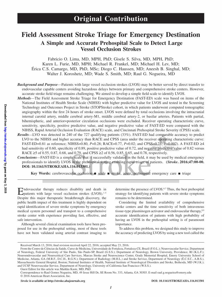
Subjects and MethodsThe FAST-ED scale (Facial Palsy [scored 0–1], Arm weakness [0–2], Speech changes [0–2], Time [documentation for decision making but no points], Eye deviation [0–2], and Denial/neglect [0–2]) was designed based on items of the National Institutes of Health Stroke Scale (NIHSS) with higher predictive value for LVOS. In addition, time was included considering its importance in the prehospital deci-sion algorithm. For the current analysis, the FAST-ED score was derived from the NIHSS score assessed by certified research person-nel at hospital admission and is shown in Table 1.10
The scale was tested on data from 741 consecutive patients enrolled in a prospective cohort study at 2 university-based hospi-tals, the Screening Technology and Outcomes Project in Stroke (STOPStroke), in which admission noncontrast computed tomog-raphy scans and computed tomographic angiography (CTA) were obtained in all patients suspected of having ischemic stroke (stroke, transient ischemic attack, or stroke mimics) in the first 24 hours of symptom onset. Patients were excluded if iodinated contrast agent administration was contraindicated (ie, history of contrast agent allergy, pregnancy, congestive heart failure, and increased creatinine level) or if there was evidence of intracranial hemorrhage on noncon-trast computed tomography. The STOPStroke study received insti-tutional review board approval at both participating institutions and was Health Insurance Portability and Accountability Act compliant.
For this study, patients with unilateral acute complete symptom-atic occlusion of the intracranial internal carotid artery (intracranial ICA), M1 and M2 segments of the middle cerebral artery (MCA) and basilar artery were selected and compared with patients without a proximal intracranial occlusion. Patients with symptomatic bilateral and anterior+posterior circulation occlusions were excluded from the analysis. Our prespecified hypothesis was that the FAST-ED would have similar or higher accuracy than other preexisting scales.
Image Protocol and ReviewThe STOPStroke noncontrast computed tomography and CTA protocol is described elsewhere.11 Image review was independently performed on a picture archiving and communication system workstation (Impax; AGFA Technical Imaging Systems, Richfield Park, NJ) by a board-certified neuroradiologist and a clinical neurologist experienced in stroke imaging interpretation. Disagreements in readings were resolved by consensus. Reviewers were blinded to follow-up clinical and imag-ing findings but had information in regard to the patients’ age, sex, and presenting clinical symptoms. Neither of the reviewers had participated in the selection of the patients. For every image, vessels were graded for the presence or absence of total occlusion according to a 5-point level of certainty score (score 5, definitely present; score 4, probably present; score 3, equivocal; score 2, probably absent; and score 1, defi-nitely absent). Those subjects with equivocal scores were excluded from the analysis. The site of intracranial occlusion was defined as the most proximal site of occlusion (intracranial ICA, MCA-M1, MCA-M2, and basilar). Functional outcomes were assessed with the use of the modi-fied Rankin scale (mRS) at 6 months.
Statistical AnalysisContinuous variables are reported as mean±SD or as median±interquartile range (IQR). Categorical variables were reported as proportions.
The Spearman test was used to test the linear correlation of the NIHSS and the FAST-ED scores. Receiver operating characteristics (ROC) curve analysis was used as the primary analysis to test whether the FAST-ED had higher discrimination ability than other similar previous published scales (the Rapid Arterial Occlusion Evaluation [RACE] Scale, the Cincinnati Prehospital Stroke Severity [CPSS] scale, and the NIHSS).12,13 The areas under the curve were compared with the FAST-ED as the reference.14 Calibration of FAST-ED was assessed graphically and by the use of the Hosmer and Lemeshow test.15 Given the potential influence of time to presentation on NIHSS, sensitivity analyses were performed including only those patients who underwent CTA within 12 hours from symptom onset and again in those patients who underwent CTA within 6 hours from symptom onset. Partial occlusions on conventional angiography are generally classified as total occlusion on CTA.16 However, as some patients were still classified as partial occlusion on CTA, we also performed a sensitivity analysis including those patients with partial occlusion on CTA.
Sensitivity, specificity, positive predictive value, negative pre-dictive value, and accuracy were calculated using several different thresholds of the FAST-ED. The Youden Index was used to evaluate the optimal threshold of the FAST-ED scale.17 Prespecified published thresholds of the other scales and a cutoff of 6 and 10 points in the NIHSS were used for comparison.10,18
The distribution of the FAST-ED was also compared according to the mRS at 6 months (dichotomized as good, mRS score of ≤2 and poor outcome, mRS score of >2). The Kruskal–Wallis test was used to compare the distribution of the FAST-ED scores according to the most proximal site of occlusion (intracranial ICA, MCA-M1, MCA-M2, and basilar). A 2-sided P value of <0.05 was considered sig-nificant. All statistical analysis was performed using SPSS software (version 20.0).
ResultsSeven hundred twenty-seven qualifying patients were selected. The mean age was 68.1±15.4 years, median baseline NIHSS
Table 1. The FAST-ED Scale and Its Correspondence to the NIHSS
Item FAST-ED ScoreNIHSS Score
Source
Facial palsy
Normal or minor paralysis 0 0–1
Partial or complete paralysis 1 2–3
Arm weakness
No drift 0 0
Drift or some effort against gravity 1 1–2
No effort against gravity or no movement
2 3–4
Speech changes
Absent 0 0
Mild to moderate 1 1
Severe, global aphasia, or mute 2 2–3
Eye deviation
Absent 0 0
Partial 1 1
Forced deviation 2 2
Denial/Neglect
Absent 0 0
Extinction to bilateral simultaneous stimulation in only 1 sensory modality
1 1
Does not recognize own hand or orients only to one side of the body
2 2
FAST-ED indicates Field Assessment Stroke Triage for Emergency Destination; and NIHSS, National Institutes of Health Stroke Scale.
by guest on May 3, 2018
http://stroke.ahajournals.org/D
ownloaded from
Lima et al FAST-ED to Detect Large Vessel Occlusion Strokes 3
was 5 (IQR, 2–12), and 52% were males. LVO was detected in 240 (33%) subjects. Fifty-three (7.3%) subjects had occlu-sion of the intracranial ICA, 98 (13.5%) of the MCA-M1, 74 (10.2%) of the MCA-M2, and 15 (2.1%) of the basilar artery. As expected, the FAST-ED had a strong correlation with NIHSS (r=0.92; P<0.001).
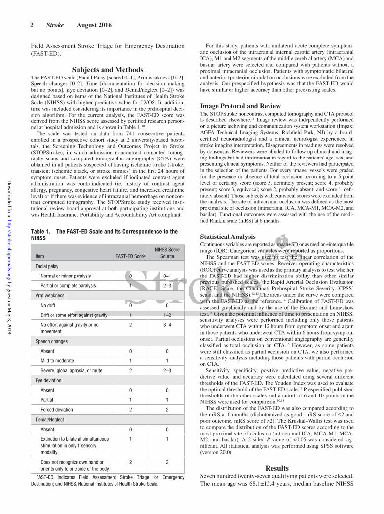
The FAST-ED scale had comparable accuracy to predict LVO to the more complex NIHSS and higher accuracy than RACE and CPSS (area under the ROC curve: FAST-ED=0.81 as reference; NIHSS=0.80, P=0.28; RACE=0.77, P=0.02; and CPSS=0.75, P=0.002; Figure 1A). A similar pattern was seen when the analysis was repeated for those patients who underwent CTA within 12 hours (n=393; area under the ROC curve: FAST-ED 0.83 as reference; NIHSS=0.81, P=0.17; RACE=0.79, P=0.03; and CPSS=0.769, P=0.001; Figure 1B) and within 6 hours from symptom onset (n=360; area under
the ROC curve: FAST-ED=0.83 as reference; NIHSS=0.81, P=0.26; RACE=0.79, P=0.08; and 0.77, P=0.02; Figure 1C).
Ninety-four patients had partial occlusions on CTA. A simi-lar pattern was observed when those patients were included with FAST-ED having a similar area under the curve when compared with the NIHSS but larger when compared with RACE and CPSS (area under the ROC curve: FAST-ED=0.79 as reference; NIHSS=0.77, P=0.24; RACE=0.74, P=0.003; and CPSS=0.73, P<0.001).
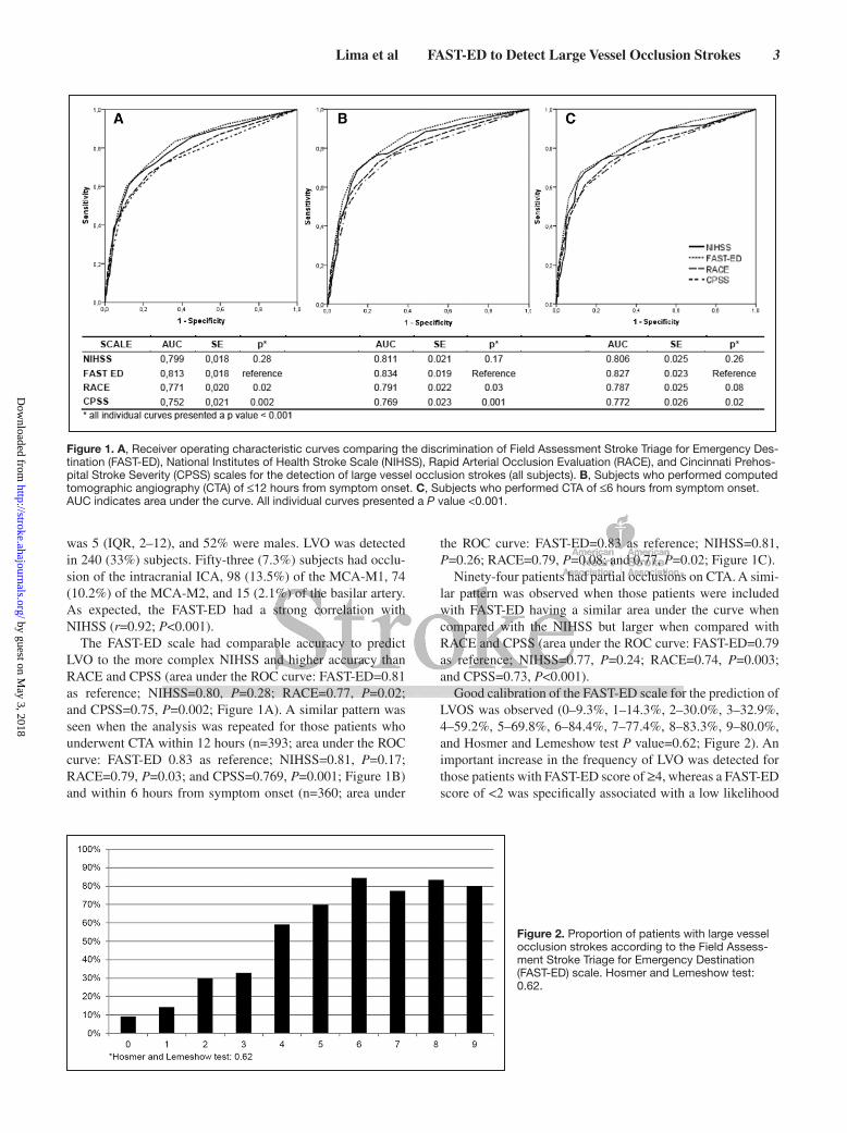
Good calibration of the FAST-ED scale for the prediction of LVOS was observed (0–9.3%, 1–14.3%, 2–30.0%, 3–32.9%, 4–59.2%, 5–69.8%, 6–84.4%, 7–77.4%, 8–83.3%, 9–80.0%, and Hosmer and Lemeshow test P value=0.62; Figure 2). An important increase in the frequency of LVO was detected for those patients with FAST-ED score of ≥4, whereas a FAST-ED score of <2 was specifically associated with a low likelihood
Figure 1. A, Receiver operating characteristic curves comparing the discrimination of Field Assessment Stroke Triage for Emergency Des-tination (FAST-ED), National Institutes of Health Stroke Scale (NIHSS), Rapid Arterial Occlusion Evaluation (RACE), and Cincinnati Prehos-pital Stroke Severity (CPSS) scales for the detection of large vessel occlusion strokes (all subjects). B, Subjects who performed computed tomographic angiography (CTA) of ≤12 hours from symptom onset. C, Subjects who performed CTA of ≤6 hours from symptom onset. AUC indicates area under the curve. All individual curves presented a P value <0.001.
Figure 2. Proportion of patients with large vessel occlusion strokes according to the Field Assess-ment Stroke Triage for Emergency Destination (FAST-ED) scale. Hosmer and Lemeshow test: 0.62.
by guest on May 3, 2018
http://stroke.ahajournals.org/D
ownloaded from
4 Stroke August 2016
of LVO. There was a steady increase in the frequency of poor outcome (6-month mRS score of >2) with higher FAST-ED scores (0–11.8%, 1–25.7%, 2–41.6%, 3–42.2%, 4–52.4%, 5–60.3%, 6–85.7%, 7–85.7%, 8%–100%, and 9% to 100%; Figure 2).
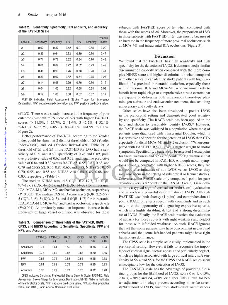
Better performance of FAST-ED according to the Youden Index could be shown at 2 distinct thresholds of ≥3 (Youden Index=0.490) and ≥4 (Youden Index=0.491; Table 2). A threshold of ≥3 and ≥4 in the FAST-ED for LVO had a sen-sitivity of 0.71 and 0.60, specificity of 0.78 and 0.89, posi-tive predictive value of 0.62 and 0.72, and negative predictive value of 0.84 and 0.82 versus RACE ≥5, 0.55, 0.87, 0.68, and 0.79 and CPSS ≥2, 0.56, 0.85, 0.65, and 0.78, NIHSS ≥6 0.76, 0.70, 0.55, and 0.85 and NIHSS ≥10 0.64, 0.85, 0.68, and 0.83, respectively (Table 3).
The median NIHSS was 14.5 (IQR, 6.2–19.7), 14 (IQR, 9.7–17), 8 (IQR, 4–15.5), and 17 (IQR, 14–32) for intracranial ICA, MCA-M1, MCA-M2, and basilar occlusion, respectively (P=0.003). The median FAST-ED score was 5 (IQR, 2.2–6.7), 5 (IQR, 3–6), 3 (IQR, 2–5), and 5 (IQR, 1–7) for intracranial ICA, MCA-M1, MCA-M2, and basilar occlusion, respectively (P<0.001). As previously noted, an important increase in the frequency of large vessel occlusion was observed for those
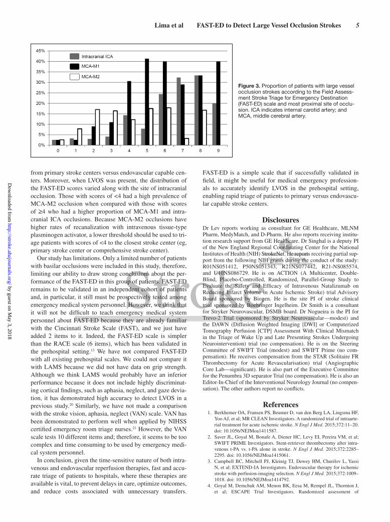
subjects with FAST-ED score of ≥4 when compared with those with the scores of <4. Moreover, the proportion of LVO in those subjects with FAST-ED of ≥4 was mostly because of an increase in the frequency of more proximal occlusions such as MCA-M1 and intracranial ICA occlusions (Figure 3).
DiscussionWe found that the FAST-ED has high sensitivity and high specificity for the detection of LVOS. It demonstrated a similar discrimination capacity when compared with the more com-plex NIHSS score and higher discrimination when compared with other scales. It can identify stroke patients with high like-lihood of a proximal intracranial occlusion, especially those with intracranial ICA and MCA-M1, who are most likely to benefit from rapid triage to comprehensive stroke centers that are capable of delivering both intravenous tissue-type plas-minogen activator and endovascular treatment, thus avoiding unnecessary and costly delays.
Other scales have also been developed to predict LVOS in the prehospital setting and demonstrated good sensitiv-ity and specificity. The RACE scale has been applied in the field and shown to reasonably identify LVOS.13 However, the RACE scale was validated in a population where most of patients were diagnosed with transcranial Doppler, which is less sensitive and specific for the detection of LVOS than CTA especially for distal MCA-M1 and M2 occlusion.19 When com-pared with FAST-ED, RACE gives a higher weight to motor symptoms. Specifically, a patient can be assessed 1 extra point for facial weakness and ≤2 extra points for leg weakness that would not be computed in FAST-ED. Although motor symp-toms strongly correlated with higher NIHSS scores, they are not good discriminators of non-LVOS versus LVOS as they may also occur in the setting of subcortical or lacunar strokes. Conversely, the RACE scale only computes 1 point for gaze deviation (versus ≤2 points in the FAST-ED scale). Gaze devi-ation is a typical sign of cortical (or brain stem) dysfunction and as such is a powerful discriminator of LVOS. Although FAST-ED tests both fluency (1 point) and comprehension (1 point), RACE only tests speech with commands and as such may miss the opportunity of diagnosing expressive aphasia, which is a highly disabling deficit and a strong discrimina-tor of LVOS. Finally, the RACE scale restricts the evaluation of aphasia for those subjects with right weakness and neglect for those with left-sided weakness. As such, RACE ignores the fact that some patients may have concomitant neglect and aphasia and that some left-handed patients might have right hemisphere dominance.
The CPSS scale is a simple scale easily implemented in the prehospital setting. However, it fails to recognize the impor-tance of cortical signs, such as aphasia and particularly neglect, which are highly associated with large cortical infarcts. A sen-sitivity of 56% and 55% for the CPSS and RACE scales seem unacceptably low for the detection of LVOS.
The FAST-ED scale has the advantage of providing 3 dis-tinct groups for the likelihood of LVOS: score 0 to 1, <15%; 2 to 3, ≈30%; and ≥4, ≈60% or higher. This allows for bet-ter adjustments in triage process according to stroke sever-ity/likelihood of LVOS, time from stroke onset, and distances
Table 2. Sensitivity, Specificity, PPV and NPV, and accuracy of the FAST-ED Scale
FAST-ED Sensitivity Specificity PPV NPV AccuracyYouden Index
≥1 0.92 0.37 0.42 0.91 0.55 0.29
≥2 0.83 0.64 0.53 0.89 0.70 0.47
≥3 0.71 0.78 0.62 0.84 0.76 0.49
≥4 0.61 0.89 0.72 0.82 0.79 0.49
≥5 0.48 0.93 0.76 0.78 0.78 0.41
≥6 0.30 0.97 0.82 0.74 0.75 0.27
≥7 0.14 0.98 0.79 0.70 0.70 0.12
≥8 0.04 1.00 0.82 0.68 0.68 0.03
≥9 0.17 1.00 0.80 0.67 0.67 0.17
FAST-ED indicates Field Assessment Stroke Triage for Emergency Destination; NPV, negative predictive value; and PPV, positive predictive value.
Table 3. Comparison of Thresholds of the FAST-ED, RACE, CPSS, and NIHSS According to Sensitivity, Specificity, PPV and NPV, and Accuracy
FAST-ED ≥3
FAST-ED ≥4
RACE ≥5
CPSS ≥2
NIHSS ≥6
NIHSS ≥10
Sensitivity 0.71 0.61 0.55 0.56 0.76 0.64
Specificity 0.78 0.89 0.87 0.85 0.70 0.85
PPV 0.62 0.72 0.68 0.65 0.55 0.68
NPV 0.84 0.82 0.79 0.78 0.85 0.83
Accuracy 0.76 0.79 0.77 0.75 0.72 0.78
CPSS indicates Cincinnati Prehospital Stroke Severity Scale; FAST-ED, Field Assessment Stroke Triage for Emergency Destination; NIHSS, National Institutes of Health Stroke Scale; NPV, negative predictive value; PPV, positive predictive value; and RACE, Rapid Arterial Occlusion Evaluation.
by guest on May 3, 2018
http://stroke.ahajournals.org/D
ownloaded from
Lima et al FAST-ED to Detect Large Vessel Occlusion Strokes 5
from primary stroke centers versus endovascular capable cen-ters. Moreover, when LVOS was present, the distribution of the FAST-ED scores varied along with the site of intracranial occlusion. Those with scores of <4 had a high prevalence of MCA-M2 occlusion when compared with those with scores of ≥4 who had a higher proportion of MCA-M1 and intra-cranial ICA occlusions. Because MCA-M2 occlusions have higher rates of recanalization with intravenous tissue-type plasminogen activator, a lower threshold should be used to tri-age patients with scores of <4 to the closest stroke center (eg, primary stroke center or comprehensive stroke center).
Our study has limitations. Only a limited number of patients with basilar occlusions were included in this study, therefore, limiting our ability to draw strong conclusions about the per-formance of the FAST-ED in this group of patients. FAST-ED remains to be validated in an independent cohort of patients and, in particular, it still must be prospectively tested among emergency medical system personnel. However, we think that it will not be difficult to teach emergency medical system personnel about FAST-ED because they are already familiar with the Cincinnati Stroke Scale (FAST), and we just have added 2 items to it. Indeed, the FAST-ED scale is simpler than the RACE scale (6 items), which has been validated in the prehospital setting.13 We have not compared FAST-ED with all existing prehospital scales. We could not compare it with LAMS because we did not have data on grip strength. Although we think LAMS would probably have an inferior performance because it does not include highly discriminat-ing cortical findings, such as aphasia, neglect, and gaze devia-tion, it has demonstrated high accuracy to detect LVOS in a previous study.20 Similarly, we have not made a comparison with the stroke vision, aphasia, neglect (VAN) scale. VAN has been demonstrated to perform well when applied by NIHSS certified emergency room triage nurses.21 However, the VAN scale tests 10 different items and; therefore, it seems to be too complex and time consuming to be used by emergency medi-cal system personnel.
In conclusion, given the time-sensitive nature of both intra-venous and endovascular reperfusion therapies, fast and accu-rate triage of patients to hospitals, where these therapies are available is vital, to prevent delays in care, optimize outcomes, and reduce costs associated with unnecessary transfers.
FAST-ED is a simple scale that if successfully validated in field, it might be useful for medical emergency profession-als to accurately identify LVOS in the prehospital setting, enabling rapid triage of patients to primary versus endovascu-lar capable stroke centers.
DisclosuresDr Lev reports working as consultant for GE Healthcare, MLNM Pharm, MedyMatch, and D-Pharm. He also reports receiving institu-tion research support from GE Healthcare. Dr Singhal is a deputy PI of the New England Regional Coordinating Center for the National Institutes of Health (NIH) StrokeNet. He reports receiving partial sup-port from the following NIH grants during the conduct of the study: R01NS051412, P50NS051343, R21NS077442, R21-NS085574, and U10NS086729. He is on ACTION (A Multicenter, Double-Blind, Placebo-Controlled, Randomized, Parallel-Group Study to Evaluate the Safety and Efficacy of Intravenous Natalizumab on Reducing Infarct Volume in Acute Ischemic Stroke) trial Advisory Board sponsored by Biogen. He is the site PI of stroke clinical trial sponsored by Boehringer Ingelheim. Dr Smith is a consultant for Stryker Neurovascular, DSMB board. Dr Nogueira is the PI for Trevo-2 Trial (sponsored by Stryker Neurovascular—modest) and the DAWN (Diffusion Weighted Imaging [DWI] or Computerized Tomography Perfusion [CTP] Assessment With Clinical Mismatch in the Triage of Wake Up and Late Presenting Strokes Undergoing Neurointervention) trial (no compensation). He is on the Steering Committee of SWIFT Trial (modest) and SWIFT Prime (no com-pensation). He receives compensation from the STAR (Solitaire FR Thrombectomy for Acute Revascularisation) trial (Angiographic Core Lab—significant). He is also part of the Executive Committee for the Penumbra 3D separator Trial (no compensation). He is also an Editor-In-Chief of the Interventional Neurology Journal (no compen-sation). The other authors report no conflicts.
References 1. Berkhemer OA, Fransen PS, Beumer D, van den Berg LA, Lingsma HF,
Yoo AJ, et al; MR CLEAN Investigators. A randomized trial of intraarte-rial treatment for acute ischemic stroke. N Engl J Med. 2015;372:11–20. doi: 10.1056/NEJMoa1411587.
2. Saver JL, Goyal M, Bonafe A, Diener HC, Levy EI, Pereira VM, et al; SWIFT PRIME Investigators. Stent-retriever thrombectomy after intra-venous t-PA vs. t-PA alone in stroke. N Engl J Med. 2015;372:2285–2295. doi: 10.1056/NEJMoa1415061.
3. Campbell BC, Mitchell PJ, Kleinig TJ, Dewey HM, Churilov L, Yassi N, et al; EXTEND-IA Investigators. Endovascular therapy for ischemic stroke with perfusion-imaging selection. N Engl J Med. 2015;372:1009–1018. doi: 10.1056/NEJMoa1414792.
4. Goyal M, Demchuk AM, Menon BK, Eesa M, Rempel JL, Thornton J, et al; ESCAPE Trial Investigators. Randomized assessment of
Figure 3. Proportion of patients with large vessel occlusion strokes according to the Field Assess-ment Stroke Triage for Emergency Destination (FAST-ED) scale and most proximal site of occlu-sion. ICA indicates internal carotid artery; and MCA, middle cerebral artery.
by guest on May 3, 2018
http://stroke.ahajournals.org/D
ownloaded from
6 Stroke August 2016
rapid endovascular treatment of ischemic stroke. N Engl J Med. 2015;372:1019–1030. doi: 10.1056/NEJMoa1414905.
5. Jovin TG, Chamorro A, Cobo E, de Miquel MA, Molina CA, Rovira A, et al; REVASCAT Trial Investigators. Thrombectomy within 8 hours after symptom onset in ischemic stroke. N Engl J Med. 2015;372:2296–2306. doi: 10.1056/NEJMoa1503780.
6. Perez de la Ossa N, Carrera D, Gorchs M, Querol M, Millan M, Gomis M, et al. Design and validation of a prehospital stroke scale to predict large arterial occlusion: the rapid arterial occlusion evaluation scale. Stroke. 2014;45:87–91.
7. Katz BS, McMullan JT, Sucharew H, Adeoye O, Broderick JP. Design and validation of a prehospital scale to predict stroke severity: Cincinnati Prehospital Stroke Severity Scale. Stroke. 2015;46:1508–1512. doi: 10.1161/STROKEAHA.115.008804.
8. Lees KR, Bluhmki E, von Kummer R, Brott TG, Toni D, Grotta JC, et al; ECASS, ATLANTIS, NINDS and EPITHET rt-PA Study Group. Time to treatment with intravenous alteplase and outcome in stroke: an updated pooled analysis of ECASS, ATLANTIS, NINDS, and EPITHET trials. Lancet. 2010;375:1695–1703. doi: 10.1016/S0140-6736(10)60491-6.
9. Sheth SA, Jahan R, Gralla J, Pereira VM, Nogueira RG, Levy EI, et al; SWIFT-STAR Trialists. Time to endovascular reperfusion and degree of disability in acute stroke. Ann Neurol. 2015;78:584–593. doi: 10.1002/ana.24474.
10. Fischer U, Arnold M, Nedeltchev K, Brekenfeld C, Ballinari P, Remonda L, et al. NIHSS score and arteriographic findings in acute ischemic stroke. Stroke. 2005;36:2121–2125. doi: 10.1161/01.STR.0000182099.04994.fc.
11. Camargo EC, Furie KL, Singhal AB, Roccatagliata L, Cunnane ME, Halpern EF, et al. Acute brain infarct: detection and delineation with CT angiographic source images versus nonenhanced CT scans. Radiology. 2007;244:541–548. doi: 10.1148/radiol.2442061028.
12. Katz BS, McMullan JT, Sucharew H, Adeoye O, Broderick JP. Design and validation of a prehospital scale to predict stroke severity: Cincinnati Prehospital Stroke Severity Scale. Stroke. 2015;46:1508–1512. doi: 10.1161/STROKEAHA.115.008804.
13. Perez de la Ossa N, Carrera D, Gorchs M, Querol M, Millan M, Gomis M, et al. Design and validation of a prehospital stroke scale to predict large arterial occlusion: the rapid arterial occlusion evaluation scale. Stroke. 2015;45:87–91.
14. Hanley JA, McNeil BJ. A method of comparing the areas under receiver operating characteristic curves derived from the same cases. Radiology. 1983;148:839–843. doi: 10.1148/radiology.148.3.6878708.
15. Cook NR. Use and misuse of the receiver operating characteristic curve in risk prediction. Circulation. 2007;115:928–935. doi: 10.1161/CIRCULATIONAHA.106.672402.
16. Frölich AM, Psychogios MN, Klotz E, Schramm R, Knauth M, Schramm P. Antegrade flow across incomplete vessel occlusions can be distin-guished from retrograde collateral flow using 4-dimensional computed tomographic angiography. Stroke. 2012;43:2974–2979. doi: 10.1161/STROKEAHA.112.668889.
17. Youden WJ. Index for rating diagnostic tests. Cancer. 1950;3:32–35. 18. Heldner MR, Zubler C, Mattle HP, Schroth G, Weck A, Mono ML, et al.
National Institutes of Health stroke scale score and vessel occlusion in 2152 patients with acute ischemic stroke. Stroke. 2013;44:1153–1157. doi: 10.1161/STROKEAHA.111.000604.
19. Suwanwela NC, Phanthumchinda K, Suwanwela N. Transcranial doppler sonography and CT angiography in patients with athero-thrombotic middle cerebral artery stroke. AJNR Am J Neuroradiol. 2002;23:1352–1355.
20. Nazliel B, Starkman S, Liebeskind DS, Ovbiagele B, Kim D, Sanossian N, et al. A brief prehospital stroke severity scale identifies ischemic stroke patients harboring persisting large arterial occlusions. Stroke. 2008;39:2264–2267. doi: 10.1161/STROKEAHA.107.508127.
21. Teleb MS, Ver Hage A, Carter J, Jayaraman MV, McTaggart RA. Stroke vision, aphasia, neglect (VAN) assessment—a novel emergent large ves-sel occlusion screening tool: pilot study and comparison with current clinical severity indices [published online ahead of print February 17, 2016]. J Neurointerv Surg. http://jnis.bmj.com/content/early/2016/02/17/neurintsurg-2015–012131.long. Accessed March 9, 2016.
by guest on May 3, 2018
http://stroke.ahajournals.org/D
ownloaded from
Raul G. NogueiraC.S. Camargo, Diogo C. Haussen, Aneesh B. Singhal, Walter J. Koroshetz, Wade S. Smith and Fabricio O. Lima, Gisele S. Silva, Karen L. Furie, Michael R. Frankel, Michael H. Lev, Érica
Prehospital Scale to Detect Large Vessel Occlusion StrokesField Assessment Stroke Triage for Emergency Destination: A Simple and Accurate
Print ISSN: 0039-2499. Online ISSN: 1524-4628 Copyright © 2016 American Heart Association, Inc. All rights reserved.
is published by the American Heart Association, 7272 Greenville Avenue, Dallas, TX 75231Stroke published online June 30, 2016;Stroke.
http://stroke.ahajournals.org/content/early/2016/06/30/STROKEAHA.116.013301World Wide Web at:
The online version of this article, along with updated information and services, is located on the
http://stroke.ahajournals.org/content/suppl/2017/07/10/STROKEAHA.116.013301.DC1Data Supplement (unedited) at:
http://stroke.ahajournals.org//subscriptions/
is online at: Stroke Information about subscribing to Subscriptions:
http://www.lww.com/reprints Information about reprints can be found online at: Reprints:
document. Permissions and Rights Question and Answer process is available in the
Request Permissions in the middle column of the Web page under Services. Further information about thisOnce the online version of the published article for which permission is being requested is located, click
can be obtained via RightsLink, a service of the Copyright Clearance Center, not the Editorial Office.Strokein Requests for permissions to reproduce figures, tables, or portions of articles originally publishedPermissions:
by guest on May 3, 2018
http://stroke.ahajournals.org/D
ownloaded from
iris
Abstract 27
緊急搬送先を決めるための現場での脳卒中トリアージ搬送前に大血管閉塞性脳卒中を見分けるための簡単で正確な評価基準Field Assessment Stroke Triage for Emergency DestinationA Simple and Accurate Prehospital Scale to Detect Large Vessel Occlusion Strokes
Fabricio O. Lima, MD, MPH, PhD1; Gisele S. Silva, MD, MPH, PhD2; Karen L. Furie, MD, MPH3, et al.1Centro de Ciências da Saúde, Curso de Medicina, Universidade de Fortaleza, Fortaleza-CE, Brazil; 2Neurovascular Service, Department of Neurology, Federal University of São Paulo, São Paulo-SP, Brazil; 3Department of Neurology, Brown University, Providence, RI
背景および目的:大血管閉塞性脳卒中(LVOS)患者は,血管内治療が可能な医療施設に直接搬送すれば一次脳卒中センターから包括的脳卒中センターへの転送による危険な治療の遅れを避けられ有益であろう。しかし,現場での正確な脳卒中トリアージはまだ難しい状況にある。我々は,現場で容易に LVOS を特定できる評価基準の開発に取り組んだ。方法:米国国立衛生研究所脳卒中スケール(NIHSS)で LVOS の陽性予測値が高い項目を参考にして Field Assessment Stroke Triage for Emergency Destination
(FAST-ED)Scale を作成し,脳卒中発症後 24 時間以内 に CT 血 管 造 影 検 査 を 受 け た Screening Technology and Outcomes Project in Stroke(STOPStroke)のコホートで検証した。LVOS は頭蓋内内頸動脈,中大脳動脈-M1,中大脳動脈 -M2,または脳底動脈の完全閉塞と定義した。部分閉塞,両側大脳半球に及ぶ閉塞,前方+後
方循環系の閉塞は除外した。FAST-ED の ROC 曲線,感度,特異度,陽性予測値,陰性予測値を NIHSS,Rapid Arterial Occlusion Evaluation(RACE)Scale,Cincinnati Prehospital Stroke Severity(CPSS)Scale と比較した。結果:適格患者 727 例中 240 例(33%)に LVOS が認められた。FAST-ED は NIHSS と同等の LVOS 予測精度を 示し,RACE および CPSS より精度が高かった(ROC 曲線下面積:FAST-ED = 0.81 を基準値として,NIHSS =0.80,P = 0.28。;RACE = 0.77,P = 0.02;CPSS = 0.75,P = 0.002)。FAST-ED が 4 以上の場合,感度 0.60,特異度 0.89,陽性予測値 0.72,陰性予測値 0.82 であったのに対し,RACE は 5 以上でそれぞれ 0.55,0.87,0.68,0.79であり,CPSS は 2 以上で 0.56,0.85,0.65,0.78 であった。結論:FAST-ED は簡単な評価尺度であり,現場での検証に成功すれば救急隊員による病院搬送前の迅速な LVOSのトリアージが可能になる。
Stroke. 2016; 47: 1997-2002. DOI: 10.1161/STROKEAHA.116.013301.
Abstract
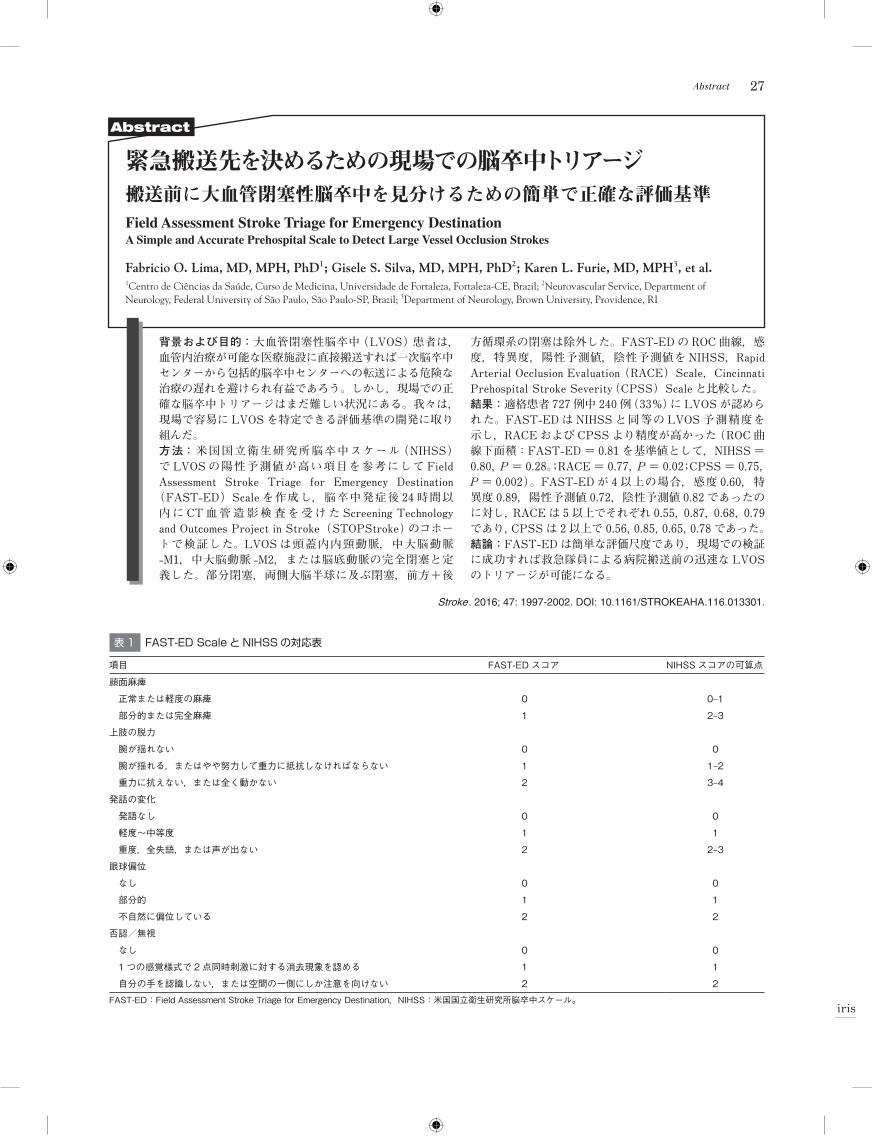
表 1 FAST-ED Scale と NIHSS の対応表
項目 FAST-EDスコア NIHSSスコアの可算点
顔面麻痺
正常または軽度の麻痺 0 0–1
部分的または完全麻痺 1 2–3
上肢の脱力
腕が揺れない 0 0
腕が揺れる,またはやや努力して重力に抵抗しなければならない 1 1–2
重力に抗えない,または全く動かない 2 3–4
発話の変化
発語なし 0 0
軽度~中等度 1 1
重度,全失語,または声が出ない 2 2–3
眼球偏位
なし 0 0
部分的 1 1
不自然に偏位している 2 2
否認/無視
なし 0 0
1つの感覚様式で 2点同時刺激に対する消去現象を認める 1 1
自分の手を認識しない,または空間の一側にしか注意を向けない 2 2
FAST-ED:FieldAssessmentStrokeTriageforEmergencyDestination,NIHSS:米国国立衛生研究所脳卒中スケール。
Recommended