1
Fecal leukocytes in children infected with diarrheagenic E. coli 1
2
Erik H. Mercado1, Theresa J. Ochoa1,2, Lucie Ecker3, Martin Cabello1, David 3
Durand1, Francesca Barletta1, Margarita Molina3, Ana I. Gil3, Luis Huicho1,4,5, 4
Claudio F. Lanata3,6 and Thomas G. Cleary2
5
6
1 Universidad Peruana Cayetano Heredia, Lima, Peru; 2 University of Texas 7
School of Public Health, Houston, Texas, United States; 3 Instituto de 8
Investigación Nutricional, Lima, Peru; 4 Universidad Nacional Mayor de San 9
Marcos, Lima, Peru; 5 Instituto Nacional de Salud del Niño, Lima, Peru; 10
6 Universidad Peruana de Ciencias Aplicadas, Lima, Peru. 11
12
*Corresponding author: 13
Theresa J. Ochoa, MD 14
Instituto de Medicina Tropical “Alexander von Humboldt” 15
Universidad Peruana Cayetano Heredia 16
Av. Honorio Delgado 430 17
San Martin de Porras, Lima 31, Perú 18
Phone 51-1-482-3910 19
Fax: 51-1-482-3404 20
E-mail: [email protected] 21
Copyright © 2011, American Society for Microbiology and/or the Listed Authors/Institutions. All Rights Reserved.J. Clin. Microbiol. doi:10.1128/JCM.02199-10 JCM Accepts, published online ahead of print on 16 February 2011
on January 12, 2019 by guesthttp://jcm
.asm.org/
Dow
nloaded from
2
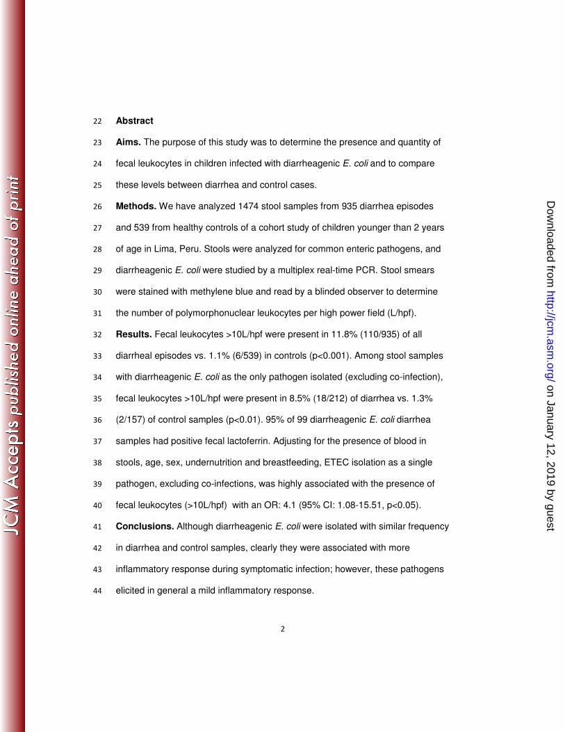
Abstract 22
Aims. The purpose of this study was to determine the presence and quantity of 23
fecal leukocytes in children infected with diarrheagenic E. coli and to compare 24
these levels between diarrhea and control cases. 25
Methods. We have analyzed 1474 stool samples from 935 diarrhea episodes 26
and 539 from healthy controls of a cohort study of children younger than 2 years 27
of age in Lima, Peru. Stools were analyzed for common enteric pathogens, and 28
diarrheagenic E. coli were studied by a multiplex real-time PCR. Stool smears 29
were stained with methylene blue and read by a blinded observer to determine 30
the number of polymorphonuclear leukocytes per high power field (L/hpf). 31
Results. Fecal leukocytes >10L/hpf were present in 11.8% (110/935) of all 32
diarrheal episodes vs. 1.1% (6/539) in controls (p<0.001). Among stool samples 33
with diarrheagenic E. coli as the only pathogen isolated (excluding co-infection), 34
fecal leukocytes >10L/hpf were present in 8.5% (18/212) of diarrhea vs. 1.3% 35
(2/157) of control samples (p<0.01). 95% of 99 diarrheagenic E. coli diarrhea 36
samples had positive fecal lactoferrin. Adjusting for the presence of blood in 37
stools, age, sex, undernutrition and breastfeeding, ETEC isolation as a single 38
pathogen, excluding co-infections, was highly associated with the presence of 39
fecal leukocytes (>10L/hpf) with an OR: 4.1 (95% CI: 1.08-15.51, p<0.05). 40
Conclusions. Although diarrheagenic E. coli were isolated with similar frequency 41
in diarrhea and control samples, clearly they were associated with more 42
inflammatory response during symptomatic infection; however, these pathogens 43
elicited in general a mild inflammatory response. 44
on January 12, 2019 by guesthttp://jcm
.asm.org/
Dow
nloaded from
3
Keywords: Fecal leukocytes, diarrheagenic E. coli, diarrhea, children, fecal 45
lactoferrin. 46
47
INTRODUCTION 48
Diarrheagenic E. coli as a group are the most common enteric pathogens 49
in children in developing countries, responsible for 30% to 40% of all diarrhea 50
episodes (20). However, some of these pathogens can be found with similar 51
frequency in asymptomatic controls, depending on several factors such as age of 52
the patient and host susceptibility. The presence of fecal leukocytes in stool 53
samples is used as an indicator of inflammatory diarrhea. A brisk inflammatory 54
response is associated with invasive pathogens such as Shigella, Salmonella or 55
Campylobacter (4). However, other non-invasive pathogens can elicit a mild 56
inflammatory response as a result of the interaction of the pathogen with the 57
host´s enteric cells. An additional method to determine an inflammatory response 58
in the gut is the measurement of fecal lactoferrin, an anti-microbial protein 59
present in several human secretions (milk, saliva, tears, etc) and in the granules 60
of the neutrophils (6). 61
Remarkable progress has been made to identify virulence determinants 62
required to mediate the pathogenesis of the different diarrheagenic E. coli 63
pathotypes. However, there are few data on the level of fecal leukocytes and 64
fecal lactoferrin as markers of inflammatory response in children infected with 65
these pathogens. Although the mechanisms of action and the pathogenesis of 66
these bacteria are diverse, our hypothesis was that children infected with 67
diarrheagenic E. coli and who develop diarrhea elicit an inflammatory response 68
on January 12, 2019 by guesthttp://jcm
.asm.org/
Dow
nloaded from

4
greater than children with asymptomatic colonization with these pathogens. 69
Therefore, we conducted this study to determine the presence and quantity of 70
fecal leukocytes and lactoferrin in children infected with diarrheagenic E. coli and 71
to compare these levels between diarrhea and control cases. 72
73
PATIENTS AND METHODS. 74
Patients. This study was part of a prospective passive surveillance cohort 75
diarrhea study in children followed from 2 to 24 months of age. The study was 76
conducted in peri-urban communities of Lima, Peru, between September 2006 77
and December 2007 (first cohort, 1034 children) (19); and from January to July 78
2008 (second cohort, 529 children). Diarrhea was defined as the presence of ≥ 3 79
liquid or semi-liquid stools in 24 hours or ≥ 1 bloody stool. Control stool samples 80
were collected from randomly selected healthy children without diarrhea 7 days 81
before and after the stool sample collection. Clinical information of the diarrheal 82
episodes was obtained from the medical records filled by study doctors. We used 83
a modified Vesikari score to determine the severity of the diarrhea episodes (23, 84
19). Weight and height measurements were obtained at the study clinic in all 85
children at 12 months of age. 86
Pathogen determination. Stool samples were analyzed for common enteric 87
pathogens; ELISA was used for rotavirus; routine stool cultures were used to 88
detect Salmonella, Shigella, Campylobacter and Vibrio sp; and the DNA from five 89
lactose positives E. coli colonies were studied by a multiplex real-time PCR to 90
identify enterotoxigenic (ETEC), enteropathogenic (EPEC), shiga toxin-producing 91
(STEC), enteroinvasive (EIEC), enteroaggregative (EAEC), and diffusely 92
on January 12, 2019 by guesthttp://jcm
.asm.org/
Dow
nloaded from

5
adherent E. coli (DAEC), searching virulence genes with primers previously 93
descript (Table 1) and following reported methods (7, 1). In this study we have 94
included data on Campylobacter and rotavirus, the two most common isolated 95
pathogens after the diarrheagenic E. coli, for comparison. We did not include 96
samples positive for Shigella or Salmonella, since these pathogens were found in 97
low frequency (19). 98
Fecal leukocytes. Fresh stool samples were examined for the presence of fecal 99
leukocytes on smears made in the field 2-4 hours after collected (13). The stool 100
for microscopic examination was chosen from an area with blood or mucus, if 101
present. Each sample was stained with methylene blue (HIMEDIA, Bombay) and 102
was read by an experienced technician who was blinded to the source of sample 103
(diarrhea or control), or the isolated pathogen. The reading was done for ten 104
minutes using an optical light microscope. The number of leukocytes per high 105
power field (L/hpf), 1000x, was determined in at least fifty fields. The results were 106
categorized as: 1-10 L/hpf, 11-20 L/hpf, 21-49 L/hpf or >50 L/hpf. Based on 107
previous studies, we chose a cutoff point of >10L/hpf to determine presence of 108
an inflammatory process associated with an infectious agent (3, 21, 15, 27, 17). 109
Fecal lactoferrin. We have randomnly selected 99 stool samples from diarrheal 110
cases, of children not breastfeeding at the time of the diarrhea episode, including 111
43 EAEC, 29 EPEC, 9 ETEC, 9 DAEC, 1 STEC and 8 co-infections, to 112
determine the presence of fecal lactoferrin measured by an 113
immunochromatographic qualitative test, according to manufacturer instructions 114
(Leuko ez value, Techlab, Virginia). 115
on January 12, 2019 by guesthttp://jcm
.asm.org/
Dow
nloaded from

6
Ethical aspects. The study was approved by the Institutional Review Boards of 116
the Universidad Peruana Cayetano Heredia, Instituto de Investigación Nutricional 117
and Instituto Nacional de Salud del Niño, all in Lima, Peru. 118
Statistical analysis. Differences between isolation rates, clinical characteristics 119
and fecal leukocytes among diarrhea and control samples were evaluated by Chi 120
square or Fisher exact tests. Anthropometric data (height-for-age and weight-for-121
height z scores) were calculated according to the World Health Organization 122
Child Growth Standards for 2006. Partial correlations between fecal leukocytes 123
and z-scores for height-for-age, weight-for-height and weight-for-age were 124
performed, controlling for full breastfeeding duration, number of diarrhea 125
episodes, and presence or absence of co-infections. To take into account within-126
individual correlation of stool samples and diarrheal episodes we used random-127
effects models. To test the odds of positive leukocytes count (>10 L/hpf) given 128
the isolation of each pathogen and adjusting for possible modifiers (blood in 129
stools, age, sex, undernutrition and breastfeeding), we used random-effects 130
logistic regressions. All the statistical analyses were performed using STATA 131
version 10.1 (Stata Corp). A significance level of p < 0.05 was used. 132
133
RESULTS 134
Samples and pathogens. We have analyzed 1474 samples from 935 diarrhea 135
episodes and 539 healthy controls. We found that diarrheagenic E. coli were 136
isolated from diarrhea samples (30.9%) as often from control samples (33.8%). 137
The most common diarrheagenic E. coli were: EAEC (14.1% and 15.4%) and 138
on January 12, 2019 by guesthttp://jcm
.asm.org/
Dow
nloaded from

7
EPEC (9.8% and 13.0%) in diarrhea and control samples respectively, including 139
co-infections (Table 2). The prevalence of Campylobacter, including co-140
infections, was 17.5% in diarrhea and 14.5% in controls. The prevalence of 141
rotavirus, including co-infections, was 13.1% (61/465) in diarrheal samples. 142
Control samples were not tested for rotavirus. 143
Fecal leukocytes and pathogens. There were no fecal leukocytes (0 L/hpf) in 144
73.7% of diarrhea samples and 89.8% of healthy control samples. Fecal 145
leukocytes (>10L/hpf) were present in 11.8% (110/935) of all diarrheal episodes 146
vs. 1.1% (6/539) of healthy controls (p<0.001) (Table 3). Among stool samples 147
with diarrheagenic E. coli as the only pathogen isolated (excluding co-infection 148
with other bacteria or viruses) fecal leukocytes >10 L/hpf were present in 8.5% 149
(18/212) of diarrhea vs. 1.3% (2/157) of controls (p<0.01). The highest 150
inflammatory response (>50 L/hpf) was present only in 1% of all diarrheagenic E. 151
coli and 4% of Campylobacter among diarrheal samples. EPEC as the sole 152
pathogen isolated, was associated with presence of fecal leukocytes (>10 L/hpf) 153
in diarrhea but not asymptomatic controls (8.3% vs. 0% respectively, p<0.05) 154
(Table 4). The presence of fecal leukocytes (>10L/hpf) was significantly more 155
common among diarrhea cases than in healthy controls in all three group ages 156
(Table 4). For comparison, in a different ongoing cohort study in children, among 157
Shigella samples, fecal leukocytes >10 L/hpf were present in 35% (18/52) of 158
diarrhea vs. 0% (0/21) of controls; the highest inflammatory response (>50 L/hpf) 159
was present only in 15% (8/52) of diarrhea samples associated to Shigella 160
infections. Similarly, among 14 Salmonella samples (6 from diarrhea and 8 from 161
controls) none had fecal leukocytes >10 L/hpf. 162
on January 12, 2019 by guesthttp://jcm
.asm.org/
Dow
nloaded from

8
Fecal leukocytes and clinical data. Clinical information was available on 626 163
diarrhea episodes; 72.7% (455 episodes) were mild, 25.6% (160 episodes) were 164
moderate, and 1.8% (11 episodes) were severe, according to the modified 165
Vesikari score. The presence of fecal leukocytes (> 10L/hpf) was not associated 166
with severity (14.5% in mild cases, 13.1% in moderate cases and 0% in severe 167
cases). Information on the presence of fecal blood was available on 750 168
diarrhea cases; 12.9% (97 samples) had visible blood, including 7 DAEC, 3 169
EPEC, 2 EAEC, 29 Campylobacter and 14 co-infections of Campylobacter and a 170
diarrheagenic E. coli (with 6 EAEC, 2 EPEC, 2 ETEC, 3 DAEC and 1 EPEC+ 171
ETEC). As expected, the presence of fecal leukocytes (> 10L/hpf) was 172
significantly more common among stool samples with visible blood (35.1%, 173
34/97) than samples without visible blood (9.2%, 60/653) (p< 0.001). We did not 174
tested for occult blood. Partial correlations between fecal leukocytes and 175
anthropometric z-scores did not reveal any relevant association. 176
Fecal lactoferrin. Among the 99 randomly selected diarrhea samples analyzed 177
for the presence of fecal lactoferrin, 11 samples had fecal leukocytes >10 L/hpf, 178
all of which were lactoferrin positive (100%); 88 samples had ≤10 L/hpf, 83 of 179
them were lactoferrin positive (94%). Overall, 95% of all diarrheagenic E. coli 180
diarrhea samples analyzed had positive fecal lactoferrin in the stools. 181
Multivariable analysis. In the bivariate analysis, blood in stool samples was 182
highly associated with the presence of fecal leukocytes (> 10L/hpf), with an OR 183
6.5 (95% CI: 3.39 - 12.43, p<0.001) (Table 5). This association persisted in the 184
multivariate analysis per pathogen. Children less than 12 months of age had a 185
higher risk of having fecal leukocytes, adjusting for blood presence in stools and 186
on January 12, 2019 by guesthttp://jcm
.asm.org/
Dow
nloaded from

9
breastfeeding, with an OR of 5.2 (95% CI: 1.95-13.79, p=0.001). This association 187
persisted in the multivariate analysis per pathogen. No association was found 188
between clinical severity score, breastfeeding or undernutrition and the presence 189
of fecal leukocytes. Adjusting for the presence of blood in stools, age, sex, 190
undernutrition and breastfeeding, ETEC isolation in stool samples was highly 191
associated with the presence of fecal leukocytes, with an OR of 3.1 (95% CI: 192
1.01- 9.72, p< 0.05). This association increased when we made the analysis per 193
single pathogen, excluding co-infections with others pathogens, to an OR of 4.1 194
(95% CI: 1.08-15.51, p<0.05). No other pathogens were associated with the 195
presence of fecal leukocytes (Table 6). 196
197
DISCUSSION 198
Although diarrheagenic E. coli were isolated with similar frequency in 199
diarrhea and control samples, illnesses were associated with more inflammatory 200
response. However, these pathogens elicited a mild inflammatory response. 201
Fecal leukocytes as a marker of inflammatory response have different sensitivity 202
and specificity in outpatients and hospitalized children (25), and also in 203
developed or resource-poor countries (4). In general, fecal leukocytes have 204
limited value in discriminating between pathogens causing watery diarrhea when 205
the inflammatory response is mild. Patients with presumably non inflammatory 206
diarrhea pathogens such as rotavirus, ETEC and cholera may have a mild 207
inflammatory response with fecal leukocytes (11-20 L/hpf), suggesting that the 208
threshold for separating patients with primary inflammatory diarrhea from those 209
with non inflammatory diarrhea may be higher in areas where multiple bacterial 210
on January 12, 2019 by guesthttp://jcm
.asm.org/
Dow
nloaded from

10
and parasitic infections are common (6, 11, 12). On the other hand, typical 211
invasive pathogens such as Shigella and EIEC have been associated with higher 212
inflammatory response (>50 L/hpf) (8, 27), suggesting that these levels may be 213
useful for discriminating invasive bacteria at the emergency room or outpatient 214
consultation. 215
In this study EAEC, EPEC and ETEC were the most prevalent E. coli 216
pathogens. Fecal leukocytes (>10 L/hpf) were found in 5.6% of EAEC diarrhea 217
cases; this is lower than previously reported (~28-40%) in EAEC traveler´s 218
diarrhea (3, 10, 2). In a study in Brazil, children with malnutrition and persistent 219
diarrhea due to EAEC had fecal lactoferrin and proinflammatory cytokines IL-8 220
and IL-1ß elevated in their stool samples (26). Of interest, patients infected with 221
EAEC carrying a group of virulence genes (aggR, aap, aatA, astA or set1A) were 222
associated with presence of fecal leukocytes and increased production of fecal 223
cytokines (IL-8, IFN-gamma, IL-1ß and IL-1ra) (14, 9, 3). 224
The second most commonly isolated pathogen was EPEC. Fecal 225
leukocytes were found in 8.3% of EPEC and were significantly associated with 226
diarrhea cases. Previous studies in children have shown higher frequency of 227
fecal leukocytes on stool samples (19%) (15). Although EPEC are not invasive 228
pathogens, they induce an inflammatory response in the gut epithelium in vivo by 229
triggering production of cytokines and chemokines, including IL-8, which recruits 230
polymorphonuclear leukocytes to the infection site (24). In vitro studies have 231
shown that intestinal epithelial cells infected with EPEC trigger IL-8 release 232
through Toll-like receptor 5 (TLR-5) and activation of NF-kB, mediated by 233
flagellin, the secreted protein of the EPEC fliC gene (16). In addition, NleE, a 234
on January 12, 2019 by guesthttp://jcm
.asm.org/
Dow
nloaded from

11
type three secretory system (T3SS) effector, is required for EPEC-induced 235
polymorphonuclear leukocytes migration (28). 236
Fecal leukocytes were found in 13.8% of ETEC diarrhea cases; this was 237
similar to other studies in children (10-34%) (15, 18, 27). Of interest, ETEC 238
isolation in stool samples was highly associated with the presence of fecal 239
leukocytes, and this association increased when we analyzed ETEC isolation as 240
a single pathogen; adjusting for the presence of blood in stools, age, sex, 241
undernutrition and breastfeeding. Infection with ETEC has traditionally been 242
considered a secretory diarrhea with little or no inflammatory response. However, 243
several studies have showed that tissue culture cells infected with ETEC cause 244
disruption of the membrane barrier plus increase of IL-8 expression, especially 245
with heat-stable enterotoxin strains (ETEC-ST) (9, 22). Similarly, increased levels 246
of IL-8, IL-1ß and IL-1ra were found in fecal samples from travelers with ETEC 247
infection (5), although these levels were lower in relation to Shigella infection. 248
Travelers with ETEC diarrhea were found to have markers of enteric 249
inflammation such as presence of occult blood in 30%, fecal leukocytes in 27% 250
and fecal lactoferrin in 27% (2). However, there is few data on the inflammatory 251
response of ETEC infection in children. 252
The relative high presence of fecal leukocyte in this study compared to 253
others is almost certainly due to the careful and rapid screening, as opposed to 254
“real-world” situations were samples sit for too long before being analyzed. 255
However, these pathogens in general elicited a mild inflammatory response, 256
measured by the number of fecal leukocytes per high power field (most samples 257
had between 11-20 L/hpf); the vast majority (95%) of diarrheagenic E. coli 258
on January 12, 2019 by guesthttp://jcm
.asm.org/
Dow
nloaded from

12
diarrhea samples had positive fecal lactoferrin. Fecal lactoferrin is a highly 259
sensitive method to detect an inflammatory process. At the screening dilution, the 260
assay detect as little as 15ng of lactoferrin per ul., or about 3,000 leukocytes/ul., 261
which correlates with >1 L/hpf (6). Further studies are needed to confirm our 262
findings and to compare the prevalence of fecal lactoferrin in control samples 263
(stool samples from children without diarrhea, with and without a diarrheagenic 264
E. coli or other enteric pathogens). It is possible that Peruvian children, as 265
children from other developing countries in general, have a chronic mild 266
inflammation in the gut (high rates of fecal lactoferrin), due to frequent and 267
recurrent exposure to enteric pathogens. It is important to clarify this in order to 268
determine the screening value of this test in developing countries. 269
As far as our partial correlation analyses revealed, it seems that fecal 270
leukocytes are not associated with significant variations in anthropometric 271
indicators. However, particularly in the aspect of association with height for age, 272
further longitudinal studies seem to be warranted before reaching a definitive 273
conclusion on the effects of an inflammatory diarrhea, particularly in the context 274
of multiple inflammatory diarrhea episodes along the period of follow-up. 275
Presence of blood in stool samples was highly associated with the 276
presence of fecal leukocytes (> 10 L/hpf) (p<0.001) and this association 277
persisted in the multivariate analysis. In addition, children less than 12 months of 278
age had a higher risk of having fecal leukocytes, adjusting by blood presence 279
and breastfeeding (p=0.001). 280
There were several limitations in our study. First, we did not search for 281
other viral pathogens other than rotavirus (calicivirus, enteric adenovirus, 282
on January 12, 2019 by guesthttp://jcm
.asm.org/
Dow
nloaded from

13
astrovirus) and therefore the samples considered as “single pathogen infection” 283
may have included some cases of co-infections with other viral pathogens. 284
Second, we did not evaluate for fecal lactoferrin in all samples. Third, we used a 285
qualitative method to determine the presence of fecal lactoferrin. A quantitative 286
method to correlate the level of lactoferrin with the amount of fecal leukocytes 287
and the clinical information might be more informative. Nevertheless, this study 288
provides important information on fecal leukocytes and lactoferrin in all currently 289
recognized groups of diarrheagenic E. coli diagnosed by molecular methods. 290
Further studies are needed to confirm the association of ETEC with the presence 291
of fecal leukocytes and to determine the level of fecal lactoferrin and other 292
inflammatory markers in stool samples of children infected with this pathogen. , 293
294
ACKNOWLEDGEMENTS 295
This work has been partially funded by: Institutional Research Funds (Fondo 296
Concursable) from Universidad Peruana Cayetano Heredia, and from Instituto 297
Nacional de Salud del Niño, Lima, Peru; and Dr. C. Lanata´s Institutional 298
Research Funds. Dr. T. Ochoa is supported by 1K01TW007405. 299
There is no conflict of interest for any of the authors. 300
301
on January 12, 2019 by guesthttp://jcm
.asm.org/
Dow
nloaded from
14
REFERENCES 302
1. Barletta F., T.J. Ochoa, L. Ecker, A.I. Gil, C.F. Lanata and T.G. Cleary. 303
2009. Validation of five-colony pool analysis using multiplex real-time PCR for 304
detection of diarrheagenic Escherichia coli. J. Clin. Microbiol. 47:1915-1917. 305
2. Bouckenooghe A.R., H.L. Dupont, Z.D. Jiang, J. Adachi, J.J. Mathewson, 306
M.P. Verenkar, S. Rodrigues and R. Steffen. 2000. Markers of enteric 307
inflammation in enteroaggregative Escherichia coli diarrhea in travelers. Am. 308
J. Trop. Med. Hyg. 62:711-713. 309
3. Cennimo D., A. Abbas, D.B. Huang and T. Chiang. 2009. The prevalence 310
and virulence characteristics of enteroaggregative Escherichia coli at an 311
urgent- care clinic in the USA: a case–control study. J. Med. Microbiol. 312
58:403–407. 313
4. Gill C., J. Lau, S.L. Gorbach and D.H. Hamer. 2003. Diagnostic accuracy of 314
stool assays for inflammatory bacterial gastroenteritis in developed and 315
resource poor countries. Clin. Infect. Dis. 37:365-375. 316
5. Greenberg D.E., Z.D. Jiang, R. Steffen, M.P. Verenker, H.L. DuPont. 2002. 317
Markers of inflammation in bacterial diarrhea among travelers, with a focus on 318
enteroaggregative Escherichia coli pathogenicity. J. Infect. Dis. 185:944-9. 319
6. Guerrant R.L., V. Araujo, E. Soares, K. Kotloff, A.A. Lima, W.H. Cooper, 320
and A.G. Lee. 1992. Measurement of fecal lactoferrin as a marker for fecal 321
leukocytes. J. Clin. Microbiol. 30:1238-1242. 322
on January 12, 2019 by guesthttp://jcm
.asm.org/
Dow
nloaded from

15
7. Guion C.E., T.J. Ochoa, C.M. Walker, F. Barletta and T.G. Cleary. 2008. 323
Detection of Diarrheagenic Escherichia coli by Use of Melting-Curve Analysis 324
and Real-Time Multiplex PCR. J. Clin. Microbiol. 46:1752-1757. 325
8. Harris J.C., H.L. DuPont and R.B. Hornick. 1972. Fecal leucocytes in 326
diarrheal illness. Ann. Intern. Med. 76:697-703. 327
9. Huang D.B., H.L. DuPont, Z.D. Jiang, L. Carlin and P.C. Okhuysen. 2004. 328
Interleukin-8 response in an intestinal HCT-8 cell line infected with 329
enteroagregative and enterotoxigenic Escherichia coli. Clin. Diagn. Lab. 330
Immunol. 11:548-551. 331
10. Huang D.B., J.A. Mohamed, J.P. Nataro, H.L. DuPont, J.Z. Jiang and P.C. 332
Okhuysen. 2007. Virulence characteristics and the molecular epidemiology 333
of enteroaggregative Escherichia coli isolates from travellers to developing 334
countries. J. Med. Microbiol. 56: 1386–1392. 335
11. Huicho L., M. Campos, J. Rivera and R.L. Guerrant. 1996. Fecal screening 336
tests in the approach to acute infectious diarrhea: a scientific overview. 337
Pediatr. Infect Dis. 15:486-494. 338
12. Huicho L., V. Garaycochea, N. Uchima, R. Zerpa and R.L. Guerrant. 1997. 339
Fecal lactoferrin, fecal leukocytes and occult blood in the diagnostic approach 340
to childhood invasive diarrhea. Pediatr. Infect. Dis. J. 16:644-647. 341
13. Jiang Z.D., M.A. Smith, K.E. Kelsey, C.P. Cortez, H.L. DuPont and J.J. 342
Mathewson. 1994. Effect of Storage Time and Temperature on Fecal 343
Leukocytes and Occult Blood in the Evaluation of Travelers' Diarrhea. J. 344
Travel. Med. 1:184-186. 345
on January 12, 2019 by guesthttp://jcm
.asm.org/
Dow
nloaded from
16
14. Jiang Z.D. , D.E. Greenberg, J.P. Nataro, R. Steffen and H.L. DuPont. 346
2002. Rate of occurrence and pathogenic effect of enteroaggregative 347
Escherichia coli virulence factors in international travelers. J. Clin. Microbiol. 348
40:4185-4190. 349
15. Jindal N. and S. Arora. 1991. Role of faecal leucocytes in the diagnostic 350
evaluation of acute diarrhea. Indian. J. Med. Sci. 45:261-264. 351
16. Khan M.A., S. Bouzari, C. Ma, C.M. Rosenberger, K.S. Bergstrom, D.L. 352
Gibson, T.S. Steiner and B.A. Vallance. 2008. Flagellin-dependent and -353
independent inflammatory responses following infection by enteropathogenic 354
Escherichia coli and Citrobacter rodentium. Infect. Immun. 76:1410-1422. 355
17. Korzeniowski O.M., F.A. Barada, J.D. Rouse and R.L. Guerrant. 1979. 356
Value of examination for faecal leucocytes in the early diagnosis of 357
shigellosis. Am. J. Trop. Med. Hyg. 28:1031-1035. 358
18. McNeely W.S., H.L. Dupont, J.J. Mathewson, R.A. Oberhelman and C.D. 359
Ericsson. 1996. Occult blood versus fecal leukocytes in the diagnosis of 360
bacterial diarrhea a study of U.S. travelers to Mexico and Mexican children. 361
Am. J. Trop. Med. Hyg. 55:430-433. 362
19. Ochoa T.J., L. Ecker, F. Barletta, M.L. Mispireta, A.I. Gil, C. Contreras, M. 363
Molina, I. Amemiya, H. Verastegui, E.R. Hall, T.G. Cleary and C.F. Lanata. 364
2009. Age-related susceptibility to infection with diarrheagenic E. coli in 365
infants from peri-urban areas of Lima, Peru. Clin. Infect. Dis. 49:1694-1702. 366
20. O’Ryan M., V. Prado and L.K. Pickering. 2005. A millennium update on 367
pediatric diarrheal illness in the developing world. Semin. Pediatr. Infect. Dis. 368
16:125–136. 369
on January 12, 2019 by guesthttp://jcm
.asm.org/
Dow
nloaded from
17
21. Patwari A.K., M. Deb, M. Dudeja, M. Jayasheela, A. Agarwal and P. 370
Singh. 1993. Clinical and laboratory predictors of invasive diarrhoea in 371
children less than five years old. J. Diarrhoeal. Dis. Res. 11:211–216. 372
22. Roselli M., A. Finamore, M.S. Britti, S.R. Konstantinov, H. Smidt, W.M. de 373
Vos and E. Mengheri. 2007. The novel porcine Lactobacillus sobrius strain 374
protects intestinal cells from enterotoxigenic Escherichia coli K88 infection 375
and prevents membrane barrier damage. J. Nutr. Dec. 137:2709-2716. 376
23. Ruuska T. and T. Vesikari. 1990. Rotavirus disease in Finnish children use 377
of numerical scores for clinical severity of diarrhoeal episodes. Scand. J. 378
Infect. Dis. 22:259-267. 379
24. Savkovic S.D., A. Koutsouris and G. Hecht. 1997. Activation of NF-kappaB 380
in intestinal epithelial cells by enteropathogenic Escherichia coli. Am. J. 381
Physiol. 273:1160–1167 382
25. Savola K.L., E.J. Baron, L.S. Tompkins and D.J. Passaro. 2001. Fecal 383
Leukocyte Stain Has Diagnostic Value for Outpatients. J. Clin. Microbiol. 384
39:266-269. 385
26. Steiner T.S., A.A. Lima, J.P. Nataro and R.L. Guerrant. 1998. 386
Enteroaggregative Escherichia coli produce intestinal inflammation and 387
growth impairment and cause interleukin-8 release from intestinal epithelial 388
cells. J. Infect. Dis. 177:88-96. 389
27. Stoll B.J., R.I. Glass, H. Banu, M.I. Huq, M.U. Khan and M. Ahmed. 1983. 390
Value of stool examination in patients with diarrhoea. Br. Med. J. 286:2037-391
2040. 392
on January 12, 2019 by guesthttp://jcm
.asm.org/
Dow
nloaded from
18
28. Zurawski D.V., K.L. Mumy, L. Badea, J.A. Prentice, E.L. Hartland, B.A. McCormick and A.T. 393
Maurelli. 2008. The NleE/OspZ Family of Effector Proteins Is Required for 394
Polymorphonuclear Transepithelial Migration, a Characteristic Shared by Enteropathogenic 395
Escherichia coli and Shigella flexneri Infections. Infect. Immun. 76:369-379. 396
on January 12, 2019 by guesthttp://jcm
.asm.org/
Dow
nloaded from

Table 1. Virulence genes and primers for multiplex real-time PCR for diarrheagenic E. coli a.
Diarrheagenic
E. coli
Gen Orientationb Primer sequence (5` → 3`)
Amplicon
size (bp)
Amplicon Tm
(mean ± SD)
EAEC AggR F
R
CGAAAAAGAGATTATAAAAATTAAC
GCTTCCTTCTTTTGTGTAT
100 77,07 ± 0,68
ETEC st (stIa)
st (stIb)
F
F
R
TTTCCCCTCTTTTAGTCAGTCAA
TGCTAAACCAGTAGAGTCTTCAAAA
GCAGGATTACAACACAATTCACAGCAG
159
138
81,45 ± 0,27
81,45 ± 0,27
lt F
R
TCTCTATGTGCATACGGAGC
CCATACTGATTGCCGCAAT
322 85,88 ± 0,34
EPEC eaeA F
R
ATGCTTAGTGCTGGTTTAGG
GCCTTCATCATTTCGCTTTC
248 83,93 ± 0,31
STEC/EHEC stx1 F
R
CTGGATTTAATGTCGCATAGTG
AGAACGCCCACTGAGATCATC
150 87,37 ± 0,32
on January 12, 2019 by guesthttp://jcm
.asm.org/
Dow
nloaded from
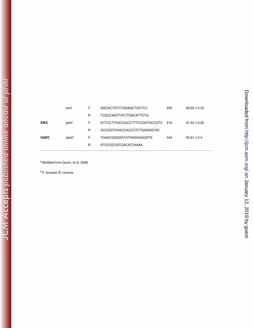
a Modified from Guion, et al, 2008.
b F, forward; R, reverse.
stx2 F
R
GGCACTGTCTGAAACTGCTCC
TCGCCAGTTATCTGACATTCTG
255 89,65 ± 0,33
EIEC ipaH F
R
GTTCCTTGACCGCCTTTCCGATACCGTC
GCCGGTCAGCCACCCTCTGAGAGTAC
619 91,54 ± 0,26
DAEC daaD F
R
TGAACGGGAGTATAAGGAAGATG
GTCCGCCATCACATCAAAA
444 93,81 ± 0,4
on January 12, 2019 by guesthttp://jcm
.asm.org/
Dow
nloaded from

Table 2. Frequency of pathogens isolated
Diarrhea Control Pathogen
n/N (%) n/N (%)
Diarrheagenic E. coli (DEC) 212/935 (22.5) 157/539 (29.5)
EAECa 89/935 (9.5) 68/539 (12.6)
EPECa 60/935 (6.4) 61/539 (113)
ETECa 29/935 (3.1) 14/539 (2.6)
DAECa 18/935 (1.9) 8/539 (1.5)
STECa 4/935 (0.4) 4/539 (0.7)
Multiple DECb 12/935 (1.3) 2/539 (0.4)
Campylobacter sp.a 99/935 (10.6) 53/539 (9.8)
Campylobacter sp. with DEC 55/935 (5.9) 25/539 (4.6)
Rotavirusa 31/465 (6.7) NAc
Rotavirus with Campylobacter 8/465 (1.7) NA
Rotavirus with DEC 20/465 (4.3) NA
Rotavirus with Campylobacter and DEC 2/465 (0.4) NA
No pathogen identified 508/935 (54.3) 304/539 (56.4)
a As the only pathogen isolated.
b Co-infection among diarrheagenic E. coli (DEC). Diarrhea (12): DAEC/EAEC
(1), EAEC/STEC (1), EAEC/EPEC (6), EAEC/ETEC (1), EPEC/EIEC (1),
EPEC/ETEC (2) and Control (2): EAEC/EPEC (1), EAEC/ETEC/DAEC (1).
c NA, not applicable. Control samples were not tested for rotavirus.
on January 12, 2019 by guesthttp://jcm
.asm.org/
Dow
nloaded from

Table 3. Distribution of fecal leukocytes in diarrheagenic E. coli among diarrhea and control samples.
Fecal leukocytes n/N (%) Pathogen
0 L/hpfc 1-10L/hpfc 11-20L/hpfc 21-50L/hpfc >50L/hpfc
DIARRHEA
All diarrhea samples 689/935(73.7) 136/935(14.5) 69/935 (7.4) 31/935(3.3) 10/935(1.1)
Diarrheagenic E. coli
EAECa 69/89(77.5) 15/89(16.9) 2/89 (2.2) 2/89 (2.2) 1/89(1.1)
EPECa 49/60(81.7) 6/60 (10.0) 4/60 (6.7) 1/60(1.7) -
ETECa 22/29(75.9) 3/29 (10.3) 3/29 (10.3) - 1/29(3.4)
DAECa 13/18(72.2) 2/18 (11.1) 3/18 (16.6) - -
STECa 1/4(25.0) 2/4 (50.0) 1/4 (25.0) - -
Multiple DECb 6/12(50.0) 6/12 (50.0) - - -
Campylobacter sp. a 61/99(61.6) 13/99(13.1) 13/99(13.1) 8/99(8.1) 4/99(4.0)
Campylobacter sp. with DEC 40/55(72.7) 7/55 (12.7) 7/55 (12.7) 1/55(1.8) -
No pathogen identified 376/508 (74.0) 81/508 (15.9) 29/508 (5.7) 19/508 (3.7) 3/508 (0.6)
on January 12, 2019 by guesthttp://jcm
.asm.org/
Dow
nloaded from
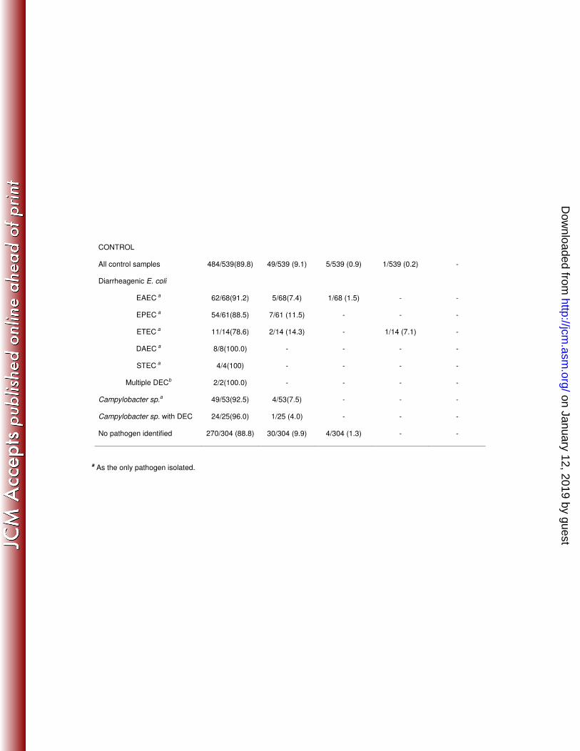
CONTROL
All control samples 484/539(89.8) 49/539 (9.1) 5/539 (0.9) 1/539 (0.2) -
Diarrheagenic E. coli
EAEC a 62/68(91.2) 5/68(7.4) 1/68 (1.5) - -
EPEC a 54/61(88.5) 7/61 (11.5) - - -
ETEC a 11/14(78.6) 2/14 (14.3) - 1/14 (7.1) -
DAEC a 8/8(100.0) - - - -
STEC a 4/4(100) - - - -
Multiple DECb 2/2(100.0) - - - -
Campylobacter sp.a 49/53(92.5) 4/53(7.5) - - -
Campylobacter sp. with DEC 24/25(96.0) 1/25 (4.0) - - -
No pathogen identified 270/304 (88.8) 30/304 (9.9) 4/304 (1.3) - -
a As the only pathogen isolated.
on January 12, 2019 by guesthttp://jcm
.asm.org/
Dow
nloaded from
b Only co-infection among diarrheagenic E. coli (DEC). Diarrhea (12): DAEC + EAEC (1), EAEC + STEC (1), EAEC +
EPEC (6), EAEC + ETEC (1), EPEC + EIEC (1), EPEC + ETEC (2) and Control (2): EAEC + EPEC (1), EAEC + ETEC +
DAEC (1).
c L/hpf, Leukocytes by high power field.
on January 12, 2019 by guesthttp://jcm
.asm.org/
Dow
nloaded from
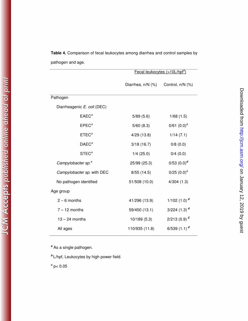
Table 4. Comparison of fecal leukocytes among diarrhea and control samples by
pathogen and age.
Fecal leukocytes (>10L/hpfb)
Diarrhea, n/N (%) Control, n/N (%)
Pathogen
Diarrheagenic E. coli (DEC)
EAECa 5/89 (5.6) 1/68 (1.5)
EPECa 5/60 (8.3) 0/61 (0.0)c
ETECa 4/29 (13.8) 1/14 (7.1)
DAECa 3/18 (16.7) 0/8 (0.0)
STECa 1/4 (25.0) 0/4 (0.0)
Campylobacter sp.a 25/99 (25.3) 0/53 (0.0)d
Campylobacter sp. with DEC 8/55 (14.5) 0/25 (0.0)c
No pathogen identified 51/508 (10.0) 4/304 (1.3)
Age group
2 – 6 months 41/296 (13.9) 1/102 (1.0) d
7 – 12 months 59/450 (13.1) 3/224 (1.3) d
13 – 24 months 10/189 (5.3) 2/213 (0.9) C
All ages 110/935 (11.8) 6/539 (1.1) d
a As a single pathogen.
b L/hpf, Leukocytes by high power field.
c p< 0.05
on January 12, 2019 by guesthttp://jcm
.asm.org/
Dow
nloaded from

Table 5. Bivariate analysis for the odds of a positive fecal leukocyte count (>10
L/hpf) per factor and pathogen.
OR (95% C.I.) p
Factor
Age (<12months) 3.63 (1.95 – 6.76) 0.000
Sex(male) 0.86 (0.57 - 1.30) 0.477
Undernutrition 0.88 (0.55 - 1.40) 0.594
Breastfeeding 2.56 (1.69 - 3.88) 0.000
Diarrhea severity (Vesikari) 0.80 (0.45 - 1.42) 0.450
Blood in stools 6.50 (3.39 - 12.43) 0.000
Pathogen
EAEC 0.51 (0.26 – 1.02) 0.056
EPEC 0.52 (0.20 – 1.33) 0.173
ETEC 2.00 (0.87 – 4.61) 0.102
DAEC 1.68 (0.61 – 4.61) 0.317
Campylobacter sp 2.20 (1.39 – 3.51) 0.001
Rotavirus 0.79 (0.74 – 0.84) 0.000
on January 12, 2019 by guesthttp://jcm
.asm.org/
Dow
nloaded from
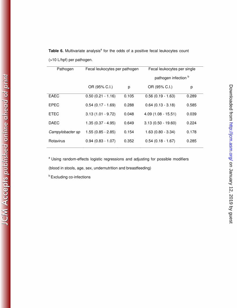
Table 6. Multivariate analysisa for the odds of a positive fecal leukocytes count
(>10 L/hpf) per pathogen.
Pathogen Fecal leukocytes per pathogen
Fecal leukocytes per single
pathogen infection b
OR (95% C.I.) p OR (95% C.I.) p
EAEC 0.50 (0.21 - 1.16) 0.105 0.56 (0.19 - 1.63) 0.289
EPEC 0.54 (0.17 - 1.69) 0.288 0.64 (0.13 - 3.18) 0.585
ETEC 3.13 (1.01 - 9.72) 0.048 4.09 (1.08 - 15.51) 0.039
DAEC 1.35 (0.37 - 4.95) 0.649 3.13 (0.50 - 19.60) 0.224
Campylobacter sp 1.55 (0.85 - 2.85) 0.154 1.63 (0.80 - 3.34) 0.178
Rotavirus 0.94 (0.83 - 1.07) 0.352 0.54 (0.18 - 1.67) 0.285
a Using random-effects logistic regressions and adjusting for possible modifiers
(blood in stools, age, sex, undernutrition and breastfeeding)
b Excluding co-infections
on January 12, 2019 by guesthttp://jcm
.asm.org/
Dow
nloaded from
Recommended