1
Fast Monitoring of Species-Specific Peptide Biomarkers Using 1
High Intensity Focused Ultrasound Assisted Tryptic Digestion and 2
Selected MS/MS Ion Monitoring Mass Spectrometry 3
4
Mónica Carrera1,*
, Benito Cañas2, Daniel López-Ferrer
3, Carmen Piñeiro
1, 5
Jesús Vázquez3, José M. Gallardo
1 6
7
1Marine Research Institute (IIM), CSIC, Vigo, Pontevedra, Spain. 8
2Complutense University of Madrid (UCM), Madrid, Spain. 9
3Severo Ochoa Molecular Biology Centre (CBMSO), CSIC, Madrid, Spain. 10
11
AUTHOR E-MAIL ADDRESS: [email protected] 12
13
TITLE RUNNING HEAD: Fast monitoring of peptide biomarkers by HIFU and SMIM 14
15
* CORRESPONDING AUTHOR: Dr. Mónica Carrera 16
Marine Research Institute (IIM), Spanish National Research Council (CSIC). Eduardo 17
Cabello 6, E-36208 Vigo, Pontevedra, Spain. Phone: +34 986 231930. Fax: +34 986 292762. 18
E-mail: [email protected] 19
20
21
22
23

2
ABSTRACT 1
2
A new strategy for the fast monitoring of peptide biomarkers is described. It is based 3
on the use of accelerated in-solution trypsin digestions under an ultrasonic field provided by 4
High-Intensity Focused Ultrasound (HIFU) and the monitoring of several peptides by 5
Selected MS/MS Ion Monitoring (SMIM) in a linear ion trap (LIT) mass spectrometer. The 6
performance of the method was established for the unequivocal identification of all 7
commercial fish species belonging to the Merlucciidae family. Using a particular combination 8
of only eleven peptides, resulting from the HIFU-assisted tryptic digestion of the thermostable 9
proteins parvalbumins (PRVBs), the workflow allowed the unequivocal identification of these 10
closely related fish species in any seafood product, including processed and precooked 11
products, in less than 2 h. The present strategy constitutes the fastest method for peptide 12
biomarker monitoring. Its application for food quality control provides to the authorities an 13
effective and rapid method of food authentication and traceability to guarantee the quality and 14
safety to the consumers. 15
16
17
18
19
KEYWORDS: mass spectrometry, biomarker, selected MS/MS ion monitoring (SMIM), 20
high-intensity focused ultrasound (HIFU), proteomics, parvalbumin, fish, food safety. 21
22
23

3
INTRODUCTION 1
Fast monitoring of biomarkers is essential to achieve a rapid response and to take a 2
precise decision in diverse life fields. It is crucial in clinical diagnosis, therapeutic 3
supervision, environmental protection and food quality control among others1, 2
. Currently, 4
the most commonly used approach for the monitoring of peptide/protein biomarkers is based 5
on immunoassays, mainly using ELISA and array techniques1. The advantages of these 6
methods are their high specificity and sensitivity. Apart of being time consuming, a flaw of 7
these techniques is that not always the right antibody is available for each biomarker, making 8
lengthy and expensive work necessary to extend the battery of specific antibodies for new 9
standardized and affordable assays. Therefore, the development of alternative and fast 10
methodologies having high reproducibility, sensitivity and specificity are necessary. 11
The emerging targeted mass spectrometry (MS)-based proteomics techniques can 12
constitute an excellent alternative methodology. When these selective and sensitive operating 13
methods are used, the MS analyzer is centered on analyzing only the compound of interest by 14
selected reaction monitoring (SRM) or multiple reaction monitoring (MRM)3, 4
. Monitoring 15
transitions (suitable pairs of precursor and fragment ion m/z), constitute a common assay to 16
identify and quantify biomarkers and by inference, the change in the corresponding biological 17
condition being studied. This setup provides high analytical reproducibility, a good signal-to-18
noise (S/N) ratio, and an increased dynamic range4. The capability of triple quadrupole (TQ) 19
instruments to selectively isolate a precursor ion and the fragment ion(s) it produces under 20
collision induced dissociation (CID) is exploited for the experiments using SRM or MRM 21
scan modes5. 22
While SRM and MRM performed on a TQ are the most sensitive scanning modes 23
(low-attomolar) with a broad dynamic range (up to five orders of magnitude)6, their 24
optimization for a definite SRM/MRM assay is time-consuming. More importantly, using 25

4
these scanning procedures, complete MS/MS spectra are not registered. The MS/MS spectrum 1
of a molecule is of paramount importance to confirm the structure of the compound detected. 2
To solve this problem, new routines, like the MRM-triggered MS/MS using hydrid 3
quadrupole/linear ion trap mass spectrometers, have been explored7. In such assays, when a 4
significant signal for a specific MRM transition is detected, the instrument switches the third 5
quadrupole automatically to the ion trap mode, collecting the full MS/MS spectrum. 6
Selected MS/MS Ion Monitoring (SMIM) in ion trap (IT) mass spectrometer is another 7
scanning mode that allows for a sensitive monitoring of specific molecules, producing 8
complete structural information8. The high scanning speed attainable in the IT allows for the 9
production of MS/MS spectra in a fraction of a second, registering the information given by 10
the complete spectra. High confident MS/MS spectra are recorded due to the possibility of an 11
averaging of the signal during acquisition. The utility of this operating mode has been 12
demonstrated in several previously published studies8-10
, where the possibility of a virtual 13
plotting of all the transitions produced has been discussed. 14
The main difficulty when LC-MS is used to monitor biomarkers arises from the 15
complexity of samples, which may result in false positives. Although the chromatographic 16
gradient time may be properly manipulated to avoid this, lowering the number of components 17
in the sample, when it is possible, may help to simplify the assays. Shorter analysis times are 18
needed when fewer components are present in the sample. Fast and easy protein fractionation 19
or purification steps conducted prior to LC-MS analysis, makes the analysis simpler and 20
faster11
. 21
Procedures to enhance the protease activity, such as the application of microwaves12
, 22
high pressure13
or the energy produced by ultrasound14
, can accelerate the time consuming 23
trypsin digestion. The application of only 1-2 minutes of High Intensity Focused Ultrasound 24

5
(HIFU) to in-solution tryptic digestions has been reported to achieve an efficiency and 1
reproducibility similar to that obtained by traditional overnight protocols14-16
. 2
A new targeted MS-based strategy for the fast monitoring of peptide biomarkers based 3
on the combination of these methodologies described is proposed and has been applied to the 4
fast authentication of all commercial species from Merlucciidae family. It is based on: (a) the 5
purification of PRVBs by heat treatment (Time: 45min), (b) their accelerated tryptic digestion 6
using HIFU (Time: 2 min) and (c) the monitoring of eleven PRVB peptide biomarkers by 7
SMIM in a linear ion trap (LIT) mass spectrometer (Time: 60 min). Each step was 8
individually adjusted to minimize the time of analysis. With this new and competitive 9
strategy, the unequivocal identification of these close-related fish species in any seafood 10
products, including processed and precooked, can be achieved in less than 2 h. 11
12
13
14
15
16
17
18
19
20
21
22
23
24
25

6
EXPERIMENTAL SECTION 1
2
1. Reference species and commercial foodstuffs 3
All the main commercial species from the Merlucciidae family were employed in this 4
study: eleven different hake species, including two different subspecies from Merluccius 5
australis and two grenadier subspecies belonging to the Macruronus novaezelandiae species 6
(Table 1). Except for European hake, the specimens were frozen on board at -30 ºC, with 7
special care in keeping their morphological characteristics in good shape, and shipped by 8
plane to the laboratory for the analyses. The weight of every specimen studied was in the 9
range of 3-6 kg. At least 10 fish belonging to each different species were subjected to 10
taxonomical study according to their anatomical and morphological features by an expert 11
marine biologist, and by genetic identification using FINS at the Food Biochemistry 12
Laboratory from the Marine Research Institute (Vigo, Pontevedra, Spain). Five correctly 13
identified individuals for each of the species were considered as reference species. In 14
addition, for the validation step using commercial real samples, a total of ten hake foodstuffs 15
were included in the work (Table 1). All samples were analyzed in triplicate. 16
17
2. Parvalbumin purification 18
Sarcoplasmic protein extraction was carried out by homogenizing 5 g of white muscle 19
in 10 mL of 10 mM Tris-HCl buffer pH 7.2, supplemented with 5 mM PMFS, during 30 s in 20
an Ultra-Turrax device (IKA-Werke, Staufen, Germany). In the case of battered precooked 21
foodstuffs the casing was first removed. The sarcoplasmic protein extracts were then 22
centrifuged at 40000 g for 20 min at 4 ºC (J221-M centrifuge; Beckman, Palo Alto, CA). 23
PRVBs were purified by taking advantage of their thermostability, heating the sarcoplasmic 24
extracts at 70ºC for 5 min10
. After centrifugation at 40000 g for 20 min (J221-M centrifuge; 25

7
Beckman, Palo Alto, CA), supernatants composed mainly by PRVBs were quantified by the 1
bicinchoninic acid (BCA) method (Sigma-Chemical Co., USA). 2
3
3. Protein digestion using HIFU 4
PRVB supernatants were subjected to HIFU-assisted trypsin digestion as previously 5
described14
. A total of 20 µg of heated extract were subjected to in-solution digestion with 1 6
µg trypsin without adding urea, DTT or iodoacetamide (Promega, Madison, WI, USA) 7
applying simultaneously HIFU. A high-intensity ultrasonic probe of 1 mm probe tip (Dr. 8
Hielscher, Teltow, Germany) was set to 50% of amplitude and was used to perform the ultra-9
fast digestion for 1 min. Another 1 µg of trypsin was added again to the sample and the HIFU 10
application was repeated for 1 min. 11
12
4. LC-MS/MS analysis 13
Peptide digests were acidified and analyzed by LC-ESI-IT-MS/MS using a Surveyor 14
LC-system coupled to an LTQ LIT mass spectrometer (Thermo Fisher, San Jose, CA). The 15
peptide separation (1µg) was performed on a 0.18 mm x 150 mm BioBasic-18 RP column 16
(ThermoHypersil-Keystone), using 0.5% acetic acid in Milli-Q-water and in 80% ACN as 17
mobile phases A and B, respectively. A 60 min linear gradient from 5 to 40% B, at a flow 18
rate of 1.5-1.7 µL/min was used. ESI parameters were: spray voltage, 3.5 kV; N2 flow, 10 19
arbitrary units; and capillary temperature, 200ºC. Peptides were analyzed in positive mode 20
from 400 to 1600 amu (3 μscans), followed by four data-dependent MS/MS scans (3 μscans), 21
using an isolation width of 3 amu and a normalized collision energy of 35%. Fragmented 22
masses were set in dynamic exclusion for 3 min after the second fragmentation event and 23
singly charged ions were excluded from MS/MS analysis. 24
25

8
5. Selected MS/MS Ion Monitoring (SMIM) 1
SMIM analysis was performed using a Surveyor LC-system coupled to an LTQ LIT 2
mass spectrometer (Thermo Fisher, San Jose, CA), as described previously8 with minor 3
modifications. The peptide separation (1μg) was performed on a 0.18 mm x 150 mm 4
BioBasic-18 RP column (ThermoHypersil-Keystone), using 0.5% acetic acid in water and in 5
80% ACN as mobile phases A and B, respectively. A 45 min linear gradient from 5 to 40% 6
B, at a flow rate of 1.5-1.7 µL/min was used. ESI parameters were as described previously. 7
Peptides were detected in the positive ion mode using the SMIM8. For this method, the MS 8
instrument was programmed to perform continuous MS/MS scans (5 μscans) of doubly-9
charged precursor ions from all candidate peptide biomarkers along the complete 10
chromatographic separation. Normalized collision energy was set to 35% and a 3 amu mass 11
window was used to fragment selected parent ions. 12
13
6. Mass spectrometry data processing 14
MS/MS spectra were searched using SEQUEST (Bioworks 3.1 package, Thermo 15
Fisher), against the Teleostei UniProt/TrEMBL database (release 2010_12; 158.545 entries), 16
which also included their respective decoy sequences. The following constraints were used 17
for the searches: semi-tryptic cleavage with up to two missed cleavage sites and tolerances 18
1.8 Da for precursor ions and 0.8 Da for MS/MS fragments ions. The variable modifications 19
allowed were methionine oxidation (Mox), carbamidomethylation of Cys (C*) and 20
acetylation of the N-terminus of the protein (N-Acyl). The database search results were 21
subjected to statistical analysis with the PeptideProphet algorithm (v.4.4)20
. The FDR was 22
kept below 1%. 23
The proteins identified in the original and heated sarcoplasmic extracts, were 24
submitted to Ingenuity Pathway Analysis (IPA; Ingenuity Systems, CA). Only pathways 25

9
scoring –log(p-value) ≥2, which have >99% confidence of not being generated by chance, 1
were selected. 2
For the SMIM mode, virtual chromatograms traces were plotted and optimized using 3
QualBrowser software (Thermo Fisher) to show the selected transitions for each parent ion. 4
In addition, MS/MS spectra collected in the SMIM mode were used to validate the peptide 5
identities using SEQUEST as is described before. 6
7
8
9
10
11
12
13
14
15
16
17
18
19
20
21
22
23
24
25

10
RESULTS AND DISCUSSION 1
2
1. Strategy for the fast monitoring of species-specific peptide biomarkers 3
The strategy for the fast monitoring of peptide biomarkers proposed in this work is 4
summarized in Figure 1. This strategy integrates three steps: (a) purification of thermostable 5
proteins (PRVBs) by short heat treatment followed by centrifugation (Time: 45 min), (b) in-6
solution trypsin digestion accelerated using HIFU (Time: 2 min) and (c) monitoring of eleven 7
species-specific PRVB peptide biomarkers by SMIM using a LIT mass spectrometer (Time: 8
60 min). With this strategy, all relevant commercial fish species belonging to the 9
Merlucciidae family can be unequivocally identified in any seafood product, including 10
precooked, in less than 2h. The detailed results for each step and the validation of this new 11
fast monitoring strategy using unknown hake commercial products are shown in the following 12
sections. 13
14
2. PRVB purification and enzymatic digestion accelerated by HIFU 15
PRVBs, considered as the best protein biomarker for the authentication of Merluciidae 16
species10, 21
, were purified from the sarcoplasmic extracts taking advantage of their 17
thermostability10
. Figure S-1 in the Supplemental Data 1 (Supporting Information) shows a 18
summary of the protein composition in the extracts before and after the treatment with heat 19
(70ºC for 5 min). The complete list of proteins and peptides for both samples, identified by 20
LC-MS/MS and Sequest search after a conventional overnight trypsin digestion22
, are 21
presented in the Supplemental Data 2. Protein composition of the original sarcoplasmic 22
extracts revealed more than 125 different proteins involved in 10 relevant functional pathways 23
(Table S-1 in the Supplemental Data 1). After treatment with heat, the majority of identified 24
peptides corresponded to PRVBs (77.87%) (Figure S-1 in the Supplemental Data 1). These 25

11
results demonstrated that the treatment with heat is a simple, fast and effective procedure to 1
purify and enrich the samples in only PRVBs. 2
Purified PRVBs were digested with trypsin using, either the conventional overnight 3
procedure, or the fast accelerated by HIFU. As reported previously14
, HIFU assisted digestion 4
produced results comparable to those obtained by the conventional overnight incubation 5
methods, but in a fraction of time. Moreover, the absence of urea in the digestion buffer 6
prevented undesired peptide side reactions, such as carbamylation of N-termini and Lys 7
residues, which may occur when HIFU is applied in the presence of urea23, 24
. 8
The combination of a fast and easy protein purification procedure (Time: 45 min) with 9
the use of HIFU for the protein digestion (Time: 2 min), considerably simplified and reduced 10
the time needed for the sample preparation, reflected in the overall time needed for 11
monitoring. 12
13
3. Selection of the species-specific peptide biomarkers 14
The next step in the proposed strategy consisted in selecting the smaller number of 15
species-specific peptides, which must be monitored, to effectively identify all the species 16
from Merlucciidae family. Parvalbumins peptide sequences with a high inter-specific 17
variability, obtained after the extensive de novo sequencing of PRVBs previously published10
, 18
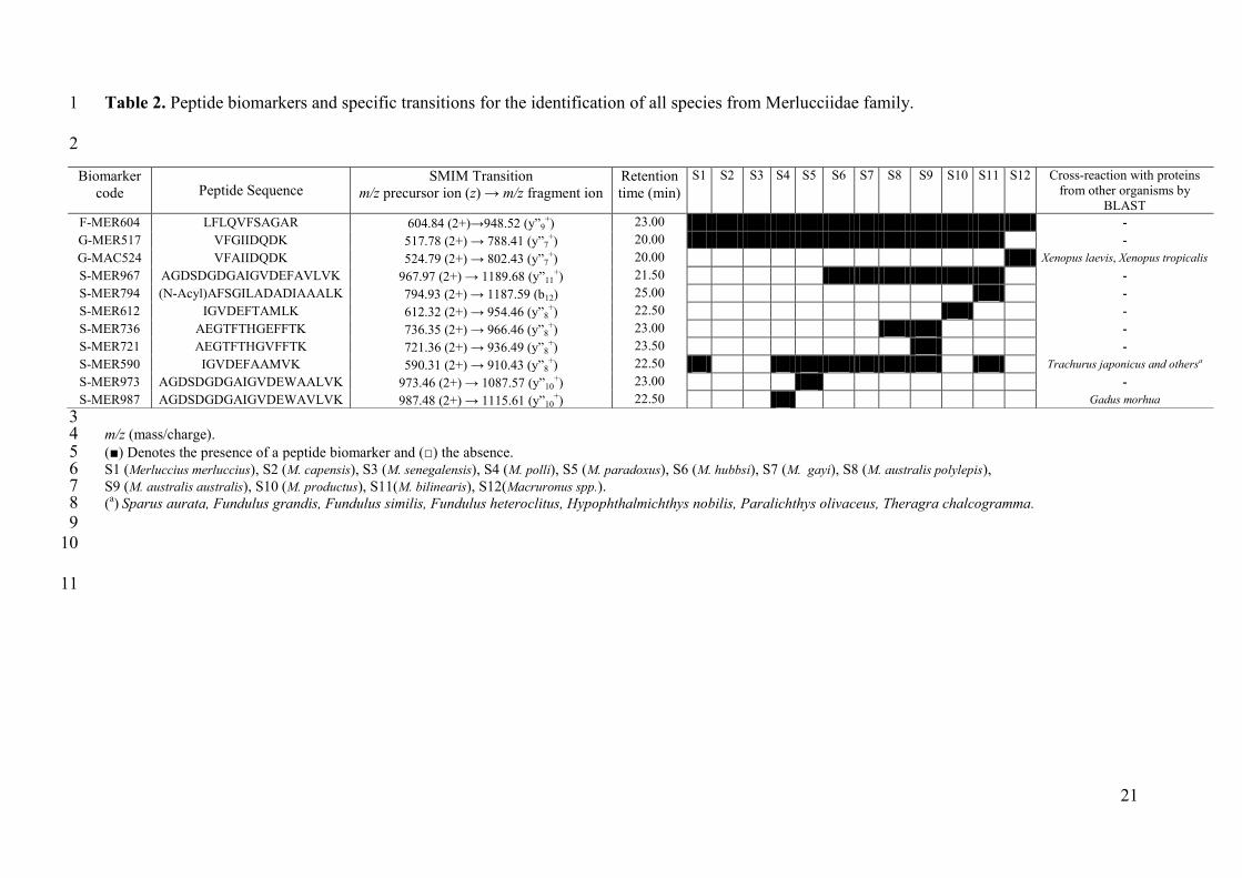
were used for the purpose. Eleven tryptic peptides were selected in basis of the information 19
that their combined presence or absence could provide to confidently identify all of the 20
species under the study (Table 2). A BLAST search was performed using the UniProtKB 21
database to validate the uniqueness of the peptide sequences selected. Four of them were 22
present in only one specific species and can be considered as a canonical peptide for each of 23
these Merlucciidae species (S-MER794, S-MER612, S-MER721, S-MER973). The sequences 24
of the rest were shared by PRVBs from several Merlucciidae species or other organisms. 25

12
However, their use following a specific and systematic combination avoids interferences and 1
allows for a correct discrimination of all the hake species under the study. Figure 2 shows the 2
flow diagram for the unambiguous systematic discrimination which can be achieved. 3
According to this scheme, the presence/absence of the peptide F-MER604 determines if any 4
member from Merlucciidae family is present in the sample. Discrimination between the 5
genera Merluccius spp. and Macruronus spp. can be achieved by the determination of 6
presence/absence of peptides G-MER517 and G-MAC524, respectively. Within the 7
Merluccius genus, the presence/absence of the peptide S-MER967 allows for the 8
classification of hake species in two groups according to their geographic distribution: 9
American hakes (M. hubbsi, M. gayi, M. australis polylepis, M. australis australis, M. 10
productus or M. bilinearis) or Euro-African hakes (M. merluccius, M. capensis, M. 11
senegalensis, M. polli or M. paradoxus). Finally, as can be seen in Figure 2, the combination 12
of the presence/absence of other eight peptides allows for the unambiguous identification of 13
any specific species from the Merlucciidae family. 14
15
4. Fast identification of hake species using SMIM 16
For each of the reference hake species, PRVBs peptide pools obtained from the 17
accelerated tryptic digestions were subjected to SMIM analysis in a LIT mass spectrometer 18
focusing the MS/MS events on the corresponding precursor ions for the eleven peptides 19
selected. The selected m/z value for each of precursor ion corresponded to the predominant 20
charge state, which was +2 for all of them (Table 2). Figure S-2 in the Supplemental Data 1 21
details the MS/MS spectra for each peptide. Once MS/MS spectra were recorded, virtual 22
chromatograms for all the different fragment ions could be obtained. For each of the peptide 23
markers, mass transitions were noted according to the criteria of sensitivity and selectivity. As 24
the peptide mixture used is not too complex, selectivity was not a matter of concern and the 25

13
transitions chosen in every case was in accordance with the maximum intensity of the 1
fragments, which mostly corresponded to y-ions. Therefore, the combination of highly 2
sensitive transitions (precursor m/z→fragment m/z) (Table 2), together with the use of simple 3
peptide mixtures (coming mainly from PRVBs), made possible the representation of specific 4
transitions with a high signal-to-noise (S/N) ratio. Tracing these transitions for each peptide 5
biomarker, according to the flow diagram described in the Figure 2, made possible to identify 6
unequivocally all the reference hake species (Figure 3). 7
The results obtained matched precisely with those obtained by DNA-analysis. 8
However, the time needed for performing both techniques was extremely different. At least 24 9
h were needed for a genetic identification by FINS, while only less than 2 h were necessary to 10
complete the identification following the proposed strategy. To our knowledge, this is the 11
fastest method to achieve the food species authentication. 12
13
5. Application to commercial samples identification 14
In order to validate this new strategy, ten commercial hake products were subjected to 15
analysis for their authentication. Table 1 summarizes the products that were tested, which had 16
been previously subjected to one or more processing treatments, even precooked. Two main 17
label categories were observed: labels with species declared (4 samples) and labels with no 18
declared species, which presented only a general commercial name (i.e. hake) (6 samples). 19
Results for the identification of the commercial samples using the designed strategy are 20
shown in the Table 3 and in the Figure S-3 in the Supplemental Data 1. Samples with 21
complete declared labelling were found to be mostly correct (CP1-CP4). However, some 22
products with no declared species were assigned to species belonging to the Macruronus 23
genus (CP8 and CP9). This genus presents a low commercial value and is not as valued by the 24
consumer25
. We have presented here a strategy that allows the fast detection of mislabelling 25

14
practices in these fish products and moreover, this work also shown that the use of 1
thermostable proteins allow the application of this fast monitoring method to battered 2
precooked products (CP6-CP10). 3
4
5
6
7
8
9
10
11
12
13
14
15
16
17
18
19
20
21
22
23
24
25

15
CONCLUSIONS 1
A new strategy for the fast monitoring of species-specific peptide biomarkers for 2
foodstuff authentication has been proposed. The principle it is based on the use of a fast 3
purification step of the target protein, the acceleration of in-solution protein digestion by 4
High-Intensity Focused Ultrasound (HIFU) and the monitoring of several peptides by 5
Selected ion MS/MS Monitoring mass spectrometry (SMIM) in a linear ion trap mass 6
spectrometer. The use of the SMIM mode of scanning allows for a more simple setting of the 7
procedures, giving the possibility of obtaining full MS/MS information necessary for the 8
validation of the structure of the component and allows the choice of the most adequate 9
transitions for each precursor ion, attending the criteria of sensitivity and selectivity. Thus, in 10
combination with a fast purification and digestion step of the target protein, the most sensitive 11
transitions may be considered for all the markers. 12
With this new strategy, all relevant commercial fish species belonging to the 13
Merlucciidae family present in any seafood product can be unequivocal identified in less than 14
2 h. The present strategy constitutes the fastest method for peptide biomarker monitoring by 15
targeted-proteomics described up to now, whose application in the food quality control area, 16
provide to the authorities an effective, competitive and rapid method of food authentication 17
and traceability that guarantee the quality and safety to the consumers. 18
19
20
21
22
23
24
25

16
REFERENCES 1
2
(1) Zangar, R. C.; Daly, D. S.; White, A. M. Expert Rev. Proteomics 2006, 3, 37-44. 3
(2) Schubert-Ullrich, P.; Rudolf, J.; Ansari, P.; Galler, B.; Führer, M.; Molinelli, A.; 4
Baumgartner, S. Anal. Bioanal. Chem. 2009, 395, 69-81. 5
(3) Gallien, S., Duriez, E., Domon, B. J. Mass Spectrom. 2011, 46, 298-312. 6
(4) Lange, V.; Picotti, P.; Domon, B.; Aebersold, R. Mol. Syst. Biol. 2008, 4, 1-14. 7
(5) Picotti, P.; Rinner, O.; Stallmach, R.; Dautel, F.; Farrah, T.; Domon, B.; Wenschuh, 8
H.; Aebersold, R. Nat. Methods 2010, 7, 43-46. 9
(6) Stahl-Zeng, J.; Lange, V.; Ossola, R.; Eckhardt, K.; Krek, W.; Aebersold, R.; Domon, 10
B. Mol. Cell Proteomics 2007, 6, 1809-1817. 11
(7) Unwin, R. D.; Griffiths, J. R.; Whetton, A. D. Nat. Protoc. 2009, 4, 870-877. 12
(8) Jorge, I.; Casas, E. M.; Villar, M.; Ortega-Pérez, I.; López-Ferrer, D.; Martínez-Ruiz, 13
A.; Carrera, M.; Marina, A.; Martínez, P.; Serrano, H.; Cañas, B.; Were, F.; Gallardo, 14
J. M.; Lamas, S.; Redondo, J. M.; García-Dorado, D.; Vázquez, J. J. Mass Spectrom. 15
2007, 42, 1391-1403. 16
(9) Carrera, M.; Cañas, B.; Piñeiro, C.; Vázquez, J.; Gallardo, J. M. J. Proteome Res. 17
2007, 6, 3070-3080. 18
(10) Carrera, M.; Cañas, B.; Vázquez, J.; Gallardo, J. M. J. Proteome Res. 2010, 9, 4393-19
4406. 20
(11) Cañas, B.; Piñeiro, C.; Calvo, E.; López-Ferrer, D.; Gallardo, J. M. J. Chromatogr. A 21
2007, 1153, 235-258. 22
(12) Sun, W.; Gao, S.; Wang, L.; Chen, Y.; Wu, S.; Wang X.; Zheng D.; Gao, Y. Mol. Cell. 23
Proteomics 2006, 5, 769-776. 24

17
(13) López-Ferrer, D.; Petritis, K.; Hixson, K. K.; Heibeck, T. H.; Moore, R. J.; Belov, M. 1
E.; Camp, D. G. 2nd
; Smith, R. D. J. Proteome Res. 2008, 7, 3276-3281. 2
(14) López-Ferrer, D.; Capelo, J. L.; Vázquez, J. J. Proteome Res. 2005, 4, 1569-1574. 3
(15) López-Ferrer, D.; Cañas, B.; Vázquez, J.; Lodeiro, C.; Rial-Otero, R.; Moura, I.; 4
Capelo, J. L. Trends Anal. Chem. 2006, 25, 996-1005. 5
(16) Capelo, J. L.; Carreira, R.; Diniz, M.; Fernandes, L.; Galesio, M.; Lodeiro, C.; Santos, 6
H. M.; Vale, G. Anal. Chim. Acta 2009, 650, 151-159. 7
(17) Civera, T. Vet. Res. Commun. 2003, 27, 481-489. 8
(18) Chapela, M. J.; Sánchez, A.; Suárez, M. I.; Pérez-Martín, R. I.; Sotelo, C. G. J. Agric. 9
Food Chem. 2007, 55, 6903-6909. 10
(19) Rasmussen, R. S.; Morrissey, M. T. Compr. Rev. Food Sci. Food Saf. 2008, 7, 280-11
295. 12
(20) Keller, A.; Eng, J.; Zhang, N.; Li, X. J.; Aebersold, R. Mol. Syst. Biol. 2005, 1, 13
2005.0017. 14
(21) Carrera, M.; Cañas, B.; Piñeiro, C.; Vázquez, J.; Gallardo, J. M. Proteomics 2006, 6, 15
5278-5287. 16
(22) Jensen, O. N.; Wilm, M.; Shevchenko, A.; Mann, M. Methods Mol. Biol. 1999, 112, 17
513-530. 18
(23) Stark, G. R.; Stein, W. H.; Moore, S. J. Biol. Chem. 1960, 235, 3177–3181. 19
(24) López-Ferrer, D.; Heibeck, T. H.; Petritis, K.; Hixson, K. K.; Qian, W.; Monroe, M. 20
E.; Mayampurath, A.; Moore, R. J.; Belov, M. E.; Camp II, D. G.; Smith, R. D. J. 21
Proteome Res. 2008, 7, 3860-3867. 22
(25) Lloris, D.; Matallanas, J.; Oliver, P. FAO Species Catalogue for Fishery Purposes No. 23
2. FAO 2005. 24
25

18
ACKNOWLEDGEMENTS 1
Authors wish to express their gratitude to Mrs. Lorena Barros for her excellent 2
technical assistance and to Freiremar SA and CETMAR for their assistance in the collection 3
of the reference species used in this study. This work was supported by the Comisión 4
Interministerial de Ciencia y Tecnología (CICyT) (Project AGL2000-0440-P4-02). 5
6
7
8
9
10
11
12
13
14
15
16
17
18
19
20
21
22
23
24
25

19
FIGURE CAPTIONS 1
Figure 1: Analytical scheme for the fast monitoring of the species-specific peptide 2
biomarkers proposed in this work. 3
4
Figure 2: Flow diagram for a systematic discrimination of Merlucciidae species using only 5
eleven specific tryptic peptides from PRVBs. Y denotes the presence and N the absence of a 6
particular peptide. 7
8
Figure 3: Reference SMIM traces for each of Merlucciidae species, plotting the 9
corresponding canonical transition for each of PRVB tryptic peptide biomarker. 10
11
12
. 13
14
15
16
17
18
19
20
21
22
23
24

20
Table 1: Reference species and commercial foodstuffs from the Merlucciidae family 1
considered in the study. 2
M. (Merluccius genus); Ma. nov. (Macruronus novaezelandiae). 3
Reference species
Species/Subspecies Common Name Origin
M. merluccius European hake Spanish coasts
M. capensis Cape hake South Africa
M. senegalensis Senegalense hake Northwest Africa
M. polli Benguela hake Northwest Africa
M. paradoxus Deep-water Cape hake South Africa
M. hubbsi Patagonian hake South America
M. gayi Peruvian or Chilean hake South America
M. australis polylepis Austral hake South America
M. australis australis Austral hake New Zealand coasts
M. productus Pacific hake North America
M. bilinearis Silver hake North America
Ma. nov. nov. Blue grenadier New Zealand coasts
Ma. nov. magellanicus Patagonian grenadier South America
Commercial products
Code Product Presentation Declared Species
CP1 Raw fillets frozen M. capensis or M. paradoxus
CP2 Raw center cuts frozen M. autralis
CP3 Raw loins frozen M. capensis or M. paradoxus
CP4 Raw center cuts frozen M. capensis or M. paradoxus
CP5 Raw center cuts frozen Hake unidentified
CP6 Battered precooked sticks frozen Hake unidentified
CP7 Battered precooked sticks frozen Hake unidentified
CP8 Battered precooked sticks frozen Hake unidentified
CP9 Battered precooked sticks frozen Hake unidentified
CP10 Battered precooked fillets frozen Hake unidentified
21
Table 2. Peptide biomarkers and specific transitions for the identification of all species from Merlucciidae family. 1
2
3 m/z (mass/charge). 4 (■) Denotes the presence of a peptide biomarker and (□) the absence. 5 S1 (Merluccius merluccius), S2 (M. capensis), S3 (M. senegalensis), S4 (M. polli), S5 (M. paradoxus), S6 (M. hubbsi), S7 (M. gayi), S8 (M. australis polylepis), 6 S9 (M. australis australis), S10 (M. productus), S11(M. bilinearis), S12(Macruronus spp.). 7 (
a) Sparus aurata, Fundulus grandis, Fundulus similis, Fundulus heteroclitus, Hypophthalmichthys nobilis, Paralichthys olivaceus, Theragra chalcogramma. 8
9
10
11
Biomarker
code Peptide Sequence SMIM Transition
m/z precursor ion (z) → m/z fragment ion
Retention
time (min)
S1 S2 S3 S4 S5 S6 S7 S8 S9 S10 S11 S12 Cross-reaction with proteins
from other organisms by
BLAST
F-MER604 LFLQVFSAGAR 604.84 (2+)→948.52 (y”9+) 23.00 -
G-MER517 VFGIIDQDK 517.78 (2+) → 788.41 (y”7+) 20.00 -
G-MAC524 VFAIIDQDK 524.79 (2+) → 802.43 (y”7+) 20.00 Xenopus laevis, Xenopus tropicalis
S-MER967 AGDSDGDGAIGVDEFAVLVK 967.97 (2+) → 1189.68 (y”11+) 21.50 -
S-MER794 (N-Acyl)AFSGILADADIAAALK 794.93 (2+) → 1187.59 (b12) 25.00 -
S-MER612 IGVDEFTAMLK 612.32 (2+) → 954.46 (y”8+) 22.50 -
S-MER736 AEGTFTHGEFFTK 736.35 (2+) → 966.46 (y”8+) 23.00 -
S-MER721 AEGTFTHGVFFTK 721.36 (2+) → 936.49 (y”8+) 23.50 -
S-MER590 IGVDEFAAMVK 590.31 (2+) → 910.43 (y”8+) 22.50 Trachurus japonicus and othersa
S-MER973 AGDSDGDGAIGVDEWAALVK 973.46 (2+) → 1087.57 (y”10+) 23.00 -
S-MER987 AGDSDGDGAIGVDEWAVLVK 987.48 (2+) → 1115.61 (y”10+) 22.50 Gadus morhua

22
Table 3: Results of the SMIM assay of commercial hake products. 1
M. (Merluccius genus). *These results were validated by genetic after 24h of analysis. 2
3
4
5
Product Declared species Species identified by HIFU + SMIM
Time: <2h (*)
CP1 M. capensis or M. paradoxus M. paradoxus
CP2 M. australis M. australis polylepis
CP3 M. capensis or M. paradoxus M. paradoxus
CP4 M. capensis or M. paradoxus M. capensis
CP5 Hake unidentified M. paradoxus
CP6 Hake unidentified M. productus
CP7 Hake unidentified M. paradoxus
CP8 Hake unidentified Macruronus spp.
CP9 Hake unidentified Macruronus spp.
CP10 Hake unidentified M. paradoxus

23
Figure 1 1
2
3
4
5
6
7
8
9
10
11
12
13
14
15
16
17
18
19
20
21
22
23
24
25
SAMPLE PROBLEM
Fresh products, frozen, processed or precooked
SARCOPLASMIC PROTEIN EXTRACTION
PURIFICATION OF THERMOSTABLE PROTEINS (PARVALBUMINS)
ACCELERATED TRYPTIC DIGESTION BY HIFU
Time: 45 min
Time: 2 min
Time: 60 min MONITORING OF 11 PEPTIDE BIOMARKERS BY SMIM
SPECIES IDENTIFICATION Total time: 107 min
70ºC for 5min

24
Figure 2 1
2
3
4 Y N
N
Merluccius genus
Y
Euro - African hakes American hakes
G-MER517
S-MER967
G-MAC524 Y
M. bilinearis
N Y
M. hubbsi or M. gayi
S-MER794
S-MER612 N Y
M. productus S-MER736
Y N
S-MER721 Y N
M. australis australis M. australis polylepis
S-MER973 N Y
M. paradoxus S-MER987 Y N
M. polli S-MER590
N Y
M. capensis or
M. senegalensis
Macruronus genus
M. merluccius
Merlucciidae family
F-MER604 Y N
Other

25
Figure 3 1
2
3
4
5
6
7
8
9
10
11
12
13
14
15
16
17
18
19
20
21
22
23
24
590.31→910.43
721.36→936.49
736.35→966.46
794.93→1187.59
612.32→954.46
973.46→1087.57
987.48→1115.61
517.78→788.41
524.79→802.43
967.97→1189.68
604.84→948.52
b) M. capensis
0 20 40 60
c) M. senegalensis
0 20 40 60
a) M. merluccius
0
100
100
0100
0
100
0
100
100
0
0
100
0100
0 20 40 60
0
100
0
100
0
100
d) M. polli
0 20 40 60
e) M. paradoxus
Rela
tive a
bundance
100
0100
0
100
0
100
0
0
100
100
0
100
0100
0 20 40 60
0
100
0
100
0
100
f) M. hubbsi
0 20 40 60
g) M. gayi
0 20 40 60
h) M. australis polylepis
0 20 40 60
i) M. australis australis
100
0100
0
100
0
100
0
0
100
100
0
100
0100
0 20 40 60
0
100
0
100
0
100
j) M. productus
0 20 40 60
k) M. bilinearis
Time (min)
0 20 40 60
l) Macruronus spp.
0 20 40 60
590.31→910.43
721.36→936.49
736.35→966.46
794.93→1187.59
612.32→954.46
973.46→1087.57
987.48→1115.61
517.78→788.41
524.79→802.43
967.97→1189.68
604.84→948.52
590.31→910.43
721.36→936.49
736.35→966.46
794.93→1187.59
612.32→954.46
973.46→1087.57
987.48→1115.61
517.78→788.41
524.79→802.43
967.97→1189.68
604.84→948.52

26
for TOC only: 1
2
3
4
5
6
7
8
9
10
11
Species id.
Time: <2h
45 min
2 min 60 min
HIFU SMIM
PRVB Purification
Recommended