International Journal of Science and Research (IJSR) ISSN (Online): 2319-7064
Index Copernicus Value (2013): 6.14 | Impact Factor (2013): 4.438
Volume 4 Issue 7, July 2015
www.ijsr.net Licensed Under Creative Commons Attribution CC BY
Evaluation of Cell Block Technique in the
Cytodiagnosis of Body Fluids
Bansode Shubhada1 , Kumbalkar D
2, Nayak S
3
1Assistant Professor, Department of Pathology, GMC& H, Latur, Maharashtra, India
2Professor & Head, Department of Pathology, GMC& H, Nagpur, Maharashtra, India
3Assistant Professor, Department of Pathology, GMC& H, Nagpur, Maharashtra, India
Abstract: Background: Conventional smear preparation is simple procedure but have many disadvantages causing the difficulties in
making the definitive diagnosis however cell block are particularly useful when the cytological abnormalities are misleading. Once
positive diagnosis is made it is often considered definitive diagnosis and it is paramount important to identify primary site and type of
malignancy. This can help obviate the proper surgical management, chemotherapy and radiotherapy so as to increase patients’ survival
rate. Aims: To study cell block preparation of all body fluids along with conventional cytology smear and to know primary site of
malignant effusions and evaluating cell block technique in cytodiagnosis of same. Material and Methods : Present prospective study was
conducted in tertiary care hospital which includes fresh body fluids minimum of 15 ml in two separate containers (from pleural and
peritoneal cavity) from 142 patients with all relevant clinical details of both sexes and all ages.scanty fluid samples were
excluded.Samples were processed by conventional smear method and cell block method. Results: 142 body fluid specimens were
evaluated by simultaneous use of smear and cell block technique of which 80% were pleural effusions and 20%were peritoneal
fluids,Male:Female was 2:1, most of the patients were in the age group of 41-60 years. In Conventional smear prepration showed
diagnostic yield 15% and accuracy 85-90% .In Cell block technique showed increase in diagnostic yield upto 21% and accuracy upto
97%. Conclusion: CB in conjunction with CS increases the accuracy of the fluid reports. The CB method provides high cellularity,
better architectural patterns and use of IHC help to indentify primary site of malignacy giving more definitive diagnosis .
Keywords: Cell block, conventional smear, IHC
1. Introduction
Cytological examination of body fluids has increasingly
gained acceptance in clinical medicine to such extent that
once positive diagnosis is made often considered definitive
diagnosis and it is paramount important to identify primary
site and type of malignancy. This can help to obviate the
proper surgical management, chemotherapy and
radiotherapy so as to increase patients’ survival rate1,2,
.
Conventional smear is a much simpler procedure than that of
cell block technique, it has lower sensitivity because of
overcrowding of cells, cell loss, lack of architecture and
also, abundance of inflammatory cells and paucity of
representative cells contribute to considerable difficulties in
making conclusive diagnosis on conventional smears3,4
. Cell
block technique is one of the oldest method later on various
modifications has been done in this method but the method
did not receive much attention due to lack of standardized
technique. The method has advantages like it concentrates
minimal amount of cellular material in one small area that
can be evaluated at a glance with all cells lying in the same
focal plane of the microscope, and as it uses of histological
techniques it gives better cellular morphology better nuclear
and cytoplasmic preservation, intact cell membrane and
crisp chromatin details , preservation of architectural pattern
like cell balls, papillae, acini, rosettes and individual cell
characteristic ,representing its primary site of malignancy,
fragment of this tissue can easily interpreted in a biopsy-like
fashion 3,4
. On the other hand as multiple section of same
material can be obtained for special stains and
immunohistochemistry . And also the possibility of storing
slides for retrospective studies also done , as storage of the
conventional smears is a practical problem 1,2
. For this
reason an attempt was made to prepare and analyze both
conventional smear preparation and cell blocks from the
same specimen by plasma-thromboplastin method to study
cellular yield, morphology, and architecture and to know
primary site in malignant effusions.
2. Material and Methods
After obtaining the approval from Institutional Ethical
Committee the present Prospective Study was conducted in
tertiary care hospital. The study included fresh body fluids
minimum of 15 ml in two separate containers (from pleural
and peritoneal cavity) from 142 patients with all relevant
clinical details of both sexes and all ages. Scanty fluid
samples were excluded from study. A detailed history
regarding age, sex, general examination, systemic
examination, relevant investigations clinical diagnosis and
the samples was collected in two different containers.
3. Technique of Aspiration
1. After explaining complete procedure, written consent
from patient was taken in each and every case before
performing any aspiration
2. Pre-medication : patient was sedated with injection
atropine 0.6mg and injection calmpose 5mg
intramuscularly as and when required
3. Under all aseptic precautions minimum of 15ml fluid
were collected in two separate sterile containers
4. After aspiration punctured skin of patient was sealed with
tincture benzoin. TPR, BP recorded.
5. Fluid was immediately brought to laboratory for further
processing.
Paper ID: SUB156104 87
International Journal of Science and Research (IJSR) ISSN (Online): 2319-7064
Index Copernicus Value (2013): 6.14 | Impact Factor (2013): 4.438
Volume 4 Issue 7, July 2015
www.ijsr.net Licensed Under Creative Commons Attribution CC BY
4. Processing of Fluid
Conventional Smear Preparation:
1) Fluid from one container transferred to centrifuge tube
labeled with the specimen identifier and centrifuged for
5 minutes at 2000 rpm
2) Supernatant fluid were discarded and the sediment was
taken on the slide with the help of glass rod and spread
by thick and thin method
3) Three slides were prepared
4) One dry smear was made and stained with MGG
5) Two slides were fixed in 95% methanol and stained
with papanicolau and hematoxylin and eosin stain
Cell Block Preparation
By Plasma- Thromboplastin method :
1) Plasma (pooled plasma from a blood bank may be
used), and thromboplastin reagent are used to prepare
cell block. The stability may be checked periodically by
adding two drops of throboplastin reagent to two drops
of plasma, which should clot in about 30 seconds.
2) Fluid from second containers transferred into centrifuge
tube labeled with the specimen identifier
3) Spin for 5 minutes at 3000rpm
4) Remove centrifuge tube decant the supernatant
5) Mix 2- 3 drops of plasma with sediment
6) Add 2-3 drops of thromboplastin reagent and mix by
tapping
7) Add 10% buffered formalin
8) Dislodge the clot from tube and let it fix in formalin for
30 minutes
9) The sediment was then wrapped in the filter paper and
processed in histokinette as part of routine paraffin
section histopathology.
10) Hematoxylin and eosin staining was done. Special
staining and immunohistochemisrty was done whenever
required.
Criteria Point score
0 1 2
1. Volume of
Blood/ clot
obscuring
background
Large :
Diagnosis
greatly
compromised
Moderate :
diagnosis
possible
Minimal :
diagnosis easy,
textbook quality
specimen
2. Amount of
diagnostic
cellular
material
present
Minimal :
diagnosis not
possible
Moderate:
sufficient for
diagnosis
Abundant:
diagnosis simple
3. Degree of
cellular
degeneration
and cellular
trauma
Marked
:diagnosis
possible
Moderate:
diagnosis
possible
Minimal: good
preservation
4. Retained
architecture /
Cellular
Arrangement
Minimal
:diagnosis not
possible
Moderate:
some
preservation
Excellent
architectural
display
Criteria for the assessment of quality of smear and cell
block
Each individual slide was objectively analysed for
background, cellularity, cytoplasmic, and nuclear details (
cellular morphology), architecture (acini, papillae, cell balls,
and proliferation spheres), using the Miar’s point scoring
system4 as shown in table no.5. According to the criteria
mentioned in the table no.5, comments were rendered on the
quality of the slides by qualitatively grouping them into
three categories.
Quality of Slide
1. Diagnostically unsuitable (0)
2. Diagnostically adequate (1-4)
3. Diagnostically superior (4-8)
These smears were observed and diagnosed on conventional
smears and cell block section.
Diagnostic Categories
1. Negative for malignancy
Scanty cellularity
Acute inflammatory infiltrate rich
Lymphocyte rich
2. Suspicious for malignancy
3. Positive for malignancy
Statistical Methods
Descriptive statistics like mean, standard deviation, median
and range were obtained for the total scores obtained for
conventional smear and cell block methods. Statistical
significance of difference in the mean scores by two
methods was evaluated using paired t-test. Similar analysis
was further performed based on the diagnostic groups i.e.
positive and negative malignant groups. To determine if
particular criterion differ significantly between methods,
Pearson’s Chi-square test was used. Agreement between
conventional smear and cell block methods was determined
using Kappa statistic. The sensitivity, specificity, positive
prediction value, negative prediction value and accuracy
were determined for the two diagnostic methods after
comparing with final diagnosis (made on the basis of
clinico-radiological and cyto-histological investigations).
The statistical significance was evaluated at 5% level, and
the analysis was carried out using SPSS ver 11.0 and R
package. Significance of diagnostic yield was determined by
Mc-Naemer’s chi-square test.
5. Results
Study material consisted of pleural and peritoneal effusion
samples which were processed by conventional smear
preparation and cell block technique. Slides were evaluated
individually. Total of 142 fluid samples were processed by
both methods.
Paper ID: SUB156104 88
International Journal of Science and Research (IJSR) ISSN (Online): 2319-7064
Index Copernicus Value (2013): 6.14 | Impact Factor (2013): 4.438
Volume 4 Issue 7, July 2015
www.ijsr.net Licensed Under Creative Commons Attribution CC BY
Table 1: Distribution of cases according to age , sex and
type of fluid
Age
(Years)
Pleural Peritoneal Total
Male Female Male Female
<20 8 1 0 0 9(6%)
21-40 26 9 2 0 37(26%)
41-60 41 17 7 12 77(54%)
>61 9 3 3 4 19(13%)
Total 84 30 12 16 142
It is evident from the table no. 1 that 77 (54%) of the
patients belonged to age group 41-60 years; while only
9(6%) cases were below age 20 years. As regards gender,
96(68%) of the cases were males and 46(32%) cases were
females, cases of pleural effusions were 114 and 28 were
peritoneal effusions.
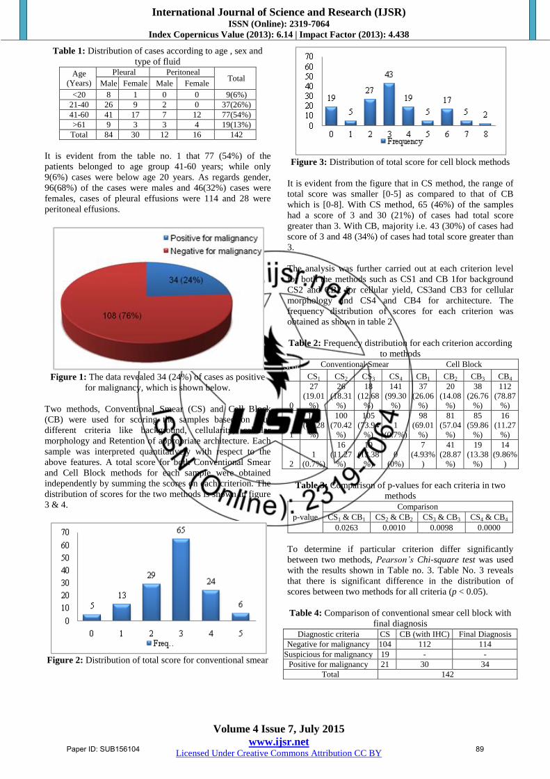
Figure 1: The data revealed 34 (24%) of cases as positive
for malignancy, which is shown below.
Two methods, Conventional Smear (CS) and Cell Block
(CB) were used for scoring the samples based on four
different criteria like background, cellularity, cellular
morphology and Retention of appropriate architecture. Each
sample was interpreted quantitatively with respect to the
above features. A total score for both Conventional Smear
and Cell Block methods for each sample were obtained
independently by summing the scores on each criterion. The
distribution of scores for the two methods is shown in figure
3 & 4.
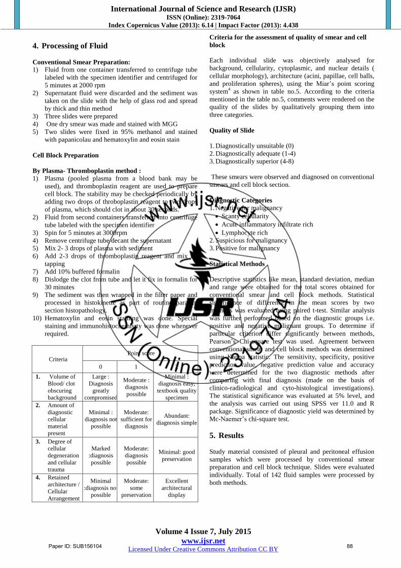
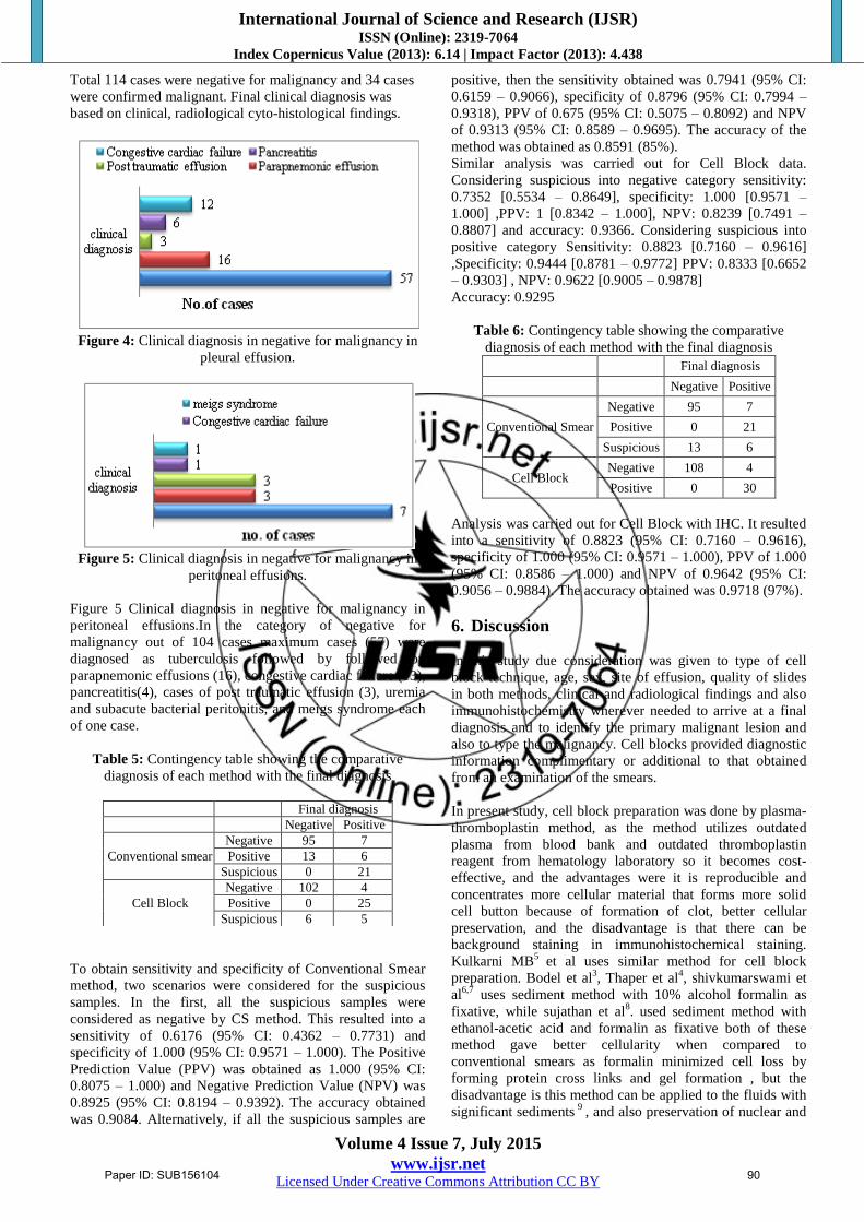
Figure 2: Distribution of total score for conventional smear
Figure 3: Distribution of total score for cell block methods
It is evident from the figure that in CS method, the range of
total score was smaller [0-5] as compared to that of CB
which is [0-8]. With CS method, 65 (46%) of the samples
had a score of 3 and 30 (21%) of cases had total score
greater than 3. With CB, majority i.e. 43 (30%) of cases had
score of 3 and 48 (34%) of cases had total score greater than
3.
The analysis was further carried out at each criterion level
for both the methods such as CS1 and CB 1for background
CS2 and CB2 for cellular yield, CS3and CB3 for cellular
morphology and CS4 and CB4 for architecture. The
frequency distribution of scores for each criterion was
obtained as shown in table 2
Table 2: Frequency distribution for each criterion according
to methods
Scor
e
Conventional Smear Cell Block
CS1 CS2 CS3 CS4 CB1 CB2 CB3 CB4
0
27
(19.01
%)
26
(18.31
%)
18
(12.68
%)
141
(99.30
%)
37
(26.06
%)
20
(14.08
%)
38
(26.76
%)
112
(78.87
%)
1
114
(80.28
%)
100
(70.42
%)
105
(73.94
%)
1
(0.7%)
98
(69.01
%)
81
(57.04
%)
85
(59.86
%)
16
(11.27
%)
2
1
(0.7%)
16
(11.27
%)
19
(13.38
%)
0
(0%)
7
(4.93%
)
41
(28.87
%)
19
(13.38
%)
14
(9.86%
)
Table 3: Comparison of p-values for each criteria in two
methods
p-value
Comparison
CS1 & CB1 CS2 & CB2 CS3 & CB3 CS4 & CB4
0.0263 0.0010 0.0098 0.0000
To determine if particular criterion differ significantly
between two methods, Pearson’s Chi-square test was used
with the results shown in Table no. 3. Table No. 3 reveals
that there is significant difference in the distribution of
scores between two methods for all criteria (p < 0.05).
Table 4: Comparison of conventional smear cell block with
final diagnosis
Diagnostic criteria CS CB (with IHC) Final Diagnosis
Negative for malignancy 104 112 114
Suspicious for malignancy 19 - -
Positive for malignancy 21 30 34
Total 142
Paper ID: SUB156104 89
International Journal of Science and Research (IJSR) ISSN (Online): 2319-7064
Index Copernicus Value (2013): 6.14 | Impact Factor (2013): 4.438
Volume 4 Issue 7, July 2015
www.ijsr.net Licensed Under Creative Commons Attribution CC BY
Total 114 cases were negative for malignancy and 34 cases
were confirmed malignant. Final clinical diagnosis was
based on clinical, radiological cyto-histological findings.
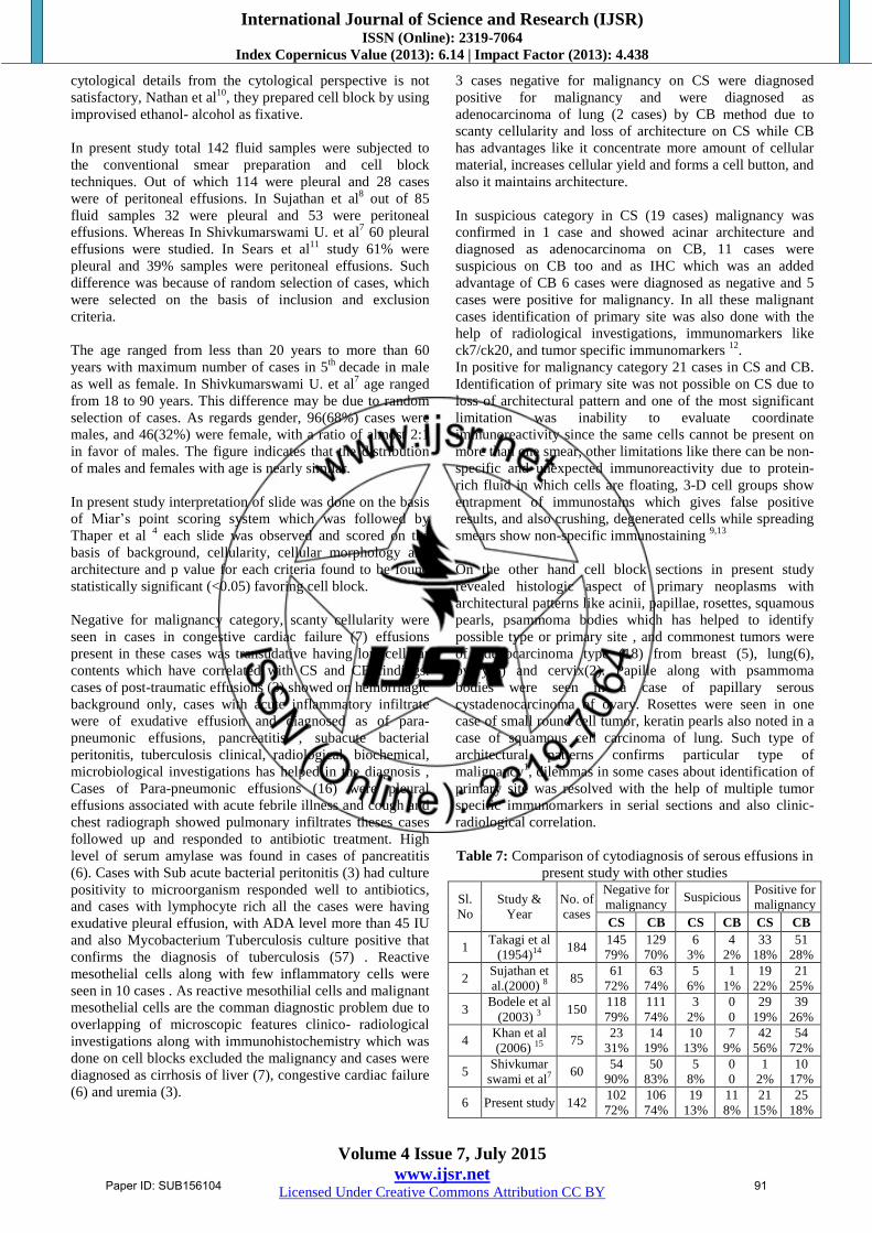
Figure 4: Clinical diagnosis in negative for malignancy in
pleural effusion.
Figure 5: Clinical diagnosis in negative for malignancy in
peritoneal effusions.
Figure 5 Clinical diagnosis in negative for malignancy in
peritoneal effusions.In the category of negative for
malignancy out of 104 cases maximum cases (57) were
diagnosed as tuberculosis followed by followed by
parapnemonic effusions (16), congestive cardiac failure (13),
pancreatitis(4), cases of post traumatic effusion (3), uremia
and subacute bacterial peritonitis, and meigs syndrome each
of one case.
Table 5: Contingency table showing the comparative
diagnosis of each method with the final diagnosis
To obtain sensitivity and specificity of Conventional Smear
method, two scenarios were considered for the suspicious
samples. In the first, all the suspicious samples were
considered as negative by CS method. This resulted into a
sensitivity of 0.6176 (95% CI: 0.4362 – 0.7731) and
specificity of 1.000 (95% CI: 0.9571 – 1.000). The Positive
Prediction Value (PPV) was obtained as 1.000 (95% CI:
0.8075 – 1.000) and Negative Prediction Value (NPV) was
0.8925 (95% CI: 0.8194 – 0.9392). The accuracy obtained
was 0.9084. Alternatively, if all the suspicious samples are
positive, then the sensitivity obtained was 0.7941 (95% CI:
0.6159 – 0.9066), specificity of 0.8796 (95% CI: 0.7994 –
0.9318), PPV of 0.675 (95% CI: 0.5075 – 0.8092) and NPV
of 0.9313 (95% CI: 0.8589 – 0.9695). The accuracy of the
method was obtained as 0.8591 (85%).
Similar analysis was carried out for Cell Block data.
Considering suspicious into negative category sensitivity:
0.7352 [0.5534 – 0.8649], specificity: 1.000 [0.9571 –
1.000] ,PPV: 1 [0.8342 – 1.000], NPV: 0.8239 [0.7491 –
0.8807] and accuracy: 0.9366. Considering suspicious into
positive category Sensitivity: 0.8823 [0.7160 – 0.9616]
,Specificity: 0.9444 [0.8781 – 0.9772] PPV: 0.8333 [0.6652
– 0.9303] , NPV: 0.9622 [0.9005 – 0.9878]
Accuracy: 0.9295
Table 6: Contingency table showing the comparative
diagnosis of each method with the final diagnosis
Final diagnosis
Negative Positive
Conventional Smear
Negative 95 7
Positive 0 21
Suspicious 13 6
Cell Block Negative 108 4
Positive 0 30
Analysis was carried out for Cell Block with IHC. It resulted
into a sensitivity of 0.8823 (95% CI: 0.7160 – 0.9616),
specificity of 1.000 (95% CI: 0.9571 – 1.000), PPV of 1.000
(95% CI: 0.8586 – 1.000) and NPV of 0.9642 (95% CI:
0.9056 – 0.9884). The accuracy obtained was 0.9718 (97%).
6. Discussion
In this study due consideration was given to type of cell
block technique, age, sex, site of effusion, quality of slides
in both methods, clinical and radiological findings and also
immunohistochemistry wherever needed to arrive at a final
diagnosis and to identify the primary malignant lesion and
also to type the malignancy. Cell blocks provided diagnostic
information complimentary or additional to that obtained
from an examination of the smears.
In present study, cell block preparation was done by plasma-
thromboplastin method, as the method utilizes outdated
plasma from blood bank and outdated thromboplastin
reagent from hematology laboratory so it becomes cost-
effective, and the advantages were it is reproducible and
concentrates more cellular material that forms more solid
cell button because of formation of clot, better cellular
preservation, and the disadvantage is that there can be
background staining in immunohistochemical staining.
Kulkarni MB5
et al uses similar method for cell block
preparation. Bodel et al3, Thaper et al
4, shivkumarswami et
al6,7
uses sediment method with 10% alcohol formalin as
fixative, while sujathan et al8. used sediment method with
ethanol-acetic acid and formalin as fixative both of these
method gave better cellularity when compared to
conventional smears as formalin minimized cell loss by
forming protein cross links and gel formation , but the
disadvantage is this method can be applied to the fluids with
significant sediments 9
, and also preservation of nuclear and
Final diagnosis
Negative Positive
Conventional smear
Negative 95 7
Positive 13 6
Suspicious 0 21
Cell Block
Negative 102 4
Positive 0 25
Suspicious 6 5
Paper ID: SUB156104 90
International Journal of Science and Research (IJSR) ISSN (Online): 2319-7064
Index Copernicus Value (2013): 6.14 | Impact Factor (2013): 4.438
Volume 4 Issue 7, July 2015
www.ijsr.net Licensed Under Creative Commons Attribution CC BY
cytological details from the cytological perspective is not
satisfactory, Nathan et al10
, they prepared cell block by using
improvised ethanol- alcohol as fixative.
In present study total 142 fluid samples were subjected to
the conventional smear preparation and cell block
techniques. Out of which 114 were pleural and 28 cases
were of peritoneal effusions. In Sujathan et al8 out of 85
fluid samples 32 were pleural and 53 were peritoneal
effusions. Whereas In Shivkumarswami U. et al7 60 pleural
effusions were studied. In Sears et al11
study 61% were
pleural and 39% samples were peritoneal effusions. Such
difference was because of random selection of cases, which
were selected on the basis of inclusion and exclusion
criteria.
The age ranged from less than 20 years to more than 60
years with maximum number of cases in 5th
decade in male
as well as female. In Shivkumarswami U. et al7 age ranged
from 18 to 90 years. This difference may be due to random
selection of cases. As regards gender, 96(68%) cases were
males, and 46(32%) were female, with a ratio of almost 2:1
in favor of males. The figure indicates that the distribution
of males and females with age is nearly similar.
In present study interpretation of slide was done on the basis
of Miar’s point scoring system which was followed by
Thaper et al 4 each slide was observed and scored on the
basis of background, cellularity, cellular morphology and
architecture and p value for each criteria found to be found
statistically significant (<0.05) favoring cell block.
Negative for malignancy category, scanty cellularity were
seen in cases in congestive cardiac failure (7) effusions
present in these cases was transudative having low cellular
contents which have correlated with CS and CB findings.
cases of post-traumatic effusions (3) showed on hemorrhagic
background only, cases with acute inflammatory infiltrate
were of exudative effusion and diagnosed as of para-
pneumonic effusions, pancreatitis , subacute bacterial
peritonitis, tuberculosis clinical, radiological, biochemical,
microbiological investigations has helped in the diagnosis ,
Cases of Para-pneumonic effusions (16) were pleural
effusions associated with acute febrile illness and cough and
chest radiograph showed pulmonary infiltrates theses cases
followed up and responded to antibiotic treatment. High
level of serum amylase was found in cases of pancreatitis
(6). Cases with Sub acute bacterial peritonitis (3) had culture
positivity to microorganism responded well to antibiotics,
and cases with lymphocyte rich all the cases were having
exudative pleural effusion, with ADA level more than 45 IU
and also Mycobacterium Tuberculosis culture positive that
confirms the diagnosis of tuberculosis (57) . Reactive
mesothelial cells along with few inflammatory cells were
seen in 10 cases . As reactive mesothilial cells and malignant
mesothelial cells are the comman diagnostic problem due to
overlapping of microscopic features clinico- radiological
investigations along with immunohistochemistry which was
done on cell blocks excluded the malignancy and cases were
diagnosed as cirrhosis of liver (7), congestive cardiac failure
(6) and uremia (3).
3 cases negative for malignancy on CS were diagnosed
positive for malignancy and were diagnosed as
adenocarcinoma of lung (2 cases) by CB method due to
scanty cellularity and loss of architecture on CS while CB
has advantages like it concentrate more amount of cellular
material, increases cellular yield and forms a cell button, and
also it maintains architecture.
In suspicious category in CS (19 cases) malignancy was
confirmed in 1 case and showed acinar architecture and
diagnosed as adenocarcinoma on CB, 11 cases were
suspicious on CB too and as IHC which was an added
advantage of CB 6 cases were diagnosed as negative and 5
cases were positive for malignancy. In all these malignant
cases identification of primary site was also done with the
help of radiological investigations, immunomarkers like
ck7/ck20, and tumor specific immunomarkers 12
.
In positive for malignancy category 21 cases in CS and CB.
Identification of primary site was not possible on CS due to
loss of architectural pattern and one of the most significant
limitation was inability to evaluate coordinate
immunoreactivity since the same cells cannot be present on
more than one smear, other limitations like there can be non-
specific and unexpected immunoreactivity due to protein-
rich fluid in which cells are floating, 3-D cell groups show
entrapment of immunostains which gives false positive
results, and also crushing, degenerated cells while spreading
smears show non-specific immunostaining 9,13
On the other hand cell block sections in present study
revealed histologic aspect of primary neoplasms with
architectural patterns like acinii, papillae, rosettes, squamous
pearls, psammoma bodies which has helped to identify
possible type or primary site , and commonest tumors were
of adenocarcinoma type (18) from breast (5), lung(6),
ovary(6) and cervix(2). Papille along with psammoma
bodies were seen in a case of papillary serous
cystadenocarcinoma of ovary. Rosettes were seen in one
case of small round cell tumor, keratin pearls also noted in a
case of squamous cell carcinoma of lung. Such type of
architectural patterns confirms particular type of
malignancy1, dilemmas in some cases about identification of
primary site was resolved with the help of multiple tumor
specific immunomarkers in serial sections and also clinic-
radiological correlation.
Table 7: Comparison of cytodiagnosis of serous effusions in
present study with other studies
Sl.
No
Study &
Year
No. of
cases
Negative for
malignancy Suspicious
Positive for
malignancy
CS CB CS CB CS CB
1 Takagi et al
(1954)14 184
145
79%
129
70%
6
3%
4
2%
33
18%
51
28%
2 Sujathan et
al.(2000) 8 85
61
72%
63
74%
5
6%
1
1%
19
22%
21
25%
3 Bodele et al
(2003) 3 150
118
79%
111
74%
3
2%
0
0
29
19%
39
26%
4 Khan et al
(2006) 15 75
23
31%
14
19%
10
13%
7
9%
42
56%
54
72%
5 Shivkumar
swami et al7 60
54
90%
50
83%
5
8%
0
0
1
2%
10
17%
6 Present study 142 102
72%
106
74%
19
13%
11
8%
21
15%
25
18%
Paper ID: SUB156104 91
International Journal of Science and Research (IJSR) ISSN (Online): 2319-7064
Index Copernicus Value (2013): 6.14 | Impact Factor (2013): 4.438
Volume 4 Issue 7, July 2015
www.ijsr.net Licensed Under Creative Commons Attribution CC BY
In present study most of cases were in negative for
malignancy category with 72% on CS while 74% on CB
similar findings were seen in study done by Sujathan et al8.
In suspicious for malignancy less no. of cases encountered
on CB( 8% ), and positive for malignancy maximum no of
cases were diagnosed on CB (18%). Similar findings were
also noted by Takagi et al14
, Sujathan et al8, Bodel et al
3 ,
Khan et al15
, and shivkumarswami et al7.
In the present study diagnostic yield for malignancy was
significantly increased by cell block method. The present
study identified additional 6.33%
(9 cases) malignant lesions by cell block method when
compared to conventional smear.Additional diagnostic yield
was noted in various studies. In study done by Bodele et al,
additional 7% (10 cases) of malignant lesions were
identified by cellblock method3. Dekkar and Bupp
et al
study, reported that samples obtained by combined cellblock
method and smear technique for malignant lesions were
double to that of conventional smear technique only. By
using cellblock method tumors were subsequently
demonstrated in 38% of the patient who had negative or
atypical cytological reports2.
In a study done by Khan et al, additional findings were
diagnostic in 16% of malignant cases15
. Additional 18 cases
for malignant lesions were diagnosed by cellblock method in
study done by Takagi F14
.Khan et al15
, in another study titled
as usefulness of cellblock verses smears in malignant
effusion cases reported that the recovery rate for malignant
lesions by cellblock preparation was 20% greater than that
obtained for specimen examined in smear only15
.
Table 8: Additional yield of malignancy in various studies
by cell block
Sl. No Study (%)
1 Dekker and Bupp et al 2 38
2 Khan et al15 20
3 Shivkumarswami et al7 15%
4 Bodele et al 3 7
5 Richardson et al16 5
6 Present study 6.33%
According to various studies additional diagnostic yield for
malignancy was noted if conventional smear technique is
supplemented by cellblock method.1,2,13
In present study, by
using cell block method we diagnosed malignant lesions in
21% of samples, where as in conventional smear method
diagnosis for malignant lesion was 15% only. In present
study identification of primary site was done in 100% cases.
In study done by Khan et al.15
could identify primary site in
81.3% while Thaper et al4
identified the same in 83.3%
cases. Clinic-radiological correlation along with
immunomarkers for identification of primary site helped to
confirm primary site in present study. In present study
sensitivity, specificity, PPV, and NPV for CS was 61-79%,
87-100%, 67-100%, 89-93% respectively, and for CB 73-
88%, 94-100%, 83-100 and 82-96% and CB with IHC 88%,
100%, 100%,and 97% respectively.
Table 26: Accuracy in different studies in CS and CB Study CS CB
Thaper et al4 71.42% 85.72%
Zemansky et al 17 - 90%
Ceelen 18 71% 89%
Present study 85-90% 97%
In present study accuracy of CB was 97%, increase accuracy
also noted by Thaper et al.4 (85%), and Ceelen et al
18 (89%)
zemansky et al17.
7. Conclusion
Cell block technique is simple and reproducible and uses
routine laboratory reagents and processing. In cell block
technique more amount of sample is required for obtaining
proper cell button. Cell block technique offers advantage
like it concentrates all the cellular material and increases
cellular yield. Though cell block show preservation of
architectural pattern. yet cellular morphology can be better
appreciated on conventional smears. Use of cell block
technique eliminated the suspicious for malignancy category
giving more definitive diagnosis and shows additional
increase in diagnostic yield. In cell block technique multiple
sections of the same material can be processed for
immunohistochemistry that help to identify primary site of
origin in malignant fluids in 100% cases.
Combined approach cell block in conjunction with
conventional smear can should be used in suspicious for
malignancy cases. Positive results, identification of primary
site in malignant effusions and further typing will have an
oblivious influence on patient management. Though cell
block technique is time consuming causes delay in issuing
report. It is balanced by its ability to increase sensitivity and
accuracy of final diagnosis
References
[1] Koss LG et al: Effusions in the presence of cancer. In
Koss’ Diagnostic Cytology and its Histopathologic
Bases, 5th edition. Edited by Koss LG, Melamed MR,
Vol 2, Philadelphia :Lippincott, Williams & wilkins
2006, 950-951.
[2] Dekker A, Bupp PA. Cytology of serous effusions. An
investigation into the usefulness of cellblocks versus
smears. Am J Clin Pathol 1978;70 (6):855-860.
[3] Bodele AK, Parate SN, Wadadekar AA, Bobhate SK,
Munshi MM. Diagnostic utility of cell block preparation
in reporting of fluid cytology. Journal of Cytology
2003;20(3):133-135.
[4] Meenu Thapar, Rajiv K Mishra, Amit Sharma, Vikas
Goyal, Vibhuti Goyal. Critical analysis of cell block
versus smear examination in effusions Journal of
Cytology.April 2009;26(2):60-64
[5] Manisha B Kulkarni,Sangeeta B Desai,Dulhan
Ajit,R.F.Chinoy.Utility of the thromboplastin-
plasma cell block technique for fine needle
aspiration and serous effusions.Diagnostic
Cytopathology 2009 Feb;37(2):86-90.
[6] Shivkumarswami U. surekha U. Arakeril, Mahesh
H. Karigowdar, B. R. Yelikar : The role of Cell
block method in the diagnosis of malignant ascetic
Paper ID: SUB156104 92
International Journal of Science and Research (IJSR) ISSN (Online): 2319-7064
Index Copernicus Value (2013): 6.14 | Impact Factor (2013): 4.438
Volume 4 Issue 7, July 2015
www.ijsr.net Licensed Under Creative Commons Attribution CC BY
fluid effusion Journal of clinical and diagnostic
research, 2012 ;September (suppl), vol-6(7):1280-
1283
[7] Shivkumarswami U. surekha U. Arakeril, Mahesh
H. Karigowdar, B. R. Yelikar : Diagnostic utility of
the cell block method versus the conventional
smear study in pleural fluid cytology: j cytol 2012;
29 : 11-5.
[8] Sujathan K, Pillai KR, Chandralekha B, Kannan S,
Mathew A, Nair MK. Cytodiagnosis of serous
effusions: A combined approach to morphological
features in Papanicolaou and May-Grunwald Giemsa
stained smears and modified cell block technique.
Journal of Cytology 2000;17(2):89-95.
[9] Shidham V, Atkinson B. Collection and processing of
effusion fluids. In Cytopathologic diagnosis of serous
fluids 1st
edition Philadelphia: Saunders Elsevier, 2007;
207-33
[10] Nathan NA, Narayan E. Smith MM, Horn MJ cell block
cytology : improved predation and its efficacy in
diagnostic cytology Am. J Clin pathol 2000; 114; 599-
606.
[11] Gia-Khanh Nguyen. Serous effusions. In Essentials Of
Fluid Cytology 1st edition Canada , 2009;9.
[12] Siddham V. Where do they come from? Evaluation of
unknown primary site of origin. In cytopathologic
diagnosis of serous fluids edn 1st Philadelphia: Saunders
Elsevier, 2007;157--169
[13] Bibbo M. pleural, peritoneal and pericardial effusions.
in Comprehensive cytopathology edn 3rd Philadelphia:
Saunders Elsevier, 1997;1: 515- 578.
[14] Takagi F. Studies on tumor cells in serious effusion, Am
J Clin Pathol 1954; 24:663-675.
[15] Khan N, Sherwani KR, Afroz N, Kapoor S. Usefulness
of cellblocks versus smears in malignant effusion cases.
Journal of Cytology 2006;23(3): 129-132.
[16] Richardson HL, Koss LG, Simon TR. An evaluation of
the concomitant use of cytological and histological
techniques in the recognition of cancer in exfoliated
material from various sources. Cancer 1995;8: 948-950.
[17] Zemansky AP Jr. The examination of fluids for tumor
cells: An analysis of m113 cases hecked against
subsequent examination of tissue. Am J M Sci
1928;175:489-504.
[18] Ceelen GH. The cytologic diagnosis of ascetic fluid.
Acta cytol 1964; 8:175-183
Author Profile Dr.Shubhada Bansode is Assistant Professor in Department of
Pathology, GMC & H,Latur, Maharashtra.India. She did MBBS –
GMC & H, Aurangabad, MD, DNB Pathology – GMC & H,
Nagpur.
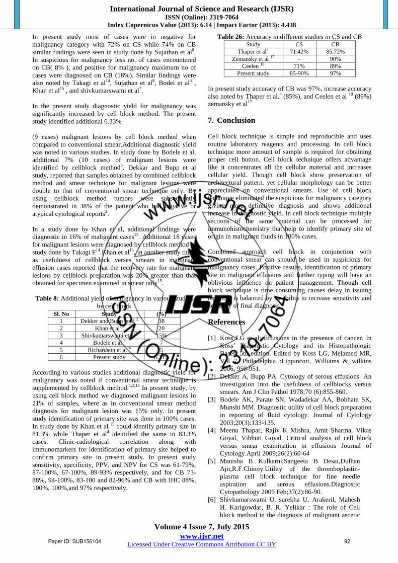
Photomicrograph 1: Atypical mesothelial cells. Suspicious
for malignancncy.on CS.
(40X, H&E)
Photomicrograph 2 : Acinar architecture . positive for
malignancncy.on CS.
(40X, H&E)
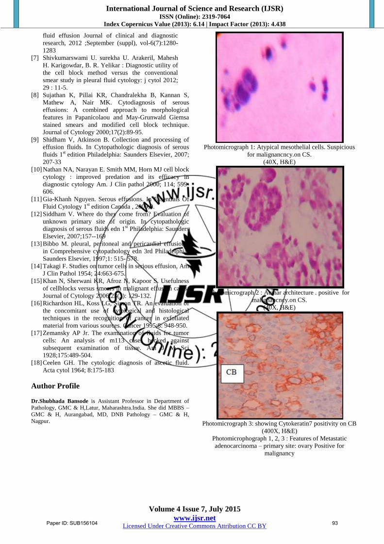
Photomicrograph 3: showing Cytokeratin7 positivity on CB
(400X, H&E)
Photomicrophograph 1, 2, 3 : Features of Metastatic
adenocarcinoma – primary site: ovary Positive for
malignancy
Paper ID: SUB156104 93
International Journal of Science and Research (IJSR) ISSN (Online): 2319-7064
Index Copernicus Value (2013): 6.14 | Impact Factor (2013): 4.438
Volume 4 Issue 7, July 2015
www.ijsr.net Licensed Under Creative Commons Attribution CC BY
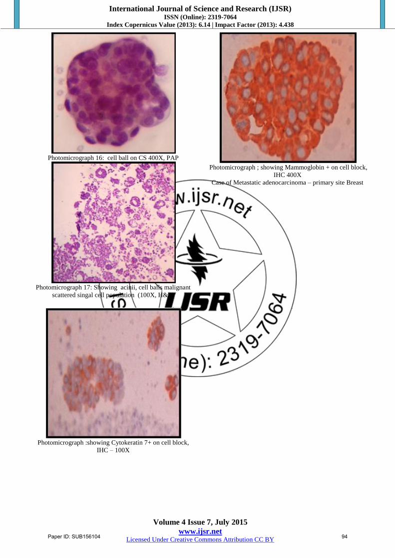
Photomicrograph 16: cell ball on CS 400X, PAP
Photomicrograph 17: Showing acinii, cell balls malignant
scattered singal cell population (100X, H&E)
Photomicrograph :showing Cytokeratin 7+ on cell block,
IHC – 100X
Photomicrograph ; showing Mammoglobin + on cell block,
IHC 400X
Case of Metastatic adenocarcinoma – primary site Breast
Paper ID: SUB156104 94
Recommended