Evaluating Anemia’s: Lets take a deeper dive
Joanne T. Eddington, MN, FNP, AOCN Providence Oncology and Hematology Care Clinic -Eastside

References
Brill, J.R., & Baumgardner, D. J. (2000). Normocytic anemia. American Family Physician, 62 (10), 2255-2263.
Cascio, M. J., & DeLoughery, T. G. (2017). Anemia. Evaluation and diagnostic tests. Medical Clinics of North America,101, 263-284. http://dx.doi.org/10.1016/j.mcna.2016.09.003
DeLoughery, T. G. (2014) Microcytic anemia. New England Journal of Medicine, 371 (14), 1324-31. http://dx.doi.org/10.1056/NEJMra1215361
Rui Li, MD- Oncology and Hematology Care Clinic – East. Thank you!

Disclosures
I have no disclosures or conflicts of interest
Overview of Anemia
Anemia is grouping of signs and symptoms, rather than a disease
Anemia is a highly significant clinical finding
Severity of the anemia does not correlate with clinical significance or causality.
Review of terms.
Anemia is a deficiency in the red blood cell mass (RBC or erythrocyte)and hemoglobin content
Anemia is revealed through a CBC (complete blood count) which is a measure of hemoglobin concentration, hematocrit, RBC mass and mean corpuscular volume (MCV)
Hematocrit is the percentage of packed RBC’s in blood or the volume occupied by RBC’s after blood is spun down in a hematocrit tube.
Various definitions of Anemia (hemoglobin and hematocrit levels) Hemoglobin (Hgb) <12 in women and Hemoglobin (Hgb) < 14 in
men
Signs and symptoms of anemia
They are unreliable in predicting the severity of the anemia
Time and onset of the anemia and health of the patient is most predictive of symptoms
Patients with anemia which develops over time and occurs gradually can tolerate lower hemoglobin levels due to compensation.
Patients with abrupt onset of anemia tend to be very symptomatic
Patients complain of fatigue and shortness of breath
On exam patients have pale mucous membranes, resting tachycardia and have a greyish hue about them
Clues to the source of anemia on exam include splenomegaly, heme positive stools
How does the body compensate for anemia? Cardiac output increases resulting in tachycardia
Plasma volume increases which allows remaining cells to move more efficiently due to decrease in red cell viscosity
Decrease in oxygen affinity for hemoglobin which increases the delivery of oxygen to the tissues This is accomplished through the increase in red cell 2,3-diphosphoglycerate
which is a 3 carbon isomer found in red blood cells. It’s increase enhances the ability of RBCs to release oxygen near tissues that need it most.
Classification of anemia
By size of red blood cellsMCV – Mean corpuscular volume refers to the volume of the
red blood cell Microcytic – low MCV
Normocyctic - normal MCV
Macrocytic - high MCV
By mechanism causing the anemia Increased loss- bleeding or hemolysis
Decreased production- impaired marrow production, nutritional deficiency
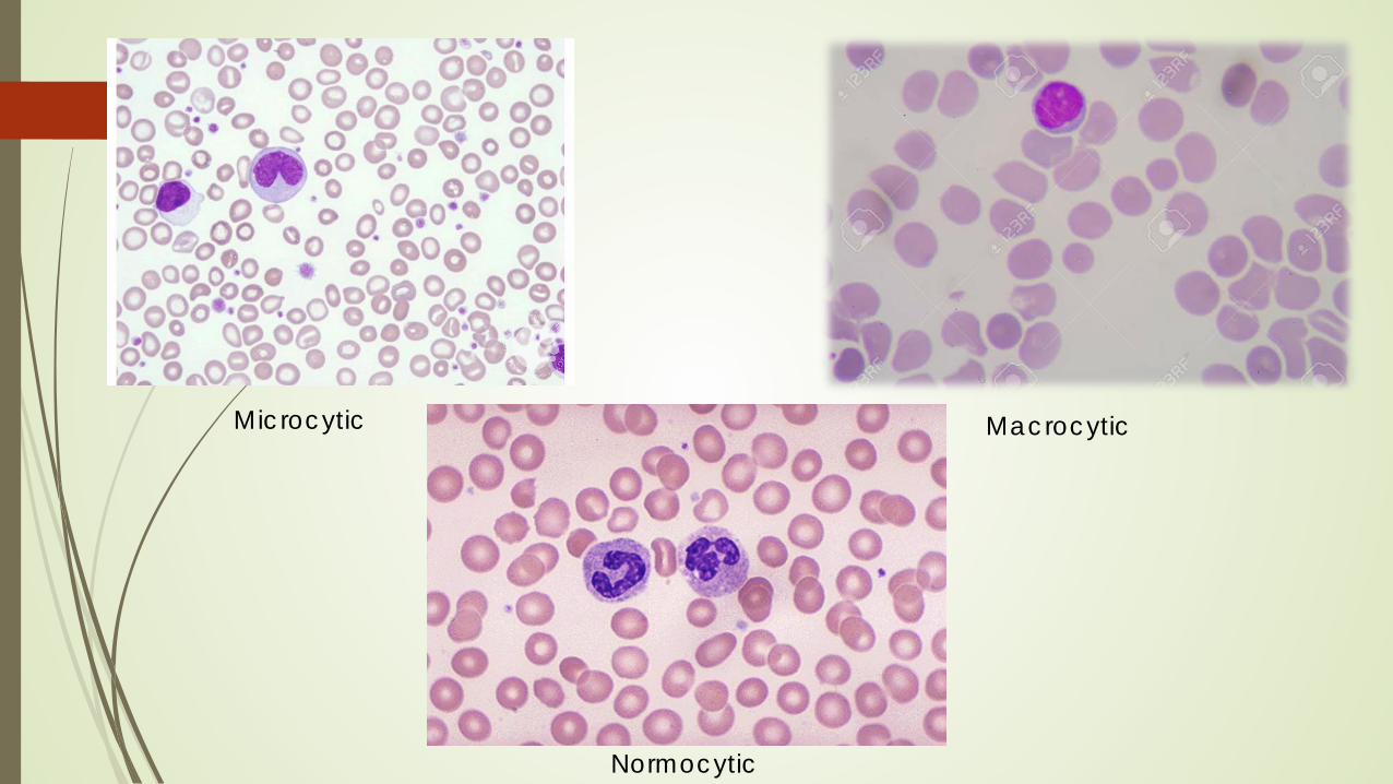
MacrocyticMicrocytic
Normocytic
Microcytosis (MCV < 80 fl)
Microcytic anemia’s reflect a defect in hemoglobin synthesis
Hemoglobin synthesis requires iron Lack of iron can be due to a deficiency or the lack of availability of iron
(sequestration of iron as part of an inflammatory process)which impairs delivery of iron to the developing red cell
Thalassemia Genetic disorder that leads to impaired production of hemoglobin
Sideroblastic anemia’s Actual defect in the synthesis of the heme molecule which leads to
underhemoglobinazation of erythroid precursors and microcytosis
Macrocytosis (MCV > 100 fl)
Caused by red cell membrane defects OR DNA synthesis defects Red cell membrane defects occur in the setting of liver disease,
hypothyroidism, aplastic anemia, renal disease and reticulocytosis Review of smear demonstrates round red blood cells
Defects in DNA synthesis is seen in B12 and Folate deficiency, chemotherapy and myelodysplastic syndrome Review of smear demonstrates oval red blood cells.
Normocytic Anemia (MCV 80-100 fl)
Red cells demonstrate normal size
Very common, and does not give a clue to potential etiologies
Can occur in early stage of what ever process is occurring
Can occur when several processes are occurring at the same time Iron deficiency and liver disease
Can see normal MCV in Anemia of inflammation
Acute hemolysis or blood loss
Renal disease
Using underlying mechanism as a tool for your differential Is there an increased RBC loss?
Is there a decreased RBC production?
The Reticulocyte count can help you differentiate this.
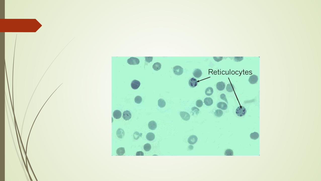
What is a reticulocyte?
Reticulocytes are immature, nonnucleated RBC’s
They circulate the peripheral blood for about 1 day before losing RNA and becoming a mature red blood cell.
The number of reticulocytes can be measured directly This is performed by automated analyzers by staining remnant RNA with
fluorescent dye.
This is known as the absolute reticulocyte count. ( ignore %)
Old methods required the percent hematocrit to perform a correction that adjusts for hematocrit.
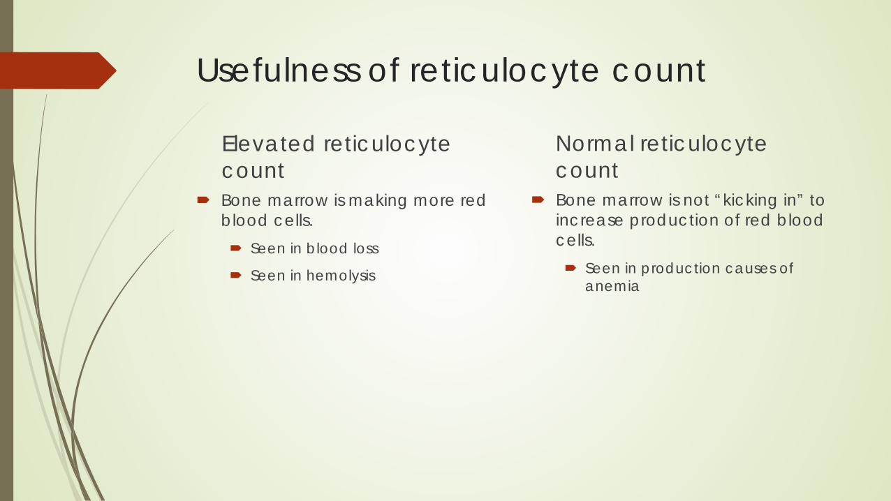
Usefulness of reticulocyte count
Elevated reticulocyte count
Bone marrow is making more red blood cells. Seen in blood loss
Seen in hemolysis
Normal reticulocyte count
Bone marrow is not “kicking in” to increase production of red blood cells. Seen in production causes of
anemia

It all comes down to the following same concepts Problems of production
Factory problem- bone marrow malfunction
Part problem- problem with the supply chain
Problems of destruction hemolysis
Problems of loss Bleeding
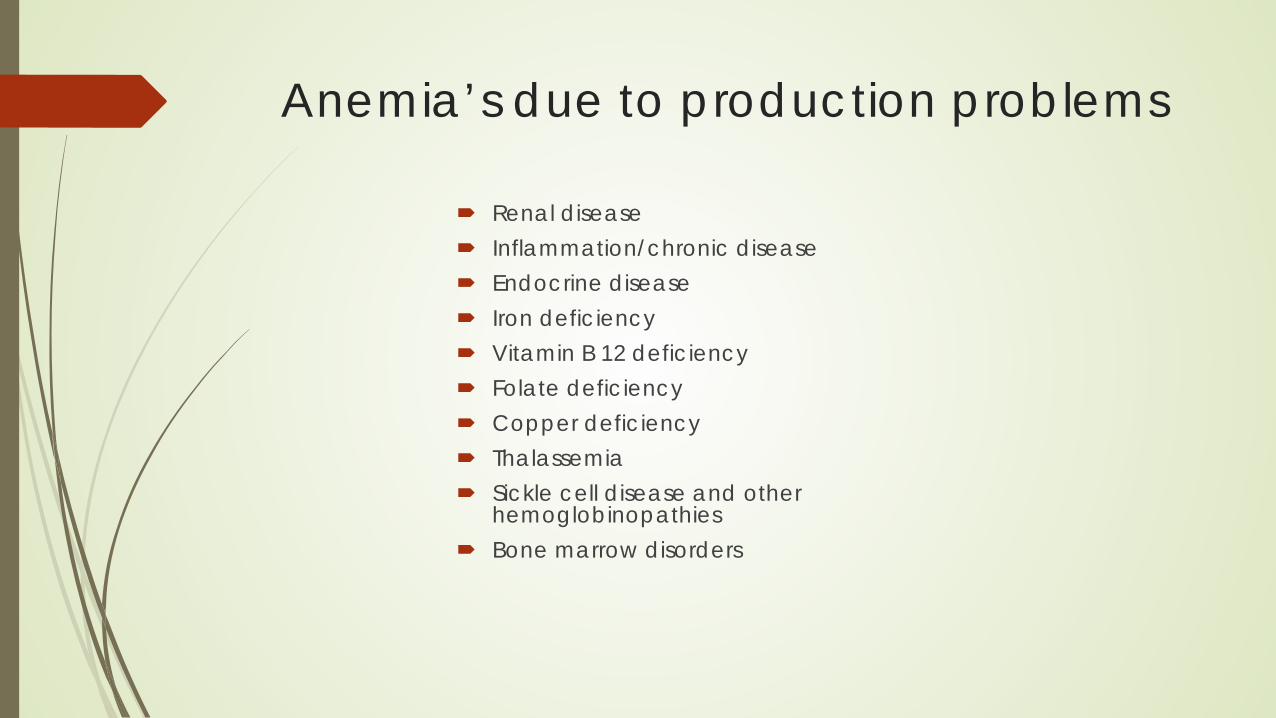
Anemia’s due to production problems
Renal disease Inflammation/chronic disease Endocrine disease Iron deficiency Vitamin B 12 deficiency Folate deficiency Copper deficiency Thalassemia Sickle cell disease and other
hemoglobinopathies Bone marrow disorders
Production of red cells
Must have a functioning marrow or “factory”
Must have appropriate supplies: iron, B12, Folate
Must have working production signals: erythropoietin
Must have working processes to utilize the supplies Acute phase reactants can interfere with the utilization process
Anemia of renal disease
Due to lack of erythropoietin
Decrease in renal function affects kidney’s ability to make erythropoietin
Generally anemia does not occur until creatinine clearance is < 30 mL/min
Patients with underlying inflammatory processes or those of advanced age can develop anemia with a creatinine clearance as high as 60 mL/min
Ace inhibitors and Angiotension II Receptor blockers can decrease Erythropoetin (EPO) production
Testing in Anemia of renal disease
CBC reveals Mild to moderate anemia
MCV is usually normal
Elevated BUN and Creatinine
Erythropoetin level maybe decreased and not compensated for level of anemia or level maybe normal based on lab range but inappropriately low for the degree of anemia.
Absolute reticulocyte count is usually normal
Anemia of inflammation or Anemia of chronic disease This is a diagnosis of exclusion
Patient will have adequate iron stores but there is impaired delivery to the developing red cells.
EPO production is depressed
If a patient has anemia, EPO level is not increased and patient has adequate iron stores, then diagnosis is anemia of chronic disease.
Ferritin will be high in this setting.
Mechanism of normal iron delivery to cells Iron is absorbed in the GI tract by enterocytes
Once absorbed by enterocytes, it is released in circulation and binds to transferrin (this is where saturation of the transferrin occurs)
Transferrin transports iron to the developing red blood cells
Excess iron is stored in hepatocytes in the liver
Mechanism of abnormal iron delivery in anemia of inflammation Inflammatory cytokines suppress production of erythropoietin by the kidney
resulting in decreased red cell production Iron availability is suppressed by hepcidin which is an acute phase reactant
Hepcidin is a key regulator of the entry of iron into the circulation in mammals. In states in which the hepcidin level is abnormally high such as inflammation, serum iron falls due to iron trapping within macrophages and liver cells and decreased gut iron absorption.
Hepcidin blocks iron absorption by preventing release of iron from enterocytes in the gut
Decreased circulating iron results in desaturation of transferrin which cannot supply enough iron to the developing red cells
Iron stored in the hepatocytes is also impacted by Hepcidin as it prevents release of iron from the hapatocyte
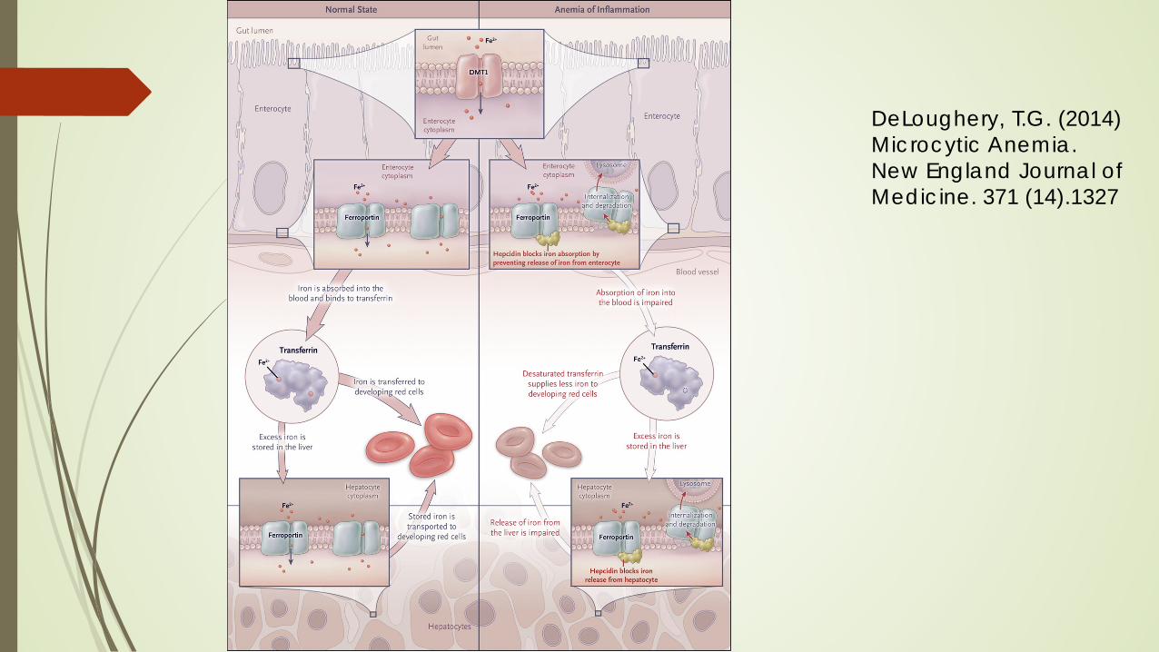
DeLoughery, T.G. (2014) Microcytic Anemia.New England Journal ofMedicine. 371 (14).1327
Testing in anemia of chronic disease
CBC shows mild anemia
MCV could be low, normal or high
Absolute reticulocyte count normal
Lack of elevation of EPO and adequate iron stores is highly suggestive of anemia of chronic disease
Anemia related to endocrine diseases
Hypogonadism can cause anemia in men.
Testosterone sensitizes erythroid precursors to the effects of EPO Explains why men have a higher hemoglobin concentration, hematocrit and
RBC count than woman
Ask older men with anemia if they have to shave anymore…….if they don’t they may have anemia related to low testosterone! (usually it is mild, Hgb 12-13)
Hypothyroidism can lead to macrocytic or normocytic anemia
Endocrine related anemia’s are considered anemia’s of chronic disease Correct the endocrine disorder and the anemia should correct.
Anemia related to iron deficiency
Iron deficiency anemia occurs when there is a negative iron balance
Negative iron balance occurs through blood loss or increase iron demand
Negative iron balance leads to a reduction in total body stores
Stage I : Iron depletion There is a decrease in iron stores, but serum iron or hemoglobin levels are WNL
Serum ferritin is low but serum iron is WNL
Stage II: Abnormal Iron test: decreased transferrin saturation, increase total iron binding
capacity
Stage III: Decreased hemoglobin concentration below the limits of normal
Causes of anemia related to iron deficiency Blood loss
GI bleed, most common reason for blood loss in men and postmenopausal women
Menstrual bleeding
Impaired absorption Gastrectomy, gastric bypass
Celiac disease
PPI use
H pylori infection
Strict vegan diet
Diagnostic testing for iron deficiency-
RBC indices, serum iron and TIBC and iron saturation have been traditionally performed
They can be confusing and are either poorly sensitive or poorly specific
Serum ferritin DIRECTLY correlates with iron stores
A serum ferritin greater than 100g/ml rules out iron deficiency in most patients
Very low ferritins are diagnostic of iron deficiency
Older patients maybe iron deficient with ferritins in the 50-80 range
Rule of thumb the ferritin should be greater than the patient’s age to rule out iron deficiency!
All unexplained cases of iron deficiency anemia need GI referral for colonoscopy/upper endoscopy. Do not assume menstruation or hemorrhoids to explain iron deficiency anemia in young patients. We are seeing a rise of colorectal cancer in young patient’s 20-40 years of age
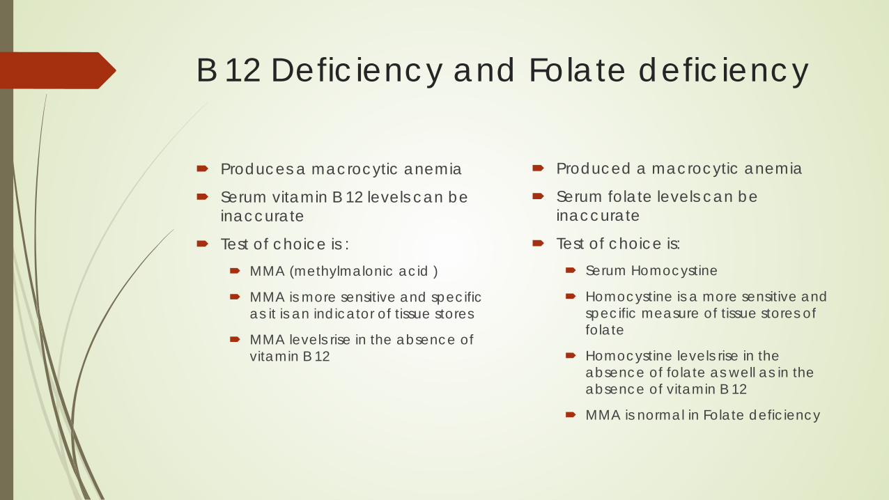
B 12 Deficiency and Folate deficiency
Produces a macrocytic anemia
Serum vitamin B 12 levels can be inaccurate
Test of choice is : MMA (methylmalonic acid )
MMA is more sensitive and specific as it is an indicator of tissue stores
MMA levels rise in the absence of vitamin B 12
Produced a macrocytic anemia
Serum folate levels can be inaccurate
Test of choice is: Serum Homocystine
Homocystine is a more sensitive and specific measure of tissue stores of folate
Homocystine levels rise in the absence of folate as well as in the absence of vitamin B 12
MMA is normal in Folate deficiency
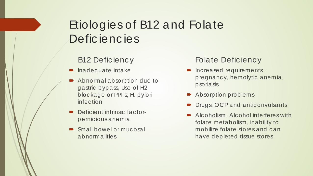
Etiologies of B12 and Folate Deficiencies
B12 Deficiency Inadequate intake
Abnormal absorption due to gastric bypass, Use of H2 blockage or PPI’s, H. pylori infection
Deficient intrinsic factor-pernicious anemia
Small bowel or mucosal abnormalities
Folate Deficiency Increased requirements :
pregnancy, hemolytic anemia, psoriasis
Absorption problems
Drugs: OCP and anticonvulsants
Alcoholism: Alcohol interferes with folate metabolism, inability to mobilize folate stores and can have depleted tissue stores
B 12 deficiency can cause neurological symptoms Paresthesia
Decrease vibratory sense
Ataxia or gait disturbances
Increase deep tendon reflexes
Memory loss
Personality changes
Orthostatic hypotension
Copper deficiencyCopper is key to normal hematopoesis
Straight forward testing Ceruloplasmin assay
Ceruloplasmin is the copper carrying protein (low result in copper deficiency)
Serum copper Seen in anorexia, bariatric surgery patients and excessive zinc intake Classic signs
Anemia
Neutropenia Thrombocytopenia (rare) Neurological findings such as peripheral neuropathy
Thalassemia
Thalassemia’s are inherited genetic disorders Thalassemia’s are diseases of hemoglobin synthesis which result in impaired
production of hemoglobin Most common types are alpha and beta thalassemia Alpha thalassemia is found in Africa, Mediterranian area and Southeast
Asia Beta thalassemia is found in the Mediterranian area and southeast asia Hemoglobin electrophoresis can assist in the diagnosis of beta thalassemia.
Hemoglobin A2 is increased
Alpha thalassemia is often a diagnosis of exclusion as there is no increase in hemoglobin A2
Sickle cell disease
Sickle cell disease is a group of genetic disorders characterized by the predominance of Hgb S
Hallmarks of the disease are chronic hemolytic anemia and vaso-occlusion
Screening test is the sickle solubility test
Definitive diagnosis is with hemoglobin electrophoresis
Bone marrow disorders
Myelodysplastic syndrome MDS Often see in older patients
Labs often show a macrocytic anemia with a non-elevated absolute reticulocyte count
Bone marrow evaluation is necessary to diagnose
Other cytopenias are also seen
Plasma cell Myeloma/Multiple Myeloma Plasma cell malignancy
Seen usually in older patient
Be suspicious if patient presents with back pain or renal disease
Testing should include: Serum protein electrophoresis with immunofixation
Serum free light chain analysis (10% of myeloma only secretes light chains)
Interpretation of SPEP and Serum Free Light chains. SPEP with immunofixation will show a monoclonal protein
IgG or IGM or IgA
Serum Free Light Chains Ratio of lambda to kappa
The ratio is the key, indicating clonal change. In a chronic renal disease patient, they may have a significant elevation in both kappa and lambda due to proteinuria, but the ratio is normal.
Anemia due to loss of red blood cells
Hemolysis
GI bleeding
Sequestration
Hemolysis
Hemolysis is destruction of red blood cells
Can be a result of an autoimmune process
Can be a result of mechanical destruction (heart valve)
Can be a result of drugs ( antibiotics, Tylenol, methyldopa)
Can be a result of paroxysmal nocturnal hemoglobinuria
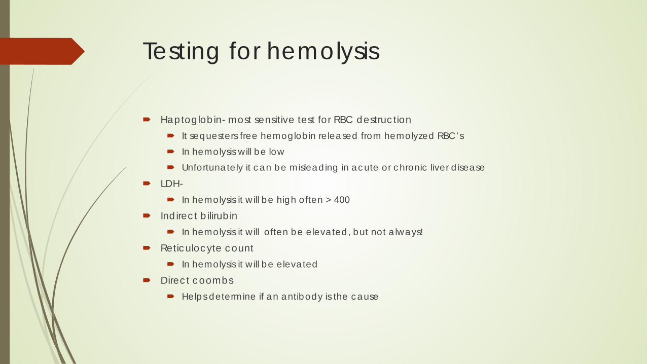
Testing for hemolysis
Haptoglobin- most sensitive test for RBC destruction It sequesters free hemoglobin released from hemolyzed RBC’s In hemolysis will be low Unfortunately it can be misleading in acute or chronic liver disease
LDH- In hemolysis it will be high often > 400
Indirect bilirubin In hemolysis it will often be elevated, but not always!
Reticulocyte count In hemolysis it will be elevated
Direct coombs Helps determine if an antibody is the cause
GI blood loss
Will cause iron deficiency over time
GI consult necessary
Patient may need both blood transfusion support and eventually iron replacement

Organ sequestration
Liver and spleen function as “filters”
Cirrhosis, chronic liver disease (hepatitis)and myeloproliferative diseases can cause hypersplenism
Blood circulates through the liver and spleen and if enlarged, can remove blood from circulation like an oil filter in your car
If the filter is bigger than it is suppose to be, mild anemia and/or pancytopenia can occur.
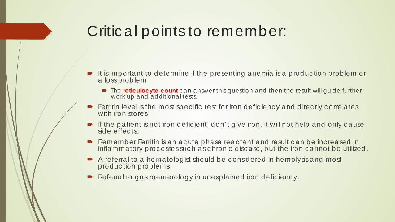
Critical points to remember:
It is important to determine if the presenting anemia is a production problem or a loss problem The reticulocyte count can answer this question and then the result will guide further
work up and additional tests.
Ferritin level is the most specific test for iron deficiency and directly correlates with iron stores
If the patient is not iron deficient, don’t give iron. It will not help and only cause side effects.
Remember Ferritin is an acute phase reactant and result can be increased in inflammatory processes such as chronic disease, but the iron cannot be utilized.
A referral to a hematologist should be considered in hemolysis and most production problems
Referral to gastroenterology in unexplained iron deficiency.


Recommended