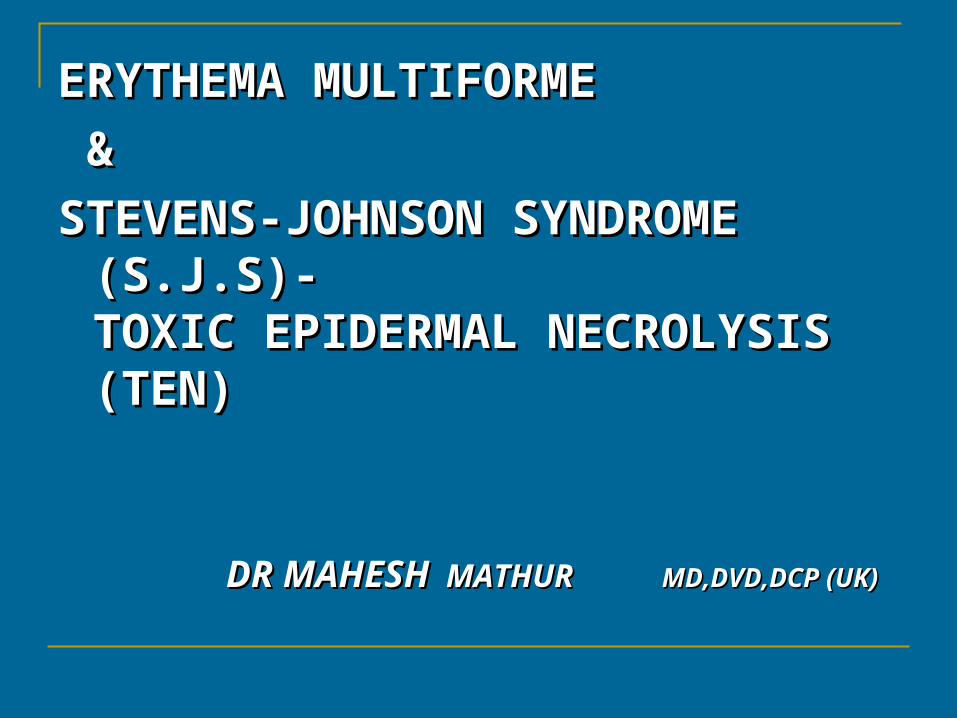
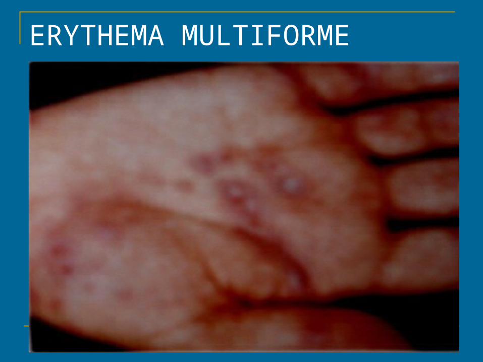
ERYTHEMA MULTIFORMEERYTHEMA MULTIFORME
&&
STEVENS-JOHNSON SYNDROME STEVENS-JOHNSON SYNDROME (S.J.S)-(S.J.S)-TOXIC EPIDERMAL NECROLYSIS TOXIC EPIDERMAL NECROLYSIS (TEN)(TEN)
DR MAHESHDR MAHESH MATHUR MATHUR MD,DVD,DCP (UK)MD,DVD,DCP (UK)
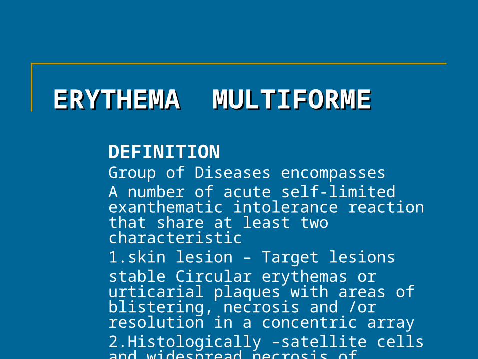
ERYTHEMA ERYTHEMA MULTIFORMEMULTIFORME
DEFINITIONGroup of Diseases encompassesA number of acute self-limited exanthematic intolerance reaction that share at least two characteristic 1.skin lesion – Target lesions stable Circular erythemas or urticarial plaques with areas of blistering, necrosis and /or resolution in a concentric array2.Histologically –satellite cells and widespread necrosis of epidermis
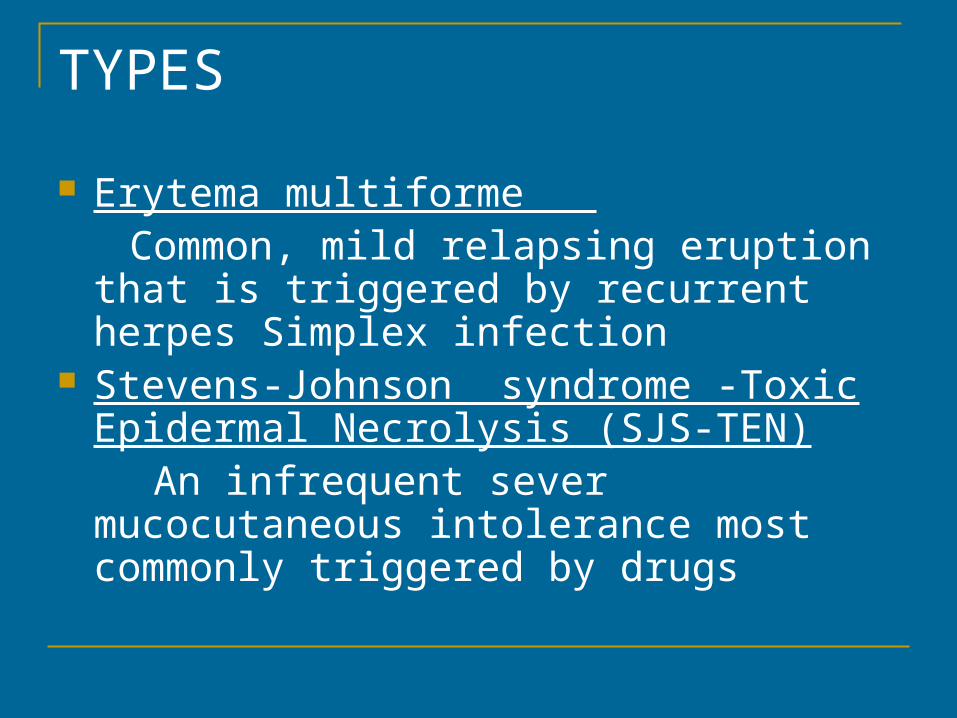
TYPES
Erytema multiforme Common, mild relapsing eruption that is
triggered by recurrent herpes Simplex infection
Stevens-Johnson syndrome -Toxic Epidermal Necrolysis (SJS-TEN)
An infrequent sever mucocutaneous intolerance most commonly triggered by drugs
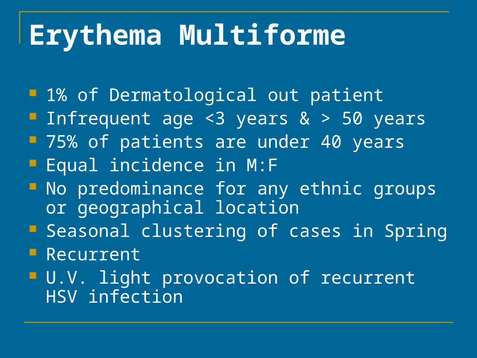
Erythema Multiforme
1% of Dermatological out patient Infrequent age <3 years & > 50 years 75% of patients are under 40 years Equal incidence in M:F No predominance for any ethnic groups or
geographical location Seasonal clustering of cases in Spring Recurrent U.V. light provocation of recurrent HSV infection
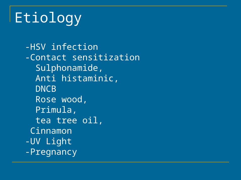
Etiology
-HSV infection -Contact sensitization Sulphonamide, Anti histaminic, DNCB Rose wood, Primula, tea tree oil, Cinnamon -UV Light -Pregnancy
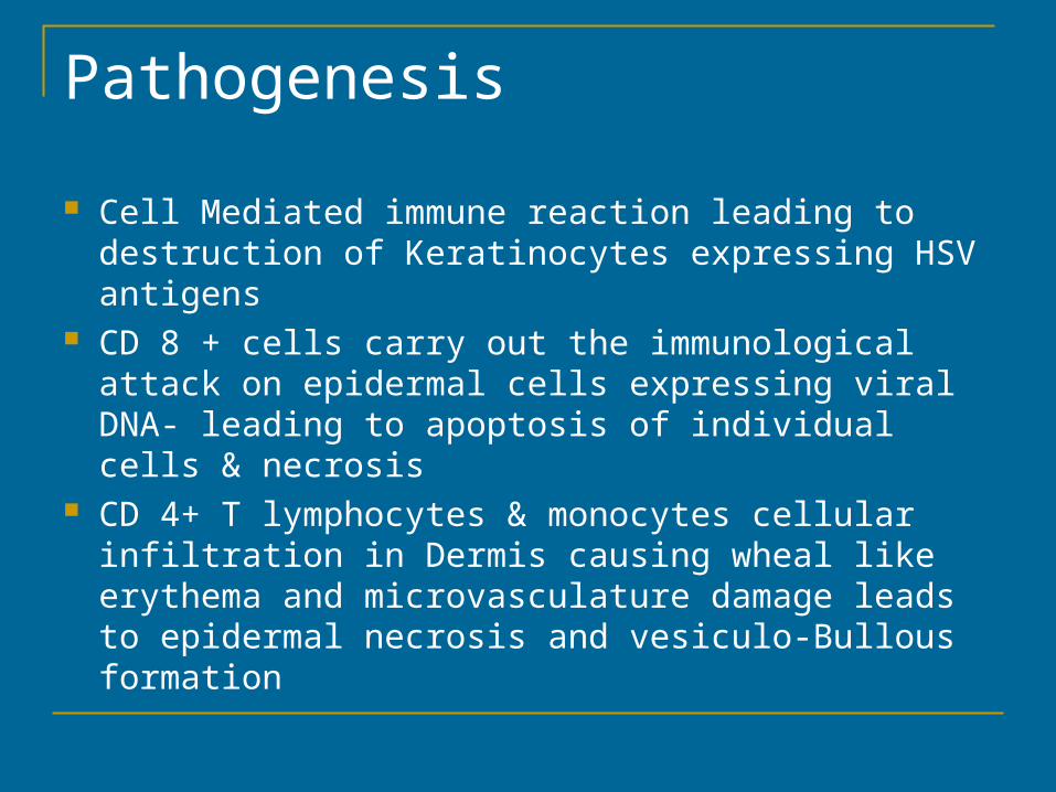
Pathogenesis
Cell Mediated immune reaction leading to destruction of Keratinocytes expressing HSV antigens
CD 8 + cells carry out the immunological attack on epidermal cells expressing viral DNA- leading to apoptosis of individual cells & necrosis
CD 4+ T lymphocytes & monocytes cellular infiltration in Dermis causing wheal like erythema and microvasculature damage leads to epidermal necrosis and vesiculo-Bullous formation
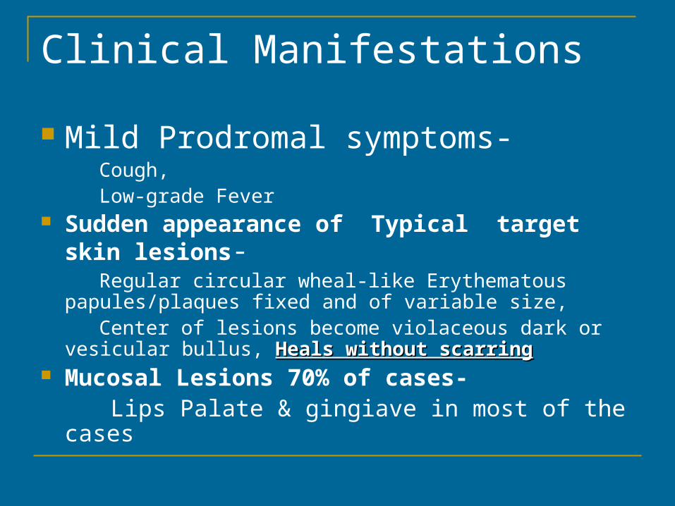
Clinical Manifestations
Mild Prodromal symptoms- Cough, Low-grade Fever Sudden appearance of Typical target skin
lesions-
Regular circular wheal-like Erythematous papules/plaques fixed and of variable size,
Center of lesions become violaceous dark or vesicular bullus, Heals without scarringHeals without scarring
Mucosal Lesions 70% of cases- Lips Palate & gingiave in most of the cases
ERYTHEMA MULTIFORME
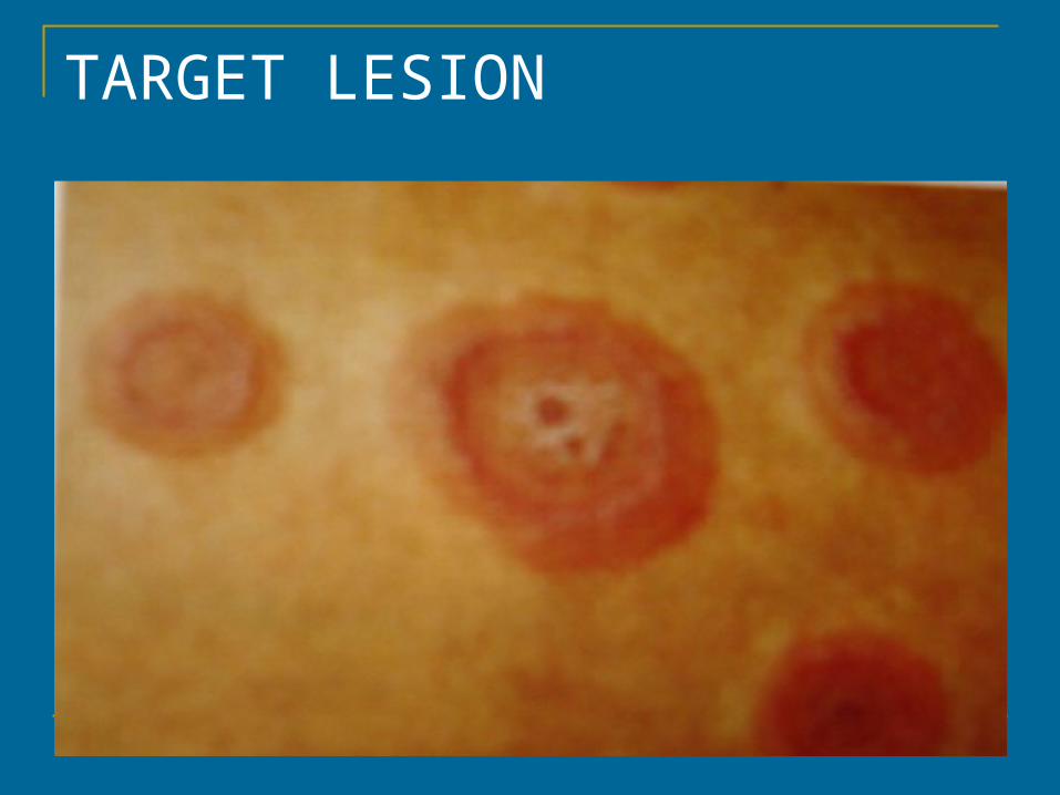
TARGET LESION
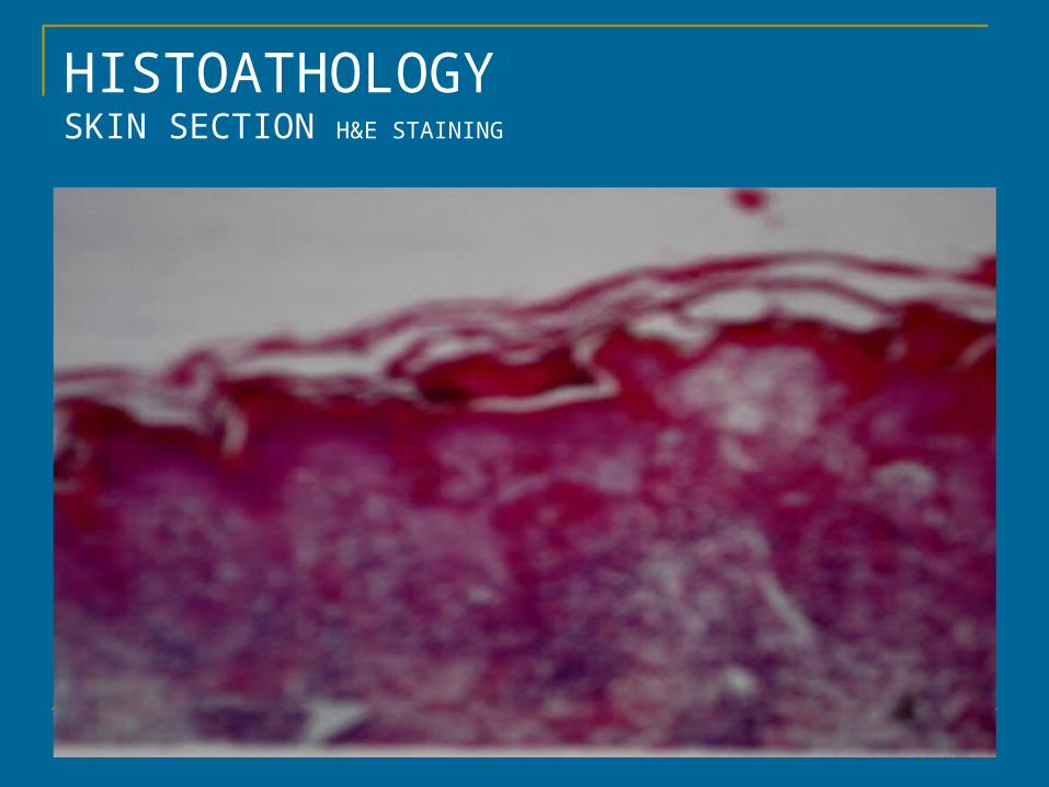
HISTOATHOLOGYSKIN SECTION H&E STAINING
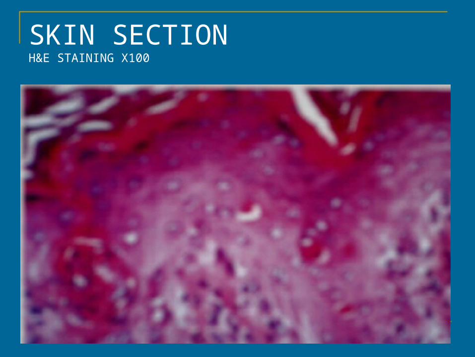
SKIN SECTIONH&E STAINING X100
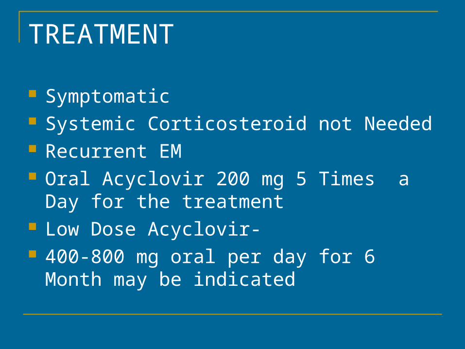
TREATMENT
Symptomatic Systemic Corticosteroid not Needed Recurrent EM Oral Acyclovir 200 mg 5 Times a Day for the
treatment Low Dose Acyclovir- 400-800 mg oral per day for 6 Month may be
indicated
STEVENS-JOHNSON SYNDROME STEVENS-JOHNSON SYNDROME (S.J.S)(S.J.S)&&TOXIC EPIDERMAL NECROLYSIS TOXIC EPIDERMAL NECROLYSIS (TEN)(TEN)DEFINITION Sever, episodic, acute mucocutaneous reaction most
commonly elicited by drugs Characterized by rapidly spreading irreregular dusky
erythematic maculs, necrosis of skin and detachment of skin resembling scalding of skin with involvement of more then one mucosal site
Constitutional symptoms and internal organ involvement often occurs and may be sever
Self-limited Significant morbidity scaring & mortality
Etiology 2 to 3 cases per million Occurs World wide M:F ratio is 2;1 Drugs * 3 fold increase in HIV infected Population Infections-Mycoplasma, Pneumoniae,Infectious
mononucleosis, Histoplasmosis, Gram Negative septicemias
Pathogenesis CD4+ & CD8+ cells & cytokines mediated injury
to epidermal cells
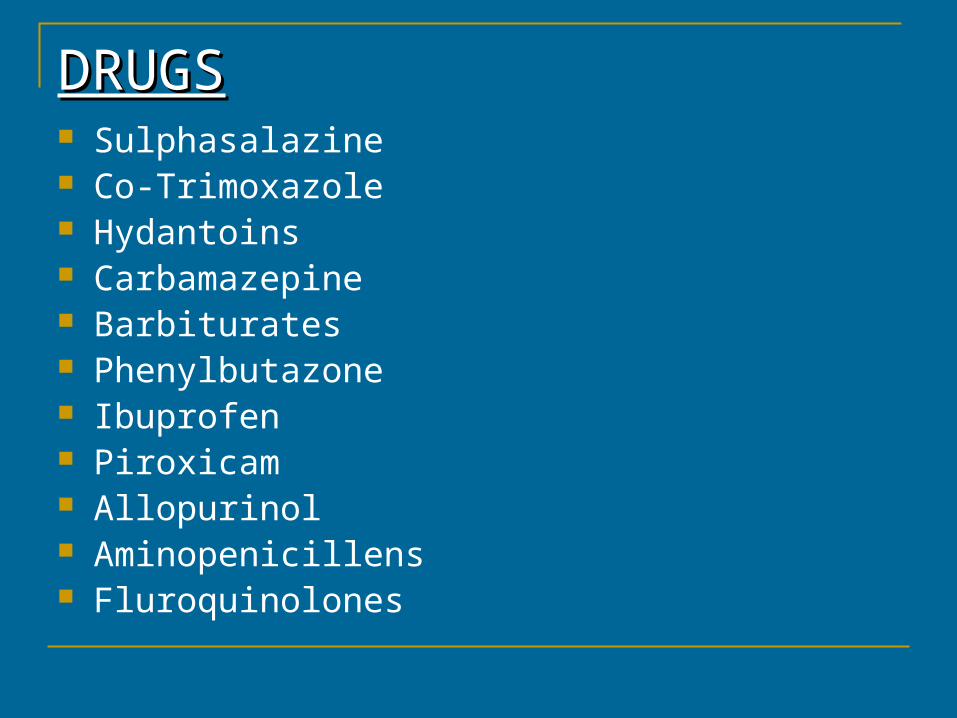
DRUGSDRUGS Sulphasalazine Co-Trimoxazole Hydantoins Carbamazepine Barbiturates Phenylbutazone Ibuprofen Piroxicam Allopurinol Aminopenicillens Fluroquinolones
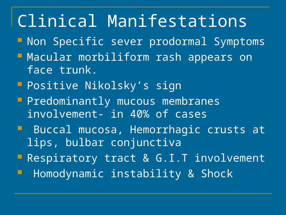
Clinical Manifestations Non Specific sever prodormal Symptoms Macular morbiliform rash appears on face
trunk. Positive Nikolsky’s sign Predominantly mucous membranes
involvement- in 40% of cases Buccal mucosa, Hemorrhagic crusts at lips,
bulbar conjunctiva Respiratory tract & G.I.T involvement Homodynamic instability & Shock
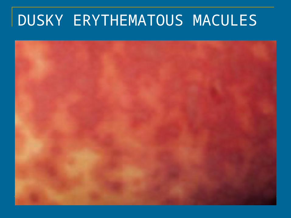
DUSKY ERYTHEMATOUS MACULES
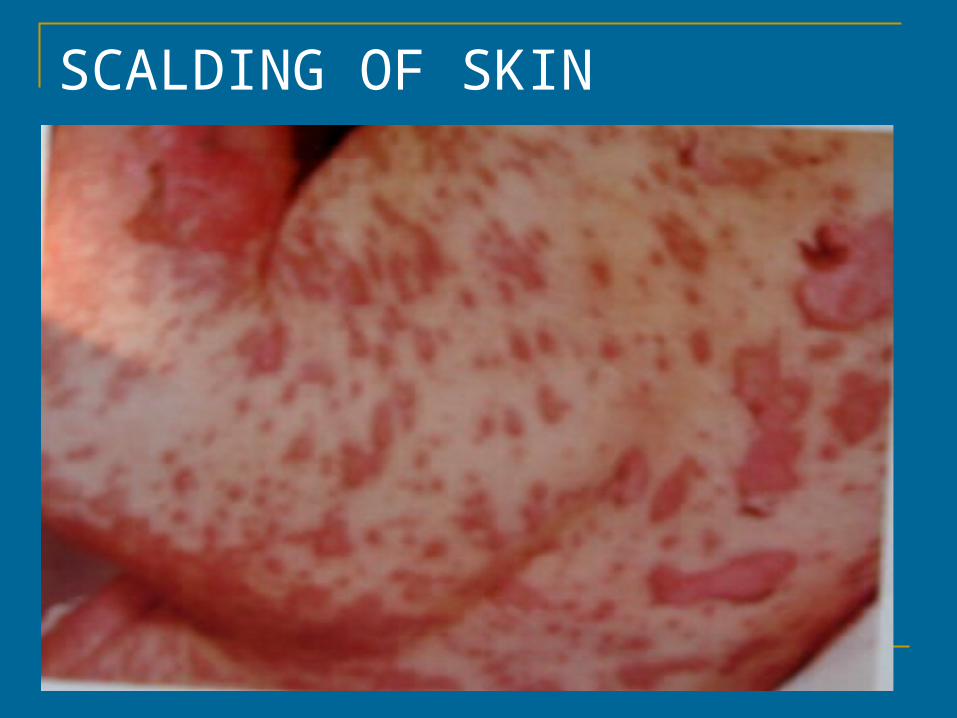
SCALDING OF SKIN
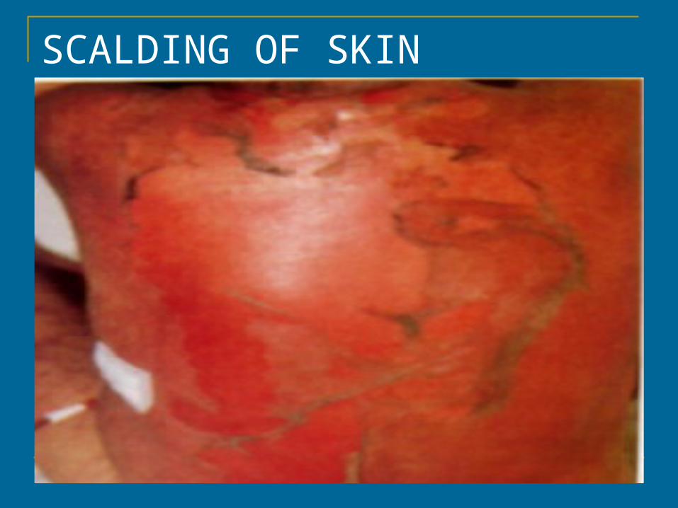
SCALDING OF SKIN
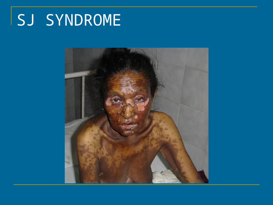
SJ SYNDROME
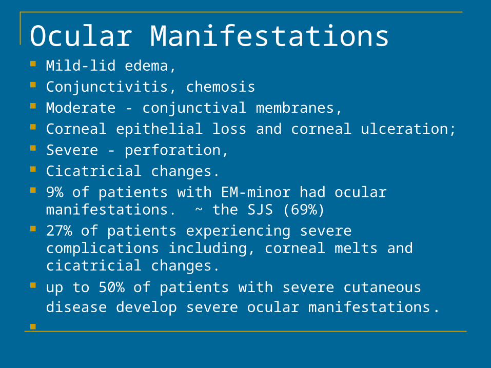
Ocular Manifestations Mild-lid edema, Conjunctivitis, chemosis Moderate - conjunctival membranes, Corneal epithelial loss and corneal ulceration; Severe - perforation, Cicatricial changes. 9% of patients with EM-minor had ocular manifestations.
~ the SJS (69%) 27% of patients experiencing severe complications
including, corneal melts and cicatricial changes. up to 50% of patients with severe cutaneous disease
develop severe ocular manifestations.
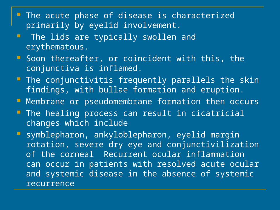
The acute phase of disease is characterized primarily by eyelid involvement.
The lids are typically swollen and erythematous. Soon thereafter, or coincident with this, the conjunctiva is
inflamed. The conjunctivitis frequently parallels the skin findings,
with bullae formation and eruption. Membrane or pseudomembrane formation then occurs The healing process can result in cicatricial changes
which include symblepharon, ankyloblepharon, eyelid margin rotation,
severe dry eye and conjunctivilization of the corneal Recurrent ocular inflammation can occur in patients with resolved acute ocular and systemic disease in the absence of systemic recurrence
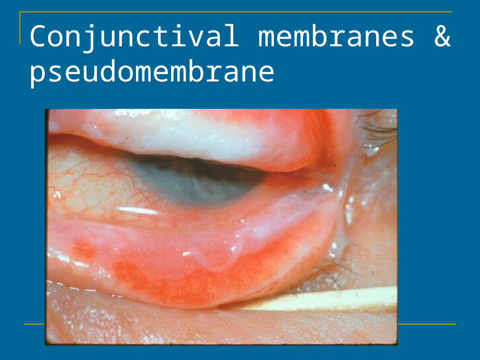
Conjunctival membranes & pseudomembrane
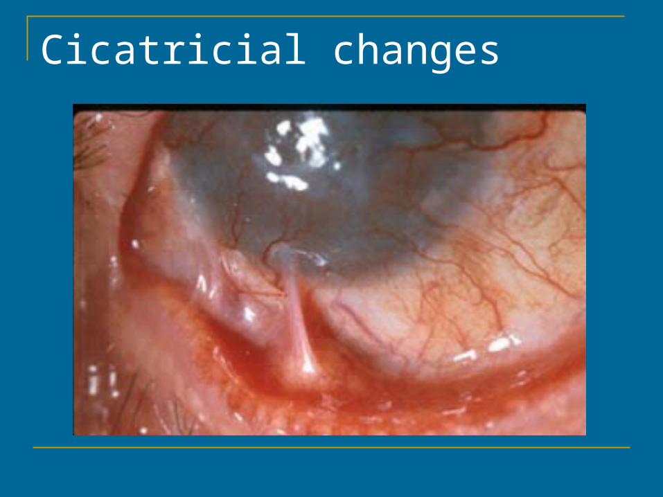
Cicatricial changes
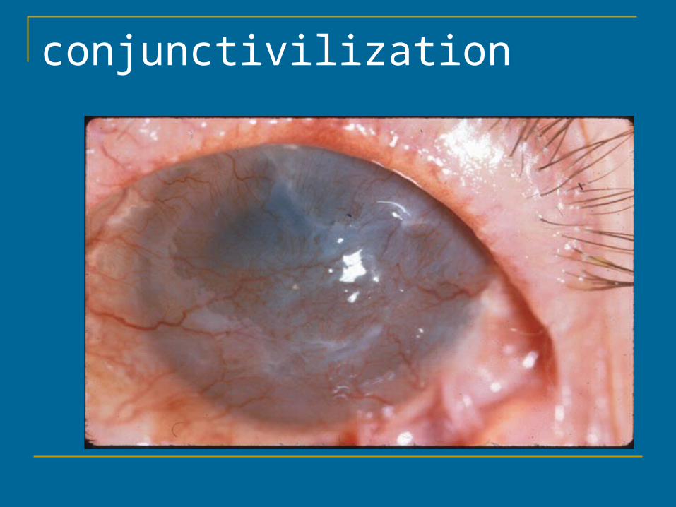
conjunctivilization
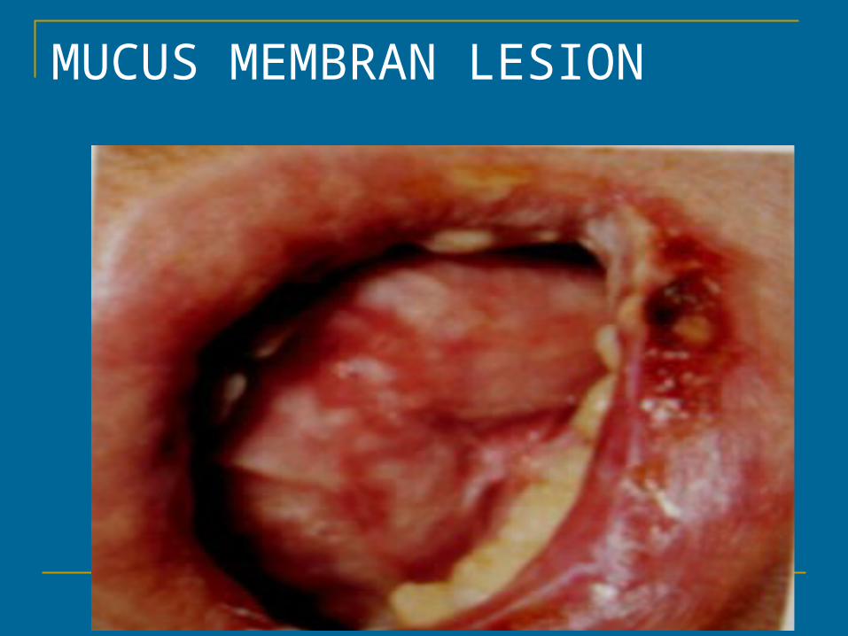
MUCUS MEMBRAN LESION
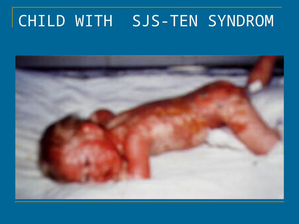
CHILD WITH SJS-TEN SYNDROM
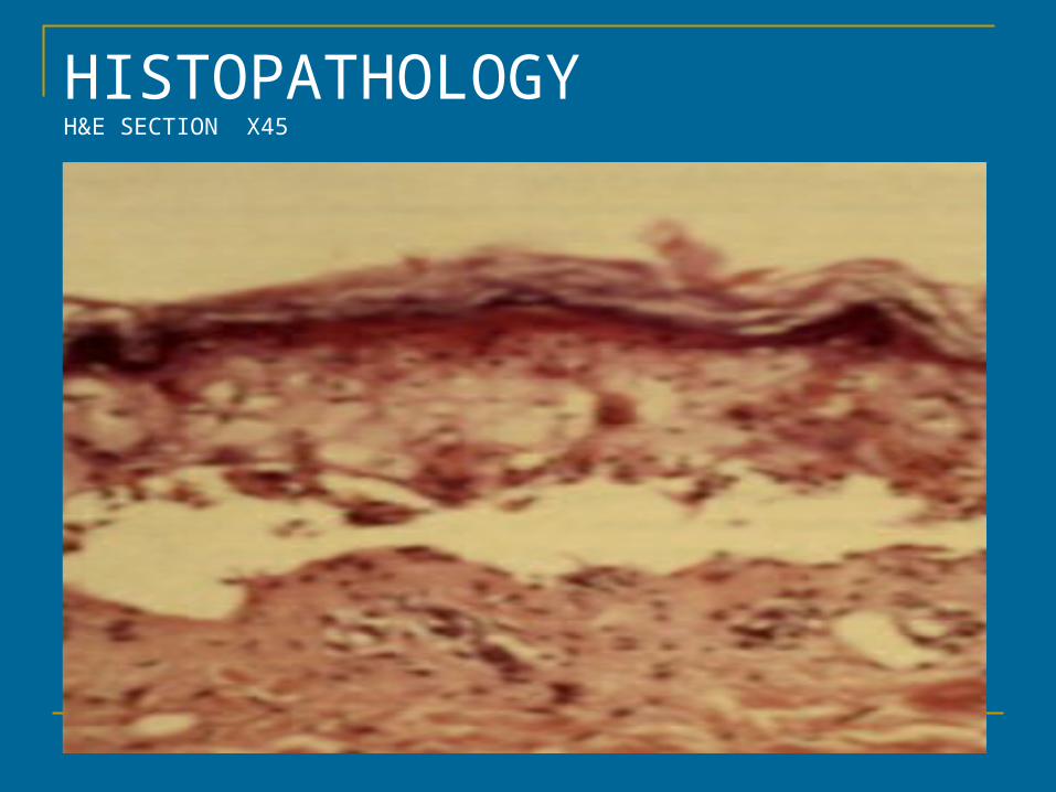
HISTOPATHOLOGYH&E SECTION X45
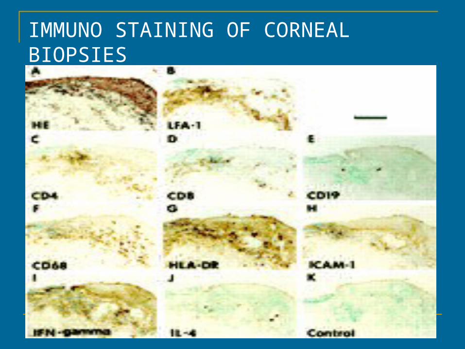
IMMUNO STAINING OF CORNEAL BIOPSIES
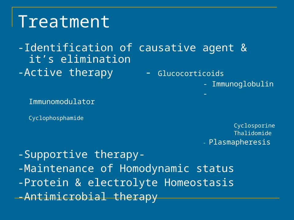
Treatment -Identification of causative agent & it’s elimination-Active therapy - Glucocorticoids
- Immunoglobulin - Immunomodulator Cyclophosphamide Cyclosporine Thalidomide
- Plasmapheresis
-Supportive therapy- -Maintenance of Homodynamic status -Protein & electrolyte Homeostasis-Antimicrobial therapy
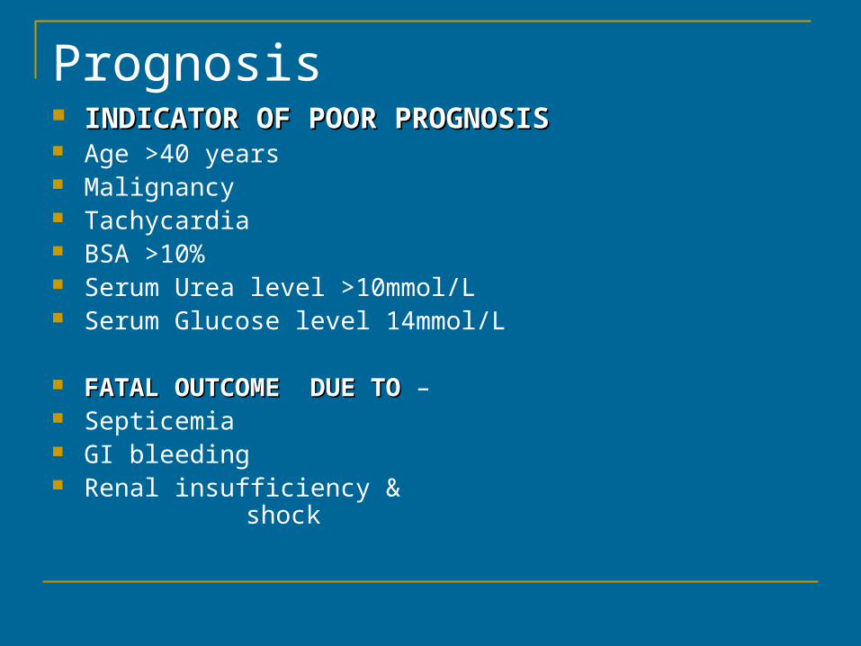
Prognosis INDICATOR OF POOR PROGNOSISINDICATOR OF POOR PROGNOSIS Age >40 years Malignancy Tachycardia BSA >10% Serum Urea level >10mmol/L Serum Glucose level 14mmol/L
FATAL OUTCOME DUE TO FATAL OUTCOME DUE TO – Septicemia GI bleeding Renal insufficiency & shock
Recommended