Epithelium-2
Hanan Jafar BDS.MSc.PhD

Relationship between CT and Epithelium
• All epithelial cells rest on connective tissue
• In case of epithelia that line internal organs, this connective tissue is called lamina propria
• Area of contact between epithelium and lamina propria increased by irregularities called papillae, most frequent in areas of stress


Basal Lamina and Basement Membrane
• Most epithelial cells are separated from the connective tissue by a sheet of extracellular material called the basal lamina (only visible under EM).
• The term basement membrane is used to specify a PAS-positive layer visible under LM present beneath some epithelia.

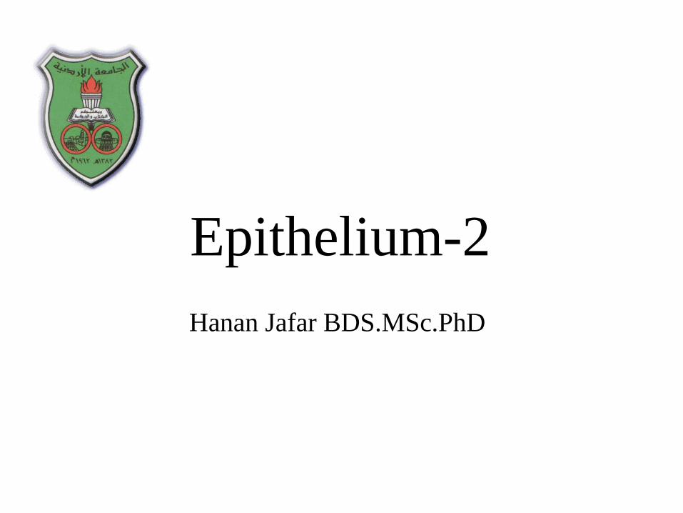
Basal lamina and basement membrane

Basement membrane is
composed of two layers:
1- Basal lamina
2- Reticular lamina
Layers of basal lamina:
Lamina Lucida (rara)
Lamina Densa

Layers of basal lamina:
Lamina Lucida
Lamina Densa
Lamina lucida: The clear layer
closer to the epithelium
Lamina densa: the dense layer
closer to the connective tissue

•Composed of lucida and densa
•Only visible with E.M
•Found also in other tissues, muscle cells,
adipocytes, peripheral neurons (external
lamina)
•Components are secreted by epithelium
Basal lamina
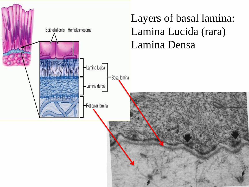
Molecular components are variable but include:
- Type IV collagen
- proteoglycans: e.g. Heparan sulfate proteoglycan called
Perlecan
- Glycoproteins (Laminin, entactin…)

Basement membrane
• Used to specify a PAS positive layer, visible
on light microscope
• It is thicker and usually formed by fusion of
two basal laminae or basal lamina and reticular
lamina
• the basement membrane is not actually a
membrane; rather, it is a matrix

Functions of basal lamina:
1- Support
2- Selective barrier
3- Influencing cell polarity
4- Regulation of proliferation and
differentiation
5- Affect cell-cell interaction
6- Pathway for cell migration

!! Remember
Every thing that enters or leaves the body must cross an epithelial sheet.
Epithelium occurs in the body
as a sheet of cells that covers a
body surface, lines a cavity, or
forms a gland.
Coverings, linings, forming
glands.

Cell Junctions
Basolateral domain


- Occluding junction forms a band (encircles epithelial cells)
- Barrier to diffusion between cells (paracellular pathway)
-Separates apical and basolateral plasma membranes,
the outer layers of 2 adjacent plasmalemma fuse together.
Tight junction/ zonula occludens

Copyright © McGraw-Hill CompaniesFigure 4-28

- TEM: is the most apical junction
- Freeze fracture of TJ reveals ridges in membranes that correspond to sites of contact
between cells
- Ridges are linear arrays of occludin and claudin proteins
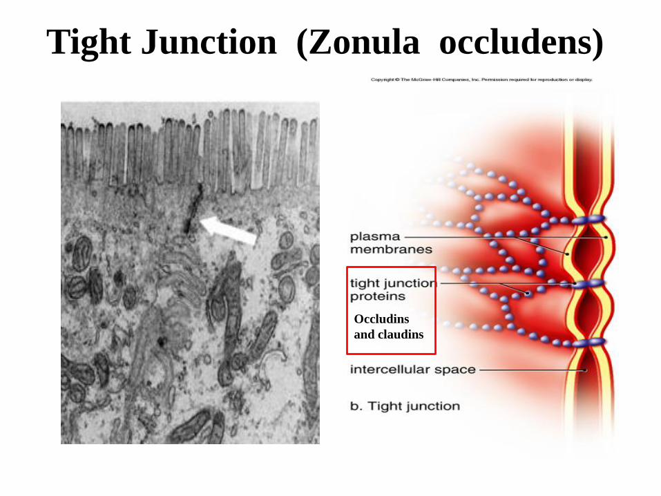
Tight Junction (Zonula occludens)
Occludins
and claudins

Each strand is a row of transmembrane proteins
in both PMs with ECDs joining together
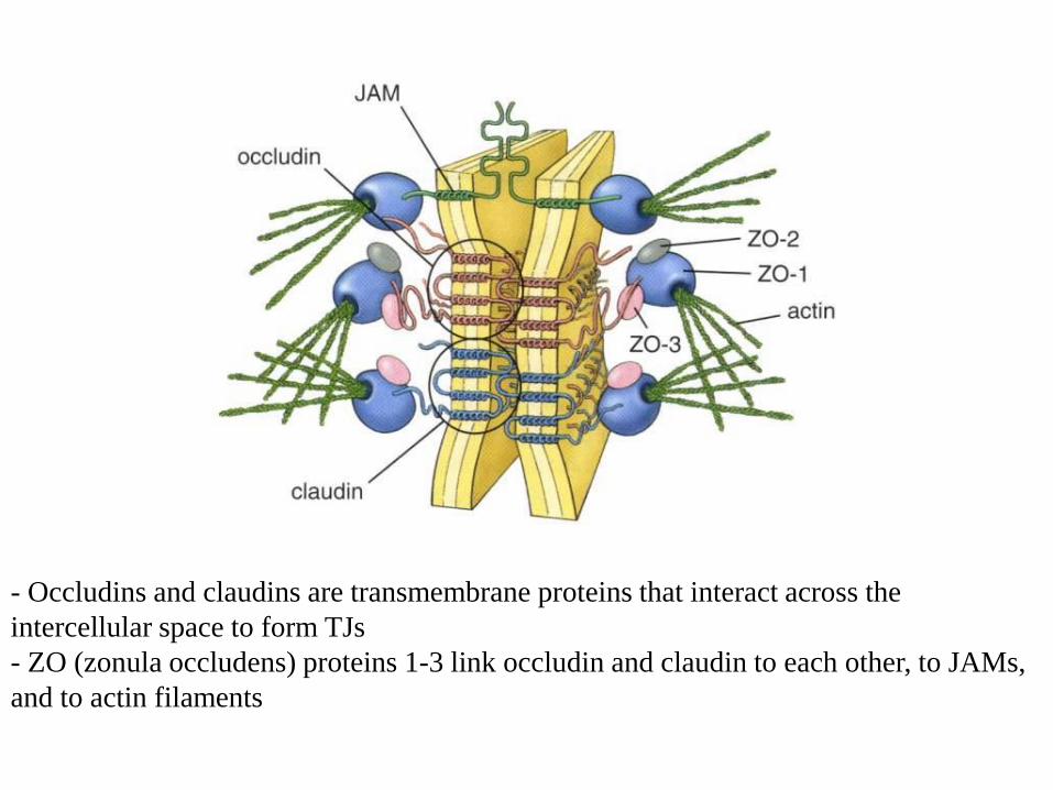
- Occludins and claudins are transmembrane proteins that interact across the
intercellular space to form TJs
- ZO (zonula occludens) proteins 1-3 link occludin and claudin to each other, to JAMs,
and to actin filaments

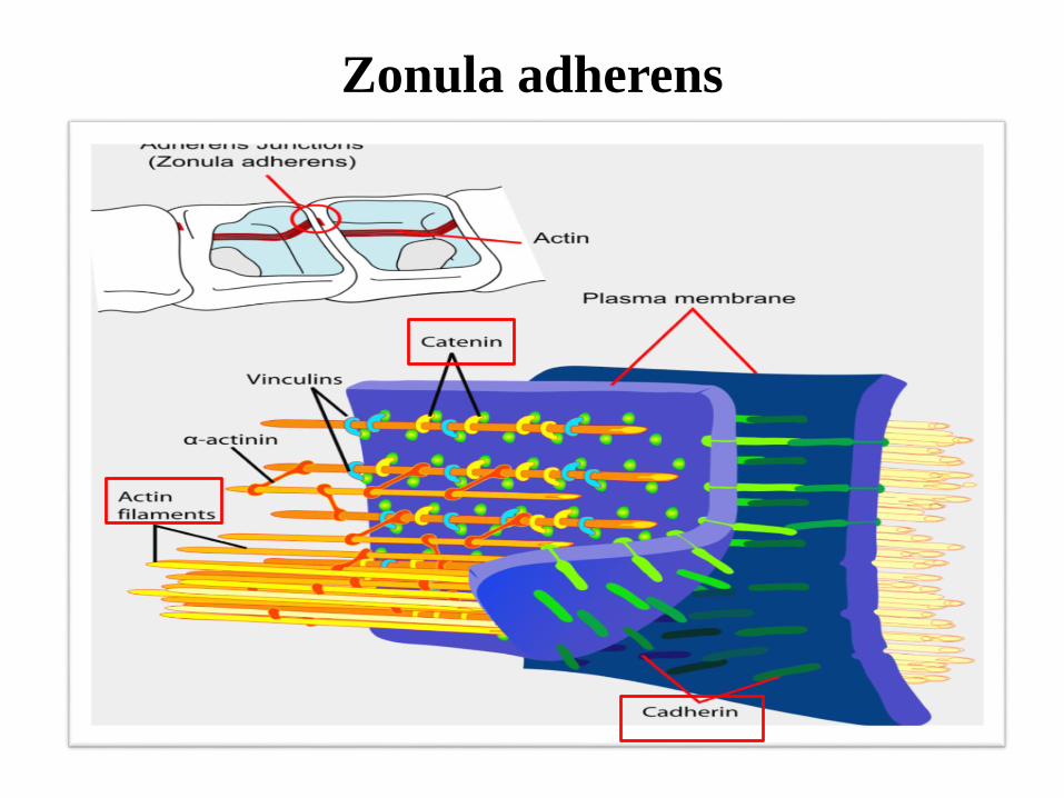
- Anchoring junction (encircles the cell) belt junction, or belt desmosome
- Located "under" tight junction in epithelial cells
-Connected to actin microfilaments that join terminal web
-Cadherin proteins attach to cross-linked actin filaments
- Mechanical support - ZA and actin filaments transmit and distribute stress throughout
cell and to neighboring cells
Zonula adherens
Zonula adherens
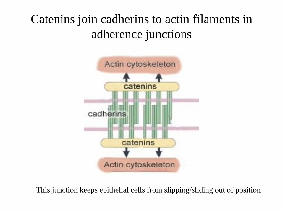
Catenins join cadherins to actin filaments in
adherence junctions
This junction keeps epithelial cells from slipping/sliding out of position

- Anchoring junctions
- Provides firm adhesion between
cells
- Function as "spot welds" to join
cells
- Located along lateral plasma
membranes of columnar epithelial
cells or on processes of squamous
cells
- Intermediate filaments associate
with plaque proteins in cytoplasm
Desmosome/ macula adherens
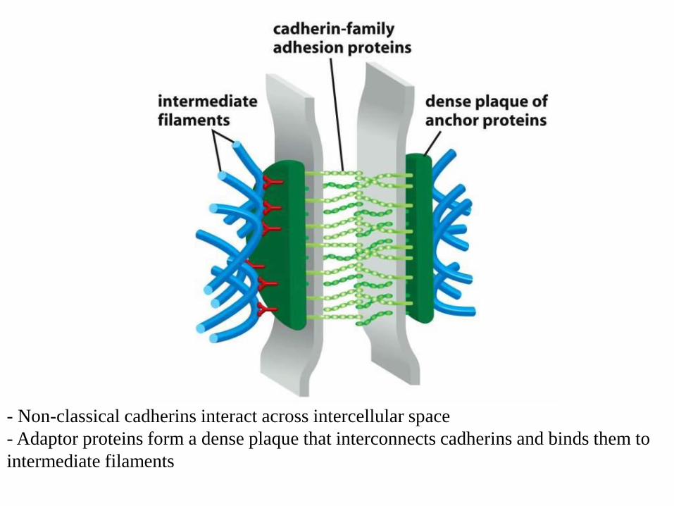
- Non-classical cadherins interact across intercellular space
- Adaptor proteins form a dense plaque that interconnects cadherins and binds them to
intermediate filaments

Desmosomes

Desmosome (Macula adherens)
- Desmoglein and desmocollin are non-classical cadherins
- Adaptor proteins such as -catenin (plakoglobin) and desmoplakin link cadherins to
intermediate filaments

•Channel-forming junction
•Named for gap of regular width
between cells visualized by TEM
•Water-filled junctions transport
molecules <1 kDal such as ions,
nucleotides (including cAMP), and
metabolites
•Rapid propagation of action potential
from one cell to another cell
Gap junction

Gap (Communicating) Junction
The gap junction is seen as an area of close plasma membrane apposition
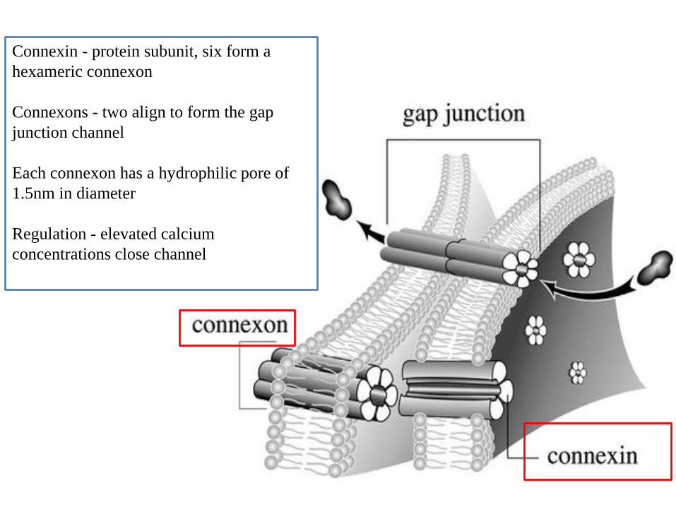
Connexin - protein subunit, six form a
hexameric connexon
Connexons - two align to form the gap
junction channel
Each connexon has a hydrophilic pore of
1.5nm in diameter
Regulation - elevated calcium
concentrations close channel

•Hemidesmosome - "half-desmosome" in appearance only
•Mediates attachment to basal lamina (extracellular matrix)
•Cytoplasmic plaque is attached to intermediate filaments
Hemidesmosomes

Integrins - membrane protein that "integrates" cell into matrix
Integrins bind to ECM (laminin and collagen 4)

Blistering Disease
- Many mechanisms
underlie blistering
disorders of the skin
- Pemphigus group -
autoimmune disease in
which autoantibodies
target desmogleins
present in desmosomes

Helicobacter pylori targets ZO-1 and disrupts
this junction
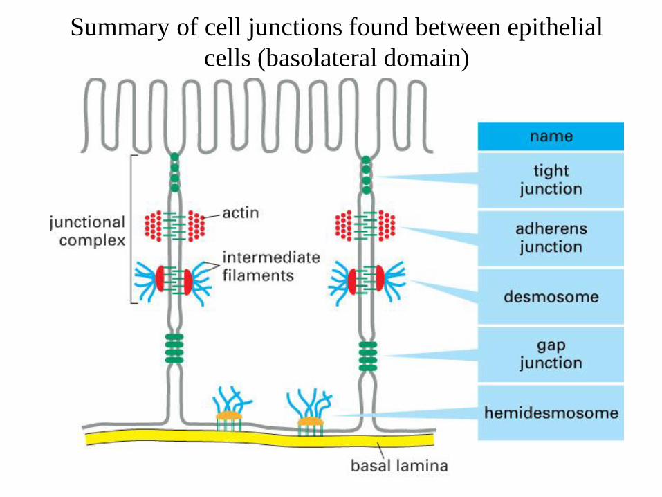
Summary of cell junctions found between epithelial
cells (basolateral domain)
Recommended