Updated 8/2018 Page
EMS System EMR-EMT-Advanced-Paramedic Standard Operating Procedures
8/9/2018
Updated 8/2018 Page i
Table of Contents INTRODUCTION FROM THE EMS MEDICAL DIRECTORS AND EMS COORDINATOR……............................... iv.
1.0 CARDIAC EMERGENCIES............................................................................................................................1
1.1 Cardiac Arrest: Termination of Resuscitation …................................................................................. 2
1.2 Cardiac Arrest: PEA and Asystole .......................................................................................................3
1.3 Cardiac Arrest: V-Fib / Pulseless V-Tach ........................................................................................... 4
1.4 Cardiac: Acute Coronary Syndrome .....................................................................................................5
1.5 Cardiac: Cardiogenic Shock .................................................................................................................6
1.6 Cardiac: Wide Complex Tachycardia with a Pulse ..............................................................................7
1.7 Cardiac: Narrow Complex Tachycardia ...............................................................................................8
1.8 Cardiac: Symptomatic Bradycardia / Heart Blocks .............................................................................9
2.0 GENERAL MEDICAL EMERGENCIES ...................................................................................................10
2.1 General: Nausea and/or Vomiting ......................................................................................................11
2.2 General: Pain Management ................................................................................................................12
2.3 General: Patient Agitation...................................................................................................................13
2.4 General: Procedural Sedation .............................................................................................................14
2.5 Medical: Allergic Reaction / Anaphylaxis .........................................................................................15
2.6 Medical: Diabetic Emergencies ......................................................................................................... 16
2.7 Medical: Overdose or Toxic Exposure .............................................................................................. 17
2.8 Medical: Seizures ............................................................................................................................... 18
2.9 Medical: Shock / Hypoperfusion ....................................................................................................... 19
2.10 Medical: Heat/Cold Related Illness ................................................................................................. 20
2.11 Medical: Suspected Stroke ............................................................................................................... 21
3.0 RESPIRATORY EMERGENCIES ............................................................................................................... 22
3.1 Respiratory: Acute Asthma and COPD with Wheezing .................................................................... 23
3.2 Respiratory: Acute Pulmonary Edema ............................................................................................... 24
3.3 Respiratory: Medication Facilitated Intubation ................................................................................. 25
4.0 TRAUMA EMERGENCIES ......................................................................................................................... 26
4.1 Trauma: Transport Guidelines ........................................................................................................... 27
4.2 Trauma: Field Triage Guidelines………............................................................................................ 28
4.3 Trauma: Trauma Center Coverage Maps ........................................................................................... 29
4.4 Trauma: Pre-established Landing Zones (Hammond and Dyer)........................................................ 30
4.5 Trauma: Helicopter Utilization .......................................................................................................... 32
Updated 8/2018 Page ii
4.6 Trauma: Burns.................................................................................................................................... 33
4.7 Trauma: Adult Rule of Nines……………………………………..…………………………………34
4.8 Trauma: Chest Trauma ...................................................................................................................... 35
4.9 Trauma: Crush Injuries ...................................................................................................................... 36
4.10 Trauma: Eye Injuries......................................................................................................................... 37
4.11 Trauma: Hypoperfusion / Hypovolemia .......................................................................................... 38
4.12 Trauma: Spinal Immobilization ....................................................................................................... 39
5.0 PEDIATRIC EMERGENCIES (pediatric triangle and normal vitals table).................................................. 40
5.1 Pediatric Trauma: Hypoperfusion / Hypovolemia ............................................................................. 41
5.2 Pediatric Trauma: Burns .................................................................................................................... 42
5.3 Pediatric Trauma: Burn Rule of Nines................................................................................................ 43
5.4 Pediatric Cardiac Arrest: Asystole or PEA ........................................................................................ 44
5.5 Pediatric Cardiac Arrest: V-Fib / Pulseless V-Tach .......................................................................... 45
5.6 Pediatric Cardiac: Symptomatic Bradycardia .................................................................................... 46
5.7 Pediatric Cardiac: Symptomatic Tachycardia .................................................................................... 47
5.8 Pediatric Medical: Acute Asthma ...................................................................................................... 48
5.9 Pediatric Medical: Anaphylaxis / Allergic Reaction.......................................................................... 49
5.10 Pediatric Medical: Seizures .............................................................................................................. 50
5.11 Pediatric Medical: Diabetic Emergencies ........................................................................................ 51
5.12 Pediatric Medical: Hypoperfusion……............................................................................................ 52
5.13 Pediatric Medical: Abdominal Pain / Vomiting ............................................................................... 53
5.14 Pediatric Medical: Overdose / Toxic Exposure ............................................................................... 54
5.15 Pediatric General: Pain Management................................................................................................ 55
5.16 Pediatric General: Procedural Sedation ........................................................................................... 56
6.0 OB/GYN ........................................................................................................................................................ 57
6.1 OB/Gyn Childbirth: Pre-delivery........................................................................................................ 58
6.2 OB/Gyn Childbirth: Delivery ............................................................................................................ 59
6.3 OB/Gyn: Eclampsia ........................................................................................................................... 61
6.4 OB/Gyn: Pre-term Labor (24 37 weeks) ............................................................................................ 62
7.0 Procedures (Scope of Practice Matrix)…………………………………..…………………………….…….63
7.1 Procedure: Airway Management ....................................................................................................... 64
7.2 Procedure: Non-visualized Airway..................................................................................................... 65
7.3 Procedure: Endotracheal Intubation.................................................................................................... 66

Updated 8/2018 Page iii
7.4 Procedure: Needle Cricothyrotomy.................................................................................................... 67
7.5 Procedure: Quick Trach Cricothyrotomy............................................................................................ 68
7.6 Procedure: CPAP (Flowsafe 2)........................................................................................................... 69
7.7 Procedure: Thoracic Decompression….............................................................................................. 70
7.8 Procedure: Intraosseous Access…...................................................................................................... 71
7.9 Procedure: 12-Lead Acquisition and Transmission………………………………………………….73
7.10 Medication Administration: Intranasal (IN)..................................................................................... 74
7.11 Medication Administration: Intramuscular (IM).............................................................................. 75
8.0 Special Procedures.......................................................................................................................................... 76
8.1 Special Procedures: Documentation................................................................................................... 77 8.2 Special Procedures: Informed Consent and Refusal of Care…………………………………………..………78
8.3 Special Procedures: Involuntary Restraint …..………………………………………………………79
8.4 Special Procedures: Ventricular Assist Device…..…………………………………………………..80
8.5 Special Procedures: Taser Removal…………..……………..……………………………………….81
8.6 Special Procedures: Care of the Dialysis Patient…............................................................................ 82
8.7 Special Procedures: System Entry and Recertification ...................................................................... 83
8.8 Special Procedures: Supply / Medication Replacement ................................................................... 85
8.9 Medication Formulary........................................................................................................................ 86
8.10 Special Procedures: District 1 Diversion Guidelines....................................................................... 87
9.0 Inter-Facility Transports…………………………………………………………………….………………89
9.1 Inter-facility Transport..……………………………………………………………………………..90
9.2 Inter-facility Drug List…..…………………………………………………………………………..91
9.3 Stroke Transfer with tPA……………………………………………………………………………94
Credits and References………………………………………………………………………………………..95
Updated 8/2018 Page iv
Introduction from the EMS Medical Directors and EMS Coordinator
Franciscan Health Hammond and Dyer Hospitals are proud to update these evidence-based
protocols for all affiliated Lake County Emergency Medical Service providers. The protocols continue
to be developed by the Medical Direction Committee after extensive review of the most recent
American Heart Association Guidelines, other regional protocols, relevant medical research, and
input from individual field providers. The following medical care guidelines are designed to improve
patient outcomes, while decreasing any potential risk to the patient as well as maximizing the
interventions appropriate for each level of care.
The color coded format of the protocols allows all EMS professionals to easily follow the
potential interventions and treatments available for each specific patient complaint. All provider levels
are highlighted, with level-appropriate care below, while the corresponding protocol STOP line is
clearly delineated.
These protocols are designed to serve the community as a whole and include all levels of field
providers. As taught in every EMT class, BLS care should be completed before ALS. Advanced
providers are responsible for all appropriate BLS interventions. If an intervention is completed at a
lower level, it should not duplicated unless specified. Ex. Epi-pen (BLS) + 1:1000 Epi SQ (ALS)
Updated 8/2018 Page v
Medical Control/Direction for all levels of EMS providers is defined as:
System Medical Director, when present at the scene and in physical contact with the patient
Base physician at the receiving hospital, by radio, landline, or cellular telephone
[When transport is to another facility, and unique orders or requests are present or communication is not
possible with the physician at the receiving facility, use Base physician at Franciscan Health Hammond
Hospital]
Statement Regarding Medication Shortages
Due to the Medications shortages that we have experienced throughout the region over the past year,
and the expectation that this may continue, we have listed medications as preferred and acceptable.
Preferred medications/concentrations should be used when available
Acceptable medications/concentrations can be used as a backup only if preferred medications
are not available
Every effort should be made to make available preferred medications/concentrations.
It is important to remember to double check the medication to be administered for proper drug and
dosage in light of the potential for new or alternative drugs and concentrations. Proper training should
take place prior to placing an unfamiliar medication, packaging, concentration, etc. on the emergency
vehicle.
Our Commitment to EMS in Lake County
Franciscan Health Hammond and Dyer will continue to evaluate current EMS and medical literature to
update the protocols to optimize the outcomes of our patients. We will continue to perform QI audits
of patient care to develop training programs that will improve care as a whole throughout the region.
We hope that this protocol format will help make your job easier and better assist you in the care of
your patients. We would like to thank everyone who provided input which contributed to these
protocols.
Protocol signatures of approval dated this 9th day of August, 2018.
________________________ ___________________________ ___________________________
Dr. Eric Cook, DO Dr. Brett Marcotte, DO Matthew Eddy, LP, PI
Franciscan Health Hammond Franciscan Health Dyer Franciscan Health H/D
EMS Medical Director EMS Medical Director EMS Coordinator
Updated 8/2018 Page 1
Medical Emergencies
1.0 Cardiac Emergencies
Updated 8/2018 Page 2
1.1 Cardiac Arrest: Termination of Resuscitation
Resuscitative efforts for patients in cardiac arrest should NOT be initiated if:
o Patient presents with significant dependent lividity, rigor mortis, decomposition and/or
injuries incompatible with life (such as decapitation)
o Family presents a signed Out of Hospital DNR (Do Not Resuscitate)
o Family presents a signed Physician Orders for Scope of Treatment (POST)
For all other patients in cardiac arrest, in whom appropriateness of resuscitation is
questionable, the EMS provider MUST start BLS care, including defibrillation awaiting
arrival of a paramedic unit.
Consider Field terminations of resuscitation ONLY if a patient meets ALL of the following:
o Completed protocol appropriate for asystole with NO response to interventions in 20
minutes
o Older than 18 years old unless obvious SIDS case with lividly and rigor mortis
o No communication failure with family
o Scene is appropriate for termination order
Consider “2 minute warning” to give family time to prepare for termination
If at any time during ALS care, appropriateness of resuscitation is questionable, consult
MEDICAL CONTROL physician for assistance.
Sections A and B outline the patient’s code status in the POST
The Temperature of the patient is not an indication of definitive death
Updated 8/2018 Page 3
1.2 Cardiac Arrest: PEA and Asystole
Recognize
CPR and AED
Check for DNR or POST directive
Perform 2 minute cycles of high quality CPR (hard and fast) Rate should be around 100-120
beats per minute, at least 2 inches deep in an adult
Secure airway with medically approved non-visualized airway
Vascular access IV/IO
Consider and treat Reversible Causes (H’s and T’s )
Epinephrine (1 mg in 10 mL) : dose 1 mg IV/IO; repeat every 3 - 5 minutes
o Optional: Epinephrine (1 mg in 1 mL) may be diluted with 9ml of normal saline.
Place advanced airway as appropriate
Refer to the 1.1 Cardiac: Termination of Resuscitation Protocol as needed
IO access should be considered and may be established as initial access for patients in cardiac arrest.
Do not allow IV/IO access, drug delivery, or advanced airway placement to cause delay > 10 sec. in chest
compressions or defibrillation
Consider and possibly treat contributing factors including: Hypoxia, Hypovolemia, Hypothermia, Hyper-
/Hypokalemia, Hydrogen Ion (Acidosis), Tension Pneumothorax, Cardiac Tamponade, Toxins, Thrombosis
Coronary and/or Pulmonary
Waveform Capnography/End-Tidal CO2 recommended for assessment of chest compression effectiveness,
advanced airway placement, and ROSC.
If sufficient personal, intubation may be performed with limited interruption of CPR.
Epinephrine needs to be given as soon as possible as ROSC is reduced by 4% for every minute it is delayed
Updated 8/2018 Page 4
1.3 Cardiac Arrest: V-Fib/Pulseless V-Tach
Recognize
CPR and AED
Check for DNR or POST directive
Perform 2 minute cycles of high quality CPR (hard and fast) Rate should be around 100-120
beats per minute, at least 2 inches deep in an adult
Secure airway with medically approved non-visualized airway
Vascular access IV/IO
Defibrillate every 2 minutes as long as rhythm persists
Epinephrine (1 mg in 10 mL): dose 1mg IV/IO; repeat every 3 - 5 minutes
o Optional: Epinephrine (1mg in 1 mL) may be diluted with 9ml of normal saline.
Administer Lidocaine 1.5 mg/kg IV/IO; may repeat .75 mg/kg in 3 - 5 minutes Max 3 mg/kg
Consider: Magnesium sulfate 1-2 grams diluted in 10 mL NS IV/IO, for Torsade’s de Pointes
Refer to the 1.1 Cardiac: Termination of Resuscitation Protocol as needed
If patient in persistent V-Fib or Pulseless V-Tach, consult MEDICAL CONTROL physician
for decision to transport or termination of field care
Defibrillate at manufacturers recommended settings
Do not allow IV/IO access, drug delivery, or advanced airway placement to cause
significant delay in chest compressions or defibrillation
Waveform Capnography/End-Tidal CO2 is recommended for assessment of chest
compression effectiveness, advanced airway placement, and ROSC.
If sufficient personal, intubation may be performed with limited interruption of CPR
Updated 8/2018 Page 5
1.4 Cardiac: Acute Coronary Syndrome
ABC
Apply appropriate oxygen therapy
Vital signs
Have AED available
Aspirin 324 mg (4 x 81 mg tabs)
o May withhold aspirin administration if patient has true allergy to ASA
Assist patient with their own prescribed Nitroglycerin (up to 3 dose maximum), if systolic BP is greater
than 90 mmHg
12 lead ECG and transmit within 5-10 mins of patient contact
Vascular access, with purple top blood draw
o IV access prior to administration of Nitroglycerin
o Nitroglycerin 0.4mg SL: repeat every 5 min up to 3 doses
o If systolic BP less than 90 mmHg; Normal Saline 500-1000 mL IV bolus
Notify MEDICAL CONTROL physician AS SOON AS POSSIBLE if STEMI identified
o If elevation in Leads II, III, aVF check for more than 1mm of ST elevation in V4R. If present,
DO NOT give nitroglycerin
If systolic BP greater than 90 mmHg
o Nitroglycerin 0.4 mg SL; repeat every 5 minutes to max 3 doses
Additional IV access as needed while enroute if time permits
Do not administer nitroglycerin if the patient has taken Sildenafil (Viagra) of Vardenafil (Levitra)
within the last 6 hours or Tadalafil (Cialis) within the last 48 hours
Franciscan Health Hammond and Dyer will never divert a STEMI patient while on bypass
Updated 8/2018 Page 6
1.5 Cardiac: Cardiogenic Shock
ABC
Apply appropriate oxygen therapy
Vital signs
Place patient supine unless dyspnea is present
12 lead ECG and transmit
Vascular access, with purple top blood draw
If no signs of pulmonary edema: Normal Saline 500-1000 mL IV bolus
If patient remains unstable following fluid bolus
o A second 500 mL bolus may be given
Check for signs of pulmonary edema, DO NOT give if rales present
Unstable is defined as systolic BP less than 90 mmHg and/or decreased level of
consciousness
Refer to appropriate Dysrhythmia protocol as needed
Monitor lung sounds if present hold/stop fluid bolus

Updated 8/2018 Page 7
1.6 Cardiac: Wide Complex Tachycardia with a Pulse
ABC
Apply appropriate oxygen therapy
Vital signs
Have AED available
12 lead ECG and transmit
Vascular access
UNSTABLE (Rate >150)
Refer to 2.4 General: Procedural Sedation protocol if time permits
Synchronized cardioversion (120-200J); may repeat (360J) if 1st attempt unsuccessful
If cardioversion fails, follow drug regiment for STABLE patient
STABLE (Rate > 150)
Lidocaine 1 - 1.5 mg/kg Slow IVP; may repeat 0.5 - 0.75 mg/kg slow IVP (Max 3 mg/kg)
o If rhythm converts hang Lidocaine Drip up to 4 mg/min
Torsade de Pointes
Magnesium Sulfate 2 grams dilute in 10mL NS over 5-10 minutes IV push
UNSTABLE is defined as ventricular rate greater than 150 bpm with symptoms of Severe
chest pain, severe dyspnea, altered mental status, pulmonary edema, or hypotension
(systolic BP less than 90 mmHg)
Wide Complex is defined as a QRS complex greater than 0.12 seconds
Cardioversion at manufacturers recommended setting
Always record initial rhythm strip and deliver to physician
Do not delay synchronous cardioversion while awaiting IV access

Updated 8/2018 Page 8
1.7 Cardiac: Narrow Complex Tachycardia
ABC
Apply appropriate oxygen therapy
Vital signs
Have AED available
12 lead ECG and transmit
Vascular access (proximal site preferred)
Valsalva Maneuvers
UNSTABLE (Rate > 150)
Refer to 2.4 General: Procedural Sedation protocol if time permits
Synchronized cardioversion (120-200J); may repeat (360J) if 1st attempt unsuccessful
If cardioversion fails, follow rhythm appropriate drug regimen for STABLE patient
STABLE (Rate > 150)
Adenosine (Adenocard) 6 mg rapid IV push followed by 20ml NaCl bolus; if unsuccessful:
o Adenosine (Adenocard) 12 mg rapid IV push; repeat 1-2 minutes 12 mg if needed
UNSTABLE is defined as ventricular rate greater than 150 bpm with symptoms of severe
chest pain, dyspnea, altered mental status, pulmonary edema, or hypotension (systolic BP
less than 90 mmHg)
Cardioversion at manufacturers recommended setting
Always record initial rhythm strip and deliver to physician
Do not delay synchronous cardioversion while awaiting IV access
Updated 8/2018 Page 9
1.8 Cardiac: Symptomatic Bradycardia / Heart Blocks
ABC
Apply appropriate oxygen therapy
Vital signs
Have AED available
12 lead ECG and transmit
Vascular access
Atropine 0.5 mg IV; repeat every 3 - 5 min to max 3 mg
Begin transcutaneous pacing if atropine is ineffective
o Refer to 2.4 General: Procedural Sedation protocol as needed
If Hypotensive, and no signs of pulmonary edema, 500 mL bolus NaCl may be given
Consider immediate pacing for 2nd degree Type II or 3rd degree Heart Blocks
Bradycardia is rate less than 60 bpm, but symptomatic is generally less than 50 bpm
Only treat bradycardia if patient is symptomatic
Use atropine with caution in ACS
Symptomatic presentation includes severe chest pain, dyspnea, altered mental status,
pulmonary edema, ischemia, infarction or hypotension (systolic BP less than 90 mmHg)
Consider and possibly treat contributing factors including: Hypoxia, Hypovolemia,
Hypothermia, Hyper-/Hypokalemia, Hydrogen Ion (Acidosis), Tension Pneumothorax,
Cardiac Tamponade, Toxins, Thrombosis- Coronary and Pulmonary
Unit will not pace unless pads AND limb leads are applied

Updated 8/2018 Page 10
Medical Emergencies
2.0 General/Medical
Emergencies

Updated 8/2018 Page 11
2.1 General: Acute Abdominal Pain / Vomiting
ABC
Apply appropriate oxygen therapy
Vital signs
Consider 12 lead ECG and transmit
Vascular access, Normal Saline 500-1000 mL IV bolus as needed
Ondansetron (Zofran) ODT 4 - 8 mg SL
Ondansetron (Zofran) 4 mg IV or IM; may repeat once in 10 minutes
Consult MEDICAL CONTROL physician if patient has any of the following: systolic BP
less than 90, pregnancy, or head trauma

Updated 8/2018 Page 12
2.2 General: Pain Management
ABC
Apply appropriate oxygen therapy
Vital signs
Consider 12 lead ECG and transmit
Consider vascular access
Continuous cardiac monitoring
Administer ONE of the following narcotic analgesics
o Morphine 2 - 5 mg IV,IM; repeat every 5 min to max 10 mg
o Fentanyl 25-100 mcg slow IV, IN; repeat every 5 min to max 200 mcg
For patients with:
o Severe burns without hemodynamic compromise
o Suspected isolated extremity injuries with severe pain
o Abdominal pain
o Back pain
For all other painful conditions, paramedics must consult MEDICAL CONTROL physician
for orders
Contraindications to pain management protocol: altered mental status, hypoventilation,
systolic BP less than 90, other traumatic injuries
This protocol may NOT be used in conjunction with the 2.4 General: Procedural Sedation
protocol, unless MEDICAL CONTROL physician is consulted.
Fentanyl should be used if there is any concern for potential hemodynamic instability.
Consult MEDICAL CONTROL physician for additional Morphine or Fentanyl
Updated 8/2018 Page 13
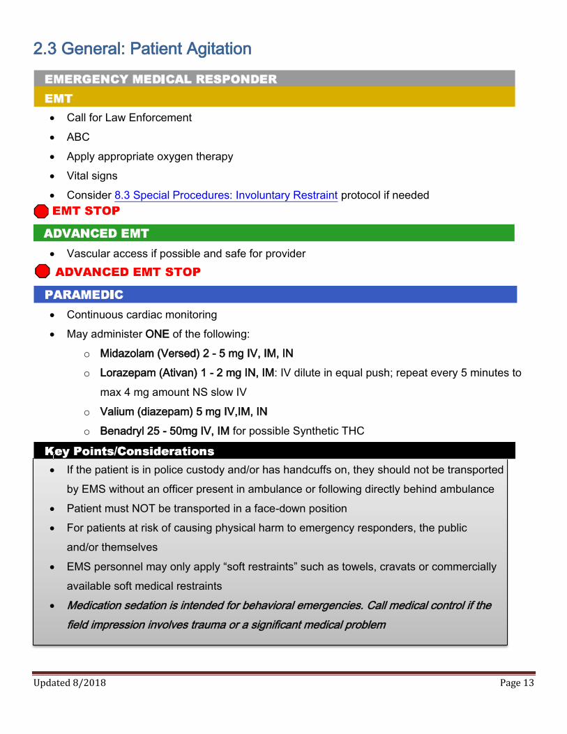
2.3 General: Patient Agitation
Call for Law Enforcement
ABC
Apply appropriate oxygen therapy
Vital signs
Consider 8.3 Special Procedures: Involuntary Restraint protocol if needed
Vascular access if possible and safe for provider
Continuous cardiac monitoring
May administer ONE of the following:
o Midazolam (Versed) 2 - 5 mg IV, IM, IN
o Lorazepam (Ativan) 1 - 2 mg IN, IM: IV dilute in equal push; repeat every 5 minutes to
max 4 mg amount NS slow IV
o Valium (diazepam) 5 mg IV,IM, IN
o Benadryl 25 - 50mg IV, IM for possible Synthetic THC
If the patient is in police custody and/or has handcuffs on, they should not be transported
by EMS without an officer present in ambulance or following directly behind ambulance
Patient must NOT be transported in a face-down position
For patients at risk of causing physical harm to emergency responders, the public
and/or themselves
EMS personnel may only apply “soft restraints” such as towels, cravats or commercially
available soft medical restraints
Medication sedation is intended for behavioral emergencies. Call medical control if the
field impression involves trauma or a significant medical problem
Updated 8/2018 Page 14
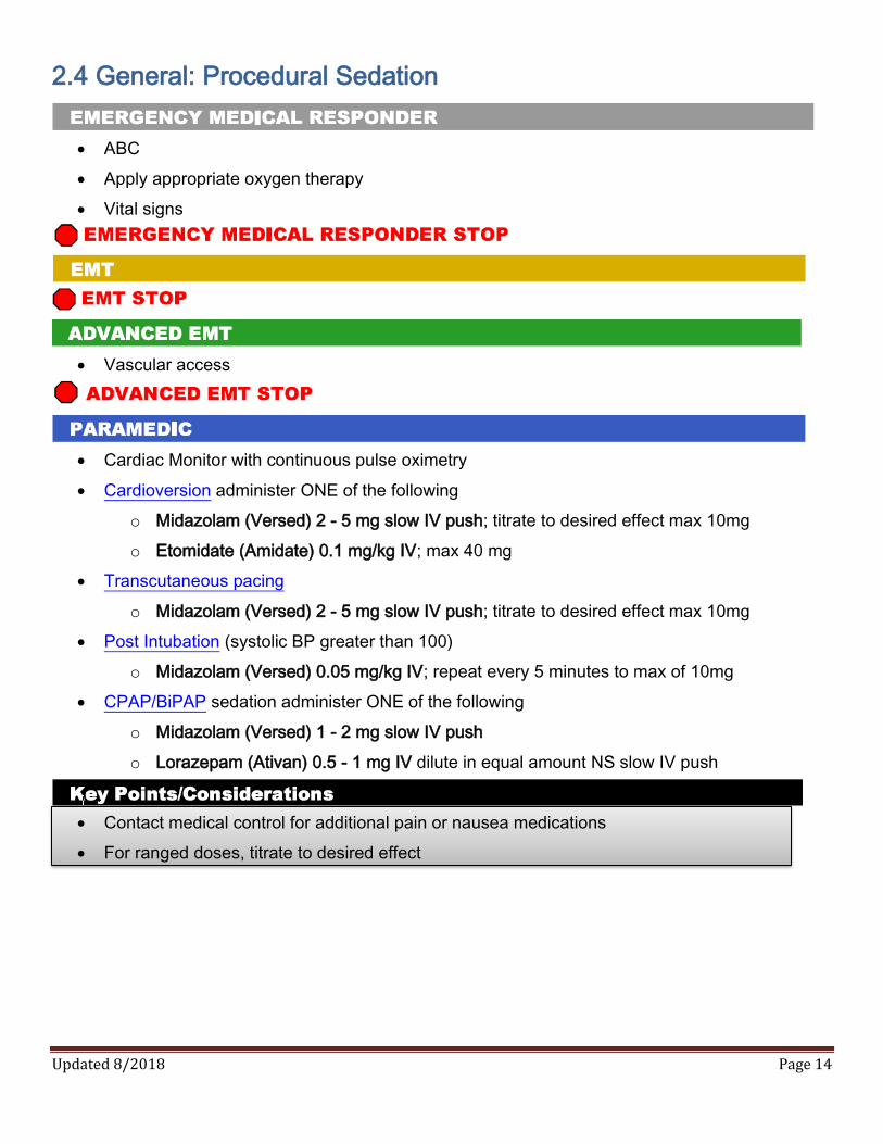
2.4 General: Procedural Sedation
ABC
Apply appropriate oxygen therapy
Vital signs
Vascular access
Cardiac Monitor with continuous pulse oximetry
Cardioversion administer ONE of the following
o Midazolam (Versed) 2 - 5 mg slow IV push; titrate to desired effect max 10mg
o Etomidate (Amidate) 0.1 mg/kg IV; max 40 mg
Transcutaneous pacing
o Midazolam (Versed) 2 - 5 mg slow IV push; titrate to desired effect max 10mg
Post Intubation (systolic BP greater than 100)
o Midazolam (Versed) 0.05 mg/kg IV; repeat every 5 minutes to max of 10mg
CPAP/BiPAP sedation administer ONE of the following
o Midazolam (Versed) 1 - 2 mg slow IV push
o Lorazepam (Ativan) 0.5 - 1 mg IV dilute in equal amount NS slow IV push
Contact medical control for additional pain or nausea medications
For ranged doses, titrate to desired effect

Updated 8/2018 Page 15
2.5 Medical: Allergic Reaction / Anaphylaxis
ABC Vital signs
Apply appropriate oxygen therapy
If severe reaction (Wheezing/stridor, face/airway swelling, altered mental status)
o Administer the Patient’s Epi Pen or
o Epinephrine (1 mg in 1 mL): dose 0.3 mg (0.3 mL) Intramuscular
Vascular access; Normal Saline 500-1000 mL IV bolus as needed
If wheezing, Albuterol 2.5 mg via nebulizer; repeat once
Cardiac Monitor
Asymptomatic
o Supportive Care
Mild symptoms: Urticaria, itching, nasal congestion, watery eyes, etc.
o Diphenhydramine (Benadryl) 50 mg IV or deep IM
Moderate symptoms: Wheezing, nausea, vomiting, diarrhea, face, neck, tongue flushing,
swelling
o DuoNeb (Albuterol 2.5 mg + Atrovent 0.5 mg in 2.5 mL mixed together), via
Nebulizer
o Methylprednisolone (Solu-Medrol) 125 mg IV, IM if no IV access
Severe reaction not relieved by initial treatment or patient presenting with Stridor,
hypotension (systolic BP less than 90 mmHg), and/or Altered Mental Status
Epinephrine (1mg in 10 mL): dose 0.01 mg/kg (0.3 mg max) IV, If no other Epi Given.
If BLS and less invasive ALS airway maneuvers fail, attempt Intubation
o 7.5 Procedure: Quick Track Cricothyrotomy may be considered only after all
other Airway interventions have been exhausted.
Updated 8/2018 Page 16
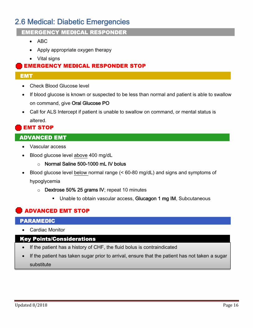
2.6 Medical: Diabetic Emergencies
ABC
Apply appropriate oxygen therapy
Vital signs
Check Blood Glucose level
If blood glucose is known or suspected to be less than normal and patient is able to swallow
on command, give Oral Glucose PO
Call for ALS Intercept if patient is unable to swallow on command, or mental status is
altered.
Vascular access
Blood glucose level above 400 mg/dL
o Normal Saline 500-1000 mL IV bolus
Blood glucose level below normal range (< 60-80 mg/dL) and signs and symptoms of
hypoglycemia
o Dextrose 50% 25 grams IV; repeat 10 minutes
Unable to obtain vascular access, Glucagon 1 mg IM, Subcutaneous
Cardiac Monitor
If the patient has a history of CHF, the fluid bolus is contraindicated
If the patient has taken sugar prior to arrival, ensure that the patient has not taken a sugar
substitute

Updated 8/2018 Page 17
2.7 Medical: Overdose / Toxic Exposure
Opiate OD: Naloxone 2mg IN; For respiratory depression only
Decontaminate as needed
ABC, apply appropriate oxygen therapy, and vital signs
Attempt to determine what was taken, when, and how much, bring containers to ED
Contact Poison Control 1-800-222-1222 for additional information and treatment
Check blood glucose level, If level is abnormal refer to 2.6 Medical: Diabetic Emergencies
protocol.
Consider 12 lead and transmit
Vascular access
o Opiate OD: Naloxone (Narcan) 0.5 mg IV, IM; repeat to max 6 mg for respiratory
depression ONLY
Cardiac Monitor
For symptomatic patients with known:
o Organophosphate poisoning: Atropine 2 - 5 mg IV; repeat every 3-5 minutes
o Calcium channel or Beta blocker OD: Glucagon 1 mg IM, Subcutaneous
o Synthetic THC OD: Benadryl 25 - 50mg IV, IM
o Tricyclic antidepressant OD: Sodium Bicarbonate 1 mEq/kg IV
o Sympathomimetic OD (cocaine/amphetamines): Midazolam (Versed) 2 - 5 mg
IV, IM, IN
Patients experiencing a Carfentanyl overdose may require multiple doses of Narcan
Use Narcan with caution with cancer patients
Do not use Narcan on intubated patients
Organophosphate poisoning: SLUDGE: Salivation, Lacrimation, Urination, Diarrhea, Gastric cramps, Emesis

Updated 8/2018 Page 18
2.8 Medical: Seizures
ABC
Apply appropriate oxygen therapy
Vital signs
Check blood glucose level; If level is abnormal refer to 2.6 Medical: Diabetic Emergencies
protocol.
Vascular access
Cardiac Monitor
Preferred
o Lorazepam (Ativan) 1 - 2 mg IN, IV dilute in equal amount NS slow IV push;
repeat every 5 minutes to max 4 mg
o Midazolam (Versed) 2 mg slow IV push; May repeat in 5 minutes
o If vascular access cannot be obtained may give ONE of the following:
Lorazepam (Ativan) 1-2 mg IM, IN; repeat every mg 5 minutes to max 4
Midazolam (Versed) 5 mg IM, IN
Acceptable
o Diazepam (valium) 5 mg slow IV; repeat 2 5 minutes to max 10 mg
If vascular access cannot be obtained may give Diazepam (valium) 5
mg slow IM, or IN with nasal atomizer
Protect the patient and EMS crew from injury during the seizure
Refer to the 6.3 OB/GYN: Eclampsia protocol if patient is pregnant or recently post-partum

Updated 8/2018 Page 19
2.9 Medical: Shock / Hypoperfusion
ABC
Apply appropriate oxygen therapy
Vital signs
Place patient in supine position unless dyspnea is present
Cover the patient to maintain body temperature
Consider 12 lead ECG and transmit
Vascular access
If no pulmonary edema (rales): Normal Saline 500 - 1000mL bolus IV
Obtain additional vascular access as time permits
Cardiac Monitor
A second 500 cc bolus may be given, if no signs of pulmonary edema
Additional fluid bolus can be administered but patient needs to be reassessed for rales
or signs of pulmonary edema.
UNSTABLE is defined as Systolic BP less than 90 mmHg and/or decreased level of
consciousness
Monitor for signs and symptoms of pulmonary edema
Consider causes of hypoperfusion, including anaphylaxis, toxic ingestions, cardiac
rhythm disturbances, myocardial infarction, sepsis, ruptured AAA, trauma, or others
ectopic pregnancy, trauma or others
Updated 8/2018 Page 20
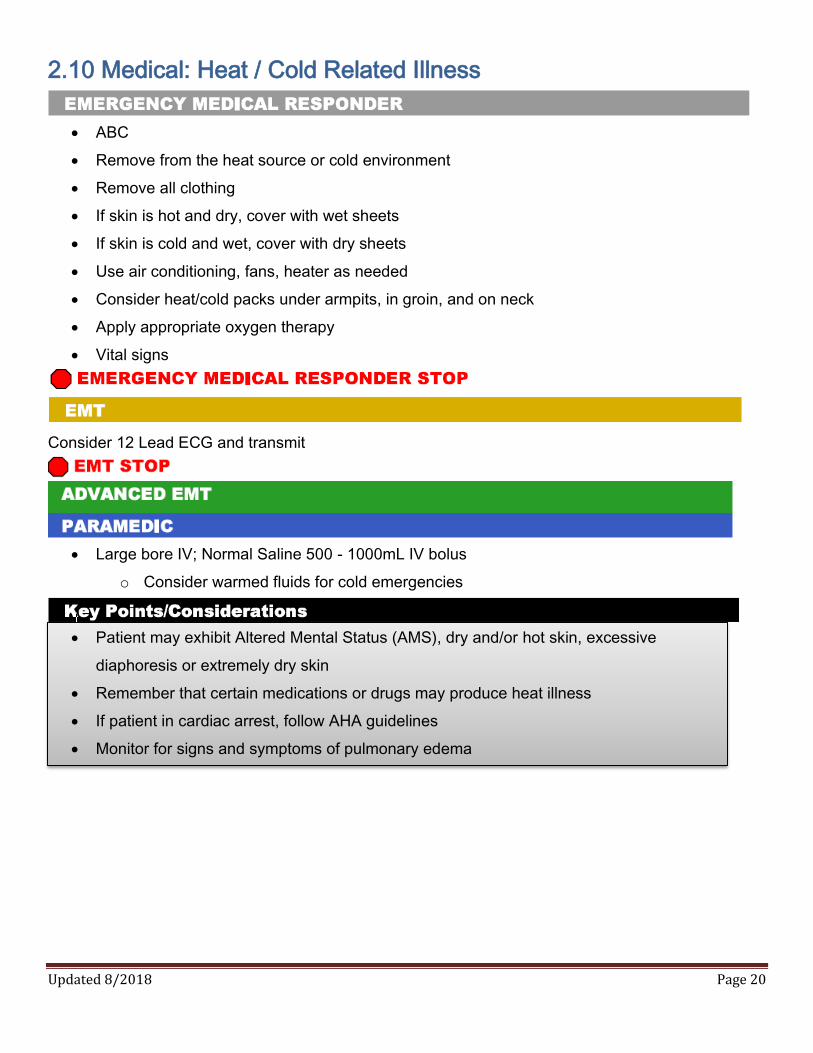
2.10 Medical: Heat / Cold Related Illness
ABC
Remove from the heat source or cold environment
Remove all clothing
If skin is hot and dry, cover with wet sheets
If skin is cold and wet, cover with dry sheets
Use air conditioning, fans, heater as needed
Consider heat/cold packs under armpits, in groin, and on neck
Apply appropriate oxygen therapy
Vital signs
Consider 12 Lead ECG and transmit
Large bore IV; Normal Saline 500 - 1000mL IV bolus
o Consider warmed fluids for cold emergencies
Patient may exhibit Altered Mental Status (AMS), dry and/or hot skin, excessive
diaphoresis or extremely dry skin
Remember that certain medications or drugs may produce heat illness
If patient in cardiac arrest, follow AHA guidelines
Monitor for signs and symptoms of pulmonary edema

Updated 8/2018 Page 21
2.11 Medical: Suspected Stroke
ABC
Apply appropriate oxygen therapy
Vital signs
Perform an initial Stroke Scale such as the Cincinnati Stroke Scale
If the patient fails the initial Stroke Scale, consider a LVO Scale such as RACE or Fast-ED
Check blood glucose level, if level is abnormal refer to 2.6 Medical: Diabetic Emergencies
protocol.
If the patient fails the initial stroke screen but passes the LVO Scale, consider transport to the
nearest Primary or tPA-ready Stroke Facility (if less than 25 minutes away)
If the patient fails both stroke scales, consider transport to a Comprehensive or
Thrombectomy-ready Stroke Facility (if less than 25 minutes away)
Advise the receiving facility of a STROKE ALERT as soon as possible
Vascular access
Rapid Arterial oCclusion Evaluation (RACE Scale) Scoring: 0-9 ≤ 4 PASS ≥5 FAIL
Aphasia: Ask the patient to 1. “Close your eyes” and 2. “Make a fist”
Agnosia: Ask the patient 1. “Whose arm is this?” and 2. “Can you lift both arms and clap?”
Updated 8/2018 Page 22
Medical Emergencies 3.0 Respiratory Emergencies

Updated 8/2018 Page 23
3.1 Respiratory: Acute Asthma and COPD with Wheezing
ABC
Apply appropriate oxygen therapy
Vital signs
Assist patient with their own meter dose inhalation medications as appropriate
Vascular access
Albuterol 2.5 mg via nebulizer; may repeat once
Epinephrine (1 mg in 1mL): dose 0.3 - 0.5 mg IM, if severe distress (call MC for order)
Cardiac Monitor
Consider starting 7.6 CPAP for moderate to severe distress.
DuoNeb (Albuterol 2.5 mg + Atrovent 0.5 mg in 2.5 mL mixed together), via nebulizer once
only, may repeat albuterol 2.5 mg once
Methylprednisolone (Solu-Medrol) 125 mg IV, IM if no IV access
If SEVERE (Status Asthmaticus) ONE of the following:
o Epinephrine (1 mg in 1 mL): dose 0.3 - 0.5 mg IM, if severe distress
o Epinephrine (1 mg in 10 mL): dose 0.5 mg IV
Not all wheezing is caused by asthma. Consider: Allergic Reaction, Airway Obstruction,
Congestive Heart Failure, Pulmonary Edema, COPD exacerbation, Acute Pulmonary
Hypertension
Caution in using Epinephrine for patients with history of CAD.
Solu-Medrol contraindicated in patients with a fever
Updated 8/2018 Page 24
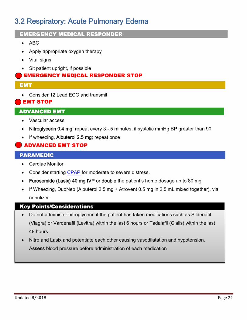
3.2 Respiratory: Acute Pulmonary Edema
ABC
Apply appropriate oxygen therapy
Vital signs
Sit patient upright, if possible
Consider 12 Lead ECG and transmit
Vascular access
Nitroglycerin 0.4 mg; repeat every 3 - 5 minutes, if systolic mmHg BP greater than 90
If wheezing, Albuterol 2.5 mg; repeat once
Cardiac Monitor
Consider starting CPAP for moderate to severe distress.
Furosemide (Lasix) 40 mg IVP or double the patient’s home dosage up to 80 mg
If Wheezing, DuoNeb (Albuterol 2.5 mg + Atrovent 0.5 mg in 2.5 mL mixed together), via
nebulizer
Do not administer nitroglycerin if the patient has taken medications such as Sildenafil
(Viagra) or Vardenafil (Levitra) within the last 6 hours or Tadalafil (Cialis) within the last
48 hours
Nitro and Lasix and potentiate each other causing vasodilatation and hypotension.
Assess blood pressure before administration of each medication

Updated 8/2018 Page 25
3.3 Respiratory: Medication Facilitated Intubation
PARAMEDIC ONLY
Medication Facilitated Intubation may be utilized on standing orders when definitive airway control
is necessary in an adult and requires the use of sedative medication and requires base station
physician approval.
Cardiac Monitor and pulse oximetry
Pre-intubation Sedation
Administer Etomidate (Amidate) 0.4 mg/kg (40 mg max) rapid IV push
Intubate if sufficient sedation has been achieved
o Confirm Placement via auscultation, CO2 detector and ETCO2 monitor if available and
secure tube
If intubation fails (2 attempts maximum) manage the airway and ventilate
Consider inserting a medically approved non-visualized airway device
If unable to adequately ventilate the patient, perform Cricothyrotomy only as a
last resort when all other Airway interventions have failed.
Post-intubation Sedation
Administer Versed 5 mg
o May repeat ONCE if needed as long as B/P is greater than 100 systolic
Pre-oxygenation and oxygenation are important when possible.
Indications for Medication Facilitated Intubation include (but are not limited to) Hypoxia
or inability to protect airway despite all other airway procedures, Traumatic injury with
GCS < 8
Updated 8/2018 Page 26
Trauma 4.0 Trauma Guidelines and
Emergencies
Updated 8/2018 Page 27
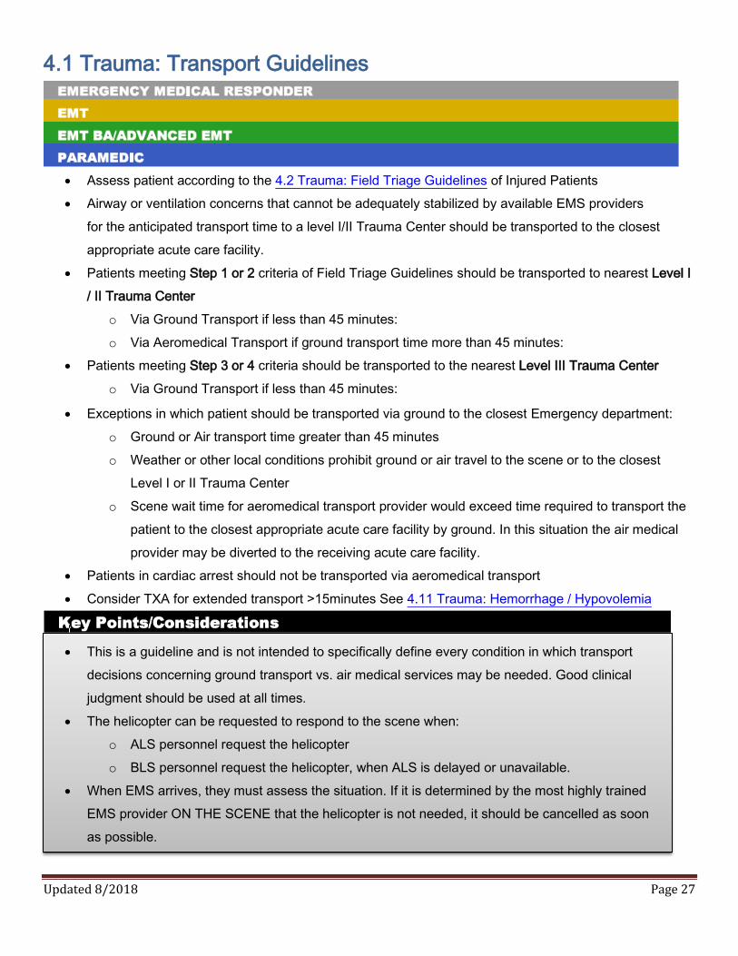
4.1 Trauma: Transport Guidelines
Assess patient according to the 4.2 Trauma: Field Triage Guidelines of Injured Patients
Airway or ventilation concerns that cannot be adequately stabilized by available EMS providers
for the anticipated transport time to a level I/II Trauma Center should be transported to the closest
appropriate acute care facility.
Patients meeting Step 1 or 2 criteria of Field Triage Guidelines should be transported to nearest Level I
/ II Trauma Center
o Via Ground Transport if less than 45 minutes:
o Via Aeromedical Transport if ground transport time more than 45 minutes:
Patients meeting Step 3 or 4 criteria should be transported to the nearest Level III Trauma Center
o Via Ground Transport if less than 45 minutes:
Exceptions in which patient should be transported via ground to the closest Emergency department:
o Ground or Air transport time greater than 45 minutes
o Weather or other local conditions prohibit ground or air travel to the scene or to the closest
Level I or II Trauma Center
o Scene wait time for aeromedical transport provider would exceed time required to transport the
patient to the closest appropriate acute care facility by ground. In this situation the air medical
provider may be diverted to the receiving acute care facility.
Patients in cardiac arrest should not be transported via aeromedical transport
Consider TXA for extended transport >15minutes See 4.11 Trauma: Hemorrhage / Hypovolemia
Key
This is a guideline and is not intended to specifically define every condition in which transport
decisions concerning ground transport vs. air medical services may be needed. Good clinical
judgment should be used at all times.
The helicopter can be requested to respond to the scene when:
o ALS personnel request the helicopter
o BLS personnel request the helicopter, when ALS is delayed or unavailable.
When EMS arrives, they must assess the situation. If it is determined by the most highly trained
EMS provider ON THE SCENE that the helicopter is not needed, it should be cancelled as soon
as possible.

Updated 8/2018 Page 28
4.2 Trauma: Field Triage Guidelines

Updated 8/2018 Page 29
4.3 Trauma: Coverage Maps
Level 1
University of Chicago
Advocate Christ
Level 3
Gary Methodist
Franciscan Health Crown Point

Updated 8/2018 Page 30
4.4 Trauma: Pre-established Landing Zones (Hammond)
1 N 41° 41' 15" W 87° 30' 2" 777 Casino Center Drive Horseshoe Casino Heliport
2 N 40° 40' 8" W 87° 30' 36" 2400 Sheffield Ave Wolf Lake Park
3 N 40° 40' 23" W 87° 30' 28" 2211 Calumet Ave Clark Athletic Field
4 N 41° 39' 43" W 87° 29' 29" 129th St. And White Oak AMOCO Park
5 N 41° 38' 11" W 87° 30' 54" 4221 Towle Lincoln Elementary School
6 N 41° 37' 41" W 87° 29' 41" 1519 Hoffman Bishop Noll High School
7 N 41° 36' 24" W 87° 30' 57" Waltham St. and Harrison Eggers Elementary School
10 N 41° 35' 12" W 87° 26' 24" 6915 Grand Ave Morton High School Parking Lot
11 N 41° 35' 9" W 87° 30' 37" 6931 Madison Ave Edison Elementary Parking Lot
12 N 41° 34' 53" W 87° 28' 30" 2239 173rd St Purdue University Parking Lot
13 N 41° 34' 35" W 87° 29' 11" 1670 175th St. Gavit High School
14 N 41° 34' 39" W 87° 27' 9" 175th St. and Parish Dowling Park
15 N 41° 34' 18" W 87° 30' 1" River Drive and Columbia Riverside Park
16 N 41° 31' 3" W 87° 28' 57" 7700 Cabela Dr. Cabela's Parking Lot
17 N 41° 36' 54" W 87° 31' 26" 5454 Hohman Ave FH Hammond Helipad
The Highlighted Zones (1 and 17) are
dedicated helipads. It is always
preferable to utilize these over
makeshift helipads.
Other areas not on this list may be
used as landing zones as long as
they meet the criteria in the 4.5
Trauma: Helicopter Utilization
Protocol
LZ 8 & 9 have been removed
(Sept 2018)
Updated 8/2018 Page 31
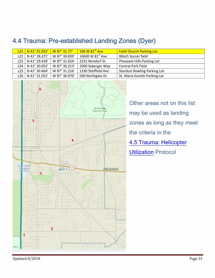
4.4 Trauma: Pre-established Landing Zones (Dyer)
LZ1 N 41° 31.052' W 87° 31.77’ 100 W 81st Ave Faith Church Parking Lot
LZ2 N 41° 28.371' W 87° 30.839' 14600 W 81st Ave Bibich Soccer field
LZ3 N 41° 29.418' W 87° 31.024' 2231 Nondorf St. Pheasant Hills Parking Lot
LZ4 N 41° 30.052' W 87° 30.213' 1000 Seberger Way Central Park Field
LZ5 N 41° 30.464' W 87° 31.216' 1330 Sheffield Ave Stardust Bowling Parking Lot
LZ6 N 41° 31.052' W 87° 30.970' 500 Northgate Dr St. Maria Goretti Parking Lot
O
Other areas not on this list
may be used as landing
zones as long as they meet
the criteria in the
4.5 Trauma: Helicopter
Utilization Protocol
Updated 8/2018 Page 32
4.5 Trauma: Helicopter Utilization
Once a request for a helicopter has been made
o Make medical direction contact and advise of your intention to initiate air transport
o Continue to follow appropriate patient care protocols
Establish an appropriate LZ (Landing Zone)
o 100’ X 100’
o Flat and clear of overhead obstructions such as trees, poles and wires
o Mark landing zone with a marker at each corner and one upwind
Communicate with flight crew
o Identify obstacles close to the landing zone and communicate all pertinent information about the
landing zone
o If it is dark, shine lights on potential obstacles such as power lines. DO NOT shine lights directly
at the aircraft
Once the aircraft has landed
o Do not approach aircraft until signaled to do so
o Always approach Helicopter from the front in a crouched position
Helicopters will not transport a patient in cardiac arrest
If using Franciscan Health Hammond’s helipad
o Contact the Emergency Department to allow security enough time to prepare the helipad
and escort the medics to the helipad
o The patient must be brought directly to the helipad (not through the Emergency Department.
The medics must park just outside of the door to the parking garage and wait for security to
escort them to the helipad
Updated 8/2018 Page 33
4.6 Trauma: Burns
Stop the burning. Remove any clothing, jewelry, etc.
ABC
High Flow Oxygen 12-15 lpm via NRB
Vital signs
Use dry sterile dressings or appropriate specialized burn dressings
Avoid wetting the patient due to the danger of hypothermia
Burns to the eye require copious irrigation with Normal Saline - do not delay irrigation
Consult MEDICAL CONTROL physician for direct transport to a Burn Center via aeromedical
transport service if needed
Vascular access at 2 sites (if severe) Normal saline 500 - 1000mL
o It is acceptable to insert through burned skin if necessary
Cardiac monitor
If patient has signs of airway involvement be prepared to intubate
o Refer to 3.3 Respiratory: Medication Facilitated Intubation protocol as needed
Refer to 2.2 General: Pain Management protocol as needed
Be alert for other injuries, including cardiac dysrhythmias
Be alert for smoke inhalation.
Assure 100% oxygen. Oxygen saturation readings may be falsely elevated.
If hazardous materials involved, notify the destination hospital immediately
When considering total area of a burn, DO NOT count first degree burns
Consider Cyanide Toxicity and Carbon Monoxide poisoning
Parkland Formula, 4ml x %BSA x weight KG : Half given in first 8 hrs

Updated 8/2018 Page 34
4.7 Trauma: Adult Rule of Nines

Updated 8/2018 Page 35
4.8 Trauma: Chest Trauma
ABC
Apply appropriate oxygen therapy
Vital signs
If sucking chest wound, cover with occlusive dressing; if dyspnea increases release
the dressing momentarily during exhalation
Consider transport to a Trauma Center using 4.1 Trauma: Transport Guidelines protocol
Vascular access, with blood draw; use the side opposite the injury if possible
Refer to 4.11 Trauma: Hypoperfusion / Hypovolemia protocol for fluid administration
Cardiac Monitor
7.7 Needle decompression if patient has signs and symptoms consistent with Tension
Pneumothorax AND hemodynamic compromise
o Prepare (14 ga. Catheter or larger, alcohol prep/Betadine)
o Locate 2nd intercostal space midclavicular line
Alternate site 5th intercostal space mid-axillary line
o Cleanse area with alcohol prep or Betadine
o Insert catheter over top of rib and into the interspace
o Advance catheter until air escapes
o Remove the needle and leave catheter in place with a one way valve in place
Refer to 2.2 General: Pain Management protocol as needed
Begin transportation as soon as possible and perform ALS treatment enroute to the hospital
Signs and symptoms of a Tension Pneumothorax: Absent lung sounds on one side, extreme
dyspnea, jugular vein distention (JVD), cyanosis (even with 100% oxygen), tracheal deviation
AND hypotension
Hemodynamic compromise is defined: hypotension, narrowed pulse pressures and tachycardia
Thoracic decompression is a serious medical intervention that requires a chest tube in the hospital
Updated 8/2018 Page 36
4.9 Trauma: Crush Injuries
ABC
Apply appropriate oxygen therapy
Vital signs every 5 minutes
Consider transport to a Trauma Center using 4.1 Trauma: Transport Guidelines protocol
Vascular access at 2 sites, with blood draw;
If blood pressure < 90 systolic or patient shows other signs of hypoperfusion
o Normal saline 500 - 1000mL IV bolus
Cardiac Monitor
If one complete extremity crushed more than 2 hours or two extremities crushed more than
1 hour:
o One minute prior to extrication: Sodium Bicarbonate 50 mEq IV
Refer to 2.2 General: Pain Management protocol as needed
“5 P’s” of crush injuries: Pain, Paresthesia, Paralysis, Pallor, Pulselessness
Consider aeromedical transport at scene if anticipated prolonged extrication.
Use one dedicated IV for Sodium Bicarbonate, the other IV for all other medications
After extrication immobilize the extremity and apply cold therapy. Do not elevate the
extremity.
Updated 8/2018 Page 37
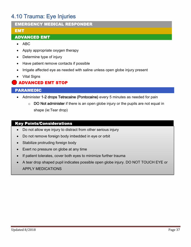
4.10 Trauma: Eye Injuries
ABC
Apply appropriate oxygen therapy
Determine type of injury
Have patient remove contacts if possible
Irrigate affected eye as needed with saline unless open globe injury present
Vital Signs
Administer 1-2 drops Tetracaine (Pontocaine) every 5 minutes as needed for pain
o DO Not administer if there is an open globe injury or the pupils are not equal in
shape (ie:Tear drop)
Do not allow eye injury to distract from other serious injury
Do not remove foreign body imbedded in eye or orbit
Stabilize protruding foreign body
Exert no pressure on globe at any time
If patient tolerates, cover both eyes to minimize further trauma
A tear drop shaped pupil indicates possible open globe injury. DO NOT TOUCH EYE or
APPLY MEDICATIONS

Updated 8/2018 Page 38
4.11 Trauma: Hemorrhage / Hypovolemia
ABC
Apply appropriate oxygen therapy
Vital signs
Control external bleeding with direct pressure. Apply a tourniquet for severe hemorrhaging
to an extremity
Wrap amputated and avulsed parts in sterile gauze and seal in a plastic bag then place in
another container with ice. Keep parts dry, sterile and cool. DO NOT place directly on ice
Consider transport to a Trauma Center using 4.1 Trauma: Transport Guidelines protocol
Vascular access, (2 sites, large-bore if possible)
Permissive hypotension: only give fluids for BP<90 SBP or other signs of hypoperfusion
o refrain from excessive amounts of saline due to clotting factor wash out.
Cardiac monitor
TXA Administration. All Trauma patients appearing to be at least 16 years of age with ongoing significant
hemorrhage (systolic BP < 90 or HR > 110 bpm) or who are considered to be a significant risk of hemorrhage
and are within 3 hours of the injury should receive TXA.
LOADING DOSE : Mix 1 gram TXA in 100 ml NS and piggyback over 10 min (see drip rates below)
CONTRAINDICATIONS FOR TXA :
Past history of thrombotic disorder such as deep venous thrombosis or pulmonary embolus.
*** Do Not Delay Transport to administer TXA
*** TXA Should not be administered to patients with less than 15 minutes of transoport
Apply tourniquet proximal to the wound and NOT across any joints
Tighten tourniquet until bleeding stops. If too loose, it may increase bleeding
Once applied correctly, a tourniquet should only be removed by the receiving hospital
Drip Rates for TXA: 10 Drop set = 1.5 drops / second
15 Drop set = 2.5 drops / second
20 Drop set = 3.5 drops / second

Updated 8/2018 Page 39
4.12 Trauma: Spinal Immobilization
Cervical collar may be appropriate without the use of Long Spine Board, if:
1. No cervical spine tenderness or anatomic abnormality. 2. No neurologic findings or complaints. 3. No significant distracting injury. 4. No intoxication with alcohol or drugs 5. No penetrating trauma to head, neck, or torso and no evidence of spinal injury. 6. Patients ambulatory on scene from blunt head trauma. 7. Isolated neck pain with no back injury.

Updated 8/2018 Page 40
5.0 Pediatric Emergencies
For these protocols, pediatric patients are defined as children having puberty (underarm hair
development in males and breast development in females Pediatric Primary Assessment Triangle:
Have Broselow Pediatric Tape or similar device available to accurately determine the correct size of
the patient
Pediatric Medication Dosages SHOULD NOT EXCEED adult dosages
Normal Vitals:

Updated 8/2018 Page 41
5.1 Pediatric Trauma: Hypovolemic Shock
ABC
Apply appropriate oxygen therapy
Control external bleeding with direct pressure
Vital signs
Keep the patient warm
Vascular access; Normal Saline 20 mL/kg IV bolus, may repeat once
Cardiac Monitor
Diagnostic criteria for UNSTABLE includes: capillary refill time greater than 2 seconds,
cool, clammy or mottled skin, inability to recognize parents, restlessness, tachycardia,
tachypnea, systolic BP less than 70 mmHg (2 years and older) or systolic, BP less than
60 mmHg (less than 2 years old).
A falling BP is a LATE sign of shock

Updated 8/2018 Page 42
5.2 Pediatric Trauma: Burns
Stop the burning. Remove any clothing, jewelry, etc.
ABC
High Flow Oxygen 12-15 lpm via NRB
Vital signs
Use dry sterile dressings or appropriate specialized burn dressing.
Avoid wetting the patient due to the danger of hypothermia
Burns to the eye require copious irrigation with Normal Saline do not delay irrigation
Vascular access at 2 sites; Normal saline 20 mL/kg IV bolus, may repeat once
Cardiac Monitor
If patient has signs of airway involvement be prepared to intubate
See 5.15 Pediatric: Pain Management OR 5.16 Pediatric: Procedural Sedation protocols
as needed
Be alert for other injuries, including cardiac dysrhythmias
Be alert for smoke inhalation
Assure 100% oxygen. Oxygen saturation readings may be falsely elevated
If hazardous materials, notify the destination hospital immediately to allow for
decontamination
When considering total area of a burn, DO NOT count first degree burns
Consider Cyanide Toxicity and Carbon Monoxide poisoning
Consider aeromedical transport to scene for transport to a pediatric burn center
Parkland Formula, 4ml x %BSA x weight KG : Half given in first 8 hrs

Updated 8/2018 Page 43
5.3 Pediatric Trauma: Burns Rule of Nines

Updated 8/2018 Page 44
5.4 Pediatric Cardiac Arrest: Asystole or PEA
ABC: Recognize the need for CPR and AED
Perform 2 minute cycles of high quality CPR (hard and fast)
Vascular / IO access; Normal Saline 20 mL/kg IV/IO bolus as needed
Cardiac monitor
Consider and treat (H’s and T’s) as appropriate
Epinephrine (1 mg in 10 mL): dose 0.01 mg/kg IV; repeat every 3-5 minutes
o Optional: Epinephrine (1 mg in 1 mL): may be diluted with 9 mL of normal saline
Place advanced airway as appropriate
Consult MEDICAL CONTROL physician and begin transport to the closest most
appropriate hospital as soon as possible
Confirm asystole in more than 1 lead
Minimize CPR interruptions
H’s and T’s Include: Hypoxia, Hypovolemia, Hypothermia, Hyper-/Hypokalemia, Hydrogen
Ion (Acidosis), Tension Pneumothorax, Cardiac Tamponade, Toxins, Thrombosis Coronary
and Thrombosis Pulmonary
Epinephrine needs to be given as soon as possible as ROSC is reduced by 4% for every
minute you delay giving it.

Updated 8/2018 Page 45
5.5 Pediatric Cardiac Arrest: V-Fib / Pulseless V-Tach
ABC: Recognize need for CPR and AED
Perform 2 minute cycles of high quality CPR (hard and fast)
Vascular / IO access; Normal Saline 20 mL/kg IV/IO bolus, as needed
Cardiac monitor
Consider (H’s and T’s) as appropriate
Initial defibrillation at 2 J/kg, repeat every two minutes at 4 J/kg
Epinephrine (1 mg in 10 mL): dose 0.01 mg/kg IV/IO; repeat every 3 5 minutes
o Optional: Epinephrine (1 mg in 1 mL): may be diluted with 9ml of normal saline
Lidocaine 1mg/kg IV/IO (Max dose 3mg/kg); repeat every 3 - 5 min
Place advanced airway when appropriate
Consult MEDICAL CONTROL physician and begin transport to the closest hospital as
soon as possible
Minimize chest compression interruptions
Use the small pediatric pads if available for patients less than 10 kg
V-fib cardiac arrest is rare in children. Consider 5.14 Pediatric Medical: Overdose / Toxic
Exposure protocol including tricyclic antidepressants.

Updated 8/2018 Page 46
5.6 Pediatric Cardiac: Symptomatic Bradycardia
ABC
Apply appropriate oxygen therapy
Vital signs
If heart rate is less than 60 bpm and patient’s mental statue and respiratory rate are
decreased, ventilate with BVM
If no improvement with ventilations, start CPR
Consider 12 lead and transmit
Vascular access (IO if CPR in progress); Normal Saline 20 mL/kg IV/IO bolus, as needed
Cardiac monitor
Consider and treat reversible H’s and T’s
Epinephrine (1 mg in 10 mL): dose 0.01 mg/kg IV/IO; repeat every 3-5 minutes
o Optional: Epinephrine (1 mg in 1 mL): may be diluted with 9ml of normal saline
If bradycardia is due to increased vagal tone or primary AV block give atropine before
giving epinephrine
o Atropine 0.02 mg/kg (0.1 mg min dose) IV/IO; repeat 5 minutes to max 0.04
mg/kg
Transcutaneous pacing
o Refer to 5.16 Pediatric: Procedural Sedation protocol
Place advanced airway as appropriate
Consult MEDICAL CONTROL physician as soon as possible
Bradycardia Newborn/Infant -- pulse less than 80 bpm; child over 1 year of age - pulse
less than 60 bpm
Symptomatic includes poor systemic perfusion, hypotension, respiratory difficulty or
altered level of consciousness
Do not treat asymptomatic bradycardia. Consult MEDICAL CONTROL physician.

Updated 8/2018 Page 47
5.7 Pediatric Cardiac: Symptomatic Tachycardia
ABC
Apply appropriate oxygen therapy
Vital signs
Consider 12 lead and transmit
Vascular access, Normal Saline 20 mL/kg IV bolus, as needed
Valsalva Maneuvers
Cardiac monitor
UNSTABLE
o Synchronized cardioversion 0.5 - 1.0 J/kg; repeat 2 J/kg if unsuccessful
Refer to 5.16 Pediatric: Procedural Sedation protocol
STABLE Wide QRS:
o Lidocaine 1mg/kg IV/IO (Max dose 3mg/kg); repeat every 3-5 min
STABLE Narrow QRS:
o Adenosine (Adenocard) 0.1 mg/kg IV; May repeat in 1-2 minutes at 0.2 mg/kg IV
Consult MEDICAL CONTROL physician as soon as possible
Newborn/Infant SVT - if pulse greater than 220 bpm; child over 1 year of age SVT - if pulse
greater than 180 bpm, with no discernable p-waves
UNSTABLE includes cardio-respiratory compromise, hypotension, or altered level of
consciousness
The most common causes of Sinus Tachycardia in children are fever and dehydration
Do not treat asymptomatic tachycardia. Consult MEDICAL CONTROL physician.

Updated 8/2018 Page 48
5.8 Pediatric Respiratory: Acute Asthma
ABC
Apply appropriate oxygen therapy
Vital signs
Determine if patient has been given his/her own asthma medications
Assist with patient prescribed metered dose inhaler
Albuterol 2.5 mg via nebulizer
(Advanced) Call Medical Control for,
o Epinephrine (1 mg in 1 mL): dose 0.01 mg/kg SQ (0.5 mg max), if in severe distress
Cardiac monitor
DuoNeb (Albuterol 2.5 mg + Atrovent 0.5 mg) via nebulizer if proceed to next step;
o Do not repeat Atrovent; may repeat Albuterol 2.5 mg via nebulizer
If patient not improving, obtain vascular access
o Methylprednisolone (Solu-Medrol) 1-2 mg/kg IV, IM if no IV access
o Epinephrine (1 mg in 1 mL): dose 0.01 mg/kg IM (0.5 mg max), if in severe distress
Consult MEDICAL CONTROL physician as soon as possible
Absence of breath sounds can be indicative of status asthmaticus. Be prepared for
imminent respiratory arrest

Updated 8/2018 Page 49
5.9 Pediatric Respiratory: Anaphylaxis / Allergic Reaction
ABC
Apply appropriate oxygen therapy
Vital signs
If localized reaction, apply ice pack to affected area
Determine if patient has been given his/her own Epi Pen
If not and patient severe distress;
o BLS administer Epi Pen Jr. or
o Epinephrine (1 mg in 1 mL): dose 0.15 mg IM
if patient weighs more than 30 kg (66 lbs.), Dose 0.3 mg IM
Vascular access; Normal Saline 20 mL/kg IV bolus as needed
If patient wheezing, Albuterol 2.5 mg via nebulizer
Cardiac Monitor
MILD SYMPTOMS: Urticaria, itching, nasal congestion, watery eye
Diphenhydramine (Benadryl) 1 - 2 mg/kg (25 mg max) IV or IM
MODERATE / SEVERE SYMPTOMS: Wheezing/stridor, swelling face, neck, tongue, hypotension, altered LOC
DuoNeb (Albuterol 2.5 mg + Atrovent 0.5 mg) via nebulizer; Do not repeat Atrovent
Methylprednisolone (Solu-Medrol) 1 - 2 mg/kg IV
If BLS airway maneuvers fail, consider intubation; If unable to intubate,
consider 7.4 Procedure: Needle Cricothyrotomy only as a last resort when all other airway
interventions have failed
Consult MEDICAL CONTROL physician as soon as possible
If the patient already used an Epi Pen, consult MEDICAL CONTROL prior to administering
additional epinephrine or allowing the legal guardian to sign a refusal
Updated 8/2018 Page 50
5.10 Pediatric Medical: Seizures
ABC
Apply appropriate oxygen therapy
Vital signs
If child is warm, remove blanket or loosen clothing
Check blood glucose level, if level is abnormal refer to 5.11 Pediatric Medical: Diabetic
protocol
o DO NOT DELAY TREATMENT OF SEIZURE TO OBTAIN BGL
Vascular access
Cardiac Monitor
Give ONE of the Following
(Ativan) 0.1 mg/kg IV, IM, IN (max 2 mg)
Midazolam (Versed) 0.05 mg/kg IV, IM, IN (max 2 mg)
Valium (Diazepam) 0.1 mg/kg IV,IM,IN (max 5 mg)
Place advanced airway as appropriate
Consult MEDICAL CONTROL physician as soon as possible
Protect the patient and EMS crew from injury during the seizure
IN administration of benzodiazepines is as effective as IV
Updated 8/2018 Page 51
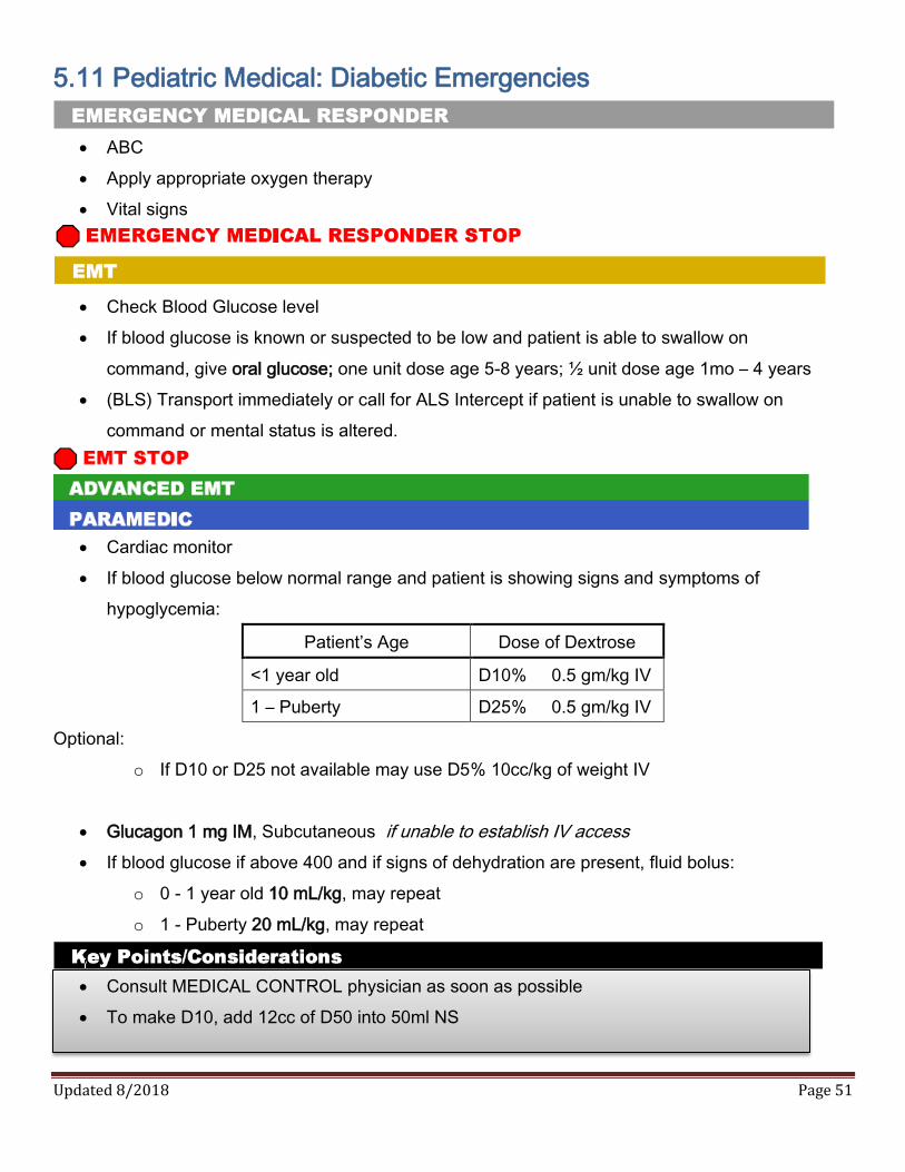
5.11 Pediatric Medical: Diabetic Emergencies
ABC
Apply appropriate oxygen therapy
Vital signs
Check Blood Glucose level
If blood glucose is known or suspected to be low and patient is able to swallow on
command, give oral glucose; one unit dose age 5-8 years; ½ unit dose age 1mo – 4 years
(BLS) Transport immediately or call for ALS Intercept if patient is unable to swallow on
command or mental status is altered.
Cardiac monitor
If blood glucose below normal range and patient is showing signs and symptoms of
hypoglycemia:
Patient’s Age Dose of Dextrose
<1 year old D10% 0.5 gm/kg IV
1 – Puberty D25% 0.5 gm/kg IV
Optional:
o If D10 or D25 not available may use D5% 10cc/kg of weight IV
Glucagon 1 mg IM, Subcutaneous if unable to establish IV access
If blood glucose if above 400 and if signs of dehydration are present, fluid bolus:
o 0 - 1 year old 10 mL/kg, may repeat
o 1 - Puberty 20 mL/kg, may repeat
Consult MEDICAL CONTROL physician as soon as possible
To make D10, add 12cc of D50 into 50ml NS

Updated 8/2018 Page 52
5.12 Pediatric Medical: Hypoperfusion
ABC
Apply appropriate oxygen therapy
Vital signs
If no fever present, keep the patient warm
Vascular access; Normal Saline 20 mL/kg IV bolus, as needed
Cardiac Monitor
Consult MEDICAL CONTROL physician as soon as possible
For patients with hypovolemia due to bleeding, vomiting, diarrhea or septic shock.
Consult MEDICAL CONTROL physician if you suspect cardiogenic shock. Diagnostic criteria for hypotension includes: capillary refill time greater than 2 seconds,
cool, clammy or mottled skin, inability to recognize parents, restlessness, tachycardia,
tachypnea, systolic BP less than 70 mmHg (2 years and older) or systolic BP less than
60 mmHg (less than 2 years old).
Updated 8/2018 Page 53
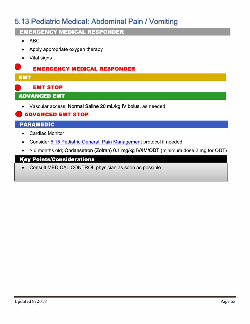
5.13 Pediatric Medical: Abdominal Pain / Vomiting
ABC
Apply appropriate oxygen therapy
Vital signs
Vascular access; Normal Saline 20 mL/kg IV bolus, as needed
Cardiac Monitor
Consider 5.15 Pediatric General: Pain Management protocol if needed
> 6 months old; Ondansetron (Zofran) 0.1 mg/kg IV/IM/ODT (minimum dose 2 mg for ODT)
Consult MEDICAL CONTROL physician as soon as possible

Updated 8/2018 Page 54
5.14 Pediatric Medical: Overdose / Toxic Exposure
Decontamination as needed
ABC
Apply appropriate oxygen therapy
Vital signs
Determine what was taken, when and how much, if possible
Consider contacting Poison Control 1-800-222-1222 for additional information
Vascular access
Opiate overdose: Naloxone (Narcan) 0.1 mg/kg IV, IM, Subcutaneous; Repeat to max 2 mg
Cardiac monitor
For symptomatic patient with:
o Organophosphate poisoning: Atropine 1 mg IV; repeat every 3 5 minutes until
secretions dry
o Sympathomimetic ingestion (cocaine/amphetamine): Midazolam (Versed) 0.1 mg/kg
IV or IM
o Tricyclic Antidepressants: Sodium Bicarb 1 mEq/kg if wide complex arrhythmia and
prolonged QRS duration (if hypotensive, 10 mL/kg NS bolus)
Consult MEDICAL CONTROL physician as soon as possible
Cocaine/Methamphetamine signs and symptoms Seizures, hypertension, tachycardia
Signs and symptoms of organophosphate poisoning consider SLUDGE
o Salivation, Lacrimation, Urination, Diarrhea, Gastric cramps, Emesis

Updated 8/2018 Page 55
5.15 Pediatric General: Pain Management
ABC
Apply appropriate oxygen therapy
Vital signs
Vascular access
Cardiac Monitor
Administer ONE of the following narcotic analgesics
o Morphine 0.05 mg/kg IV or IM; repeat once to max 0.1 mg/kg
o Fentanyl 0.5 - 1 mcg/kg Slow IV, IM, or IntraNasal (IN)
Ondansetron (Zofran) 0.1 mg/kg IV/ODT/IM, if patient becomes nauseous (minimum
dose 2 mg for ODT)
Consult MEDICAL CONTROL physician as soon as possible
For patients with: o Severe burns without hemodynamic compromise
o Suspected isolated extremity injuries, fractures or dislocations with severe pain
o Abdominal pain
o Back pain
For all other painful conditions, providers must consult MEDICAL for orders CONTROL
physician
Contraindications to pain management protocol: altered mental status, hypoventilation,
hypotension, other traumatic injuries
This protocol may NOT be used in conjunction with the Pediatric: Procedural Sedation
protocol, unless MEDICAL CONTROL physician is consulted.
Consult MEDICAL CONTROL physician for additional pain or nausea medication
Updated 8/2018 Page 56
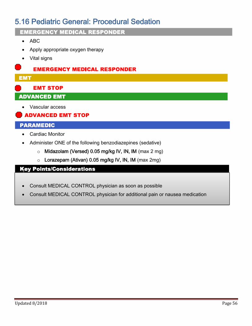
5.16 Pediatric General: Procedural Sedation
ABC
Apply appropriate oxygen therapy
Vital signs
Vascular access
Cardiac Monitor
Administer ONE of the following benzodiazepines (sedative)
o Midazolam (Versed) 0.05 mg/kg IV, IN, IM (max 2 mg)
o Lorazepam (Ativan) 0.05 mg/kg IV, IN, IM (max 2mg)
Consult MEDICAL CONTROL physician as soon as possible
Consult MEDICAL CONTROL physician for additional pain or nausea medication

Updated 8/2018 Page 57
OB/GYN 6.0 OB/GYN EMERGENCIES

Updated 8/2018 Page 58
6.1 OB/GYN Childbirth: Pre-delivery
Vital Signs
Determine the estimated date of expected birth, the number of previous pregnancies and
number of live births (Gravida / Para / Abortio)
Determine if the amniotic sac (bag of waters) has broken, if there is vaginal bleeding or
mucous discharge.
Determine the duration and frequency of uterine contractions
If labor seems active based on above information:
o Examine the patient for crowning or bulging of the perineum with contractions
If delivery is imminent, prepare for an on-scene delivery.
Prepare equipment and drape the mother
Consider Vascular Access
If the amniotic sac has ruptured, the birth may not be imminent, but the mother must
deliver the baby within 24 hours
Gravida: number of pregnancies;
Para: number of live births;
Abortio: number of demised births (abortion, miscarriage, still-birth, etc.)

Updated 8/2018 Page 59
6.2 OB/GYN Childbirth: Delivery
Support the baby’s head over the perineum
If the membranes cover the head after it emerges, tear the sac with your fingers or
forceps to permit escape of the amniotic fluid.
Suction oropharynx then nostrils with a bulb syringe.
Feel for the presence of the umbilical cord around the neck
o If the cord is around the neck and cannot be easily removed, clamp it with two
clamps, cut the cord between the clamps, and unwrap the cord from around the neck.
Gently guide the head downward until the shoulder appears. The other shoulder is
delivered by gentle upward traction. The infant’s face should be upward at this point.
Carefully hold (catch) the infant as it continues to deliver
Provide tactile stimulation by drying and wrapping the infant in a blanket
Once the infant is crying or breathing adequately, clamp the umbilical cord, >60 seconds after
birth, with a clamp at 4 inches and one at 6 inches from umbilicus and cut the cord between
them.
Assess APGAR score at 1 minute and 5 minutes after birth (See next page)
Support the buttocks or extremities until the back appears.
Grasp the baby’s ILIAC WINGS and apply gentle downward traction. DO NOT pull
on the legs or back, as this may cause spine dislocation or adrenal hemorrhage.
Gently move the infant’s body in the direction of least resistance. By moving anteriorly and
posteriorly, both shoulders should deliver posteriorly.
Splint the humerus bones with your two fingers and apply gentle traction with your fingers.
Gentle downward compression of the uterus will assist in head delivery. Swing the legs upward
until the body is in a vertical position. This will permit delivery of the head.

Updated 8/2018 Page 60
6.2 OB/GYN Childbirth: Delivery (continued)
Place the mother in a face-up position with hips elevated
Place a gloved hand in the vagina and attempt to hold the baby’s head away from the cord
Keep the cord moist using a sterile dressing and sterile water
Transport as soon as possible
Score should be recorded at 1 minute and 5 minutes after birth
Do not withhold resuscitation efforts to determine APGAR score
Sign 0 1 2
Appearance
(skin color) Central Cyanosis Pink torso with Distal
Cyanosis Completely Pink
Pulse Absent <100 >100
Grimace
(flick soles of feet) No response Grimace, shudders or
flinches Vigorous cry
Activity
(muscle tone) Limp Some flexion Active motion
Respirations No effore Weak, Irregular Strong Cry
If multiple births are anticipated but the subsequent births do not occur within 10 minutes
of the previous delivery transport immediately.
After delivery of the placenta gently massage the uterus
Bring the placenta and any other tissue to the hospital for inspection
NEONATAL RESUSCITATION
o If the Infant is not breathing adequately or heart rate is < 100 after 30 seconds of
tactile stimulation, consider assisted ventilations.
o If the infant’s heartrate is less than 60 after one minute, start chest compressions

Updated 8/2018 Page 61
6.3 OB/GYN: Eclampsia
ABC
Apply appropriate oxygen therapy
Vital signs
Check blood glucose level, if level is abnormal refer to 2.6 Medical: Diabetic Emergencies
protocol.
Vascular access
Cardiac Monitor
If patient is seizing administer Magnesium Sulfate 4 g over 2 minutes IV (IM buttock if
unable to establish IV)
No response, administer Versed 2 - 5 mg IV
Pre-eclampsia is defined as BP greater than 140/90 in a pregnant patient (or one who
has recently given birth) with severe headache, confusion and/or hyper-reflexia
Eclampsia includes the above information and includes seizure activity
Females should be considered pregnant up to 6 weeks after delivery
Updated 8/2018 Page 62
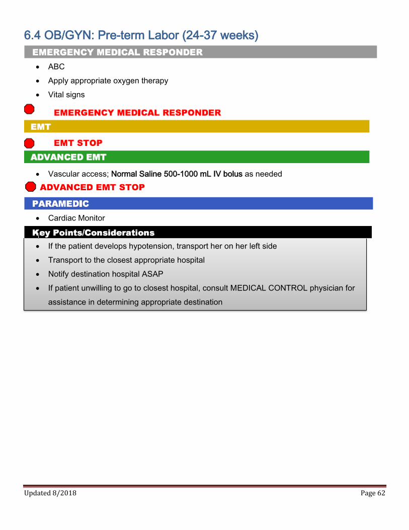
6.4 OB/GYN: Pre-term Labor (24-37 weeks)
ABC
Apply appropriate oxygen therapy
Vital signs
Vascular access; Normal Saline 500-1000 mL IV bolus as needed
Cardiac Monitor
If the patient develops hypotension, transport her on her left side
Transport to the closest appropriate hospital
Notify destination hospital ASAP
If patient unwilling to go to closest hospital, consult MEDICAL CONTROL physician for
assistance in determining appropriate destination
Updated 8/2018 Page 63
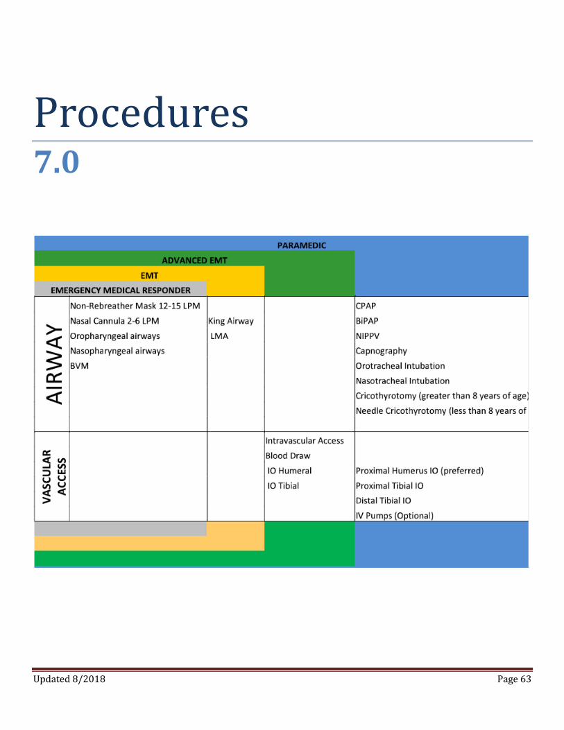
Procedures 7.0

Updated 8/2018 Page 64
7.1 Procedure: Airway Management
Oxygen therapy: The goal of oxygen therapy is to achieve adequate tissue oxygenation using the
lowest possible FiO2
o Non-rebreather mask 12 - 15 lpm, NRB
o Nasal cannula, 2 - 6 lpm
o Nasopharyngeal and/or Oropharyngeal airways
o BVM assisted ventilation
Medically approved non-visualized airway;
o 7.2 Procedure: Non-visualized Airway
LMA
King Airway
Oral endotracheal intubation in unresponsive Adults and Pediatric patients
o 7.3 Procedure: Endotracheal Intubation
Continuous Positive Airway Pressure (CPAP) or Bi-Level Positive Airway Pressure (BiPAP)
o 7.6 Procedure: CPAP
Medication facilitated intubation;
o 3.3 Respiratory: Medication Facilitated Intubation
Pediatric Needle cricothyrotomy (< 8 yo)
o 7.4 Procedure: Needle cricothyrotomy
Quick Trach Cricothyrotomy (> 8 yo);
o 7.5 Procedure: Quick Trach Cricothyrotomy
Always have a BVM available when using a portable transport ventilator
Intubation may be attempted on a patient 2 times. If unsuccessful utilize a medically approved non-
visualized airway or ventilate with BVM.
Re-confirm endotracheal placement after any patient transfer with at least two assessments and
continuous Waveform/Quantitative Capnography (if available)
Updated 8/2018 Page 65
7.2 Procedure: Non-visualized Airway
Indications:
Airway control in the absence of other effective methods (e.g. failed airway) Situations involving a difficult mask (BVM) fit.
Contraindications: Patients at risk of aspiration Patients with massive thoracic injury Patients who are not profoundly unconscious and who may resist insertion Severe maxillofacial or oropharyngeal trauma Greater than 14-16 weeks pregnant
Insertion: Step 1: Size selection
- Verify correct size of LMA for the patient Step 2: Examine the LMA
- Visually inspect the LMA cuff for tears - Inspect the tube to ensure that it is free of blockage - Deflate cuff to ensure that it will maintain a vacuum - Inflate cuff to ensure that it does not leak
Step 3: Lubrication
- Lubricate LMA with a water soluble lubricant.
Step 4: Positioning of Airway - Extend the head and flex the neck - Avoid LMA fold over - Assistant pulls the lower jaw downwards - Visualize the posterior oral airway - Ensure that the LMA is not folding over in the oral cavity as it is inserted
Step 5: Insertion
- Place tip of LMA against the inner surface of the patient’s upper teeth - Using the index finger, keep pressing upwards as you advance the mask into the pharynx to
ensure the tip remains flattened and avoids the tongue - Guide the mask down into position - Inflate the mask with recommended volume of air
Use Caution with a possible foreign body airway obstruction.
Attempt to remove obstruction before placing device
Updated 8/2018 Page 66
7.3 Procedure: Endotracheal Intubation
Assure that the patient is being adequately oxygenated/ventilated prior to intubation
Have suction ready
Continuous pulse oximetry and cardiac monitoring
Select and prepare proper ETT: ET Tube is determined by comparison of the patients nares or little
finger ( refer to the Length Based Pediatric Tape for Pediatric population)
Insert stylet
Check ETT for cuff leakage
Lubricate tube
Prepare laryngoscope: select proper blade and check light
Place patient in “sniffing position”, unless contraindicated
Pre-oxygenate patient with 100% OXYGEN via BVM
Apply continuous cricoid pressure (Sellick Manuever) if needed
Insert laryngoscope: hold in left hand, insert blade into right side of mouth and sweep tongue to the left
Visualize vocal cords:
o Straight Blade- Direct blade below epiglottis and lift handle up and away from you
o Curved Blade- Direct blade below into vallecula and tilt your hand towards yourself
DO NOT USE TEETH AS A FULCRUM
Using right hand, insert tube between vocal cords
Remove stylet, if used
Verify tube placement: auscultation of epigastrium and both axillae, obtain pulse oximetry and use
appropriate CO2 detector
Inflate cuff with 5-10 mL of air, if appropriate
Secure ETT appropriately
Reassess tube placement while ventilating patient
Intubation may be attempted on a patient 2 times. If unsuccessful utilize a medically approved
non-visualized airway or ventilate with BVM.
Utilize 3.3 Respiratory: Medication Facilitated Intubation as needed

Updated 8/2018 Page 67
7.4 Procedure: Needle Cricothyrotomy
Establish that complete obstruction of airway has occurred.
o Attempt BLS measures to relieve obstruction.
o Attempt to visualize obstruction with laryngoscope.
o If obstruction visible, remove with Magill Forceps.
If still complete obstruction:
o Prepare large bore angiocath (14 gauge) with 50cc syringe.
o Palpate crico-thyroid membrane.
o Prep area with antibacterial solution.
o With angiocath on syringe, insert through crico-thyroid membrane, maintaining
negative pressure on plunger of syringe.
o Entry into trachea will be confirmed by escape of air into syringe.
o Advance Catheter only in caudal direction (towards feet).
o Attach bag-ventilator with 3.5mm endotracheal tube adaptor;
o Ventilate patient with supplemental oxygen; listen for breath sounds bilaterally;
visualize chest movements;
Transport as soon as possible.
Needle cricothyrotomy may provide ventilation Assistance for a short period of time only.
Do not delay transport.
3.5mm ett adapter
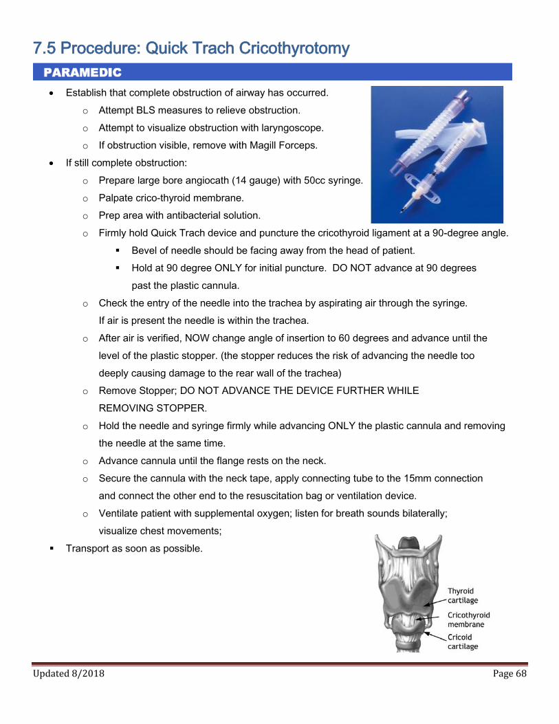
Updated 8/2018 Page 68
7.5 Procedure: Quick Trach Cricothyrotomy
Establish that complete obstruction of airway has occurred.
o Attempt BLS measures to relieve obstruction.
o Attempt to visualize obstruction with laryngoscope.
o If obstruction visible, remove with Magill Forceps.
If still complete obstruction:
o Prepare large bore angiocath (14 gauge) with 50cc syringe.
o Palpate crico-thyroid membrane.
o Prep area with antibacterial solution.
o Firmly hold Quick Trach device and puncture the cricothyroid ligament at a 90-degree angle.
Bevel of needle should be facing away from the head of patient.
Hold at 90 degree ONLY for initial puncture. DO NOT advance at 90 degrees
past the plastic cannula.
o Check the entry of the needle into the trachea by aspirating air through the syringe.
If air is present the needle is within the trachea.
o After air is verified, NOW change angle of insertion to 60 degrees and advance until the
level of the plastic stopper. (the stopper reduces the risk of advancing the needle too
deeply causing damage to the rear wall of the trachea)
o Remove Stopper; DO NOT ADVANCE THE DEVICE FURTHER WHILE
REMOVING STOPPER.
o Hold the needle and syringe firmly while advancing ONLY the plastic cannula and removing
the needle at the same time.
o Advance cannula until the flange rests on the neck.
o Secure the cannula with the neck tape, apply connecting tube to the 15mm connection
and connect the other end to the resuscitation bag or ventilation device.
o Ventilate patient with supplemental oxygen; listen for breath sounds bilaterally;
visualize chest movements;
Transport as soon as possible.

Updated 8/2018 Page 69
7.6 Procedure: CPAP (Flowsafe 2)
Oxygenate the patient with 15 liters via non-rebreather mask while setting up CPAP
Chose correct size CPAP
Attach CPAP tubing to oxygen source and obtain desired pressure (see chart below)
Encourage patient to place mask over mouth and nose, then firmly attach mask using
final connection on side of mask
o Refer to 2.4 General: Procedural Sedation protocol as needed
Monitor patient’s level of consciousness and vital signs continuously.
o If patient develops decreased mental status or decreased blood pressure-
DISCONTINUE CPAP.
Continuous cardiac monitoring and pulse oximetry required
If port is available for Albuterol administration, follow manufactures guidelines
Contraindications for CPAP include Unconsciousness, Children under 14, Hypotension
facial / chest / upper airway trauma, apnea, inability to protect airway, vomiting,
pneumothorax
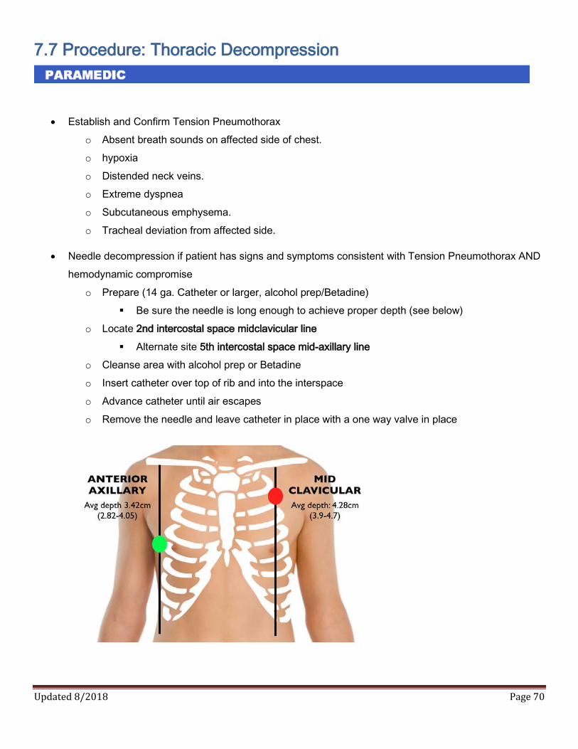
Updated 8/2018 Page 70
7.7 Procedure: Thoracic Decompression
Establish and Confirm Tension Pneumothorax
o Absent breath sounds on affected side of chest.
o hypoxia
o Distended neck veins.
o Extreme dyspnea
o Subcutaneous emphysema.
o Tracheal deviation from affected side.
Needle decompression if patient has signs and symptoms consistent with Tension Pneumothorax AND
hemodynamic compromise
o Prepare (14 ga. Catheter or larger, alcohol prep/Betadine)
Be sure the needle is long enough to achieve proper depth (see below)
o Locate 2nd intercostal space midclavicular line
Alternate site 5th intercostal space mid-axillary line
o Cleanse area with alcohol prep or Betadine
o Insert catheter over top of rib and into the interspace
o Advance catheter until air escapes
o Remove the needle and leave catheter in place with a one way valve in place
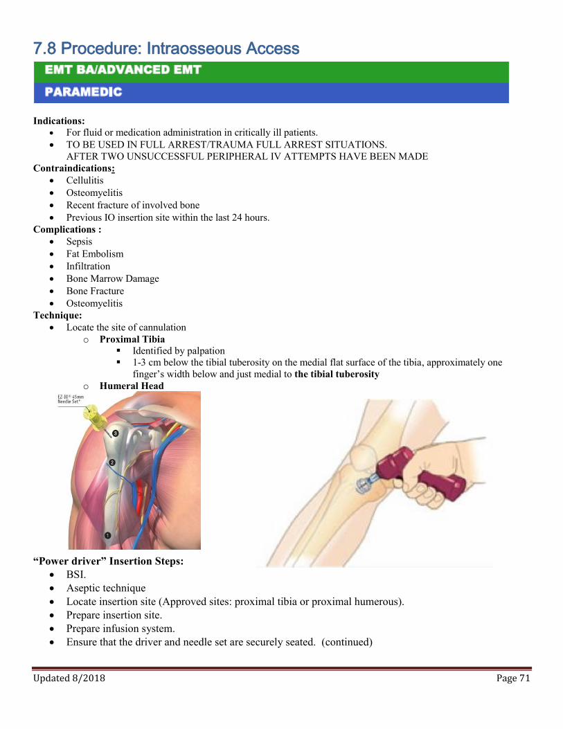
Updated 8/2018 Page 71
7.8 Procedure: Intraosseous Access
Indications: For fluid or medication administration in critically ill patients. TO BE USED IN FULL ARREST/TRAUMA FULL ARREST SITUATIONS.
AFTER TWO UNSUCCESSFUL PERIPHERAL IV ATTEMPTS HAVE BEEN MADE Contraindications:
Cellulitis Osteomyelitis Recent fracture of involved bone Previous IO insertion site within the last 24 hours.
Complications : Sepsis Fat Embolism Infiltration Bone Marrow Damage Bone Fracture Osteomyelitis
Technique: Locate the site of cannulation
o Proximal Tibia Identified by palpation 1-3 cm below the tibial tuberosity on the medial flat surface of the tibia, approximately one
finger’s width below and just medial to the tibial tuberosity o Humeral Head
“Power driver” Insertion Steps:
BSI. Aseptic technique Locate insertion site (Approved sites: proximal tibia or proximal humerous). Prepare insertion site. Prepare infusion system. Ensure that the driver and needle set are securely seated. (continued)
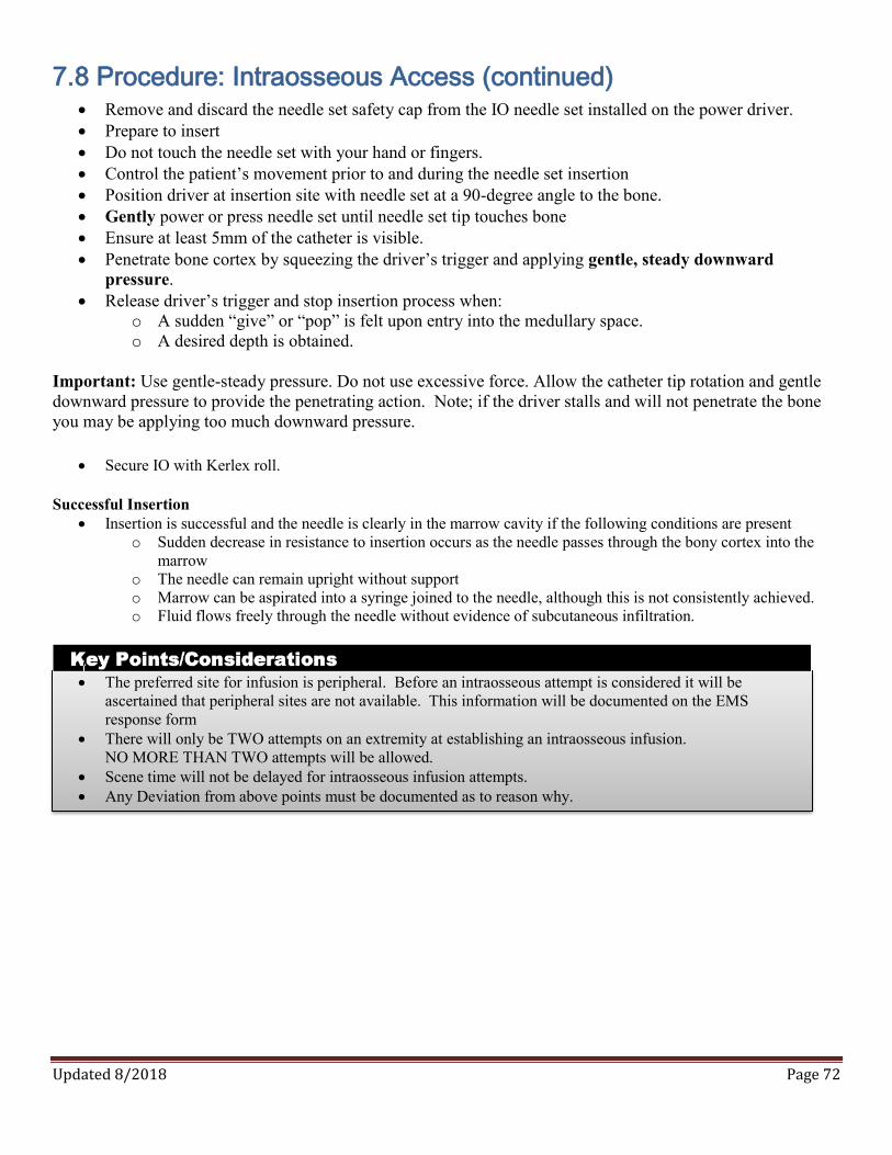
Updated 8/2018 Page 72
7.8 Procedure: Intraosseous Access (continued) Remove and discard the needle set safety cap from the IO needle set installed on the power driver. Prepare to insert Do not touch the needle set with your hand or fingers. Control the patient’s movement prior to and during the needle set insertion Position driver at insertion site with needle set at a 90-degree angle to the bone. Gently power or press needle set until needle set tip touches bone Ensure at least 5mm of the catheter is visible. Penetrate bone cortex by squeezing the driver’s trigger and applying gentle, steady downward
pressure. Release driver’s trigger and stop insertion process when:
o A sudden “give” or “pop” is felt upon entry into the medullary space. o A desired depth is obtained.
Important: Use gentle-steady pressure. Do not use excessive force. Allow the catheter tip rotation and gentle downward pressure to provide the penetrating action. Note; if the driver stalls and will not penetrate the bone you may be applying too much downward pressure.
Secure IO with Kerlex roll.
Successful Insertion Insertion is successful and the needle is clearly in the marrow cavity if the following conditions are present
o Sudden decrease in resistance to insertion occurs as the needle passes through the bony cortex into the marrow
o The needle can remain upright without support o Marrow can be aspirated into a syringe joined to the needle, although this is not consistently achieved. o Fluid flows freely through the needle without evidence of subcutaneous infiltration.
The preferred site for infusion is peripheral. Before an intraosseous attempt is considered it will be
ascertained that peripheral sites are not available. This information will be documented on the EMS response form
There will only be TWO attempts on an extremity at establishing an intraosseous infusion. NO MORE THAN TWO attempts will be allowed.
Scene time will not be delayed for intraosseous infusion attempts. Any Deviation from above points must be documented as to reason why.

Updated 8/2018 Page 73
7.9 Procedure: 12-Lead Acquisition and Transmission
Clinical Indications: Chest pain or other angio-equivalents such as dizziness, shortness of breath or other referred pain Electrical Injuries Precautions: Care must be taken to avoid an unnecessary extension of scene time. Do not delay scene time more than four (4) minutes to perform 12 lead ECG. Patients who require a 12 lead ECG should not refuse treatment. Treatment of patient with Aspirin, Nitroglycerin or any other medication should not be delayed while preparing to acquire 12 lead ECG Contraindications: Trauma Patient Unstable Patient Procedure: Place patient in position of comfort. Push “12” button, then “acquire” button. Enter patient demographic data if time allows. Remove patient clothing above waist. Use a gown or sheet to preserve patient modesty. Prepare patient’s skin for electrode placement. (shave hair if necessary, clean oily skin with alcohol and drying briskly) Apply chest and limb leads as follows: Limb Leads RA-RIGHT ARM LA-LEFT ARM RL-RIGHT LEG (lower right abdomen, groin, thigh) LL-LEFT LEG (lower left abdomen, groin, thigh)
Precordial Leads V1- 4TH INTERCOSTAL SPACE RIGHT OF THE STERNUM V2- 4TH INTERCOSTAL SPACE LEFT OF THE STERNUM V3- DIRECTLY BETWEEN LEADS 2 AND 4. V4- 5TH LEFT INTERCOSTAL SPACE AT MIDCLAVICULAR LINE
V5- LEVEL WITH V4 AT THE LEFT ANTERIOR AXILLARY LINE V6- LEVEL WITH V5 AT THE LEFT MIDAXILLARY LINE Instruct patient to remain still. Obtain 12-lead ECG reading. Avoid acquiring ECG in a moving vehicle. TRANSMIT 12-LEAD IF AVAILABLE Document TIME of when12 lead acquired. Do Not remove limb/precordial electrodes after ECG acquisition; leave in place upon ED arrival. Contact Medical Control to notify them of the patient and the incoming 12 lead ECG. (Alert medical control ASAP if STEMMI is present) Monitor and assess the patient enroute and continue treatment protocol. Upon arrival to the ED, the 12 lead must be handed to the attending physician only. Document TIME, DATE and PHYSICIAN who received the 12 lead at facility.

Updated 8/2018 Page 74
7.10 Medication Administration: Intranasal (IN)
Using a 1 ml or 3 ml syringe and needle, draw the appropriate amount of medication into the
syringe. (Unless medication is already in a quick jet)
Remove the needle and place the MAD tip onto the syringe. The MAD is a Luer lock device and twists
into place
Use your free hand to hold the crown of the head stable.
Place the tip of the atomizer snugly against the nostril aiming slightly up and outward (toward the
top of the ear
Briskly compress the syringe plunger and deliver approximately half of the medication
Move the device over to the opposite nostril and administer the remainder of the medication as before.
If an amount greater than 1 ml per nostril is needed, wait 2 3 minutes and administer the remaining
medication
The Mucosal Atomization Device (MAD) can be used for the Fentanyl, Midazolam, Ativan (ALS)
and Naloxone in the event an IV a has not or cannot be initiated.
Prior to using the IN route, inspect the patient’s nostrils for significant amounts of blood or mucous
discharge. The presence of these will limit medication absorption. Considering suctioning the nasal
passages before IN administration in these cases
Always deliver half of the medication dose up each nostril.
Be aware there is approximately 0.1 ml of dead space in the MAD. It is important to make
allowances for this dead space when calculating drug dosages
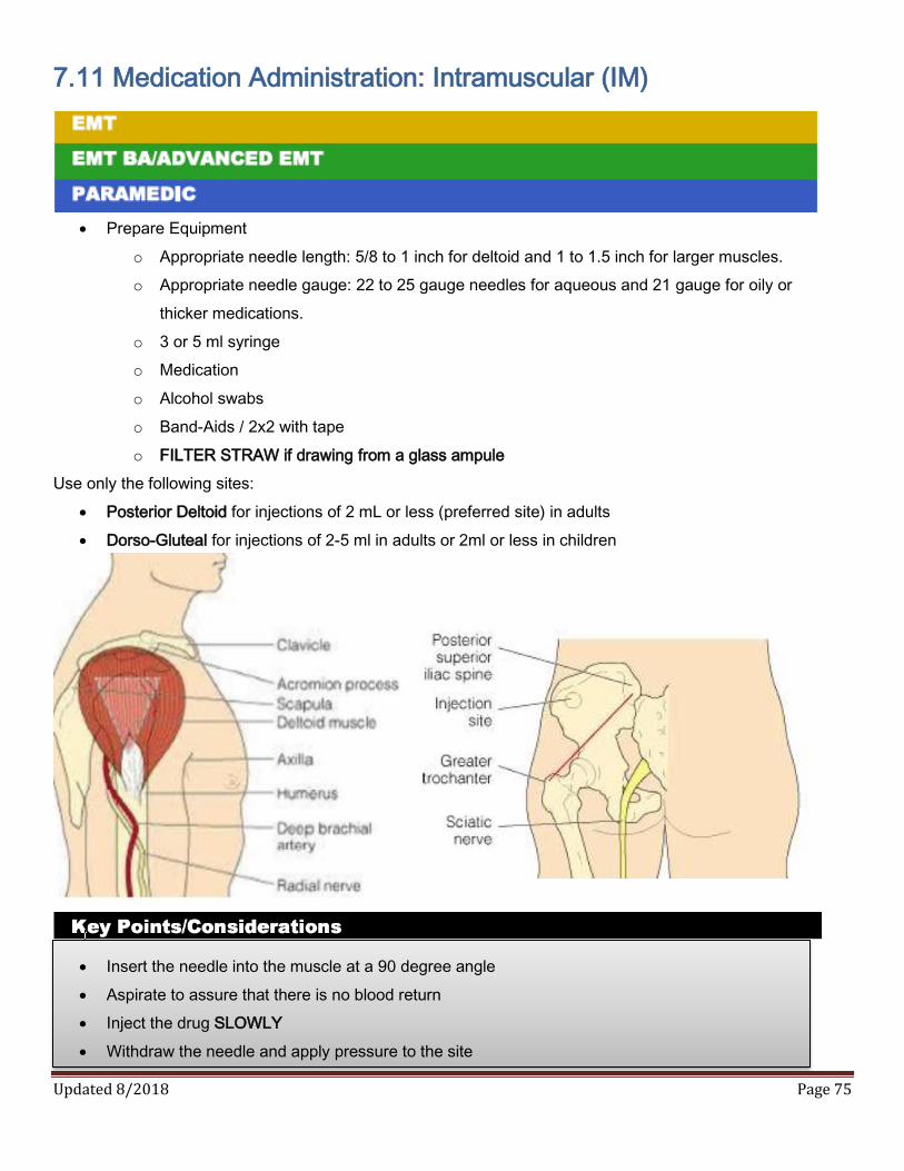
Updated 8/2018 Page 75
7.11 Medication Administration: Intramuscular (IM)
Prepare Equipment
o Appropriate needle length: 5/8 to 1 inch for deltoid and 1 to 1.5 inch for larger muscles.
o Appropriate needle gauge: 22 to 25 gauge needles for aqueous and 21 gauge for oily or
thicker medications.
o 3 or 5 ml syringe
o Medication
o Alcohol swabs
o Band-Aids / 2x2 with tape
o FILTER STRAW if drawing from a glass ampule
Use only the following sites:
Posterior Deltoid for injections of 2 mL or less (preferred site) in adults
Dorso-Gluteal for injections of 2-5 ml in adults or 2ml or less in children
Insert the needle into the muscle at a 90 degree angle
Aspirate to assure that there is no blood return
Inject the drug SLOWLY
Withdraw the needle and apply pressure to the site

Updated 8/2018 Page 76
Special Procedures 8.0
Updated 8/2018 Page 77
8.1 Special Procedures: Documentation
Standard procedure: After completion of each patient transport, the following information needs to be
documented. A copy of the run form needs to be left with or faxed to the Emergency Department. Please
send reports as soon as possible. The state reports to be sent to the hospital within 24 hours. A $500 fine
per day can be levied the department for failure to turn in run report
Chief complaint: The patient's major presenting problem.
History of present illness or injury (HPI): This should include events leading up to the chief
complaint, a description of the onset of the promlem and further explanation of the chief
complaint of presenting symptoms: Provocation, Quality, Radiation, Severity, and Time.
Past pertinent medical history: As it relates to the current problem: Symptoms, Allergies,
Medications, Pertinent medical history, Last oral intake and Events leading to the injury or
Illness.
Physical exam: Initial, rapid, focused and / detailed exams, including vital signs and EKG
interpretations. Documentation of findings should be complete and detailed to thoroughly and
objectively describe the condition of the patient. Be sure to include pertinent negatives and
time stamps for assessments such as vitals and 12-lead acquisitions.
Treatment and Response: All treatments must be documented chronologically to include Who,
what where when, number of attempts and patient response. Also be sure to document and
obtain proper refusals if a patient refuses a specific procedure. Document scene delays.
Transportation: You should document how you moved the patient from the scene to the
Ambulance. It is recommended that you document how the patient was secured to the stretcher,
when and how the hospital was contacted, any orders received or denied and changes in the patient’s
condition. Also document which room you placed the patient in and who you transferred care to.
Be sure to document any delays in transport either to the scene or to the hospital

Updated 8/2018 Page 78
8.2 Special Procedures: Informed Consent and Refusal of care
Conduct the following assessments:
o Legal competence
o Mental competence
o Medical or situational competence
Patient Incompetent:
o Treat and transport with “implied consent”
o Do not jeopardize safety of self or crew
o Call for law enforcement if needed
See 8.3 Special Procedures: Involuntary Restraint protocol
Who may sign for refusal?
o Patient of legal age who is competent and not a threat to themselves or others
o Parent
o Legal Guardian
o Medical Power of Attorney
Refusal Assessment and Documentation:
o Complete Assessment
o Obtain complete set of vital signs
o Explain risks and dangers
o Advise them to seek medical attention
o Complete patient refusal form
o Review form with signer
o Have patient sign form if possible
o Obtain witness signature (preferably not your partner)
o Complete Patient Care Report
o IF needed, contact medical control
Updated 8/2018 Page 79
8.3 Special Procedures: Involuntary Restraint
Indication: If the mental status is judged to be abnormal, prehospital personnel must carry out
treatment and transport in the patients’ best interest.
Prehospital personnel must ALWAYS CONSIDER THEIR OWN SAFETY FIRST!
Consider 2.3 General: Patient Agitation protocol
Procedure:
Attempt to verbally reassure the patient and seek their willing cooperation before committing to
restraint
If it is necessary to physically restrain a patient, perform the following:
o Prepare all the necessary equipment
o Always utilize police and/or fire personnel if possible.
o If available, have one person assigned to each extremity and one to hold equipment.
o Apply the restraints as loosely as possible to maintain a safe situation while preventing
neurovascular compromise and undue patient discomfort. Apply restraints over clothing
when possible.
o Never place restraints over a patient’s chest or on the abdomen of a pregnant patient.
o Perform routine and specific medical care as indicated by the patient’s condition.
o Routinely document the neurovascular status of the patient’s extremities distal to the restraints.
o Notify the receiving the hospital of the situation, and request security assistance upon arrival.
o Continue to attempt to verbally reassure the patient and seek their cooperation. Inform the
patient’s family of the reasons for the use of restraints.
o Thoroughly document the situation including the reasons for using restraints and how they
were applied.
o At no time will towels, washcloths or other devices be placed over the mouth and/or nose of a
restrained patient for any reason.
o Never restrain a patient in the prone position.
For reasons of medical safety, any patient who is under police hold and requires handcuffs and must have a
police officer accompany the patient in the back of the ambulance while enroute to the hospital or provide the
transporting EMS personnel with keys to the handcuffs.

Updated 8/2018 Page 80
8.4 Special Procedures: Ventricular Assist Device
ABC
Apply appropriate oxygen therapy
LVAD Functioning?
o Auscultate left upper abdominal quadrant. Continuous Humming sound = pump IS working
o Vital signs (must use a Doppler or Elemano device to obtain HR and BP)
Have AED available
If the pump has stopped for 5 minutes or more, assess patient:
o If patient hemodynamically unstable, re-establish power and contact the VAD coordinator
or ED physician immediately.
o If patient is hemodynamically stable, DO NOT re-establish power (a clot may have formed
in the pump) and contact the VAD Coordinator or ED physician immediately.
Continue to monitor.
Transport emergently to Emergency Department
12 lead ECG and transmit
Vascular access, with blood draw
Controller Alarming (red heart)
o Treat for cardiogenic shock per 1.5 Cardiac: Cardiogenic Shock protocol
Cardiac monitor
Go to appropriate protocol as needed
o Patient may be defibrillated/cardioverted and/or paced safely
Contact Medical Control as soon as possible so they are aware of this special patient
Compressions will likely result in dislodgement of the pump and are considered intervention of
last resort
The LVAD is a continuous flow device and you may or may not feel pulse
Transport caregiver and all equipment. (The caregiver is the expert on the device)
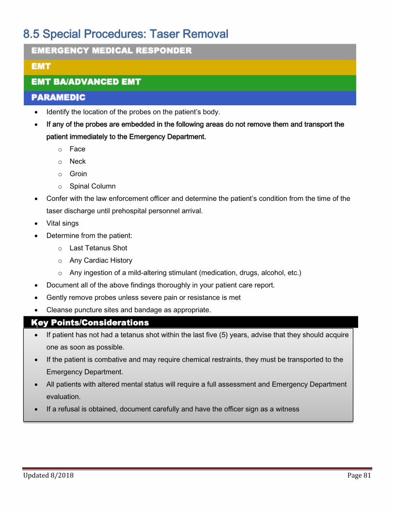
Updated 8/2018 Page 81
8.5 Special Procedures: Taser Removal
Identify the location of the probes on the patient’s body.
If any of the probes are embedded in the following areas do not remove them and transport the
patient immediately to the Emergency Department.
o Face
o Neck
o Groin
o Spinal Column
Confer with the law enforcement officer and determine the patient’s condition from the time of the
taser discharge until prehospital personnel arrival.
Vital sings
Determine from the patient:
o Last Tetanus Shot
o Any Cardiac History
o Any ingestion of a mild-altering stimulant (medication, drugs, alcohol, etc.)
Document all of the above findings thoroughly in your patient care report.
Gently remove probes unless severe pain or resistance is met
Cleanse puncture sites and bandage as appropriate.
If patient has not had a tetanus shot within the last five (5) years, advise that they should acquire
one as soon as possible.
If the patient is combative and may require chemical restraints, they must be transported to the
Emergency Department.
All patients with altered mental status will require a full assessment and Emergency Department
evaluation.
If a refusal is obtained, document carefully and have the officer sign as a witness

Updated 8/2018 Page 82
8.6 Special Procedures: Care of the Dialysis Patient
ABC
Consider appropriate oxygen therapy
Vitals
o DO NOT take a blood pressure in an extremity containing a graft or fistula.
Control bleeding with direct pressure
o DO NOT apply a tourniquet to an extremity with an AV fistula
Vascular access if needed
o Consider IO in full arrest or critical situations
o May utilize an extremity with an AV graft if unable to maintain establish or maintain IV/IO
Access in another location

Updated 8/2018 Page 83
8.7 Special Procedures: System Entry and Recertification
State of Indiana and National Registry Certifications/Licenses are valid for a period of 2 years. Didactic,
skill session, and audit and review hours must signed by the person responsible for the program.
For initial system entry, the candidate must:
Become familiar with Franciscan Health Hammond/Dyer system protocols
Complete a BLS protocol examination with a minimum score of 80%
Update their Acadis Portal and National Registry accounts to reflect their new agency alignment
Must have and maintain a valid AHA Healthcare Provider CPR certification
For recertification, the candidate must:
Maintain a valid AHA Healthcare Provider CPR certification
Maintain continuing education hours, audit hours and skills sessions as required by the State of
Indiana certifications division
The agency’s training officer is required to maintain recertification paperwork
For initial system entry, the candidate must do all of the above PLUS:
Complete a State Form 67 (Application for EMS Reciprocity and/or ALS Affiliation)
Provide a letter of good standing from the Medical Director of their previous system.
A medic transferring from another state must follow the appropriate procedures regarding temporary
licensures and National Registry testing procedures as outlined by the IDPH Certifications Division.
Complete the protocol test with a score of at least 80%.
Successfully perform a skills session with the EMS Medical Director, EMS Coordinator or authorized
educational staff
Complete adequate, 3rd person ride-time with a system medic who is currently in good standing. The
length of this preceptorship will be no less than 48 hours. These shifts are to be documented by the
preceptor.
Present current ACLS and PALS certifications
Updated 8/2018 Page 84
8.7 Special Procedures: System Entry and Recertification (Cont.)
For recertification, the candidate must:
Attend a minimum of 3 (75%) audit and review meetings a year. If this is not possible, the
Medical Direction Committee may determine a possible alternative.
Successfully complete (80%) the current ALS protocol test at least once every 2 years
Be in good standing with the system.
All continuing education hours must be approved by the EMS Coordinator. Hours that are
acquired elsewhere will be accepted on a case-by-case basis.
Hold a current ACLS, PALS certification.
All medics must acquire a signature by the system Coordinator and the System Medical
Director. Any falsification of documentation will be reported to the IDHS Certifications
Division.
A copy of all appropriate system documentation is to be maintained by the System
Coordinator
Updated 8/2018 Page 85
8.8 Special Procedures: Supply / Medication Replacement
General Supplies
Medics may utilize the Omnicell to replace the supplies used on the patient they transported
into the hospital
Medics may replace a reasonable cache of supplies and linen used on other patients
o Omnicell transactions will be closely monitored by the EMS Coordinator
To use the Omnicell
o Log in
o Choose “FLOOR STOCK SUPPLY” DO NOT add a patient
o Press the corresponding green button for every item you take
o Press exit
Non-controlled Medications
Non-controlled medications (including Etomidate) may be replaced by an ED nurse out of the ED
Pyxis as long as it was used on a patient
Expired medications must be replaced in the pharmacy
Controlled Medications
The unused medication must be wasted by an ED nurse who will then fill out the controlled
substance log which the medic will also sign
The run sheet must be taken to the pharmacy with the nurse’s signature confirming that the unused
portion was wasted
A non-controlled medication must be replaced by the hospital to which the patient was transported
A controlled substance taken to another hospital or medications not transported (i.e. Dextrose
Refusals, cardiac meds used on terminated resuscitation) must be replaced by supervising hospital
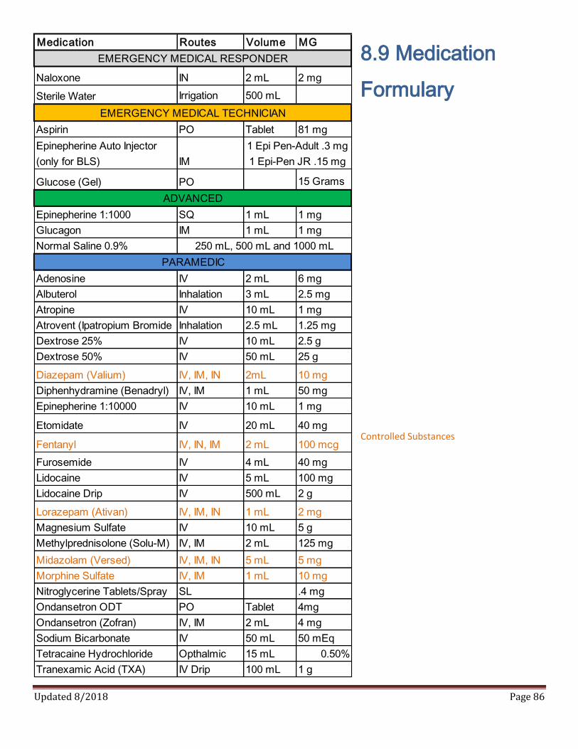
Updated 8/2018 Page 86
8.9 Medication
Formulary
Controlled Substances
Medication Routes Volume MG
Naloxone IN 2 mL 2 mg
Sterile Water Irrigation 500 mL
Aspirin PO Tablet 81 mg
Epinepherine Auto Injector
(only for BLS) IM
Glucose (Gel) PO 15 Grams
Epinepherine 1:1000 SQ 1 mL 1 mg
Glucagon IM 1 mL 1 mg
Normal Saline 0.9%
Adenosine IV 2 mL 6 mg
Albuterol Inhalation 3 mL 2.5 mg
Atropine IV 10 mL 1 mg
Atrovent (Ipatropium Bromide Inhalation 2.5 mL 1.25 mg
Dextrose 25% IV 10 mL 2.5 g
Dextrose 50% IV 50 mL 25 g
Diazepam (Valium) IV, IM, IN 2mL 10 mg
Diphenhydramine (Benadryl) IV, IM 1 mL 50 mg
Epinepherine 1:10000 IV 10 mL 1 mg
Etomidate IV 20 mL 40 mg
Fentanyl IV, IN, IM 2 mL 100 mcg
Furosemide IV 4 mL 40 mg
Lidocaine IV 5 mL 100 mg
Lidocaine Drip IV 500 mL 2 g
Lorazepam (Ativan) IV, IM, IN 1 mL 2 mg
Magnesium Sulfate IV 10 mL 5 g
Methylprednisolone (Solu-M) IV, IM 2 mL 125 mg
Midazolam (Versed) IV, IM, IN 5 mL 5 mg
Morphine Sulfate IV, IM 1 mL 10 mg
Nitroglycerine Tablets/Spray SL .4 mg
Ondansetron ODT PO Tablet 4mg
Ondansetron (Zofran) IV, IM 2 mL 4 mg
Sodium Bicarbonate IV 50 mL 50 mEq
Tetracaine Hydrochloride Opthalmic 15 mL 0.50%
Tranexamic Acid (TXA) IV Drip 100 mL 1 g
PARAMEDIC
EMERGENCY MEDICAL RESPONDER
EMERGENCY MEDICAL TECHNICIAN
1 Epi Pen-Adult .3 mg
1 Epi-Pen JR .15 mg
ADVANCED
250 mL, 500 mL and 1000 mL

Updated 8/2018 Page 87
8.10 Special Procedures: District 1 Diversion Guidelines (copied)
I. Purpose
To establish guidelines under which Hospital Emergency Departments divert ambulance patients when it has
been determined, through pre-established criteria, that the hospital is unable to accommodate additional
patients.
To define procedures for communicating changes in diversion status.
To establish guidelines for ambulance provider operations when a Hospital is on diversion.
To define exceptions to the Diversion Guidelines when hospital(s) follow procedures as outlined herein.
II. DEFINITIONS
A. Diversion: Is an alert from an overwhelmed hospital to EMS Providers to consider the possible diversion of incoming
ambulance patients to the next closest hospital. This request should be reasonably honored as a COURTESY in order to
provide better care for our patients. Hospital resources, including emergency services may occasionally be
overwhelmed. The hospital, based on its own criteria, may designate itself as being on “diversion or bypass”.
EMS Guidelines require that unstable patients be transported to the nearest facility regardless of diversion status.
Unstable Patient:
Unable to maintain an airway
Unable to ventilate
Unremitting shock
Any patient presenting with signs and/or symptoms of acute cardiac syndrome or stroke
As otherwise defined in appropriate EMS System protocols, (including as determined by medical control
contact).
If in the EMS personnel’s judgment in consultation with on-line medical direction, a patient’s condition may be
jeopardized by bypassing a facility on diversion, a unit may override the hospital’s request for diversion for emergency
stabilization of a patient. Any diversion override should be documented.
B. Total Diversion: When a hospital determines through pre-established criteria, that the Emergency Department is
unable to provide care to additional ambulance patients. Resources of the ED/Hospital are overwhelmed and they are
alerting pre-hospital providers to divert patients to the next closest facilities, if possible. Hospital must still accept
unstable patients, if transport to the next closest facility jeopardizes the patient’s condition.
C. Hospital Disaster Diversion: Request to divert ALL patients to the next closest facility due to the hospital/ED being
unable to take any patients due to a disaster situation. This diversion is only used in cases (quarantine, damage to the
facility, etc.) that require the hospital to call an external or internal disaster.
a. Overwhelming is not a valid cause for DISASTER DIVERSION.
b. An internal or external disaster occurs which compromises patient safety.
D. Specialty Diversion: This is an informational diversion based on equipment that is not available at the ED/hospital.
Examples include, but are not limited to CT and MRI. The hospital MUST identify the specialty area on diversion when
giving EMS notification with definition if needed.

Updated 8/2018 Page 88
8.10 Special Procedures: District 1 Diversion Guidelines (continued)
III. POLICY
This policy shall not override or interfere with each facility’s Emergency Preparedness Policies.
The decision to initiate diversion shall be made according to each facility’s policies and procedures.
A record will be kept indicating the reasons for diversion according to hospital criteria. This record, including
times of notification and termination of diversion, will be kept by the hospital. The record should include
documentation of communication of diversion status to the Local 911 Centers, EMS Services, and Hospitals.
Once diversion in initiated, the criteria and status for continued diversion will be reviewed no less frequently
than every four hours.
Unstable patients are to be transported to the closest hospital unless that hospital is on Hospital Disaster
Diversion.
If ALL geographically contiguous hospitals (catchment area) go on diversion, they will automatically negate each
other relative to their shared provider area.
Diversion status may NOT be honored if transport time by an ambulance to the next closest hospital jeopardizes
patient care. EMS personnel will determine most appropriate facility based on the patient’s best interest.
If a patient adamantly requests a hospital on bypass, they must sign a refusal to accept hospital diversion form
DIVERSION COMMUNCIATION PROCEDURE
Hospitals and EMS shall follow the procedures outlined below whenever Hospital diversion status changes are deemed
necessary.
A. Communication Procedure
1. The Hospital shall notify all Local 911 Centers, EMS Services, Hospitals, and other stakeholders of any diversion status
changes every 4 hours.
V. SUSPENSION OF DIVERSION
Hospitals may determine that continuation of diversion might result in a danger to public health and safety. In
consultation with other hospital administrations, the administrators may suspend diversion and require all hospitals to
accept both critical and non-critical patients. This step would be taken only after consultation with the hospital
administrators or designee and only if it was determined to represent an imminent danger to public health and safety.
VI. QUALITY ASSURANCE AND RECORD KEEPING
A. Problems related to the implementation of this policy shall be reported to the Franciscan Health Hammond EMS
Coordinator

Updated 8/2018 Page 89
Intra-Facility Transports 9.0
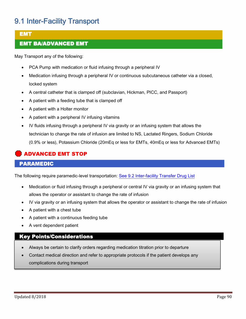
Updated 8/2018 Page 90
9.1 Inter-Facility Transport
May Transport any of the following:
PCA Pump with medication or fluid infusing through a peripheral IV
Medication infusing through a peripheral IV or continuous subcutaneous catheter via a closed,
locked system
A central catheter that is clamped off (subclavian, Hickman, PICC, and Passport)
A patient with a feeding tube that is clamped off
A patient with a Holter monitor
A patient with a peripheral IV infusing vitamins
IV fluids infusing through a peripheral IV via gravity or an infusing system that allows the
technician to change the rate of infusion are limited to NS, Lactated Ringers, Sodium Chloride
(0.9% or less), Potassium Chloride (20mEq or less for EMTs, 40mEq or less for Advanced EMTs)
The following require paramedic-level transportation: See 9.2 Inter-facility Transfer Drug List
Medication or fluid infusing through a peripheral or central IV via gravity or an infusing system that
allows the operator or assistant to change the rate of infusion
IV via gravity or an infusing system that allows the operator or assistant to change the rate of infusion
A patient with a chest tube
A patient with a continuous feeding tube
A vent dependent patient
Always be certain to clarify orders regarding medication titration prior to departure
Contact medical direction and refer to appropriate protocols if the patient develops any
complications during transport
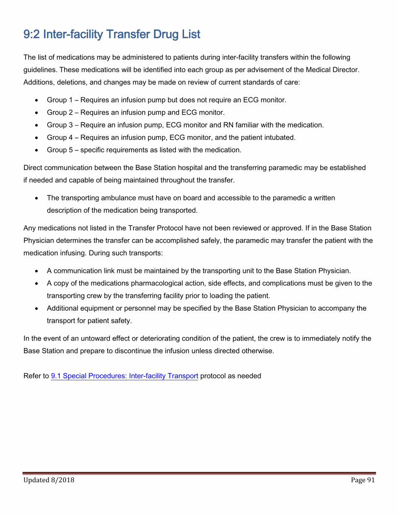
Updated 8/2018 Page 91
9:2 Inter-facility Transfer Drug List
The list of medications may be administered to patients during inter-facility transfers within the following
guidelines. These medications will be identified into each group as per advisement of the Medical Director.
Additions, deletions, and changes may be made on review of current standards of care:
Group 1 – Requires an infusion pump but does not require an ECG monitor.
Group 2 – Requires an infusion pump and ECG monitor.
Group 3 – Require an infusion pump, ECG monitor and RN familiar with the medication.
Group 4 – Requires an infusion pump, ECG monitor, and the patient intubated.
Group 5 – specific requirements as listed with the medication.
Direct communication between the Base Station hospital and the transferring paramedic may be established
if needed and capable of being maintained throughout the transfer.
The transporting ambulance must have on board and accessible to the paramedic a written
description of the medication being transported.
Any medications not listed in the Transfer Protocol have not been reviewed or approved. If in the Base Station
Physician determines the transfer can be accomplished safely, the paramedic may transfer the patient with the
medication infusing. During such transports:
A communication link must be maintained by the transporting unit to the Base Station Physician.
A copy of the medications pharmacological action, side effects, and complications must be given to the
transporting crew by the transferring facility prior to loading the patient.
Additional equipment or personnel may be specified by the Base Station Physician to accompany the
transport for patient safety.
In the event of an untoward effect or deteriorating condition of the patient, the crew is to immediately notify the
Base Station and prepare to discontinue the infusion unless directed otherwise.
Refer to 9.1 Special Procedures: Inter-facility Transport protocol as needed

Updated 8/2018 Page 92
9:2 Inter-facility Transfer Drug List (Continued)
The following medications require No Special requirements during ALS Transport:
5W0.9% Sodium Chloride
D5/0.2% Sodium Chloride
Vitamins
Antibiotics
Cortisone
Lactated Ringers
Plasmanate
Morphine ( self contained pump)
Group 1 – Infusion Pump
5% Albumin
10% Dextrose infusion
Acetylcysteine (Mucomyst)
Heparin
Hetastarch (Hespan)
Hyperalimentation
Octretide Acetate (Sandostatin)
Pantoprazole Sodium (Protonix)
Potassium chloride less than 40 mEq/L
Regular Insulin infusions
Group 2 –Infusion pump + ECG monitor
Amiodarone (Cordarone)
Amrinone (Inocor)
Blood/Blood products
Diltiazem (Cardizem)
Dobutamine (Dobutrex)
Fentanyl
Isoproterenol (Isuprel)
Lidocaine (Xylocaine)
Magnesium Sulfate [Cardiac patient]
Mannitol (Osmitrol)
Methylpredisolone (Solumedrol)
Milrinone Lactate (Primacor)

Updated 8/2018 Page 93
9:2 Inter-facility Transfer Drug List (Continued)
Group 2 –Infusion pump + ECG monitor (continued)
Nesiriteride (Natrecor)
Morphine Sulfate
Nitroprusside (Nipride)
Abciximab (ReoPro)
Eptifibatide (Integrilin)
Tirofiban (Aggrastat)
Potassium Chloride >or = 40 mEq/L
Procainamide (Pronestyl)
Vasopressin (Pitressin)
Group 3 – Infusion pump + ECG monitor + RN familiar with the medication
Alteplase tPA (Activase)
Cardene
Dopamine (Intropin)
Epinephrine Infusion (Adrenalin)
Esmolol (Brevibloc)
Fendolopam Mesylate (Corlopam)
Fibrinolytic infusion
Labetolol trandate (Normodyne)
Nitroglycerine (Tridil)
Norepinephrine (Levophed)
Urokinase (Abbokinase)
Phenylephrine (Neosynephrine)
Group 4 –Infusion pump + ECG monitor + RN + the patient must be intubated.
Midazolam infusion (Versed)
Pentobarbital infusion (Nembutal)
Propofol (Diprivan)
Skeletal muscle paralyzers
Cisatracurium Besylate (Nimbex)
Pancuronium Bromide (Pavulon)
Vencuronium Bromide (Nocuron)
Group 5 – Specific requirements as listed. Infusion Pump + Fetal Monitor
oxytocin (Pitocin) Magnesium Sulfate infusion (for maternity patient)

Updated 8/2018 Page 94
9:3 Stroke Transfer with tPA
Purpose:
To provide guidance for safe transport of patients who are receiving or have received intravenous tissue
plasminogen activator (tPA) for treatment of ischemic stroke. Transport must be done in a safe, smooth, and
expedited manner, making sure the patient remains in a stable and comfortable position during transport.
History & Physical Exam:
Perform and document initial neurological exam
Perform and document vital signs prior to transport. If SBP >180 or DBP > 105 discuss treatment of
Hypertension with sending hospital prior to transport and obtain necessary medications.
Treatment:
Oxygen to maintain pulse oximetry > 95%
Cardiac Monitor – must be placed on the patient prior to transport.
If patient becomes hypoglycemic, refer to 2.6 Medical: Diabetic Emergencies protocol. Initially, it is
preferable to use the blood glucose measurement obtained by the transferring hospital in order to
avoid unnecessary delay.
o Reminder patient must remain NPO. No oral glucose can be given to patient.
Establish or maintain IV access.
Patient must remain NPO, this includes any medications
The Paramedic is NOT authorized to give tPA bolus, but IS authorized to maintain the tPA infusion.
tPA may only be given if ordered and started at the sending facility.
Once tPA has completed, Normal Saline Solution should be infused at a TKO rate for the remainder
of the transport.
Monitor and document neurological exam every 10 minutes. If patient develops worsened neurologic
condition or if patient develops severe headache, acute hypertension, difficulty breathing, evidence of
allergic reaction, or major bleeding then STOP the tPA infusion (if still infusing) and contact medical
direction.
Monitor and document vital signs every 10 minutes. If antihypertensive medication (Labetalol,
Nicardipine, Metoprolol) are started or ordered at the sending facility, they may be continued for SBP >
180 or DBP > 105.
Notify receiving hospital of any changes that occur while transporting.
Updated 8/2018 Page 95
Credits / References
Medication Formularies, Inter facility Drug List
Meghan Jordan, PharmD, BCPS
o Emergency Medicine Clinical Pharmacist, Franciscan Health Dyer ED
Glenn Allen, PharmD, BCPS
o Emergency Medicine Clinical Specialist, Franciscan Health Hammond ED
https://www.fda.gov/downloads/Drugs/DrugSafety/InformationbyDrugClass/UCM562901.pdf
Overall Protocol Format
Elkhart County EMS System Protocols
Inter-Facility Transport Protocols
Tom Fentress, MBA, NRP, PI, CFI, Gary Methodist EMS System Protocols
John Munier, NRP, Regional Operations Manager, Midwest Medical Transport
Christopher Bollinger, EMT, Station Manager, Midwest Medical Transport
Delineation of Scope of Practice and General Consultation
Mikel Fort, EMS District Manager, IN Department of Homeland Security
https://www.in.gov/dhs/files/EMS%20Scope%20of%20Practice%202017.pdf
Helicopter Landing Zones
Jeff Kochis, Assistant Chief, Hammond Fire Department
David Hay, EMS Chief, Dyer Fire Department
Trauma Protocols
https://www.in.gov/dhs/files/Indiana_Trauma_Field_Triage_and_Transport_Destination_Protocol.pdf
45 Minute Trauma Center Coverage Maps
Maptitude 2016, Caliper Corporation
Unfortunately, the above list does not contain the large number of supervisory and front-line staff from
Franciscan Health, the Hammond Fire Department, the Dyer Fire Department and Midwest Medical Transport
who offered valuable recommendations and insight throughout the years which were used in the development
of these protocols. Thank you all for your input.
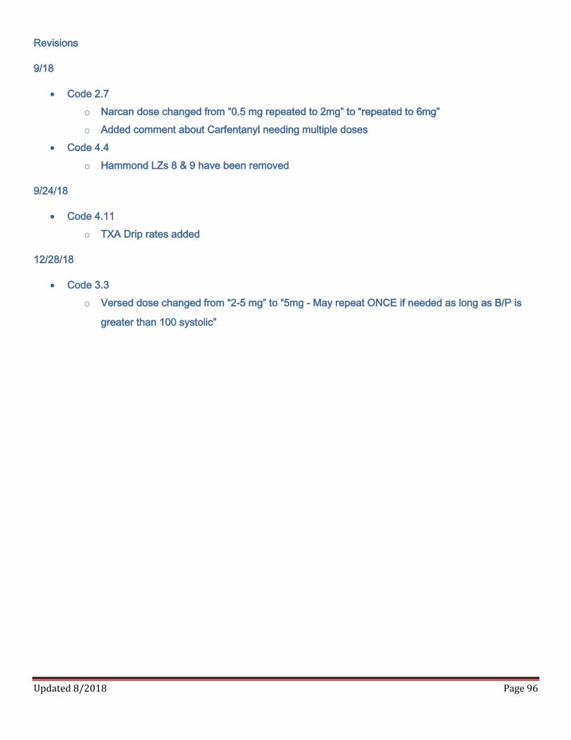
Updated 8/2018 Page 96
Revisions
9/18
Code 2.7
o Narcan dose changed from “0.5 mg repeated to 2mg” to “repeated to 6mg”
o Added comment about Carfentanyl needing multiple doses
Code 4.4
o Hammond LZs 8 & 9 have been removed
9/24/18
Code 4.11
o TXA Drip rates added
12/28/18
Code 3.3
o Versed dose changed from “2-5 mg” to “5mg - May repeat ONCE if needed as long as B/P is
greater than 100 systolic”
Recommended