RESEARCH Open Access
Effects of Aedes aegypti salivary components ondendritic cell and lymphocyte biologyBruna Bizzarro1, Michele S Barros1, Ceres Maciel1, Daniele I Gueroni1, Ciro N Lino1, Júlia Campopiano2,Michalis Kotsyfakis3, Gustavo P Amarante-Mendes2,4, Eric Calvo5, Margareth L Capurro6,7 and Anderson Sá-Nunes1,7*
Abstract
Background: Saliva is a key element of interaction between hematophagous mosquitoes and their vertebratehosts. In addition to allowing a successful blood meal by neutralizing or delaying hemostatic responses, the salivarycocktail is also able to modulate the effector mechanisms of host immune responses facilitating, in turn, thetransmission of several types of microorganisms. Understanding how the mosquito uses its salivary componentsto circumvent host immunity might help to clarify the mechanisms of transmission of such pathogens anddisease establishment.
Methods: Flow cytometry was used to evaluate if increasing concentrations of A. aegypti salivary gland extract(SGE) affects bone marrow-derived DC differentiation and maturation. Lymphocyte proliferation in the presenceof SGE was estimated by a colorimetric assay. Western blot and Annexin V staining assays were used to assessapoptosis in these cells. Naïve and memory cells from mosquito-bite exposed mice or OVA-immunized mice andtheir respective controls were analyzed by flow cytometry.
Results: Concentration-response curves were employed to evaluate A. aegypti SGE effects on DC and lymphocytebiology. DCs differentiation from bone marrow precursors, their maturation and function were not directly affected byA. aegypti SGE (concentrations ranging from 2.5 to 40 μg/mL). On the other hand, lymphocytes were very sensitive tothe salivary components and died in the presence of A. aegypti SGE, even at concentrations as low as 0.1 μg/mL. Inaddition, A. aegypti SGE was shown to induce apoptosis in all lymphocyte populations evaluated (CD4+ and CD8+ Tcells, and B cells) through a mechanism involving caspase-3 and caspase-8, but not Bim. By using different approachesto generate memory cells, we were able to verify that these cells are resistant to SGE effects.
Conclusion: Our results show that lymphocytes, and not DCs, are the primary target of A. aegypti salivarycomponents. In the presence of A. aegypti SGE, naïve lymphocyte populations die by apoptosis in a caspase-3- andcaspase-8-dependent pathway, while memory cells are selectively more resistant to its effects. The present workcontributes to elucidate the activities of A. aegypti salivary molecules on the antigen presenting cell-lymphocyteaxis and in the biology of these cells.
Keywords: Dendritic cells, T cells, Aedes aegypti, Saliva, Apoptosis
* Correspondence: [email protected]ório de Imunologia Experimental, Departamento de Imunologia,Instituto de Ciências Biomédicas, Universidade de São Paulo, São Paulo05508-900, SP, Brazil7Instituto Nacional de Ciência e Tecnologia em Entomologia Molecular,Conselho Nacional de Desenvolvimento Científico e Tecnológico(INCT-EM/CNPq), Rio de Janeiro, BrazilFull list of author information is available at the end of the article
© 2013 Bizzarro et al.; licensee BioMed Central Ltd. This is an Open Access article distributed under the terms of the CreativeCommons Attribution License (http://creativecommons.org/licenses/by/2.0), which permits unrestricted use, distribution, andreproduction in any medium, provided the original work is properly cited. The Creative Commons Public Domain Dedicationwaiver (http://creativecommons.org/publicdomain/zero/1.0/) applies to the data made available in this article, unless otherwisestated.
Bizzarro et al. Parasites & Vectors 2013, 6:329http://www.parasitesandvectors.com/content/6/1/329

BackgroundMosquitoes are the most important vectors of humanpathogens, transmitting a wide number of emerging andre-emerging diseases. In particular, Aedes aegypti mos-quitoes are the primary vectors of yellow fever, denguefever and Chikungunya fever [1-4]. The key interactionelement between A. aegypti and its vertebrate host is themosquito saliva and a successful blood meal is achievedby the action of salivary anti-hemostatic and immuno-modulatory molecules present in this pharmacologicalcocktail. The former are responsible for anticoagulant,anti-platelet aggregation and vasodilatory activities [5,6],while the latter is thought to modulate immune functions,which in turn, facilitates pathogen transmission. Indeed,a growing number of recent pieces of evidence havedemonstrated that A. aegypti salivary components increaseviral infection in vitro and in vivo [7-12] and an increasedreactivity with mosquito salivary proteins is observedin sera from infected individuals, suggesting a positivecorrelation between mosquito exposure and risk ofinfection [13,14].
As a result of their strategic location in the skin, den-dritic cells (DCs) interact directly with the mosquitosalivary components during and after the blood meal.They are also among the first cells to encounter pathogenstransmitted by these vectors and initiate the adaptiveimmune response against them. Following this initialcontact, DCs mature and migrate to the draining lymphnodes, becoming effective stimulators of T cell responses[15]. Despite DCs essential role in connecting innateand adaptive immune responses, very little is knownabout the effects of A. aegypti salivary components onthese cells. A previous study has demonstrated that A.aegypti salivary gland extract (SGE) does not affect theviability or IL-12 production by a fetal skin-derived DCline (FSDC) [16]. Therefore, A. aegypti SGE has noeffect on the basal expression of IFN-β by DCs, but itdecreases the production of this cytokine in the presenceof West Nile Virus infection [9]. In addition to its putativeeffects on DCs, A. aegypti SGE was shown to affect theproliferation of murine lymphocytes in vitro [16-18] andinhibit the production of proinflammatory (GM-CSF andTNF-α) and Th1 cytokines (IL-2 and IFN-γ), but modesteffects were observed on Th2 cytokines levels (IL-4 andIL-5) [16]. On the other hand, it has been shown thatthe salivary protein SAAG-4 induces CD4+ T cells toexpress IL-4 [19]. Accordingly, splenocytes from micepreviously exposed to A. aegypti bites produced higherlevels of IL-4 and IL-10 and decreased IFN-γ production[20]. Additionally, recent literature has demonstratedan important functional relationship between coagulationand immunity [21-23] and, in fact, some of the salivaryanti-hemostatic molecules described in hematophagousarthropods are also involved in the modulation of host
inflammation and immune responses through differentmechanisms and pathways [20,24-26].
However, despite the effects described above and themosquitoes relevance as disease vectors, the immuno-modulatory activities of A. aegypti saliva on the antigenpresenting cell-lymphocyte axis is still very limited. Inthe current study, we examined the activity of A. aegyptiSGE on several parameters of DC and lymphocyte biology.Employing murine cells, we demonstrated that modulationof DC maturation, differentiation or function does notseem to be a priority for A. aegypti salivary components.Conversely, direct inhibition of naïve T cell proliferationcaused by apoptosis is already achieved with low amountsof A. aegypti SGE, through a mechanism involving cleavageof pro-caspase-3 and pro-caspase-8, but not the proa-poptotic Bcl-2 homolog Bim. Interestingly, memory cellsgenerated by different approaches are selectively resistantto this activity.
MethodsMiceAll the experiments were carried out in accordance withinternationally recognized guidelines and approved bythe Animal Care of the Institute of Biomedical Sciencesof University of São Paulo (CEUA-ICB/USP) and underthe protocol number 91/2009. Female BALB/c, C57BL/6,DO11.10 (expressing a TCR transgenic for the sequenceof OVA 323–339), Bim+/− and Bim−/− (a proapoptoticmember of the Bcl-2 family) mice at 6–16 weeks oldwere bred and maintained at the Department of Immun-ology, ICB/USP, Brazil.
Mosquitoes and sandfliesMale and female A. aegypti and Anopheles aquasalismosquitoes were reared in an insectary at the Departmentof Parasitology, ICB/USP, Brazil. Temperature was main-tained at 26°C, 80% humidity and a 12/12-h photoperiod.Larvae were fed on powdered fish food and adult mos-quitoes were given continuous access to a 10% sucrosesolution. Salivary glands from Phlebotomus duboscqisandflies were kindly provided by Dr. Jesus G. Valenzuela,from the Vector Molecular Biology Section, Laboratoryof Malaria and Vector Research, National Institutes ofAllergy and Infectious Diseases, National Institutes ofHealth (LMVR/NIAID/NIH, USA).
Salivary gland extractsFemale mosquitoes aged 4–6 days had their surfacesterilized by brief immersion in 70% ethanol, prior todissection. The salivary glands were dissected in PBSand transferred to a microtube containing 50 μL of coldPBS. Salivary glands of female P. duboscqi were collectedunder the same conditions described above. The tubescontaining salivary glands were sonicated to release the
Bizzarro et al. Parasites & Vectors 2013, 6:329 Page 2 of 13http://www.parasitesandvectors.com/content/6/1/329

soluble material and centrifuged at 14,000 g for 10minutes to remove particulate material. The resultingsupernatant, referred to as SGE, was collected and ster-ilized by passage through a nitrocellulose membranefilter with 0.2 μm pores (Millipore, Carrigtwohill, CountyCork, Ireland). Protein concentration was determined byNanoDrop 2000 (Thermo Fisher Scientific, Wilmington,DE, USA) and aliquots were stored at −80°C until use.
Differentiation and maturation of bone marrow-derivedDCs (BMDCs)BMDCs were employed as a model to study DC biology.Bone marrow cells collected from the femurs of micewere cultured in complete medium (RPMI 1640 mediumwith 10% heat-inactivated FBS, 2 mM L-glutamine,100 U/mL penicillin, 100 μg/mL streptomycin, 25 mMHepes, 2.5 × 10⁵ M 2-mercaptoethanol) and 20 ng/mLof murine GM-CSF to induce DC differentiation[24,27]. In order to define if the salivary componentswould affect the differentiation process, cells wereincubated (37°C and 5% CO2) in aliquots of 1 mL insterile 24 well plates in the presence of medium only or A.aegypti SGE at 2.5, 5, 10, 20 and 40 μg/mL concentrations(6 replicates per group). After 4 days of incubation,non-adherent cells from three replicates of each groupwere collected by washing with complete medium. Onthe three remaining replicates, half the volume wasremoved and replaced by an equal volume of completemedium again containing 40 ng/mL GM-CSF in thepresence or absence of the same concentrations ofSGE. At 7 days of incubation, non-adherent cells of theremaining three replicates were collected as describedabove. In both cases, cells were counted and differentiationof bone marrow cells into DCs in different culturetimes was assessed by flow cytometry to evaluate theexpression of CD11b and CD11c surface markers.
For the maturation assays, BMDCs were obtained anddifferentiated for 6 days with GM-CSF as describedabove. Non-adherent cells were collected, resuspendedat 106 cells/mL and distributed into sterile 24 wellplates in aliquots of 1 mL per well. These cells wereincubated overnight with medium only or mediumcontaining SGE. Then, cells were stimulated withultrapure LPS (100 ng/mL - final concentration) for 24h to promote their maturation and then labeled withantibodies to MHC class II, CD40, CD80 and CD86[24,27].
Antigen presentation by DCsIn order to evaluate the antigen presentation by BMDCs,non-adherent cells were collected at 7 days of incubationand CD11c+ cells were purified using magnetic MACScolumns (Miltenyi Biotec Inc., Auburn, CA, USA). In brief,BMDC suspensions were incubated with anti-CD11c
MicroBeads (Miltenyi Biotec Inc.) for 15 min at 4°C,followed by a PBS wash, and then sorted using MACScolumns (MS). Analysis of the sorted cells showed purityof 80–90% CD11c+ cells (data not shown). CD11c+ cellswere preincubated overnight (37°C and 5% CO2) withmedium or SGE at various concentrations (2.5, 5, 10,20 and 40 μg/mL). After this period, cells were incubatedfor 4 h in the presence of LPS (100 ng/mL) plus OVA(100 μg/mL). After three washes, 2.5 × 10⁵ cells/mLwere distributed in aliquots of 100 μL per well into a96-well plate. CD4+ T lymphocytes from DO11.10 mice(which express a specific transgenic TCR for the OVApeptide 323–339) were purified using magnetic MACScolumns. A suspension containing 106 cells/mL was pre-pared and 100 μL were added to wells on the culture plate(DC:lymphocyte ratio 1:4) and maintained at 37°C and 5%CO2 for 72 h.
Similar experiments were performed, but after DCwashing, A. aegypti SGE was added again in the culture,followed by addition of CD4+ T cells from DO11.10 mice(that express a specific transgenic TCR for the OVApeptide 323–339). Cells were stimulated with OVA (100μg/mL) plus LPS (100 ng/mL) or Con A (0.5 μg/mL). Inthe last 24 h of the culture, 25 μL of 0.01% resazurinwere added to all wells. Cell proliferation was evaluatedby reading the culture absorbance at 570 and 600 nm,and results are expressed as the difference between thosereadings as previously described [24,27,28].
Spleen cell proliferationFollowing euthanasia, spleens of BALB/c mice wereaseptically removed and a cell suspension containing106 cells/mL was prepared. Cells were then dividedinto aliquots of 100 μL/well in 96-well plates. Next,50 μl of A. aegypti, An. aquasalis or P. duboscqi atvarious concentrations were added in the culture andincubated for 30 min. After pre-incubation, 50 μL ofCon A was added to each well (0.5 μg/mL – final con-centration), and cultures were incubated for 72 h at 37°Cand 5% CO2. Proliferation was evaluated as described[24,27,28].
ApoptosisTo assess the proportion of viable cells and ‘debris’, spleencells were incubated in the presence of A. aegypti SGE(final concentration 10 μg/mL) or medium for 72 h. Afterthis period, the percentage of viable cells was evaluatedby flow cytometry using the forward scatter (cell size)and side scatter (granularity) parameters and comparedto fresh cells.
For apoptosis quantification, spleen cells were incubatedin the presence of A. aegypti SGE or medium for 4, 8, 24and 72 h. Cells were labeled with lymphocyte markers(CD4, CD8 and CD19) for subpopulation analysis. Cells
Bizzarro et al. Parasites & Vectors 2013, 6:329 Page 3 of 13http://www.parasitesandvectors.com/content/6/1/329
were then washed twice with PBS and resuspendedwith 500 μL of Annexin-binding buffer (0.1 M Hepes,1.4 M NaCl, 25 mM CaCl2). Subsequently, 5 μL ofAnnexin V-FITC was added to the cell suspension,which was then incubated in the dark for 10 min at roomtemperature. Cells were evaluated by flow cytometryand apoptosis was measured by the percentage ofAnnexin V+ cells.
For Western blot analysis, spleen cells from BALB/cmice were prepared as described previously. Cell sus-pensions containing 20 × 106 cells/ml were incubatedwith medium only or in the presence of Ae. aegyptiSGE (final concentration: 10 μg/mL) and stimulatedwith Con A (final concentration 0.5 μg/mL) for 4 h at37°C and 5% CO2. Cells were then collected, washedtwice with PBS and lysed with 100 μL of RIPA buffer(150 mM NaCl, 1% NP40, 0.1% SDS, 50 mM Tris;pH 8.0) supplemented with 1% of protease inhibitor(Sigma- Aldrich, St. Louis, MO, USA). The lysatesupernatant was collected after 10 minutes incubation,centrifuged at 14000 g for 5 minutes and the proteinconcentration was determined using BCA Protein AssayKit according to the manufacturer's instructions (ThermoFisher Scientific, Rockford, IL, USA).
The equivalent of 30 μg protein from each samplewere diluted v/v in Laemmli buffer (2% SDS, 10%glycerol, 1% β -mercaptoethanol, 0.02% bromophenolblue, 0.4 M Tris), heated to 100°C for 5 minutes andseparated by electrophoresis in a 12% Sodium DodecylSulfate (SDS) polyacrylamide gel under a constant currentof 20 mA. The separated proteins were transferred to anitrocellulose membrane which was then blocked for1 hour with 5% FBS diluted in Tris buffer containing 0.1%Tween −20 (TBST). Membranes were washed 3 times withTBST (10 minutes per wash) and incubated overnight at4°C with the following rabbit monoclonal antibodies: anti-pro-caspase-3 (1:500) and anti-pro-caspase-8 (1:1,000),both from Cell Signaling Technology, Inc. (Danvers,MA, USA). After further washing, the membranes wereincubated for 1 hour at room temperature with anti-rabbit secondary antibodies (1:2,000) conjugated withhorseradish peroxidase for detection (Dako, Glostrup,Denmark). Immunoreactive bands were stained using thechemiluminescent ECL Detection Kit (Thermo FisherScientific) and visualized in a photodocumentation sys-tem. The membranes were then washed and incubatedovernight with anti β-actin (1:10,000), followed by an-other washing and subsequent incubation with a anti-mouse secondary antibody (1:60,000). The density ofthe bands was analyzed using ImageJ software(available free at http://rsb.info.nih.gov/nih-image).The values were normalized by the total of β-actinpresent in each sample and expressed as arbitraryunits.
Mice sensitization by mosquito biteMale and female mosquitoes A. aegypti and An. aquasaliswere kept in plastic containers of about 12 cm diametercovered with a fine mesh screen. Mice were anesthetizedand placed on the screen for 20–30 min so that femalemosquitoes had direct contact with the animal’s skin forenough time to complete the blood meal, as previouslystandardized (data not published). This whole procedurewas performed four times at 15 day intervals, usingapproximately 50 female mosquitoes per mice and theproportion of mosquitoes fed at each sensitization wastypically ≥ 80%. Mice from the control group were alsoanesthetized, but had no contact with mosquitoes. Fif-teen days after the last sensitization, proliferation wasperformed as described above.
Adoptive transfer and mice immunizationSpleen cells from DO11.10 mice were prepared in com-plete medium and incubated at 37°C and 5% CO2 for 2 hin Petri dishes. Non-adherent cells were collected and thecell suspension was prepared in PBS containing 1% fetalbovine serum. These cells were inoculated i.v. to BALB/cmice in a volume of 200 μL containing 3 × 10⁶ cells. After24 h, these mice were immunized subcutaneously at twosites of the dorsal region with 100 μL of an emulsioncontaining OVA and complete Freund's adjuvant v/v(40 μg of OVA per animal). After 7 days, spleens cells ofcontrol and immunized animals were collected and cellsuspensions (106 cells/mL) prepared. In vitro re-stimulationassays were performed with cells pre-incubated withmedium alone or SGE for 30 minutes followed bystimulation with OVA (100 μg/mL) or Con A (0.5 μg/mL). The proliferation of these cells was assessed asdescribed above. Naïve and memory phenotyping wasperformed in CD4+ T cells by flow cytometry as fol-lows: CD62LHIGH/CD44LOW (naïve cells), CD62LHIGH/CD44HIGH (TCM cells), and CD62LLOW/CD44HIGH
(TEM cells).
Flow cytometryCells were prepared for flow cytometry as previouslydescribed [24,27]. Fluorochrome-conjugated monoclonalantibodies against the following molecules were employed:CD4-APC, CD19-PE, CD11b-FITC, CD11c-PE, CD40-FITC, CD80-PerCP-Cy5.5, CD86-PE-Cy7 (BD Biosciences,San Jose, CA); CD8-PerCP, Annexin-FITC, I-A/I-E-APC,CD44-Pacific Blue, CD62L-PE (Biolegend, San Diego,CA, USA). Cells were acquired in a FACSCanto II (BDBiosciences) and analysis was performed using FlowJosoftware, version 7.5.5 (Tree Star, Ashland, OR, USA).
Statistical analysisStatistical analysis of differences between means of ex-perimental groups was performed using Student’s t test
Bizzarro et al. Parasites & Vectors 2013, 6:329 Page 4 of 13http://www.parasitesandvectors.com/content/6/1/329
(for two groups comparisons) or analysis of variance(ANOVA) followed by Tukey as a post-test (for threeor more groups). A value of p ≤ 0.05 was consideredstatistically significant.
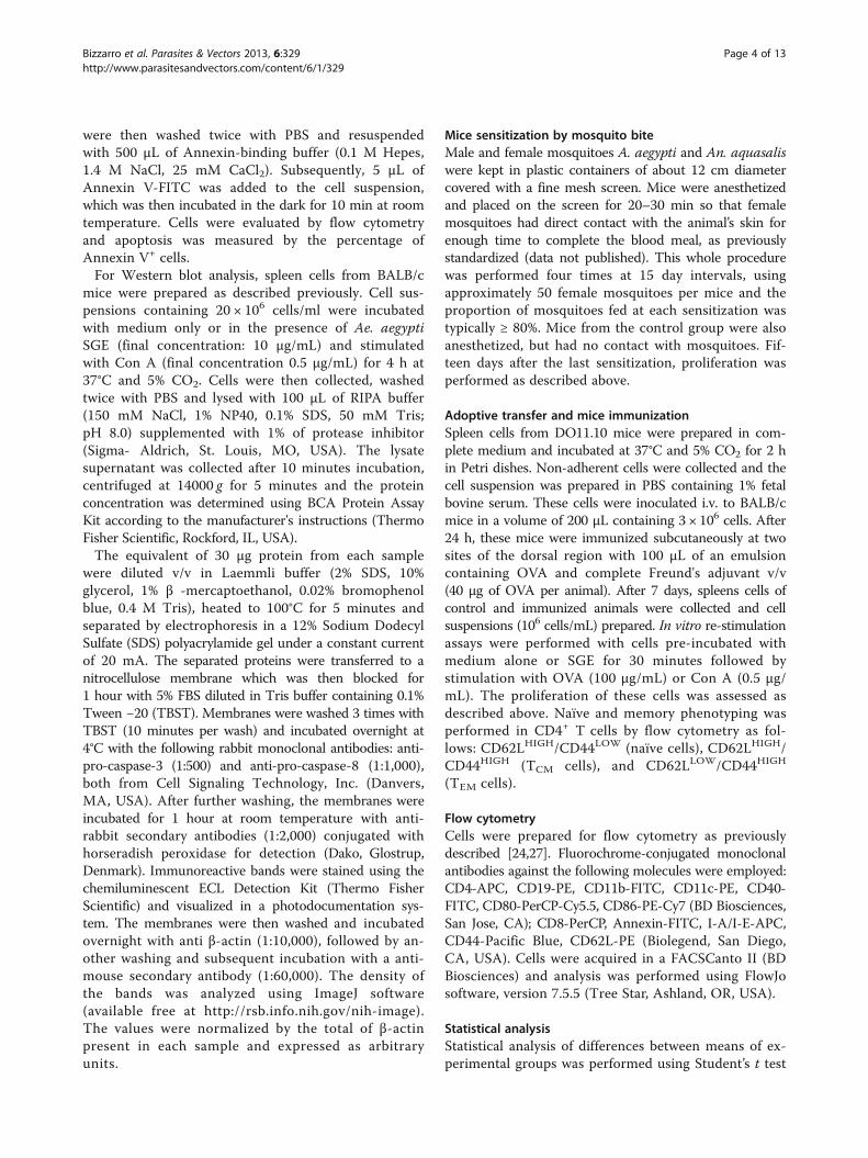
ResultsA. aegypti SGE does not interfere with dendritic celldifferentiation or maturationBMDCs from BALB/c mice were cultured with GM-CSFin the presence of different concentrations of A. aegyptisaliva and their differentiation was evaluated by flowcytometry through the percentage of CD11c+/CD11b+
cells. Figure 1 shows that A. aegypti SGE did not affectthe differentiation of BMDCs at 4 or 7 days of culture(Figure 1A), as comparable percentages of CD11c+/CD11b+
cells were observed in all groups on both days (Figures 1Band 1C). These sets of experiments were also performedwith BMDC from C57BL/6 mice and similar results wereachieved (data not shown).
To assess whether A. aegypti saliva has an effect onDCs maturation, we analyzed the expression pattern ofMHC class II and costimulatory molecules on DCsafter incubation with LPS in the presence of SGE. Asexpected, immature DCs present variable levels ofextracellular MHC class II expression (low, mid and high)and, upon LPS stimulation, the majority of these cellsshift to MHC class IIHIGH, confirming DC maturation.
However, the presence of SGE did not alter the expres-sion of MHC class II either in immature or matureDCs (Figure 2A). No differences were found in thesample’s mean fluorescent intensity (MFI), indicatingsimilar levels of expression of this marker (data notshown). The expression of CD40 was also upregulatedin CD11c+ cells upon LPS stimulation when comparedto control cells (incubated with medium only), butagain, the presence of SGE in the culture did not affectCD40 expression (Figure 2B). The same was observedwith CD80 and CD86 expression (data not shown).
A. aegypti SGE inhibits T cell proliferation in aDC-independent fashionIn order to investigate the effect of A. aegypti SGE onantigen presentation by DCs, purified CD11c+ cellswere incubated with SGE, pulsed with OVA plus LPSand after repeated washings to remove SGE, OVA andLPS residues, these cells were coincubated with CD4+ Tlymphocytes from DO11.10 mice and the proliferationwas evaluated [24,27,28]. OVA-pulsed DCs stimulatedCD4+ T cell proliferation when compared to control (DCsincubated with medium alone). Nonetheless, when DCswere preincubated with A. aegypti SGE, the specificproliferation of CD4+ T cells was not affected (Figure 3A).
In another set of experiments, SGE was added again tothe culture after DC washing, followed by coincubation
Figure 1 A. aegypti SGE does not interfere with DC differentiation. BMDC from BALB/c mice were cultured with GM-CSF in the presence orabsence of A. aegypti SGE (final concentration: 2.5, 5, 10, 20 and 40 μg/mL) for 4 and 7 days and analyzed by flow cytometry. Dot plots (A) andthe mean of relative percentage of CD11b+/CD11c+ cells present in culture at 4 days (B) and 7 days (C) are presented.
Bizzarro et al. Parasites & Vectors 2013, 6:329 Page 5 of 13http://www.parasitesandvectors.com/content/6/1/329

with T cells from DO11.10 mice. Under these conditions,antigen-specific CD4+ T cell proliferation was completelyabolished (Figure 3B). The same approach was performed,but stimulating the cultures with Con A, a polyclonalactivator of T cells. Figure 3C shows that if SGE ismaintained in the culture, polyclonal activation of Tcells is also completely inhibited. These results wereconfirmed through CFSE staining (Additional file 1).Together, these data show that A. aegypti salivary compo-nents affect T lymphocytes in a DC-independent manner.
Next, we evaluated the potency of this inhibitory activityby incubating spleen cells with increasing concentrationsof SGE followed by Con A stimulation. Figure 3D showsthat SGE induced a concentration-dependent decreaseof T cell proliferation, reaching maximal inhibition at10 μg/mL. Of note, similar inhibitory activity was foundfor spleen cells from C57BL/6 mice (data not shown). Wehave also tested if salivary components of hematophagous
species other than A. aegypti possessed such inhibitoryactivity. Interestingly, neither SGE from An. aquasalismosquitoes (Figure 3E) nor from P. duboscqi sandflies(Figure 3F) were able to affect lymphocyte proliferationunder the same conditions.
A. aegypti SGE induces caspase-3 and caspase-8dependent lymphocyte apoptosisAs expected, when a spleen cell culture stimulated withCon A for 3 days is analyzed by flow cytometry, mostlymphocytes appear larger in “size” and with moregranularity compared to fresh cultures, indicating theiractivation (Additional file 2). The presence of A. aegyptiSGE in this culture drastically changes cell phenotype, asthe amount of “viable cells” is inversely proportional toSGE concentration, suggesting cell death (Additional file 2).In order to investigate the mechanism by which cells wouldbe dying in the presence of SGE, we evaluated several
Figure 2 DC maturation is not affected by A. aegypti SGE. DCs were differentiated with GM-CSF for 6 days, preincubated overnight in thepresence or absence of A. aegypti SGE (final concentration: 5 and 40 μg/mL) and stimulated or not with LPS (100 ng/mL), as indicated. Expressionof MHC class II (A) and CD40 (B) was evaluated in CD11c+ cells by flow cytometry.
Bizzarro et al. Parasites & Vectors 2013, 6:329 Page 6 of 13http://www.parasitesandvectors.com/content/6/1/329

apoptosis parameters. Figure 4A shows that annexin Vstaining is increased in total spleen cells stimulatedby Con A in the presence of A. aegypti SGE when com-pared to cells stimulated with Con A only. A similar pheno-type is observed in CD4+ and CD8+ T cells population aswell as in CD19+ B cells (Figure 4A).
In order to further characterize the molecular pathway(s) involved in the apoptosis induced by A. aegypti salivarycomponents, we evaluated the expression of pro-caspase-3and pro-caspase-8 in total spleen cell lysates. As observedin Figure 4B, levels of both pro-caspases are reduced incells incubated with Con A plus SGE when compared tocells stimulated with Con A alone.
Naïve and memory T cells are differentially affected byA. aegypti SGEMemory cells are known to be more resistant to apoptosisthan naïve T cells [29]. Because Bim, a proapoptotic
member of the Bcl-2 family, is involved in T cellhomeostasis and memory T cells are resistant to Bim-induced apoptosis [30], we tested whether spleen cellsfrom Bim knockout mice (Bim−/−) were as sensitive astheir heterozygous counterparts (Bim+/−) to the inhibitoryeffects of A. aegypti SGE. Figure 4C shows that spleencells from either Bim−/− or Bim+/− are equally affectedby SGE, suggesting that the intrinsic apoptosis pathwaydoes not play a role in SGE-induced apoptosis.
To further investigate the observed phenomenon, wegenerated memory cells by natural sensitization of miceto A. aegypti bites and evaluated the proliferation of spleencells from these animals. As previously demonstrated(Figure 3D), A. aegypti SGE inhibits both basal cellmetabolism and Con A-induced proliferation of spleencells (Figure 5A). Contrarily, cells from mice sensitizedwith A. aegypti bites display antigen-specific proliferationin the presence of SGE (Figure 5B). Interestingly, whenstimulated by Con A in the presence of SGE, proliferation
Figure 3 A. aegypti SGE inhibits T cell proliferation in a DC-independent fashion. DCs were pre-incubated overnight in the presence orabsence of A. aegypti SGE (final concentrations indicated) and stimulated for 4 h with OVA (100 μg/mL) plus LPS (100 ng/mL). After 3 washings,DCs were co-incubated with CD4+ cells from DO11.10 mice for 72 h (A and B). Similar DC/CD4+ cultures were also stimulated by Con A for 72 h(C). In some groups, SGE was added again after washing the cells (B and C). Concentration-response effect of A. aegypti SGE on Con A-inducedspleen cells proliferation (D). Absence of effect of An. aquasalis (E) and P. dubosqi SGE (F) on Con A-induced spleen cells proliferation.*p < 0.05 versus “C” (control); #p < 0.05 versus stimulated with OVA + LPS or Con A.
Bizzarro et al. Parasites & Vectors 2013, 6:329 Page 7 of 13http://www.parasitesandvectors.com/content/6/1/329

of these cells was only partially inhibited, and the propor-tion of surviving cells corresponded to the amount of anti-gen specific proliferating cells (Figure 5B – dotted line).The secretion of neutralizing antibodies against salivarycomponents by B lymphocytes present in the culturecould not explain the lack of inhibition observed, sinceculture supernatants from spleen cells of sensitized mice,used as conditioned media, did not block (even par-tially) the effect of SGE on T cells from non-sensitizedmice (Additional file 3).
We also investigated the effects of A. aegypti SGE onspleen cell proliferation of mice sensitized with bites ofAn. aquasalis, a species belonging to a different mosquitosubfamily. Spleen cells from An. aquasalis-sensitized miceproliferate in the presence of SGE from this species,but not in the presence of A. aegypti SGE, indicatingantigen-specific stimulation (Figure 5C). When these cellsare incubated with both SGE (from A. aegypti and An.aquasalis), the proliferative response persists, confirmingthe lack of A. aegypti SGE activity on memory cellsgenerated against An. aquasalis salivary components(Figure 5C). Additionally, when spleen cells from An.aquasalis-sensitized mice are stimulated with Con A orAn. aquasalis SGE, a significant proliferation is observed,possibly from both naïve and memory cells. Stimulationwith Con A in the presence of A. aegypti SGE only, ortogether with An. aquasalis SGE, induces a partial inhib-ition of the proliferative response. The partial blockage
of proliferation observed suggests again that while thememory cell population survives and continues to prolifer-ate, the naïve population dies in the culture (Figure 5C).This finding implies that A. aegypti SGE does not affectthe memory cells generated against salivary componentsof other mosquito species (e.g. mice sensitized withAn. aquasalis bites).
In order to further test our hypothesis, we generatedmemory cells against OVA, an antigen not related tomosquito salivary molecules. Recipient BALB/c micewere adoptively transferred with cells from DO11.10mice and immunized with OVA to induce expansion ofthe donor cells and allow the generation of memorycells. One week later, spleen cells from recipient animalswere preincubated in vitro with A. aegypti SGE andstimulated with Con A or OVA to assess the polyclonaland antigen-specific proliferation, respectively. Figure 5Dshows that basal metabolism of cells incubated withmedium or OVA, as well as Con A-induced proliferationfrom naïve mice are all significantly inhibited by A.aegypti SGE, as expected. On the contrary, spleen cellsfrom mice adoptively transferred with DO11.10 cellsand immunized with OVA had the Con A-induced pro-liferation only partially inhibited and, more important,antigen-specific proliferation was not affected at all(Figure 5E).
Finally, we evaluated the naïve and activated/memorymarkers in CD4+ T cell populations in medium only or
A
B C
Figure 4 A. aegypti SGE induces lymphocyte apoptosis and cleavage of pro-caspase 3 and pro-caspase-8. Total spleen cells wereincubated with Con A in medium only or in presence of A. aegypti SGE for 4 h. Annexin V staining was evaluated by flow cytometry in total cells,CD4+, CD8+ and CD19+ cells from WT mice (A) or CD4+ T cells from Bim+/− and Bim−/− mice (C). Lysates of similar cell cultures were blottedagainst a monoclonal antibody against pro-caspase-3 and pro-caspase-8 (B).
Bizzarro et al. Parasites & Vectors 2013, 6:329 Page 8 of 13http://www.parasitesandvectors.com/content/6/1/329

under OVA stimulation. CD62LHIGH/CD44LOW wereconsidered naïve T cells while memory cells were dividedinto two subsets: effector memory T cells (TEM) thatexpress CD62LLOW and CD44HIGH; and central memorycells (TCM) which express CD62LHIGH and CD44HIGH
[31-35]. Figure 5 shows that both, TEM and TCM subsets,were increased in OVA-sensitized mice when comparedto non-sensitized mice (upper left panels - Figures 5Fand 5G). The addition of A. aegypti SGE to the cultures,affected naïve cell populations in both groups (lowerleft panels). The presence of OVA expanded both memorysubsets in OVA-sensitized spleen cell cultures when
compared to cultures of spleens from non-sensitizedmice as expected (upper right panels – Figures 5F and5G). Remarkably, coincubation with OVA plus SGEstrongly affected CD62LHIGH/CD44LOW naïve T cellpopulation in both groups, while memory subsets werepreserved or even increased, especially in the culturesfrom OVA-sensitized mice (lower right panels – Figures 5Fand 5G). All these findings were concentration-dependent,as observed in Additional file 4. Taken together, thesedata show that naïve CD4+ T cells are susceptible, whilememory T cells are selectively more resistant, to theapoptotic effect of A. aegypti SGE.
Figure 5 Memory cells are resistant to A. aegypti SGE effects. Spleen cells from non-sensitized (A) and A. aegypti-sensitized BALB/c mice(B) were incubated with medium only or with A. aegypti SGE (5 μg/mL) and/or stimulated with Con A (0.5 μg/mL) for 72 h. Cells from An.aquasalis-sensitized mice were cultured with medium only or in the presence of 5 μg/mL An. aquasalis SGE (An.) and/or 5 μg/mL A. aegyptiSGE (Ae.) and then stimulated with 0.5 μg/mL Con A (C). Non-adherent DO11.10 spleen cells were adoptively transferred to BALB/c mice andafter 7 days, recipient mice were sensitized with OVA and complete Freund’s adjuvant (40 μg/animal). Spleen cells from non-sensitized (D)and sensitized mice (E) were obtained after 7 days and cultured in the presence of medium or A. aegypti SGE and stimulated with Con A (0.5 μg/mL)or OVA (100 μg/mL). Phenotype of naïve cells (CD62LHIGH/CD44LOW), TEM subset (CD62LLOW and CD44HIGH) and TCM subset (CD62LHIGH and CD44HIGH)from non-sensitized (F) or sensitized mice (G) were evaluated by flow cytometry after 72 h cultured in presence of medium or A. aegypti SGE andstimulated with Con A or OVA. *p < 0.05 versus respective control group.
Bizzarro et al. Parasites & Vectors 2013, 6:329 Page 9 of 13http://www.parasitesandvectors.com/content/6/1/329

DiscussionThe blood-feeding behaviour is present in several ordersof insects that have acquired the genetic and morpho-functional resources to suck, digest and use the bloodof their vertebrate hosts [36]. Over millions of years ofevolution, hematophagous mosquitoes have developeda complex pharmacological cocktail in their saliva, whichclearly modulates host vascular and immune systems.Nevertheless, our knowledge of these processes is in-complete. The present study aimed to investigate theputative immunomodulatory effects of salivary compo-nents of A. aegypti mosquito vector on the differentiation,maturation and function of DCs and on the proliferationof T lymphocytes.
Pioneering work has shown that saliva of Rhipicephalussanguineus, the “brown dog tick”, is able to inhibit diffe-rentiation and maturation of murine DCs [37]. In addition,Sá-Nunes et al. (2007) were the first to isolate andcharacterize prostaglandin E2 (PGE2) as the major DCmodulator in saliva of Ixodes scapularis ticks, the Lymedisease vector [24]. Recently, PGE2 found in the saliva ofDermacentor variabilis ticks was also shown to regulatemacrophage migration and cytokine production bythese cells [38]. In addition, the presence of PGE2 in R.sanguineus saliva was also demonstrated, although insmaller amounts than I. scapularis [39]. However, thecapacity of R. sanguineus saliva to modulate DCs iscomplemented by the presence of adenosine [39]. Add-itional studies have demonstrated the immunoregulatoryand anti-inflammatory activity of crude tick saliva [40,41]and other proteinaceous components capable of modu-lating the function of DCs, such as Salp15 [42] and sia-lostatin L [27], both identified in the I. scapularissaliva. Although these previous pieces of evidence showa clear effect of the tick saliva on DCs, very little isknown about the modulation of these cells by saliva ofblood feeding insects. Costa et al. (2004) demonstratedthat SGE of Lutzomyia longipalpis sandflies, one of theleishmaniasis vectors in the new world, affects cytokineproduction and costimulatory activity of human DCs [43].Some years later, it was shown that SGE of P. duboscqiand P. papatasi sandfly species induced the productionof PGE2 and IL-10 by DCs [44]. The observed effects ofP. papatasi SGE on DCs was due to the presence ofadenosine and adenosine monophosphate (5’ AMP) andthis seems to be, at least partially, the mechanism bywhich the SGE of this species was able to decrease thearthritis symptoms in an autoimmune model inducedby collagen [25].
DCs comprise distinct developmental and functionalsubsets present in lymphoid and non-lymphoid tissuesand are involved in the activation of adaptive immuneresponses, but also in tolerance to self-antigens [45].However, their frequencies in the tissues limit their
experimental use. For example, Langerhans cells (the DCpopulation from the epidermal layer of the skin) accountfor 3-5% of epidermal cells [46]. Accordingly, classicalDCs such as those found in the dermis, represent 1-5%of total cells from peripheral tissues [45]. In addition,the increasing number of DC phenotypes described andisolation protocols employing enzymatic digestion whichtemporarily destroy surface markers, are other factors toconsider [45]. Thus, although the BMDCs preparationsemployed in the present work do not precisely representthe population of epidermal and dermal DCs that possiblyinteract with mosquito saliva, the use of these cells toinvestigate the biological effects of salivary preparationsor their purified components is accepted by most studiesin the field [9,24,27,37,44,47]. To our surprise, A. aegyptiSGE had no effect on DCs differentiation (Figure 1),maturation (Figure 2) and antigen presentation to Tlymphocytes (Figure 3A). Corroborating with this data,it has been demonstrated that A. aegypti SGE did notaffect the viability of a murine DC line [16]. These resultscontrast greatly with data described in other species ofarthropod vectors and, in the specific case of A. aegypti,our results are original in demonstrating that directmodulation of DCs by salivary components does notseem to be a property of the saliva from this mosquitospecies.
Interestingly, addition of A. aegypti SGE to cultures ofCD11c+ cells after washing caused a significant inhibitionin antigen specific (Figure 3B) and polyclonal (Figure 3C)proliferation of CD4+ T lymphocytes. This data confirmsthat SGE acts directly on T lymphocytes and not onantigen-presenting cells, and corroborates with findingsin the literature showing the negative modulation oflymphocyte function by A. aegypti salivary components[16-18]. We also observed that SGE of other insect species(namely An. aquasalis, and P. duboscqi) had no inhibitoryeffect on T cell proliferation (Figures 3E and 3F). Wanasenet al. [18] also observed the absence of effects on T cellproliferation employing SGE of Culex quinquefasciatus,which belongs to the same mosquito subfamily. To ourknowledge, such inhibitory activity was only found inthe SGE of another nematoceran species, the black flySimulium vittatum [48].
We have also explored the mechanisms by which theA. aegypti SGE inhibits lymphocyte proliferation. Ourresults show that this inhibition occurs due to inductionof apoptosis in spleen cells, more specifically on T (CD4+
and CD8+) and B (CD19+) cells (Figure 4A). The specifi-city of such biological activity is evidenced by DC assays,since differentiation, maturation and function were notaffected, even when these cells were incubated with 40 μg/mL SGE, a 4-fold increase in the maximum concentrationeffect on the proliferative response. As previously reported,decreased T cell proliferation induced by A. aegypti SGE
Bizzarro et al. Parasites & Vectors 2013, 6:329 Page 10 of 13http://www.parasitesandvectors.com/content/6/1/329

was due to diminished cell viability, as evaluated by PIand 7-AAD expression, both DNA markers [16,18]. It isimportant to emphasize that these markers are notspecific for apoptotic cell death. Our findings unveiledthat A. aegypti SGE induces apoptosis in T and B lym-phocytes as assessed by exposure of phosphatidylserine(labeled with annexin V) at the surface of these cells(Figure 4A). Furthermore, our results suggest thatcaspase-3 (an executor caspase) and caspase-8 (an ini-tiator caspase), but not Bim (a proapoptotic member ofthe intrinsic pathway), are likely to be involved in theapoptosis signaling induced by A. aegypti SGE (Figures 4Band 4C). As T and B cells are components of the adaptiveimmune response, it is reasonable to imagine that theireffector functions (antibody secretion, cytotoxic granulesor helper activities) are somehow deleterious to themosquito life cycle. In fact, a classical study has dem-onstrated a decrease in the fecundity of mosquitoes fedon rabbits or guinea pigs immunized with a A. aegyptiwhole body homogenate [49].
Memory T cells are known to be more resistant toapoptosis than naïve T cells due to the increased expres-sion of anti-apoptotic proteins [29]. Because cells fromBim−/− mice are as susceptible to apoptosis as cells fromBim+/− mice (Figure 4C), and it is well known that thisdifferential resistance is considerably dependent on theneutralization of Bim-mediated apoptosis by increasedlevels of Bcl-2 [29,30], we investigated whether the A.aegypti salivary components would also have an effecton memory cells. Initially, mice were sensitized with A.aegypti bites and the effect of SGE on proliferative re-sponse of spleen cells from these animals was evaluated.Unlike naïve spleen cells (Figure 5A), those derived fromsensitized animals proliferate in the presence of SGE(Figure 5B). In addition, when these cells were stimu-lated with Con A, only partial inhibition was achieved,suggesting that the memory cells present in the culturecontinue to proliferate even in the presence of SGEinhibitory factor(s). We rule out the role of neutralizingantibodies produced by B lymphocytes present in theculture on this effect, since spleen cell supernatants fromsensitized mice, used as conditioned media, did not blockthe effect of SGE on T cells from non-sensitized mice(Additional file 3). Other explanations/mechanismscannot be ruled out, such as peripheral tolerance anddevelopment of regulatory T cells, and will be exploredin future work. We also demonstrated that SGE doesnot inhibit proliferation if memory cells are generatedto other antigens. For example, spleen cells fromAn. aquasalis-sensitized mice proliferate when culturedwith SGE of the same species, even in the presence of A.aegypti SGE. In addition, when these cells are co-culturedwith Con A plus A. aegypti SGE, the inhibition of theproliferative response is only partial, reinforcing again
our assumption that only naïve cells are affected by thepresence of SGE in culture (Figure 5C).
This hypothesis is further supported by analyzing theproliferation of spleen cells from BALB/c mice receivingcells from a DO11.10 donor and subsequently immunizedwith OVA. When these cells are co-cultured with Con Aplus SGE, the proliferation is partially inhibited (Figure 5B),whereas in cells from non-sensitized animals, SGEcompletely inhibits proliferation (Figure 5A). Remark-ably, antigen-specific proliferation induced by OVAonly occurs in sensitized mice, as expected, and it is notaffected by A. aegypti SGE (Figure 5D and 5E). Regardingcell phenotype, SGE affects naïve cells (CD62LHIGH/CD44LOW) in both, control and immunized animals(Figure 5F and Figure 5G, respectively), while TCM cells(CD62LHIGH/CD44HIGH) and TEM cells (CD62LLOW/CD44HIGH) are proportionally more resistant in immunizedanimals (Figure 5G). So far, there is a single study showinga selective action of a salivary component in subpopulationsof T cells. Such work demonstrated that Salp15, fromthe I. scapularis tick, binds to CD4 but not CD8 T cellco-receptor [50]. However, this is the first time thatarthropod saliva has been shown to discriminate betweennaïve and memory T cells.
The blood feeding strategies greatly diverge betweenmany hematophagous arthropods. While hard ticks main-tain prolonged contact with host skin, some others, likemosquitoes and sandflies, are transient feeders and leavetheir host in minutes or even seconds. Undoubtedly,their strategies to modulate the host vascular and immunesystem may vary as well.
ConclusionsTogether, these results provide evidence for a complexinteraction between A. aegypti salivary constituents andthe host immune system. This pioneer study shows thatsaliva of a hematophagous arthropod is able to distinguishamong different cell types (dendritic cells versus lympho-cytes) and even subpopulations of the same cell type(naïve versus memory T cells). Whether this selectivityis important to mosquitoes feeding and reproductionremains to be determined. Therefore, the results generatedby this work contributes to clarifying some of the fea-tures of the vector-host interaction providing a betterunderstanding of the mechanisms used by the mosquitoA. aegypti to circumvent the immune system of their hostsand successfully feed.
Additional files
Additional file 1: A. aegypti SGE inhibits T cell proliferation. BMDCswere pre-incubated with medium or 40 μg/mL of A. aegypti SGE overnight,washed 3 times and co-incubated with CD4+ from DO11.10 mice stainedwith CFSE. A. aegypti SGE was replaced in culture after washing and T cells
Bizzarro et al. Parasites & Vectors 2013, 6:329 Page 11 of 13http://www.parasitesandvectors.com/content/6/1/329

were stimulated with Con A for 72 h. Cells were evaluated by flowcytometry as described in Methods.
Additional file 2: A. aegypti SGE induces changes in total spleencell phenotype. Indirect evaluation of the cell viability by examining thesize (FSC) and internal complexity (SSC) of spleen cells incubated with ConA (B-G) for 72 h in the presence of increasing concentrations of A. aegyptiSGE (0.1, 0.5, 1, 5 and 10 μg/mL) and compared with fresh cells (A).
Additional file 3: Antibodies produced by B lymphocytes do notneutralize SGE activity. Three-day culture supernatants from differentnumbers of spleen cells from non-sensitized (A) and A. aegypti-sensitized(B) mice were used as a conditioned medium for cell cultures from acontrol mice spleen. These cells were then pre-incubated with mediumalone or A. aegypti SGE and then stimulated with Con A for 72 h.
Additional file 4: Memory cells are resistant to A. aegypti SGEeffects. Non-adherent DO11.10 spleen cells were adoptively transferredto BALB/c mice and after 7 days, recipient mice were sensitized withOVA and complete Freund’s adjuvant (40 μg/animal). Spleen cells fromnon-sensitized and sensitized mice were obtained after 7 days and culturedin the presence of medium or A. aegypti SGE and stimulated with Con A (0.5μg/mL) or OVA (100 μg/mL). Phenotype of naïve cells (CD62LHIGH/CD44LOW),TEM subset (CD62LLOW and CD44HIGH) and TCM subset (CD62LHIGH andCD44HIGH) from non-sensitized or sensitized mice were evaluated by flowcytometry after 72 h cultured in presence of medium or A. aegypti SGE andstimulated with Con A or OVA.
AbbreviationsSGE: Salivary gland extract; FSDC: Fetal skin-derived DC line; BMDC: Bonemarrow-derived dendritic cell; PGE2: Prostaglandin E2; 5’AMP: Adenosinemonophosphate..
Competing interestsThe authors declare that they have no competing interests.
Authors’ contributionsBB, MK, GPAM, EC, MLC and AS-N conceived the experimental design andassisted data interpretation. BB, MSB, CM, DIG, CNL, JC, MLC and AS-N carriedout laboratory work. BB and AS-N wrote the manuscript’s draft. All authorsread, reviewed and approved the final manuscript.
AcknowledgmentsThe authors would like to thank Dr. Momtchilo Russo and Dr. Sonia Jancar(Department of Immunology, ICB/USP, Brazil) for providing some reagentsand equipments used in this work; Dr. Philippe Bouillet (Walter and ElisaHall Institute of Medical Research, Australia) for providing the BIM−/− mice;and Dr. José M. Ribeiro (Laboratory of Malaria and Vector Research, NIAID/NIH, USA) for encouragement and support. The authors also thank Van M.Pham (Laboratory of Malaria and Vector Research, NIAID/NIH, USA) andSandra Alexandre (Department of Immunology, ICB/USP) for their technicalassistance. The following authors were recipients of fellowships: B.B.(FAPESP 2009/12247-5); M.S.B. (CNPq 134660/2010-2); C.M. (FAPESP 2010/18216-1); D.I.G. (FAPESP 2011/06626-3); C.N.L. (FAPESP 2011/15569-3). Thiswork was supported by grants from FAPESP (2009/09892-6 and 2009/53637-0), CNPq (302194/2009-6), Casadinho/PROCAD (MCTI/CNPq/MEC/CAPES 552258/2011-3), Brazilian Malaria Network (MCT/CNPq/MS/SCTIE/DECIT/PRONEX 555648/2009-5), and Research Network on BioactiveMolecules from Arthropod Vectors (NAP-MOBIARVE, University of SãoPaulo). We dedicated this paper to the memory of Alexandre A. Peixoto.
Author details1Laboratório de Imunologia Experimental, Departamento de Imunologia,Instituto de Ciências Biomédicas, Universidade de São Paulo, São Paulo05508-900, SP, Brazil. 2Laboratório de Biologia Celular e Molecular,Departamento de Imunologia, Instituto de Ciências Biomédicas, Universidadede São Paulo, São Paulo, SP 05508-900, Brazil. 3Laboratory of Genomics andProteomics of Disease Vectors, Institute of Parasitology, Biology Centre of theAcademy of Sciences of Czech Republic, Ceske Budejovice 37005, CzechRepublic. 4Instituto de Investigação em Imunologia, Instituto Nacional deCiência e Tecnologia, INCT, São Paulo, Brazil. 5Section of Vector Biology,Laboratory of Malaria and Vector Research, National Institute of Allergy and
Infectious Diseases, National Institutes of Health, Rockville, MD 20852, USA.6Laboratório de Mosquitos Geneticamente Modificados, Departamento deParasitologia, Instituto de Ciências Biomédicas, Universidade de São Paulo,São Paulo, SP 05508-900, Brazil. 7Instituto Nacional de Ciência e Tecnologiaem Entomologia Molecular, Conselho Nacional de DesenvolvimentoCientífico e Tecnológico (INCT-EM/CNPq), Rio de Janeiro, Brazil.
Received: 28 September 2013 Accepted: 5 November 2013Published: 15 November 2013
References1. Morens DM, Folkers GK, Fauci AS: The challenge of emerging and
re-emerging infectious diseases. Nature 2004, 430(6996):242–249.2. Tolle MA: Mosquito-borne diseases. Curr Probl Pediatr Adolesc Health Care
2009, 39(4):97–140.3. Scott TW, Takken W: Feeding strategies of anthropophilic mosquitoes
result in increased risk of pathogen transmission. Trends Parasitol 2012,28(3):114–121.
4. Fontaine A, Diouf I, Bakkali N, Misse D, Pages F, Fusai T, Rogier C, Almeras L:Implication of haematophagous arthropod salivary proteins in host-vector interactions. Parasit Vectors 2011, 4:187.
5. Francischetti IM, Sa-Nunes A, Mans BJ, Santos IM, Ribeiro JM: The role ofsaliva in tick feeding. Front Biosci 2009, 14:2051–2088.
6. Ribeiro JM, Francischetti IM: Role of arthropod saliva in blood feeding:sialome and post-sialome perspectives. Annu Rev Entomol 2003, 48:73–88.
7. Edwards JF, Higgs S, Beaty BJ: Mosquito feeding-induced enhancement ofCache Valley Virus (Bunyaviridae) infection in mice. J Med Entomol 1998,35(3):261–265.
8. Limesand KH, Higgs S, Pearson LD, Beaty BJ: Effect of mosquito salivary glandtreatment on vesicular stomatitis New Jersey virus replication andinterferon alpha/beta expression in vitro. J Med Entomol 2003, 40(2):199–205.
9. Schneider BS, Soong L, Coffey LL, Stevenson HL, McGee CE, Higgs S: Aedesaegypti saliva alters leukocyte recruitment and cytokine signaling byantigen-presenting cells during West Nile virus infection. PLoS One 2010,5(7):e11704.
10. Styer LM, Bernard KA, Kramer LD: Enhanced early West Nile virus infectionin young chickens infected by mosquito bite: effect of viral dose.Am J Trop Med Hyg 2006, 75(2):337–345.
11. Styer LM, Lim PY, Louie KL, Albright RG, Kramer LD, Bernard KA: Mosquitosaliva causes enhancement of West Nile virus infection in mice. J Virol2011, 85(4):1517–1527.
12. Surasombatpattana P, Patramool S, Luplertlop N, Yssel H, Misse D: Aedesaegypti Saliva Enhances Dengue Virus Infection of Human Keratinocytesby Suppressing Innate Immune Responses. J Invest Dermatol 2012,132(8):2103–2105.
13. Machain-Williams C, Mammen MP Jr, Zeidner NS, Beaty BJ, Prenni JE, NisalakA, Blair CD: Association of human immune response to Aedes aegyptisalivary proteins with dengue disease severity. Parasite Immunol 2012,34(1):15–22.
14. Schneider BS, Higgs S: The enhancement of arbovirus transmission anddisease by mosquito saliva is associated with modulation of the hostimmune response. Trans R Soc Trop Med Hyg 2008, 102(5):400–408.
15. Banchereau J, Steinman RM: Dendritic cells and the control of immunity.Nature 1998, 392(6673):245–252.
16. Wasserman HA, Singh S, Champagne DE: Saliva of the Yellow Fevermosquito, Aedes aegypti, modulates murine lymphocyte function.Parasite Immunol 2004, 26(6–7):295–306.
17. Cross ML, Cupp EW, Enriquez FJ: Differential modulation of murinecellular immune responses by salivary gland extract of Aedes aegypti.Am J Trop Med Hyg 1994, 51(5):690–696.
18. Wanasen N, Nussenzveig RH, Champagne DE, Soong L, Higgs S: Differentialmodulation of murine host immune response by salivary gland extractsfrom the mosquitoes Aedes aegypti and Culex quinquefasciatus.Med Vet Entomol 2004, 18(2):191–199.
19. Boppana VD, Thangamani S, Adler AJ, Wikel SK: SAAG-4 is a novelmosquito salivary protein that programmes host CD4 T cells to expressIL-4. Parasite Immunol 2009, 31(6):287–295.
20. Zeidner NS, Higgs S, Happ CM, Beaty BJ, Miller BR: Mosquito feedingmodulates Th1 and Th2 cytokines in flavivirus susceptible mice: aneffect mimicked by injection of sialokinins, but not demonstrated inflavivirus resistant mice. Parasite Immunol 1999, 21(1):35–44.
Bizzarro et al. Parasites & Vectors 2013, 6:329 Page 12 of 13http://www.parasitesandvectors.com/content/6/1/329

21. Opal SM, Esmon CT: Bench-to-bedside review: functional relationshipsbetween coagulation and the innate immune response and theirrespective roles in the pathogenesis of sepsis. Crit Care 2003, 7(1):23–38.
22. Niessen F, Schaffner F, Furlan-Freguia C, Pawlinski R, Bhattacharjee G, Chun J,Derian CK, Andrade-Gordon P, Rosen H, Ruf W: Dendritic cell PAR1-S1P3signalling couples coagulation and inflammation. Nature 2008,452(7187):654–658.
23. Ruf W, Furlan-Freguia C, Niessen F: Vascular and dendritic cell coagulationsignaling in sepsis progression. J Thromb Haemost 2009, 7(Suppl 1):118–121.
24. Sa-Nunes A, Bafica A, Lucas DA, Conrads TP, Veenstra TD, Andersen JF,Mather TN, Ribeiro JM, Francischetti IM: Prostaglandin E2 is a majorinhibitor of dendritic cell maturation and function in Ixodes scapularissaliva. J Immunol 2007, 179(3):1497–1505.
25. Carregaro V, Sa-Nunes A, Cunha TM, Grespan R, Oliveira CJ, Lima-Junior DS,Costa DL, Verri WA Jr, Milanezi CM, Pham VM, et al: Nucleosides fromPhlebotomus papatasi salivary gland ameliorate murine collagen-induced arthritis by impairing dendritic cell functions. J Immunol 2011,187(8):4347–4359.
26. Collin N, Assumpcao TC, Mizurini DM, Gilmore DC, Dutra-Oliveira A, KotsyfakisM, Sa-Nunes A, Teixeira C, Ribeiro JM, Monteiro RQ, et al: Lufaxin, a novelfactor Xa inhibitor from the salivary gland of the sand fly Lutzomyialongipalpis blocks protease-activated receptor 2 activation and inhibitsinflammation and thrombosis in vivo. Arterioscler Thromb Vasc Biol 2012,32(9):2185–2198.
27. Sa-Nunes A, Bafica A, Antonelli LR, Choi EY, Francischetti IM, Andersen JF,Shi GP, Chavakis T, Ribeiro JM, Kotsyfakis M: The immunomodulatoryaction of sialostatin L on dendritic cells reveals its potential to interferewith autoimmunity. J Immunol 2009, 182(12):7422–7429.
28. Ahmed SA, Gogal RM Jr, Walsh JE: A new rapid and simple non-radioactiveassay to monitor and determine the proliferation of lymphocytes: analternative to [3H]thymidine incorporation assay. J Immunol Methods 1994,170(2):211–224.
29. Wojciechowski S, Tripathi P, Bourdeau T, Acero L, Grimes HL, Katz JD,Finkelman FD, Hildeman DA: Bim/Bcl-2 balance is critical for maintainingnaive and memory T cell homeostasis. J Exp Med 2007, 204(7):1665–1675.
30. Kurtulus S, Tripathi P, Hildeman DA: Protecting and rescuing the effectors:roles of differentiation and survival in the control of memory T celldevelopment. Front Immunol 2012, 3:404.
31. Dutton RW, Bradley LM, Swain SL: T cell memory. Annu Rev Immunol 1998,16:201–223.
32. Reinhardt RL, Khoruts A, Merica R, Zell T, Jenkins MK: Visualizing thegeneration of memory CD4 T cells in the whole body. Nature 2001,410(6824):101–105.
33. Masopust D, Vezys V, Marzo AL, Lefrancois L: Preferential localization ofeffector memory cells in nonlymphoid tissue. Science 2001,291(5512):2413–2417.
34. Sallusto F, Lenig D, Forster R, Lipp M, Lanzavecchia A: Two subsets ofmemory T lymphocytes with distinct homing potentials and effectorfunctions. Nature 1999, 401(6754):708–712.
35. Medeiros AI, Sa-Nunes A, Turato WM, Secatto A, Frantz FG, Sorgi CA, SerezaniCH, Deepe GS Jr, Faccioli LH: Leukotrienes are potent adjuvant during fungalinfection: effects on memory T cells. J Immunol 2008, 181(12):8544–8551.
36. Lehane M: The Biology of Blood-Sucking in Insects. 2nd edition. Cambridge:Cambridge University Press; 2005.
37. Cavassani KA, Aliberti JC, Dias AR, Silva JS, Ferreira BR: Tick saliva inhibitsdifferentiation, maturation and function of murine bone-marrow-deriveddendritic cells. Immunology 2005, 114(2):235–245.
38. Poole NM, Mamidanna G, Smith RA, Coons LB, Cole JA: Prostaglandin E2 intick saliva regulates macrophage cell migration and cytokine profile.Parasit Vectors 2013, 6(1):261.
39. Oliveira CJ, Sa-Nunes A, Francischetti IM, Carregaro V, Anatriello E, Silva JS,de Miranda Santos IK, Ribeiro JM, Ferreira BR: Deconstructing tick saliva:non-protein molecules with potent immunomodulatory properties.J Biol Chem 2011, 286(13):10960–10969.
40. Chen G, Severo MS, Sohail M, Sakhon OS, Wikel SK, Kotsyfakis M, Pedra JH:Ixodes scapularis saliva mitigates inflammatory cytokine secretion duringAnaplasma phagocytophilum stimulation of immune cells. Parasit Vectors2012, 5:229.
41. Brake DK, Perez de Leon AA: Immunoregulation of bovine macrophagesby factors in the salivary glands of Rhipicephalus microplus.Parasit Vectors 2012, 5:38.
42. Hovius JW, de Jong MA, den Dunnen J, Litjens M, Fikrig E, van der Poll T,Gringhuis SI, Geijtenbeek TB: Salp15 binding to DC-SIGN inhibits cytokineexpression by impairing both nucleosome remodeling and mRNAstabilization. PLoS Pathog 2008, 4(2):e31.
43. Costa DJ, Favali C, Clarencio J, Afonso L, Conceicao V, Miranda JC, Titus RG,Valenzuela J, Barral-Netto M, Barral A, et al: Lutzomyia longipalpis salivarygland homogenate impairs cytokine production and costimulatorymolecule expression on human monocytes and dendritic cells.Infect Immun 2004, 72(3):1298–1305.
44. Carregaro V, Valenzuela JG, Cunha TM, Verri WA Jr, Grespan R, Matsumura G,Ribeiro JM, Elnaiem DE, Silva JS, Cunha FQ: Phlebotomine salivas inhibitimmune inflammation-induced neutrophil migration via an autocrineDC-derived PGE2/IL-10 sequential pathway. J Leukoc Biol 2008,84(1):104–114.
45. Merad M, Sathe P, Helft J, Miller J, Mortha A: The dendritic cell lineage:ontogeny and function of dendritic cells and their subsets in the steadystate and the inflamed setting. Annu Rev Immunol 2013, 31:563–604.
46. Merad M, Ginhoux F, Collin M: Origin, homeostasis and function ofLangerhans cells and other langerin-expressing dendritic cells. Nat RevImmunol 2008, 8(12):935–947.
47. Oliveira CJ, Sa-Nunes A, Francischetti IM, Carregaro V, Anatriello E, Silva JS,Santos IK, Ribeiro JM, Ferreira BR: Deconstructing tick saliva: non-proteinmolecules with potent immunomodulatory properties. J Biol Chem 2011,286(13):10960–10969.
48. Tsujimoto H, Gray EW, Champagne DE: Black fly salivary gland extractinhibits proliferation and induces apoptosis in murine splenocytes.Parasite Immunol 2010, 32(4):275–284.
49. Sutherland GB, Ewen AB: Fecundity decrease in mosquitoes ingesting bloodfrom specifically sensitized mammals. J Insect Physiol 1974, 20(4):655–660.
50. Garg R, Juncadella IJ, Ramamoorthi Ashish N, Ananthanarayanan SK, ThomasV, Rincon M, Krueger JK, Fikrig E, Yengo CM: Cutting edge: CD4 is thereceptor for the tick saliva immunosuppressor, Salp15. J Immunol 2006,177(10):6579–6583.
doi:10.1186/1756-3305-6-329Cite this article as: Bizzarro et al.: Effects of Aedes aegypti salivarycomponents on dendritic cell and lymphocyte biology. Parasites &Vectors 2013 6:329.
Submit your next manuscript to BioMed Centraland take full advantage of:
• Convenient online submission
• Thorough peer review
• No space constraints or color figure charges
• Immediate publication on acceptance
• Inclusion in PubMed, CAS, Scopus and Google Scholar
• Research which is freely available for redistribution
Submit your manuscript at www.biomedcentral.com/submit
Bizzarro et al. Parasites & Vectors 2013, 6:329 Page 13 of 13http://www.parasitesandvectors.com/content/6/1/329
Recommended


![Potential of Aedes albopictus and Aedes aegypti (Diptera ...mosquitoes, Aedes aegypti [3] or potentially, Aedes albopictus [4,5]. Indeed, between 2015 and 2016 in Central Africa, major](https://img.pdfslide.us/doc/110x75/60e30e3483720c1b6128c2b9/potential-of-aedes-albopictus-and-aedes-aegypti-diptera-mosquitoes-aedes-aegypti.jpg)