British Journal of Ophthalmology, 1984, 68, 367-369
Ectodermal dysplasia associated with autoimmunediseaseS. 0. HUNG AND A. PATTERSON
From St Paul's Eye Hospital, Old Hall, Street, Liverpool L3 9PF
SUMMARY A case is reported of hereditary ectodermal dysplasia with corneal involvement associ-ated with polyendocrinopathy and pernicious anaemia. The presence of autoantibodies to variousendocrine organs and ocular tissues is confirmed. This suggests that ectodermal dysplasia may bean autoimmune disease.
Ectodermal dysplasia is known to affect varioustissues of embryonic ectodermal origin. The mainabnormality affects skin and its appendages and teeth.There are various forms of ocular manifestation,principally involving ectodermal structures such asthe cornea, lacrimal systems, lens, and retina. Relateddisorders of skeletal abnormality and cleft palate havebeen described. This paper reports an association ofectodermal dysplasia with autoimmune diseases.
Case report
The patient, a male aged 22 years, was born at fullterm following normal pregnancy. At the age of 18months he was seen by dermatologist for loss of hair,eyebrows, andeyelashestogetherwith nail dystrophy.At the age of 10 years he presented to a paediatricianwith pallor and anorexia; pernicious anaemia wasdiagnosed. Four years later he developed symptomsof excessive water drinking, salt craving, nocturia,and enuresis. A water deprivation test confirmed thediagnosis of cranial diabetes insipidus. He was firstseen by an ophthalmologist at the age of 14 years withsymptoms of photophobia and chronic irritation ofthe eyes. The initial sign was superficial punctatekeratitis, which did not respond to various topicalantibiotics and steroids. He was followed up in theeye clinic for 2 years, during which his vision remainedgood at 6/6 in each eye. He was then lost to follow-upin the eye clinic because of his poor general health.At the age of 17 years Addison's disease wasdiagnosed. Two years later he developed attacks oftetany and was found to have hypoparathyroidism.He was seen again by an ophthalmologist at the age of18 years, when upper-third corneal pannus was notedCorrcspondcnce to S. 0. Hung.
in both eyes; since then the corneal pannus hadprogressed very slowly.The parents are normal, but an elder brother had
Addision's disease and diabetes insipidus and died atthe age of 15 years after fulminating chicken pox.
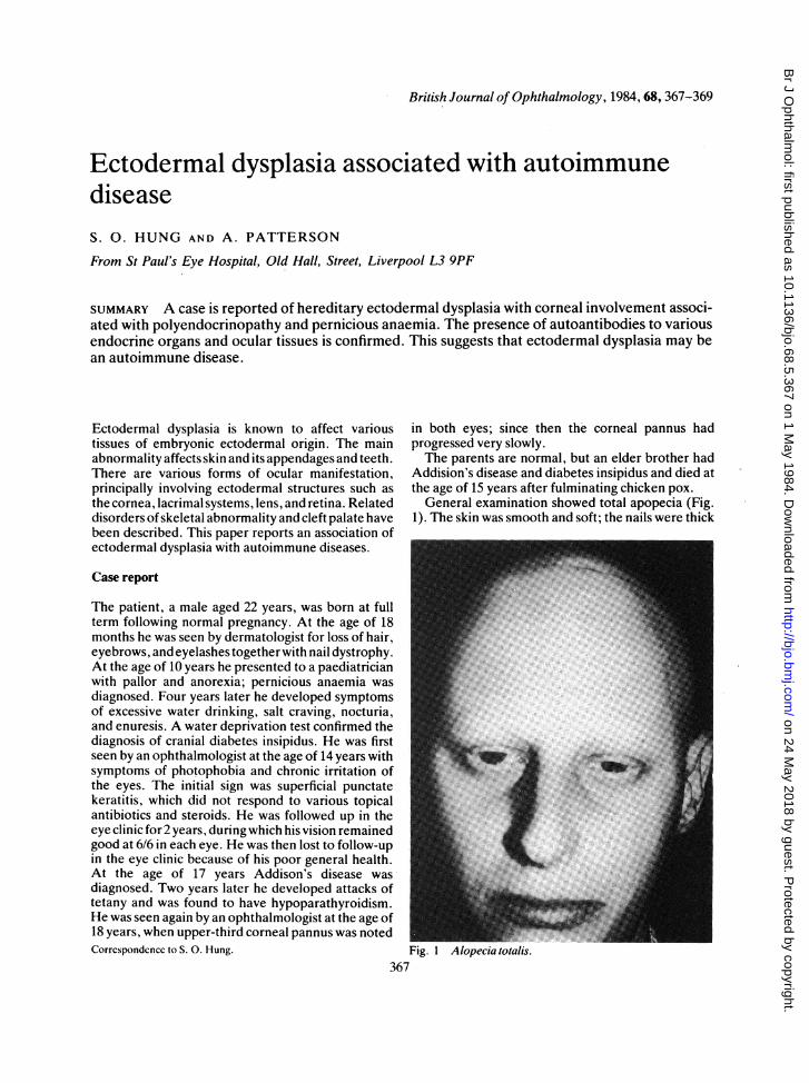
General examination showed total apopecia (Fig.1). The skin was smooth and soft; the nails were thick
367
on 24 May 2018 by guest. P
rotected by copyright.http://bjo.bm
j.com/
Br J O
phthalmol: first published as 10.1136/bjo.68.5.367 on 1 M
ay 1984. Dow
nloaded from
S. 0. Hung and A. Patterson
Fig. 2 Nail dystrophy.
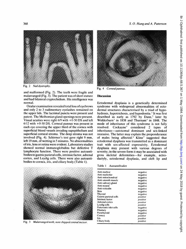
and malformed (Fig. 2). The teeth were fragile andmalarranged (Fig. 3). The patient was of short statureand had bilateral cryptochidism. His intelligence wasnormal.
Ocular examination revealed total loss ofeyebrowsand only 2 to 3 rudimentary eyelashes remained onthe upper lids. The lacrimal puncta were present andpatent. The Meibomian gland openings were present.Visual acuities were right 6/9 with +0-50 DS and left6/12 with +0-50 DS. Corneal pannus was present ineach eye covering the upper third of the cornea withsuperficial blood vessels invading supepithelium andsuperficial corneal stroma. The deep stroma was notinvolved (Fig. 4). Schirmer's test gave right 9 mm,left 19 mm, of wetting in 5 minutes. No abnormalitiesof iris, lens or retina were evident. Laboratory studiesshowed normal immunoglobulins but defective Tlymphocyte function. There were positive autoanti-bodies'to gastric parietal ce'lls, intrinsic factor, adrenalcortex, and Leydig cells. There were also autoanti-bodies to cornea, iris, and ciliary body (Table 1).
Fig.63-Malarranged chipped
Fig. 3 Malarranged teeth; note chipped central incisor.
rig. 4 Cornealpannus.
Discussion
Ectodermal dysplasia is a genetically determinedsyndrome with widespread abnormalities of ecto-dermal structures characterised by a triad of hypo-hydrosis, hypotrichosis, and hypodontia. ' It was firstdescribed as early as 1792 by Danz, later byWidderburn3 in 1838 and Thurnam4 in 1848. Themode of inheritance of this syndrome is not fullyresolved. Cockayne5 considered 2 types ofinheritance-autosomal dominant and sex-linkedrecessive. The latter may explain the preponderanceof males being affected.6 Kline7 suggested thatectodermal dysplasia was transmitted as a dominanttrait with sex-affected expressivity. Ectodermaldysplasia may present with various degrees ofseverity; in the severe form it may be associated withgross skeletal deformities-for example, ectro-dactyly, ectodermal dysplasia, and cleft lip and
Table 1 Autoantibodies
Anti-nuclearAnti-nucleolarAnti-mitochondrialAnti-smooth muscleAnti-salivary glandAnti-neuralAnti-rcticulinSkinThyroidGastric parietal cellsIntrinsic factorAdrenal cortexLeydig cellsPancreatic islet cellsPituitaryParathyroidCorneaIrisCiliary body
ncgativenegativcnegativcncgativcncgativcncgativenegativenegativcnegativepositivepositivepositivepositivenegativencgativcnegativepositivepositivepositive
368
on 24 May 2018 by guest. P
rotected by copyright.http://bjo.bm
j.com/
Br J O
phthalmol: first published as 10.1136/bjo.68.5.367 on 1 M
ay 1984. Dow
nloaded from
Ectodermal dysplasia associated with autoimmune disease
palate.8-"' Ectodermal dysplasia is associated withFriedreich's ataxia and Horner's syndrome of thecentral type together with nystagmus." However,the association with autoimmune disease has notbeen reported hitherto.
Disordered structures derived from embryonicectoderm cause various ocular symptoms. Cornealchanges with pannus formation,'2 as in this case, andGroenouw type I granular dystrophy7 have beendescribed. Lacrimal system anomalies with lacrimalpunctal atresia,8 1314 cataracts, '3 5 and chorioretinalatrophy'5 have also been reported.Wilson et al. 12 recorded the histopathology of the
corneal pannus removed at superficial keratectomy:eosinophils and plasma cells were found in thepannus. They suggested that immune mechanismsmay be a factor in the corneal changes. Alopeciaareata had been found to be associated with a highincidence of autoantibodies to smooth muscle,'"indicating its association with autoimmunity. It isinteresting that the present case has demonstratedthe association of alopecia totalis and corneal pannuswith autoimmune polyendocrine disease andpernicious anaemia. Indeed, the presence of auto-antibodies to gastric parietal cells, intrinsic factor,adrenal cortex, Leydig cells, cornea, iris, and ciliarybody were confirmed. Thus ectodermal dysplasia maybe an autoimmune disease affecting structuresdeveloped from embryonic ectodermal cells.
We thank Dr C. S. Smith of Alder Hey Children's Hospital,Liverpool, for allowing us to quote the result of autoantibodies toendocrine organs. We are also grateful to Dr A. H. S. Rahi of TheInstitute of Ophthalmology for carrying out the test for ocularautoantibodies.
References
I Wecch AA. Hereditary ectodermal dysplasia (congenital ecto-dermal defect): a report of two cases. Am J Dis Child 1929; 37:766-90.
2 Danz. Concerning men without hair or teeth. Stark's ArchGeburt 1792; 5: 684 (quoted in Wilson FM, et al. 12).
3 Widdenburn W. Letter to Charles Darwin. In: Darwin CR: Thevariations of animals and plant under domestication. New York:Appleton, 1894: 2: 319.
4 Thurnam J. Two cases in which the skin, hair, and teeth arc veryimperfectly developed. R Med Chir Soc Trans (London) 1848;31:71.
5 Cockayne EA. Inherited abnormalities of the skin and itsappendages. London: Oxford University Press, 1933: 218.
6 Seagle TB. Anhidrotic hereditary ectodermal dysplasia. JPediatr 1954; 45: 688-91.
7 Kline AH, Sidbury JB, Richter CP. The occurrence of ecto-dermal dysplasia and corneal dysplasia in one family. J Pediatr1959; 55: 355-66.
8 Kaiser-Kupper M. Ectrodactyly, ectodermal dysplasia, andclefting syndrome. Am J Ophthalmol 1973; 76: 993-8.
9 Rudiger RA, Hasse W, Passarge E. Association of ectrodactyly,ectodermal dysplasia and cleft lip-palate. Am J Dis Child 1970;120: 160-3.
10 Baum JL, Bull MJ. Ocular manifestations of the ectrodactyly,ectodermal dysplasia, cleft lip-palate syndrome. Am J Oph-thalmol 1974; 78: 211-6.
11 Rook A, Wildinson DS, Ebling FJG. Textbook of dermatology.3rd ed. Oxford: Blackwell, 1979: 1: 113-5.
12 Wilson FM, Grayson M, Pieroni D. Corneal changes in ecto-dermal dysplasia, case report, histopathology, and differentialdiagnosis. Am J Ophthalmol 1973; 75: 17-26.
13 Beckerman BL. Lacrimal anomalies in anhidrotic ectodermaldysplasia. Am J Ophthalmol 1979; 75: 728-30.
14 Liakos GM. Anhidrotic ectodermal dysplasia with lacrimalanomalies. BrJ Ophthalmol 1979; 63: 520-2.
15 Gregory IDR. Congenital ectodermal dysplasia. Br J Oph-thalmol 1955; 39: 44-7.
16 Main RA, Robbie RB, Gray ES, Donald D, Horne CHW.Smooth muscle antibodies and alopecia areata. Br J Dermatol1975; 92: 389-93.
369
on 24 May 2018 by guest. P
rotected by copyright.http://bjo.bm
j.com/
Br J O
phthalmol: first published as 10.1136/bjo.68.5.367 on 1 M
ay 1984. Dow
nloaded from
Recommended