3
Early EmbryogenesisEmbryogenesis: —formationofbodystructures&organs(organogenesis) —requirescelldivision(proliferation)andcelldifferentiation(specialization) —producesthegreatvarietyofcelltypesandextracellularproductsfoundinthebody.
Cell specialization: —selectivegeneexpression(andresultantproteinproduction)istheultimateexplanationforthecelldifferentiationprocessduringembryogenesis. —geneticexpressionbyaparticularcelldependsonthecell’spreviousgenetichistory(com-mitmentlineage)anditscurrentcellularenvironment(intercellularcommunications).
Cell differentiation istheresultofcellsexpressingsomegenesandsuppressingotherswithinacommongenome.Cellsdifferbecausetheyproduceddifferentproteins/peptides.
Proteins&peptidesare: —structuralcomponents(cytoskeletonorextracellularstructures) —enzymes(controllingcellmetabolism) —secretoryproducts(e.g.,hormones;digestiveenzymes;etc.) —channels&pumps(passageofmoleculesacrossmembranes) —receptors(communication,etc.)
Periods: Embryonic Period — definedasthetimefromfertilizationtotheearliest(primordial)stagesoforgandevelopment(about30daysindog,cat,sheep,pig;almost60daysinhorse,cattle,human).
Fetal Period — thetimebetweentheembryonicperiodandparturition(theendofgesta-tion),duringwhichorgansgrowandbegintofunction.Fertilization: —unionofahaploidoocyteandahaploidspermatozoon,producingadiploidzygote (apleuripotentcellcapableofdevelopingintoanewindividual) —fertilizationbeginswithgametefusion(zygoteformation) —fertilizationendswiththeinitiationofzygotecelldivision(thestartofcleavage) Fertilization related details: —fusionofaspermatozoonwithanoocytetakesplaceintheuterinetube,neartheovary —thespermatozoonmustbindtoaspecificglycoproteinonthezonapellucidasurrounding theoocyte[thisspeciesrecognitionprocesspreventsunionwithforeignsperm];
specialized cell
stellate neuron, etc.
stem cell committed cells
neural epitheliumneuroblast
glioblast
pyramidal neuron
astrocyte
oligodendrocyte
ectoderme.g.,
Cell Differentiation
4
—thenthespermatozoonreleasesdegradativeenzymes(acrosomalreaction)[theenzymes denaturethezonapellucida,allowingthespermcelltopenetratethebarrier] —spermatozoonandoocyteplasmamembranesfuse(secondaryoocytecompletesmeiosis) —theoocyteimmediatelycancelsitsmembranepotential(viaCa++influx)andthen denaturesitszonapellucida(viaenzymesarereleasedbyexocytosisfromoocyte cytoplasmicgranules)[thispreventsfusionbyadditionalsperm] —male&femalehaploidpronucleimakecontact,losetheirnuclearmembranes,andbegin mitosis(mitosisbegins12hoursafterspermfusion;DNAsynthesistakesplacebeforemitosis)
Oocyte(envelopedbyazonapellucida(glycoproteinmembrane)andcoronaradiata(granulosacells)atovulation) —selectivefolliclesmatureateachcycle(inresponsetocirculatingFSHhormonefromthepituitary) —oogonia(germcells)giverisetoprimaryoocytesbymitosiswithintheembryo —primaryoocytesinitiateMeiosisI(reductiondivision)withintheembryoandonlyresumeMeiosisI followingovulation(beingsuspendedinMeiosisIbyinhibitorysecretionoffolliclegranulosacells) —secondaryoocytescompletemeiosis(MeiosisII)followingfertilization(ifunfertilizedtheydegenerate), producingafertilizedoocyte(ovum).
Spermatozoa(severalhundredmillionperejaculate) —propelledfromvaginatouterinetubebycontractionoffemalegenitaltract —spermatogonia(germcells)giverisetoprimaryspermatocytesbymitosisrepetitivelyfollowingpuberty —primaryspermatocytesundergoMeiosisI(reductiondivision)producingsecondaryspermatocytes —secondaryspermatocytescompletemeiosis(MeiosisII),producingspermatidsthatundergo transformationintospermatozoa(spermiogenesis) —subsequently,spermatozoaundergocapacitation(removalofsurfaceproteinsthatwouldimpedecontact withanoocyte)
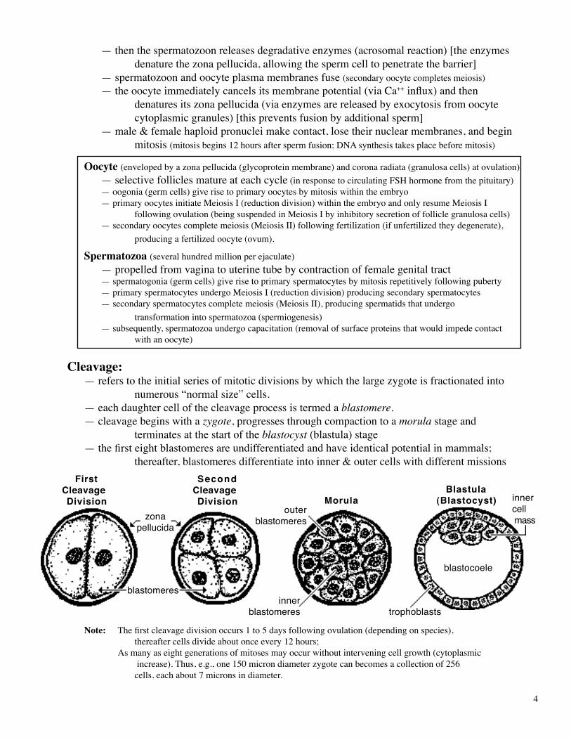
Cleavage: —referstotheinitialseriesofmitoticdivisionsbywhichthelargezygoteisfractionatedinto numerous“normalsize”cells. —eachdaughtercellofthecleavageprocessistermedablastomere. —cleavagebeginswithazygote,progressesthroughcompactiontoamorulastageand terminatesatthestartoftheblastocyst (blastula)stage —thefirsteightblastomeresareundifferentiatedandhaveidenticalpotentialinmammals; thereafter,blastomeresdifferentiateintoinner&outercellswithdifferentmissions
Note: Thefirstcleavagedivisionoccurs1to5daysfollowingovulation(dependingonspecies), thereaftercellsdivideaboutonceevery12hours;Asmanyaseightgenerationsofmitosesmayoccurwithoutinterveningcellgrowth(cytoplasmic increase).Thus,e.g.,one150microndiameterzygotecanbecomesacollectionof256 cells,eachabout7micronsindiameter.
FirstCleavage Division
SecondCleavage Division Morula
Blastula(Blastocyst)
zonapellucida
blastomeres
outerblastomeres
innerblastomeres trophoblasts
blastocoele
innercell mass
5
Morula[L.=smallmulberry] —asolidballofblastomereswithinazonapellucida(typicallyconsistingof16to64blastomeres) —blastomeresbecomecompacted;cellsontheinsidedifferentiatefromthosealongthe surfaceofthemorula: —outer blastomeresbecomeflattenedandformtightjunctions(reducingfluidpermeability); theydevelopthecapacitytosecretefluid(internally);theyaredestinedtobecome trophoblastswhichformthechorion&amnion(fetalmembranes)oftheconceptus; —inner blastomeresformgapjunctionstomaximizeintercellularcommunication;theyare destinedtobecomeinner cell masswhichformstheembryoitself(plustwo fetalmembranes).
Note: •Asfewasthreeinnerblastomeresaresufficienttoproduceanentireembryo(andadult).•Whenamorulaleavestheuterinetubeandenterstheuterus(uterinehorn)itisatabout the16-cellstage,around4to7daysafterfertilization(dependingonspecies).•The32-cellstagemorula(5-7dayspostovulation)isidealforembryotransferincattle.
Blastocyst(orBlastula) —developsduringthesecondweek,afterthezonapellucidaruptures —consistsofalargenumberofblastomeresarrangedtoformahollow,fluid-filled,spherical orcylindricalstructure —containsaninner cell mass(embryoblast),evidentasacollectionofcellslocalizedinside onepolarendoftheblastula —surfacecellsoftheblastocystaredesignatedtrophoblasts(futurechorionoftheconceptus) —thecavityoftheblastocystiscalledablastocoele —eventuallytheblastocystattachestoorimplantswithintheuterinewall(pendingspecies).
Cleavage in fish, reptiles, and birds: Largequantitiesofyolkimpedecelldivisionduringcleavage.Thusablastodisc(ratherthanasphericalorellipticalblastocyst)isformedattheanimalpoleoftheegg. A telolecithal ovum (eggwithlargeamountsofasymmetricallydistributedyolk)hasananimal polewherethenucleusislocatedandanoppositevegetal polewhereyolkisconcentrated.Cleavageispar-tial(meroblastic):cellsdividemorerapidlyattheanimalpolethanatthevegetalpole,resultinginmany,smallblastomeresattheanimalpoleandafew,largemacromeresatthevegetalpole. Incontrast,mammalianovumhasmeageramountsofyolk(oligolecithalovum)whichisuni-formlydistributed(isolecithal).Cleavageisholoblastic(total)andeachblastomeredivisionproducestwoequal-sizedaughtercells.Thusanimalandvegetalpolesarenotevidentinmammalianova.
TwiNS Monozygotic:identical(samegeneticcomposition)twinscanresultfromeither:1]separationofearlyblastomeres(uptothe8-cellstage)—eachoftheseparate
blastomere(s)developsintoanindependentconceptus;or2]separationofinnerblastomereswithinasinglemorula—eachoftheseparate
blastomere(s)developsintoanindependentembryoandbothembryosshareacommonplacenta(thisislesscommonthanthefirstpossibility).
Note:Separationslaterinembryonicdevelopmentresultinconjoinedtwins(diplopagus;Siamesetwins),ordoubleheads,etc.typesofanomalies.
Dizygotic:fraternaltwinsresultwhentwozygotesdevelop“independently”duringthesamepregnancy(independencecanbecompromisedbyfusionoffetalmembranesandbloodsupplies).Itispossibleforfraternalblastomerestomergeandproduceasingleconceptuswithtwodifferentgenotypes(achimera).
6
GERM LAYERSEctoderm,mesodermandendodermaredesignatedprimary germ layersbecause originsofallorganscanbetracedbacktothesethreelayers.Ectodermformsepidermisoftheskin,epitheliumoftheoralandnasalcavities,and thenervoussystemandsenseorgans.Mesodermformsmuscleandconnectivetissue,includingbone,andcomponentsof thecirculatory,urinaryandgenitalsystems.Endodermformsmucosalepitheliumandglandsofrespiratoryanddigestivesystems.
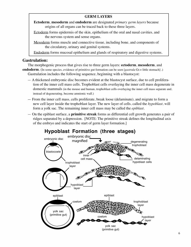
Gastrulation: Themorphogenicprocessthatgivesrisetothreegermlayers:ectoderm,mesoderm,andendoderm.(Insomespecies,evidenceofprimitivegutformationcanbeseen[gastrulaGr.=littlestomach].) Gastrulationincludesthefollowingsequence,beginningwithablastocyst:
—Athickenedembryonicdiscbecomesevidentattheblastocystsurface,duetocellprolifera-tionoftheinnercellmasscells.Trophoblastcellsoverlayingtheinnercellmassdegenerateindomesticmammals(inthemouseandhuman,trophoblastcellsoverlayingtheinnercellmassseparateand,insteadofdegenerating,becomeamnionicwall.)
—Fromtheinnercellmass,cellsproliferate,breakloose(delaminate),andmigratetoformanewcelllayerinsidethetrophoblastlayer.Thenewlayerofcells,calledthehypoblast,willformayolksac.Theremaininginnercellmassmaybecalledtheepiblast.
—Ontheepiblastsurface,aprimitive streak formsasdifferentialcellgrowthgeneratesapairofridgesseparatedbyadepression.[NOTE:Theprimitivestreakdefinesthelongitudinalaxisoftheembryoandindicatesthestartofgermlayerformation.]
trophoblastlayer
degeneratingtrophoblast
hypoblastlayer
inner cell mass
embryonic disc
epiblast
Hypoblast Formation (three stages)
delaminatinghypoblast cells
blastocoele
embryonic discmagni�ed
trophoblastlayer
hypoblastlayer
coelom
yolk sac(primitive gut)
coelom
yolk sac(primitive gut)
epiblast

7
—Deeptotheprimitivestreak,aspace(coelom/celom)becomesevidentbetweenthehypoblastlayerandepiblast.Subsequently,thecoelomisfilledbymesodermthatundergoescavitationandgivesrisetobodycavities.
—Epiblastcellsproliferatealongprimitivestreakmarginsandmi-gratethroughthestreakintothecoelom.Themigratingcellsformendoderm&mesodermlayers.
—Initialmigratingcellsjointhehypoblastlayer,formingembry-onic endoderm (hypoblastcellsconstitutesyolksacendoderm).
—Themajorityofmigratingcellsenterthecoelomasprimarymesenchymeandbecomemeso-derm.Theprimarymesenchymemigrateslaterallyandcranially(butnotalongthemidlineregiondirectlycranialtotheprimitivestreakwherenotochordwillform).Note:Mesodermdividesinto:paraxial,intermediate,andlateralmesodermalregions.
—Withinthelateralmesoderm,cavitationre-establishesacoelom(hoseshoe-shaped).Themesodermsplitsintotwolayersborderingthecoelom—somatic mesodermisattachedtotheectodermandsplanchnic mesodermisjoinedtoendoderm.
—Theremainingepiblastbecomesectoderm whichformsskinepidermis&nervoussystem.
Dorsal View of Embryonic Disc
NOTE: Arrows indicate the spread of primary mesenchyme through the primitive streak
and between the epiblast and hypoblast
primitivestreak
primitivenode
primarymesenchyme
notochord
epiblast (ectoderm)
hypoblast endoderm primary mesenchyme (mesoderm)
primitive streak

8
NOTE: Mesodermcanexistintwomorphologicforms:mesenchymeandepithelioid: Mesenchymefeaturesaggregatesofstellatecellswithinanabundantextracel-
lularmatrixcomposedoffluidandmacromolecules(polymers). Epithelioid referstoorganizedcellshavingdistinctapicalandbasalsurfaces;
thelattercommonlyrestsonabasallaminaproducedbyepithelioidsecretion.Mesodermcantransformfromamesenchymetoepithelioidandviceversa:The
mesodermthatstreamsthroughtheprimitivestreakisprimary mesenchyme.Somatic,splanchnic,andsomitemesodermcanbetemporarilyepithelioid.Thetemporaryepithelioidtransformstoasecondary mesenchymewhichulti-matelyformsmuscleandconnectivetissue(includingcartilage,bone,liga-ments,tendons,dermis,fascia,andadiposetissue).
Thus,theterm“mesenchyme”referstothemorphologicappearanceofembryonictis-sue.Althoughmostmesenchymeismesoderm,theothergermlayerscanalsoformmesenchyme,e.g.,ectomesenchymefromneuralcrestectoderm.
Formation of the Notochord:•Thenotochordisarod-shapedaggregateofcellslocatedbetweenectodermandendoderm
anteriortotheprimitivestreakoftheembryo.Itoccupiesthemidlinecoelomicspacethatwasnotinvadedbymigratingprimarymesenchyme.
•Thenotochordisimportantbecauseitinduces: formationoftheheadprocess, developmentofthenervoussystem,and formationofsomites•Thenotochordmarksthefuturelocationofthevertebralcolumnandthebaseofthecranium.•Theultimatefateofthenotochordistobecomenucleuspulposusofintervertebraldiscs.Note:Thenotochorddevelopsfromtheprimitive nodelocatedatthecranialendoftheprimitivestreak.Fromthe
node,mesoderm-formingcellsproliferateandmigrateforwardintothefutureheadregionwheretheybecometherod-shapednotochord.
9
Note: Eachorgansystemhasacriticalperiodduringdevelopmentwhenitismostsensitivetoexternalagents(teratogens)thatproducebirthdefects.
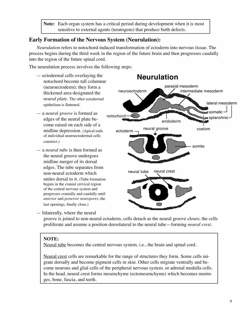
Early Formation of the Nervous System (Neurulation): Neurulationreferstonotochord-inducedtransformationofectodermintonervoustissue.Theprocessbeginsduringthethirdweekintheregionofthefuturebrainandthenprogressescaudallyintotheregionofthefuturespinalcord.Theneurulationprocessinvolvesthefollowingsteps:
—ectodermalcellsoverlayingthenotochordbecometallcolumnar(neuroectoderm);theyformathickenedareadesignatedtheneural plate. Theotherectodermalepitheliumisflattened.
—aneural grooveisformedasedgesoftheneuralplatebe-comeraisedoneachsideofamidlinedepression.(Apicalendsofindividualneuroectodermalcellsconstrict.)
—aneural tubeisthenformedastheneuralgrooveundergoesmidlinemergerofitsdorsaledges.Thetubeseparatesfromnon-neuralectodermwhichunitesdorsaltoit.(Tubeformationbeginsinthecranialcervicalregionofthecentralnervoussystemandprogressescraniallyandcaudallyuntilanteriorandposterior neuropores,thelastopenings,finallyclose.)
—bilaterally,wheretheneuralgrooveisjoinedtonon-neuralectoderm,cellsdetachastheneuralgroovecloses;thecellsproliferateandassumeapositiondorsolateraltotheneuraltube—formingneural crest.
NOTE:Neuraltubebecomesthecentralnervoussystem,i.e.,thebrainandspinalcord.
Neuralcrestcellsareremarkablefortherangeofstructurestheyform.Somecellsmi-gratedorsallyandbecomepigmentcellsinskin.Othercellsmigrateventrallyandbe-comeneuronsandglialcellsoftheperipheralnervoussystem,oradrenalmedullacells.Inthehead,neuralcrestformsmesenchyme(ectomesenchyme)whichbecomesmenin-ges,bone,fascia,andteeth.
Neurulation
ectoderm
neuroectoderm
notochord
neural groove
neural tube
paraxial mesodermintermediate mesoderm
lateral mesoderm
coelom
somite
neural crest
somaticsplanchnic
endoderm
10
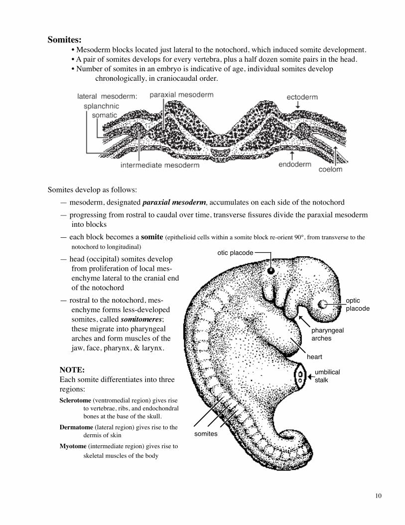
Somites: •Mesodermblockslocatedjustlateraltothenotochord,whichinducedsomitedevelopment. •Apairofsomitesdevelopsforeveryvertebra,plusahalfdozensomitepairsinthehead. •Numberofsomitesinanembryoisindicativeofage,individualsomitesdevelop
chronologically,incraniocaudalorder.
Somitesdevelopasfollows:—mesoderm,designatedparaxial mesoderm,accumulatesoneachsideofthenotochord—progressingfromrostraltocaudalovertime,transversefissuresdividetheparaxialmesoderm
intoblocks—eachblockbecomesasomite(epithelioidcellswithinasomiteblockre-orient90°,fromtransversetothe
notochordtolongitudinal)
—head(occipital)somitesdevelopfromproliferationoflocalmes-enchymelateraltothecranialendofthenotochord
—rostraltothenotochord,mes-enchymeformsless-developedsomites,calledsomitomeres;thesemigrateintopharyngealarchesandformmusclesofthejaw,face,pharynx,&larynx.
NOTE: Eachsomitedifferentiatesintothreeregions:Sclerotome(ventromedialregion)givesrise
tovertebrae,ribs,andendochondralbonesatthebaseoftheskull.
Dermatome(lateralregion)givesrisetothedermisofskin
Myotome(intermediateregion)givesrisetoskeletalmusclesofthebody
otic placode
optic placode
heart
umbilicalstalk
somites
pharyngealarches
11
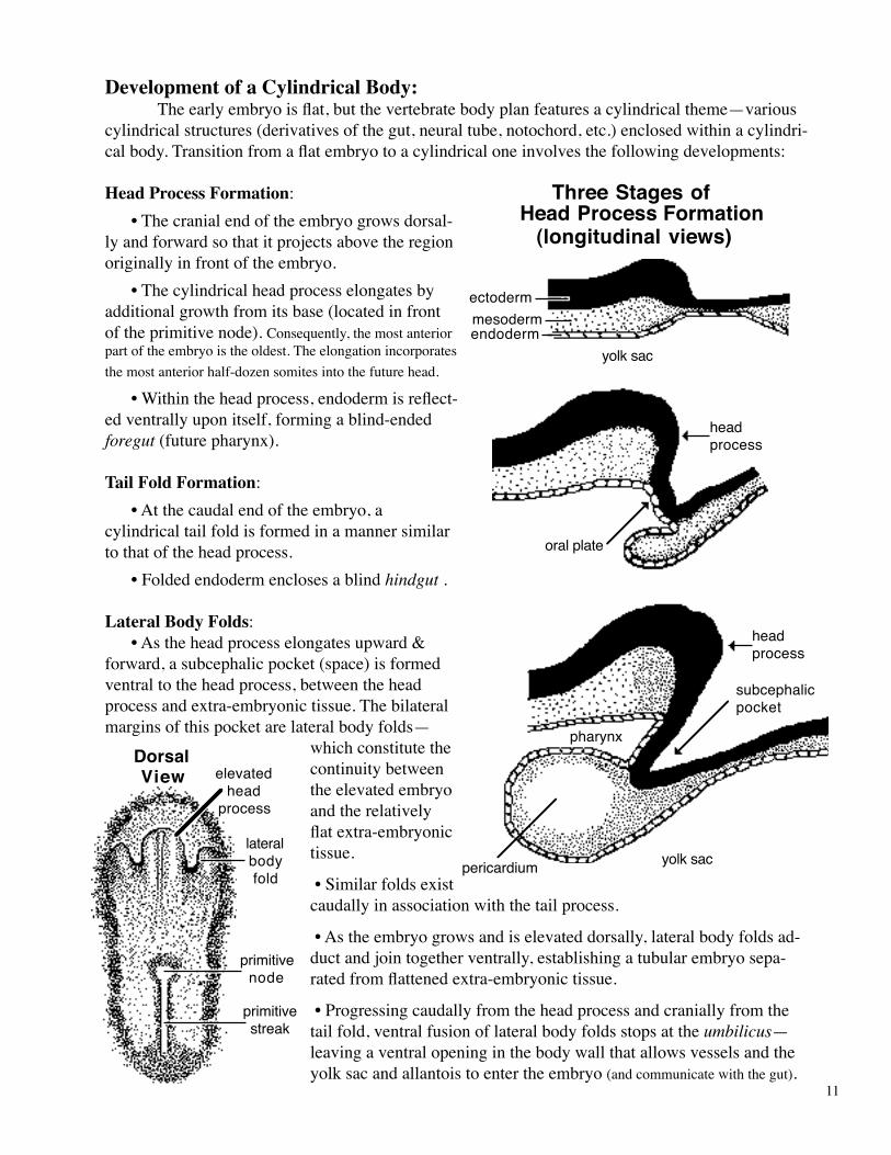
Development of a Cylindrical Body: Theearlyembryoisflat,butthevertebratebodyplanfeaturesacylindricaltheme—variouscylindricalstructures(derivativesofthegut,neuraltube,notochord,etc.)enclosedwithinacylindri-calbody.Transitionfromaflatembryotoacylindricaloneinvolvesthefollowingdevelopments:
Head Process Formation: •Thecranialendoftheembryogrowsdorsal-lyandforwardsothatitprojectsabovetheregionoriginallyinfrontoftheembryo. •Thecylindricalheadprocesselongatesbyadditionalgrowthfromitsbase(locatedinfrontoftheprimitivenode).Consequently,themostanteriorpartoftheembryoistheoldest.Theelongationincorporatesthemostanteriorhalf-dozensomitesintothefuturehead.
•Withintheheadprocess,endodermisreflect-edventrallyuponitself,formingablind-endedforegut(futurepharynx).
Tail Fold Formation: •Atthecaudalendoftheembryo,acylindricaltailfoldisformedinamannersimilartothatoftheheadprocess. •Foldedendodermenclosesablindhindgut.
Lateral Body Folds: •Astheheadprocesselongatesupward&forward,asubcephalicpocket(space)isformedventraltotheheadprocess,betweentheheadprocessandextra-embryonictissue.Thebilateralmarginsofthispocketarelateralbodyfolds—
whichconstitutethecontinuitybetweentheelevatedembryoandtherelativelyflatextra-embryonictissue.
•Similarfoldsexistcaudallyinassociationwiththetailprocess.
•Astheembryogrowsandiselevateddorsally,lateralbodyfoldsad-ductandjointogetherventrally,establishingatubularembryosepa-ratedfromflattenedextra-embryonictissue.
•Progressingcaudallyfromtheheadprocessandcraniallyfromthetailfold,ventralfusionoflateralbodyfoldsstopsattheumbilicus—leavingaventralopeninginthebodywallthatallowsvesselsandtheyolksacandallantoistoentertheembryo(andcommunicatewiththegut).
oral plate
head process
subcephalicpocket
pharynx
ectoderm
endodermmesoderm
yolk sac
Three Stages ofHead Process Formation
(longitudinal views)
head process
yolk sacpericardium
DorsalView
primitivestreak
primitivenode
elevatedhead
process
lateralbodyfold
12
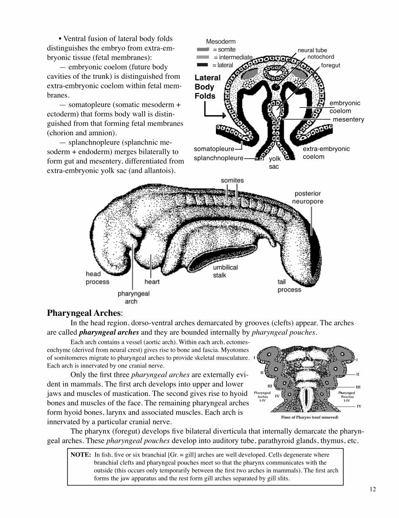
•Ventralfusionoflateralbodyfoldsdistinguishestheembryofromextra-em-bryonictissue(fetalmembranes): —embryoniccoelom(futurebodycavitiesofthetrunk)isdistinguishedfromextra-embryoniccoelomwithinfetalmem-branes. —somatopleure(somaticmesoderm+ectoderm)thatformsbodywallisdistin-guishedfromthatformingfetalmembranes(chorionandamnion). —splanchnopleure(splanchnicme-soderm+endoderm)mergesbilaterallytoformgutandmesentery,differentiatedfromextra-embryonicyolksac(andallantois).
Pharyngeal Arches: Intheheadregion,dorso-ventralarchesdemarcatedbygrooves(clefts)appear.Thearchesarecalledpharyngeal arches andtheyareboundedinternallybypharyngeal pouches. Eacharchcontainsavessel(aorticarch).Withineacharch,ectomes-enchyme(derivedfromneuralcrest)givesrisetoboneandfascia.Myotomesofsomitomeresmigratetopharyngealarchestoprovideskeletalmusculature.Eacharchisinnervatedbyonecranialnerve. Onlythefirstthreepharyngeal arches areexternallyevi-dentinmammals.Thefirstarchdevelopsintoupperandlowerjawsandmusclesofmastication.Thesecondgivesrisetohyoidbonesandmusclesoftheface.Theremainingpharyngealarchesformhyoidbones,larynxandassociatedmuscles.Eacharchisinnervatedbyaparticularcranialnerve. Thepharynx(foregut)developsfivebilateraldiverticulathatinternallydemarcatethepharyn-gealarches.Thesepharyngeal pouchesdevelopintoauditorytube,parathyroidglands,thymus,etc.
NOTE:Infish,fiveorsixbranchial[Gr.=gill]archesarewelldeveloped.Cellsdegeneratewherebranchialcleftsandpharyngealpouchesmeetsothatthepharynxcommunicateswiththeoutside(thisoccursonlytemporarilybetweenthefirsttwoarchesinmammals).Thefirstarchformsthejawapparatusandtherestformgillarchesseparatedbygillslits.
Mesoderm = somite = intermediate = lateral
neural tubenotochord
foregut
mesentery
yolksac
embryoniccoelom
extra-embryoniccoelom
somatopleuresplanchnopleure
LateralBody Folds
13
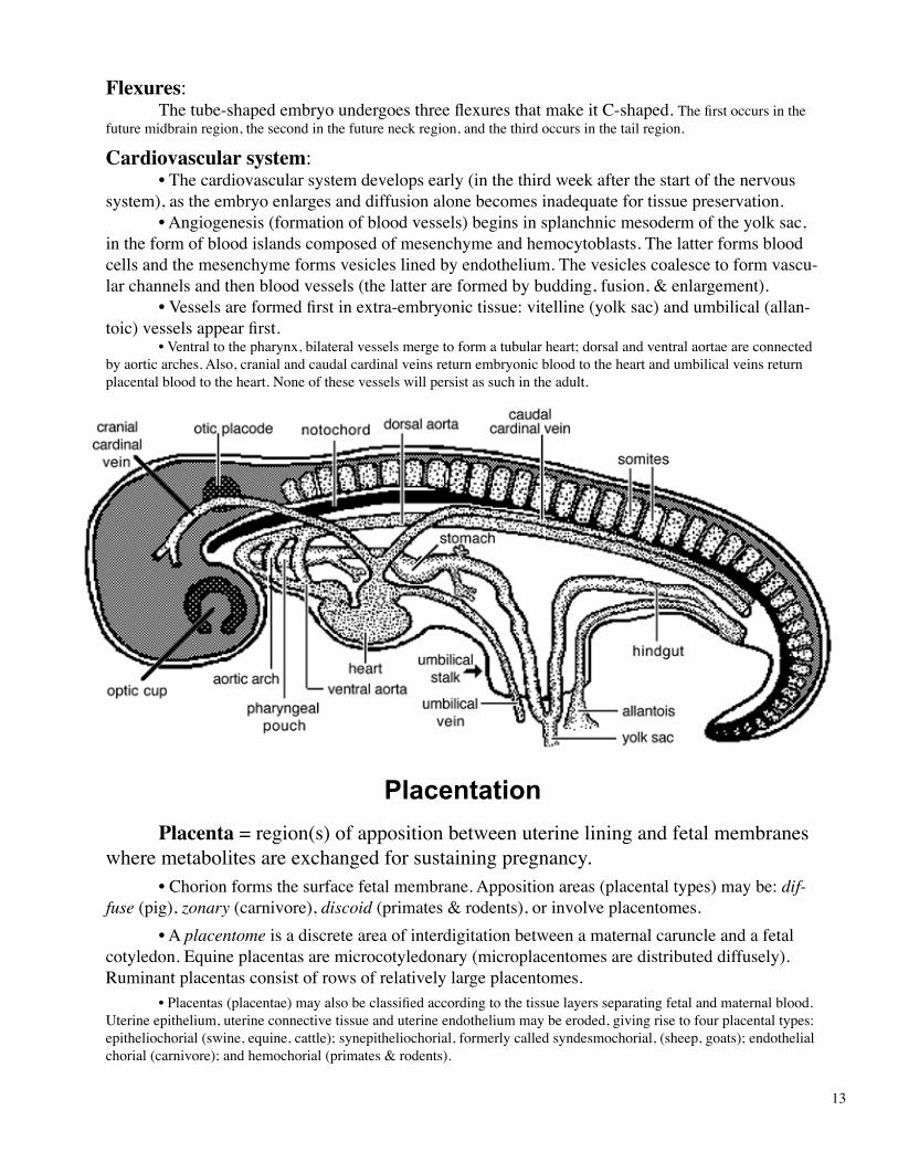
Flexures: Thetube-shapedembryoundergoesthreeflexuresthatmakeitC-shaped.Thefirstoccursinthefuturemidbrainregion,thesecondinthefutureneckregion,andthethirdoccursinthetailregion.
Cardiovascular system: •Thecardiovascularsystemdevelopsearly(inthethirdweekafterthestartofthenervous
system),astheembryoenlargesanddiffusionalonebecomesinadequatefortissuepreservation. •Angiogenesis(formationofbloodvessels)beginsinsplanchnicmesodermoftheyolksac,
intheformofbloodislandscomposedofmesenchymeandhemocytoblasts.Thelatterformsbloodcellsandthemesenchymeformsvesicleslinedbyendothelium.Thevesiclescoalescetoformvascu-larchannelsandthenbloodvessels(thelatterareformedbybudding,fusion,&enlargement).
•Vesselsareformedfirstinextra-embryonictissue:vitelline(yolksac)andumbilical(allan-toic)vesselsappearfirst.
•Ventraltothepharynx,bilateralvesselsmergetoformatubularheart;dorsalandventralaortaeareconnectedbyaorticarches.Also,cranialandcaudalcardinalveinsreturnembryonicbloodtotheheartandumbilicalveinsreturnplacentalbloodtotheheart.Noneofthesevesselswillpersistassuchintheadult.
Placentation Placenta=region(s)ofappositionbetweenuterineliningandfetalmembraneswheremetabolitesareexchangedforsustainingpregnancy. •Chorionformsthesurfacefetalmembrane.Appositionareas(placentaltypes)maybe:dif-fuse (pig),zonary (carnivore),discoid (primates&rodents),orinvolveplacentomes. •Aplacentome isadiscreteareaofinterdigitationbetweenamaternalcaruncleandafetalcotyledon.Equineplacentasaremicrocotyledonary(microplacentomesaredistributeddiffusely).Ruminantplacentasconsistofrowsofrelativelylargeplacentomes. •Placentas(placentae)mayalsobeclassifiedaccordingtothetissuelayersseparatingfetalandmaternalblood.Uterineepithelium,uterineconnectivetissueanduterineendotheliummaybeeroded,givingrisetofourplacentaltypes:epitheliochorial(swine,equine,cattle);synepitheliochorial,formerlycalledsyndesmochorial,(sheep,goats);endothelialchorial(carnivore);andhemochorial(primates&rodents).

14
Porcine Chorionic Surface(folds; diffuse placental contact)
Equine Chorionic Surface(microcotyledons)
Bovine Chorionic Surface(rows of cotyledons)
Carnivore Chorionic Surface(zonary placental contact)
Human/Rodent Chorionic Surface(discoid placental contact)
Fetal Components of Placentae
marginalhematoma
cervical star(region over cervix)ne
crot
ic ti
p(c
horio
n w
ithou
t alla
ntoi
s)
marginalhematoma

15
Fetal membranes: Fourfetalmembranesdevelopinaconceptus.Twoarisefromthetrophoblastlayeroftheblastocyst(andarecontinuouswiththesomatopleureoftheembryo).Twoarisefromtheinnercellmassoftheblastocyst(andarecontinuouswithsplanchnopleureoftheembryo);thesetwosplanch-nopleuremembranesarevascular.Thefourfetalmembranesare:
1.Chorion—formstheouterboundaryoftheentireconceptus(fromtrophoblast) 2.Amnion—enclosestheembryowithinafluid-filledamnioniccavity;formedbyfoldsofchorionindomesticmammals(inhumans,amnionformsbycavitationdeeptoapersistenttrophoblast). 3.Allantois—developsasanoutgrowthofhindgutsplanchnopleure(originatesfrominnercellmass).Allantoisgrowstofilltheentireextra-embryoniccoelom,withfluid-filledallantoiccavityindomesticmammals.Theoutersurfaceofallantoisbindstotheinnersurfaceofchorion(andtheoutersurfaceofamnion).Theallantoisishighlyvascularandprovidesthefunctionalvesselsoftheplacenta,viaumbilicalvessels. 4.Yolk sac—continuouswithmidgutsplanchnopleure(developsearlywithhypoblastfor-mationfrominnercellmass).Suppliedbyvitellinevessels,itformsanearlytemporaryplacentainthehorseanddog.Yolksacismostimportantinegglayingvertebrates.Note: Thetermconceptusreferstotheembryoorfetusplusitsfetalmembranes.
implantation Theblastocystisinitiallyfreeintheuterinelumen(nourishedbyuterineglands).Im-
plantationoftheblastocyctisagradualprocess,beginningwithapposition,leadingtoadhesion(orinvasioninthecaseofthehuman&GuineaPig).
Approximateimplantationtimesare:oneweek(human);twoweeks(dog,cat,sheep),3-5weeks(cattle),3-8weekshorse;ordelayedupto4mons(deer,bears).
amnion
somatopleure
splanchnopleure
gut
yolk sac
allantoischorion
coelom
embryo
Recommended