Dually Fluorescent Core-Shell Microgels for RatiometricImaging in Live Antigen-Presenting CellsXianfeng Zhou, Fengyu Su, Yanqing Tian*, Deirdre R. Meldrum
Center for Biosignatures Discovery Automation, Biodesign Institute, Arizona State University, Tempe, Arrizona, United States of America
Abstract
Core-shell microgels containing sensors/dyes in a matrix were fabricated by two-stage free radical precipitationpolymerization method for ratiometric sensing/imaging. The microgels composing of poly(N-isopropylacrylamide) (PNIPAm)shell exhibits a low critical solution temperature (LCST), underwent an entropically driven transition from a swollen state to adeswollen state, which exhibit a hydrodynamic radius of ,450 nm at 25uC (in vitro) and ,190 nm at 37uC (in vivo). Themicrogel’s ability of escaping from lysosome into cytosol makes the microgel be a potential candidate for cytosolic deliveryof sensors/probes. Non-invasive imaging/sensing in Antigen-presenting cells (APCs) was feasible by monitoring the changesof fluorescence intensity ratios. Thus, these biocompatible microgels-based imaging/sensing agents may be expected toexpand current molecular imaging/sensing techniques into methods applicable to studies in vivo, which could further driveAPC-based treatments.
Citation: Zhou X, Su F, Tian Y, Meldrum DR (2014) Dually Fluorescent Core-Shell Microgels for Ratiometric Imaging in Live Antigen-Presenting Cells. PLoSONE 9(2): e88185. doi:10.1371/journal.pone.0088185
Editor: Vipul Bansal, RMIT University, Australia
Received November 1, 2013; Accepted January 5, 2014; Published February 4, 2014
Copyright: � 2014 Zhou et al. This is an open-access article distributed under the terms of the Creative Commons Attribution License, which permitsunrestricted use, distribution, and reproduction in any medium, provided the original author and source are credited.
Funding: This work was supported by NIH 5P50 HG002360. The funders had no role in study design, data collection and analysis, decision to publish, orpreparation of the manuscript.
Competing Interests: The authors have declared that no competing interests exist.
* E-mail: [email protected]
Introduction
Antigen-presenting cells (APCs) internalize antigens, present
antigen-derived peptides to T cells containing major histocom-
patibility complexes (MHCs) on their surfaces, and play a pivotal
role in both initiation and regulation of immune responses [1].
Intracellular variables such as pH and oxygen levels are important
factors in regulation of the antigen-presenting processes [2]. The
interferon-gamma (IFN-c) and pro-inflammatory cytokine pro-
duced by APCs is dictated by intracellular local oxygen tension
[3]; therefore, there is much interest in the development of
methods suitable for detection of essential analytes (such as
dissolved oxygen and pH) in the clinical sitting of APCs. The
development of cellular imaging and sensing techniques is
imperative to ultimately advancing APCs-based therapy. Fluores-
cent sensor-based imaging of cells is an alternative, noninvasive
imaging modality with the capability of cellular events tracking [4–
6]. The approach has intrinsic value because asynchronous and
single-cell level behaviors of APCs are not indicated by population
measurements [7].
Polymer-based fluorescent nanosensors [8,9], or probes encap-
sulated by biologically localized embedding sensors (PEBBLEs),
were first created to be used for intracellular measurements by
creating a biocompatible shell around the probes [10,11]. The real
power of particle-based nanosensors was realized when PEBBLEs
were embedded with a sensing probe and a reference dye [12].
This feature makes the sensor ratiometric. By plotting the ratio of
sensing probe over reference dye emission vs. analyte concentra-
tion, the fluorescence signal can be handled in a quantitative
manner. For these early sensors, however, the optical probes were
physically incorporated into the polymer matrix. The possible
leaching of these probes from the matrices might be a significant
problem which may result in signal instability, inaccuracy of the
measurement, decreased long-term applicability, and potential
cytotoxicity for cells [13]. To address this problem, the probes/
dyes have been covalently attached to the matrix to give sensing/
imaging microgels [14,15].
APCs can take up nanoparticles by ligand-mediated endocyto-
sis. However, many other cell types also are capable of ingesting
these small-sized particulate matters (typically ,200 nm in
diameter) by mechanisms such as pinocytosis and endocytosis
[16]. The uptake of particles with diameters up to several
micrometers is generally restricted to phagocytic APCs; therefore,
tuning the size of particulate delivery systems can enable passive
targeting of optical sensors to APCs. Several reports have
highlighted the impact that particle sizes may have interactions
with APCs [17]. For example, early studies on the functional
application of particulate carriers indicated that carriers with
diameters between 0.5 and 3 mm were effective for APCs in vitro
[18]. However, in vivo studies using a similar polystyrene-based
system suggest that particles less than 200 nm in diameter are
effective at activating APCs [19].
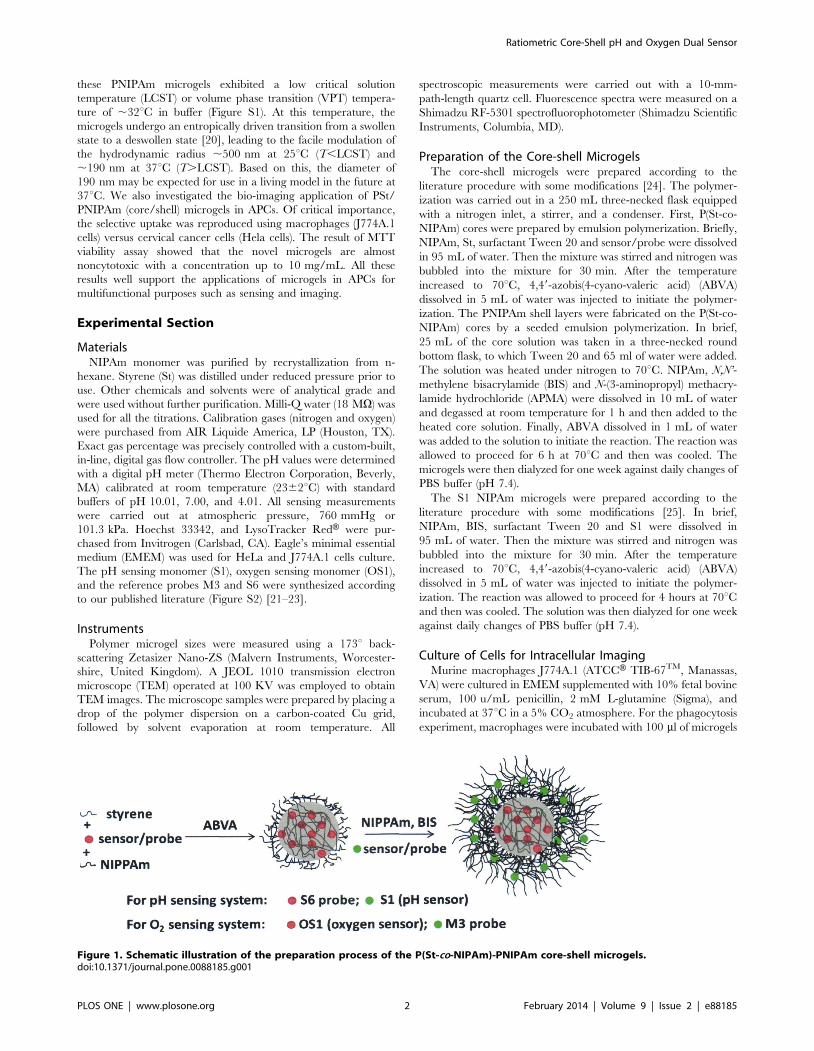
Herein, we report on the fabrication of core-shell microgels with
covalently incorporated sensors/dyes in the matrix for ratiometric
pH and oxygen sensing (Figure 1). The microgels are mainly
composed of a polystyrene (PSt) core and a poly(N-isopropylacry-
lamide) (PNIPAm) shell. Such a structure makes it possible for the
microgels to be stable in aqueous solutions and act as efficient
solubilizers of hydrophobic sensors/dyes to enable their applica-
tions in biological environments. This versatile matrix allows for
chemical immobilization of the probes either into the core or the
shell for ratiometric imaging/sensing. Furthermore, we found that
PLOS ONE | www.plosone.org 1 February 2014 | Volume 9 | Issue 2 | e88185
these PNIPAm microgels exhibited a low critical solution
temperature (LCST) or volume phase transition (VPT) tempera-
ture of ,32uC in buffer (Figure S1). At this temperature, the
microgels undergo an entropically driven transition from a swollen
state to a deswollen state [20], leading to the facile modulation of
the hydrodynamic radius ,500 nm at 25uC (T,LCST) and
,190 nm at 37uC (T.LCST). Based on this, the diameter of
190 nm may be expected for use in a living model in the future at
37uC. We also investigated the bio-imaging application of PSt/
PNIPAm (core/shell) microgels in APCs. Of critical importance,
the selective uptake was reproduced using macrophages (J774A.1
cells) versus cervical cancer cells (Hela cells). The result of MTT
viability assay showed that the novel microgels are almost
noncytotoxic with a concentration up to 10 mg/mL. All these
results well support the applications of microgels in APCs for
multifunctional purposes such as sensing and imaging.
Experimental Section
MaterialsNIPAm monomer was purified by recrystallization from n-
hexane. Styrene (St) was distilled under reduced pressure prior to
use. Other chemicals and solvents were of analytical grade and
were used without further purification. Milli-Q water (18 MV) was
used for all the titrations. Calibration gases (nitrogen and oxygen)
were purchased from AIR Liquide America, LP (Houston, TX).
Exact gas percentage was precisely controlled with a custom-built,
in-line, digital gas flow controller. The pH values were determined
with a digital pH meter (Thermo Electron Corporation, Beverly,
MA) calibrated at room temperature (2362uC) with standard
buffers of pH 10.01, 7.00, and 4.01. All sensing measurements
were carried out at atmospheric pressure, 760 mmHg or
101.3 kPa. Hoechst 33342, and LysoTracker RedH were pur-
chased from Invitrogen (Carlsbad, CA). Eagle’s minimal essential
medium (EMEM) was used for HeLa and J774A.1 cells culture.
The pH sensing monomer (S1), oxygen sensing monomer (OS1),
and the reference probes M3 and S6 were synthesized according
to our published literature (Figure S2) [21–23].
InstrumentsPolymer microgel sizes were measured using a 173u back-
scattering Zetasizer Nano-ZS (Malvern Instruments, Worcester-
shire, United Kingdom). A JEOL 1010 transmission electron
microscope (TEM) operated at 100 KV was employed to obtain
TEM images. The microscope samples were prepared by placing a
drop of the polymer dispersion on a carbon-coated Cu grid,
followed by solvent evaporation at room temperature. All
spectroscopic measurements were carried out with a 10-mm-
path-length quartz cell. Fluorescence spectra were measured on a
Shimadzu RF-5301 spectrofluorophotometer (Shimadzu Scientific
Instruments, Columbia, MD).
Preparation of the Core-shell MicrogelsThe core-shell microgels were prepared according to the
literature procedure with some modifications [24]. The polymer-
ization was carried out in a 250 mL three-necked flask equipped
with a nitrogen inlet, a stirrer, and a condenser. First, P(St-co-
NIPAm) cores were prepared by emulsion polymerization. Briefly,
NIPAm, St, surfactant Tween 20 and sensor/probe were dissolved
in 95 mL of water. Then the mixture was stirred and nitrogen was
bubbled into the mixture for 30 min. After the temperature
increased to 70uC, 4,49-azobis(4-cyano-valeric acid) (ABVA)
dissolved in 5 mL of water was injected to initiate the polymer-
ization. The PNIPAm shell layers were fabricated on the P(St-co-
NIPAm) cores by a seeded emulsion polymerization. In brief,
25 mL of the core solution was taken in a three-necked round
bottom flask, to which Tween 20 and 65 ml of water were added.
The solution was heated under nitrogen to 70uC. NIPAm, N,N’-
methylene bisacrylamide (BIS) and N-(3-aminopropyl) methacry-
lamide hydrochloride (APMA) were dissolved in 10 mL of water
and degassed at room temperature for 1 h and then added to the
heated core solution. Finally, ABVA dissolved in 1 mL of water
was added to the solution to initiate the reaction. The reaction was
allowed to proceed for 6 h at 70uC and then was cooled. The
microgels were then dialyzed for one week against daily changes of
PBS buffer (pH 7.4).
The S1 NIPAm microgels were prepared according to the
literature procedure with some modifications [25]. In brief,
NIPAm, BIS, surfactant Tween 20 and S1 were dissolved in
95 mL of water. Then the mixture was stirred and nitrogen was
bubbled into the mixture for 30 min. After the temperature
increased to 70uC, 4,49-azobis(4-cyano-valeric acid) (ABVA)
dissolved in 5 mL of water was injected to initiate the polymer-
ization. The reaction was allowed to proceed for 4 hours at 70uCand then was cooled. The solution was then dialyzed for one week
against daily changes of PBS buffer (pH 7.4).
Culture of Cells for Intracellular ImagingMurine macrophages J774A.1 (ATCCH TIB-67TM, Manassas,
VA) were cultured in EMEM supplemented with 10% fetal bovine
serum, 100 u/mL penicillin, 2 mM L-glutamine (Sigma), and
incubated at 37uC in a 5% CO2 atmosphere. For the phagocytosis
experiment, macrophages were incubated with 100 ml of microgels
Figure 1. Schematic illustration of the preparation process of the P(St-co-NIPAm)-PNIPAm core-shell microgels.doi:10.1371/journal.pone.0088185.g001
Ratiometric Core-Shell pH and Oxygen Dual Sensor
PLOS ONE | www.plosone.org 2 February 2014 | Volume 9 | Issue 2 | e88185

(1 mg/mL) overnight in EMEM. The macrophages were then
washed three times with a PBS buffer to remove excess microgels.
To confirm the subcellular distribution of microgels, LysoTracker
RedH was added to co-stain lysosomes. Cells were incubated for an
additional 30 min for observation of colocalization of the
microsgels and the LyoTracker RedH. The medium was removed
and the cells were washed once with cold phosphate buffered
saline (PBS). Hoechst 33342 dissolved in fresh medium was then
added into the medium to stain cell nuclei for 30 min.
Concentrations of LysoTracker RedH and Hoechst 33342 were
100 nM and 1 mM, respectively. Under Nikon Eclipse TE2000E
confocal fluorescence microscope (Melville, NY), Hoechst 33342
was excited at 402 nm and its blue emission was collected using a
450/35 nm filter set; microgels were excited at 440 nm and their
green emissions were collected using a 515/30 nm filter set;
LysoTracker RedH was excited at 561 nm and its red emission was
collected using a 605/75 nm filter set. As the control, HeLa cells
(ATCCH CCL-2TM) were cultured in EMEM supplemented with
10% fetal bovine serum, 5% penicillin, 2 mM L-glutamine
(Sigma), and incubated at 37uC in a 5% CO2 atmosphere. Then
HeLa cells were incubated with microgels for 24 h.
Results and Discussion
Firstly, we optimized the size of the microgel by altering the
compositions of either the core or the shell (supporting informa-
tion, Table S1, Table S2, and Table S3). Figure 2 shows a size
distribution of the representative PSt core with a platinum
porphyrin based oxygen sensor of OS1 (Figure S2) and core-shell
microgels of MS1 with a reference fluorophore of M3 (Figure S2)
in the PNIPAm shell determined by dynamic light scattering
(DLS) and transmission electron microscopy (TEM). The hydro-
dynamic size of the PSt core varies from 50 to 140 nm with an
average diameter of 95 nm. The hydrodynamic size of the core-
shell microgels varies from 200 to 800 nm with an average
diameter of 500 nm.
Microgels for Sensing OxygenOS1 was chosen as an oxygen-sensitive probe because of its
excellent photostability and good brightness. The oxygen sensor is
a monomer, which emits in the red spectral window. The OS1 was
polymerized with another monomer (herein styrene) to form OS1-
containing PSt cores. An oxygen-insensitive green emitter (M3)
was chosen as a reference probe and was polymerized into the
PNIPAm shell. Figure 3a shows the oxygen response of the OS1/
M3 (core/shell) microgel (MS1) measured in buffer at ambient
temperature. A marked dependence of fluorescence intensity on
dissolved oxygen concentrations [O2] was observed, showing that
the emission of the oxygen probe was physically quenched by
oxygen. The oxygen quenching process is ideally described by the
linear Stern-Volmer equation:
I0
I~1zKSV ½O2� ð1Þ
where KSV is the Stern-Volmer quenching constant and [O2] is the
dissolved oxygen concentration. At 23uC, [O2] in water is
8.57 ppm at atmospheric pressure corresponding to an oxygen
partial pressure of 21 kPa. I0 and I are the steady-state
fluorescence signals measured in the presence of nitrogen and
various oxygen concentrations generated by controlled gas
bubbling, respectively. The fluorescence intensity ratio in the
absence and presence of oxygen at 12 ppm (30% of oxygen in the
mixture of oxygen and nitrogen), I0/I12, was approximately 2.4 at
Figure 2. Size and distribution of the core (OS1 in PSt) and thecore-shell microgels (MS1) prepared by microemulsion poly-merization determined by TEM (a, b) and DLS (c).doi:10.1371/journal.pone.0088185.g002
Ratiometric Core-Shell pH and Oxygen Dual Sensor
PLOS ONE | www.plosone.org 3 February 2014 | Volume 9 | Issue 2 | e88185

room temperature with a KSV of 0.126 ppm21 (R2.0.988). This
data is in agreement with PSt thin hydrogel film oxygen sensor
designed in our group [26]. The calibration curve for the
ratiometric microgels shows the response to dissolved oxygen as
displayed in Figure 3c. Fluorescence emission intensity maximums
of amino-naphthalimide of M3 (525 nm) and platinum porphyrin
of OS1 (650 nm) were used to determine the ratios
(lexc = 402 nm). The linearity (R2.0.992) of the Stern-Volmer
plot gave a KSV’ of 0.127, which implies that a single probe class is
accessible to molecular oxygen [27]. Many oxygen sensing films,
hydrogels, and silica particles consisting of the oxygen sensors
trapped in the matrix do not have linear Stern-Volmer constants.
The non-linearity of the Stern-Volmer plot is a result of some
probe molecules lacking oxygen because of the inability of oxygen
to penetrate into the matrix [28]. In other words, the MS1
microgel allows the oxygen to interact uniformly with a greater
proportion of probes, thus resulting in a linear range in the Stern-
Volmer plot. Figure 3d shows the change of the fluorescence
intensity ratio (F525/F650), where the dissolved oxygen concentra-
tion is changed repeatedly between 0 and 12 ppm. The data
showed that the reversibility of the sensor for at least 5 cycles.
Microgels for pH SensingAn amino-naphthalimide-based monomeric compound S1 [4]
(Figure S2) was chosen as a typical pH sensor. It was polymerized
into the PNIPAm shell. A pH-insensitive red emitter (S6 [21],
Figure S2) was chosen as a reference dye and was polymerized into
the PSt core. Figure 4a shows the emission spectra of the S6/S1
(core/shell) microgels (MS2) in PBS buffers at different pH.
Fluorescence intensity increased with a decrease in pH value. The
fluorescence intensity changes are described well by a sigmoidal
function (Boltzmann fitting) as shown in equation 2.
I
I0
~m1{m2
1z exp (pH{pK
0a
p)
zm2 ð2Þ
where, I and I0 are the fluorescence intensities measured at varying
pH values and at the highest pH value used during the calibration,
respectively. Empirical parameters, m1, m2, pKa’, and p describe
the initial value (m1), the final value (m2), the point of inflection
(pKa’), and the width (p) of the sigmoidal curve. The fluorescence
intensity changes at 500 nm and their curve fittings are shown in
Figure 4b. The apparent pKa value was 6.81 for the microgels
(MS2) in PBS buffers. The fitting was reliable with a correlation
coefficient (R2) of 0.992. Figure 4c shows the ratios of fluorescence
intensities at 500 and 665 nm (lexc = 402 nm) at different pH. A
minor change is observed with the apparent pKa value 6.92 for the
microgels (MS2) at ratiometric sensing mode as compared to the
pKa value of 6.91 using the pH sensor only, suggesting that the
MS2 microgels be suitable for pH measurements in physiological
conditions. It should be noted here that the sensitivity (Fmax/
Fmin = 2.4) of MS2 decreased compared with that of S1 only in
PNIPAm microgels (Fmax/Fmin = 6.0, Figure S3). This may be due
to the fluorescence energy resonance transfer (FRET) from S1 to
S6 or the aggregation quenching of S1 [29]. Figure 4d shows the
change of the fluorescence intensity ratios (F500/F665), where the
pH is changed repeatedly between 3 and 11. The data clearly
Figure 3. Response of the core-shell microgels (MS1) to dissolved oxygen in PBS buffer. (a) Typical response to dissolved oxygen in PBSpH 7.4 buffer. (b–c) Fits of the Stern-Volmer plots, whichwere performed using eq 1 with/without ratiometric calibration. (d) Change in fluorescenceintensity ratio (F525/F650) of MS1 in PBS buffer, where the oxygen concentration was changed repeatedly between 0 and 12 ppm.doi:10.1371/journal.pone.0088185.g003
Ratiometric Core-Shell pH and Oxygen Dual Sensor
PLOS ONE | www.plosone.org 4 February 2014 | Volume 9 | Issue 2 | e88185

shows that the fluorescence ratio (F500/F665) is reversibly changed
at least 10 times.
Cellular uptakeMurine macrophages J774A.1 (American Type Culture Col-
lection, ATCC, Manassas, VA) were chosen to investigate the
cellular uptake of the core-shell microgels. Macrophages are the
professional APCs which are most prominent in inflammatory sites
and are specialized for clearing necrotic and apoptotic materials.
Furthermore, macrophages can also play either pro- or anti-
inflammatory roles, depending on the means by which they are
activated [30]. The core-shell microgels (MS1) were incubated
with J774A.1 cells. Confocal fluorescent microscopic images
showed that the green fluorescence from the M3 segment is
colocalized completely with the red fluorescence from OS1
moieties (Pearson’s sample correlation factors, Rr .99.1%) [31].
This indicated that the core-shell microgels embedded with two
fluorophores were taken up by macrophages and the core-shell
microgels were localized in cells (Figure 5). In order to avoid
confusion, when we performed the colocalization study, we only
use the fluorescence in the green channel to represent the
microgels.
To determine the subcellular distributions of microgels, a
secondary dye-staining method was used to track the nucleus and
the acidic compartments in the cells. First of all, the nuclei specific
staining probe Hoechst 33342 was used to co-stain the cells with
core-shell microgels. Small spherical green emissions distributed
mainly in the cytoplasm region were observed under confocal
microscopy, which was confirmed because of only minimal
colocalization of the green emissions (from microgels) with blue
emissions (from Hoechst 33342) (Figure 6a–c). Cells were also co-
stained using a commercially available lysosome specific staining
probe LysoTracker RedH and the core-shell microgel. It is evident
that significant amount of the microgels lie outside of the
lysosomes, since the green fluorescence is largely anticorrelated
with the red fluorescence channel (Figure 6d–f). A magnified high
resolution Figure 6f was given in the supplementary information as
Figure S4. This result can be further confirmed from the
colocalization efficiency calculation. The Pearson’s sample corre-
lation factor is 56% and overlap coefficient is 62%. This behavior
of the microgels, that they were phagocytosed but were not
retained in the lysosomes, greatly increases the potential applica-
bility of these microgels for cytosolic sensing/imaging. Similar
behavior has been observed previously for block copolymer
micelles [32,33]. For most particulate carriers, it is generally
assumed that a triggering mechanism must occur in the lysosome
to release the particulate matters in the cytosol [34]. Cationic lipids
may possess some intrinsic bilayer-disrupting property, especially
when forming non-lamellar phase (e.g. lipopolyamines from direct
hexagonal phase [35] ) . Cat ionic polymers possess no
fusogenic property and this is why polylysine and similar polymers
require chloroquine, a lysosomotropic drug used to unmask the
intravacuolar malaria parasite, to become an effective agent for
cytosol delivery [36]. Interestingly, our microgels lack any
purposefully designed mechanism and are located in the cytosol
after being phagocyted. Hence, the structures of the microgels
have potentials to be used as carriers for cytosolic delivery of
sensors to cells.
Figure 4. Response of the core-shell microgels (MS2) to pH in PBS buffer. (a) Typical fluorescence intensity change at different pH values inPBS buffer. (b–c) Boltzmann fittings, which were performed using eq 2 with/without ratiometric calibration. (d) Change in fluorescence intensity ratio(F500/F665) of MS2 in PBS buffer, where the pH was changed repeatedly between 3 and 11.doi:10.1371/journal.pone.0088185.g004
Ratiometric Core-Shell pH and Oxygen Dual Sensor
PLOS ONE | www.plosone.org 5 February 2014 | Volume 9 | Issue 2 | e88185
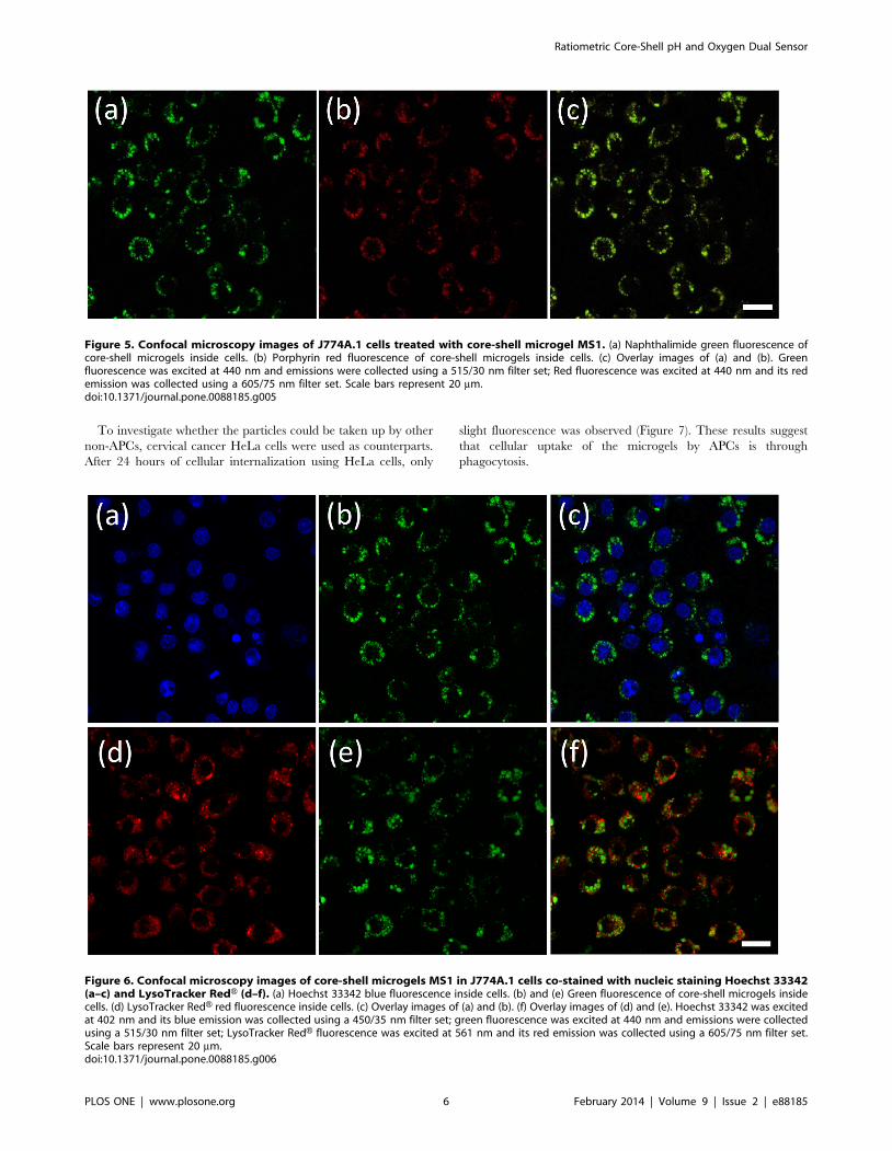
To investigate whether the particles could be taken up by other
non-APCs, cervical cancer HeLa cells were used as counterparts.
After 24 hours of cellular internalization using HeLa cells, only
slight fluorescence was observed (Figure 7). These results suggest
that cellular uptake of the microgels by APCs is through
phagocytosis.
Figure 5. Confocal microscopy images of J774A.1 cells treated with core-shell microgel MS1. (a) Naphthalimide green fluorescence ofcore-shell microgels inside cells. (b) Porphyrin red fluorescence of core-shell microgels inside cells. (c) Overlay images of (a) and (b). Greenfluorescence was excited at 440 nm and emissions were collected using a 515/30 nm filter set; Red fluorescence was excited at 440 nm and its redemission was collected using a 605/75 nm filter set. Scale bars represent 20 mm.doi:10.1371/journal.pone.0088185.g005
Figure 6. Confocal microscopy images of core-shell microgels MS1 in J774A.1 cells co-stained with nucleic staining Hoechst 33342(a–c) and LysoTracker RedH (d–f). (a) Hoechst 33342 blue fluorescence inside cells. (b) and (e) Green fluorescence of core-shell microgels insidecells. (d) LysoTracker RedH red fluorescence inside cells. (c) Overlay images of (a) and (b). (f) Overlay images of (d) and (e). Hoechst 33342 was excitedat 402 nm and its blue emission was collected using a 450/35 nm filter set; green fluorescence was excited at 440 nm and emissions were collectedusing a 515/30 nm filter set; LysoTracker RedH fluorescence was excited at 561 nm and its red emission was collected using a 605/75 nm filter set.Scale bars represent 20 mm.doi:10.1371/journal.pone.0088185.g006
Ratiometric Core-Shell pH and Oxygen Dual Sensor
PLOS ONE | www.plosone.org 6 February 2014 | Volume 9 | Issue 2 | e88185

Microgel-induced cytotoxicity was investigated by evaluating
the cell viability. Cytotoxicity was determined as a function of
concentration of the microgels by a standard MTT cell viability
assay. More than 85% of the cells were viable after the cells were
incubated for 24 h with the core-shell microgels concentrations of
2.5–10 mg/mL (Figure 8). These observations demonstrated the
biocompatibility of the core-shell microgels.
Conclusions
In summary, biocompatible core-shell microgels containing
optical sensors/reference probes as a novel ratiometric imaging/
sensing system for APCs was developed. The microgels can easily
be prepared using the two-stage free radical precipitation
polymerization method. The sensors/probes can be immobilized
either into the core or into the shell. In our lab we have been
developing new fluorescent sensors including pH, O2, Zn2+, DNA,
and temperature sensors [4,26,37–44] for not only new materials
but also applications for intracellular and extracellular sensing,
especially at the single cell level [43,44]. Our long term goal is to
investigate cellular metabolism, disease/cancer detection and
diagnosis using multi-sensor platforms. Herein, the core-shell
microgels can have the potential for optical sensing and imaging of
important analytes, such as O2, pH, and/or temperature.
Furthermore, simultaneous sensing of two analytes (pH and
oxygen) is also possible using the core-shell microgels platform
(Figure S5). The ability of the microgels to escape from the
lysosome into the cytosol makes them a potential candidate for
cytosolic delivery of sensors/probes. With the help of the
microgels, the noninvasive imaging/sensing in APCs was feasible.
The biocompatible microgel-based imaging/sensing agents are
expected to expand current molecular imaging techniques into
Figure 7. Confocal microscopy images of core-shell microgels MS1 in Hela cells (a–c, human cervical cancer) or J774A.1 cells (d–f,Mouse macrophage). (a) and (d) Green fluorescence of core-shell microgels inside cells. (b) and (e) Bright field images of cells. (c) and (d) Overlayimages of (a)/(b) and (d)/(e). Green fluorescence was excited at 440 nm and emissions were collected using a 515/30 nm filter set. Scale barsrepresent 20 mm.doi:10.1371/journal.pone.0088185.g007
Figure 8. Cytotoxicity of the microgels MS1 to J774A.1 (blue)and Hela (red) cells after incubation at 37uC for 24 h. Theconcentration of ‘‘control’’ in the x-axis means the control cells withoutadding any of the microgels.doi:10.1371/journal.pone.0088185.g008
Ratiometric Core-Shell pH and Oxygen Dual Sensor
PLOS ONE | www.plosone.org 7 February 2014 | Volume 9 | Issue 2 | e88185

methods applicable to studies in vivo, which will further drive
APCs-based treatments.
Supporting Information
Figure S1 Temperature-dependent change in the hy-drodynamic radius (Rh) of the microgels measured bylaser scattering analysis. The sigmoidal fitting was
performed for LCST determination (32.26C).(TIF)
Figure S2 Structures of oxygen sensor (OS1) and pHsensor (S1). Oxygen-insensitive green dye (M3) and pH-insensitive red dye (S6) were used as the references forratiometric sensing. Red background represents the probe
exhibiting red emission. Green background represents the probe
exhibiting green emission.
(TIF)
Figure S3 Response of the S1 in PNIPAm microgels topH in PBS buffer.(TIF)
Figure S4 Magnified Figure 6f.(TIF)
Figure S5 Response to dissolved oxygen in buffer. Fitsof the Stern-Volmer plot with/without ratiometriccalibration. (d–f) The emission profile of core-shell microgels
changes as a function of pH. Intensity ratio variations with pH
with/without ratiometric calibration.
(TIF)
Table S1 Size of Poly(St-co-NIPAm) core (OS1) particlesobtained by DLS at 25uC. In the systematic preparationof Poly(St-co-NIPAm) core particles, the dosages of St,NIPAm, and ABVA were kept constant as 1.0 g, 0.2 g and0.01 mM, respectively.
(DOC)
Table S2 Hydrodynamic mean diameters of the core-shell microgels (MS1) prepared with different NIPAmdosages. For all samples, the w/w percentage of BIS is3%.
(DOC)
Table S3 Hydrodynamic diameters of the core-shellmicrogels (MS1) prepared with different BIS dosages.For all samples, the dosage of NIPAm is 400 mg.
(DOC)
Acknowledgments
Dr. Hao Yan and Dr. Zhengtao Deng at Single Molecule Biophysics
(Biodesign Institute, ASU) are acknowledged for their kind help on TEM
measurements. The authors would also like to thank Patti Senechal-Willis
for the cell culture.
Author Contributions
Conceived and designed the experiments: XZ YT. Performed the
experiments: XZ FS YT. Analyzed the data: XZ FS YT. Wrote the
paper: XZ YT. Critical manuscript review: DRM Overall research
guidance: DRM.
References
1. Balagopalan L, Sherman E, Barr VA, Samelson LE (2011) Imaging techniques
for assaying lymphocyte activation in action. Nat Rev Immunol 11: 21–33.
2. Unanue ER (1984) Antigen-presenting function of the macrophage. Annu Rev
Immunol 2: 395–428.
3. Murata Y, Ohteki T, Koyasu S, Hamuro J (2002) IFN-c and pro-inflammatory
cytokine production by antigen-presenting cells is dictated by intracellular thiol
redox status regulated by oxygen tension. European Journal of Immunology 32:
2866–2873.
4. Tian Y, Su F, Weber W, Nandakumar V, Shumway BR, et al. (2010) A series of
naphthalimide derivatives as intra and extracellular pH sensors. Biomaterials 31:
7411–7422.
5. Gottlieb RA, Giesing HA, Zhu JY, Engler RL, Babior BM (1995) Cell
acidification in apoptosis: granulocyte colony-stimulating factor delays pro-
grammed cell death in neutrophils by up-regulating the vacuolar H(+)-ATPase.
Proceedings of the National Academy of Sciences of the United States of
America 92: 5965–5968.
6. Chudakov DM, Lukyanov S, Lukyanov KA (2005) Fluorescent proteins as a
toolkit for in vivo imaging. Trends in Biotechnology 23: 605–613.
7. Cahalan MD, Parker I, Wei SH, Miller MJ (2002) Two-photon tissue imaging:
seeing the immune system in a fresh light. Nat Rev Immunol 2: 872–880.
8. Burns A, Sengupta P, Zedayko T, Baird B, Wiesner U (2006) Core/shell
fluorescent silica nanoparticles for chemical sensing: towards single-particle
laboratories. Small 2: 723–726.
9. Rastogi SK, Pal P, Aston DE, Bitterwolf TE, Branen AL (2011) 8-Aminoquino-
line functionalized silica nanoparticles: a fluorescent nanosensor for detection of
divalent zinc in aqueous and in yeast cell suspension. ACS Applied Materials &
Interfaces 3: 1731–1739.
10. Clark HA, Hoyer M, Philbert MA, Kopelman R (1999) Optical nanosensors for
chemical analysis inside single living cells. 1. Fabrication, characterization, and
methods for intracellular delivery of PEBBLE sensors. Analytical Chemistry 71:
4831–4836.
11. Clark HA, Kopelman R, Tjalkens R, Philbert MA (1999) Optical nanosensors
for chemical analysis inside single living cells. 2. Sensors for pH and calcium and
the intracellular application of PEBBLE sensors. Analytical Chemistry 71: 4837–
4843.
12. Cao Y, Lee Koo Y-E, Kopelman R (2004) Poly(decyl methacrylate)-based
fluorescent PEBBLE swarm nanosensors for measuring dissolved oxygen in
biosamples. Analyst 129: 745–750.
13. Kim TH, Nah JW, Cho M-H, Park TG, Cho CS (2006) Receptor-mediated
gene delivery into antigen presenting cells using mannosylated chitosan/DNA
nanoparticles. Journal of Nanoscience and Nanotechnology 6: 2796–2803.
14. Chen YC, Ostafin A, Mizukami H (2010) Synthesis and characterization of pH
sensitive carboxySNARF-1 nanoreactors. Nanotechnology 21: 215503–215512.
15. Gan D, Lyon LA (2001) Interfacial nonradiative energy transfer in responsive
core-shell hydrogel nanoparticles. Journal of the American Chemical Society123: 8203–8209.
16. Rejman J, Oberle V, Zuhorn IS, Hoekstra D (2004) Size-dependent
internalization of particles via the pathways of clathrin- and caveolae-mediated
endocytosis. Biochem J 377: 159–169.
17. Cohen JA, Beaudette TT, Tseng WW, Bachelder EM, Mende I, et al. (2008) T-
cell activation by antigen-loaded pH-sensitive hydrogel particles in vivo: the effectof particle size. Bioconjugate Chemistry 20: 111–119.
18. Kovacsovics-Bankowski M, Clark K, Benacerraf B, Rock KL (1993) Efficient
major histocompatibility complex class I presentation of exogenous antigen upon
phagocytosis by macrophages. Proceedings of the National Academy of Sciencesof the United States of America 90: 4942–4946.
19. Harding CV, Song R (1994) Phagocytic processing of exogenous particulate
antigens by macrophages for presentation by class I MHC molecules. The
Journal of Immunology 153: 4925–4933.
20. Jones CD, Lyon LA (2000) Synthesis and characterization of multiresponsive
core-shell microgels. Macromolecules 33: 8301–8306.
21. Zhou X, Su F, Gao W, Tian Y, Youngbull C, et al. (2011) Triazacryptand-basedfluorescent sensors for extracellular and intracellular K+ sensing. Biomaterials
32: 8574–8583.
22. Grabtschev IK, Moneva IT, Wolarz E, Bauman D (1996) New unsaturated 1,8-
naphthalimide dyes for use in nematic liquid crystals. Journal of Physical Science
51: 1185–1191.
23. Filipova T, Grabchev I, Petkov I (1997) Synthesis and spectral properties of newN-substituted naphthalimide luminophores for structural coloration of poly-
methylmethacrylate and polystyrene. Journal of Polymer Science Part A:
Polymer Chemistry 35: 1069–1076.
24. Zhu L, Wu W, Zhu M-Q, Han JJ, Hurst JK, et al. (2007) Reversibly
photoswitchable dual-color fluorescent nanoparticles as new tools for live-cellimaging. Journal of the American Chemical Society 129: 3524–3526.
25. Nayak S, Lee H, Chmielewski J, Lyon LA (2004) Folate-mediated cell targeting
and cytotoxicity using thermoresponsive microgels. Journal of the American
Chemical Society 126: 10258–10259.
26. Tian Y, Shumway BR, Meldrum DR (2010) A new cross-linkable oxygen sensorcovalently bonded into poly(2-hydroxyethyl methacrylate)-co-polyacrylamide
thin film for dissolved oxygen sensing. Chemistry of Materials 22: 2069–2078.
27. Xu W, Schmidt R, Whaley M, Demas JN, DeGraff BA, et al. (1995) Oxygen
sensors based on luminescence quenching: interactions of pyrene with the
polymer supports. Analytical Chemistry 67: 3172–3180.
Ratiometric Core-Shell pH and Oxygen Dual Sensor
PLOS ONE | www.plosone.org 8 February 2014 | Volume 9 | Issue 2 | e88185
28. McDonagh C, MacCraith BD, McEvoy AK (1998) Tailoring of sol-gel films for
optical sensing of oxygen in gas and aqueous phase. Analytical Chemistry 70:
45–50.
29. Jakubiak R, Collison CJ, Wan WC, Rothberg LJ, Hsieh BR (1999) Aggregation
quenching of luminescence in electroluminescent conjugated polymers. The
Journal of Physical Chemistry A 103: 2394–2398.
30. DiPietro LA, Burdick M, Low QE, Kunkel SL, Strieter RM (1998) MIP-1alpha
as a critical macrophage chemoattractant in murine wound repair. The Journal
of Clinical Investigation 101: 1693–1698.
31. Nikolic D, Muresan RC, Feng W, Singer W (2012) Scaled correlation analysis: a
better way to compute a cross-correlogram. European Journal of Neuroscience
35: 742–762.
32. Savic R, Luo L, Eisenberg A, Maysinger D (2003) Micellar nanocontainers
distribute to defined cytoplasmic organelles. Science 300: 615–618.
33. Tian Y, Wu W-C, Chen C-Y, Strovas T, Li Y, et al. (2010) 2,1,3-
Benzothiadiazole (BTD)-moiety-containing red emitter conjugated amphiphilic
poly(ethylene glycol)-block-poly(e-caprolactone) copolymers for bioimaging.
Journal of Materials Chemistry 20: 1728–1736.
34. Vogel K, Wang S, Lee RJ, Chmielewski J, Low PS (1996) Peptide-mediated
release of folate-targeted liposome contents from endosomal compartments.
Journal of the American Chemical Society 118: 1581–1586.
35. Labat-Moleur F, Steffan AM, Brisson C, Perron H, Feugeas O, et al. (1996) An
electron microscopy study into the mechanism of gene transfer with
lipopolyamines. Gene Therapy 3: 1010–1017.
36. Zuber G, Dauty E, Nothisen M, Belguise P, Behr J-P (2001) Towards synthetic
viruses. Advanced Drug Delivery Reviews 52: 245–253.
37. Tian Y, Shumway BR, Gao W, Youngbull C, Holl MR, et al. (2010) Influence of
matrices on oxygen sensing of three sensing films with chemically conjugatedplatinum porphyrin probes and preliminary application for monitoring of
oxygen consumption of escherichia coli (E. coli). Sensors and Actuators B:
Chemical 150: 579–587.38. Jin Y, Tian Y, Zhang W, Jang S-H, Jen AY, et al. (2010) Tracking bacterial
infection of macrophages using a novel red-emission pH sensor. Analytical andBioanalytical Chemistry 398: 1375–1384.
39. Tian Y, Shumway BR, Cody Youngbull A, Li Y, Jen AKY, et al. (2010) Dually
fluorescent sensing of pH and dissolved oxygen using a membrane made frompolymerizable sensing monomers. Sensors and Actuators B: Chemical 147: 714–
722.40. Tian Y, Chen C-Y, Yang C-C, Young AC, Jang S-H, et al. (2008) 2-(29-
Hydroxyphenyl)benzoxazole-containing two-photon-absorbing chromophoresas sensors for zinc and hydroxide ions. Chemistry of Materials 20: 1977–1987.
41. Yang C-C, Tian Y, Chen C-Y, Jen AKY, Chen W-C (2007) A novel
benzoxazole-containing poly(N-isopropylacrylamide) copolymer as a multifunc-tional sensing material. Macromolecular Rapid Communications 28: 894–899.
42. Yang C-C, Tian Y, Jen AKY, Chen W-C (2006) New environmentallyresponsive fluorescent N-isopropylacrylamide copolymer and its application to
DNA sensing. Journal of Polymer Science Part A: Polymer Chemistry 44: 5495–
5504.43. Lidstrom ME, Meldrum DR (2003) Life-on-a-chip. Nature Reviews Microbi-
ology 1: 158–164.44. Molter TW, McQuaide SC, Suchorolski MT, Strovas TJ, Burgess LW, et al.
(2009) A microwell array device capable of measuring single-cell oxygenconsumption rates. Sensors and Actuators B: Chemical 135: 678–686.
Ratiometric Core-Shell pH and Oxygen Dual Sensor
PLOS ONE | www.plosone.org 9 February 2014 | Volume 9 | Issue 2 | e88185
Recommended