17 ACRYLAMIDE
3 HEALTH EFFECTS
31 INTRODUCTION
The primary purpose of this chapter is to provide public health officials physicians toxicologists and
other interested individuals and groups with an overall perspective on the toxicology of acrylamide It
contains descriptions and evaluations of toxicological studies and epidemiological investigations and
provides conclusions where possible on the relevance of toxicity and toxicokinetic data to public health
A glossary and list of acronyms abbreviations and symbols can be found at the end of this profile
32 DISCUSSION OF HEALTH EFFECTS BY ROUTE OF EXPOSURE
To help public health professionals and others address the needs of persons living or working near
hazardous waste sites the information in this section is organized first by route of exposure (inhalation
oral and dermal) and then by health effect (death systemic immunological neurological reproductive
developmental genotoxic and carcinogenic effects) These data are discussed in terms of three exposure
periods acute (14 days or less) intermediate (15ndash364 days) and chronic (365 days or more)
Levels of significant exposure for each route and duration are presented in tables and illustrated in
figures The points in the figures showing no-observed-adverse-effect levels (NOAELs) or lowest-
observed-adverse-effect levels (LOAELs) reflect the actual doses (levels of exposure) used in the studies
LOAELs have been classified into less serious or serious effects Serious effects are those that
evoke failure in a biological system and can lead to morbidity or mortality (eg acute respiratory distress
or death) Less serious effects are those that are not expected to cause significant dysfunction or death
or those whose significance to the organism is not entirely clear ATSDR acknowledges that a
considerable amount of judgment may be required in establishing whether an end point should be
classified as a NOAEL less serious LOAEL or serious LOAEL and that in some cases there will be
insufficient data to decide whether the effect is indicative of significant dysfunction However the
Agency has established guidelines and policies that are used to classify these end points ATSDR
believes that there is sufficient merit in this approach to warrant an attempt at distinguishing between
less serious and serious effects The distinction between less serious effects and serious effects is
considered to be important because it helps the users of the profiles to identify levels of exposure at which
major health effects start to appear LOAELs or NOAELs should also help in determining whether or not
18 ACRYLAMIDE
3 HEALTH EFFECTS
the effects vary with dose andor duration and place into perspective the possible significance of these
effects to human health
The significance of the exposure levels shown in the Levels of Significant Exposure (LSE) tables and
figures may differ depending on the users perspective Public health officials and others concerned with
appropriate actions to take at hazardous waste sites may want information on levels of exposure
associated with more subtle effects in humans or animals (LOAELs) or exposure levels below which no
adverse effects (NOAELs) have been observed Estimates of levels posing minimal risk to humans
(Minimal Risk Levels or MRLs) may be of interest to health professionals and citizens alike
Levels of exposure associated with carcinogenic effects (Cancer Effect Levels CELs) of acrylamide are
indicated in Table 3-4 and Figure 3-2 Because cancer effects could occur at lower exposure levels
Figure 3-2 also shows a range for the upper bound of estimated excess risks ranging from a risk of 1 in
10000 to 1 in 10000000 (10-4 to 10-7) as developed by EPA
A Users Guide has been provided at the end of this profile (see Appendix B) This guide should aid in
the interpretation of the tables and figures for Levels of Significant Exposure and the MRLs
321 Inhalation Exposure
3211 Death
Human data are available from two cohort mortality studies of occupational exposure to acrylamide one
by Collins et al (1989) with most recent follow up by Marsh et al (2007) and one by Sobel et al (1986)
with follow up by Swaen et al (2007) In these studies no significant associations were found between
occupational exposure to acrylamide and incidences of death from all causes See Section 3127
(Cancer) for more detailed information regarding these cohorts and assessments of death due to cancers
Reliable information regarding death in animals following inhalation exposure to acrylamide is limited
In a study performed for the American Cyanamid Company (1953a) two dogs seven rats and seven
guinea pigs (sex and strain unspecified) were exposed to acrylamide dust at 156 mgm3 for 6 hoursday
5 daysweek for up to 12 exposures in a 16-day period Four of the seven rats died overnight following
the first exposure period and two of the remaining rats died a few days later One of the dogs died on
study day 15 there were no deaths among the guinea pigs The study authors stated that gt90 of the
particles were in the respirable range (03ndash12 μm)
19 ACRYLAMIDE
3 HEALTH EFFECTS
Reliable inhalation mortality data for each species are recorded in Table 3-1 and plotted in Figure 3-1
3212 Systemic Effects
No human or animal data were located regarding cardiovascular gastrointestinal musculoskeletal
hepatic renal endocrine or dermal effects following inhalation exposure to acrylamide
Respiratory Effects Available information regarding acrylamide-associated respiratory effects is
restricted to complaints of nose and throat irritation in a group of tunnel workers who had been
occupationally exposed to acrylamide and N-methylolacrylamide in a chemical grouting agent for
2 months (Hagmar et al 2001) Increasing incidences of complaints were associated with increasing
levels of hemoglobin adducts of acrylamide
Hematological Effects No data were located regarding hematological effects in humans following
inhalation of acrylamide Available information in animals is limited to a study in which four rats were
exposed to acrylamide as a ldquosaturatedrdquo vapor for 6 hoursday 5 daysweek for 3 months at a mean
analytical concentration of 165 ppm (48 mgm3) the exposures did not affect hematology results
(American Cyanamid Company 1954)
Ocular Effects Available information regarding acrylamide-associated ocular effects is restricted to
complaints of eye irritation in a group of tunnel workers who had been occupationally exposed to
acrylamide and N-methylolacrylamide in a chemical grouting agent for 2 months (Hagmar et al 2001)
Increasing incidences of complaints were associated with increasing levels of hemoglobin adducts of
acrylamide
Body Weight Effects No data were located regarding body weight effects in humans following
inhalation of acrylamide Available information in animals is limited to a study in which four rats were
exposed to acrylamide as a ldquosaturatedrdquo vapor for 6 hoursday 5 daysweek for 3 months at a mean
analytical concentration of 165 ppm (48 mgm3) the exposures did not affect body weights (American
Cyanamid Company 1954)
159156
166156
25156
161
48
48
160
156
23156
Table 3-1 Levels of Significant Exposure to Acrylamide - Inhalation
Exposure LOAEL Duration
a FrequencyKey to Species NOAEL Less Serious(Route)Figure (Strain) System (mgmsup3) (mgmsup3)
ACUTE EXPOSURE Death 1 Rat 4 d
6 hrd(NS)
Neurological 2 Rat 4 d
6 hrd(NS)
INTERMEDIATE EXPOSURE Death 3 Dog 16 d
5 dwk(NS) 6 hrd
Systemic 4 Cat 3 mo Hemato 485 dwk
6 hrd
Bd Wt 48
Neurological 5 Gn Pig 16 d 1565 dwk(NS)
6 hrd
6 Dog 16 d 5 dwk(NS) 6 hrd
Serious (mgmsup3)
156 (death of 47 rats in 1 day and 2 others before day 8)
156 (loss of coordination and equilibrium on exposure day 4)
156 (death of 12 dogs on day 15)
156 (CNS effects including loss of equilibrium and coordination)
Reference Chemical Form Comments
American Cyanamid Company 1953a
American Cyanamid Company 1953a
American Cyanamid Company 1953a
American Cyanamid Company 1954 Acrylamide
American Cyanamid Company 1953a
American Cyanamid Company 1953a
ACRYLA
MIDE
3 HE
ALTH
EFFE
CTS
20
Table 3-1 Levels of Significant Exposure to Acrylamide - Inhalation (continued)
a Key to Species Figure (Strain)
Exposure Duration
Frequency (Route)
System NOAEL (mgmsup3)
LOAEL
Reference Chemical Form Comments
Less Serious (mgmsup3)
Serious (mgmsup3)
82
48
7 Cat 3 mo 48 American Cyanamid Company5 dwk 19546 hrd Acrylamide
a The number corresponds to entries in Figure 3-1
Bd Wt = body weight CNS = central nervous system d = day(s) Gn pig = guinea pig Hemato = hematological hr = hour(s) LOAEL = lowest-observed-adverse-effect level mo = month(s) NOAEL = no-observed-adverse-effect level NS = not specified wk = week(s)
ACRYLA
MIDE
3 HE
ALTH
EFFE
CTS
21
Death
Neurologi
mgm3
100
1r 2r
10
c-Cat -Humans f-Ferret n-Mink Cancer Effect Level-Animals Cancer Effect Level-Humans LD50LC50d-Dog k-Monkey j-Pigeon o-Other LOAEL More Serious-Animals LOAEL More Serious-Humans Minimal Risk Level r-Rat m-Mouse e-Gerbil LOAEL Less Serious-Animals LOAEL Less Serious-Humans for effects p-Pig h-Rabbit s-Hamster NOAEL - Animals NOAEL - Humans other than q-Cow a-Sheep g-Guinea Pig Cancer
cal
Figure 3-1 Levels of Significant Exposure to Acrylamide - InhalationAcute (le14 days) A
CRYLA
MIDE
3 HE
ALTH
EFFE
CTS
22
Death
Hematol
Body We
Neurolog
mgm3
100
3d 6d 5g
10
4c 4c 7c
1
c-Cat -Humans f-Ferret n-Mink Cancer Effect Level-Animals Cancer Effect Level-Humans LD50LC50d-Dog k-Monkey j-Pigeon o-Other LOAEL More Serious-Animals LOAEL More Serious-Humans Minimal Risk Level r-Rat m-Mouse e-Gerbil LOAEL Less Serious-Animals LOAEL Less Serious-Humans for effects p-Pig h-Rabbit s-Hamster NOAEL - Animals NOAEL - Humans other than q-Cow a-Sheep g-Guinea Pig Cancer
ogic igh
tical al
Systemic
Figure 3-1 Levels of Significant Exposure to Acrylamide - Inhalation (Continued) Intermediate (15-364 days) A
CRYLA
MIDE
3 HE
ALTH
EFFE
CTS
23
24 ACRYLAMIDE
3 HEALTH EFFECTS
3213 Immunological and Lymphoreticular Effects
No data were located regarding immunological or lymphoreticular effects in humans or animals following
inhalation exposure to acrylamide
3214 Neurological Effects
Information in humans is available from numerous case reports in which acrylamide exposure has been
associated with signs of impaired neurological performance in central and peripheral nervous systems that
include impaired motor function and muscle weakness (Auld and Bedwell 1967 Davenport et al 1976
Dumitru 1989 Fullerton 1969 Garland and Patterson 1967 Gjerloslashff et al 2001 Igisu et al 1975 Kesson
et al 1977 Mapp et al 1977 Mulloy 1996 Takahashi et al 1971) Human data are also available from
cross-sectional studies that included self-reported symptoms and neurological evaluations of acrylamide-
exposed workers with potential for inhalation and dermal (and possibly oral) exposure (Bachmann et al
1992 Calleman et al 1994 Hagmar et al 2001 He et al 1989 Myers and Macun 1991) Although the
case reports and cross-sectional studies provide supportive evidence of acrylamide-induced neurotoxicity
they lack information regarding relative contributions of natural exposure routes (inhalation oral
dermal) exposure-response relationships and other confounding exposures They are therefore
unsuitable for meaningful quantitative risk analysis
He et al (1989) evaluated health end points in workers employed for 1ndash18 months at a factory in China
that began producing acrylamide and polyacrylamide in 1984 A referent group consisted of unexposed
workers from the same town Concentrations of acrylamide in the workplace air (determined by gas
chromatography) reached 556ndash902 mgm3 between March and June 1985 during polymerization when
there was an exceptional increase in production and decreased to an average of 00324 mgm3 (range not
specified) after July 1985 The workers were evaluated in October 1985 The study authors reported that
heavy skin contamination by aqueous acrylamide monomer was common among the workers An
acrylamide level of 410 mgL was measured in the water in which three of the workers washed their
hands Personal interviews were conducted to obtain information on demographic factors occupational
history symptoms past illnesses and family history Physical and neurological examinations visual
acuity and visual field testing skin temperature measurements electrocardiography and
electroencephalography were performed Sixty-nine of the exposed workers and 48 of the referent
workers were subjected to electroneuromyographic examinations
25 ACRYLAMIDE
3 HEALTH EFFECTS
As shown in Table 3-2 significantly greater percentages of the acrylamide-exposed group reported skin
peeling from the hands anorexia numbness and coldness in hands and feet lassitude sleepiness muscle
weakness clumsiness of the hands unsteady gait difficulty in grasping and stumbling and falling The
authors stated that initial symptoms of skin peeling were the result of dermal exposure to aqueous
acrylamide and that other symptoms appeared following 3ndash10 months of occupational exposure Greater
percentages of acrylamide-exposed workers exhibited erythema of the hands sensory impairments
(vibration pain and touch sensation) diminished reflexes and intention tremor (Table 3-2) Electrical
activity monitored in both the abductor pollicis brevis and abductor digiti minimi muscles of the hand
revealed electromyographic abnormalities in the acrylamide-exposed workers that included denervation
potentials (369 exposed workers) prolonged duration of motor units (4069) increased polyphasic
potentials (2969) and discrete pattern of recruitment (969) These abnormalities were not seen in
referent workers with the exception of prolonged duration of motor units (448 referents) Significantly
increased mean duration and mean amplitude of motor unit potentials and significantly decreased mean
amplitude of sensory unit potentials were seen in the acrylamide-exposed group compared to the referent
group Assessment of visual acuity and visual field nerve conduction velocity electrocardiography and
electroencephalography revealed no significant exposure-related effects
Calleman et al (1994) performed a cross-sectional analysis of hemoglobin adduct formation and
neurological effects in a group of 41 factory workers who were exposed to acrylamide (and acrylonitrile
from which acrylamide is formed) for 1 month to 115 years (mean 3 years) during the production of
acrylamide and polyacrylamide at a factory in China As determined by station sampling and gas
chromatography mean acrylamide air concentrations were 107 and 327 mgm3 in the synthesis and
polymerization rooms respectively during the summer of 1991 Mean exposure concentrations during
the time of collection of biomarker data (September 1991) were lower averaging 061 and 058 mgm3 in
synthesis and polymerization rooms respectively Information regarding demographic factors smoking
and drinking habits height and weight occupational history past illnesses current symptoms and
reproductive history were collected by questionnaire Neurological examinations were performed
approximately 1 hour after a work shift Vibration sensitivity thresholds were measured in fingers and
toes Physical and neurological examinations and electroneuromyographic (ENMG) testing were
performed For each test a nonexposed referent group was included Quantitative assessment of
contributions of dermal and inhalation exposure were not made although in the synthesis area of the
factory where neurological symptoms were most severe dermal contact was considered to have been the
major exposure route
26 ACRYLAMIDE
3 HEALTH EFFECTS
Table 3-2 Self-Reported Neurological Symptoms and Observed Clinical SignsAmong Acrylamide Workers and Nonexposed Workers
Symptom Acrylamide Number
group (n=71) Percent
Reference Number
group (n=51) Percent
Skin peeling from the hands 38 535a 2 39 Numbness in the hands and feet 15 211b 2 39 Lassitude 14 197b 1 19 Sleepiness 12 169b 0 0 Muscle weakness 11 154b 0 0 Clumsiness of the hands 8 112a 0 0 Anorexia 8 112a 1 19 Unsteady gait 6 84a 0 0 Coldness of the hands and feet 6 84a 0 0 Difficulty in grasping 5 70a 0 0 Stumbling and falling 5 70a 0 0 Sweating 27 380 14 274 Dizziness 7 98 2 39 Cramping pain 6 84 5 98 Sign Erythema of hands 16 225b 0 0 Skin peeling from the hands 16 225b 1 19 Sensory impairments
Vibration sensation 12 169b 0 0 Pain sensation 7 98a 0 0 Touch sensation 6 84a 0 0 Position sensation 1 14 0 0
Muscle atrophy in hands 4 56 0 0 Diminished reflexes
Biceps 12 169b 0 0 Triceps 10 140 4 78 Knee 11 154a 1 19 Ankle 8 112a 1 19
Loss of reflexes Biceps 3 42 0 0 Triceps 5 70 1 19 Knee 5 70a 0 0 Ankle 17 239b 0 0
Intention tremor 13 183a 2 39 Positive Rombergrsquos sign 15 211 3 58
aplt005bplt001 (χ2 test)
Source He et al 1989
27 ACRYLAMIDE
3 HEALTH EFFECTS
As shown in Table 3-3 a variety of symptoms and signs of adverse health effects were noted in
acrylamide-exposed workers (Calleman et al 1994) Other significant (plt001) effects in the exposed
workers included increased (magnitude ge60) vibration threshold decreased (10ndash20) conduction
velocity in the peroneal and sural nerves and increased (25ndash36) latency in median ulnar and peroneal
nerves Neurotoxicity index scores a quantitative expression of the severity of peripheral neuropathy
decreased with physical distance from the synthesis room where the monomer itself was handled This
relationship was not reflected by results of hand or foot vibration sensitivity measurements
Hagmar et al (2001) performed a health examination on a group of 210 tunnel construction workers who
had been occupationally exposed for 2 months to a chemical grouting agent containing acrylamide and
N-methylolacrylamide Workers were expected to have experienced dermal exposure as well as
inhalation exposure Venous blood samples were drawn and questionnaires and physical examinations
were administered 1ndash5 weeks after exposure was stopped Quantitative exposure data were limited to two
personal air samples showing concentrations of 027 and 034 mgm3 for the sum of acrylamide and
N-methylolacrylamide further analysis suggested that the air contained a 5050 mixture of these
compounds The health examination included an extensive questionnaire and a physical examination that
included unspecified tests of peripheral nerve function Blood samples for the analysis of adducts of
acrylamide with N-terminal valines in hemoglobin were drawn within a month after construction work
was completed A group of 50 subjects who claimed recently developed or deteriorated peripheral
nervous function at the initial physical examination was subjected to more detailed neurophysiologic
examinations and 6-month follow-up clinical (n=29) and neurophysiological (n=26) examinations Those
with remaining symptoms were examined for up to 18 months postexposure
Hemoglobin adduct levels for 18 nonsmoking unexposed referents varied between 002 and 007 nmolg
globin Adduct levels in 47 of the 210 tunnel workers did not exceed the highest level of the referents
The remaining workers were divided into three categories according to adduct levels as follows 89 with
008ndash029 nmolg globin 36 with 03ndash10 nmolg globin and 38 with 10ndash177 nmolg globin The study
authors noted a significant (plt005) association between self-reported exposure categories and adduct
levels Significant positive correlations (plt005) between prevalence of self-reported peripheral nervous
symptoms irritant symptoms and symptoms of general discomfort with adduct levels were found For
example in the groups with adduct levels lt008 nmolg globin 008ndash029 nmolg globin 03ndash10 nmolg
globin and gt10 nmolg globin incidences of reported numbness or tingling in the feet or legs were
247 (4) 1089 (11) 936 (25) and 1438 (37) respectively Irritant symptoms and symptoms of
28 ACRYLAMIDE
3 HEALTH EFFECTS
Table 3-3 Prevalence of Symptoms and Signs of Adverse Health Effects in Acrylamide-Exposed Workers and Controls
Symptom or sign Exposed (percent) Controls (n=10) Numbness of extremities 2941 (71)a 0 Fatigue 2941 (71)a 0 Sweating of hands and feet 2841 (71)a 0 Skin peeling 2441 (59)a 0 Menstruation disorders 47 (57) NA Loss of pain sensation 2241 (54)a 0 Loss of touch sensation 1941 (46)b 0 Dizziness 1841 (44)b 0 Anorexia 1741 (41)b 0 Loss of vibration sensation 1741 (41)b 0 Nausea 1641 (39)b 0 Loss of ankle reflexes 1241 (29) 0 Headache 1141 (27) 0 Unsteady gait 941 (22) 0 Loss of knee jerk 841 (20) 0 Unsteady Romberg sign 841 (20) 0 Loss of triceps reflexes 441 (10) 0 Loss of biceps reflexes 441 (10) 0
aplt001 (χ2 test) bplt005
Source Calleman et al 1994
29 ACRYLAMIDE
3 HEALTH EFFECTS
general discomfort typically disappeared following the end of a workday whereas peripheral nervous
symptoms persisted Follow-up examinations revealed that 58 of the subjects with early signs of
impaired peripheral nervous function improved while only 4 showed signs of deterioration
Myers and Macun (1991) investigated peripheral neuropathy in a cohort of 66 workers in a South African
factory that produced polyacrylamide The investigation followed clinical diagnosis of peripheral
neuropathy in five workers at the factory The workforce was divided into a number of exposure
categories based on environmental sampling and discussions with workers Exposure levels for the
various tasks ranged from 007 to 25 times the National Institute of Occupational Safety and Health
(NIOSH) recommended exposure limit (REL) of 03 mgm3 Workers were then classified as being
exposed to airborne acrylamide when exposure levels exceeded the REL (n=22) and unexposed when
exposure levels were below the REL (n=41) Workers completed a questionnaire that was designed to
capture social medical and occupational history A standard blind neurological examination was also
performed
The exposed group showed higher prevalences of abnormalities for all symptoms (weakness sensation
balance fatigue visual loss of weight urogenital and fingertip skin) most signs (fingertip effects light
touch tactile discrimination pain) and reflexes coordination motor weakness gait and Rombergism
Statistically significant differences between exposed and unexposed groups for individual effects were
seen only for abnormal sensation symptoms and signs in fingertip skin (including color peeling and
sweating) The overall prevalence of acrylamide-related abnormalities among the exposed was 667
which was statistically significantly higher (plt005) than that of the unexposed group (prevalence of
143) The authors stated that most workers observed to have abnormalities (number not reported) were
employed in areas where exposures were highest (16ndash25 times the REL)
Bachmann et al (1992) performed a follow-up investigation in July 1990 at the same South African
factory that had been examined in 1986 by Myers and Macun (1991) The study design was similar to
that of Myers and Macun (1991) but included measurements of vibration sensation threshold Among
82 workers employed at follow-up increased prevalences of symptoms of tingling and numbness in hands
and feet weakness and pain in arms and legs peeling hand skin and sweating hands were reported by
exposed workers compared with those classified as being unexposed The symptoms of numbness limb
pain and peeling and sweating of hands were statistically significantly increased in exposed workers
Results of clinical examinations provided supporting evidence for the reported increased symptoms of
peeling and sweating of the hands No gross neurological abnormalities were found Mean vibration
30 ACRYLAMIDE
3 HEALTH EFFECTS
sensation thresholds were similar among unexposed and exposed groups even when adjusting for age
and no association was found between vibration thresholds and any symptoms
Information regarding neurological effects in animals exposed to acrylamide by the inhalation route is
limited to a single study report in which seven rats seven guinea pigs and two dogs were exposed to dust
of acrylamide at an analytical concentration of 156 mgm3 for 6 hoursday 5 daysweek for up to
12 exposures in a 16-day period (American Cyanamid Company 1953a) Reported signs of neurological
effects in the three rats that survived to exposure termination on day 4 included loss of equilibrium and
coordination There was no mention of neurological signs in the exposed dogs although one of the dogs
lost weight and died on day 15 No toxic signs were seen in the guinea pigs
3215 Reproductive Effects
No data were located regarding reproductive effects in humans or animals following inhalation exposure
to acrylamide
3216 Developmental Effects
No data were located regarding developmental effects in humans or animals following inhalation
exposure to acrylamide
3217 Cancer
Human data are available from two cohort mortality studies of occupational exposure to acrylamide one
by Collins et al (1989) with most recent follow up by Marsh et al (2007) and one by Sobel et al (1986)
with follow up by Swaen et al (2007) Exposure to acrylamide was considered to have occurred
primarily via inhalation and dermal exposure
Collins et al (1989) conducted a cohort mortality study of all male workers (8854 of which 2293 were
exposed to acrylamide) who had been hired between January 1 1925 and January 31 1973 at four
American Cyanamid factories three in the United States (Fortier Louisiana [1295 workers] Warners
New Jersey [7153 workers] and Kalamazoo Michigan [60 workers]) and one in the Netherlands (Botlek
[346 workers]) Estimations of acrylamide exposure were based on available monitoring data and worker
knowledge of past jobs and processes Mortality rates among the factory workers were compared with
the expected number of deaths among men of the United States from 1925 to 1980 or the Netherlands
31 ACRYLAMIDE
3 HEALTH EFFECTS
from 1950 to 1982 to derive standardized mortality ratios (SMRs) as a measure of relative risk for each
cohort No statistically significantly elevated all cause or cause-specific SMRs were found among
acrylamide-exposed workers (including cancer of the digestive or respiratory systems bone skin
reproductive organs bladder kidney eye central nervous system thyroid or lymphatic system) Trend
tests showed no increased risk of mortality due to cancer at several sites (digestive tract respiratory
system prostate central nervous system or lymphopoietic system) with increasing level of exposure to
acrylamide
The most recent update report of the cohort of Collins et al (1989) includes study periods of 1925ndash2002
for the 8508 workers in the three facilities in the United States and 1965ndash2004 for the 344 workers at the
Botlek plant in the Netherlands (the original cohort of 346 people included 2 females who were excluded
in the follow up) (Marsh et al 2007) Among the workers at the three facilities in the United States
(during which 4650 deaths occurred among the 8508 workers in the period of 1925ndash2002) excess and
deficit overall mortality risks were observed for cancer sites implicated in oral studies in experimental
animals brain and other central nervous system (SMR 067 95 confidence interval [CI] 040ndash105)
thyroid gland (SMR 138 95 CI 028ndash402) and testis and other male genital organs (SMR 064 95
CI 008ndash230) and for sites selected in the original report (Collins et al 1989) of this cohort respiratory
system cancer (SMR 117 95 CI 106ndash127) esophagus (SMR 120 95 CI 086ndash163) rectum (SMR
125 95 CI 084ndash178) pancreas (SMR 094 95 CI 070ndash122) and kidney (SMR 101 95 CI 066ndash
146) None of the mortality excesses were statistically significant except for respiratory system cancer
which Collins et al (1989) attributed to muriatic acid (hydrochloric acid) exposure No significantly
elevated SMRs were found for rectal pancreatic or kidney cancers in exploratory exposure-response
analyses conducted according to the following exposure parameters and categories duration of
employment (lt1 1ndash and 15+ years) time since first employment (lt20 20ndash and 30+ years) duration of
exposure (unexposed 0001ndash 5ndash and 20+ years) cumulative exposure (lt0001 0001ndash 003ndash and 030+
mgm3-years) and estimated mean exposure concentrations (unexposed 0001ndash 002ndash and 03+ mgm3)
Sobel et al (1986) conducted a mortality study on a cohort of 371 workers assigned to acrylamide and
polymerization operations at a Dow Chemical facility in the United States between 1955 and 1979
Analysis and review of air monitoring data and job classifications resulted in estimates of personal 8-hour
time-weighted average acrylamide concentrations of 01ndash10 mgm3 before 1957 01ndash06 mgm3 from
1957 to 1970 and 01 mgm3 thereafter SMRs calculated for categories in which at least two deaths
were observed were based on mortality of white males in the United States No significantly increased
incidences of cancer-related deaths were observed within the cohort
32 ACRYLAMIDE
3 HEALTH EFFECTS
Followup to the Sobel et al (1986) study cohort was expanded to include employees hired through 2001
(Swaen et al 2007) Exposure to acrylamide was retrospectively assessed based on personal samples
from the 1970s onwards and area samples from the entire study period Fewer acrylamide workers died
(n=141) than expected (n=1721) No cause-specific SMR for any of the investigated types of cancer was
exposure related The authors reported more total pancreatic cancer deaths (n=5) than expected (n=23)
(SMR 2222 95 CI 721ndash5185) however three of the five were in the low-dose group with no
apparent dose-response relationship with acrylamide exposure
Meta-analyses of the most recent results from the two major cohort studies that assessed cancer risk and
occupational exposure to acrylamide (Marsh et al 2007 Swaen et al 2007) were performed by Pelucchi
et al (2011b) The results indicate a lack of increased risk of cancers of the digestive tract pancreas
lung or kidney among these cohorts
No data were located regarding cancer in animals following inhalation exposure to acrylamide EPA
(IRIS 2012) calculated an inhalation unit risk of 1x10⁻⁴ per microgm3 for acrylamide based on results of a
2-year cancer bioassay in orally-exposed male and female F344 rats (Johnson et al 1986) and route-to-
route extrapolation (see Section 3227 for details of the oral study) The air concentrations associated
with risk of 1x10⁻⁴ 1x10⁻⁵ 1x10⁻⁶ and 1x10⁻⁷ are 1 01 001 and 0001 mgm3 respectively These risk
levels are presented in Figure 3-1
322 Oral Exposure
3221 Death
There are no reports of human deaths associated with oral exposure to acrylamide
Acrylamide has been demonstrated to be lethal to laboratory animals following a single oral dose
Reported oral LD50 values in rats range from 150 to 413 mgkg (American Cyanamid Company 1973
1977 Dow Chemical Company 1957 Fullerton and Barnes 1966 McCollister et al 1964 Tilson and
Cabe 1979 Union Carbide Corporation 1947) Reported LD50 values in mice guinea pigs and rabbits
range from 107 to 195 mgkg (American Cyanamid Company 1951 Dow Chemical Company 1957
Hashimoto et al 1981 McCollister et al 1964)
33 ACRYLAMIDE
3 HEALTH EFFECTS
Repeated oral exposure to acrylamide has also been associated with death in laboratory animals In one
rat study a single 100 mgkg dose was not lethal but two 100 mgkg doses administered 24 hours apart
resulted in mortalities (Fullerton and Barnes 1966) Sublet et al (1989) reported death in a group of male
Long-Evans hooded rats administered acrylamide by daily gavage for 5 days at a dose of 75 mgkgday
Longer repeated-dose exposure periods to lower daily doses are lethal as well For example a dose level
of 50 mgkg was lethal to rats receiving 12 daily gavage doses of 50 mgkg in a 15-day period the deaths
occurred within a few days following the cessation of dosing (Fullerton and Barnes 1966) All mice
(4sex) given acrylamide in the drinking water at a concentration resulting in an estimated dose of
150 mgkgday were sacrificed moribund on the 10th day of treatment (NTP 2011b) No deaths occurred
in groups given acrylamide in the drinking water for 14 days at exposure levels resulting in estimated
doses ranging from 2 to 76 mgkgday or in other mice receiving acrylamide from the food for 14 days at
estimated doses up to 75 mgkgday (NTP 2011b) Similar treatment of rats to acrylamide in the drinking
water or food resulted in the death of one high-dose (77 mgkgday) male from the drinking water study
there were no deaths in the female rats exposed via the drinking water or food (doses up to 70 and
63 mgkgday) or the male rats exposed via the food (doses up to 52 mgkgday) An acrylamide gavage
dose of 30 mgkgday resulted in the death of 410 male and 210 female rats during the third week of
daily dosing (Schulze and Boysen 1991) No deaths occurred in rats or mice receiving acrylamide from
the drinking water for 13 weeks at estimated doses as high as 22ndash26 mgkgday (rats) and 70ndash
83 mgkgday (mice) (NTP 2011b) Thirteen weeks of exposure to acrylamide in the food resulted in the
death of one male mouse each at estimated dose levels of 32 and 59 mgkgday there were no deaths in
female mice at estimated doses as high as 64 mgkgday or among similarly-treated male and female rats
at estimated doses as high as 14 and 18 mgkgday respectively (NTP 2011b) During the last 4 months
of a 2-year study in which male and female rats received acrylamide from the drinking water at an
estimated dose level of 2 mgkgday decreased survival of both sexes was noted the increased mortality
was statistically significant by study termination (Johnson et al 1984 1986) In other studies of rats and
mice administered acrylamide in the drinking water for 2 years significantly decreased survival was
noted at estimated doses of ge09 mgkgday (female rats) ge46 mgkgday (female mice) and
9 mgkgday (male rats) (NTP 2011b)
Reliable acute oral LD50 values for death and other mortality data for each species are recorded in
Table 3-4 and plotted in Figure 3-2
14
294
12
413
1
180
119
203
149100
175
766
125
175
21
316
Exposure Duration
a FrequencyKey to Species (Route)Figure (Strain)
ACUTE EXPOSURE Death 1 Rat Once
(Wistar) (GW)
2 Rat Once (Sprague- (GW) Dawley)
3 Rat Once (NS) (GW)
4 Rat Once (albino) (GW)
5 Rat 2 d 1 xd(albino) (GW)
6 Rat 14 d (Fischer- 344) (W)
7 Rat Once (Fischer- 344) (GW)
8 Rat Once (albino) (GW)
LOAEL
NOAEL Less Serious Serious System (mgkgday) (mgkgday) (mgkgday)
294 M
413 M
180
203 F
100
766 M
175 M
316 M
(LD50)
(LD50)
(LD50)
(LD50)
(most rats died within 3 days following 2 days of dosing)
(14 died)
(LD50)
(LD50)
Reference Chemical Form Comments
American Cyanamid Company 1973
American Cyanamid Company 1977
Dow Chemical Company 1957 McCollister et al 1964
Fullerton and Barnes 1966 Acrylamide
Fullerton and Barnes 1966 Acrylamide
NTP 2011
Tilson and Cabe 1979 Acrylamide
Union Carbide Corporation 1947
Table 3-4 Levels of Significant Exposure to Acrylamide - Oral
ACRYLA
MIDE
3 HE
ALTH
EFFE
CTS
34
18
195
135
107
182150
4
180
5
150
167
5
20
183
374
39470 766
Table 3-4 Levels of Significant Exposure to Acrylamide - Oral (continued)
LOAEL
a Key to Species NOAEL Less Serious
System (mgkgday) (mgkgday)
Bd Wt 5 M 20 M (8 decreased mean body weight)
Bd Wt 374 M 70 F (15 lower terminal body weight)
394 F
Serious (mgkgday)
195 M (LD50)
107 M (LD50)
150 (moribund sacrifice of 44 males and 44 females on treatment day 10)
180 (LD50)
150 (LD50)
766 M (44 lower terminal body weight)
Reference Chemical Form Comments
American Cyanamid Company 1951
Hashimoto et al 1981 Acrylamide
NTP 2011 Acrylamide
Dow Chemical Company 1957 McCollister et al 1964
Dow Chemical Company 1957 McCollister et al 1964
Burek et al 1980 Acrylamide
NTP 2011
Figure
9
10
11
12
13
(Strain)
Mouse (albino)
Mouse
Mouse (B6C3F1)
Gn Pig (NS)
Rabbit (NS)
Systemic 14 Rat
Exposure Duration
Frequency (Route)
Once (G)
Once (NS)
14 d (W)
Once (GW)
Once (GW)
13 d (Fischer- 344) (W)
15 Rat 14 d (Fischer- 344) (W)
ACRYLA
MIDE
3 HE
ALTH
EFFE
CTS
35
184
224
634517
95
5
15
117
20
186
667
758
150
187
728
757
98
100
150
3126
Table 3-4 Levels of Significant Exposure to Acrylamide - Oral (continued)
Exposure LOAEL Duration
a FrequencyKey to Species NOAEL Less Serious Serious(Route)Figure (Strain) System (mgkgday) (mgkgday) (mgkgday)
16 Rat 14 d Bd Wt 224 M 517 M (28 lower terminal body (Fischer- 344) (F) weight)
634 F
17 Rat 5 d Bd Wt 5 M 15 M (significantly depressed (Long- Evans) 1 xd body weight gain during
(GW) 5 days of acrylamide administration)
18 Rat Gd 6-17 Bd Wt 20 F (Fischer- 344) (GW)
19 Mouse 14 d Bd Wt 667 M 150 (marked weight loss and(B6C3F1) (W) moribund sacrifice at
758 F treatment day 10)
20 Mouse 14 d Bd Wt 728 M (B6C3F1) (F)
757 F
21 Mouse Once Bd Wt 100 M 150 M (significantly depressed ddY (NS) body weight)
22 Gn Pig Once Bd Wt 126 (very slight initial body(NS) (GW) weight loss)
Reference Chemical Form Comments
NTP 2011 Acrylamide
Tyl et al 2000b Acrylamide
Walden et al 1981 Maternal body weight
Acrylamide
NTP 2011 Acrylamide
NTP 2011 Acrylamide
Sakamoto et al 1988 Acrylamide
Dow Chemical Company 1957 McCollister et al 1964
ACRYLA
MIDE
3 HE
ALTH
EFFE
CTS
36
6
63
168
20
15725
120
203
146
100
148
100
47
126
Exposure LOAEL Duration
a FrequencyKey to Species NOAEL Less Serious Serious(Route)Figure (Strain) System (mgkgday) (mgkgday) (mgkgday)
Reference Chemical Form Comments
Dow Chemical Company 1957 McCollister et al 1964
Burek et al 1980 Acrylamide
Dixit et al 1981 Acrylamide
Fullerton and Barnes 1966 Acrylamide
Fullerton and Barnes 1966 Acrylamide
Fullerton and Barnes 1966 Acrylamide
McCollister et al 1964 Acrylamide
23 Rabbit (NS)
Neurological 24 Rat
(Fischer- 344)
25 Rat (Wistar)
26 Rat (albino)
27 Rat (albino)
28 Rat (albino)
29 Rat (NS)
Once (GW)
Bd Wt 63 (slight initial weight loss)
7 d (W)
20 M
Up to 21 d 1 xd 25 M
(G)
Once (GW)
203 F
Once (GW)
100
2 d 1 xd 100
(GW)
Once (GW)
126 F
(convulsions and ataxia as early as treatment day 14)
(fine tremors)
(fine tremors)
(generalized weakness)
(lethargy)
Table 3-4 Levels of Significant Exposure to Acrylamide - Oral (continued)
ACRYLA
MIDE
3 HE
ALTH
EFFE
CTS
37
176
374
394766
70
178
224
294517
634
126
100
200
94
30
45
118
20
Table 3-4 Levels of Significant Exposure to Acrylamide - Oral (continued)
Exposure LOAEL Duration
a FrequencyKey to Species NOAEL Less Serious Serious(Route)Figure (Strain) System (mgkgday) (mgkgday) (mgkgday)
30 Rat 14 d 374 M 766 M (hind-leg paralysis in 44 (Fischer- 344) (W) males)
394 F
70 F (hind-leg paralysis in 44 females)
31 Rat 14 d 224 M 517 M (hind-leg paralysis in 44 (Fischer- 344) (F) males)
294 F
634 F (hind-leg paralysis in 44 females)
32 Rat Once 100 M 200 M (decreases in hindlimb (Fischer- 344) (GW) grip strength and
locomotory performance)
33 Rat 5 d 30 M 45 M (clinical signs of (Long- Evans) 1 xd neurotoxicity)
(GW)
34 Rat Gd 6-17 20 F (Fischer- 344) (GW)
Reference Chemical Form Comments
NTP 2011
NTP 2011 Acrylamide
Tilson and Cabe 1979 Acrylamide
Tyl et al 2000b Acrylamide
Walden et al 1981 Acrylamide
ACRYLA
MIDE
3 HE
ALTH
EFFE
CTS
38
Table 3-4 Levels of Significant Exposure to Acrylamide - Oral (continued)
a Key to Figure
Species (Strain)
Exposure Duration
Frequency (Route)
System NOAEL
(mgkgday)
LOAEL
Reference Chemical Form Comments
Less Serious (mgkgday)
Serious (mgkgday)
35 Mouse (BALBc)
134
36 Mouse (B6C3F1)
180
37 Mouse (B6C3F1)
188
38 Dog (Mongrel)
37
39 Rabbit (NS) 50
Reproductive 40 Rat
(Fischer- 344)
12 d (W)
14 d (W)
14 d (F)
Once (C)
Once (GW)
14 d (W)
667
758
728
757
374
667 M
758 F
728 M
757 F
374 M
258
150
100
126
258 F (decreased rotarod performance increased hindlimb splay as early as days 6-8)
150 (hind-leg paralysis in 14 males and 14 females prior to moribund sacrifice)
100 (severe neurological impairment of the limbs)
126 (tremors)
766 M (seminiferous tubule degeneration in 44 males)
Gilbert and Maurissen 1982 Acrylamide
NTP 2011 Acrylamide
NTP 2011 Acrylamide
American Cyanamid Company 1953c
McCollister et al 1964 Acrylamide
NTP 2011
177766
ACRYLA
MIDE
3 HE
ALTH
EFFE
CTS
39
179
224
517
92
5
15
93
30
45
8730
181
667
189
728
Table 3-4 Levels of Significant Exposure to Acrylamide - Oral (continued)
Exposure LOAEL Duration
a FrequencyKey to Species NOAEL Less Serious Serious(Route)Figure (Strain) System (mgkgday) (mgkgday) (mgkgday)
41 Rat 14 d 224 M 517 M (seminiferous tubule (Fischer- 344) (F) degeneration in 24
males)
42 Rat 5 d b 5 M 15 M (depressed fertility
(Long- Evans) 1 xd increased(GW) preimplantation loss)
43 Rat 5 d 30 M 45 M (significantly increased (Long- Evans) 1 xd postimplantation losses)
(GW)
44 Rat 5 d 30 M (significantly elevated (Fischer- 344) 1 xd pre- and
(GW) post-implantation losses)
45 Mouse 14 d 667 M(B6C3F1) (W)
46 Mouse 14 d 728 M(B6C3F1) (F)
Reference Chemical Form Comments
NTP 2011 Acrylamide
Sublet et al 1989 Acrylamide
Tyl et al 2000b A significant trend for increasedAcrylamide postimplantation loss was observed at doses from 15 to 60 mgkgday
Working et al 1987 Acrylamide
NTP 2011 Acrylamide
NTP 2011 Acrylamide
ACRYLA
MIDE
3 HE
ALTH
EFFE
CTS
40
97100
3247
28
25
24
12250
14130
3447
Table 3-4 Levels of Significant Exposure to Acrylamide - Oral (continued)
Exposure LOAEL Duration
a FrequencyKey to Species NOAEL Less Serious(Route)Figure (Strain) System (mgkgday) (mgkgday)
47 Mouse Once ddY (NS)
INTERMEDIATE EXPOSURE Death 48 Rat 21 d
(albino) (F)
49 Rat 28 d (Fischer- 344) (W)
50 Rat 12 doses in 15 d(albino) (GW)
51 Rat 5 wk 7 dwk(Sprague-1 xdDawley) (GW)
Systemic 52 Rat 21 d Bd Wt
(albino) (F)
Serious (mgkgday)
100 M (histologic abnormalities in spermatids)
47 M (410 deaths during 21 days of exposure)
25 M (death of 810 male rats during exposure week 4)
24 F (death of 510 female rats during exposure week 4)
50 (death within a few days posttreatment)
30 (death of 410 males and 210 females during treatment week 3)
47 M (severely depressed body weight gain)
Reference Chemical Form Comments
Sakamoto et al 1988 Acrylamide
American Cyanamid Company 1953b
American Cyanamid Company 1991
Fullerton and Barnes 1966 Acrylamide
Schulze and Boysen 1991 Acrylamide
American Cyanamid Company Magnitude not 1953b specified
ACRYLA
MIDE
3 HE
ALTH
EFFE
CTS
41
114
382
30
12
19
12
9
19
165
1
5
231
5
100
25
75
Table 3-4 Levels of Significant Exposure to Acrylamide - Oral (continued)
Exposure LOAEL Duration
a Key to Species Frequency NOAEL Less Serious Serious Reference Figure (Strain) (Route)
System (mgkgday) (mgkgday) (mgkgday) Chemical Form Comments
53 Rat (Sprague-Dawley)
54 Rat (Fischer- 344)
55 Rat (Fischer- 344)
56 Rat (Fischer- 344)
57 Rat (Sprague-Dawley)
2 wk premating and Gd 0-19 (F)
28 d (W)
up to 93 d (W)
16 d (dams) Gd 6-21 38 d (pups) Gd 6-Ppd 22 (GW)
Gd 6-20 1 xd (GW)
Bd Wt 382 F
Endocr 12 M 19 M (decreased serum testosterone 73 less than controls)
Bd Wt 12 M
9 F
Hemato 1 F 5 F (decreases in packed cell volume erythrocytes hemoglobin)
Bd Wt 5 F
Bd Wt 25 F 75 F (12 decreased maternal weight gain)
19 (emaciation)
American Cyanamid Company 1979 Acrylamide
American Cyanamid Company 1991
Burek et al 1980 Acrylamide
Ferguson et al 2010
Field et al 1990 Acrylamide
Weight gain minus gravid uterine weight
ACRYLA
MIDE
3 HE
ALTH
EFFE
CTS
42
11025
192
86
123
223
263
86
6123 223
193
55
66142
179
227
789
1456
Table 3-4 Levels of Significant Exposure to Acrylamide - Oral (continued)
a Key to Figure
Species (Strain)
Exposure Duration
Frequency (Route)
System NOAEL
(mgkgday)
58 Rat (Wistar)
21 d lactation period 1 xd (G)
Bd Wt
59 Rat (Fischer- 344)
13 wk (W)
Hemato 86 M
123 F
60 Rat (Fischer- 344)
13 wk (F)
Bd Wt
Bd Wt
86 M
6 F
55 M
66 F
61 Rat (CD)
6 wk Gd 6-Ld 21 (W)
Bd Wt 789 F
LOAEL
ReferenceLess Serious Serious (mgkgday) (mgkgday) Chemical Form Comments
25 F (net maternal weight loss Friedman et al 1999 during 21-day lactation Acrylamidetreatment period)
223 M (congestion and pigment NTP 2011 in spleen erythroid cell hyperplasia in bone marrow)
263 F (congestion and pigment in spleen erythroid cell hyperplasia in bone marrow)
123 F (10 lower mean 223 M (29 lower mean terminal body weight) terminal body weight)
142 M (15 lower mean NTP 2011 terminal body weight)
179 F (14 lower mean terminal body weight)
1456 F (8 depressed mean Ogawa et al 2011 body weight)
ACRYLA
MIDE
3 HE
ALTH
EFFE
CTS
43
140
229
218
138
224
10610
)
Bd Wt 30 (decreased mean body Sweight 27 lower than Acontrols)
30
Bd Wt 15 M 30 M (14 lower mean body S15 weight than controls)
30
Bd Wt 44 M T 44
49 F 49
Bd Wt 237 M T237
A
Bd Wt 5 M (gt10 depressed mean body weight during most of the 8-week treatment period)
5
Bd Wt 5 F 10 F (approximately 33 5 depressed maternal body A
weight gain during 10 days of postpartum exposure)
ccr
hi
ak
acr
Wa
Wiscr
Table 3-4 Levels of Significant Exposure to Acrylamide - Oral (continued)
Exposure LOAEL Duration
a Key to Species Frequency NOAEL Less Serious Serious Reference Figure (Strain) (Route)
System (mgkgday) (mgkgday) (mgkgday) Chemical Form Comments
62 Rat (Sprague-Dawley)
63 Rat (Sprague-Dawley)
64 Rat (Fischer- 344)
65 Rat (Wistar)
66 Rat (Sprague-Dawley)
67 Rat (Sprague-Dawley)
5 wk 7 dwk 1xd (GW)
4 wk 5 dwk 1 xd (G)
12 wk Ld 1-21 (dams9 wk postweaning (pups) (W)
90 d (W)
8 wk 1xd (GW)
Gd 6- Ld 10 1 xd (GW)
hulze and Boysen 1991 ylamide
et al 2011
ami et al 2011
nii and Hashimoto 1983 ylamide
ng et al 2010
e et al 1995 ylamide
ACRYLA
MIDE
3 HE
ALTH
EFFE
CTS
44
104
45
143
908
197
321
139594 64
202
328
31470
831
131
7
13315
3910
Table 3-4 Levels of Significant Exposure to Acrylamide - Oral (continued)
a Key to Species Figure (Strain)
Exposure Duration
Frequency (Route)
System NOAEL
(mgkgday) Less Serious
(mgkgday)
LOAEL
Serious (mgkgday)
Reference Chemical Form Comments
68 Mouse (CD-1)
Gd 6-20 1 xd (GW)
Bd Wt 45 F Field et al 1990 Acrylamide
69
70
Mouse (ICR)
Mouse (B6C3F1)
Up to 38 d (W)
13 wk (F)
Bd Wt
Bd Wt 321 M
139 F
594 M (12 lower mean terminal body weight)
908 M (body weight loss)
64 F (22 lower mean terminal body weight)
Ko et al 1999 Acrylamide
NTP 2011
71 Mouse (B6C3F1)
13 wk (W)
Bd Wt 328 M
314 F
70 M (13 lower mean terminal body weight)
831 F (12 lower mean terminal body weight)
NTP 2011
72
73
Dog
Cat
8 wk (F)
Up to 16 wk (F)
Bd Wt
Bd Wt 15 (body weight loss magnitude unspecified)
7 (up to 12 weight loss) Satchell and McLeod 1981 Acrylamide
Post and McLeod 1977a Acrylamide
Neurological 74 Monkey 44-61 d
5 dwk 1 xd
10 F (clinical signs of peripheral neuropathy)
Dow Chemical Company 1981 Maurissen et al 1983 Acrylamide
Histopathological evaluation of nerve tissue not performed
ACRYLA
MIDE
3 HE
ALTH
EFFE
CTS
45
12910
45
30
52
3
10
4310
33
47
2625
115
382
Table 3-4 Levels of Significant Exposure to Acrylamide - Oral (continued)
Exposure LOAEL Duration
System
Frequency (Route)
NOAEL (mgkgday)
Less Serious (mgkgday)
Serious (mgkgday)
6-10 wk 5 dwk 1 xd
10 (degenerative changes in visual nerve fibers)
NS 1 xd 30 (peripheral neuropathy)
up to 363 d 5 dwk (F)
3 F 10 F (clinical signs of peripheral neuropathy)
33-47 d 5 dwk 1 xd
10 F (ataxia adverse visual effects)
21 d (F)
47 M (paralysis of hind limbs)
NS 5 dwk (G)
25 M (clinical signs of peripheral neuropathy)
2 wk premating and Gd 0-19 (F)
382 F
Reference Chemical Form Comments
Eskin et al 1985 Acrylamide
Leswing and Ribelin 1969 Acrylamide
McCollister et al 1964 Only 1 animal per dose group no clear signs ofAcrylamide toxicity at 3 mgkgday
Merigan et al 1985 Acrylamide
American Cyanamid Company 1953b
American Cyanamid Company 1959
American Cyanamid Company 1979 Acrylamide
a Key to Figure
75
76
77
78
79
80
81
Species (Strain)
Monkey
Monkey
Monkey (NS)
Monkey
Rat (albino)
Rat (albino)
Rat (Sprague-Dawley)
ACRYLA
MIDE
3 HE
ALTH
EFFE
CTS
46
27
12
919
74
02
1
20
15325
10925
121
50
1236
124
25
Table 3-4 Levels of Significant Exposure to Acrylamide - Oral (continued)
Exposure LOAEL Duration
a Key to Figure
82
83
84
85
86
87
88
Species (Strain)
Frequency (Route)
System NOAEL
(mgkgday) Less Serious
(mgkgday) Serious (mgkgday)
Rat (Fischer- 344)
28 d (W)
12 M
9 F
19 (clinical signs of peripheral neuropathy)
Rat (Fischer- 344)
up to 93 d (W)
c 02 M 1 M (reversible ultrastructural
degeneration in sciatic nerve fibers)
20 (progressive signs of peripheral neuropathy)
Rat (Wistar)
Up to 21 d 1 xd (G)
25 M (complete hindlimb paralysis by treatment day 21)
Rat (Wistar)
21 d lactation period 1 xd (G)
25 F (progressive clinical signs of neuropathy beginning as early as treatment day 4)
Rat (albino)
Rat (albino)
12 doses in 15 d (GW)
variable (F)
50
6
(severe weakness)
(slight leg weakness after 40 weeks of exposure)
Rat (albino)
38 d (F)
25 (severe leg weakness)
Reference Chemical Form Comments
American Cyanamid Company Histopathological 1991 evaluation of nerve
tissue not performed
Burek et al 1980 Acrylamide
Dixit et al 1981 Acrylamide
Friedman et al 1999 electron microscopic examinations were notAcrylamide performed on sciatic nerve
Fullerton and Barnes 1966 Acrylamide
Fullerton and Barnes 1966 Acrylamide
Fullerton and Barnes 1966 Acrylamide
ACRYLA
MIDE
3 HE
ALTH
EFFE
CTS
47
150
25
151
10
152100
31
3
10
10
30
81
05
2
Table 3-4 Levels of Significant Exposure to Acrylamide - Oral (continued)
Exposure LOAEL Duration
a FrequencyKey to Species NOAEL Less Serious Serious(Route)Figure (Strain) System (mgkgday) (mgkgday) (mgkgday)
89 Rat Variable 25 (severe leg weakness by5 dwk(albino) 28 days of treatment)1 xd (GW)
90 Rat 55 doses 10 F 5 dwk(albino) 1 xd (GW)
91 Rat Variable 100 (signs of severe1 dwk(albino) neuropathy by the third1 xd dose)(GW)
92 Rat 21 d 3 M 10 M (clinical and (Fischer- 344) (W) histopathologic evidence
10 F of peripheral neuropathy)
30 F (clinical and histopathologic evidence of peripheral neuropathy)
93 Rat 3 mo 05 M 2 M (electron microscopic (Fischer- 344) 6 mo degenerative effects in
(W) sciatic nerve fibers)
Reference Chemical Form Comments
Fullerton and Barnes 1966 Acrylamide
Fullerton and Barnes 1966 Histopathological evaluation of nerveAcrylamide tissue not performed
Fullerton and Barnes 1966 Younger rats appeared to be less severelyAcrylamide affected
Gorzinski et al 1979
Johnson et al 1984 1986 Acrylamide
ACRYLA
MIDE
3 HE
ALTH
EFFE
CTS
48
190
194
225
139
228
86
6
55
66
372
5
86 M 223 M (hind-leg paralysis in 88 NTP 2011 males)
6 F 223
123 F (hind-leg paralysis in 48 females)
123
55 M 142 M (hind-leg paralysis) NTP 2011 142
66 F 179 F (hind-leg paralysis) 179
372 F 789 F (slightly abnormal gait) 1456 F (severely abnormal gait) Ogawa et al 2011 789 1456
10 (degenerative effects in Schulze and Boysen 1991 nerve fibers) Acrylamide
10
5 M 15 M (neurotoxicity evidenced Shi et al 2011 by clinical signs and biochemical and histologic lesions in cerebellum)
15
Table 3-4 Levels of Significant Exposure to Acrylamide - Oral (continued)
Exposure LOAEL Duration
a Key to Species Frequency NOAEL Less Serious Serious Reference Figure (Strain) (Route)
System (mgkgday) (mgkgday) (mgkgday) Chemical Form Comments
94 Rat (Fischer- 344)
95 Rat (Fischer- 344)
96 Rat (CD)
97 Rat (Sprague-Dawley)
98 Rat (Sprague-Dawley)
13 wk (W)
13 wk (F)
6 wk Gd 6-Ld 21 (W)
5 wk 7 dwk 1xd (GW)
4 wk 5 dwk 1 xd (G)
ACRYLA
MIDE
3 HE
ALTH
EFFE
CTS
49
173
372 789
220
44
49
137
88
127
156
5
107
10
Takahashi et al 2009 Acrylamide
Takami et al 2011
145 M (decreased rotarod Tanii and Hashimoto 1983 Electron microscopic evaluations of nerve performance) Acrylamide tissues not performed
145
10 M (hindlimb dysfunction) Tilson and Cabe 1979 10
Acrylamide
Assessment included 5 M (slight axonal Tyl et al 2000a clinical signs fragmentation in Acrylamide histopathological peripheral nerves of 66 evaluations of males assessed) peripheral nerves
5
15 F (hindlimb splay in 100 Wise et al 1995 Histopathology of peripheral nerve tissue of dams during the first Acrylamide was not performed few days of postpartum
exposure)
15
Table 3-4 Levels of Significant Exposure to Acrylamide - Oral (continued)
Exposure LOAEL Duration
a Key to Species Frequency NOAEL Less Serious Serious Reference Figure (Strain) (Route)
System (mgkgday) (mgkgday) (mgkgday) Chemical Form Comments
99 Rat (Sprague-Dawley)
100 Rat (Fischer- 344)
101 Rat (Wistar)
102 Rat (Fischer- 344)
103 Rat (Fischer- 344)
104 Rat (Sprague-Dawley)
Gd 6-Ppd 21 (W)
12 wk Ld 1-21 (dams) 9 wk postweaning (pups) (W)
90 d (W)
4 wk 5 dwk (GW)
16 wk (W)
Gd 6- Ld 10 1 xd (GW)
372 F 789 F (abnormal gait)
44 M
49 F
88 M
5 F
10 F
ACRYLA
MIDE
3 HE
ALTH
EFFE
CTS
50
216
15
89
79
146
103
15
45
13636
142908
Table 3-4 Levels of Significant Exposure to Acrylamide - Oral (continued)
Exposure LOAEL Duration
a FrequencyKey to Species NOAEL Less Serious(Route)Figure (Strain) System (mgkgday) (mgkgday)
105 Rat 4 wk 5 dwk(Sprague-1 xdDawley) (G)
106 Rat 10 wk (Long- Evans) (W)
107 Mouse Gd 6-20 15 F 1 xd(CD-1) (GW)
108 Mouse 8 wk 2 xwk (NS)
109 Mouse Up to 38 d (ICR) (W)
Serious (mgkgday)
15 M (hind-leg splay)
79 M (increased incidences of hindlimb splay)
146 F (increased incidences of foot splay)
45 F (up to 48 incidence of hindlimb splay)
36 M (clinical signs of peripheral neuropathy testicular atrophy)
908 M (clinically and histopathologically confirmed peripheral neuropathy)
Reference Chemical Form Comments
Yuxin et al 2011
Zenick et al 1986 NOAEL for neurological effects not establishedAcrylamide due to lack of histopathologic assessment of peripheral nerve tissue
Field et al 1990 Acrylamide
Hashimoto et al 1981 Acrylamide
Ko et al 1999 Acrylamide
ACRYLA
MIDE
3 HE
ALTH
EFFE
CTS
51
144918
198
321
139594
64
199
328
31470
831
36
1
8
3810
Table 3-4 Levels of Significant Exposure to Acrylamide - Oral (continued)
Exposure LOAEL Duration
a Key to Species Figure (Strain)
110 Mouse (ICR)
111 Mouse (B6C3F1)
112 Mouse (B6C3F1)
113 Dog (Mongrel)
114 Dog (Mongrel)
Frequency (Route)
System NOAEL
(mgkgday) Less Serious
(mgkgday) Serious (mgkgday)
NS (W)
918 M (clinical signs of peripheral neuropathy ultrastructural degeneration in cutaneous nerve terminals)
13 wk (F)
321 M
139 F
594 M (hind-leg paralysis in 88 males)
64 F (hind-leg paralysis in 88 females)
13 wk (W)
328 M
314 F
70 M (hind-leg paralysis in 88 males)
831 F (hind-leg paralysis in 88 females)
19 wk 6 dwk 1 xday (C)
1 8 (loss of coordination in the rear extremities)
29 d 7 dwk 1 xday (C)
10 F (incoordination and weakness of the hind legs)
Reference Chemical Form Comments
Ko et al 2000 Ko et al 2002 Acrylamide
NTP 2011
NTP 2011
American Cyanamid Company 1953c
American Cyanamid Company 1953c
ACRYLA
MIDE
3 HE
ALTH
EFFE
CTS
52
14557
1307
44
20
158
1
3
13215
53
10
Table 3-4 Levels of Significant Exposure to Acrylamide - Oral (continued)
Exposure LOAEL Duration
a Key to Species Frequency NOAEL Less Serious Serious Reference Figure (Strain) (Route)
System (mgkgday) (mgkgday) (mgkgday) Chemical Form Comments
115 Dog 6-7 wk 57 (clinical evidence of Hersch et al 1989a (C) neuropathy) Acrylamide
116 Dog 8 wk 7 (clinical signs of Satchell and McLeod 1981 (F) peripheral neuropathy) Acrylamide
117 Cat NS 1 xd 20 (peripheral neuropathy) Leswing and Ribelin 1969
Acrylamide
118 Cat (NS)
up to 367 d 5 dwk (F)
1 3 (twitching motion in hindquarters at 26 days slightly unsteady gait at 47 days definite weakness in the hindquarters at 68 days)
McCollister et al 1964 Acrylamide
Histopathological evaluation of nerve tissue not performed
119 Cat Up to 16 wk (F)
15 (initial weakness of hindlimbs and subsequent paralysis of fore- and hind-limbs)
Post and McLeod 1977a Acrylamide
120 Baboon up to 137 d 1 xd (F)
10 (peripheral neuropathy) Hopkins 1970 Acrylamide
ACRYLA
MIDE
3 HE
ALTH
EFFE
CTS
53
29
12
19
191
21
123
45
263
195
14
17928
85
15
28
219
21
44
Table 3-4 Levels of Significant Exposure to Acrylamide - Oral (continued)
Exposure LOAEL Duration
a FrequencyKey to Species (Route)Figure (Strain)
Reproductive 121 Rat 28 d
(Fischer- 344) (W)
122 Rat 13 wk (Fischer- 344) (W)
123 Rat 13 wk (Fischer- 344) (F)
124 Rat 80 d (Long- Evans) (W)
125 Rat 12 wk Ld 1-21 (dams)(Fischer- 344) 9 wk postweaning (pups) (W)
System NOAEL
(mgkgday)
12 M
21 M
123 F
14 M
179 F
15 M
21 M
Less Serious (mgkgday)
Serious (mgkgday)
19 M (atrophy of the testes andor seminal vesicles)
45 M (germinal epithelium degeneration in testes of 58 males)
263 F (88 females in anestrus)
28 M (germinal epithelium degeneration in testes)
28 M (male-mediated increased postimplantation loss)
44 M (degenerative effects in seminiferous epithelium of testis and epididymis)
Reference Chemical Form Comments
American Cyanamid Company 1991
NTP 2011
NTP 2011
Smith et al 1986 Acrylamide
Takami et al 2011
ACRYLA
MIDE
3 HE
ALTH
EFFE
CTS
54
83
2
5
5
2235
217
5
15
8879
Table 3-4 Levels of Significant Exposure to Acrylamide - Oral (continued)
a Key to Figure
Species (Strain)
Exposure Duration
Frequency (Route)
System NOAEL
(mgkgday)
LOAEL
Less Serious (mgkgday)
Serious (mgkgday)
Reference Chemical Form Comments
126 Rat (Fischer- 344)
16 wk (W)
2 Tyl et al 2000a Acrylamide
5 M (dominant lethal mutation effects)
5 (decreases in implantations and number of live pups)
127 Rat (Sprague-Dawley)
8 wk 1xd (GW)
Wang et al 20105 M (decreased epididymal sperm concentration)
128 Rat (Sprague-Dawley)
4 wk 5 dwk 1 xd (G)
5 M Yuxin et al 201115 M (decreased sperm count increased sperm abnormality)
129 Rat (Long- Evans)
10 wk (W)
Zenick et al 1986 Acrylamide
79 M (male-mediated reproductive effects including decreased percentage impregnation of nonexposed females and increased postimplantation loss)
ACRYLA
MIDE
3 HE
ALTH
EFFE
CTS
55
84
214
196
201
16-22 wk 31 M 75 M (increased early Chapin et al 1995 (W) 31 resorptions total Acrylamide
postimplantation loss decreased numbers of live fetuses decreased numbers of live pups apparently male mediated)
75
2 mo 5 M (decreased sperm Kermani-Alghoraishi et al 2010 (W) motility increased
percentage of immotile sperm)
5
13 wk 321 M 594 M (germinal epithelium NTP 2011 (F) 321 degeneration in testes)
351 F 351
594
64 F (88 females in anestrus) 64
13 wk 328 M 70 M (germinal epithelium NTP 2011 (W) 328 degeneration in testes of
314 F 68 males) 314
70
831 F (68 females in anestrus) 831
Table 3-4 Levels of Significant Exposure to Acrylamide - Oral (continued)
a Key to Species Figure (Strain)
130 Mouse (CD-1)
131 Mouse NMRI
132 Mouse (B6C3F1)
133 Mouse (B6C3F1)
Exposure LOAEL Duration
Frequency NOAEL Less Serious Serious Reference (Route)
System (mgkgday) (mgkgday) (mgkgday) Chemical Form Comments
ACRYLA
MIDE
3 HE
ALTH
EFFE
CTS
56
96
9
187133
113
382
230
5
99
15
112
13
6
Table 3-4 Levels of Significant Exposure to Acrylamide - Oral (continued)
a Key to Figure
Species (Strain) System
Exposure Duration
Frequency (Route)
NOAEL (mgkgday)
LOAEL
Less Serious (mgkgday)
Serious (mgkgday) Comments
Reference Chemical Form
134 Mouse ddY
4 wk (W)
Developmental 135 Rat
(Sprague-Dawley)
2 wk premating and Gd 0-19 (F)
9 M
187 F
382
133 M (decreased number of fetusesdam)
Sakamoto and Hashimoto 1986 Acrylamide
Assessment included mating and pregnancy indices litter data and offspring growth and survival
American Cyanamid Company 1979 Acrylamide
136 Rat (Fischer- 344)
16 d (dams) Gd 6-21 38 d (pups) Gd 6-Ppd 22 (GW)
5 (30-49 decreased open field activity)
Ferguson et al 2010
137 Rat (Sprague-Dawley)
Gd 6-20 1 xd (GW)
15 Assessment included external visceral and skeletal examinations of offspring
Field et al 1990 Acrylamide
138 Rat (Fischer- 344)
15 wk Gd 1-21 Ppd 1-84 (GW)
13 6 (effects on measures of cognitive motivation)
Garey and Paule 2007 Acrylamide
ACRYLA
MIDE
3 HE
ALTH
EFFE
CTS
57
232
1
233
5
10
108
25
226372
1 5 (decreased performance in an incremental repeated acquisition task a measure of learning ability)
5
5 10 (deficient negative geotaxis and rotarod performance)
Table 3-4 Levels of Significant Exposure to Acrylamide - Oral (continued)
Exposure LOAEL Duration
a Key to Species Frequency NOAEL Less Serious Serious Reference Figure (Strain) (Route)
System (mgkgday) (mgkgday) (mgkgday) Chemical Form Comments
139 Rat (Fischer- 344)
140 Rat (Fischer- 344)
141 Rat (Wistar)
142 Rat (CD)
Gd 6 through 8 mo of age (GW)
5 wk Gd 7-Ppd 22 (GW)
Throughout lactation via mothers or 5 d 1 xd (G)
6 wk Gd 6-Ld 21 (W)
25 M (neurochemical changes in brain regions)
372 (biochemical indicators of compensatory regulation to repair acrylamide-impaired neurogenesis in the pup brain)
Garey and Paule 2010
Garey et al 2005
Husain et al 1987 Acrylamide
Ogawa et al 2011
ACRYLA
MIDE
3 HE
ALTH
EFFE
CTS
58
174
789
1456
105
5
15
215
51
88
101
15
45
772
Table 3-4 Levels of Significant Exposure to Acrylamide - Oral (continued)
Exposure LOAEL Duration
a Key to Species Frequency NOAEL Less Serious Serious Reference Figure (Strain) (Route)
System (mgkgday) (mgkgday) (mgkgday) Chemical Form Comments
143 Rat (Sprague-Dawley)
Gd 6-Ppd 21 (W)
789 1456 (gt40 decreased mean pup body weight)
Takahashi et al 2009 Acrylamide
144 Rat (Sprague-Dawley)
Gd 6- Ld 10 1 xd (GW)
5 F (significantly decreased pup body weight during the preweaning period as much as 9 lower than controls)
Wise et al 1995 Acrylamide
15 (increased overall horizontal activity and decreased auditory startle response)
145 Rat (Long- Evans)
10 wk (W)
51 F 88 F (female-mediated decreased pup body weight)
Zenick et al 1986 Acrylamide
146 Mouse (CD-1)
Gd 6-20 1 xd (GW)
15 45 (decreased mean fetal body weight 15 lower than controls)
Field et al 1990 Acrylamide
Assessment included external visceral and skeletal examinations of offspring
CHRONIC EXPOSURE Death 147 Rat
(Fischer- 344) 2 yr (W)
2 (significantly decreased survival after 24 months of treatment)
Johnson et al 1984 1986 Acrylamide
ACRYLA
MIDE
3 HE
ALTH
EFFE
CTS
59
204402
208
893
465
Table 3-4 Levels of Significant Exposure to Acrylamide - Oral (continued)
a Key to Species Figure (Strain)
Exposure Duration
Frequency (Route)
System NOAEL
(mgkgday) Less Serious
(mgkgday)
LOAEL
Serious (mgkgday)
Reference Chemical Form Comments
148 Rat 2 yr (Fischer- 344) (W)
402 F (significantly decreased survival to terminal sacrifice)
NTP 2011
149 Mouse (B6C3F1)
2 yr (W)
893 M (decreased survival)
465 F (decreased survival)
NTP 2011
ACRYLA
MIDE
3 HE
ALTH
EFFE
CTS
60
Systemic 150 Rat 2 yr Resp 2 M Friedman et al 1995
(Fischer- 344) (W) 2
d Acrylamide 3 F
3
164
Cardio 2 M 2
d 3 F
3
Gastro 2 M 2
d 3 F
3
Hemato 2 M 2
d 3 F
3
Muscskel 2 M 2
d 3 F
3
Hepatic 2 M 2
d 3 F
3
Renal 2 M 2
d 3 F
3
Endocr 2 M 2
d 3 F
3
Table 3-4 Levels of Significant Exposure to Acrylamide - Oral (continued)
Exposure LOAEL Duration
a Key to Species Frequency NOAEL Less Serious Serious Reference Figure (Strain) (Route)
System (mgkgday) (mgkgday) (mgkgday) Chemical Form Comments
ACRYLA
MIDE
3 HE
ALTH
EFFE
CTS
61
163
2
2
2
2
2
2
2
2
Table 3-4 Levels of Significant Exposure to Acrylamide - Oral (continued)
Exposure LOAEL Duration
a Key to Species Frequency NOAEL Less Serious Serious Reference Figure (Strain) (Route)
System (mgkgday) (mgkgday) (mgkgday) Chemical Form Comments
151 Rat (Fischer- 344)
2 yr (W)
Resp 2 Johnson et al 1984 1986 Acrylamide
Cardio 2
Gastro 2
Hemato 2
Muscskel 2
Hepatic 2
Renal 2
Endocr 2
ACRYLA
MIDE
3 HE
ALTH
EFFE
CTS
62
206
271
402
271
402
271
402
184
402
271
402
132
184
271
402
Table 3-4 Levels of Significant Exposure to Acrylamide - Oral (continued)
a Key to Species Figure (Strain)
Exposure Duration
Frequency (Route)
System NOAEL
(mgkgday) Less Serious
(mgkgday)
LOAEL
Serious (mgkgday)
Reference Chemical Form Comments
152 Rat (Fischer- 344)
2 yr (W)
Resp 271 M
402 F
NTP 2011
Gastro 271 M
402 F
Hemato 271 M
402 F
Endocr 184 F 402 F (cytoplasmic vacuolation in adrenal cortex)
Ocular 271 M
402 F
Bd Wt 132 M
184 F
271 M (14 lower mean body weight than controls at week 104)
402 F (15 lower mean body weight than controls at week 104)
ACRYLA
MIDE
3 HE
ALTH
EFFE
CTS
63
209
411
893
411
893
411
223
893
465
411
223
893
465
80
05
2
75
05
2
Table 3-4 Levels of Significant Exposure to Acrylamide - Oral (continued)
LOAELExposure Duration
a FrequencyKey to Species NOAEL(Route)Figure (Strain) System (mgkgday)
153 Mouse 2 yr Resp 411 M (B6C3F1) (W)
Gastro 411 M
Hemato 411 M
223 F
Ocular 411 M
223 F
Neurological 154 Rat 2 yr e
05 M(Fischer- 344) (W)
155 Rat 2 yr 05(Fischer- 344) (W)
Less Serious Serious (mgkgday) (mgkgday)
893 M (alveolar epithelium hyperplasia)
893 M (hyperplasia in forestomach epithelium)
893 M (hematopoietic cell proliferation in the spleen)
465 F (hematopoietic cell proliferation in the spleen)
893 M (cataracts)
465 F (cataracts)
2 M (light microscopic evidence of peripheral nerve degeneration)
2 (light microscopic evidence of moderate to severe degeneration in sciatic nerve fibers)
Reference Chemical Form Comments
NTP 2011
Friedman et al 1995 electron microscopy not conductedAcrylamide
Johnson et al 1984 1986 Acrylamide
ACRYLA
MIDE
3 HE
ALTH
EFFE
CTS
64
205
132
184271
402
222
893
996
51
1
3
155
2
154
2
Table 3-4 Levels of Significant Exposure to Acrylamide - Oral (continued)
Exposure LOAEL Duration
a Key to Species Frequency NOAEL Less Serious Serious Reference Figure (Strain) (Route)
System (mgkgday) (mgkgday) (mgkgday) Chemical Form Comments
156 Rat (Fischer- 344)
2 yr (W)
132 M
184 F
271 M (degenerative effects in retina and sciatic nerve)
NTP 2011
402 F (degenerative effects in sciatic nerve)
157 Mouse (B6C3F1)
2 yr (W)
893 M
996 F
NTP 2011
158 Cat (NS)
up to 367 d 5 dwk (F)
1 3 (clinical signs of peripheral neuropathy)
McCollister et al 1964 Acrylamide
Reproductive 159 Rat
(Fischer- 344) 2 yr (W)
2 Friedman et al 1995 Acrylamide
Gross and histopathological evaluations of reproductive organs and tissues
160 Rat (Fischer- 344)
2 yr (W)
2 Johnson et al 1984 1986 Acrylamide
Gross and histopathological evaluations of reproductive organs and tissues
ACRYLA
MIDE
3 HE
ALTH
EFFE
CTS
65
210
893
223
465
79
1
76
05
Table 3-4 Levels of Significant Exposure to Acrylamide - Oral (continued)
Exposure LOAEL Duration
a Key to Species Frequency NOAEL Less Serious Serious Reference Figure (Strain) (Route)
System (mgkgday) (mgkgday) (mgkgday) Chemical Form Comments
161 Mouse (B6C3F1)
2 yr (W)
893 M
223 F
465 F (ovarian cysts) NTP 2011
Cancer 162 Rat
(Fischer- 344) 2 yr (W)
1 F (CEL mammary gland fibroadenomas and adenomas or carcinomas combined)
Friedman et al 1995 Acrylamide
Tunica vaginalis mesotheliomas and thyroid gland tumors observed at high dose (2 and 3 mgkgday in males and females respectively)
163 Rat (Fischer- 344)
2 yr (W)
05 M (CEL testicular sac mesotheliomas)
Johnson et al 1984 1986 Acrylamide
At 2 mgkgday increased incidences of tumors at other sites in both males and females
ACRYLA
MIDE
3 HE
ALTH
EFFE
CTS
66
203
271
402
Table 3-4 Levels of Significant Exposure to Acrylamide - Oral (continued)
Exposure Duration
a Key to Species Frequency Figure (Strain) (Route)
System NOAEL
(mgkgday) Less Serious
(mgkgday)
LOAEL
Serious Reference
(mgkgday) Chemical Form Comments
164 Rat (Fischer- 344)
2 yr (W)
271 M (CEL tumors in epididymis heart pancreas thyroid gland)
NTP 2011
402 F (CEL tumors in clitoral gland mammary gland oral mucosa or tongue skin thyroid gland)
ACRYLA
MIDE
3 HE
ALTH
EFFE
CTS
67
207
411
104
893
465
996
223
11
Table 3-4 Levels of Significant Exposure to Acrylamide - Oral (continued)
Exposure Duration
a FrequencyKey to Species (Route)Figure (Strain) System NOAEL
(mgkgday) Less Serious
(mgkgday)
LOAEL
ReferenceSerious (mgkgday) Chemical Form Comments
165 Mouse 2 yr (B6C3F1) (W)
411 M (CEL forestomach NTP 2011 squamous cell papilloma)
104 M (CEL Harderian gland adenoma)
893 M (CEL alveolarbronchiolar adenoma)
465 F (CEL skin tumors)
996 F (CEL benign ovarian granulose cell tumors)
223 F (CEL tumors in lung mammary gland)
11 F (CEL Harderian gland adenoma)
ACRYLA
MIDE
3 HE
ALTH
EFFE
CTS
68
Table 3-4 Levels of Significant Exposure to Acrylamide - Oral (continued)
Exposure LOAEL Duration
a Key to Species Frequency NOAEL Less Serious Serious Reference Figure (Strain) (Route)
System (mgkgday) (mgkgday) (mgkgday) Chemical Form Comments
a The number corresponds to entries in Figure 3-2
b Study results used to derive an acute-duration oral minimal risk level (MRL) of 001 mgkgday for acrylamide as described in detail in Appendix A PBPK modeling and benchmark dose (BMD) analysis were performed using administered acrylamide doses and corresponding male-mediated unsuccessful impregnation incidence data to identify a human equivalent dose (HED) of 031 mgkgday which was divided by an uncertainty factor of 30 (3 for extrapolation from animals to humans using PBPK modeling and 10 for human variability)
c Study results used to derive an intermediate-duration oral minimal risk level (MRL) of 0001 mgkgday for acrylamide as described in detail in Appendix A The rat NOAEL of 02 mgkgday was subjected to PBPK modeling to identify an HED of 0038 mgkgday which was divided by an uncertainty factor of 30 (3 for extrapolation from animals to humans using PBPK modeling and 10 for human variability)
d Differences in levels of health effects and cancer effects between male and females are not indicated in Figure 3-2 Where such differences exist only the levels of effect for the most sensitive gender are presented
e Study results used to derive a chronic-duration oral minimal risk level (MRL) of 0001 mgkgday for acrylamide as described in detail in Appendix A PBPK modeling and BMD analysis were performed using administered acrylamide doses and corresponding incidence data for degenerative sciatic nerve changes to identify a HED of 0042 mgkgday which was divided by an uncertainty factor of 30 (3 for extrapolation from animals to humans using PBPK modeling and 10 for human variability)
Bd Wt = body weight (C) = capsule Cardio = cardiovascular CEL = cancer effect level d = day(s) Endocr = endocrine (F) = feed F = Female (G) = gavage Gastro = gastrointestinal Gd = gestation day Gn Pig = guinea pig (GW) = gavage in water Hemato = hematological Ld = lactation day LD50 = lethal dose 50 kill LOAEL = lowest-observed-adverse-effect level M = male mo = month(s) Muscskel = musculoskeletal NOAEL = no-observed-adverse-effect level NS = not specified Ppd = post-parturition day Resp = respiratory (W) = drinking water wk = week(s) x = time(s) yr = year(s)
ACRYLA
MIDE
3 HE
ALTH
EFFE
CTS
69
Death
Body Wei
Neurologi
mgkgday
1000
2r 1r 8r
9m 4r 26r 32r12g 3r 7r11m 13h 19m 21m 36m22g 29r 39h10m100 5r 21m 38d 27r 28r 32r 6r 15r 30r19m 20m 36m 37m20m 37m15r 30r19m 36m16r 31r23h
16r 31r 33r15r 30r15r 30r 33r31r35m 25r16r 31r14r 18r 24r 34r
17r10
14r 17r
1
01
001
c-Cat d-Dogr-Rat p-Pigq-Cow
h-Humans k-Monkeym-Mouse h-Rabbit a-Sheep
f-Ferret j-Pigeone-Gerbil s-Hamster g-Guinea Pig
n-Mink o-Other Cancer Effect Level-Animals
LOAEL More Serious-AnimalsLOAEL Less Serious-AnimalsNOAEL - Animals
Cancer Effect Level-Humans LOAEL More Serious-HumansLOAEL Less Serious-HumansNOAEL - Humans
Minimal Risk Level for effects other than Cancer
LD50LC50
ght
cal
Figure 3-2 Levels of Significant Exposure to Acrylamide - OralAcute (le14 days)
Systemic
AC
RY
LAM
IDE
3 HE
ALTH
EFFE
CTS
70
Reprodu
mgkgday
1000
100 47m 46m 40r45m
41r 43r40r
43r 44r 41r 42r
10
42r
1
01
001
c-Cat d-Dogr-Rat p-Pigq-Cow
h-Humans k-Monkeym-Mouse h-Rabbit a-Sheep
f-Ferret j-Pigeone-Gerbil s-Hamster g-Guinea Pig
n-Mink o-Other Cancer Effect Level-Animals
LOAEL More Serious-AnimalsLOAEL Less Serious-AnimalsNOAEL - Animals
Cancer Effect Level-Humans LOAEL More Serious-HumansLOAEL Less Serious-HumansNOAEL - Humans
Minimal Risk Level for effects other than Cancer
LD50LC50
ctive
Figure 3-2 Levels of Significant Exposure to Acrylamide - Oral (Continued) Acute (le14 days) A
CR
YLA
MID
E
3 HE
ALTH
EFFE
CTS
71
Death
Hematologi
Endocrine
Body Weig
mgkgday
1000
100 69m 71m71m70m 70m50r48r 52r 51r
68m 71m70m 71m 62r 63r59r49r 58r49r 65r59r 59r54r 54r 60r73c 63r61r60r70m59r 59r54r 54r10 67r54r59r 59r 61r57r72d 60r59r 60r55r 56r 66r 67r64r 64r53r 57r
1 55r
01
001
0001
c-Cat d-Dogr-Rat p-Pigq-Cow
h-Humans k-Monkeym-Mouse h-Rabbit a-Sheep
f-Ferret j-Pigeone-Gerbil s-Hamster g-Guinea Pig
n-Mink o-Other Cancer Effect Level-Animals
LOAEL More Serious-AnimalsLOAEL Less Serious-AnimalsNOAEL - Animals
Cancer Effect Level-Humans LOAEL More Serious-HumansLOAEL Less Serious-HumansNOAEL - Humans
Minimal Risk Level for effects other than Cancer
LD50LC50
cal
ht
Figure 3-2 Levels of Significant Exposure to Acrylamide - Oral (Continued) Intermediate (15-364 days)
Systemic
AC
RY
LAM
IDE
3 HE
ALTH
EFFE
CTS
72
Neurologi
mgkgday
1000
100 91r110m109m 112m 112m111m111m 86r79r107m108m 112m111m 112m76k 92r80r 84r 85r 88r 89r 94r117c 83r82r 95r119c 107m 98r96r95r111m 94r82r10 120c 114d 74k 75k 77k 78k 90r 92r 92r 97r82r 94r113d 96r 99r116d 95r87r 94r115d 95r 98r 100r
81r 96r 99r118c 77k 92r
93r
1 118c 113d 83r
93r
83r
01
001
0001
c-Cat d-Dogr-Rat p-Pigq-Cow
h-Humans k-Monkeym-Mouse h-Rabbit a-Sheep
f-Ferret j-Pigeone-Gerbil s-Hamster g-Guinea Pig
n-Mink o-Other Cancer Effect Level-Animals
LOAEL More Serious-AnimalsLOAEL Less Serious-AnimalsNOAEL - Animals
Cancer Effect Level-Humans LOAEL More Serious-HumansLOAEL Less Serious-HumansNOAEL - Humans
Minimal Risk Level for effects other than Cancer
LD50LC50
cal
Figure 3-2 Levels of Significant Exposure to Acrylamide - Oral (Continued) Intermediate (15-364 days) A
CR
YLA
MID
E
3 HE
ALTH
EFFE
CTS
73
Neurolo
Reprod
Develop
mgkgday
1000
100 132m
132m
104r 105r 106r101r 10 102r 104r101r 106r 130m
103r 103r 131m100r 130m
1
01
001
0001
133m133m132m
132m 133m133m 122r 134m 121r 123r 134m 122r 134m
121r
122r 123r 122r 123r
c-Cat d-Dogr-Rat p-Pigq-Cow
h-Humans k-Monkeym-Mouse h-Rabbit a-Sheep
f-Ferret j-Pigeone-Gerbil s-Hamster g-Guinea Pig
n-Mink o-Other Cancer Effect Level-Animals
LOAEL More Serious-AnimalsLOAEL Less Serious-AnimalsNOAEL - Animals
Cancer Effect Level-Humans LOAEL More Serious-HumansLOAEL Less Serious-HumansNOAEL - Humans
Minimal Risk Level for effects other than Cancer
LD50LC50
146m
128r 146m
129r 126r 126r 127r 128r 136r125r 135r
124r 125r 126r 124r
137r 140r
138r 139r
138r 139r
gical
uctive
mental
Figure 3-2 Levels of Significant Exposure to Acrylamide - Oral (Continued) Intermediate (15-364 days) A
CR
YLA
MID
E
3 HE
ALTH
EFFE
CTS
74
Developmental
mgkgday
1000
100
141r
143r 144r 10 145r143r
140r 145r144r 142r
1
01
001
0001
c-Cat d-Dogr-Rat p-Pigq-Cow
h-Humans k-Monkeym-Mouse h-Rabbit a-Sheep
f-Ferret j-Pigeone-Gerbil s-Hamster g-Guinea Pig
n-Mink o-Other Cancer Effect Level-Animals
LOAEL More Serious-AnimalsLOAEL Less Serious-AnimalsNOAEL - Animals
Cancer Effect Level-Humans LOAEL More Serious-HumansLOAEL Less Serious-HumansNOAEL - Humans
Minimal Risk Level for effects other than Cancer
LD50LC50
Figure 3-2 Levels of Significant Exposure to Acrylamide - Oral (Continued) Intermediate (15-364 days) A
CR
YLA
MID
E
3 HE
ALTH
EFFE
CTS
75
Death
Respirat
Cardiova
Gastroint
Hematol
Musculo
Hepatic
Renal
Endocrin
mgkgday
10 149m 153m 153m 153m 149m 153m153m 153m 153m148r 152r 152r 152r 152r150r 150r 150r 150r 150r 150r 150r 150r152r 152r 152r153m147r 150r 151r 150r 151r 150r 151r 150r 151r 150r 151r 150r 151r 150r 151r 150r 151r 152r
1
01
001
0001
00001
1E-5
1E-6
1E-7
1E-8
c-Cat d-Dogr-Rat p-Pigq-Cow
h-Humans k-Monkeym-Mouse h-Rabbit a-Sheep
f-Ferret j-Pigeone-Gerbil s-Hamster g-Guinea Pig
n-Mink o-Other Cancer Effect Level-Animals
LOAEL More Serious-AnimalsLOAEL Less Serious-AnimalsNOAEL - Animals
Cancer Effect Level-Humans LOAEL More Serious-HumansLOAEL Less Serious-HumansNOAEL - Humans
Minimal Risk Level for effects other than Cancer
LD50LC50
ory scu
lar estinal
ogical
skeletal
e
Figure 3-2 Levels of Significant Exposure to Acrylamide - Oral (Continued) Chronic (ge365 days)
Systemic
AC
RY
LAM
IDE
3 HE
ALTH
EFFE
CTS
76
Ocular
Body Weight
Neurological
Reproductive
Cancer
mgkgday
10 157m 165m153m 157m 161m 165m 153m 153m 161m 165m 165m152r 152r 156r 164r158c 164r152r 152r 156r153m 161m 165m155r 159r 160r156r152r 154r
152r 156r 165m 165m1 158c 162r 154r 155r 163r
01
001
0001
00001
10-4
Estimated Upper-Bound
10-5 Human Cancer
1E-6
1E-5
Risk Levels
10-6
1E-7
10-7Doses represent the lowest dose tested per study that produced a tumorigenic 1E-8 response and do not imply the existence of a threshold for the cancer endpoint
c-Cat d-Dogr-Rat p-Pigq-Cow
h-Humans k-Monkeym-Mouse h-Rabbit a-Sheep
f-Ferret j-Pigeone-Gerbil s-Hamster g-Guinea Pig
n-Mink o-Other Cancer Effect Level-Animals
LOAEL More Serious-AnimalsLOAEL Less Serious-AnimalsNOAEL - Animals
Cancer Effect Level-Humans LOAEL More Serious-HumansLOAEL Less Serious-HumansNOAEL - Humans
Minimal Risk Level for effects other than Cancer
LD50LC50
Figure 3-2 Levels of Significant Exposure to Acrylamide - Oral (Continued) Chronic (ge365 days)
Systemic
AC
RY
LAM
IDE
3 HE
ALTH
EFFE
CTS
77
78 ACRYLAMIDE
3 HEALTH EFFECTS
3222 Systemic Effects
The highest NOAEL values and all LOAEL values from each reliable study for systemic effects in each
species and duration category are recorded in Table 3-4 and plotted in Figure 3-2
Respiratory Effects No data were located regarding respiratory effects in humans following oral
exposure to acrylamide
No clinical signs or gross or histopathological evidence of respiratory effects were observed in male or
female F344 rats administered acrylamide in the drinking water for up to 93 days at concentrations
resulting in doses up to 20 mgkgday (Burek et al 1980) or for up to 2 years at concentrations resulting
in doses up to 2 or 3 mgkgday (Friedman et al 1995 Johnson et al 1984 1986) NTP (2011b) reported
increased incidences of alveolar epithelium hyperplasia in male B6C3F1 mice receiving acrylamide from
the drinking water for 2 years at an estimated dose of 9 mgkgday
Cardiovascular Effects No data were located regarding cardiovascular effects in humans following
oral exposure to acrylamide
No clinical signs or gross or histopathological evidence of cardiovascular effects were observed in male
or female F344 rats administered acrylamide in the drinking water for up to 93 days at concentrations
resulting in doses up to 20 mgkgday (Burek et al 1980) or up to 2 years at concentrations resulting in
doses up to 2 or 3 mgkgday (Friedman et al 1995 Johnson et al 1984 1986)
Gastrointestinal Effects No data were located regarding gastrointestinal effects in humans following
oral exposure to acrylamide
No clinical signs or gross or histopathological evidence of gastrointestinal effects were observed in male
or female F344 rats administered acrylamide in the drinking water for up to 93 days at concentrations
resulting in doses up to 20 mgkgday (Burek et al 1980) or up to 2 years at concentrations resulting in
doses up to 2 or 3 mgkgday (Friedman et al 1995 Johnson et al 1984 1986) No clinical signs or gross
or histopathological evidence of gastrointestinal effects were observed in male or female F344N rats
receiving acrylamide from the drinking water for 2 years at estimated doses as high as 271 and
79 ACRYLAMIDE
3 HEALTH EFFECTS
402 mgkgday respectively among similarly-treated B6C3F1 mice high-dose males (estimated dose of
9 mgkgday) exhibited increased incidence of hyperplasia in the forestomach epithelium (NTP 2011b)
Hematological Effects No data were located regarding hematological effects in humans following
oral exposure to acrylamide
Burek et al (1980) reported significant decreases in packed cell volume (PCV) erythrocyte counts and
hemoglobin in male and female F344 rats at day 76 during administration of acrylamide in their drinking
water at a dose level of 20 mgkgday These effects were also noted in females (but not males) dosed at
5 mgkgday for 93 days Hematological evaluations at 3 6 12 18 and 24 months on male and female
F344 rats receiving acrylamide doses as high as 2 mgkgday from the drinking water for up to 2 years
revealed no evidence of treatment-related effects (Johnson et al 1984 1986) However NTP (2011b)
reported congestion and pigmentation in the spleen and erythroid cell hyperplasia in the bone marrow of
male and female F344N rats receiving acrylamide from the drinking water for 13 weeks at estimated
doses in the range of 22ndash26 mgkgday
Musculoskeletal Effects No data were located regarding musculoskeletal effects in humans
following oral exposure to acrylamide
Atrophy of skeletal muscles in the posterior area was noted in F344 rats receiving acrylamide at a dose
level of 20 mgkgday from the drinking water for up to 93 days however this effect was considered the
likely result of acrylamide-induced peripheral neuropathy (Burek et al 1980)
Renal Effects No data were located regarding renal effects in humans following oral exposure to
acrylamide
Urinalysis and gross and histopathological evaluations revealed no evidence of renal effects in male or
female F344 rats administered acrylamide in the drinking water for up to 93 days at concentrations
resulting in doses up to 20 mgkgday (Burek et al 1980) or up to 2 years at concentrations resulting in
doses up to 2 or 3 mgkgday (Friedman et al 1995 Johnson et al 1984 1986) Repeated oral exposure
to acrylamide has been associated with distention of the urinary bladders in some animal studies For
example in 14-day repeated-dose studies NTP (2011b) reported dilatation of the urinary bladder in most
or all male and female F344N rats receiving acrylamide from the drinking water or food at estimated
doses in the range of 52ndash77 mgkgday this effect was not reported in similarly-treated male and female
80 ACRYLAMIDE
3 HEALTH EFFECTS
B56C3F1 mice at estimated doses as high as 73ndash150 mgkgday Dilatation of the urinary bladder was
observed in most F344N rats and B6C3F1 mice receiving acrylamide from the drinking water or food for
13 weeks at estimated doses in the range of 14ndash26 mgkgday (rats) and 59ndash83 mgkgday (mice) Most
of the animals that exhibited dilatation of the urinary bladder also showed hind-leg paralysis and
degenerative histopathologic lesions in the sciatic nerve It has been suggested that bladder distension
may be the result of acrylamide-induced effects on nerve fibers that innervate the bladder (Fullerton and
Barnes 1966 NTP 2011b)
Endocrine Effects No data were located regarding endocrine effects in humans following oral
exposure to acrylamide
Significantly decreased mean serum testosterone levels (27 and 9 of the control mean) were noted in
adult male F344 rats administered acrylamide in the drinking water for 28 days at concentrations resulting
in estimated acrylamide doses of 19 and 25 mgkgday respectively there were no statistically significant
effects on serum testosterone levels at 14-day interim assessment (American Cyanamid Company 1991)
In the same study serum levels of the thyroid hormones thyroxin (T4) and triiodothyronine (T3) and
serum prolactin were assessed in both male and female F344 rats receiving acrylamide from the drinking
water at estimated doses up to 24ndash25 mgkgday High-dose males exhibited significantly increased mean
T4 concentration compared to controls at 14-day interim assessment and significantly decreased mean T3
and T4 concentrations at study termination Terminal (28-day) mean T3 level was significantly decreased
in 19 mgkgday males as well Mean T3 and T4 levels in treated female rats were not significantly
different from controls at either time point Serum prolactin levels were significantly decreased in 19 and
25 mgkgday males at the 14-day interim assessment but not at terminal assessment No significant
effects on serum prolactin levels were seen in treated female rats At terminal sacrifice no significant
treatment-related effects on thyroid weights were seen in males or females at any dose level
Gross and histopathological assessments revealed no evidence of endocrine effects in male or female
F344 rats administered acrylamide in the drinking water for up to 93 days at concentrations resulting in
doses up to 20 mgkgday (Burek et al 1980) or up to 2 years at concentrations resulting in doses up to
2 or 3 mgkgday (Friedman et al 1995 Johnson et al 1984 1986) Cytoplasmic vacuolation was
reported in the adrenal cortex of female F344N rats receiving acrylamide from the drinking water for
2 years at an estimated dose of 4 mgkgday (NTP 2011b)
81 ACRYLAMIDE
3 HEALTH EFFECTS
Dermal Effects No data were located regarding dermal effects in humans or animals following oral
exposure to acrylamide
Ocular Effects No data were located regarding ocular effects in humans following oral exposure to
acrylamide
Ophthalmological evaluations revealed no evidence of ocular effects in male or female F344 rats
administered acrylamide in the drinking water for up to 93 days at concentrations resulting in doses up to
20 mgkgday (Burek et al 1980) or up to 2 years at concentrations resulting in doses up to 2 or
3 mgkgday (Friedman et al 1995 Johnson et al 1984 1986) NTP (2011b) reported significantly
increased incidences of cataracts in male and female B6C3F1 mice receiving acrylamide from the
drinking water for 2 years at estimated doses of 9 mgkgday (males) and 46 and 10 mgkgday (females)
Acrylamide-induced neurological effects on the visual system of orally-exposed primates are discussed in
Section 3224 (Neurological Effects)
Body Weight Effects No data were located regarding body weight effects in humans following oral
exposure to acrylamide
Depressed body weight including actual body weight loss was consistently reported in laboratory
animals following single or repeated oral exposure to acrylamide
Slight initial weight loss (magnitude not specified) was reported following a single oral dose of 126 mg
acrylamidekg in rats and guinea pigs and 63 mgkg in rabbits (Dow Chemical Company 1957)
Significantly depressed body weight was observed for several days in mice dosed once at 150 mgkg
(lethal dose) but not at 100 mgkg (sublethal dose) (Sakamoto et al 1988)
Depressed body weight or actual body weight loss was observed in repeat-dosing oral studies of rats and
mice (American Cyanamid Company 1953b 1991 Burek et al 1980 Ko et al 1999 NTP 2011b Ogawa
et al 2011 Schulze and Boysen 1991 Shi et al 2011 Tyl et al 2000b Wang et al 2010a) For example
at treatment day 13 of a 3-month study significantly decreased mean body weights (8 lower than
controls) were observed in male and female rats receiving acrylamide at 20 mgkgday from the drinking
water (Burek et al 1980) Significantly depressed body weight gain (approximately 40 less than
controls) was noted in male Long-Evans rats receiving 15 mgkgday of acrylamide for 5 consecutive
82 ACRYLAMIDE
3 HEALTH EFFECTS
days there were no apparent acrylamide-induced effects at 5 mgkgday (Tyl et al 2000b) Wang et al
(2010a) reported between 10 and 20 depressed body weight in male Sprague-Dawley weanling rats
administered acrylamide by gavage at 5 or 10 mgkgday for up to 8 weeks Male and female F344N rats
receiving acrylamide from the drinking water for 14 days at estimated doses in the range of 70ndash
77 mgkgday exhibited 44 (males) and 15 (females) lower mean terminal body weights than their
controls Similar effects on terminal body weight were observed in rats receiving acrylamide from the
drinking water for 13 weeks at estimated doses in the range of 14ndash18 mgkgday (NTP 2011b) Male and
female B6C3F1 mice were less sensitive to acrylamide-induced body weight effects the lowest estimated
doses resulting in significantly depressed mean terminal body weight in the mice were 150 mgkgday for
14-day treatment and 70ndash83 mgkgday for 13-week treatment (NTP 2011b) In a 2-year oral study in
F344N rats (NTP 2011b) estimated doses of 27 mgkgday (males) and 4 mgkgday (females) resulted
in 14ndash15 lower mean body weight at treatment week 104 Two years of drinking water exposure in
B6C3F1 mice did not affect body weight at estimated doses as high as 9ndash10 mgkgday (NTP 2011b)
Dogs and cats are also susceptible to the body weight effects of orally-administered acrylamide An
8-week dosing period to dogs at 7 mgkgday resulted in weight loss (Satchell and McLeod 1981) Cats
dosed for 12ndash16 weeks at 15 mgkgday exhibited weight loss of unspecified magnitude (Post and
McLeod 1977a)
Several groups of investigators assessed effects of maternal body weight on rat or mouse dams receiving
oral acrylamide during gestation andor lactation A daily dose as low as 75 mgkgday during gestation
days 6ndash20 resulted in approximately 12 depressed maternal weight gain in rats but a NOAEL of
45 mgkgday was identified in similarly-treated mouse dams (Field et al 1990) Net maternal weight
loss was observed in rat dams administered acrylamide at 25 mgkgday during 21 days of lactation
(Friedman et al 1999) Wise et al (1995) treated rat dams with 5 or 15 mgkgday from gestation day 6
through lactation day 10 no body weight effects were seen at the 5 mgkgday dose level but
approximately 33 depressed maternal weight gain was noted during the 10-day lactation period in the
15 mgkgday group
3223 Immunological and Lymphoreticular Effects
No data were located regarding immunological or lymphoreticular effects in humans or animals following
oral exposure to acrylamide
83 ACRYLAMIDE
3 HEALTH EFFECTS
3224 Neurological Effects
Neurological deficits are a hallmark of acrylamide toxicity Most evidence in humans derives from
occupational exposures that predominantly involved inhalation and dermal routes (see Section 3214)
Available information regarding the neurological effects of oral exposure in humans is limited to a case
report of persistent peripheral neuropathy in a subject who intentionally ingested 18 g of acrylamide
crystals (Donovan and Pearson 1987) and signs of central and peripheral neurological deficits in family
members exposed (likely via oral and dermal routes) to acrylamide in well water at a concentration of
400 ppm (Igisu and Matsuoka 2002 Igisu et al 1975) Epidemiologic studies designed to evaluate
noncancer health effects in groups of orally-exposed subjects have not been conducted
Neurological effects associated with oral exposure to acrylamide have been well characterized in
laboratory animals and include clinical signs such as twitching loss of balance tremors lethargy and
general weakness and more subtle indicators of functional deficits such as decreased rotarod performance
and increased limb or foot splay Evidence of degenerative lesions in peripheral nerve fibers as observed
by light and electron microscopy have been detected at oral doses lower than those eliciting clinical signs
and other overt indications of functional deficit
Clinical signs have been elicited following single oral exposure of rats rabbits and dogs to doses in the
range of 100ndash200 mgkg (American Cyanamid Company 1953c Fullerton and Barnes 1966 McCollister
et al 1964 Tilson and Cabe 1979) Five daily doses to rats at 45 mgkg (Tyl et al 2000b) or 75 mgkg
(Sublet et al 1989) elicited typical clinical signs of peripheral neuropathy Ko et al (1999) noted the
onset of altered gait as early as treatment day 5 in 3- and 8-week-old mice receiving acrylamide from the
drinking water at approximately 90 mgkgday Friedman et al (1999) and Dixit et al (1981) observed
clinical signs of neurological effects as early as treatment days 4 and 14 respectively in rats receiving
acrylamide at 25 mgkgday Gilbert and Maurissen (1982) reported decreased rotarod performance and
increased hindlimb splay as early as days 6ndash8 of a 12-day exposure of mice receiving acrylamide from the
drinking water at an estimated dose of 258 mgkgday NTP (2011b) noted hind-leg paralysis in all male
and female F344N rats receiving acrylamide for 14 days from the drinking water at 70ndash77 mgkgday or
from the food at 52ndash63 mgkgday (NTP 2011b) Among similarly-treated male and female B6C3F1
mice hind-leg paralysis was observed in one of four males and one of four females receiving acrylamide
from the drinking water at approximately 150 mgkgday there were no indications of neurological
effects in the male or female mice receiving acrylamide from the food at doses as high as 73ndash
76 mgkgday (NTP 2011b)
84 ACRYLAMIDE
3 HEALTH EFFECTS
Clinical signs and other indicators of acrylamide-induced functional neurological deficit such as
increased hindlimb splay and decreased rotarod performance during intermediate-duration (15ndash364 days)
exposure have been observed in several animal species In rats and cats dose levels in the range of 8ndash
25 mgkgday elicited overt signs of neurological deficit as early as 1ndash8 weeks following the initiation of
treatment (American Cyanamid Company 1991 Burek et al 1980 Fullerton and Barnes 1966 Gorzinski
et al 1979 Leswing and Ribelin 1969 McCollister et al 1964 Ogawa et al 2011 Post and McLeod
1977a Shi et al 2011 Takahashi et al 2009 Tanii and Hashimoto 1983 Tilson and Cabe 1979 Wise et
al 1995 Zenick et al 1986) Slight leg weakness was reported after 40 weeks of oral dosing at
6 mgkgday in one rat study (Fullerton and Barnes 1966) In dogs dose levels of 57ndash10 mgkgday
elicited clinical signs within 3ndash4 weeks (American Cyanamid Company 1953c Hersch et al 1989a
Satchell and McLeod 1981) Hashimoto et al (1981) noted typical signs of peripheral neuropathy and
diminished rotarod performance in male mice treated at 36 mgkgday 2 daysweek for 8 weeks Hind-
leg paralysis and degenerative histopathologic lesions of sciatic nerve and spinal cord fibers were
observed in male and female F344N rats receiving acrylamide from the drinking water for 13 weeks at
approximately 22ndash26 mgkgday hind-leg paralysis was also noted in two of eight females receiving
acrylamide at approximately 12 mgkgday (NTP 2011b) Most of the rats exhibiting hind-leg paralysis
and degenerative lesions of sciatic nerve also exhibited atrophy of hind-leg skeletal muscle as well Hind-
leg paralysis degenerative lesions of the sciatic nerve and atrophy of hind-leg skeletal muscle were
observed in most male and female F344N rats receiving acrylamide from the food at approximately 14ndash
18 mgkgday for 13 weeks (NTP 2011b) Similar effects were noted in male and female B6C3F1 mice
receiving acrylamide for 13 weeks at approximately 70ndash83 mgkgday from the drinking water and 59ndash
64 mgkgday from the food (NTP 2011b) Histopathological evidence of degenerative effects was noted
in fibers from cervical and lumbar spinal cord gasserian and dorsal root ganglia and sciatic tibial and
sural nerves of male and female Sprague-Dawley rats that had received acrylamide by gavage for 5 weeks
at doses of 10 and 30 mgkgday (Schulze and Boysen 1991) The most severe changes were observed in
the large fibers of the tibial and sural nerves of high-dose rats In a 2-generation study of F-344 rats Tyl
et al (2000a) reported degenerative effects in peripheral nerve fibers of F0 (but not female or F1 male or
female) rats that had received acrylamide from the drinking water at 5 mgkgday for 16 weeks Shi et al
(2011) reported treatment-related biochemical and histopathologic changes in the cerebellum of rats
administered acrylamide by gavage at doses of 15 or 30 mgkgday for 4 weeks In primates (monkeys
baboons) clinical signs were elicited by doses of 10ndash30 mgkgday for periods of 42ndash61 days (Eskin et al
1985 Hopkins 1970 Leswing and Ribelin 1969 Maurissen et al 1983 Merigan et al 1985) Higher
85 ACRYLAMIDE
3 HEALTH EFFECTS
levels of oral dosing typically result in earlier onset of clinical signs and more severe effects with
continued dosing
Histopathological assessment of acrylamide-induced neurological effects includes studies in which light
and electron microscope evaluations were performed on peripheral nerve fibers of rats administered
acrylamide in the drinking water (Burek et al 1980 Johnson et al 1984 1985 1986) In the study of
Burek et al (1980) the rats received estimated doses of 005ndash20 mgkgday for up to 93 days This study
identified a NOAEL of 02 mgkgday and a LOAEL of 1 mgkgday for increased incidences of
ultrastructural degeneration (axolemma invaginations with cell organelles andor dense bodies) in the
sciatic nerve higher doses elicited more pronounced degenerative effects that could also be detected by
light microscopy Clinical signs of peripheral neuropathy were elicited at the 20 mgkgday dose level
The study of Johnson et al (1984 1985 1986) employed estimated dose levels of 001 01 05 and
20 mgkgday for up to 2 years Interim evaluations of peripheral nerve fibers were performed at 3 6 12
and 18 months however electron microscope evaluations at interims gt12 months were inconclusive due
to high incidences of degenerative results attributed in part to aging The studies of Johnson et al (1984
1985 1986) identified a NOAEL of 05 mgkgday and a LOAEL of 2 mgkgday for increased incidences
of ultrastructural degeneration (axolemma invaginations) in the sciatic nerve at 3 6 and 12 months and
for light microscopy evidence of degenerative effects in peripheral nerve fibers at 12ndash24 months there
were no clinical signs of neurological effects at any dose level Friedman et al (1995) duplicated the
study design of Johnson et al (1986) but excluded electron microscopy in histopathological evaluations
of peripheral nerve fibers The study of Friedman et al (1995) also identified a NOAEL of 05 mgkgday
and a LOAEL of 2 mgkgday for peripheral nerve degeneration as revealed by light microscopy in the
absence of clinical signs of neurological effects
NTP (2011b) observed degenerative effects in sciatic nerve preparations from male and female F344N
rats receiving acrylamide for 2 years from the drinking water at approximately 27 mgkgday (males) and
4 mgkgday (females) some of the males exhibited degenerative effects in the retina as well There were
no indications of degenerative lesions in nerve preparations from male or female B6C3F1 mice receiving
acrylamide from the drinking water for 2 years at approximate doses as high as 9ndash10 mgkgday (NTP
2011b) The NTP (2011b) studies did not include electron microscopic evaluation of nerve preparations
Information regarding the neurodevelopmental toxicity of acrylamide is discussed in Section 3226
86 ACRYLAMIDE
3 HEALTH EFFECTS
The highest NOAEL values and all LOAEL values from each reliable study for neurological effects in
each species and duration category are recorded in Table 3-4 and plotted in Figure 3-2
3225 Reproductive Effects
No studies were located regarding acrylamide-induced reproductive effects in humans
Animal studies designed to assess the reproductive toxicity of orally-administered acrylamide revealed
pre- and postimplantation losses and decreased numbers of live fetuses in rats and mice at repeated doses
in the range of 3ndash60 mgkgday (Chapin et al 1995 Sakamoto and Hashimoto 1986 Smith et al 1986
Sublet et al 1989 Tyl et al 2000a 2000b Working et al 1987 Zenick et al 1986)
Results of dominant lethality testing (Chapin et al 1995 Smith et al 1986 Tyl et al 2000a 2000b) and
crossover trials (Chapin et al 1995 Sakamoto and Hashimoto 1986 Zenick et al 1986) indicate that
acrylamide induces male-mediated reproductive effects at repeated oral doses in the range of 28ndash
19 mgkgday Sublet et al (1989) reported statistically significant moderately decreased sperm mobility
in Long-Evans rats administered acrylamide at a gavage dose of 45 mgkgday for 5 days but suggested
that this effect was not solely responsible for poorer reproductive performance In apparent contrast Tyl
et al (2000b) found no significant effects on sperm parameters in Long-Evans hooded rats following
repeated oral dosing at levels as high as 60 mgkgday and suggested that indicators of acrylamide-
induced reproductive toxicity such as pre-and post-implantation loss and decreased numbers of live
fetuses may be at least partly the result of impaired mating performance due to acrylamide neurotoxicity
Adverse effects on sperm parameters were observed in Sprague-Dawley rats administered acrylamide by
oral gavage onceday 5 daysweek for 4 weeks (Yuxin et al 2011) and in NMRI mice receiving
acrylamide from the drinking water for 2 months at an estimated dose of 5 mgkgday (Kermani-
Alghoraishi et al 2010) In a study of F344 rat pups whose mothers were exposed to acrylamide in the
drinking water during 3 weeks of lactation followed by 9 weeks of exposure of the pups directly via their
drinking water degenerative effects on seminiferous epithelium of testis and epididymis were observed at
an estimated pup dose of 44 mgkgday the NOAEL was 22 mgkgday (Takami et al 2011) In a study
of male Sprague-Dawley weanling rats administered acrylamide by gavage at 0 5 or 10 mgkgday for
8 weeks mean epididymal sperm concentrations in the 5 and 10 mgkgday dose groups were
approximately 24 and 40 lower respectively than that of controls (Wang et al 2010a) The study
authors also reported significantly increased concentrations of Leydig cells and serum testosterone at
5 mgkgday and approximately 2-fold increases in these concentrations at 10 mgkgday
87 ACRYLAMIDE
3 HEALTH EFFECTS
Histologic indicators of degenerative effects were reported in spermatids of ddY mice administered
acrylamide by daily gavage for 5 days at dose levels of 100 or 150 mgkgday (Sakamoto et al 1988)
Other investigators reported evidence of acrylamide-induced testicular atrophy in F344 rats receiving
acrylamide in the drinking water for 28 or 90 days at concentrations resulting in estimated acrylamide
doses of 19 or 5 mgkgday respectively (American Cyanamid Company 1991 Burek et al 1980) NTP
(2011b) noted degeneration in the seminiferous tubules of male F344N rats receiving acrylamide from
the drinking water for up to 14 days at an approximate dose of 77 mgkgday the NOAEL for this effect
was 37 mgkgday In other males similarly exposed (via the food) degeneration in the seminiferous
tubules was observed at an approximate dose of 52 mgkgday the NOAEL for this effect was
22 mgkgday (NTP 2011b) In similar 14-day studies of male B6C3F1 mice (NTP 2011b) no
histopathologic evidence of reproductive toxicity was observed at acrylamide doses from the drinking
water or food as high as 67 and 73 mgkgday respectively Results of 13-week oral studies include
findings of degeneration of testicular germinal epithelium of male F344N rats and B6C3F1 mice at doses
in the range of 28ndash45 and 59ndash70 mgkgday respectively and anestrus in female F344N rats and
B6C3F1 mice at doses of 26 and 64ndash83 mgkgday respectively (NTP 2011b) Atrophy of the testes
andor seminal vesicles was reported in F344 rats receiving acrylamide at 19 or 25 mgkgday from the
drinking water for 28 days this effect was not seen at 12 mgkgday (American Cyanamid Company
1991)
Gross and histopathologic examinations of reproductive organs and tissues from male rats receiving
acrylamide from the drinking water for up to 2 years at estimated doses as high as 2 mgkgday revealed
no signs of acrylamide-induced effects (Friedman et al 1995 Johnson et al 1984 1986) However NTP
(2011b) observed increased numbers of ovarian cysts in female B6C3F1 mice receiving acrylamide from
the drinking water for 2 years at an estimated dose of 46 mgkgday
Prebreeding exposure of female mice to acrylamide at 187 mgkgday (Sakamoto and Hashimoto 1986)
or female Long-Evans rats to doses up to 146 mgkgday (Zenick et al 1986) did not adversely affect
reproductive performance variables such as fertility or implantation when the animals were bred with
nonexposed males Gross and histopathologic examinations of reproductive organs and tissues from
female rats receiving acrylamide from the drinking water for up to 2 years at estimated doses as high as
2ndash3 mgkgday revealed no signs of acrylamide-induced effects (Friedman et al 1995 Johnson et al
1984 1986)
88 ACRYLAMIDE
3 HEALTH EFFECTS
The highest NOAEL values and all LOAEL values from each reliable study for reproductive effects in
each species and duration category are recorded in Table 3-4 and plotted in Figure 3-2
3226 Developmental Effects
No studies were located regarding acrylamide-induced developmental effects in humans
Several reports are available in which developmental toxicity was assessed in the offspring of rat or
mouse dams administered acrylamide via daily gavage during gestation andor lactation (American
Cyanamid Company 1979 Field et al 1990 Friedman et al 1999 Husain et al 1987 Takahashi et al
2009 Walden et al 1981 Wise et al 1995)
Body weight decreases and decreased auditory startle response were noted in offspring of female
Sprague-Dawley rats exposed to 5 and 15 mgkg-day respectively on gestation days 6ndash10 (Wise et al
1995) No exposure-related fetal malformations or variations (gross visceral or skeletal) were found in
offspring of Sprague-Dawley rats administered acrylamide at doses of 25 75 or 15 mgkgday on
gestation days 6ndash20 or in CD-1 mice at doses of 3 15 or 45 mgkgday on gestation days 6ndash17 (Field et
al 1990) The highest dose in each species was maternally toxic as evidenced by depressed maternal
weight gain in the rats and mice and increased hindlimb splay in the mice There were no indications of
treatment-related developmental effects in the offspring of Sprague-Dawley rat dams administered
acrylamide at doses up to nearly 4 mgkgday for 2 weeks premating and throughout gestation (American
Cyanamid Company 1979) Decreased mean fetal body weight (approximately 15 lower than controls)
was noted in the offspring of CD-1 mouse dams administered acrylamide by gavage at 45 mgkgday
during gestation days 6ndash20 a NOAEL for this effect was 15 mgkgday (Field et al 1990) Significantly
depressed mean body weights were reported in offspring of Sprague-Dawley rat dams administered
acrylamide in the drinking water at 100 ppm (mean acrylamide intake 1456 mgkgday) from gestation
day 6 through postnatal day 21 the male and female pups exhibited approximately 42 and 46 lower
mean body weights respectively than unexposed control pups (Takahashi et al 2009) At lower mean
maternal exposure levels (25 and 50 ppm mean doses of 372 and 789 mgkgday respectively) pup
body weights did not differ significantly from those of controls There were no signs of acrylamide-
induced neurotoxicity or testicular toxicity in pups of any exposure group The 100 ppm dams exhibited
increasing severity of gait abnormalities from postnatal day 2 onward abnormal gait was observed in
50 ppm dams from postnatal day 18 onward
89 ACRYLAMIDE
3 HEALTH EFFECTS
Decreased performance in an operant test of cognitive motivation was reported in adolescent F344 rats of
the 5 mgkgday dose group exposed via their gavaged mothers during gestation followed by direct
gavage treatment through weaning at postnatal day 22 and subsequent exposure to acrylamide via the
drinking water until the pups were 12 weeks of age (Garey and Paule 2007) In a similarly-designed
study in which the pups were exposed to acrylamide until they were 8 months of age decreased
performance in an incremental repeated acquisition task (a measure of learning ability) was reported in
the 5 mgkgday dose group (Garey and Paule 2010)
Delayed pinnae detachment (a developmental landmark) and deficient negative geotaxis and rotarod
performance were reported in F344 rat pups that had been exposed via their mothers (10 mg
acrylamidekgday by gavage) during gestation followed by gavage of the pups at the same dose until
postnatal day 22 these effects were not seen at doses le5 mgkgday (Garey et al 2005) In a similarly-
designed study that included 5 mgkgday as the highest dose tested there were no effects on pup
developmental landmarks or most behavioral tests however the high-dose pups exhibited 30ndash49 less
open field activity than controls (Ferguson et al 2010)
Periodic significantly decreased brain levels of selected catecholamines (noradrenaline dopamine
5-hydroxytryptamine) were noted in pups of rat dams administered acrylamide at 25 mgkgday during
lactation only (Husain et al 1987) Similar effects on brain catecholamines were observed in rat pups 12ndash
21 days of age at the beginning of a 5-day period in which they were administered acrylamide by gavage
at 25 mgkgday these effects were not seen in rat pups that were 60 days of age at the initiation of dosing
(Husain et al 1987) Significant decreases in whole brain levels of noradrenaline dopamine and
5-hydroxytryptamine were observed in pups of rat dams administered acrylamide at 25 mgkgday during
lactation Walden et al (1981) found significant alterations in intestine enzyme levels in young Sprague-
Dawley rat pups whose mothers had received acrylamide via gavage on gestation days 6ndash17 at
20 mgkgday the toxicological significance of these findings is uncertain Friedman et al (1999)
reported increased mortality and reduced body weights in pups of rat dams dosed at 25 mgkgday during
lactation however serious maternal toxicity was noted as well Ogawa et al (2011) reported dose-
dependent increases in indicators of a compensatory regulatory mechanism for correcting impaired
neurogenesis in the brain of rat pups whose mothers had received acrylamide from the drinking water
between gestation day 6 and lactation day 21 at estimated doses ge372 mgkgday The pups from the
high-dose dams (1456 mgkgday estimated dose) exhibited nearly 50 depressed mean body weight as
well
90 ACRYLAMIDE
3 HEALTH EFFECTS
The highest NOAEL values and all LOAEL values from each reliable study for developmental effects in
each species and duration category are recorded in Table 3-4 and plotted in Figure 3-2
3227 Cancer
Available epidemiology studies on increased risk of cancer from acrylamide in food include case-control
studies (Lin et al 2010 Michels et al 2006 Mucci et al 2003 2004 2005 Pelucchi et al 2006 2007
2011a Wilson et al 2009a) prospective cohort studies (Hogervorst et al 2007 2008a 2008b 2009a
2009b Larsson et al 2009a 2009b 2009c 2009d 2009e Mucci et al 2006 Schouten et al 2009
Wilson et al 2009b 2010) These studies provide mixed results regarding possible associations between
dietary acrylamide intake and selected cancer types See Section 3217 (Cancer) for information
regarding cancer among acrylamide workers primarily exposed via inhalation and dermal routes
Case-control Studies No statistically significant associations were found between self-reported
consumption of foods with high (300ndash1200 μgkg) or moderate (30ndash299 μgkg) acrylamide
concentrations and increased risk of large bowel kidney or bladder cancer in a population-based case-
control study (692 controls and 875 391 and 186 large bowel kidney and bladder cancer cases
respectively) (Augustsson et al 1999 Mucci et al 2003) Information on intake of various foods and
nutrients was assessed by questionnaire Cancer cases were born in Sweden between 1918 and 1942 and
resided in Stockholm for at least 1 month between November 1992 and December 1994 Controls were
selected from a Swedish national population registry and matched to cases by age and gender
Mucci et al (2005) assessed acrylamide intake of gt43000 women including 667 breast cancer cases
who were enrolled in the Swedish Womenrsquos Lifestyle and Health Cohort The estimated average daily
acrylamide intake among the participants was 259 μgday and was based on results of food frequency
questionnaires (FFQs) and the Swedish National Food Administration database of information on
acrylamide content of selected food items After ranking the women into quintiles of estimated
acrylamide intake (means of 12 20 25 31 and 44 μgday) there was no significant increased risk of
breast cancer in the higher quintiles compared to the lowest quintile
A Swedish nationwide population-based case-control study reported a significant association between
dietary acrylamide intake and risk of esophageal cancer (Lin et al 2010) The study included 189 cases
of exophageal adenocarcinoma 262 cases of gastroexophageal junctional adenocarcinoma 167 cases of
esophageal squamous cell carcinoma and 820 control participants Participation rates ranged from 73 to
91 ACRYLAMIDE
3 HEALTH EFFECTS
88 Dietary acrylamide intake was assessed by questionnaire and categorized into quartiles For the
highest quartile the adjusted risk of all esophageal tumors combined was increased (odds ratio [OR] 123
95 CI 102ndash175) and was higher among overweight or obese patients (OR 188 95 CI 106-334)
The association appeared to be strongest for esophageal squamous cell carcinoma particularly among
nonsmokers in the highest quartile of acrylamide exposure (OR 282 95 CI 116ndash687)
Within an integrated network of Italian and Swiss hospital-based case-control studies to investigate the
relation between dietary acrylamide intake and cancers at several sites no significant associations were
found between estimated dietary acrylamide intake and cancers of the oral cavity and pharynx esophagus
large bowel larynx breast ovary or prostate (Pelucchi et al 2006) Dietary acrylamide intake was
estimated based on results of FFQs and average content of acrylamide in foods from resources of the
World Health Organization and Swiss Federal Office of Public Health In case-control study that
included four areas of Italy no significant association was found between total dietary acrylamide intake
and renal cell cancer (Pelucchi et al 2007) In this study statistically significantly elevated ORs (149
95 CI 118ndash187 170 95 CI 125ndash230) were noted for weekly white bread portions of 7ndashlt21 and
ge21 respectively however the study authors indicated that the relationship between white bread
consumption and renal cell cancer might be explained by a high glycemic content and consequent effect
on levels of insulin-like growth factors Another case-control study performed by Pelucchi and
coworkers in northern Italy (Pelucchi et al 2011a) found no significant association between dietary
acrylamide and pancreatic cancer (OR 101 95 CI 092ndash110 for a 10 microgday increase in acrylamide
intake)
Wilson et al (2009a) conducted a case-control study to assess possible associations between acrylamide
and prostate cancer risk using two measures of acrylamide exposure intake from FFQs and acrylamide-
hemoglobin adduct levels in blood samples Dietary data were available for 1499 prostate cancer cases
and 1118 controls from a Cancer of the Prostate in Sweden (CAPS) population-based case-control study
Acrylamide-hemoglobin adduct levels were measured in blood samples from a subset of 170 prostate
cancer cases and 161 controls Controls were randomly selected from the Swedish Population Registry
and were frequency matched to cases by 5-year age groups and region of residence No significant
association was found between acrylamide exposure (as measured by FFQ or acrylamide-hemoglobin
adduct levels) and risk of prostate cancer
Michels et al (2006) conducted a case-control study to evaluate whether diet during preschool age
affected a womanrsquos risk of breast cancer later in life Cases and controls were selected from participants
92 ACRYLAMIDE
3 HEALTH EFFECTS
in two prospective cohort studies the Nursesrsquo Health Study and the Nursesrsquo Health Study II Information
concerning childhood diet of the nurses at ages 3ndash5 years was obtained from FFQs filled out by the
mothers of the participants The median year of birth of the mothers was 1914 for case mothers and 1913
for control mothers The results indicated an increased risk of breast cancer among woman who had
frequently consumed French fries at preschool age For one additional serving of French fries per week
the OR for breast cancer adjusted for adult life breast cancer risk factors was 127 (95 CI=112ndash144)
Consumption of whole milk was associated with a slightly decreased risk of breast cancer (covariate-
adjusted OR for every additional glass of milk per day=090 95 CI=082ndash099) Intake of none of the
nutrients calculated was related to the breast cancer risk in this study The authors noted that they did not
observe a similar association of breast cancer with frequent consumption of hot dogs or ground beef
suggesting that French fry consumption was not a marker of ldquofast foodrdquo habits The study results suggest
a possible association between diet before puberty and the subsequent risk of breast cancer but the
conclusions and the study are of limited use No information is available on cooking methods or
acrylamide content in the foods being evaluated and the ability of mothers to accurately recall preschool
diets of their daughters is questionable
No significant associations were found between acrylamide-hemoglobin or glycidamide-hemoglobin
adduct levels and total breast cancer in a Danish nested case-control study that examined breast cancer
and acrylamide exposure using acrylamide-hemoglobin and glycidamide-hemoglobin adduct levels in red
blood cells as presumed biomarkers for oral exposure to acrylamide (Olesen et al 2008) After adjusting
for confounding factors including smoking behavior the study authors noted that a 10-fold increase in
acrylamide-hemoglobin adduct levels was associated with a 19 (95 CI 09ndash40) times higher risk of
breast cancer and a 5-fold increase (which corresponds to the range in acrylamide-hemoglobin adduct
levels among nonsmokers) was associated with a 16 (95 CI 09ndash26) times higher risk A significant
positive association was observed between acrylamide-hemoglobin adduct level and ER+ breast cancer a
10-fold increase in adduct level was associated with a 49 (95 CI 12ndash20) times increased risk in
smokers and 27 (95 CI 11ndash66) times increased risk after adjustment for smoking However this
study is limited by the relatively small number of subjects (374 cases and 374 controls) and uncertainty
regarding extrapolation of acrylamide exposure as assessed by a few months of acrylamide-hemoglobin
adduct measurements to a lifetime of exposure
Mucci et al (2004) analyzed data from a large population-based Swedish case-control study of renal cell
cancer FFQs were used to collect information on intake of 11 food items with elevated acrylamide levels
as ascertained through extensive food databases in Sweden and the United States and quartiles of daily
93 ACRYLAMIDE
3 HEALTH EFFECTS
food and acrylamide intake were created This study found no evidence that food items with elevated
acrylamide were associated with a higher risk of renal cell cancer risk
Prospective Cohort Studies Recent and ongoing prospective studies designed to evaluate possible
associations between acrylamide in food and risk of cancers at various sites include cohorts from Sweden
(Larsen et al 2009a 2009b 2009c 2009d Mucci et al 2006) the United States (Wilson et al 2009b
2010) and the Netherlands (Hogervorst et al 2007 2008a 2008b) Most studies found no statistically
significant associations between acrylamide in food and risks of cancers of the oro-hypopharynx larynx
or thyroid gland (Schouten et al 2009) esophagus stomach or pancreas (Hirvonen et al 2010
Hogervorst et al 2008b) colon or rectum (Hirvonen et al 2010 Hogervorst et al 2008b Larsen et al
2009c Mucci et al 2006) bladder or prostate (Hirvonen et al 2010 Hogervorst et al 2008a Larsson et
al 2009e) lung (Hogervorst et al 2009b) brain (Hogervorst et al 2009a) breast (Hogervorst et al 2007
Larsson et al 2009d Wilson et al 2009b 2010) endometrium (Hogervorst et al 2007 Larsson et al
2009a) ovarian epithelium (Larsson et al 2009b) or lymphomas (Hirvonen et al 2010)
However Wilson et al (2010) reported increased risk for endometrial cancer among ldquohighrdquo acrylamide
consumers (relative risk [RR] for highest versus lowest quintile=141 95 CI 101ndash197 p for
trend=003) among women in the Nursesrsquo Health Study Wilson et al (2010) also reported a slightly
increased risk for ovarian serous tumors (RR 158 95 CI 099ndash252 p for trend=004) Hirvonen et al
(2010) reported increased risk for lung cancer in the highest quintile (compared to the lowest quintile) of
dietary acrylamide intake (RR 118 95 CI 101ndash138 p for trend=011) within a cohort of 27111 male
smokers identified through the Finnish Cancer Registry without history of cancer prior to a 102-year
follow-up period Two prospective studies of a Dutch population reported increased risks of
postmenopausal endometrial and ovarian cancer (Hogervorst et al 2007) and renal cell cancer
(Hogervorst et al 2008a) with increasing dietary acrylamide in prospective studies of a Dutch population
but in these studies estimations of dietary acrylamide levels in foods on the market at baseline in 1986
were based on food samples analyzed since 2001 and questionnaires did not include details regarding
specifics of food preparation Some of the tumor sites observed in animal studies (thyroid testis central
nervous system) have not been evaluated in humans and there are limitations in some of the study
methods and cohort sizes in the prospective studies
Pelucchi et al (2011b) performed meta-analyses for various cancer end points from a number of
prospective cohort and case-control studies that assessed dietary intake of acrylamide The meta-analyses
results indicated a lack of increased risk for cancers of the esophagus colorectum colon rectum breast
94 ACRYLAMIDE
3 HEALTH EFFECTS
endometrium ovary prostate bladder and kidney The relative risk of 112 (95CI 080ndash157 p for
heterogeneity=0055) was considered to indicate that a possible association between dietary acrylamide
intake and kidney cancer should not be excluded
Information regarding the carcinogenicity of acrylamide in orally-exposed animals is available from two
similarly-designed 2-year bioassays (Friedman et al 1995 Johnson et al 1984 1985 1986) in which
groups of male and female F344 rats were administered acrylamide in the drinking water at
concentrations calculated to provide doses up to 2 mgkgday (3 mgkgday for high-dose female rats of
the Friedman et al 1995 study) Selected tumor types with significantly increased incidences in
acrylamide-treated rats from both studies are summarized in Table 3-5 Significantly increased
incidences of mesotheliomas of the tunica vaginalis testis were observed by Johnson et al (1984 1986) in
male rats at the two highest dose levels (05 and 2 mgkgday) Additional findings included significantly
increased incidences of thyroid (follicular cell) adenomas (no carcinomas) mesotheliomas of the tunica
vaginalis testis and benign adrenal pheochromocytoma in high-dose males and mammary gland benign
tumors (adenoma fibroadenomas or fibroma) central nervous system tumors of glial origin thyroid
(follicular cell) adenomas or adenocarcinomas squamous papillomas of the oral cavity uterine
adenocarcinomas benign clitoral gland adenomas and pituitary gland adenomas in high-dose females
The study of Friedman et al (1995) noted increased incidences of tunica vaginalis mesothelioma and
thyroid gland (follicular cell) adenoma (and adenoma or carcinoma) in high-dose males and thyroid gland
follicular cell neoplasms (adenomas and carcinomas combined) in high-dose females and mammary gland
tumors (fibroadenomas or combined fibroadenomas and carcinomas) in low- and high-dose (1 and
3 mgkgday) females The findings of statistically significant increased incidences of adrenal
pheochromocytomas in male rats oral cavity tumors in female rats central nervous system tumors of glial
origin and clitoral or uterine tumors in female rats in the earlier bioassay (Johnson et al 1986) were not
replicated in the second bioassay (Friedman et al 1995) however Rice (2005) reported that the study of
Friedman et al (1995) did not include examination of all the brains or spinal cords in the treatment groups
and that seven reported cases of a morphologically distinctive category of primary brain tumor described
as ldquomalignant reticulosisrdquo were excluded from the analysis
Table 3-6 (rats) and Table 3-7 (mice) summarize relevant tumor incidence data obtained in more recent
cancer bioassays of male and female F344N rats and B6C3F1 mice administered acrylamide in the
drinking water for up to 2 years (NTP 2011b) Treatment-related increased incidences of cancers of the
epididymis heart pancreas and thyroid gland were noted in the high-dose male rats (271 mgkgday)
and increased incidences of cancers of the clitoral gland mammary gland oral mucosa or tongue skin
95 ACRYLAMIDE
3 HEALTH EFFECTS
Table 3-5 Incidence of Tumors with Statistically Significant Increases in 2-Year Bioassays with F344 Rats Exposed to Acrylamide in Drinking Water
Dose (mgkgday) Referencetumor type 0 0 001 01 05 10 20 30 Johnson et al 1986 males
Follicular cell adenoma 160 ndash 058 259 159 ndash 759a ndash Tunica vaginalis mesothelioma 360 ndash 060 760 1160a ndash 1060a ndash Adrenal pheochromocytoma 360 ndash 759 760 560 ndash 1060a ndash
Johnson et al 1986 females Follicular cell adenoma 158 ndash 059 159 158 ndash 560b ndash carcinoma Mammary adenocarcinoma 260 ndash 160 160 258 ndash 661 ndash Mammary benign 1060 ndash 1160 960 1958 ndash 2361a ndash Mammary benign + malignantc 1260 ndash 1260 1060 2158 ndash 2961a ndash Central nervous system 160 ndash 259 160 160 ndash 961a ndash tumors of glial origin Oral cavity malignant + benign 060 ndash 360 260 360 ndash 860a ndash Uterus adenocarcinoma 160 ndash 260 160 059 ndash 560b ndash Clitoral adenoma benign 02 ndash 13 34 24 ndash 55b ndash Pituitary gland adenoma 2559 ndash 3060 3260 2760 ndash 3260b ndash
Friedman et al 1995 malesd
Follicular cell 2102f ndash 12203 5101 ndash 1775a ndash adenomacarcinoma Tunica vaginalis mesotheliomae 4102 4102 ndash 9204 8102 ndash 1375a ndash
Friedman et al 1995 femalesd
Follicular cell adenoma carcinoma
150 746
150 450
ndash ndash
ndash ndash
ndash ndash
10100 2194a
ndash ndash
23100a
3095a
Mammary benign + malignant
aStatistically significantly (plt005) different from control Fisherrsquos Exact testbStatistically significantly (plt005) different from control after Mantel-Haenszel mortality adjustment cIncidences of benign and adenocarcinoma were added herein based on an assumption that rats assessed with adenocarcinoma were not also assessed with benign mammary gland tumorsdTwo control groups were included in the study design to assess variability in background tumor responseseIncidences reported herein are those originally reported by Friedman et al (1995) and not those reported in the reevaluation study by Latropoulos et al (1998)fThe data reported in Table 4 in Friedman et al (1995) noted one follicular cell adenoma in the second control group however the raw data obtained in the Tegeris Laboratories (1989) report (and used in the time-to-tumor analysis) listed no follicular cell adenomas in this group The corrected number for adenomas (0) and the total number (2) of combined adenomas and carcinomas in the second control group are used in the tables of this assessment
Sources Friedman et al 1995 Johnson et al 1986
96 ACRYLAMIDE
3 HEALTH EFFECTS
Table 3-6 Incidence of Tumors with Statistically Significant Increases in a 2-Year Bioassay with F344 Rats Exposed to Acrylamide in Drinking Water
Concentration of acrylamide in the drinking water (mM) Tumor type 0 00875 0175 035 070 Males (dose in mgkgday) 0 033 066 132 271
Epididymis Malignant mesothelioma 248a 248 148 548 848b
Heart Malignant schwannoma 148 248 348 448 648b
Pancreatic islets Adenoma 146 248 448 148 648b
Thyroid gland (follicular cell) Carcinoma 147 248 347 648 648b
Adenoma or carcinoma 147 348 447 648 948c
Females (dose in mgkgday) 0 044 088 184 402 Clitoral gland
Carcinoma 148 648 1247c 348 847c
Mammary gland Fibroadenoma 1648 1848 2446b 2247b 3148c
Oral mucosa or tongue Squamous cell papilloma or carcinoma 048 248 148 348 548b
Skin Subcutaneous fibroma fibrosarcoma or sarcoma 148 048 048 148 548b
Thyroid gland (follicular cell) Adenoma or carcinoma 048 048 248 348 447b
aNumber of animals with neoplasm per number of animals examined microscopicallybStatistically significantly (ple005) different from control with adjustment for intercurrent mortality in Poly-3 test cStatistically significantly (plt001) different from control with adjustment for intercurrent mortality in Poly-3 test
Source NTP 2011b
97 ACRYLAMIDE
3 HEALTH EFFECTS
Table 3-7 Incidence of Tumors with Statistically Significant Increases in a 2-Year Bioassay with B6C3F1 Mice Exposed to Acrylamide in Drinking Water
Concentration of acrylamide in the drinking water (mM) Gender (dose)tumor type 0 00875 0175 035 070 Males (dose in mgkgday) 0 104 220 411 893
Harderian gland Adenoma 246a 1346b 2747b 3647b 3947b
Lung Alveolarbronchiolar adenoma 547 646 1347c 1045 1948b
Forestomach (squamous cell) Papilloma 046 245 246 647c 644b
Papilloma or carcinoma 046 245 246 747b 844b
Females (dose in mgkgday) 0 110 223 465 996 Harderian gland
Adenoma 045 844b 2048b 3247b 3143b
Lung Alveolarbronchiolar adenoma 147 447 648 1145b 1945b
Mammary gland Adenocanthoma or adenocarcinoma 047 446 748b 445c 1742b
Ovary Benign granulose cell tumor 046 145 048 145 542c
Skin All tumor morphologies 148 046 448 1145b 943b
Forestomach (squamous cell) Papilloma 446 046 248 545 842c
aNumber of animals with neoplasm per number of animals examined microscopicallybStatistically significantly (plt001) different from control with adjustment for intercurrent mortality in Poly-3 test cStatistically significantly (plt005) different from control with adjustment for intercurrent mortality in Poly-3 test
Source NTP 2011b
98 ACRYLAMIDE
3 HEALTH EFFECTS
and thyroid gland were observed in the high-dose female rats (402 mgkgday) Significantly increased
incidences of Harderian gland adenoma and Harderian gland adenoma or carcinoma (combined) were
observed in all acrylamide-treated groups of male and female B6C3F1 mice beginning at doses of 104ndash
11 mgkgday (NTP 2011b) Significantly increased incidences of cancers at other sites included
squamous cell papilloma and squamous cell papilloma or carcinoma (combined) of the forestomach in
males at doses ge411 mgkgday alveolarbronchiolar adenoma or carcinoma (combined) in males at
doses ge220 mgkgday alveolarbronchiolar adenoma and adenoma or carcinoma (combined) in females
at doses ge465 mgkgday skin tumors (including sarcoma fibrosarcoma fibrous histocytoma
myxosarcoma and neurofibrosarcoma) in females at doses ge465 mgkgday mammary gland
adenocanthoma or adenocarcinoma (combined) in females at doses ge223 mgkgday and benign
granulose tumor of the ovary in high-dose females (996 mgkgday) (NTP 2011b)
The cancer effect levels (CELs) from the rat cancer bioassays of Johnson et al (1984 1986) and
Friedman et al (1995) and the rat and mouse cancer bioassays of NTP (2011b) are recorded in Table 3-4
and plotted in Figure 3-2
The potential for acrylamide to initiate skin tumors was examined in two studies of female mice
administered acrylamide at oral dose levels of 0 125 25 or 50 mgkgday 6 times during a 2-week
period followed 2 weeks later by dermal applications of 12-O-tetradecanoylphorbol-13-acetate (TPA a
tumor promoter) to the shaved back 3 timesweek for 20 weeks (Bull et al 1984a 1984b) Incidences of
skin papillomas and skin carcinomas were significantly elevated in the mice receiving acrylamide at oral
dose levels of 25 andor 50 mgkgday indicating that acrylamide initiated skin tumors under the
conditions of the studies
EPA IARC and the Department of Health and Human Services have concluded that acrylamide is likely
to be carcinogenic to humans This conclusion is based on lack of adequate human data and sufficient
evidence of carcinogenicity in the animal studies summarized above The Department of Health and
Human Services (NTP 2011a) assigned the cancer descriptor ldquoreasonably anticipated to be a human
carcinogenrdquo IARC (1994 2011) assigned acrylamide to Group 2A (probably carcinogenic to humans)
99 ACRYLAMIDE
3 HEALTH EFFECTS
EPA characterized acrylamide as ldquolikely to be carcinogenic to humansrdquo (EPA 2010 IRIS 2012) and
derived an oral slope factor of 05 per mgkgday) for acrylamide based on the summed risks for increased
incidence of thyroid tumors and tunica vaginalis mesotheliomas in male F344 rats exposed to acrylamide
in the drinking water for 2 years (Johnson et al 1986) For cancer risks of 1x10⁻⁴ 1x10⁻⁵ 1x10⁻⁶ and
1x10⁻⁷ the corresponding dose levels are 2x10⁻⁵ 2x10⁻⁶ 2x10⁻⁷ and 2x10⁻⁸ mgkgday respectively
These risk levels are presented in Figure 3-2 The human oral slope factor was derived using the rat
BMDL10 of 015 mgkgday determined from BMD analysis of the summed tumor incidence data from
Johnson et al (1986) as the point of departure which was converted to a human equivalent dose
(HEDBMDL10) of 0194 mgkgday using glycidamide area under the curve (AUC) as the dose metric EPA
noted that the slope factor for acrylamide should not be used with exposures exceeding the HEDBMDL10
and that age-dependent adjustment factors should be applied to the slope factor when assessing cancer
risks to individuals lt16 years of age (EPA 2010 IRIS 2012)
323 Dermal Exposure
3231 Death
There are no reports of human deaths associated with dermal exposure to acrylamide
Acrylamide has been demonstrated to be lethal to laboratory animals following a single dermal dose
Reported dermal LD50 values are 252 mgkg in rats (American Cyanamid Company 1973) and 941 mgkg
in rabbits (American Cyanamid Company 1977) Dow Chemical Company (1957) reported that one of
two rabbits administered a single 24-hour dermal dose of 1000 mgkg died 2 days following dosing
Reliable acute dermal LD50 values for death and other mortality data for each species are recorded in
Table 3-8
3232 Systemic Effects
No human or animal data were located regarding respiratory cardiovascular gastrointestinal
hematological musculoskeletal hepatic renal or endocrine effects following dermal exposure to
acrylamide
15mgkg
15
170
mgkgday
170
13 mgkg
13
7mgkg
7
171 mgkgday171
19mgkg
19
Species (Strain)
Exposure Duration
Frequency (Route)
ACUTE EXPOSURE Death
(albino) Rat Once
Table 3-8 Levels of Significant Exposure to Acrylamide
System NOAEL Less Serious
- Dermal
LOAEL
252 M mgkg
Serious
(LD50)
Reference Chemical Form
American Cyanamid Company 1973
Comments
Mouse 5 d 1 xd 200 M
mgkgday (48 died during 30 days following cessation of dosing)
Gutierrez-Espeleta et al 1992 Acrylamide
(New Zealand)
Rabbit Once 941 M
mgkg (LD50)
American Cyanamid Company 1977
(NS) Rabbit Once
1000 mgkg
(death of 12 rabbits) Dow Chemical Company 1957 McCollister et al 1964
Systemic Mouse 5 d
1 xd Dermal 125 M mgkgday
Gutierrez-Espeleta et al 1992 Acrylamide
Histopathological evaluations of peripheral nerve fibers not performed
(albino) Rabbit Once
18 h Dermal 1120 M mgkg
(slight dermal irritation in 13 rabbits)
American Cyanamid Company 1951
AC
RY
LAM
IDE
3 HE
ALTH
EFFE
CTS
100
16mgkg
16
169 mgkg
mgkg
169
41 mgkgday
41
Table 3-8 Levels of Significant Exposure to Acrylamide - Dermal (continued)
Species (Strain)
Exposure Duration
Frequency (Route) System NOAEL Less Serious
LOAEL
Serious Reference Chemical Form Comments
Neurological
(albino) Rat Once
200 M mgkg
(unspecified CNS symptoms)
American Cyanamid Company 1973
Reproductive Mouse 5 d
1 xd 25 M mgkg
50 M mgkg
(significantly increased number of dead implants)
Gutierrez-Espeleta et al 1992 Acrylamide
INTERMEDIATE EXPOSURE Neurological
(New Zealand)
Rabbit 5 wk 3 xd 50
mgkgday (clinical signs of peripheral neuropathy)
Rohm and Haas Company 1975Acrylamide
d = day(s) hr = hour(s) LD50 = lethal dose 50 kill LOAEL = lowest-observed-adverse-effect level M = male NOAEL = no-observed-adverse-effect level wk = week(s) x = time(s)
AC
RY
LAM
IDE
3 HE
ALTH
EFFE
CTS
101
ACRYLAMIDE 102
3 HEALTH EFFECTS
The highest NOAEL values and all LOAEL values from each reliable study for systemic effects in each
species and duration category are recorded in Table 3-8
Dermal Effects Peeling of the skin was a common complaint among workers in factories with
measurable acrylamide levels in the air and probable dermal exposure as well (Bachmann et al 1992
Calleman et al 1994 He et al 1989 Myers and Macun 1991) Available information in animals is
limited Slight dermal irritation was reported in rats and rabbits following single dermal application of
acrylamide in the range of 200ndash1120 mgkg (American Cyanamid Company 1951 1973 1977 Dow
Chemical Company 1957) There was no evidence of dermal irritation in male mice receiving dermal
applications of acrylamide for 5 days at doses ranging from 25 to 125 mgkgday (Gutierrez-Espeleta et
al 1992)
Ocular Effects No data were located regarding ocular effects in humans following dermal exposure to
acrylamide Mild to moderate irritation was noted in the eyes of rabbits following ocular instillation of
acrylamide (in water) irritation resolved during the ensuing 24 hours (American Cyanamid Company
1951 Dow Chemical Company 1957)
Body Weight Effects No data were located regarding body weight effects in humans following
dermal exposure to acrylamide Available information in animals is restricted to a report of very slight
initial weight loss (magnitude not specified) among a group of two rabbits administered a single
500 mgkg dermal dose of acrylamide (Dow Chemical Company 1957) and another report of weight loss
Table 3-8 in rabbits administered acrylamide dermally at 50 mgkgday for 5 weeks (Rohm and Haas
Company 1975)
3233 Immunological and Lymphoreticular Effects
No human or animal data were located regarding immunological or lymphoreticular effects following
dermal exposure to acrylamide
3234 Neurological Effects
Available human data consist of occupational exposure scenarios with suspected inhalation and dermal
exposure See Section 3214 for information regarding neurological effects in occupationally-exposed
workers Unspecified clinical signs of central nervous system effects were reported in rats that died
following single dermal application of acrylamide at doses in the range of 200ndash800 mgkg (American
ACRYLAMIDE 103
3 HEALTH EFFECTS
Cyanamid Company 1973) Clinical signs that included shaking and loss of coordination were reported
in rabbits following single dermal application at doses in the range of 784ndash1568 mgkg (American
Cyanamid Company 1977) Clinical signs of hindlimb neuropathy became apparent during treatment
week 4 in a study of rabbits administered acrylamide dermally for 5 weeks at 50 mgkgday (Rohm and
Haas Company 1975)
The highest NOAEL values and all LOAEL values from each reliable study for neurological effects in
each species and duration category are recorded in Table 3-8
3235 Reproductive Effects
No data were located regarding reproductive effects in humans following dermal exposure to acrylamide
Dermal applications of acrylamide to male mice for 5 days at doses ranging from 25 to 125 mgkgday
followed by matings to unexposed female mice resulted in significantly increased numbers of dead
implants (Gutierrez-Espeleta et al 1992) At doses ge50 mgkgday significantly decreased numbers of
living embryos were noted as well this effect is indicative of dominant lethality
The highest NOAEL values and all LOAEL values from each reliable study for reproductive effects in
each species and duration category are recorded in Table 3-8
3236 Developmental Effects
No data were located regarding developmental effects in humans or animals following dermal exposure to
acrylamide
3237 Cancer
No data were located regarding cancer associated with dermal exposure to acrylamide in humans
The potential for acrylamide to initiate skin tumors was examined in a study of female mice administered
acrylamide at dermal dose levels of 0 125 25 or 50 mgkgday 6 times during a 2-week period
followed 2 weeks later by dermal applications of 12-O-tetradecanoylphorbol-13-acetate (TPA a tumor
promoter) to the shaved back 3 timesweek for 20 weeks (Bull et al 1984a) Incidences of skin
papillomas and skin carcinomas were not significantly elevated at any acrylamide dose level It was
concluded that acrylamide did not act as a skin tumor initiator under the conditions of the study
ACRYLAMIDE 104
3 HEALTH EFFECTS
33 GENOTOXICITY
The genotoxicity of acrylamide has been studied both in vivo and in vitro Studies are limited almost
exclusively to laboratory rodents and nonmammalian species with the exception of a few in vitro assays
of human cells Results indicate that acrylamide is genotoxic and most potent in its ability to induce
clastogenic effects (including heritable translocations in offspring of acrylamide-exposed male rodents
mated with untreated females) deoxyribonucleic acid (DNA) damage and gene mutations (including
male-germ-cell-mediated dominant lethal mutations and heritable specific-locus mutations)
Table 3-9 summarizes the results of in vivo assays for acrylamide Numerous assays have been
performed in rodents Dominant lethal mutations were induced consistently following administration of
acrylamide via oral dermal or intraperitoneal injection in rodents (Adler et al 2000 Chapin et al 1995
Gutierrez-Espeleta et al 1992 Sakamoto and Hashimoto 1986 Shelby et al 1987 Smith et al 1986
Sublet et al 1989 Tyl et al 2000a 2000b Working et al 1987 Zenick et al 1986) Exposure via
intraperitoneal injection was also associated with specific locus mutations in offspring of male mice
exposed to 50ndash125 mgkg acrylamide before mating to untreated females (Ehling and Neuhaumluser-Klaus
1992 Russell et al 1991) and in offspring of pregnant female mice exposed to 50 or 75 mgkg
(Neuhaumluser-Klaus and Schmahl 1989) Heritable or reciprocal translocations were noted in offspring of
male mice exposed to 50ndash100 mgkg acrylamide via intraperitoneal injection or dermal application before
mating to untreated females (Adler 1990 Adler et al 1994 2004 Shelby et al 1987) Intraperitoneal
exposure to acrylamide also increased mutations at the tk and HPRT loci in lymphocytes of mice (von
Tungeln et al 2009) and at lac z loci in transgenic mice (Hoorn et al 1993 Krebs and Favor 1997)
Significantly increased mutation rates at the HPRT locus in splenic lymphocytes from Big Blue
transgenic rats that had received acrylamide from the drinking water for 2 months at an estimated dose of
10 mgkgday (Mei et al 2010) and in Big Blue transgenic mice that had received acrylamide from the
drinking water for 3ndash4 weeks at estimated doses of 19ndash25 and 98ndash107 mgkgday (Manjanatha et al
2006) Manjanatha et al (2006) also noted a positive mutagenic response at the cII locus in liver cells at
the high dose (98ndash107 mgkgday) Mei et al (2010) also observed weakly positive mutagenic responses
at the cII locus of bone marrow and thyroid cells from the acrylamide-treated rats and no significant effect
on mutation rates at the cII locus of cells from testis liver or mammary gland Significantly increased
mutation rates were observed at the cII locus of testicular germ cells from Big Blue transgenic mice
receiving acrylamide from the drinking water for 4 weeks at 19 or 98 mgkgday (Wang et al 2010b)
Species (test system) End point Result Test conditions Reference Mammalian gene mutation
Mouse Dominant lethal + 1x125 mgkg intraperitoneal Adler et al 2000 mutation injection of males before
mating with untreated females
Mouse Dominant lethal + 5x40 or 50 mgkg Shelby et al 1987 mutation intraperitoneal injection of
males before mating with untreated females
Mouse Dominant lethal + 6 weeks 081 319 or Chapin et al 1995 mutation 722 mgkgday drinking
water of males before mating with untreated females
Mouse Dominant lethal + 4 weeks 33ndash163 mgkgday Sakamoto and mutation estimated drinking water Hashimoto 1986
doses of males before mating with untreated females
Rat Dominant lethal + 64 days 05 or Tyl et al 2000a mutation 50 mgkgday drinking water 2000b
of males before mating with untreated females
Rat Dominant lethal + 5 days 5 15 30 45 or Sublet et al 1989 mutation 60 mgkg drinking water of
males before mating with untreated females
Rat Dominant lethal + 5 days 30 mgkg gavage of Working et al 1987 mutation males before mating with
untreated females Rat Dominant lethal + 80 days 15 28 or Smith et al 1986
mutation 58 mgkgday drinking water of males before mating
with untreated females Rat Dominant lethal + 10 weeks 465 786 or Zenick et al 1986
mutation 1153 mgkgday estimated drinking water doses of
males before mating with untreated females
Mouse Dominant lethal + 5 days 25ndash125 mgkgday Gutierrez-Espeleta mutation dermal exposure of males et al 1992
before mating with untreated females
Mouse (spleen Gene mutation ndash 0 014 or 070 mmolkg Von Tungeln et al lymphocytes tk and intraperitoneal injection on 2009 HPRT loci) postnatal days 1 8 or 15 Mouse (spleen Gene mutation + 0 014 or 070 mmolkg Von Tungeln et al lymphocytes tk and intraperitoneal injection on 2009 HPRT loci) postnatal days 1ndash8
ACRYLAMIDE 105
3 HEALTH EFFECTS
Table 3-9 Genotoxicity of Acrylamide In Vivo
Species (test system) End point Result Test conditions Reference Mutareg Mouse (lac z Gene mutation + 5x50 mgkg intraperitoneal Hoorn et al 1993
loci) injection Mutareg Mouse (lac z Gene mutation ndash 1x50ndash100 mgkg Krebs and Favor
loci) intraperitoneal injection 1997 Mouse (offspring coat Gene mutation + 1x50 or 75 mgkg Neuhaumluser-Klaus color loci) intraperitoneal injection of and Schmahl 1989
pregnant females Mouse (offspring coat Gene mutation + 3x50 or 75 mgkg Neuhaumluser-Klaus color loci) intraperitoneal injection of and Schmahl 1989
pregnant females Mouse (offspring coat Gene mutation + 5x50 mgkg intraperitoneal Russell et al 1991 color loci) injection of males before
mating with untreated females
Mouse (offspring coat Gene mutation + 1x100ndash125 mgkg Ehling and color loci) intraperitoneal injection of Neuhaumluser-Klaus
males before mating with 1992 untreated females
Mouse Big Blue Gene mutation (+) 3ndash4 weeks 0 or 19ndash Manjanatha et al transgenic (splenic 25 mgkgday drinking water 2006 lymphocytes HPRT locus) Mouse Big Blue Gene mutation + 3ndash4 weeks 0 or 98ndash Manjanatha et al transgenic (splenic 107 mgkgday drinking 2006 lymphocytes HPRT water locus) Mouse Big Blue Gene mutation + 3ndash4 weeks 0 or 98ndash Manjanatha et al
transgenic (liver cells 107 mgkgday drinking 2006 cII locus) water
Mouse male Big Blue Gene mutation + 4 weeks 0 19 or Wang et al 2010b transgenic (testicular both 98 mgkgday drinking water
germ cells cII locus) doses Rat male and female Big Blue transgenic (splenic lymphocytes
Gene mutation (+) high dose
2 months 0 5 or Mei et al 2010 10 mgkgday drinking water
HPRT locus) Rat male Big Blue transgenic (testis liver cII locus)
Gene mutation ndash 2 months 0 5 or Mei et al 2010 10 mgkgday drinking water
Rat male Big Blue transgenic (bone marrow thyroid cII
Gene mutation (+) 2 months 0 5 or Mei et al 2010 10 mgkgday drinking water
locus) Rat female Big Blue transgenic (mammary gland liver cII locus)
Gene mutation ndash 2 months 0 5 or Mei et al 2010 10 mgkgday drinking water
ACRYLAMIDE 106
3 HEALTH EFFECTS
Table 3-9 Genotoxicity of Acrylamide In Vivo
Species (test system) End point Result Test conditions Reference Rat female Big Blue Gene mutation transgenic (bone
marrow thyroid cII
(+) 2 months 0 5 or 10 mgkgday drinking water
Mei et al 2010
locus) Rat 3-week-old male Gene mutation ndash 4 weeks 0 301 595 or Koyama et al
gpt delta transgenic (gpt (liver) 1219 mgkgday drinking 2011a locus) water Rat 11-week-old male Gene mutation ndash 4 weeks 0 183 354 or Koyama et al
gpt delta transgenic (gpt (liver) 705 mgkgday drinking 2011a locus) water Rat 3-week-old male Gene mutation + 4 weeks 0 301 595 or Koyama et al
gpt delta transgenic (gpt (testis) high 1219 mgkgday drinking 2011a locus) dose water Rat 11-week-old male Gene mutation ndash 4 weeks 0 183 354 or Koyama et al
gpt delta transgenic (gpt (testis) 705 mgkgday drinking 2011a locus) water
Chromosomal alterations in mammalian cells Mouse (bone marrow) Chromosomal + 1x50ndash150 mgkg Adler et al 1988
aberration intraperitoneal injection Mouse (bone marrow) Chromosomal + 1x100 mgkg intraperitoneal Čihaacutek and
aberration injection Vontorkovaacute 1988 Mouse (bone marrow) Chromosomal ndash 1x100ndash200 mgkg Shiraishi 1978
aberration intraperitoneal injection Mouse (bone marrow) Chromosomal ndash 7ndash21 days 78 mgkg-day Shiraishi 1978
aberration diet Rat (bone marrow) Chromosomal ndash 1x100 mgkg intraperitoneal Krishna and Theiss
aberration injection 1995 Mouse (spleen Chromosomal ndash 1x50ndash125 mgkg Backer et al 1989 lymphocyte) aberration intraperitoneal injection Mouse (splenocyte) Chromosomal ndash 1x100 mgkg intraperitoneal Kligerman et al
aberration injection 1991 Mouse (spermatogonia) Chromosomal ndash 1x50ndash150 mgkg Adler et al 1988 aberration intraperitoneal injection
Mouse (spermatogonia) Chromosomal ndash 1x50ndash125 mgkg Backer et al 1989 aberration intraperitoneal injection
Mouse (spermatogonia) Chromosomal ndash 5x50 mgkgday Adler 1990 aberration intraperitoneal injection
Mouse (spermatocyte) Chromosomal + 1x100 mgkg intraperitoneal Adler 1990 aberration injection
Mouse (first cleavage Chromosomal + 1x75 or 125 mgkg or Pacchierotti et al one-cell zygote) aberration 5x50 mgkgday 1994
intraperitoneal injection of males before mating with
untreated females
ACRYLAMIDE 107
3 HEALTH EFFECTS
Table 3-9 Genotoxicity of Acrylamide In Vivo
Table 3-9 Genotoxicity of Acrylamide In Vivo
Species (test system) End point Result Test conditions Reference Mouse (first cleavage zygote)
Mouse (offspring spermatocyte)
Mouse (offspring spermatid)
Mouse (offspring spermatocyte)
Mouse (offspring spermatocyte)
Mouse (bone marrow)
Mouse (bone marrow)
Mouse (bone marrow)
Mouse (bone marrow)
Mouse (bone marrow)
Mouse (bone marrow)
Mouse (bone marrow)
Mouse (bone marrow)
Sprague-Dawley rat (bone marrow) Rat (bone marrow)
Rat (bone marrow)
Rat 3-week-old male gpt delta transgenic
(bone marrow)
Chromosomal aberration
Heritable translocation
Heritable translocation
Heritable translocation
Heritable translocation
Polyploidy or aneuploid Polyploidy or aneuploid Spindle disturbance Micronucleus
Micronucleus
Micronucleus
Micronucleus
Micronucleus
Micronucleus
Micronucleus
Micronucleus
Micronucleus
+
+
+
+
+
+
+
ndash
+
+
+
+
+
ndash
ndash
+
ndash
5x50 mgkgday intraperitoneal injection of
males before mating with untreated females
5x40ndash50 mgkgday intraperitoneal injection of males before mating with untreated females
1x50ndash100 mgkg intraperitoneal injection of
males before mating with untreated females
5x50 mgkgday dermal exposure of males before mating with untreated females
5x50 mgkgday intraperitoneal injection
1x100ndash200 mgkg intraperitoneal injection 7ndash21 days 78 mgkgday diet
1x120 mgkg intraperitoneal injection
1x50ndash125 mgkg intraperitoneal injection
1x100 mgkg intraperitoneal injection
2 days 25ndash100 mgkgday intraperitoneal injection
1x136 mgkg intraperitoneal injection
1 2 or 3 days 425ndash 100 mgkgday
intraperitoneal injection 1x100 mgkg intraperitoneal
injection 1x100 mgkg intraperitoneal
injection 1x0 125 150 or 175 mgkg
gavage 4 weeks 0 301 595 or 1219 mgkgday drinking
water
Marchetti et al 1997
Shelby et al 1987
Adler et al 1994
Adler et al 2004
Adler 1990
Shiraishi 1978
Shiraishi 1978
Adler et al 1993
Adler et al 1988
Čihaacutek and Vontorkovaacute 1988
Čihaacutek and Vontorkovaacute 1988 Knaap et al 1988
Čihaacutek and Vontorkovaacute 1990
Paulsson et al 2002
Krishna and Theiss 1995
Yener and Dikmenli 2009
Koyama et al 2011a
ACRYLAMIDE 108
3 HEALTH EFFECTS
Species (test system) End point Result Test conditions Reference Rat 11-week-old male
gpt delta transgenic (bone marrow) Rat 3-week-old male
gpt delta transgenic (bone marrow) Rat 11-week-old male
gpt delta transgenic (bone marrow)
Rat Big Blue transgenic (reticulocyte)
Mouse male Big Blue transgenic (reticulocyte)
Mouse (reticulocyte)
Mouse (reticulocyte)
Mouse (reticulocyte and nonchromatic erythrocyte)
Mouse (reticulocyte and nonchromatic erythrocyte)
Mouse (reticulocyte)
Mouse (normochromatic erythrocyte) Mouse (erythrocyte)
Mouse (spleen lymphocyte)
Mouse (splenocyte)
Mouse (spermatid)
Mouse (spermatid)
Lewis rat (spermatid)
Micronucleus
Micronucleus
Micronucleus
Micronucleus
Micronucleus
Micronucleus
Micronucleus
Micronucleus
Micronucleus
Micronucleus
Micronucleus
Micronucleus
Micronucleus
Micronucleus
Micronucleus
Micronucleus
Micronucleus
ndash
+ high dose
ndash
ndash
(+)
+
+
ndash
ndash
+
+
+
+
+
+
+
+
4 weeks 0 183 354 or 705 mgkgday drinking water
4 weeks 0 301 595 or 1219 mgkgday drinking
water 4 weeks 0 183 354 or
705 mgkgday drinking water
2 months 0 5 or 10 mgkgday drinking water
3ndash4 weeks 0 or 98ndash 107 mgkgday drinking
water
1x50ndash100 mgkg intraperitoneal injection
1x25ndash100 mgkg intraperitoneal injection
0 014 or 070 mmolkg postnatal days 1 8 or 15
intraperitoneal injection
0 014 or 070 mmolkg postnatal day 1ndash8
intraperitoneal injection
28 days 0ndash24 mgkgday gavage
28 days 0ndash24 mgkgday gavage
5 days 0 25 50 mgkgday intraperitoneal injection
1x50ndash125 mgkg intraperitoneal injection
1x100 mgkg intraperitoneal injection
1x10ndash100 mgkg intraperitoneal injection
1x50ndash100 mgkg or 4x50 mgkgday
intraperitoneal injection
1x50ndash100 mgkg or 4x50 mgkgday
intraperitoneal injection
Koyama et al 2011a
Koyama et al 2011a
Koyama et al 2011a
Mei et al 2010
Manjanatha et al 2006
Russo et al 1994
Paulsson et al 2002
Von Tungeln et al 2009
Von Tungeln et al 2009
Zeiger et al 2009
Zeiger et al 2009
Ghanayem et al 2005b
Backer et al 1989
Kligerman et al 1991
Collins et al 1992
Russo et al 1994
Xiao and Tates 1994
Table 3-9 Genotoxicity of Acrylamide In Vivo
ACRYLAMIDE 109
3 HEALTH EFFECTS
Species (test system) End point Result Test conditions Reference Sprague-Dawley rat
(spermatid) Micronucleus + 1x50ndash100 mgkg or
4x50 mgkgday Laumlhdetie et al 1994
intraperitoneal injection Mouse (germ cell) Synaptonemal
complex aberration
ndash 1x50ndash150 mgkg intraperitoneal injection
Backer et al 1989
Mouse (germ cell) Synaptonemal + 1x50ndash150 mgkg Backer et al 1989 complex intraperitoneal injection asynapsis Asynapsis in meiotic
prophase
Sister chromatid exchange Mouse (spleen lymphocytes)
Chromatid exchange
+ 1x50ndash125 mgkg intraperitoneal injection
Backer et al 1989
Mouse (splenocytes) Chromatid exchange
+ 1x100 mgkg intraperitoneal injection
Kligerman et al 1991
DNA damage Mouse (spermatocytes
and early spermatids) DNA breakage + 1x0ndash125 mgkg
intraperitoneal injection Sega and Generoso 1990
Mouse (bone marrow spleen liver kidney
lungs testes)
DNA breakage + 1x0ndash125 mgkg intraperitoneal injection
Dobrzynska 2007
Mouse (leukocytes liver lung)
DNA breakage + 5 days 0 25 or 50 mgkgday intraperitoneal
Ghanayem et al 2005b
injection (wild type mice) B6C3F1 mouse (blood
leukocytes liver cells duodenal cells
DNA damage (Comet assay)
+ 4 days 0 125 25 375 or 50 mgkgday gavage
Recio et al 2010
testicular somatic cells) F344N rat (blood leukocytes thyroid cells duodenal cells
DNA damage (Comet assay)
+ 4 days 0 125 25 375 or 50 mgkgday gavage
Recio et al 2010
testicular somatic cells) F344N rat (liver cells
presumptive sperm cells)
DNA damage (Comet assay)
ndash 4 days 0 125 25 375 or 50 mgkgday gavage
Recio et al 2010
Rat 3-week-old male DNA damage + 4 weeks 0 301 595 or Koyama et al gpt delta transgenic (Comet assay) high 1219 mgkgday drinking 2011a
(liver cells) dose water Rat 11-week-old male DNA damage + 4 weeks 0 183 354 or Koyama et al
gpt delta transgenic (Comet assay) two 705 mgkgday drinking 2011a (liver cells) high water
doses Rat (hepatocyte) Unscheduled
DNA synthesis ndash 1x100 mgkg or
5x30 mgkgday gavage Butterworth et al
1992
ACRYLAMIDE 110
3 HEALTH EFFECTS
Table 3-9 Genotoxicity of Acrylamide In Vivo
Species (test system) End point Result Test conditions Reference Rat (spermatocyte) Unscheduled
DNA synthesis + 1x100 mgkg or
5x30 mgkgday gavage Butterworth et al
1992 Mouse (germ cell) Unscheduled
DNA synthesis + 1x78ndash125 mgkg
intraperitoneal injection Sega et al 1990
Mouse (testis) DNA adduct (Alkylation)
+ 1x46 mgkg intraperitoneal injection
Sega et al 1990
Mouse (liver) DNA adduct (Alkylation)
+ 1x46 mgkg intraperitoneal injection
Sega et al 1990
Sprague-Dawley rat (liver lung kidney
brain testis)
DNA adduct (N7shyGA-Gua)
+ 1x46 mgkg intraperitoneal injection
Segerbaumlck et al 1995
Mouse (liver kidney brain)
DNA adduct (N7shyGA-Gua)
+ 1x53 mgkg intraperitoneal injection
Segerbaumlck et al 1995
Neonatal mouse (whole body)
DNA adduct (N7shyGA-Gua N3-GA-Ade)
+ 1x50 mgkg intraperitoneal injection
Gamboa da Costa et al 2003
Mouse (liver lung
kidney) DNA adduct (N7shyGA-Gua N3-GA-Ade)
+ 1x50 mgkg intraperitoneal injection
Gamboa da Costa et al 2003
Mouse (liver lung) DNA adduct (N7shy
GA-Gua N3-GA-Ade)
+ 1x1ndash10 mgkg intraperitoneal injection
Gamboa da Costa et al 2003
Mouse (lung liver spleen bone marrow)
DNA adduct (N7shyGA-Gua N3-GA-Ade)
+ 0 014 or 070 mmolkg intraperitoneal injection
postnatal day 1 8 15
Von Tungeln et al 2009
Mouse (lung liver spleen)
DNA adduct (N7shyGA-Gua N3-GA-Ade)
+ 0 014 or 070 mmolkg postnatal days 1ndash8
intraperitoneal injection
Von Tungeln et al 2009
Mouse (liver) DNA adduct (GA-Gua)
+ 28 days 0ndash24 mgkgday gavage
Zeiger et al 2009
Mouse (liver lung kidney leukocyte
testis)
DNA adduct (N7shyGA-Gua N3-GA-Ade)
+ 1x50 mgkg intraperitoneal injection
Doerge et al 2005a
Mouse (liver) DNA adduct (N7shyGA-Gua N3-GA-Ade)
+ 14 days 1 mgkgday drinking water
Doerge et al 2005a
Rat (liver brain thyroid leukocyte mammary
gland testis)
DNA adduct (N7shyGA-Gua N3-GA-Ade)
+ 1x50 mgkg intraperitoneal injection
Doerge et al 2005a
Rat (liver) DNA adduct (N7shyGA-Gua)
+ 14 days 1 mgkgday drinking water
Doerge et al 2005a
ACRYLAMIDE 111
3 HEALTH EFFECTS
Table 3-9 Genotoxicity of Acrylamide In Vivo
Species (test system) End point Result Test conditions Reference Rat 3-week-old male DNA adduct (N7shy +a 4 weeks 0 301 595 or Koyama et al
gpt delta transgenic GA-Gua) 1219 mgkgday drinking 2011a (preparations from liver water)
testis mammary thyroid) Rat 11-week-old male DNA adduct (N7shy +b 4 weeks 0 183 354 or Koyama et al
gpt delta transgenic GA-Gua) 705 mgkgday drinking 2011a (preparations from liver water
testis mammary thyroid)
Nonmammalian gene mutation Drosophila Sex-linked ndash 1x40ndash50 mM abdominal Knaap et al 1988 melanogaster recessive lethal injection
D melanogaster Sex-linked + 48 hours 025ndash5 mM larvae Tripathy et al 1991 recessive lethal feeding
D melanogaster Somatic mutation + 1ndash15 mM larvae feeding Knaap et al 1988 and until pupation recombination
D melanogaster Somatic mutation + 10ndash30 mM larvae feeding Batiste-Alentorn et and until pupation al 1991 recombination
D melanogaster Somatic mutation + 48 hours 025ndash5 mM larvae Tripathy et al 1991 and feeding recombination
ACRYLAMIDE 112
3 HEALTH EFFECTS
Table 3-9 Genotoxicity of Acrylamide In Vivo
aThe high-dose young rats exhibited a significantly higher concentration of N7-GA-Gua adducts in the liver than the older rats bAll groups of acrylamide-dosed young rats exhibited significantly higher concentrations of N7-GA-Gua adducts in the testis than the older rats
ndash = negative result + = positive result (+) = weakly positive result DNA = deoxyribonucleic acid
ACRYLAMIDE 113
3 HEALTH EFFECTS
Koyama et al (2011a) assessed the effects of age on the rate of mutation at the gpt locus from liver and
testicular cells of 3- and 11-week-old male gpt delta transgenic rats administered acrylamide in the
drinking water for 4 weeks The test was positive for gene mutation in testicular cells from the younger
rats receiving acrylamide at the highest dose tested (approximately 12 mgkgday) but negative for gene
mutation in liver cells from both age groups and negative for gene mutation in testicular cells from the
older rats up to and including the highest dose tested (approximately 7 mgkgday)
Prominent clastogenic effects consistently associated with in vivo exposure include sister chromatid
exchanges (Backer et al 1989 Kligerman et al 1991) micronucleus formation (Backer et al 1989 Čihaacutek
and Vontorkovaacute 1988 1990 Collins et al 1992 Ghanayem et al 2005b Kligerman et al 1991 Knaap et
al 1988 Koyama et al 2011a Laumlhdetie et al 1994 Manjanatha et al 2006 Paulsson et al 2002 Russo
et al 1994 Yener and Dikmenli 2009 Xiao and Tates 1994 Zeiger et al 2009) and aneuploidy or
polyploidy (Shiraishi 1978) Acrylamide induced DNA damage in various rodent tissues (including testes
and male germ cells) (Butterworth et al 1992 Dobrzynska 2007 Ghanayem et al 2005b Koyama et al
2011a Recio et al 2010 Sega and Generoso 1990 Sega et al 1990) Acrylamide also induced DNA
adduct formation (Doerge et al 2005a Gamboa da Costa et al 2003 Koyama et al 2011a Sega et al
1990 Segerbaumlck et al 1995 Von Tungeln et al 2009 Zeiger et al 2009) Increases in chromosomal
aberrations were observed in the first cleavage zygote of acrylamide-treated male mice mated with
untreated females (Marchetti et al 1997 Pacchierotti et al 1994)
Assays for acrylamide-induced chromosomal aberrations in bone marrow spleen or spermatogonia of
acrylamide-treated rodents produced both positive (Adler 1990 Adler et al 1988 Čihaacutek and Vontorkovaacute
1988) and negative (Adler 1990 Adler et al 1988 Backer et al 1989 Kligerman et al 1991 Krishna and
Theiss 1995 Shiraishi 1978) results Acrylamide did not induce micronuclei in a small number of rat and
mouse assays (Krishna and Theiss 1995 Paulsson et al 2002 Von Tungeln et al 2009) Results for
synaptonemal complex aberrations in male mouse germ cells were negative or only weakly positive
(Backer et al 1989) Acrylamide did not induce spindle disturbances in the bone marrow of
intraperitoneally-injected mice (Adler et al 1993)
Somatic mutations and recombination (Batiste-Alentorn et al 1991 Knaap et al 1988 Tripathy et al
1991) and sex-linked recessive lethal mutations (Tripathy et al 1991) were induced in drosophila larval
feeding assays Negative results were obtained for sex-linked recessive lethal mutations in another assay
that employed abdominal injection (Knaap et al 1988)
ACRYLAMIDE 114
3 HEALTH EFFECTS
Table 3-10 summarizes the results of in vitro assays Acrylamide induced mutations at the tk and HPRT
loci in several assays with mammalian cells including mouse lymphoma L5178Y cells (Barfknecht et al
1988 Knaap et al 1988 Mei et al 2008b Moore et al 1987) and human promyelocytic leukemia HL-60
and NB4 cells (Ao et al 2008) Koyama et al (2011b) reported a weakly positive result for gene
mutation in human lymphoblastoid cell lines (TK6 AHH-1 and h2E1v2) at acrylamide concentrations in
the range of 3ndash15 mM and in the absence of exogenous activation in assays of the TK6 cell line with
exogenous activation it was noted that human liver microsomes induced a more strongly positive
response than S9 mix In Chinese hamster V79H3 cells (which do not express genes for CYP enzymes)
acrylamide did not induce mutations at the HPRT locus (Tsuda et al 1993) Acrylamide did not induce
reverse mutations in multiple strains of Salmonella typhimurium or in Escherichia coli WP2 uvrA- with or
without metabolic activation (Hashimoto and Tanii 1985 Jung et al 1992 Knaap et al 1988 Lijinsky
and Andrews 1980 Muumlller et al 1993 Tsuda et al 1993 Zeiger et al 1987) One exception was a
weakly positive result in a few trials of TA98 and TA100 but only with S9 activation (Zeiger et al 1987)
Acrylamide did not cause DNA damage in S typhimurium strain OY10022E1 (a strain that expresses
human CYP2E1 reductase and O-acetyl-transferase) in the absence of exogenous metabolic activation
or in S typhimurium strain TA1535pSK1002 (a strain that does not express human CYP2E1 reductase
or O-acetyl-transferase) either with or without exogenous metabolic activation (Koyama et al 2011b)
Clastogenic effects associated with in vitro exposure to acrylamide include chromosomal aberrations in
mammalian cells (Knaap et al 1988 Moore et al 1987 Oliveira et al 2009 Tsuda et al 1993)
polyploidy sister chromatid exchanges and spindle disturbances in Chinese hamster cells (Adler et al
1993 Knaap et al 1988 Martins et al 2007 Tsuda et al 1993 Warr et al 1990) and micronuclei in
human hepatoma G2 cells (Jiang et al 2007) Although acrylamide did not induce micronuclei in one in
vitro assay using seminiferous tubule segments from rats (Laumlhdetie et al 1994) micronuclei were induced
in spermatids of rodents exposed to acrylamide in vivo (Collins et al 1992 Laumlhdetie et al 1994 Russo et
al 1994 Xiao and Tates 1994) Koyama et al (2011b) exposed human lymphoblastoid cell lines (TK6
AHH-1 and h2E1v2 lines) to acrylamide at concentrations in the range of 5ndash15 mM (TK6 cells) or up to
3 mM (AHH-1 and h2E1v2 cells) The assay of TK6 cells included tester groups with and without
exogenous S9 mix acrylamide induced micronuclei in the absence but not the presence of S9 A weak
induction of micronuclei was observed in the AHH-1 and h2E1v2 cell preparations that were assayed
only in the absence of exogenous metabolic activation
Result Activation
With-
Species (test system) End point With out Test conditions Reference Prokaryotic
Bacterial gene mutation Salmonella Reverse ndash ndash 10ndash10000 μgplate Zeiger et al 1987 typhimurium TA98 mutation +- S9 activation
TA100 TA1535 Weakly positive in TA98 TA1537 and TA100 in only a few
trials with activation all other strains negative
S typhimurium TA97 Reverse ndash ndash 100ndash10000 μgplate Zeiger et al 1987 TA98 TA100 mutation +- S9 activation
TA1535 S typhimurium TA98 Reverse ndash ndash 1ndash100 mgplate Knaap et al 1988
TA100 TA102 mutation +- S9 activation TA1535 TA1537
S typhimurium TA98 Reverse ndash ndash 05ndash50 mgplate Tsuda et al 1993 TA100 TA1535 mutation +- S9 activation
TA1537 S typhimurium Reverse ndash ndash Up to 5 mgplate Muumlller et al 1993
TA1535 mutation +- S9 activation Jung et al 1992 S typhimurium TA98 Reverse ndash ndash Up to 1 mgplate Lijinsky and
TA100 TA1535 mutation +- S9 activation Andrews 1980 TA1537 TA1538
S typhimurium TA98 Reverse ndash ndash 05ndash5000 μgplate Hashimoto and TA100 TA 1535 mutation +- S9 activation Tanii 1985
TA1537 TA1538 Escherichia coli WP2 Reverse ndash
uvrA- mutation ndash 05ndash50 mgplate
+- S9 activation Tsuda et al 1993
Klebsiella Fluctuation No - -pneumoniae ur pro data
ndash 2ndash10 mgL Knaap et al 1988
DNA damage and repair and DNA adduct formation S typhimurium DNA damage ndash ndash 2ndash10 mM Koyama et al
TA1535pSK1002 +- S9 activation 2011b S typhimurium DNA damage No ndash 2ndash10 mM Koyama et al
OY10022E1 data 2011b Bacillus subtilis DNA damage + + 1ndash50 mg disk Tsuda et al 1993
+- S9 activation Mammalian gene mutation assays
Mouse lymphoma Gene mutation + L5178Y TK+- tk locus
+ 10 mM Barfknecht et al 1988
Mouse lymphoma Gene mutation No L5178Y TK+- tk locus data
+ 0ndash085 mgmL No activation
Moore et al 1987
Clastogenic response
Table 3-10 Genotoxicity of Acrylamide In Vitro
ACRYLAMIDE 115
3 HEALTH EFFECTS
Result Activation
With-Species (test system) End point With out Test conditions Reference
Mouse lymphoma Gene mutation No + 0ndash18 mM Mei et al 1988 L5178Y TK+- tk locus data No activation Mouse lymphoma Gene mutation ndash ndash 05ndash75 mgmL Knaap et al 1988 L5178Y TK+- tk and +- S9 activation HPRT loci Mutation only at cytotoxic
concentrations Mouse lymphoma Gene mutation + + 01ndash05 mgmL Knaap et al 1988 L5178Y TK+- HPRT Cocultivated activation locus Chinese hamster Gene mutation No ndash 1ndash7 mM Tsuda et al 1993 V79H3 HPRT locus data No activation
Human Gene mutation (+) (+) 5ndash15 mM Koyama et al lymphoblastoid cell +- S9 activation 2011b line (TK6) Human Gene mutation + (+) 5ndash15 mM Koyama et al lymphoblastoid cell +- human liver 2011b line (TK6) microsomal activation
Human Gene mutation No (+) Up to 3 mM Koyama et al lymphoblastoid cell data 2011b line (AHH-1) Human Gene mutation No (+) Up to 3 mM Koyama et al lymphoblastoid cell data 2011b line (h2E1v2) Human promyelocytic Gene mutation No + 0ndash700 mgL Ao et al 2008 leukemia HL-60 and data NB4 HPRT locus
Chromosomal alteration in mammalian cells Chinese hamster Chromosomal No + 05ndash5 mM Tsuda et al 1993 V79H3 aberration data No activation Chinese hamster V79 Chromosomal + + 01ndash3 mgmL Knaap et al 1988
aberration +- S9 activation Chinese hamster V79 Chromosomal No ndash 0ndash2000 μM Martins et al 2007
aberration data No activation Chinese hamster V79 Chromosomal No + 2 mM Oliveira et al 2009
aberration data Mouse lymphoma Chromosomal No + 065ndash085 mgmL Moore et al 1987 L5178Y TK+- aberration data No activation Chinese hamster Polyploidy No + 05ndash5 mM Tsuda et al V79H3 data 1993 Chinese hamster Polyploidy No + Warr et al 1990 00125ndash05 mgmL LUC2 p5 data
Table 3-10 Genotoxicity of Acrylamide In Vitro
ACRYLAMIDE 116
3 HEALTH EFFECTS
Table 3-10 Genotoxicity of Acrylamide In Vitro
Result Activation
With-Species (test system) End point With out Test conditions Reference
Chinese hamster V79 Spindle No + 001ndash1 mgmL Adler et al 1993 disturbance data
Chinese hamster Spindle No + 02ndash2 mgmL Warr et al 1990 DONWg3h disturbance data Chinese hamster Spindle No + 001ndash1 mgmL Warr et al 1990 LUC2 p5 disturbance data Rat seminiferous Micronucleus No ndash 5ndash50 μgmL Laumlhdetie et al tubular segments data 1994 Human hepatoma G2 Micronucleus No + 0ndash25 mM Jiang et al 2007
data Human Micronucleus ndash + 5ndash15 mM Koyama et al lymphoblastoid cell +- S9 activation 2011b line (TK6) Human Micronucleus ndash + 5ndash15 mM Koyama et al lymphoblastoid cell +- human liver 2011b line (TK6) microsomal activation
Human Micronucleus No (+) Up to 3 mM Koyama et al lymphoblastoid cell data 2011b line (AHH-1) Human Micronucleus No (+) Up to 3 mM Koyama et al lymphoblastoid cell data 2011b line (h2E1v2)
Sister chromatid exchange Chinese hamster V79 Chromatid + + 01ndash1 mgmL Knaap et al 1988
exchange Chinese hamster V79 Chromatid No + 05ndash25 mgmL Tsuda et al 1993
exchange data Chinese hamster V79 Chromatid No + 0ndash2000 μM Martins et al 2007
exchange data DNA damage and repair and DNA adduct formation
Human hepatoma G2 DNA breakage No + 0ndash20 mM Jiang et al 2007 data
Human hepatoma G2 Oxidative DNA No + 0ndash20 mM Jiang et al 2007 damage data
F344 Rat primary Unscheduled No ndash 001ndash1 mM Butterworth et al hepatocytes DNA synthesis data 1992 Human mammary Unscheduled No + 1ndash10 mM Butterworth et al epithelial DNA synthesis data 1992 Chinese hamster V79 DNA adducts No + 0ndash2000 μM Martins et al 2007
(N7-GA-Gua data N3-GA-Ade)
ACRYLAMIDE 117
3 HEALTH EFFECTS
ACRYLAMIDE 118
3 HEALTH EFFECTS
Table 3-10 Genotoxicity of Acrylamide In Vitro
Result Activation
With-Species (test system) End point With out Test conditions Reference
Mouse lymphoma L5178Y TK+shy
DNA adducts (N7-GA-Gua
No data
ndash 0ndash20 mM Mei et al 2008b
N3-GA-Ade) Big Blue mouse DNA adducts No + 0 00032 0320 and Besaratinia and embryonic fibroblasts (N7-GA-Gua data 16 mM Pfeifer 2004
N1-GA-Ade N3-GA-Ade)
Human bronchial DNA adducts No + 0 0320 and 32 mM Besaratinia and epithelial (N7-GA-Gua data Pfeifer 2004
N1-GA-Ade N3-GA-Ade)
Human DNA adducts + + Up to 15 mM Koyama et al lymphoblastoid cell line (TK6)
(N7-GA-Gua) +- human liver microsomal activation
2011b
Human DNA adducts No ndash 07ndash28 mM Koyama et al lymphoblastoid cell line (AHH-1)
(N7-GA-Gua) data 2011b
Human DNA adducts No ndash 07ndash28 mM Koyama et al lymphoblastoid cell line (h2E1v2)
(N7-GA-Gua) data 2011b
Cell transformation Mouse C3H10T12 Morphological No + 25ndash200 μgmL Banerjee and
transformation data Segal 1986 Mouse NIH3T3 Morphological No + 2ndash200 μgmL Banerjee and
transformation data Segal 1986 Mouse C3H10T12 Morphological No ndash 10ndash300 μgmL Abernethy and
transformation data Boreiko 1987 Mouse BALBc 3T3 Morphological No + 05ndash2 mM Tsuda et al 1993
transformation data Syrian hamster Morphological No + 01ndash07 mM Park et al 2002 embryo transformation data Syrian hamster Morphological No ndash 0001ndash10 mM Kaster et al 1998 embryo transformation data
ndash = negative result + = positive result (+) = weakly positive result DNA = deoxyribonucleic acid
ACRYLAMIDE 119
3 HEALTH EFFECTS
DNA damage associated with in vitro exposure to acrylamide includes DNA damage in Bacillus subtilis
(Tsuda et al 1993) DNA breakage and oxidative DNA damage in human hepatoma G2 cells (Jiang et al
2007) unscheduled DNA synthesis in human mammary epithelial cells (Butterworth et al 1992) and
glycidamide-DNA adducts in Chinese hamster V79 cells (Martins et al 2007) mouse embryonic
fibroblasts (Besaratinia and Pfeifer 2004) human bronchial epithelial cells (Besaratinia and Pfeifer 2004)
and a TK6 human lymphoblastoid cell line (Koyama et al 2011b) DNA adducts were not detected in
AHH-1 or h2E1v2 human lymphoblastoid cell lines (Koyama et al 2011b) DNA adducts were not
detected when mouse lymphoma cells were incubated with up to 20 mM acrylamide but glycidamide
concentrations in the range of 05ndash4 mM induced DNA adducts in these cells (Mei et al 2008b)
Acrylamide in vitro exposure also caused cell morphological transformations in several mouse cell lines
and Syrian hamster embryo cells (Abernethy and Boreiko 1987 Banerjee and Segal 1986 Park et al
2002 Tsuda et al 1993)
34 TOXICOKINETICS
Acrylamide and its principal and toxicologically significant (epoxide) metabolite glycidamide react with
various biologically significant targets The chemical basis for these interactions is strongly associated
with the degree of electrophilicity (electron deficiency) with nucleophilic centers (ie unshared
electrons) Electrophiles and nucleophiles are generally characterized as being either ldquohardrdquo or ldquosoftrdquo
corresponding to a spectral range of high or low charge densities or electronegativity for reactivity
(Pearson and Songstad 1967) Due to its άβ-unsaturated structure and ready capacity to undergo
Michael-type additions acrylamide may be classified as a ldquosoftrdquo electrophile Soft electrophiles like
acrylamide react readily with soft nucleophiles such as the thiol groups of proteins or glutathione
Glycidamide on the other hand has a relatively high positive charge density and acts as a hard
electrophile more capable of reacting with centers of high electronegativity (ie hard nucleophiles) such
as the purine and pyrimidine bases in DNA (Dearfield et al 1995 Lopachin and DeCaprio 2005)
Hemoglobin adducts have been used as biomarkers of exposure to acrylamide and are based on the
assumption that a measured adduct level represents a steady-state level from a continuous exposure to
acrylamide over the previous 120 days which is the average life span of a red blood cell Hemoglobin
adduct levels provide a direct measure of the total amount of acrylamide and its reactive metabolite
glycidamide in the blood over a given time period which is quantified as the area under the curve (AUC
in amount-unit timevolume) AUC is the integral of ldquoconcentrationrdquo (eg mg or mmolL) times ldquotimerdquo (eg
minutes or hours) Under the reasonable assumption that the amount of parent or reactive toxicant in
ACRYLAMIDE 120
3 HEALTH EFFECTS
blood indicates the amount available to bind to tissue macromolecules or DNA hemoglobin adducts
provide a relevant internal metric for use in estimating the risk of acrylamide toxicity
341 Absorption
Detection of hemoglobin adducts of acrylamide in exposed workers and volunteers provides qualitative
evidence of absorption Controlled human studies of the formation of hemoglobin adducts of acrylamide
following oral and dermal exposure provide some information regarding the extent of absorption via these
exposure routes Available animal data indicate that acrylamide is readily and rapidly absorbed following
inhalation and oral exposure and somewhat less readily absorbed following dermal exposure
3411 Inhalation Exposure
Hagmar et al (2001) measured hemoglobin adducts of acrylamide in a group of 210 tunnel construction
workers who were occupationally exposed for 2 months to a chemical grouting agent containing
acrylamide and N-methylolacrylamide Within 1 month after construction work was completed blood
samples were drawn for the analysis of adducts of acrylamide with N-terminal valines (acrylamideVal) in
hemoglobin Workers were expected to have experienced both inhalation and dermal exposure
Quantitative exposure data were limited to two personal air samples showing concentrations of 027 and
034 mgm3 for the sum of acrylamide and N-methylolacrylamide further analysis suggested that the air
contained a 5050 mixture of these compounds Hemoglobin adduct levels for 18 nonsmoking unexposed
reference subjects varied between 002 and 007 nmolg globin The frequency distribution of adduct
levels in the 210 tunnel workers was as follows 47 with lt008 nmolg globin 89 with 008ndash029 nmolg
36 with 03ndash10 nmolg and 38 with 10ndash177 nmolg Adduct levels were determined in blood samples
collected at intervals up to 5 months after cessation of exposure from five workers with initial levels
ranging from about 22 to 44 nmolg Adduct levels decreased to background levels within 120 days
consistent with the approximate 120-day life of red blood cells
Hemoglobin adduct levels were measured in 41 Chinese workers who were exposed to acrylamide for
01ndash8 years (Bergmark et al 1993) AcrylamideVal hemoglobin levels were measured Workers were
involved in the production of acrylamide (via the hydration of acrylonitrile) and polyacrylamide The
adduct levels in exposed workers ranged from 03 to 34 nmol acrylamideg hemoglobin AcrylamideVal
levels were not detected in blood samples of 10 control workers from the same city who had not been
occupationally exposed to acrylamide (or acrylonitrile) Blood samples from 5 of the 41 exposed workers
were also analyzed for hemoglobin adducts of glycidamide (glycidamideVal) a principal metabolite of
ACRYLAMIDE 121
3 HEALTH EFFECTS
acrylamide in animals (see also Section 343 Metabolism) A statistically significant linear relationship
was observed between levels of acrylamideVal and glycidamideVal in these five workers the ratio
between acrylamideVal and glycidamideVal was approximately 310 Average levels of acrylamide in air
samples were 152 and 073 mgm3 for workplaces involved with polymerization and synthesis processes
respectively Workers involved in these processes showed average acrylamideVal levels of 73plusmn34
(n=12 polymerization) and 147plusmn106 nmolg hemoglobin (n=14 synthesis)
Bergmark et al (1993) made the following assumptions based on a central assumption that these workers
may have experienced only inhalation exposure (1) adducts are stable during the life of erythrocytes
(2) the lifespan of human erythrocytes is about 120 days (17 weeks) (3) the second-order reaction rate
constant for the reaction of acrylamideVal in human hemoglobin is 44x10-6 [L (g Hb)-1hour -1] (based on
in vitro experiments) (4) the human ventilation rate is 02 L minute-1 kg bw-1 and (5) inhaled acrylamide
is 100 absorbed Using these assumptions the predicted levels of acrylamideVal were only 093 and
044 nmolg hemoglobin respectively The large disparity between the observed and predicted adduct
levels resulted in the conclusion that dermal contact may have been the predominant source of absorption
in these workers
Animal studies indicate that inhaled acrylamide is readily absorbed (Sumner et al 2003) Male F344 rats
and B6C3F1 mice were exposed to approximately 3 ppm of a mixture of [13C]-labeled acrylamide and
[14C]-labeled acrylamide vapor via nose-only inhalation for 6 hours Selected rats and mice were
sacrificed immediately following the exposure period for determination of [14C] content in tissues an
indicator of the extent of absorption of inhaled acrylamide The remaining rats and mice were monitored
for 24-hour elimination of acrylamide and metabolites via urine feces and expired air Immediately
following the 6-hour exposure period approximately 18 and 8 micromol of [14C]-equivalents were recovered
from tissues and carcasses of the rats and mice respectively At the end of the 24-hour postexposure
period 42 of the total recovered radioactivity was in urine feces and nose-tube and cage washes of
rats lt3 was in exhaled air and 56 remained in the body In mice 51 was recovered in urine feces
and nose-tube and cage washes lt3 was in exhaled air and 46 remained in the body Fractional
absorption could not be determined from the presented data because ventilation rates were apparently not
measured
ACRYLAMIDE 122
3 HEALTH EFFECTS
3412 Oral Exposure
Fennell and coworkers evaluated metabolism and hemoglobin adduct formation (Fennell et al 2005b) and
kinetics of elimination of urinary metabolites of acrylamide (Fennell et al 2006) following oral
administration of [123-13C3]-acrylamide to 24 adult male volunteers exposed under controlled
conditions All volunteers were aspermic (ie clinically sterile because of the potential for adverse
effects of acrylamide on sperm) and had not used tobacco products for the prior 6 months The health of
the volunteers was continually monitored Acrylamide was administered in an aqueous solution (single
dose of 05 1 or 3 mgkg) to the volunteers For the 3 mgkg dose group approximately 40 of the
administered dose was recovered as urinary metabolites in the 24-hour urine Approximately 86 of the
urinary metabolites were derived from GSH conjugation and excreted as N-acetyl-S-(3-amino-3-
oxopropyl)cysteine and its S-oxide the remainder included glycidamide glyceramide and low levels of
N-acetyl-S-(3-amino-2-hydroxy-3-oxopropyl)cysteine Mean levels of hemoglobin adducts of acrylamide
(13C3-acrylamideVal) and glycidamide (13C3-glycidamideVal) in the 3 mgkg group at 24 hours
postadministration were 2479 and 1076 fmolmg globin respectively These findings demonstrate that
orally-administered acrylamide is rapidly and readily absorbed by the gastrointestinal tract
Fuhr et al (2006) evaluated the toxicokinetics of acrylamide in six young healthy volunteers after the
consumption of a meal containing 094 mg of acrylamide Urine was collected up to 72 hours thereafter
Unchanged acrylamide its mercapturic acid metabolite N-acetyl-S-(2-carbamoylethyl)cysteine (AAMA)
its epoxy derivative glycidamide and the respective metabolite of glycidamide N-acetyl-S-(2-hydroxy-
2-carbamoylethyl)cysteine (GAMA) were quantified in the urine by liquid chromatography-mass
spectrometry Toxicokinetic variables were obtained by noncompartmental methods Overall
approximately 60 of the dose was recovered in the urine Although no glycidamide was found
unchanged acrylamide AAMA and GAMA accounted for urinary excretion of approximately 45 50
and 6 of the dose respectively These results indicate that most of the acrylamide ingested with food is
absorbed in humans
Boettcher et al (2006a) investigated the human metabolism of acrylamide to AAMA and GAMA in a
healthy male volunteer who received a single dose of about 1 mg deuterium-labeled (d(3)) acrylamide
representing 13 microgkg body weight in drinking water Urine samples before dosing and within 46 hours
after the dose were analyzed for d(3)-AAMA and d(3)-GAMA Total recovery of AAMA and GAMA in
the 24-hour urine was about 51 of the administered dose which provides additional demonstration of
the rapid and extensive absorption of ingested acrylamide
ACRYLAMIDE 123
3 HEALTH EFFECTS
Boettcher et al (2006b) reported the influence of an acrylamide-free diet on the excretion of urinary
mercapturic acid metabolites derived from acrylamide in three healthy volunteers who fasted for
48 hours Urinary acrylamide mercapturic acid metabolites were considerably reduced after 48 hours of
fasting with levels even well below the median level in nonsmokers These results indicate that the
acrylamide in the diet is the main source of environmental acrylamide exposure in humans apart from
smoking
Bjellaas et al (2007a) reported urinary mercapturic acid derivatives of acrylamide in a clinical study
comprised of 53 subjects Median intakes (range) of acrylamide were estimated based on 24-hour dietary
recall as 21 (13ndash178) μg for nonsmokers and 26 (12ndash67) μg for smokers The median dietary exposure to
acrylamide was estimated to be 047 (range 017ndash116) μgkg body weight per day The median (range)
total excretion of acrylamide in urine during 24 hours was 16 (7ndash47) μg for nonsmokers and 74 (38ndash106)
μg for smokers In a multiple linear regression analysis a statistically significant correlation was found
between the urinary excretion of acrylamide metabolites and intake of aspartic acid protein starch and
coffee
Studies in rats and mice indicate that orally administered acrylamide is rapidly and extensively absorbed
by the gastrointestinal tract (Dixit et al 1982 Doerge et al 2005b 2005c Fennell et al 2005a Kadry et al
1999 Miller et al 1982 Ramsey et al 1984) The time course of urinary elimination of radioactivity from
male F344 rats during a 7-day period following single oral doses of 10 mgkg [23-14C]-acrylamide was
essentially the same as that observed with male F344 rats following single intravenous dosing at 10 mgkg
[23-14C]-acrylamide (Miller et al 1982) This observation suggests that 100 of the oral dose was
absorbed The time courses of urinary elimination of radioactivity were similar for groups of rats given
single 1 10 or 100 mgkg oral doses of radiolabeled acrylamide suggesting that the extent of absorption
was not affected by dose level in the tested range The rapidity of absorption was demonstrated by
observations that peak plasma levels of radioactivity were attained by 1 hour after administration and that
53ndash67 of administered radioactivity was detected in the urine collected within 24 hours of administration
(Miller et al 1982) Similar results indicating rapid and extensive oral absorption were reported for studies
in which male Sprague-Dawley rats were given single 50 mgkg oral doses of [1-14C]-acrylamide (Kadry et
al 1999) Radioactivity was detected in blood 5 minutes after administration and peak plasma levels of
radioactivity occurred at 38 minutes after administration Approximately 51 of administered
radioactivity was detected in urine collected within 24 hours of administration (Kadry et al 1999) Fennell
et al (2005a) administered 3 mgkg of [123-13C3]-acrylamide by gavage to male F344 rats The total
ACRYLAMIDE 124
3 HEALTH EFFECTS
amount of acrylamide metabolites recovered in urine by 24 hours after dosing was 50 which is similar to
that reported by Miller et al (1982) and Kadry et al (1999)
Doerge and coworkers evaluated the toxicokinetics of acrylamide and glycidamide in serum and tissues of
male and female Fischer 344 rats (Doerge et al 2005c) and B6C3F1 mice (Doerge et al 2005b) following
single 01 mgkg acrylamide dosing by intravenous injection or gavage or a comparable dose of
acrylamide from a feeding exposure for 30 minutes Study groups also received an equimolar amount of
glycidamide from either intravenous injection or gavage dosing Following oral dosing acrylamide was
rapidly absorbed widely distributed to tissues and efficiently converted to glycidamide in the rats and
mice Oral glycidamide dosing also resulted in rapid absorption and wide distribution to tissues in the
rats and mice Evaluation of livers from the orally-treated rats at 10 hours posttreatment revealed
significantly higher glycidamide-DNA adduct levels in both the acrylamide- and glycidamide-treated
groups of male and female rats relative to controls The glycidamide-DNA adduct level in the
glycidamide-treated male rats was significantly (2-fold) higher than that in the acrylamide-treated male
rats there was no significant difference in glycidamide-DNA adduct levels from the acrylamide- and
glycidamide-treated female rats In the mice treatment with glycidamide produced a 15-fold increase in
glycidamide-DNA adduct formation compared to that produced from treatment with acrylamide there
were no apparent gender-related differences in glycidamide-DNA adduct formation in the mice For the
rats oral administration of acrylamide from the diet attenuated acrylamide bioavailability to 32ndash44 of
the intravenous dose and aqueous gavage resulted in approximately 60ndash98 of the acrylamide
bioavailability from the intravenous dose For the mice oral administration of acrylamide from the diet
attenuated acrylamide bioavailability to 23 of the intravenous dose and aqueous gavage attenuated
acrylamide bioavailability to 32ndash52 For the rats and mice oral exposure to acrylamide resulted in
higher relative internal glycidamide levels compared with levels following an intravenous injection likely
due to a first-pass effect but possibly the result of some other kinetic change
3413 Dermal Exposure
Fennell and coworkers evaluated metabolism and hemoglobin adduct formation (Fennell et al 2005b) and
kinetics of elimination of urinary metabolites of acrylamide (Fennell et al 2006) following dermal
administration of [123-13C3]-acrylamide to 24 adult male volunteers exposed under controlled
conditions All volunteers were aspermic (ie clinically sterile because of the potential for adverse
effects of acrylamide on sperm) and had not used tobacco products for the prior 6 months The health of
the volunteers was continually monitored Acrylamide was administered as three daily 24-hour occluded
ACRYLAMIDE 125
3 HEALTH EFFECTS
dermal doses of 3 mgkg Mean levels of hemoglobin adducts of acrylamide (13C3-acrylamideVal) and
glycidamide (13C3-glycidamideVal) increased from 116 and 55 fmolmg globin respectively following
the first exposure period to 464 and 316 fmolmg globin respectively following the last exposure period
Based on total amount administered formation of acrylamideVal after dermal exposure was much lower
than after oral administration (49 vs 747 nmolg globinmmol acrylamidekg) Approximately 45 of
the administered dose was recovered as urinary metabolites through day 4 Dermal exposure also resulted
in much lower formation of glycidamideVal (97 of that formed following oral exposure)
Animal studies confirm that acrylamide is readily absorbed following dermal exposure (Ramsey et al
1984 Sumner et al 2003) In male F344 rats an average of 22 of an occluded dermal dose of
162 mgkg [23-14C]-labeled acrylamide was absorbed during a 24-hour exposure period (Sumner et al
2003) Following dermal administration of [13-14C]-labeled acrylamide to other F344 rats peak plasma
concentrations of acrylamide occurred at approximately 2 and 5 hours postadministration respectively
(Ramsey et al 1984) The peak concentration at the high dose was about 20-fold higher than that of the
low dose
Results of an in vitro study describe dermal absorption of acrylamide Marty and Vincent (1998) applied
[14C]-labeled acrylamide (in an aqueous gel of 2 polyacrylamide) to biopsied human abdominal skin for
24 hours at acrylamide concentrations of 128 or 2 ppm Approximately 28 and 21 of the applied doses
respectively was recovered in the receptor fluid Between 16 and 34 of applied doses was recovered
in dermis and epidermis The authors estimated total absorption of acrylamide to be 332 and 267 at
low and high concentration respectively based on radioactivity recovered from the receptor phase
epidermis and dermis
342 Distribution
Results from several animal studies indicate that following absorption radioactivity from radiolabeled
acrylamide is widely distributed with no specific accumulation in any tissues other than red blood cells
(Barber et al 2001a Crofton et al 1996 Edwards 1975 Hashimoto and Aldridge 1970 Ikeda et al 1985
Kadry et al 1999 Marlowe et al 1986 Miller et al 1982 Ramsey et al 1984) and late-staged spermatids
(Sega et al 1989) Results of intravenous injection and gavage studies in pregnant animals indicate that
acrylamide andor its metabolites readily cross the placenta and are distributed within the developing fetus
in a manner similar to that of the pregnant mother (Ferguson et al 2010 Ikeda et al 1983 1985 Marlowe
et al 1986) In a recent study designed to assess hemoglobin adduct levels of acrylamide in blood
ACRYLAMIDE 126
3 HEALTH EFFECTS
samples of pregnant mothers and umbilical cord blood of neonates the concentration in umbilical cord
blood was approximately 50 of that found in the blood of the mother indicating that acrylamide readily
passes from mother to developing fetus (Schettgen et al 2004a)
3421 Inhalation Exposure
Immediately following a 6-hour inhalation exposure of male F344 rats to 3 ppm of [14C][13C]-acrylamide
vapor the rank of acrylamide equivalent concentrations was blood cells (~7 microgg) gt testes skin liver
and kidneys (~6 microgg) gt brain spleen lung and epididymis (~4 microgg) (Sumner et al 2003) Acrylamide
equivalent concentrations in similarly-treated male B6C3F1 mice were testes (~14 microgg) gt skin and liver
(~11 microgg) gt epididymis (~8 microgg) gt brain (~7 microgg) gt lung and blood (~6 microgg) gt fat (~5 microgg) (Sumner
et al 2003)
3422 Oral Exposure
Following 13 daily oral doses of [13-14C]-labeled acrylamide at 005 or 30 mgkgday tissue
concentrations of acrylamide in male F344 rats were similar among tissues with the exception of red
blood cells which showed higher concentrations (Ramsey et al 1984) presumably due to the hemoglobin
binding of acrylamide to cysteine and the formation of acrylamideVal andor glycidamideVal hemoglobin
adducts Mean concentrations (microg equivalents [14C]-acrylamide per gram of tissue) at the high dose were
3837 microgg in red blood cells 8774 microgg in liver 7043 microgg in kidneys 7060 microgg in epididymis
6714 microgg in testis 5400 microgg in sciatic nerve 5352 microgg in brain 4756 microgg in carcass 3911 microgg in
skin and 1645 microgg in plasma At the low dose the mean concentration in red blood cells was 126 microgg
(approximately 61 of the total recovered dose) compared with a range of 007ndash013 microgg in other
tissues
In Sprague-Dawley rats given single 50 mgkg oral doses of [1-14C]-labeled acrylamide tissue
concentrations of radioactivity at 28 and 144 hours postadministration were indicative of wide
distribution of acrylamide metabolites among tissues with no evidence for accumulation in toxicity targets
(Kadry et al 1999) These results suggest the binding of acrylamide in the absence of its accumulation in
erythrocytes or neural tissue At 28 hours brain thyroid testes adrenal pancreas thymus liver kidney
heart and spleen showed a narrow range of mean concentrations approximately 005ndash010 of the initial
doseg Higher concentrations were noted in the skin bone marrow stomach and lung (015ndash018 of
the initial doseg) only the gastric contents showed a markedly higher concentration (137 of the initial
doseg At 144 hours after administration tissue concentrations were uniformly low for tissues including
ACRYLAMIDE 127
3 HEALTH EFFECTS
the gastric contents ranging from 001 to 005 of the initial doseg with the exception of skin bone
marrow and lung which exhibited mean concentrations of 006 008 and 019 of the initial doseg
respectively
3423 Dermal Exposure
Following 24-hour dermal exposure of male F344 rats to 162 mgkg [14C]-labeled acrylamide the highest
concentration of acrylamide equivalents (excluding skin at the application site) was found in blood cells
(71 microgg) followed by skin at nondosing sites (~28 microgg) liver spleen testes and kidneys (~21 microgg)
lungs thymus brain and epididymis (~14 microgg) and fat (lt4 microgg) (Sumner et al 2003)
343 Metabolism
Results from rat and mouse studies indicate that acrylamide is rapidly metabolized and excreted
predominantly in the urine as metabolites (Dixit et al 1982 Edwards 1975 Miller et al 1982 Ramsey et
al 1984 Sumner et al 1992 1999 2003 Twaddle et al 2004) Figure 3-3 depicts a metabolic scheme
for acrylamide adapted from reports of Calleman (1996) IARC (1994) and Sumner et al (1992 1999)
According to the metabolic scheme acrylamide reacts readily with glutathione to form a glutathione
conjugate which is further metabolized to N-acetyl-S-(3-amino-3-oxopropyl)cysteine or S-(3-amino-
3-oxopropyl)cysteine Another initial step catalyzed by CYP2E1 involves oxidation of acrylamide to the
epoxide derivative glycidamide Glycidamide can react with glutathione to form conjugates that are
further metabolized to N-acetyl-S-(3-amino-2-hydroxy-3-oxopropyl)cysteine or N-acetyl-S-
(1-carbamoyl-2-hydroxyethyl)cysteine Glycidamide may also undergo hydrolysis perhaps catalyzed by
epoxide hydrolases leading to the formation of 23-dihydroxypropionamide and 23-dihydroxypropionic
acid
Both acrylamide and glycidamide react with nucleophilic sites in macromolecules (including hemoglobin
and DNA) in Michael-type additions (Bergmark et al 1991 1993 Segerbaumlck et al 1995 Solomon et al
1985) In rats the binding index of acrylamide to cysteine in hemoglobin was extremely high
(6400 pmolg Hbmicromol acrylamidekg) the binding index of glycidamide to cysteine was 1820 pmolg
Hbmicromol glycidamidekg The lower binding index for glycidamide may be the result of lower reactivity
toward hemoglobin-cysteine and shorter half-life in blood Rate constants of 00054 and 0021 M-1s-1 for
reactions of acrylamide with human serum albumin and glutathione respectively were reported by Tong
et al (2004) suggesting that these reactions account for most of the elimination of absorbed acrylamide
Figure 3-3 Metabolic Scheme for Acrylamide and Its Metabolite Glycidamide
CH2 C H
C NH2
O
CH2 C H
O
CONH2
GS C H
CH2OH
CONH2
CH2OH
acrylamide Hb adducts
acrylamide
Hb
GS-CH2-CH2-CONH2
Cys-S-CH2-CH2-CONH2 S-(3-amino-3-oxypropyl)cysteine
N-AcCys-S-CH2-CH2-CONH2 N-acetyl-S-(3-amino-3-oxypropyl)cysteine
CYP2E1
GSH
glycidamide Hb adducts
Hb
glycidamide
GSH GSH
GS-CH2-CHOH-CONH2
N-AcCys-S-CH2-CHOH-CONH2 N-acetyl-S-(3-amino-2-hydroxy-3-oxopropyl)cysteine
DNA adducts
HOCH2-CHOH-CONH2 23-dihydroxypropionamide
HOCH2-CHOH-COOH 23-dihydroxpropionic acid
2 2 2N-acetyl-S-(3-amino-3-oxypropyl)cysteine-sulfoxide H 2
N-acetyl-S-(1-carbamoyl-2-hydroxyethyl)cysteine)
N-AcCys S C CONHN-AcCys-S-CH -CH -CONH -sulfoxide
Processes involving several steps are represented with broken arrows GSH = reduced glutathione Hb = hemoglobin N-AcCys = N-acetylcysteine
ACRYLAMIDE 128
3 HEALTH EFFECTS
Sources Calleman 1996 Fennell et al 2006 IARC 1994 Sumner et al 1992 1999
ACRYLAMIDE 129
3 HEALTH EFFECTS
Doerge et al (2005a) measured DNA adducts following a single intraperitoneal administration of
acrylamide to adult B6C3F1 mice and F344 rats at 50 mgkg Glycidamide-derived DNA adducts of
adenine and guanine formed in all tissues examined including target tissues identified in rodent
carcinogenicity bioassays and nontarget tissues Dosing rats and mice with an equimolar amount of
glycidamide (61 mgkg) typically produced higher levels of DNA adducts than those observed from the
acrylamide dose Gamboa da Costa et al (2003) measured DNA adduct formation in selected tissues of
adult and whole body DNA of 3-day-old neonatal mice treated with acrylamide and glycidamide In adult
mice DNA adduct formation was observed in liver lung and kidney glycidamide treatment produced
modestly higher adduct levels than acrylamide treatment However glycidamide-treated neonates
exhibited 5ndash7-fold higher whole-body DNA adduct levels than the acrylamide-treated neonates
Results from studies of CYP2E1 null and wild-type mice demonstrate the importance of CYP2E1 in
catalyzing the oxidative formation of glycidamide from acrylamide Following oral administration of
single 50 mgkg doses of [123-13C]-acrylamide to wild-type mice considerable amounts of metabolites
derived from glycidamide were found in the 24-hour urine (Sumner et al 1999) Approximately 22 of
the excreted metabolites were derived from glutathione conjugation with glycidamide (N-acetyl-
S-[3-amino-2-hydroxy-3-oxopropyl]cysteine and N-acetyl-S-[1-carbamoyl-2-hydroxyethyl]cysteine) and
28 were derived from glycidamide and its hydrolysis products (23-dihydroxypropionamide and
23-dihydroxypropionic acid) In contrast no evidence was found for the formation of these metabolites
in the 24-hour urine of CYP2E1 null mice or wild-type mice treated with the CYP2E1 inhibitor
aminobenzotriazole The wild-type and CYP2E1-null mice excreted a similar percentage of the
administered dose in the urine within 24 hours (about 30) suggesting that the CYP2E1-null mice
compensated for the CYP2E1 deficiency by metabolizing more of the administered acrylamide via direct
conjugation with glutathione
Ghanayem et al (2005c) investigated the formation of hemoglobin adducts of acrylamide and
glycidamide and DNA adducts of glycidamide in wild-type and CYP2E1-null mice following the
administration of a single 50 mgkg dose of acrylamide via intraperitoneal injection At 6 hours
posttreatment mean plasma levels of acrylamide and glycidamide were 084 and 330 microM respectively
in the wild-type mice and 115 and 17 microM respectively in the CYP2E1-null mice Levels of hemoglobin
adducts of acrylamide were approximately 2-fold higher in the CYP2E1-null mice compared to the wild-
type mice Levels of hemoglobin adducts of glycidamide were approximately 33-fold lower in CYP2E1-
null mice compared to the wild type mice Although only traces of DNA adducts of glycidamide were
seen in the CYP2E1-null mice levels were 52ndash66 times higher in the wild type mice These results
ACRYLAMIDE 130
3 HEALTH EFFECTS
demonstrate the importance of CYP2E1 in the epoxidation of acrylamide to glycidamde and the formation
of hemoglobin adducts and glycidamide-DNA adducts
N-Acetyl-S-(3-amino-3-oxopropyl)cysteine has been identified as the major urinary metabolite of
acrylamide in male F344 rats exposed to oral doses of 1ndash100 mgkg [23-14C]-labeled acrylamide (Miller
et al 1982) and in male F344 rats and B6C3F1 mice exposed to oral doses of 50 mgkg
[123-13C]-acrylamide (Sumner et al 1992)
Following oral administration of [123-13C]-acrylamide (50 mgkg) to rats and mice glycidamide and
glycidamide-derived metabolites accounted for about 33 (rats) and 59 (mice) of the total metabolites
excreted in the urine within 24 hours indicating that acrylamide is transformed to glycidamide to a
greater extent in mice than in rats (Sumner et al 1992) Similar results were reported in a study of
metabolites in urine collected for 24 hours after 6-hour inhalation exposure (nose only) of rats and mice to
3 ppm [14C][13C]-acrylamide where glycidamide and glycidamide-derived metabolites accounted for 36
(rats) and 73 (mice) of total metabolites excreted in the urine within 24 hours (Sumner et al 2003)
Following dermal application of 138 mgkg [123-13C]-acrylamide male F344 rats excreted
approximately 2 of the applied dose in the 24-hour urine as GSH-acrylamide derived metabolites
(~50 of the urinary metabolites) glycidamide (~17) and GSH-glycidamide derived metabolites
(~31) (Sumner et al 2003)
Figure 3-3 does not include a possible minor pathway in which CO2 may be released from the hydrolysis
products of glycidamide because of conflicting results from several studies Following intravenous
administration of 100 mgkg [1-14C]-labeled acrylamide to male albino Porton rats about 6 of the
injected dose of radioactivity was exhaled as CO2 in 8 hours (Hashimoto and Aldridge 1970) but
following administration of [23-14C]-labeled acrylamide to male Fischer 344 rats no radioactivity was
detected in exhaled breath (Miller et al 1982) Sumner et al (1992) hypothesized that these results may
be consistent with the existence of a minor pathway involving metabolism of 23-dihydroxypropionamide
to glycerate and hydroxypyruvate with the subsequent release of CO2 and production of glycoaldehyde
but they did not detect two-carbon metabolites in urine of mice exposed to [123-13C]-acrylamide In
other experiments no exhaled 14CO2 was detected following 50 mgkg oral dosing of [1-14C]-labeled
acrylamide to male Sprague-Dawley rats (Kadry et al 1999) whereas 3ndash4 of intravenously injected
[13-14C]-acrylamide (2 or 100 mgkg) was detected as 14CO2 in exhaled breath of male Fischer 344 rats
(Ramsey et al 1984)
ACRYLAMIDE 131
3 HEALTH EFFECTS
Species differences in acrylamide metabolism are apparent Twaddle et al (2004) administered
acrylamide at approximately 50 mgkg via gavage to adult male and female B6C3F1 mice Serum
concentrations of acrylamide and glycidamide were taken at 05 1 2 4 and 8 hours postdosing Livers
were removed from control and acrylamide-treated mice and analyzed for glycidamide-derived DNA
adducts The results indicated no systematic sex differences in acrylamide or glycidamide serum levels at
each time point Twaddle et al (2004) estimated an acrylamide half-life of elimination from plasma at
073 hours This value in mice can be compared to an estimate of 2 hours in F344 rats following a
subchronic oral administration of 28 mM acrylamide in drinking water for 34 days or subacute
intraperitoneal doses at 50 mgkgday for 11 days (Barber et al 2001) Miller et al (1982) estimated a
17-hour half-life for acrylamide in rat blood following a 10 mgkg intravenous dose Twaddle et al
(2004) reported an elimination half-life of 19 hours for the acrylamide-treated mice which is identical to
that measured by Barber et al (2001) in rats Barber et al (2001) also reported a glycidamide
acrylamide-areas under the curve (AUC) ratio of 018 for Sprague-Dawley rats treated with 20 mgkg
acrylamide by gavage This contrasts to the observation of equal AUCs for glycidamide and acrylamide
in B6C3F1 mice (Twaddle et al 2004) Since rats and mice had a comparable glycidamide elimination
half-life this approximately 5-fold difference in internal exposure to glycidamide for mice compared with
rats is considered to be the result of an increased rate of glycidamide formation in the mouse Based on
quantitation of metabolites in the urine of acrylamide-treated rats and mice Sumner et al (1992)
estimated that acrylamide is converted to glycidamide to a greater extent in the mouse (59) than in the
rat (33)
The metabolism of acrylamide has also been investigated in controlled human studies (Boettcher et al
2006a Fennell et al 2005b 2006 Fuhr et al 2006)
Fennell and coworkers evaluated metabolism and hemoglobin adduct formation (Fennell et al 2005b) and
kinetics of elimination of urinary metabolites (Fennell et al 2006) following oral and dermal
administration of [123-13C3)-acrylamide andor [23-14C]-acrylamide to 24 adult male volunteers
Metabolism of the administered acrylamide was investigated by 13C nuclear magnetic resonance (NMR)
spectroscopy (Fennell et al 2005b) and by liquid chromatography-tandem mass spectroscopy (Fennell et
al 2006) Urinary metabolites accounted for approximately 40 of a 3 mgkg oral dose of acrylamide
and 45 of a 3 mgkgday repeated (3 consecutive days) dermal dose (Fennell et al 2006)
Approximately 86 of the recovered urinary metabolites were derived from glutathione conjugation and
excreted as N-acetyl-S-(3-amino-3-oxopropyl)cysteine and its S-oxide Glycidamide glyceramide
(23-dihydroxypropionamide) and low levels of N-acetyl-S-(3-amino-2-hydroxy-3-oxopropyl)cysteine
ACRYLAMIDE 132
3 HEALTH EFFECTS
were also detected in urine (Fennell et al 2005b) On oral administration a linear dose response was
observed for acrylamideVal and glycidamideVal in hemoglobin The main pathway of metabolism in
humans was via direct glutathione conjugation forming N-acetyl-S-(3-amino-3-oxopropyl)cysteine and
its S-oxide which has not been reported previously (Fennell et al 2006) Epoxidation to glycidamide
was the other important pathway with glyceramide formed as a major metabolite in humans
Glycidamide was detected in low amounts The glutathione conjugation of glycidamide which is a major
pathway in rodents appeared to occur at very low levels in humans Metabolism via glycidamide in
humans was approximately 12 of the total urinary metabolites This is considerably lower than the
amount of glycidamide-derived metabolites reported for oral administration of acrylamide in rats (28 at
50 mgkg [Sumner et al 2003]) and in mice (59 at 50 mgkg [Sumner et al 1992]) It should be noted
that glyceramide has only been quantitated in human urine by NMR spectroscopy in combination with
administration of [123-13C3]-acrylamide
The Fennell et al (2005b) study also provided data on the amount of hemoglobin adducts derived from
acrylamide and glycidamide following administration of a defined dose of acrylamide to adult male
volunteers Both acrylamideVal and glycidamideVal increased linearly with increasing dose of
acrylamide administered orally suggesting that saturation of the conversion of acrylamide to glycidamide
is not reached in the dosing range of 05ndash30 mgkg The ratio of glycidamideValacrylamideVal
produced by administration of acrylamide was similar to the ratio of the background adducts prior to
exposure Compared with the equivalent oral administration in rats (3 mgkg) the ratio of
glycidamideValacrylamideVal was lower in humans (044 compared to 084 for the rat) and the absolute
amounts of acrylamideVal and glycidamideVal formed were approximately 27- and 14-fold higher
respectively in humans compared to rats
Fennell et al (2005b) calculated the expected amount of adduct that would accumulate in men from
continuous exposure based on the amount of adduct formedday of exposure and from the lifespan of the
erythrocyte Oral intake of 1 microgkg acrylamideday (105 fmol acrylamideValmg globinday) for the
lifespan of the erythrocyte (120 days) was estimated to result in the accumulation of adducts to
63 fmolmg globin Daily dermal exposure to 1 microgkg acrylamide (018 fmol acrylamideValmg
globinday) for the lifespan of the erythrocyte (120 days) would result in the accumulation of adducts to
108 fmol acrylamideValmg globin With workplace exposure of 5 daysweek this would decrease to
approximately 78 fmol acrylamideValmg globin
ACRYLAMIDE 133
3 HEALTH EFFECTS
Some investigators have reported on formation of AAMA and GAMA in humans following controlled
administration of acrylamide (Boettcher et al 2006a Fuhr et al 2006) However it should be noted that
GAMA and AAMA sulfoxide metabolites are isomeric which means that the formation of GAMA may
be overestimated since the sulfoxide can account for a major portion of the metabolism of acrylamide
Boettcher et al (2006a) investigated the human metabolism of acrylamide to AAMA and GAMA in a
healthy male volunteer who received a single dose of about 1 mg deuterium-labeled (d(3)) acrylamide
representing 13 microgkg body weight in drinking water Urine samples before dosing and within 46 hours
after the dose were analyzed for d(3)-AAMA and d(3)-GAMA using liquid chromatography-mass
spectrometry (LC-MS) Total recovery in urine after 24 hours was about 51 as the sum of AAMA and
GAMA and was similar to recoveries in rats (53ndash66) given a gavage dose of 01 mgkg (Doerge et al
2007) After 2 days AAMA accounted for 52 of the total acrylamide dose and was the major
metabolite of acrylamide in humans GAMA accounted for 5 and appeared as a minor metabolite of
acrylamide A urinary ratio of 01 was observed for GAMAAAMA compared to previously reported
values of 02 for rats and 05 for mice (Doerge et al 2005b 2005c) The authors concluded that the
metabolic fate of acrylamide in humans was more similar to that in rats than in mice as previously
demonstrated in terms of hemoglobin adducts Fuhr et al (2006) evaluated the urinary levels of
acrylamide AAMA glycidamide and GAMA (using LC-MS) in six young healthy volunteers after the
consumption of a meal containing 094 mg of acrylamide Urine was collected up to 72 hours thereafter
No glycidamide was found Unchanged acrylamide AAMA and GAMA accounted for urinary excretion
of approximately 45 50 and 6 of the dose respectively Conjugation with glutathione exceeded the
formation of the reactive metabolite glycidamide The data suggest 2- and 4-fold lower relative internal
exposures for glycidamide from dietary acrylamide in humans compared with rats and mice respectively
Hemoglobin adducts of acrylamide and glycidamide and urinary metabolites have been used as
biomarkers of exposure to acrylamide (Bergmark 1997 Bergmark et al 1993 Boettcher et al 2005
Calleman et al 1994) Refer to Section 38 for detailed information regarding biomarkers of exposure to
acrylamide
344 Elimination and Excretion
Available human and animal data indicate that urinary excretion of acrylamide metabolites is the primary
route of elimination of absorbed acrylamide (Barber et al 2001a Boettcher et al 2006a Doerge et al
2007 Fennell et al 2005b 2006 Fuhr et al 2006 Hashimoto and Aldridge 1970 Kadry et al 1999
Miller et al 1982 Ramsey et al 1984 Sumner et al 1992 1999 2003)
ACRYLAMIDE 134
3 HEALTH EFFECTS
3441 Inhalation Exposure
No human data were located regarding excretion and elimination following inhalation exposure to
acrylamide Sumner et al (2003) exposed male F344 rats and B6C3F1 mice nose-only to a mixture of
[123-13C]-acrylamide (90) and [23-14C]-acrylamide (10) vapors at an average analytical
concentration of 117 ppm for 6 hours During a 24-hour postexposure period the distribution of the
inhaled dose in the rats was 31 in the urine 56 retained in the body 3 in the feces and 2 as
exhaled organic volatiles and 14CO2 The distribution of the inhaled dose in the mice was 27 in the
urine 46 in tissues 5 in feces 2 as organic volatiles and 1 as14CO2
3442 Oral Exposure
Approximately 34 of an orally-administered 3 mgkg dose of acrylamide to adult male volunteers was
recovered in the total urinary metabolites within 24 hours of administration (Fennell et al 2005b) In a
male volunteer who received a single dose of about 1 mg deuterium-labeled acrylamide (representing
13 microgkg body weight) in drinking water elimination via urinary AAMA and GAMA followed a
two-phase pattern (Boettcher et al 2006a) Elimination half-lives of both AAMA and GAMA were
estimated to be approximately 35 hours for the first phase and gt10 hours up to few days for the second
phase After 2 days urinary AAMA and GAMA accounted for 52 and 5 respectively of the total
acrylamide dose The ratio GAMAAAMA was approximately 02
Fuhr et al (2006) measured acrylamide and metabolite levels in a 72-hour urine collection from six young
healthy volunteers after the consumption of a meal containing 094 mg of acrylamide Unchanged
acrylamide AAMA and GAMA accounted for urinary excretion of approximately 4 50 and 10
respectively of the administered dose glycidamide was not detected Estimated elimination half-lives for
the unchanged acrylamide AAMA and GAMA were 24 174 and 251 hours respectively
Heudorf et al (2009) reported median levels of 36 microg AAMAL and 13 microg GAMAL in the urine of
110 children (63 boys and 47 girls 5ndash6 years of age) Children who reported higher regular consumption
of French fries had significantly higher urinary levels of acrylamide metabolites Based on the urinary
levels of AAMA and GAMA mean estimated acrylamide dietary intakes were 113 microgkgday (creatinine
excretion based model) and 081 microgkgday (volume based model) The ratio GAMAAAMA was
approximately 04 which is 2-fold higher than that observed by Boettcher et al (2006a) in adults Based
ACRYLAMIDE 135
3 HEALTH EFFECTS
on this finding Heudorf et al (2009) suggest that acrylamide may undergo oxidative metabolism to a
greater extent in children than adults
In male Fischer 344 rats given oral (1 10 or 100 mgkg) doses of [23-14C]-acrylamide about 60 and
70 of the administered radioactivity was excreted in urine collected within 24 hours and 7 days
respectively (Miller et al 1982) Less than 2 of radioactivity in the urine was accounted for by
acrylamide Elimination of radioactivity from tissues was biphasic with half-lives of about 5 and 8 hours
respectively The elimination time course of parent compound from tissues exhibited single phase
exponential elimination with a half-life of about 2 hours Fecal excretion accounted for 48 and 6 of
administered radioactivity at 24 hours and 7 days respectively Bile-duct cannulated rats given single
intravenous 10 mgkg doses of [23-14C]-acrylamide excreted about 15 of the administered radioactivity
as metabolites within about 6 hours lt1 of radioactivity in the bile was in the form of acrylamide
(Miller at al 1982) These results are consistent with the existence of enterohepatic circulation of
metabolites
Doerge et al (2007) administered acrylamide to male and female rats and mice at 01 mgkg by oral
gavage or via the diet and measured the percentages of parent compound and metabolites in the 24-hour
urine For rats and mice combined the percentage of total dose excreted as the sum of acrylamide and its
metabolites (glycidamide AAMA and GAMA) averaged 49 Excretion of glycidamide and
glycidamide-derived species was typically greater in mice than rats
No radiolabeled CO2 was captured from rats given [23-14C]-acrylamide orally (Kadry et al 1999 Miller
et al 1982) However intravenous injection of rats with a 100 mgkg dose of [1-14C]-labeled acrylamide
(Hashimoto and Aldridge 1970) or a 2 mgkg dose of [13-14C]-labeled acrylamide (Ramsey et al 1984)
resulted in the capture of about 6 and 4 respectively of the radioactivity as CO2 in the expired air
during the subsequent 6ndash8 hours
3443 Dermal Exposure
Fennell et al (2006) exposed 24 adult male volunteers to dermal applications of [123-13C3]-acrylamide
under controlled conditions Acrylamide was administered as three daily 24-hour occluded dermal doses
of 3 mgkg Approximately 45 of the administered dose (1235 of the absorbed dose) was recovered
as urinary metabolites through day 4 Urinary recovery included 13C3-acrylamide (~4 of total urinary
recovery) and metabolites 13C3-cysteine-S-propionamide (13C3-CP ~08) 13C3-N-acetyl cysteine-
ACRYLAMIDE 136
3 HEALTH EFFECTS
S-propionamide (13C3-NACP ~70) 13C3-NACP sulfoxide (~225) 13C3-GAMA3 (~26) and 13C3-GAMA2 (~04) Glycidamide was not detected in the majority of the urine samples 13C3-Acrylamide and 13C3-CP were detected in 2ndash4 hour urine samples collected on the first day 13C3-NACP and its sulfoxide were first detectable in the urine between 4 and 8 hours posttreatment The
mercapturic acids of glycidamide (13C3-GAMA2 and 13C3-GAMA2) were first detectable in the urine
between 8 and 16 hours posttreatment The metabolite glycidamide was not detectable in the majority of
the urine samples collected over the 4-day period
Sumner et al (2003) exposed male F344 rats to [23-14C]-labeled acrylamide under occluded dermal
conditions for 24 hours During a 24-hour postexposure period approximately 8 of the applied dose
(36 of the absorbed dose) was excreted in the urine 3 as volatiles and 14CO2 in exhaled air lt1 in
feces and53 remained in tissues
345 Physiologically Based Pharmacokinetic (PBPK)Pharmacodynamic (PD) Models
Physiologically based pharmacokinetic (PBPK) models use mathematical descriptions of the uptake and
disposition of chemical substances to quantitatively describe the relationships among critical biological
processes (Krishnan et al 1994) PBPK models are also called biologically based tissue dosimetry
models PBPK models are increasingly used in risk assessments primarily to predict the concentration of
potentially toxic moieties of a chemical that will be delivered to any given target tissue following various
combinations of route dose level and test species (Clewell and Andersen 1985) Physiologically based
pharmacodynamic (PBPD) models use mathematical descriptions of the dose-response function to
quantitatively describe the relationship between target tissue dose and toxic end points
PBPKPD models refine our understanding of complex quantitative dose behaviors by helping to
delineate and characterize the relationships between (1) the externalexposure concentration and target
tissue dose of the toxic moiety and (2) the target tissue dose and observed responses (Andersen and
Krishnan 1994 Andersen et al 1987) These models are biologically and mechanistically based and can
be used to extrapolate the pharmacokinetic behavior of chemical substances from high to low dose from
route to route between species and between subpopulations within a species The biological basis of
PBPK models results in more meaningful extrapolations than those generated with the more conventional
use of uncertainty factors
ACRYLAMIDE 137
3 HEALTH EFFECTS
The PBPK model for a chemical substance is developed in four interconnected steps (1) model
representation (2) model parameterization (3) model simulation and (4) model validation (Krishnan and
Andersen 1994) In the early 1990s validated PBPK models were developed for a number of
toxicologically important chemical substances both volatile and nonvolatile (Krishnan and Andersen
1994 Leung 1993) PBPK models for a particular substance require estimates of the chemical substance-
specific physicochemical parameters and species-specific physiological and biological parameters The
numerical estimates of these model parameters are incorporated within a set of differential and algebraic
equations that describe the pharmacokinetic processes Solving these differential and algebraic equations
provides the predictions of tissue dose Computers then provide process simulations based on these
solutions
The structure and mathematical expressions used in PBPK models significantly simplify the true
complexities of biological systems If the uptake and disposition of the chemical substance(s) are
adequately described however this simplification is desirable because data are often unavailable for
many biological processes A simplified scheme reduces the magnitude of cumulative uncertainty The
adequacy of the model is therefore of great importance and model validation is essential to the use of
PBPK models in risk assessment
PBPK models improve the pharmacokinetic extrapolations used in risk assessments that identify the
maximal (ie the safe) levels for human exposure to chemical substances (Andersen and Krishnan 1994)
PBPK models provide a scientifically sound means to predict the target tissue dose of chemicals in
humans who are exposed to environmental levels (for example levels that might occur at hazardous waste
sites) based on the results of studies where doses were higher or were administered in different species
Figure 3-4 shows a conceptualized representation of a PBPK model
If PBPK models for acrylamide exist the overall results and individual models are discussed in this
section in terms of their use in risk assessment tissue dosimetry and dose route and species
extrapolations
Physiologically based pharmacokinetic (PBPK) models for acrylamide are available (Calleman et al
1992 1993 Kirman et al 2003 Sweeney et al 2010 Walker et al 2007 Young et al 2007)
ACRYLAMIDE 138
3 HEALTH EFFECTS
Figure 3-4 Conceptual Representation of a Physiologically Based Pharmacokinetic (PBPK) Model for a
Hypothetical Chemical Substance
Lungs
Liver
Fat
Slowly perfused tissues
Richly perfused tissues
Kidney
Skin
V E N O U S
B L O O D
A R T E R I A L
B L O O D
Vmax Km
Ingestion
Inhaled chemical Exhaled chemical
GI Tract
Feces
Urine
Chemicals contacting skin
Note This is a conceptual representation of a physiologically based pharmacokinetic (PBPK) model for a hypothetical chemical substance The chemical substance is shown to be absorbed via the skin by inhalation or by ingestion metabolized in the liver and excreted in the urine or by exhalation
Source adapted from Krishnan and Andersen 1994
ACRYLAMIDE 139
3 HEALTH EFFECTS
Calleman et al (1992 1993) Model
Calleman et al (1992 1993) developed a nonlinear dosimetric model to simultaneously determine
hemoglobin adduct formation by acrylamide and its genotoxic metabolite glycidamide in rats The
model is not useful for human health risk assessment
Kirman et al (2003) Model
Kirman et al (2003) developed a PBPK model (Figure 3-5) to predict the behavior of acrylamide and its
epoxide metabolite glycidamide in the rat for intravenously- intraperitoneally- or orally-administered
acrylamide The model includes components for both acrylamide and glycidamide each consisting of
five compartments (arterial blood venous blood liver lung and all other tissues) The acrylamide
component is linked to the glycidamide portion of the model via metabolism in the liver Model
parameters are listed in Table 3-11 Rat physiological parameters were selected from measured data
(Brown et al 1997 Kedderis et al 1996) Partition coefficients for acrylamide were estimated based on
an algorithm derived by Poulin and Krishnan (1995 1996a 1996b) using specific chemical properties
Partition coefficients for glycidamide were derived by application of a proportionality constant of 32 to
the values calculated for acrylamide (Kedderis et al 1996) Metabolism of acrylamide and glycidamide
are represented only in the liver Estimated values for metabolism and tissue binding were based on
fitting of the model to metabolism and urinary elimination data from Miller et al (1982) Ramsey et al
(1984) Raymer et al (1993) and Sumner at al (1992) Depletion and resynthesis of glutathione were
included in the model structure (DrsquoSouza et al 1988)
The PBPK model of Kirman et al (2003) was developed to predict the behavior of acrylamide and
glycidamide in the rat The model has only been partially validated in rats and is not useful for human
risk assessment
Sweeney et al (2010) Model
Sweeney et al (2010) extended the Kirman et al (2003) model to include the following functionality
(1) separate compartments representing brain fat liver kidney slowly perfused tissues and other richly
perfused tissues (2) production and urinary excretion of AAMA GAMA and glycidamide (3) a human
model and (4) calibration and evaluation of the model based on newer data not available at the time the
Kirman et al (2003) model was developed The structure of the Sweeney et al (2010) is shown in
ACRYLAMIDE 140
3 HEALTH EFFECTS
Figure 3-5 Schematic of the Kirman et al (2003) Physiologically Based Pharmacokinetic (PBPK) Model for Acrylamide
Acrylamide Glycidamide
Tissues
Liver
Tissues
Liver
Arte
rial B
lood
Arte
rial B
lood
Ven
ous
Blo
od
Ven
ous
Blo
od
Lung Lung
Oral dose P450
GST GST EH
ivip dose
EH = epoxide hydrolase GST = glutathione S-transferase ip = intraperitoneal injection iv = intravenous injection
Source Kirman et al 2003
Parameter
group Parameter Symbol (units) Value Referencesource Rat physiology Body weight BW (kg) Study specific Study specific
Cardiac output QCC (Lhourshy 14 Kedderis et al 1996 kg)
Alveolar ventilation QPC (Lhourshy 14 Kedderis et al 1996 kg)
Liver blood flow QLC (fraction 025 Kedderis et al 1996 QCC)
Fraction arterial FABC (fraction 035 Kedderis et al 1996 VB)
Fraction venous FVBC (fraction 065 Kedderis et al 1996 VB)
Liver volume VLC (fraction 004 Brown et al 1997 BW)
Tissue volume VTC (fraction 087 Calculated (091-VLC) BW)
Tissue blood flow QTC (fraction 075 Calculated (1-QLC) QCC)
Volume blood VBC (fraction 006 Brown et al 1997 BW)
Hemoglobin HGB (gL) 15 Kedderis et al 1996 concentration
Fraction blood cells FBC (fraction 044 Kedderis et al 1996 VB)
Fraction blood serum FBS (fraction 056 Kedderis et al 1996 VB)
Absorption Absorption rate from KA (hour) 5 Model simulations fit to gastrointestinal tract Miller et al 1982
(oral dose) or Ramsey et al 1984 intraperitoneal cavity Raymer et al 1993
(intraperitoneal dose) Infusion time TINF (hour) 0003 Model simulations fit to
(intravenous dose) Miller et al 1982 Ramsey et al 1984
Raymer et al 1993
ACRYLAMIDE 141
3 HEALTH EFFECTS
Table 3-11 Original Model Parameter Values for Rats in the Kirman et al (2003) Physiologically Based Pharmacokinetic Model
Parameter
group Parameter Symbol (units) Value Referencesource Partition Bloodair AA PB1 (unitless) 31000000 Estimated coefficients Liverblood AA PL1 (unitless) 083 Poulin and Krishnan
1995 1996a 1996b Tissueblood AA PL1 095 Poulin and Krishnan
1995 1996a 1996b Bloodair GA PB2 (unitless) 98000000 Poulin and Krishnan
1995 1996a 1996b Liverblood GA PL2 (unitless) 27 Poulin and Krishnan
1995 1996a 1996b Tissueblood GA PT2 (unitless) 30 Poulin and Krishnan
1995 1996a 1996b Metabolism Cytochrome P-450 VMAXC1 16 Model simulations fit to
oxidation rate AA (mghour-kg) Miller et al 1982 Raymer et al 1993 Sumner et al
1992 Cytochrome P-450 KMC1 (mgL) 10 Model simulations fit to
Michaelis-Menten Miller et al 1982 Raymer constant AA et al 1993 Sumner et al
1992 Epoxide hydrolase VMAXC2 19 Model simulations fit to
hydrolysis rate GA (mghour-kg) Miller et al 1982 Raymer et al 1993 Sumner et al
1992 Epoxide hydrolase KMC2 (mgL) 100 Model simulations fit to
Michaelis-Menten Miller et al 1982 Raymer constant GA et al 1993 Sumner et al
1992 Reaction with KGSTC1 055 Model simulations fit to
glutathione AA (Lmmol GSHshy Miller et al 1982 Raymer hr) et al 1993 Sumner et al
1992 Reaction with KGSTC2 08 Model simulations fit to
glutathione GA (Lmmol GSH- Miller et al 1982 Raymer hour) et al 1993 Sumner et al
1992
ACRYLAMIDE 142
3 HEALTH EFFECTS
Table 3-11 Original Model Parameter Values for Rats in the Kirman et al (2003) Physiologically Based Pharmacokinetic Model
Table 3-11
Original Model Parameter Values for Rats in the Kirman et al (2003) Physiologically Based Pharmacokinetic Model
Parameter group Parameter Symbol (units) Value Referencesource
Tissue binding
Glutathione
Binding to hemoglobin AA
Binding to hemoglobin GA
Binding to liver macromolecules
acrylamide Binding to liver macromolecules GA
Binding to tissue macromolecules AA
Binding to tissue macromolecules GA
Binding to blood macromolecules AA
Binding to blood macromolecules GA
Protein turnover
GSH production rate
GSH loss rate Initial GSH concentration in liver
KHGB1 (LgHGB)
KHGB2 (LgHGB-hour)
KFEEL1 (hour)
KFEEL2 (hour)
KFEET1 (hour)
KFEET1 (hour)
KFEEB1 (hour)
KFEEB2 (hour)
KPT (hour)
KGSHP (mmolhour) KGSHL (hour) GSHL0 (mmolL)
05
025
02
01
008
004
001
0005
0008
0025
035 70
Model simulations fit to Miller et al 1982 Raymer
et al 1993 Model simulations fit to
Miller et al 1982 Raymer et al 1993 Model simulations fit to
Miller et al 1982 Raymer et al 1993 Model simulations fit to
Miller et al 1982 Raymer et al 1993 Model simulations fit to
Miller et al 1982 Raymer et al 1993 Model simulations fit to
Miller et al 1982 Raymer et al 1993 Model simulations fit to
Miller et al 1982 Raymer et al 1993 Model simulations fit to
Miller et al 1982 Raymer et al 1993 Model simulations fit to
Miller et al 1982 Raymer et al 1993 DrsquoSouza et al 1988
DrsquoSouza et al 1988 DrsquoSouza et al 1988
ACRYLAMIDE 143
3 HEALTH EFFECTS
AA = acrylamide GA = glycidamide GSH = glutathione
Source Kirman et al 2003
ACRYLAMIDE 144
3 HEALTH EFFECTS
Figure 3-6 parameters are listed in Table 3-12 Like the Kirman et al (2003) model the Sweeney et al
(2010) model consists of separate models for acrylamide and glycidamide that are connected at the liver
compartments where acrylamide is assumed to undergo oxidation to glycidamide by cytochrome P-450
(Km Vmax) Conjugation of acrylamide and glycidamide with GSH mediated by glutathione transferase
(Km Vmax) and hydrolysis of glycidamide mediated by epoxide hydrolase (Km Vmax) are also assumed to
occur solely in the liver The conjugation and epoxide hydrolase reactions yield the urinary metabolites
AAMA GAMA and glyceramide (first-order clearance) Values for metabolism parameters were
derived from measurements made in rat hepatocytes (eg Km) or optimized based on data for blood or
urinary kinetics of acrylamide glycidamide or metabolites AAMA and GAMA (eg Vmax first-order
clearance) Tissueblood partition coefficients were based on in vivo measurements of concentration
ratios measured in rats exposed to acrylamide or glycidamide (Doerge et al 2005c) Partition coefficients
estimated for rats were adopted for the human model Data used to derive model parameters are
presented in Table 3-12 The rat and human models were calibrated and then validated with different sets
of data Sources of data used to calibrate and validate the models are described in Sweeney et al (2010)
Sweeney et al (2010) also describe the results of sensitivity analyses conducted to ascertain the
sensitivity of internal dose metrics (eg blood AUC for glycidamide) to variation in model parameters
The various improvements and extensions made to the Kirman et al (2003) model including
development of a human model and calibration and evaluation of the rat and human models against newer
data from rat bioassays and human studies enabled application of the Sweeney et al (2010) model to
interspecies dose-response extrapolation Tardiff et al (2010) applied the Sweeney et al (2010) model to
derive tolerable daily intakes for acrylamide The rat model was used to estimate internal doses (eg
blood AUC for acrylamide or glycidamide) corresponding to external drinking water doses in 2-year rat
bioassays (eg Friedman et al 1995 Johnson et al 1986) from which internal dose-response
relationships were derived The human model was used to estimate human equivalent external doses
corresponding to rat BMDLs
Young et al (2007) Model
Young et al (2007) produced a PBPKtoxicodynamic (TD) model to predict the behavior of acrylamide
glycidamide and their respective glutathione conjugates in rats mice and humans (Figure 3-7) The
model was developed using PostNatal a windows-based program from the US FDArsquos National Center
for Toxicological Research (NCTR) The program controls up to four PBPK model units (depicted as
PBPK 1ndash4 in Figure 3-7) under one shell with multiple input and output options Each PBPK unit is
ACRYLAMIDE 145
3 HEALTH EFFECTS
Figure 3-6 Structure of the Physiologically Based Pharmacokinetic Model for Acrylamide and Glycidamide
Gas Exchange
Other Richly Perfused
KPBR1
Brain
KPBBR1
Slowly Perfused
KPBS1
Veno
us B
lood
K
FOR
MA
AVA
L
KP
BP
L1K
PB
RB
1
Liver
KPBL1
Fat
KPBF1
Kidneys
KPBK1
Inhaled Exhaled AA
Arte
rial
Blo
od
KFO
RM
AAV
AL
KP
BP
L1K
PB
RB
1
CV1 CA1
Gas Exchange
Other Richly Perfused
KPBR2
Brain
KPBBR2
Slowly Perfused
KPBS2
Veno
us B
lood
K
FOR
MG
AVA
L
KP
BP
L2K
PB
RB
2
Liver
KPBL2
Fat
KPBF2
Kidneys
KPBK2
Inhaled Exhaled GA
Arte
rial B
lood
K
FOR
MG
AVA
L
KP
BP
L2K
PB
RB
2
CV2 CA2
P-450 Oxidation
Oral Dose Oral Dose IV Dose IV Dose
GSH GSH EH Conjugation Conjugation Hydrolysis
AA = acrylamide EH = epoxide hydrolase GA = glycidamide GSH = glutathione IV = intravenous KFORMAAVAL = Rate of formation of adducts of AA with the N-terminal valine of hemoglobin KFORMGAVAL = Rate of formation of adducts of GA with the N-terminal valine of hemoglobin KPBBR = Rate of binding of AA in brain KPBF = Rate of binding of AA in fat KPBK = Rate of binding of AA in kidney KPBL = Rate of binding of AA in liver KPBPL = Rate of binding of AA in plasma KPBR = Rate of binding of AA in richly perfused tissues KPBRB = Rate of binding of AA in red blood cells KPBS = Rate of binding of AA in slowly perfused tissues
Source Sweeney et al 2010
Parameter
(abbreviation) Units Male F344 rat a Human Sourcecomment Body weight kg 025 70 Default values study-specific
values used when available Cardiac output L(hour kg 074) 14 14 Kirman et al 2003
normalized to body weight (QCC)
Alveolar ventilation L(hour kg 074) 14 14 QPC = QCC normalized to body
weight (QPC) Blood flow to the liver None 018 0175 Brown et al 1997 (fraction of cardiac output) (QLC) Blood flow to the None 013 0175 Brown et al 1997 kidneys (fraction of cardiac output) (QKC) Blood flow to the brain None 002 0114 Brown et al 1997 (fraction of cardiac output) (QBrC)
Blood flow to fat None 0087 0085 Brown et al 1997 (fraction of cardiac output) (QFC) Blood flow to slowly None 034 0249 Brown et al 1997 muscle and skin perfused tissues (fraction of cardiac output) (QSC)
Blood flow to other richly None 025 0202 QRC = 1 ndash (QLC + QKC + QBrC + perfused tissues QFC + QSC) (fraction of cardiac output) (QRC)
Liver volume (fraction of None 0037 0026 Brown et al 1997 body weight) (VLC)
Kidney volume (fraction None 00073 00044 Brown et al 1997 of body weight) (VKC)
Brain volume (fraction of None 0006 002 Brown et al 1997 body weight) (VBrC)
Fat volume (fraction of None 0087 021 Schoeffner et al 1999 body weight) (VFC)
Slowly perfused tissue None 059 0511 Brown et al 1997 muscle and skin volume (fraction of body
weight) (VSC) Unperfused tissue None 005 009 Brown et al 1997
volume (fraction of body weight) (VUC)
ACRYLAMIDE 146
3 HEALTH EFFECTS
Table 3-12 Model Parameter Values in the Sweeney et al (2010) Physiologically Based Pharmacokinetic Model
ACRYLAMIDE 147
3 HEALTH EFFECTS
Table 3-12 Model Parameter Values in the Sweeney et al (2010) Physiologically Based Pharmacokinetic Model
Parameter (abbreviation) Units Male F344 rat Humana Sourcecomment Volume of other richly perfused tissues (fraction of body weight) (VRC)
None 01487 01386 VRC = 1 ndash (VLC + VKC + VBrC + VFC + VSC + VBC + VUC)
Blood volume (fraction of body weight) (VBC)
None 0074 0079 Brown et al 1997
Arterial blood volume None 035 035 Kirman et al 2003 (fraction of total blood volume) (VABC) Red blood cell volume None 044 044 Kirman et al 2003 (fraction of blood volume) (FBC) Liverblood partition coefficient for AA (PL1)
None 04 Doerge et al 2005c liverblood concentration ratio
Kidneyblood partition coefficient for AA (PK1)
None 08 Miller et al 1982 kidneyblood concentration ratio
Brainblood partition coefficient for AA (PBr1)
None 12 Doerge et al 2005c brainblood concentration ratio
Fatblood partition coefficient for AA (PF1)
None 02 Estimated from Doerge et al 2005c mammaryblood concentration ratio and mammary tissue composition (percent lipid Duck 1990)
Slowly perfused tissuesblood partition coefficient for AA (PS1)
None 069 Doerge et al 2005c muscleblood concentration ratio
Other richly perfused tissuesblood partition coefficient for AA (PR1)
None 04 Same as liver
Bloodair partition coefficient for AA (PB1)
None 31000000 Kirman et al 2003
Liverblood partition coefficient for GA (PL2)
None 05 Doerge et al 2005c liverblood concentration ratio
Kidneyblood partition coefficient for GA (PK2)
None 1 Average of liverblood and brainblood partition coefficients
Brainblood partition coefficient for GA (PBr2)
None 14 Doerge et al 2005c brainblood concentration ratio
Fatblood partition coefficient for GA (PF2)
None 02 Estimated from Doerge et al 2005c mammaryblood concentration ratio and mammary tissue composition (percent lipid Duck 1990)
ACRYLAMIDE 148
3 HEALTH EFFECTS
Table 3-12 Model Parameter Values in the Sweeney et al (2010) Physiologically Based Pharmacokinetic Model
Parameter (abbreviation) Units Male F344 rat Humana Sourcecomment Slowly perfused tissuesblood partition coefficient for GA (PS2)
None 1 Doerge et al 2005c muscleblood concentration ratio
Other richly perfused tissuesblood partition coefficient for GA (PR2)
None 05 Same as liver
Bloodair partition coefficient for GA (PB2)
None 98000000 Kirman et al 2003
Maximum rate of AA epoxidation to GA (body weight normalized) (VmaxC1)
mg(hour kg07)
15 (gavage diet ip) 043 (iv)
05 (oral) Rat fit to Doerge et al 2005c human fit to Fennell et al 2005b
KM for epoxidation of AA to GA (KMC1)
mgL 10 Kirman et al 2003 fit to Sumner et al 1992
Maximum rate of AA conjugation with GSH (body weight normalized) (VmaxGC1)
mg(hour kg07)
13 (gavage diet ip) 82 (iv)
22 Rat fit to Doerge et al 2005c 2007 human fit to Fennell et al 2005b Kopp and Dekant 2009
KM for conjugation of AA with GSH (KMGC1)
mgL 100 Kurebayashi and Ohno 2006 (Sprague-Dawley rat hepatocytes)
Maximum rate of GA hydrolysis (body weight normalized) (VmaxC2)
mg(hour kg07)
13 20 Rat fit to Doerge et al 2005c 2007 human fit to Fennell et al 2005b
KM for hydrolysis of GA to glyceramide (KMGC2)
mgL 100 Kirman et al 2003
Maximum rate of GA conjugation with GSH (body weight normalized) (VmaxGC2)
mg(hour kg07)
19 20 Rat fit to Doerge et al 2005c 2007 human fit to Kopp and Dekant 2009
KM for conjugation of GA with GSH (KMGC1)
mgL 100 Kurebayashi and Ohno 2006 (Sprague-Dawley rat hepatocytes)
Urinary elimination of GA (fraction of kidney blood flow) (KUC2)
None 0025 Fit to Doerge et al 2005c 2007
Urinary elimination of AAMA (KUAAMA)
Hour-1 013 Fit to Kopp and Dekant 2009
Urinary elimination of GAMA (KUGAMA)
Hour-1 0077 Fit to Kopp and Dekant 2009
Urinary elimination of glyceramide (KUGAOH)
Hour-1 0077 Assumed equal to KUGAMA
Parameter (abbreviation) Units Male F344 rat Humana Sourcecomment Rate of binding of AA in liver (KPBL1)
Rate of binding of AA in kidney (KPBK1)
Rate of binding of AA in brain (KPBBr1)
Rate of binding of AA in fat (KPBF1)
Rate of binding of AA in slowly perfused tissues (KPBS1) Rate of binding of AA in richly perfused tissues (KPBR1) Rate of binding of AA in plasma (KPBPl1)
Rate of binding of AA in red blood cells (KPBRB1) Ratio of AA binding rate to GA binding rate for tissues and blood components Loss rate for bound material in liver (KPTL) Loss rate for bound material in kidney (KPTK) Loss rate for bound material in brain (KPTBr) Loss rate for bound material in fat (KPTF) Loss rate for bound material in slowly perfused tissues (KPTS) Loss rate for bound material in other richly
Hour-1
Hour-1
Hour-1
Hour-1
Hour-1
Hour-1
Hour-1
Hour-1
None
Hour-1
Hour-1
Hour-1
Hour-1
Hour-1
Hour-1
055
027
0093
010
0079
0093
00065
034
2
0017
0016
00091
00051
00051
00091
025
013
0041
0045
0036
0041
0003
015
Rat fit to Miller et al 1982 human adjusted to fit Kopp and Dekant 2009 urinary recovery Rat fit to Miller et al 1982 human adjusted to fit Kopp and Dekant 2009 urinary recovery Rat fit to Miller et al 1982 human adjusted to fit Kopp and Dekant 2009 urinary recovery Rat fit to Miller et al 1982 human adjusted to fit Kopp and Dekant 2009 urinary recovery Rat fit to Miller et al 1982 human adjusted to fit Kopp and Dekant 2009 urinary recovery Assumed equal to rate of binding in brain
Rat fit to Miller et al 1982 human adjusted to fit Kopp and Dekant 2009 urinary recovery Rat fit to Miller et al 1982 human adjusted to fit Kopp and Dekant 2009 urinary recovery Kirman et al 2003 Bergmark et al 1991
Fit to Miller et al 1982
Fit to Miller et al 1982
Fit to Miller et al 1982
Fit to Miller et al 1982
Fit to Miller et al 1982
Assumed equal to loss rate in brain
Table 3-12 Model Parameter Values in the Sweeney et al (2010) Physiologically Based Pharmacokinetic Model
perfused tissues (KPTR)
ACRYLAMIDE 149
3 HEALTH EFFECTS
ACRYLAMIDE 150
3 HEALTH EFFECTS
Table 3-12 Model Parameter Values in the Sweeney et al (2010) Physiologically Based Pharmacokinetic Model
Parameter (abbreviation) Units Male F344 rat Humana Sourcecomment Loss rate for bound Hour-1 0012 Fit to Miller et al 1982 material in plasma (KPTPl) Loss of bound material See Section 4 in Sweeney et al in red blood cells 2010 Rate of formation of fmol 7500 Tareke et al 2006 adducts of AA with the adductmg N-terminal valine of globin per mM hemoglobin hour AA (KFORMAAVAL) Rate of formation of fmol 34000 Tareke et al 2006 adducts of GA with the adductmg N-terminal valine of globin per mM hemoglobin hour GA (KFORMGAVAL) Rate of AAVal clearance Hour-1 000231 NA Tareke et al 2006 from red blood cells (KREMAAVAL) Rate of GAVal Hour-1 000231 Tareke et al 2006 clearance from red blood cells (KREMGAVAL) Absorption rate for AA Hour-1 046 027 Fit to Doerge et al 2005c (KA) from gavagewater diet Absorption rate for GA Hour-1 11 Fit to Doerge et al 2005c (KA2) Infusion duration Hour 0003 Estimate (intravenous) (TINF)
AA = acrylamide AAMA = N-acetyl-S-(2-carbamoylethyl)cysteine GA = glycidamide GAMA = N-acetylshyS-(2-hydroxy-2-carbamoylethyl)cysteine NA = not applicable
aWhere no value is listed under ldquohumanrdquo the human value was assumed to be the same as the rat value
Source Sweeney et al 2010
ACRYLAMIDE 151
3 HEALTH EFFECTS
Figure 3-7 Schematic of the Young et al (2007) Physiologically Based PharmacokineticToxicodynamic (PBPKTD) Model For
Acrylamide and Glycidamide
Input Input
PBPK-1 PBPK-21st order Acrylamide Glycidamidemetabolism(AA) (GA)
PD
Hemoglobin Adducts
DNA Adducts
PBPK-4 Gylcidamide metabolites
(GA-GS)
urine
PBPK-3 Acrylamide metabolite
(AA-GS)
urine
urine
1st order metabolism
1st order metabolism
Kf Kf Kf
KdKd
turn over repair
urine
Block diagram for the metabolism of AA to GA and further metabolism of both to their glutathione conjugates All pharmacokinetic and pharmacodynamic (PD) processes are first order
ACRYLAMIDE 152
3 HEALTH EFFECTS
comprised of 28 organtissuefluid components maintained independently or connected through metabolic
pathways Pharmacodynamic components for liver glycidamide-DNA adducts and hemoglobin adducts
with acrylamide and glycidamide are included in the model as well Dose administration can be either
acrylamide or glycidamide All metabolism and urinary elimination are considered first order
Physiological parameters were assigned values from within the PostNatal program based on animal
species gender and total body weight Serum acrylamide and glycidamide concentrations and urinary
elimination levels for male and female rats and mice were simulated from intravenous and oral
administration of 01 or 012 mgkg glycidamide Adduct formation and decay rates were determined
from a 6-week exposure to 1 mgkg acrylamide in the drinking water followed by 6 weeks of
nontreatment
The data used to calibrate the Young et al (2007) model for rats and mice include acrylamide serum
levels in rats following intraperitoneal administration (Raymer et al 1993) plasma acrylamide and
glycidamide levels and acrylamide and glycidamide hemoglobin adduct levels following relatively high
(50 mgkg bw) repeat intraperitoneal dosing in rats for 11 days or 28 mM of acrylamide in drinking
water for 47 days (Barber et al 2001) urinary excretion profile and acrylamide and glycidamide
hemoglobin adduct levels following dosing via intraperitoneal injection (50 mgkg) gavage (50 mgkg)
dermal application (150 mgkg) or inhalation (3 ppm for 6 hours) (Sumner et al 2003) and serum and
tissue levels of acrylamide and glycidamide and liver glycidamide-DNA adduct data in rats and mice
following relatively low-dose administration via intravenous injection (acrylamide and glycidamide at
01ndash012 mgkg) gavage (acrylamide and glycidamide at 012 and 50 mgkg) diet (~01 mgkg over
30 minutes) and drinking water (~1 mgkg acrylamide over 42 days) (Doerge et al 2005a 2005b
2005c) The single and multiple oral data from Barber et al (2001) were combined with the urinary
elimination data of Sumner et al (1992 2003) and simulated with the model The Raymer et al (1993)
data were also combined with the urinary elimination data of Sumner et al (1992 2003) and simulated in
a similar manner The NCTR tissue data (Doerge et al 2005a 2005b 2005c) were used to develop
partition coefficients Only those tissues specifically analyzed for acrylamide or glycidamide were
partitioned differently from the blood compartment Values for the human parameters were calibrated
against urinary excretion data (Fennell et al 2005b Fuhr et al 2006) and hemoglobin adduct data from a
dietary exposure (Boettcher et al 2005)
Values for the metabolism and elimination of acrylamide or glycidamide for acrylamide or glycidamide
binding to hemoglobin and for glycidamide-DNA adduct formation were derived by optimizing the fit of
ACRYLAMIDE 153
3 HEALTH EFFECTS
the simulation results to individual animal data All rate constants for the metabolic and elimination
processes the binding and decay of acrylamide or glycidamide to hemoglobin and the binding of
glycidamide to liver macromolecule are represented as first order Although Young et al (2007)
calibrated their model parameter values in a logical sequence against the data identified in the paper a
number of sensitive parameters were allowed to vary when fitting the individual animal data so as to
optimize the model fit to each set of data The authors evaluated the resulting differences among the
model parameter values relative to gender and study conditions for insights into the toxicokinetics of
acrylamide and glycidamide and to assess the uncertainty in the model parameter values Although there
are statistically significant differences in some cases in the fitted model parameter values for basic
physiological functions such as excretion of acrylamide-GSH conjugates in urine (which varies as much
as 4ndash6 for model fits to different studies) the authors argued that the ranges of values are not exceedingly
wide considering that different routes of administration for different chemicals are all being compared
and that there is very little difference for each metabolic rate constant when comparing across gender
dose and route
For the purpose of quantitative risk assessment a PBPK model is generally developed to produce a single
set of parameter values that fits at least the most relevant data for a particular application Evaluating the
importance of uncertainty in parameter values also depends upon the choice of the dose metric and its
sensitivity to parameters of interest For useful application of the PBPK model of Young et al (2007) to
human health risk additional studies are needed to identify a single set of parameters and to evaluate the
sensitivity of various dose metrics to the parameters that are the most uncertain
Walker et al (2007) Model
Walker et al (2007) described an adaptation of the PBPK model of Kirman et al (2003) to account for
(1) hemoglobin adduct data for rats (Fennell et al 2003) and (2) extrapolation to adult humans using
human adduct data (Fennell et al 2004 2005b) The Walker et al (2007) model incorporates available
information regarding childrenrsquos changing physiology and metabolic capacity for selected processes
involved in acrylamide disposition (ie CYP2E1 glutathione conjugation epoxide hydrolase) However
this model has not been calibrated or validated with respect to acrylamide dosimetry in children
ACRYLAMIDE 154
3 HEALTH EFFECTS
35 MECHANISMS OF ACTION
351 Pharmacokinetic Mechanisms
Acrylamide is readily absorbed via all natural routes of exposure (inhalation oral dermal) and is
distributed throughout the body via the blood No studies were located regarding mechanisms for
acrylamide absorption across lung gut or skin processes may include passive andor facilitative
mechanisms Acrylamide and its reactive metabolite glycidamide readily bind to hemoglobin but do not
accumulate appreciably in any tissues Acrylamide metabolism is relatively rapid both parent compound
and the epoxide glycidamide appear to be involved in acrylamide toxicity Animal data demonstrate the
importance of CYP2E1 in acrylamide metabolism Metabolism is assumed to take place primarily in the
liver Urinary excretion of conjugated acrylamide derivatives represents the major excretory pathway for
acrylamide
352 Mechanisms of Toxicity
Neurotoxic Effects The neurotoxicity of acrylamide has been assessed since the 1950s in numerous
animal studies notable signs and symptoms include hindfoot splay decreased grip strength and
increasingly impaired mobility with continued exposure (see Sections 3214 3224 and 3234 for
detailed information regarding neurological effects in animals administered acrylamide by inhalation
oral and dermal exposure routes respectively) Some of the neurological effects observed in animals can
be elicited by administration of acrylamide or its epoxide metabolite (glycidamide) However in one
study of male rats administered acrylamide (25 or 50 mgkgday) or glycidamide (50 or 100 mgkgday)
via intraperitoneal injection for 8 days only acrylamide elicited poor performance on the hindlimb splay
test (Costa et al 1995) Observations of neurological effects in acrylamide-exposed humans include
muscle weakness and other signs of functional impairments (Calleman et al 1994 Hagmar et al 2001
He et al 1989) Early histopathologic investigations performed on acrylamide-intoxicated animals
revealed degenerative effects in distal portions of large peripheral nerve fibers and associated myelin
sheath (Spencer and Schaumberg 1974 1977) The degeneration was observed to progress proximally in
a process termed ldquodying backrdquo Early hypotheses to explain this ldquodying backrdquo effect involved damage to
axonal neurofilaments impairment of cellular metabolism effects on axonal transport mechanisms and
disruption of axon cytoskeleton (Cavanaugh 1964 Harris et al 1994 Harry 1992 Lapadula et al 1989
LoPachin and Lehning 1994 Padilla et al 1993 Pleasure et al 1969 Sickles 1991 Spencer et al 1979)
ACRYLAMIDE 155
3 HEALTH EFFECTS
Specific mechanisms of acrylamide neurotoxicity have not been clearly elucidated However results of
numerous studies designed to assess mechanisms of acrylamide-induced neurological effects have led to
two major hypotheses (1) acrylamide-induced disruption of fast axonal transport (Sickles et al 2002a)
and (2) acrylamide-induced disruption of nitric oxide signaling at nerve terminals (LoPachin and Barber
2006 LoPachin et al 2008) In addition recent work by Zhu et al (2008) provides some support to a
hypothetical mode of action whereby acrylamide exposure results in increases in reactive oxygen species
damage to cellular macromolecules and subsequent degeneration of neural tissues
Disruption of Fast Axonal Transport The hypothesis that acrylamide induces peripheral neuropathy via
disruption of fast axonal transport proposes the binding of acrylamide to kinesin which leads to
impairment of the fast axonal transport system responsible for the distal delivery of macromolecules
This would result in deficiencies in proteins responsible for maintaining axonal structure and function
The particular vulnerability of distal axons and nerve terminals is based on the large axonal volume and
transport distance from cell body to distal regions Kinesin which plays an integral role in intracellular
transport along microtubules is inhibited by neurotoxic doses of acrylamide Evidence was provided
from results of a microtubule motility assay in which preincubation of purified kinesin with acrylamide
produced a dose-dependent loss of numbers of microtubules moving over a bed of kinesin and less-steady
locomotory activity (Sickles et al 1996) These effects were proposed to result from covalent adduction
through sulfhydryl alkylation by acrylamide Comparable effects were produced by glycidamide
Disruption of Nitric Oxide Signaling at Nerve Terminals The hypothesis that acrylamide induces
peripheral neuropathy via disruption of nitric oxide signaling at nerve terminals was proposed by
LoPachin and coworkers (LoPachin and Barber 2006 LoPachin et al 2008) based on critical review of
available data for conjugated αβ-unsaturated carbonyl derivatives a group of type-2 alkenes that includes
acrylamide The hypothesis is based on evidence that (1) acrylamide inhibits neurotransmission in
peripheral and central synapses and (2) soft electrophiles such as acrylamide form Michael-type adducts
with soft nucleophilic sulfhydryl groups on protein cysteine residues
According to the review of LoPachin and Barber (2006) the functional status of many synaptic processes
is determined by proteins whose activity is regulated by the redox state of highly nucleophilic sulfhydryl
groups within corresponding catalytic triads Acrylamide (a soft electrophile) forms adducts with soft
nucleophilic sulfhydryl groups on cysteine residues (reviewed in LoPachin and DeCaprio 2005) Results
of early electrophysiological studies demonstrated acrylamide-induced neurotransmitter inhibition at
peripheral and central synapses (reviewed in LoPachin et al 2002 2003) Results of subsequent
ACRYLAMIDE 156
3 HEALTH EFFECTS
mechanistic studies indicate that acrylamide causes decreases in neurotransmitter release uptake and
storage (Barber and LoPachin 2004 LoPachin et al 2004 2006) and that these effects may be mediated
by acrylamide-sulfhydryl adduction of specific cysteine residues on functionally critical proteins (Barber
and LoPachin 2004) Because the cysteine-acrylamide adduction sites are also sites of nitric oxide
nitrosylation (Jaffrey et al 2001 Stamler et al 2001) it has been suggested that acrylamide inhibits
synaptic activity by disrupting nitric oxide signaling (LoPachin and Barber 2006) LoPachin and
coworkers suggest that acrylamide itself and not its major electrophilic metabolite glycidamide is
responsible for the neurotoxicity because glycidamide is a hard electrophile that forms adducts with hard
nucleophiles (nitrogen carbon oxygen) consistent with glycidamide adducts on adenine and guanine
bases
Reactive Oxygen Species Hypothesis Results of Zhu et al (2008) provide some indication that
acrylamide-induced neurotoxicity may involve enhancement of lipid peroxidation and decreased
antioxidative capacity depletion of neural glutathione levels and antioxidant enzyme activities leading to
increased levels of reactive oxygen species damage to cellular macromolecules and subsequent
degeneration of neural tissues Time-dependent decreased glutathione levels and anti-reactive oxygen
species activities and increased malondialdehyde levels in sciatic nerve preparations were highly
correlated with changes in electrophysiological indices of acrylamide-induced neurotoxicity in rats
receiving repeated intraperitoneally-injected doses of 40 mgkg acrylamide
Bowyer et al (2009) designed a study to assess whether gene expression or overt histological signs of
neurotoxicity in specific regions of the forebrain might be involved in acrylamide-induced neurological
effects by administering acrylamide to male Fischer 344 rats via their drinking water for 14 days at
concentrations up to 500 microgmL which delivered an overtly neurotoxic dose (44 mgkgday) The results
indicate that the forebrain is not a likely source of acrylamide neurotoxicity in the rat because there were
no treatment-related prominent changes in gene expression or histopathological evidence of axonal
dendritic or cell body damage in the forebrain
Reproductive Effects Mechanisms of acrylamide-induced reproductive toxicity are poorly
understood There is some indication that mutagenic effects on male germ cells may play a significant
role (see Section 33 Genotoxic Effects) Available data provide suggestions that acrylamide-induced
male dominant lethal mutations may involve clastogenic events from binding of acrylamide andor
glycidamide to spermatid protamines or spindle fiber proteins andor direct alkylation of DNA by
glycidamide (Adler et al 2000 Perrault 2003 Sega et al 1989 Tyl and Friedman 2003 Tyl et al 2000b)
ACRYLAMIDE 157
3 HEALTH EFFECTS
Tyl and Friedman (2003) also suggested that adverse effects on mounting sperm motility and
intromission could be related to distal axonopathy resulting from binding of acrylamide to motor proteins
Results of one study using male CYP2E1-null and wild-type mice demonstrate the importance of
metabolism in acrylamide-induced germ cell mutations (Ghanayem et al 2005a) The male mice were
administered acrylamide via intraperitoneal injection for 5 consecutive days at dose levels of 0 125 24
or 50 mgkgday and subsequently mated with untreated B6C3F1 female mice Dose-related increased
incidences of dominant lethal mutations and decreases in numbers of pregnant females and proportion of
living fetuses were observed in females mated to wild-type male mice No significant changes in any
fertility parameters were seen in females mated to CYP2E1-null male mice These results demonstrate
the importance of CYP2E1-mediated epoxidation to glycidamide for acrylamide-induced germ cell
mutations in male mice
GenotoxicCarcinogenic Effects Specific mechanisms whereby acrylamide induces tumors in
laboratory animals are not understood at present However the weight of evidence supports a mutagenic
mode of action (Besaratinia and Pfeiffer 2005 2007 Dearfield et al 1995 Moore et al 1987 Segerbaumlck
et al 1995) Evidence for a mutagenic mode of action includes findings that (1) acrylamide is
metabolized by CYP2E1 to DNA-reactive glycidamide (2) acrylamide and glycidamide induce mutations
in lymphocyte HPRT and liver cII cells (3) DNA adducts of glycidamide have been detected in tissues of
all relevant tumor targets of acrylamide-and glycidamide-exposed male and female rats and mice
(4) glycidamide is mutagenic to bacteria and male mouse germ cells and male and female mouse somatic
cells in vivo (5) acrylamide induces heritable translocations and specific locus mutations in germ cells of
exposed male mice (6) acrylamide induces clastogenic effects in mouse lymphoma assays and
(7) dominant lethal effects in rodents occur at subchronic oral exposure levels comparable to those
associated with carcinogenic effects in chronically-exposed rats These findings support a proposed mode
of action whereby acrylamide is metabolized to the relatively long-lived epoxide glycidamide which
reacts with proteins and DNA causing mutations that persist in viable somatic cells resulting in tumor
formation Ghanayem et al (2005b) observed significant dose-related increases in micronucleated
erythrocytes and DNA damage in somatic cells (leukocytes liver lung) of acrylamide-treated wild-type
mice but not in CYP2E1-null mice indicating that genetic damage in somatic cells is dependent on
metabolism of acrylamide by CYP2E1 Allen et al (2005) investigated dose-response relationships from
in vivo genotoxicity data (chromosomal damage gene mutations and recombinations in somatic and
germ cells) for acrylamide using three different mathematical models according to end point the
investigation included BMD calculations The results were not consistent with a genotoxic mode of
action for the thyroid tumors reported in 2-year cancer bioassays in rats (Friedman et al 1995 Johnson et
ACRYLAMIDE 158
3 HEALTH EFFECTS
al 1984 1986) Based on these results Allen et al (2005) suggested that a nongenotoxic mode of action
may be primarily responsible for acrylamide-induced thyroid tumors in rats
Another hypothetical mode of action involves disruption of hormone levels or hormone signaling for
acrylamide-induced tumors in hormonally-sensitive tissues including mammary gland and thyroid or
tissues adjacent to hormonally sensitive tissue such as tunica vaginalis mesothelium (DFG 2009
Dourson et al 2008 Environ 2002 Haber et al 2009 Klaunig 2008 Shipp et al 2006) In support of
this hypothetical mode of action American Cyanamid Company (1991) reported decreases in serum
testosterone and thyroid hormone (T3 and T4) levels in rats following oral exposure to acrylamide
The induction of cellular proliferation is yet another proposed mode of action for acrylamide
carcinogenicity in selected target tissues although limited supporting data are available Lafferty et al
(2004) demonstrated that tumorigenic doses of acrylamide administered to male rats induced cell
proliferation in thyroid tunica vaginalis and adrenal medulla (tumor target tissues) but not in liver or
adrenal cortex (nontarget tissues)
Studies designed by Bowyer et al (2008a) found no evidence to support hormonal disruption as a
plausible mechanism for acrylamide-induced thyroid cancer in male Fischer 344 rats Groups of male rats
were administered acrylamide in the drinking water for 14 days at concentrations designed to deliver
doses of 25 10 and 50 mgkgday The low dose was selected to represent a dose level that is
carcinogenic with lifetime exposure the high dose is neurotoxic when administered acutely End points
assessed included serum levels of thyroid and pituitary hormones target tissue expression of genes
involved in hormone synthesis release and receptors neurotransmitters in the central nervous system
that affect hormone homeostasis and histopathological evaluation of target tissues The study authors
stated that the negative results are consistent with a genotoxic mechanism of acrylamide carcinogenicity
based on metabolism to glycidamide and DNA adduct formation
353 Animal-to-Human Extrapolations
Available data from rats and mice indicate that acrylamide is transformed to glycidamide to a greater
extent in mice than rats Following oral administration of radiolabeled acrylamide (50 mgkg)
glycidamide and glycidamide-derived metabolites accounted for about 33 (rats) and 59 (mice) of the
total metabolites excreted in the urine within 24 hours (Sumner et al 1992) Similar results were reported
in a study of metabolites in urine collected for 24 hours after 6-hour inhalation exposure (nose only) of
rats and mice to 3 ppm acrylamide where glycidamide and glycidamide-derived metabolites accounted for
ACRYLAMIDE 159
3 HEALTH EFFECTS
36 (rats) and 73 (mice) of total metabolites excreted in the urine within 24 hours (Sumner et al
2003)
Available PBPK models for acrylamide (Kirman et al 2003 Walker et al 2009 Young et al 2007) have
not been adequately calibrated and validated for useful extrapolation from animals to humans
36 TOXICITIES MEDIATED THROUGH THE NEUROENDOCRINE AXIS
Recently attention has focused on the potential hazardous effects of certain chemicals on the endocrine
system because of the ability of these chemicals to mimic or block endogenous hormones Chemicals
with this type of activity are most commonly referred to as endocrine disruptors However appropriate
terminology to describe such effects remains controversial The terminology endocrine disruptors
initially used by Thomas and Colborn (1992) was also used in 1996 when Congress mandated the EPA to
develop a screening program for ldquocertain substances [which] may have an effect produced by a
naturally occurring estrogen or other such endocrine effect[s]rdquo To meet this mandate EPA convened a
panel called the Endocrine Disruptors Screening and Testing Advisory Committee (EDSTAC) and in
1998 the EDSTAC completed its deliberations and made recommendations to EPA concerning endocrine
disruptors In 1999 the National Academy of Sciences released a report that referred to these same types
of chemicals as hormonally active agents The terminology endocrine modulators has also been used to
convey the fact that effects caused by such chemicals may not necessarily be adverse Many scientists
agree that chemicals with the ability to disrupt or modulate the endocrine system are a potential threat to
the health of humans aquatic animals and wildlife However others think that endocrine-active
chemicals do not pose a significant health risk particularly in view of the fact that hormone mimics exist
in the natural environment Examples of natural hormone mimics are the isoflavinoid phytoestrogens
(Adlercreutz 1995 Livingston 1978 Mayr et al 1992) These chemicals are derived from plants and are
similar in structure and action to endogenous estrogen Although the public health significance and
descriptive terminology of substances capable of affecting the endocrine system remains controversial
scientists agree that these chemicals may affect the synthesis secretion transport binding action or
elimination of natural hormones in the body responsible for maintaining homeostasis reproduction
development andor behavior (EPA 1997) Stated differently such compounds may cause toxicities that
are mediated through the neuroendocrine axis As a result these chemicals may play a role in altering
for example metabolic sexual immune and neurobehavioral function Such chemicals are also thought
to be involved in inducing breast testicular and prostate cancers as well as endometriosis (Berger 1994
Giwercman et al 1993 Hoel et al 1992)
ACRYLAMIDE 160
3 HEALTH EFFECTS
No studies were located regarding endocrine disruption in humans after exposure to acrylamide
One repeated-dose oral study in rats reported acrylamide-induced decreases in serum testosterone levels
thyroid hormone (T3 and T4) levels and prolactin (American Cyanamid Company 1991)
No in vitro studies were located regarding endocrine disruption of acrylamide
37 CHILDRENrsquoS SUSCEPTIBILITY
This section discusses potential health effects from exposures during the period from conception to
maturity at 18 years of age in humans when all biological systems will have fully developed Potential
effects on offspring resulting from exposures of parental germ cells are considered as well as any indirect
effects on the fetus and neonate resulting from maternal exposure during gestation and lactation
Relevant animal and in vitro models are also discussed
Children are not small adults They differ from adults in their exposures and may differ in their
susceptibility to hazardous chemicals Childrenrsquos unique physiology and behavior can influence the
extent of their exposure Exposures of children are discussed in Section 66 Exposures of Children
Children sometimes differ from adults in their susceptibility to hazardous chemicals but whether there is
a difference depends on the chemical (Guzelian et al 1992 NRC 1993) Children may be more or less
susceptible than adults to health effects and the relationship may change with developmental age
(Guzelian et al 1992 NRC 1993) Vulnerability often depends on developmental stage There are
critical periods of structural and functional development during both prenatal and postnatal life and a
particular structure or function will be most sensitive to disruption during its critical period(s) Damage
may not be evident until a later stage of development There are often differences in pharmacokinetics
and metabolism between children and adults For example absorption may be different in neonates
because of the immaturity of their gastrointestinal tract and their larger skin surface area in proportion to
body weight (Morselli et al 1980 NRC 1993) the gastrointestinal absorption of lead is greatest in infants
and young children (Ziegler et al 1978) Distribution of xenobiotics may be different for example
infants have a larger proportion of their bodies as extracellular water and their brains and livers are
proportionately larger (Altman and Dittmer 1974 Fomon 1966 Fomon et al 1982 Owen and Brozek
1966 Widdowson and Dickerson 1964) The infant also has an immature blood-brain barrier (Adinolfi
ACRYLAMIDE 161
3 HEALTH EFFECTS
1985 Johanson 1980) and probably an immature blood-testis barrier (Setchell and Waites 1975) Many
xenobiotic metabolizing enzymes have distinctive developmental patterns At various stages of growth
and development levels of particular enzymes may be higher or lower than those of adults and
sometimes unique enzymes may exist at particular developmental stages (Komori et al 1990 Leeder and
Kearns 1997 NRC 1993 Vieira et al 1996) Whether differences in xenobiotic metabolism make the
child more or less susceptible also depends on whether the relevant enzymes are involved in activation of
the parent compound to its toxic form or in detoxification There may also be differences in excretion
particularly in newborns who all have a low glomerular filtration rate and have not developed efficient
tubular secretion and resorption capacities (Altman and Dittmer 1974 NRC 1993 West et al 1948)
Children and adults may differ in their capacity to repair damage from chemical insults Children also
have a longer remaining lifetime in which to express damage from chemicals this potential is particularly
relevant to cancer
Certain characteristics of the developing human may increase exposure or susceptibility whereas others
may decrease susceptibility to the same chemical For example although infants breathe more air per
kilogram of body weight than adults breathe this difference might be somewhat counterbalanced by their
alveoli being less developed which results in a disproportionately smaller surface area for alveolar
absorption (NRC 1993)
Specific information regarding acrylamide-induced health effects in children was not located
Neurotoxic end points have been examined in acrylamide-exposed mature and immature animals
however with respect to possible age-related differences in susceptibility to acrylamide neurotoxicity
results are conflicting Some reports indicate that young animals may be less susceptible than older ones
(Fullerton and Barnes 1966 Kaplan et al 1973) whereas other reports present evidence that young
animals may be more sensitive (Ko et al 1999 Suzuki and Pfaff 1973) In rats administered acrylamide
orally at 100 mgkgday Fullerton and Barnes (1966) noted that 26-week-old rats experienced earlier and
more severe neurotoxic effects than 5-week-old rats In contrast Ko et al (1999) reported that oral
administration of acrylamide at 91 mgkgday resulted in earlier onset and more rapid progression of
neuropathy in 3-week-old mice compared to 8-week-old mice Studies that employed intraperitoneal
injection of acrylamide also yielded conflicting results (Kaplan et al 1973 Suzuki and Pfaff 1973) In
rats repeatedly injected with 50 mgkg acrylamide earlier and more prominent degenerative
histopathologic changes were noted in peripheral nerves of 1-day-old pups compared to adults Kaplan et
al (1973) found the opposite in rats repeatedly injected with the same dose 14-week-old rats experienced
ACRYLAMIDE 162
3 HEALTH EFFECTS
impaired rotarod performance earlier than 5-week-old rats (although the younger rats recovered more
slowly)
Cancer is also a possible human health effect based on evidence that carcinogenic responses in rats
chronically exposed to acrylamide throughout adulthood are likely mediated through a mutagenic mode
of action that could occur in humans In the absence of direct evidence that early-life exposure leads to
increased risk for cancer EPA (2005b) assumes that increased risk occurs with early-life exposure to
agents that act through a mutagenic mode of action For acrylamide no human or animal studies were
located that examined whether early-life exposure to acrylamide increased the risk for cancer compared
with exposure during adulthood alone however there is evidence that acrylamide acts through a
mutagenic carcinogenic mode of action CYP2E1 which catalyzes acrylamide to its DNA-reactive
metabolite glycidamide is not expressed in the developing fetus but attains levels of expression during
early postnatal periods that are similar to levels in adults (Johnsrud et al 2003)
There is no information regarding developmental health effects as a result of acrylamide exposure in
humans Developmental effects observed in acrylamide-exposed rats include decreased brain levels of
catecholamines decreased auditory startle response deficient motor skills decreased open field activity
decreased performance in operant testing of cognitive motivation and learning ability and decreased body
weight (Field et al 1990 Ferguson et al 2010 Garey and Paule 2007 2010 Garey et al 2005 Husain et
al 1987 Wise et al 1995) Decreased brain levels of catecholamine resulted from various lengths of
exposure via lactation or gavage (Husain et al 1987) Decreased auditory startle response followed
exposure during gestation (Wise et al 1995) and decreased performance in an operant test of cognitive
motivation followed either exposure during gestation lactation or through 12 weeks of age (Garey and
Paule 2007) Garey and Paule (2010) reported decreased performance in an incremental repeated
acquisition task (a measure of learning ability) followed exposure during gestation lactation and up to
8 months of age Delayed pinnae detachment (a developmental landmark) and deficient negative geotaxis
and rotarod performance were reported in F344 rat pups that had been exposed via their mothers (10 mg
acrylamidekgday by gavage) during gestation followed by gavage of the pups at the same dose until
postnatal day 22 these effects were not seen at doses le5 mgkgday (Garey et al 2005) In a similarly-
designed study that included 5 mgkgday as the highest dose tested there were no effects on pup
developmental landmarks or most behavioral tests however the high-dose pups exhibited 30ndash49 less
open field activity than controls (Ferguson et al 2010)
ACRYLAMIDE 163
3 HEALTH EFFECTS
Acrylamide has been shown to distribute across the placenta in exposed pregnant animals Two hours
following intravenous administration of acrylamide to pregnant beagle dogs concentrations of
radioactivity in blood brain heart and lung were similar in both maternal and fetal tissues (Ikeda et al
1983 1985 Marlowe et al 1986) Ferguson et al (2010) reported similar levels of acrylamide in the
serum of rat dams and their fetuses at 60 minutes postadministration on gestation day 20 in a study where
the dams had been treated from gestation day 6 Humans are unlikely to be exposed by intravenous
routes but these results show that acrylamide could cross the placenta if exposure was great enough to
achieve comparable maternal blood levels
Soumlrgel et al (2002) reported the detection of low levels of acrylamide (317ndash188 ngmL) in breast milk
samples taken from mothers (n=2) during 8 hours following the ingestion of potato chips (estimated
acrylamide dose of 08ndash1 mg) predose acrylamide in the breast milk was below the level of
quantification (5 ngmL)
In studies of offspring of nursing rat dams receiving acrylamide orally at 25 mgkgday throughout
lactation the dams exhibited severe toxic effects (some mortalities body weight loss and hindlimb splay
in 90 of the dams) (Friedman et al 1999) Starting at lactation day 4 offspring showed progressively
decreased body weight compared with control offspring (at lactation day 21 mean body weight of
exposed pups was about 43 of the control pup weight) many of the pups died or became moribund
during the lactational period without exhibiting signs of peripheral neuropathy (Friedman et al 1999) In
surviving acrylamide-exposed male pups grip strength was decreased at postnatal day (PND) 30
compared with controls but was not significantly different from control values at PNDs 60 and 90
(Friedman et al 1999) The absence of milk in the stomachs of exposed pups that died or became
moribund during lactation suggests that inadequate milk supply caused these effects in the offspring An
earlier study by Husain et al (1987) reported complete hindlimb paralysis in male offspring of
comparably-exposed rat dams of the same strain but neither of the studies examined breast milk for the
presence of acrylamide Free acrylamide was not detected in the serum of Sprague-Dawley rat dams
receiving acrylamide from the drinking water from gestation day 6 through postnatal day 21 at
concentrations resulting in mean daily acrylamide doses as high as 1456 mgkg (Takahashi et al 2009)
No free acrylamide was detected in stomach contents and serum of the pups at postnatal day 14
However dose-related increases in acrylamide-Hb adduct levels of both dams and their pups were noted
the levels in the pups were ge10-fold lower than those of their respective dams
ACRYLAMIDE 164
3 HEALTH EFFECTS
38 BIOMARKERS OF EXPOSURE AND EFFECT
Biomarkers are broadly defined as indicators signaling events in biologic systems or samples They have
been classified as markers of exposure markers of effect and markers of susceptibility (NASNRC
1989)
The National Report on Human Exposure to Environmental Chemicals provides an ongoing assessment
of the exposure of the US population to environmental chemicals using biomonitoring This report is
available at httpwwwcdcgovexposurereport The biomonitoring data for acrylamide from this report
are discussed in Section 65 A biomarker of exposure is a xenobiotic substance or its metabolite(s) or the
product of an interaction between a xenobiotic agent and some target molecule(s) or cell(s) that is
measured within a compartment of an organism (NASNRC 1989) The preferred biomarkers of exposure
are generally the substance itself substance-specific metabolites in readily obtainable body fluid(s) or
excreta However several factors can confound the use and interpretation of biomarkers of exposure
The body burden of a substance may be the result of exposures from more than one source The
substance being measured may be a metabolite of another xenobiotic substance (eg high urinary levels
of phenol can result from exposure to several different aromatic compounds) Depending on the
properties of the substance (eg biologic half-life) and environmental conditions (eg duration and route
of exposure) the substance and all of its metabolites may have left the body by the time samples can be
taken It may be difficult to identify individuals exposed to hazardous substances that are commonly
found in body tissues and fluids (eg essential mineral nutrients such as copper zinc and selenium)
Biomarkers of exposure to acrylamide are discussed in Section 381
Biomarkers of effect are defined as any measurable biochemical physiologic or other alteration within an
organism that depending on magnitude can be recognized as an established or potential health
impairment or disease (NASNRC 1989) This definition encompasses biochemical or cellular signals of
tissue dysfunction (eg increased liver enzyme activity or pathologic changes in female genital epithelial
cells) as well as physiologic signs of dysfunction such as increased blood pressure or decreased lung
capacity Note that these markers are not often substance specific They also may not be directly
adverse but can indicate potential health impairment (eg DNA adducts) Biomarkers of effects caused
by acrylamide are discussed in Section 382
A biomarker of susceptibility is an indicator of an inherent or acquired limitation of an organisms ability
to respond to the challenge of exposure to a specific xenobiotic substance It can be an intrinsic genetic or
ACRYLAMIDE 165
3 HEALTH EFFECTS
other characteristic or a preexisting disease that results in an increase in absorbed dose a decrease in the
biologically effective dose or a target tissue response If biomarkers of susceptibility exist they are
discussed in Section 310 Populations That Are Unusually Susceptible
381 Biomarkers Used to Identify or Quantify Exposure to Acrylamide
Several biomarkers of exposure to acrylamide have been reported in the literature Unchanged
acrylamide its mercapturic acid metabolite AAMA its epoxy derivative glycidamide and the respective
metabolite of glycidamide GAMA were quantified in the urine of six volunteers after the consumption
of a meal containing 094 mg of acrylamide (Fuhr et al 2006) Urinary mercapturic acid derivatives of
acrylamide andor glycidamide were quantified in other human studies as well (Bjellaas et al 2007a
Boettcher et al 2005 2006a 2006b Huang et al 2011a 2011b) Results of epidemiological studies
support the use of hemoglobin adducts of acrylamide andor glycidamide as biomarkers of exposure to
acrylamide (Bergmark et al 1993 Boettcher et al 2005 Calleman et al 1994 Fennell et al 2005b 2006
Hagmer et al 2001 Olesen et al 2008) Results of animal studies indicate similar biomarkers of
exposure to acrylamide For example Doerge and coworkers demonstrated the usefulness of metabolites
glycidamide-hemoglobin adducts and glycidamide-DNA adducts as biomarkers of exposure to
acrylamide in rats and mice (Doerge et al 2005a 2005b 2005c Tareke et al 2006) Metabolites can be
measured for assessment of recent exposure Hemoglobin adducts provide a biomarker of exposure for
longer periods See the introductory paragraph in Section 34 (Toxicokinetics) for a more detailed
discussion of hemoglobin adduct levels as biomarkers of exposure
It should be noted that hemoglobin adducts of N-methylolacrylamide are indistinguishable from
hemoglobin adducts of acrylamide (Fennell et al 2003 Paulsson et al 2002) Furthermore assays to
identify exposure to acrylamide are not readily available to clinicians
382 Biomarkers Used to Characterize Effects Caused by Acrylamide
Glycidamide-derived DNA adduct formation has been quantified in rats and mice exposed to acrylamide
(Doerge et al 2005a Gamboa da Costa et al 2003) There are no other known biomarkers of effect that
are considered to be specific to acrylamide exposure
ACRYLAMIDE 166
3 HEALTH EFFECTS
39 INTERACTIONS WITH OTHER CHEMICALS
Nesterova et al (1999) demonstrated an enhanced effect of acrylamide-induced clastogenicity in male
mice administered acrylamide in combination with Verapamil (a calcium antagonist) No other
information was located regarding health effects attributed to interactions between acrylamide and other
substances
310 POPULATIONS THAT ARE UNUSUALLY SUSCEPTIBLE
A susceptible population will exhibit a different or enhanced response to acrylamide than will most
persons exposed to the same level of acrylamide in the environment Reasons may include genetic
makeup age health and nutritional status and exposure to other toxic substances (eg cigarette smoke)
These parameters result in reduced detoxification or excretion of acrylamide or compromised function of
organs affected by acrylamide Populations who are at greater risk due to their unusually high exposure
to acrylamide are discussed in Section 67 Populations with Potentially High Exposures
No human data were located regarding populations that would be particularly sensitive to acrylamide
toxicity
Available animal data demonstrate increased susceptibility of males to acrylamide-induced reproductive
effects expressed as male-mediated implantation loss reduced number of fetuses and testicular atrophy
The dominant lethal effects observed in male but not female rodents may be caused by acrylamide-
induced alkylation of sperm protamine during spermiogenesis (Adler et al 2000 Generoso et al 1996
Perrault 2003 Sega et al 1989 Sublet et al 1989) Key determinants of male reproductive performance
such as copulatory behavior (Zenick et al 1986) and sperm motility (Sublet et al 1989 Tyl et al 2000b)
may also be adversely affected by acrylamide
Available animal data do not suggest gender-related differences in susceptibility to acrylamide
neurotoxicity Male and female rats experience similar neurological effects at comparable dose levels
(Burek et al 1980 Friedman et al 1995 Fullerton and Barnes 1996 Johnson et al 1984 1986) Results
of animal cancer bioassays do not indicate any gender-related differences in susceptibility to acrylamide
carcinogenicity Chronic exposure of F344 rats to acrylamide in drinking water induced increased
incidences of thyroid follicular cell tumors in both genders scrotal sac mesotheliomas in males and
mammary gland fibroadenomas in females (Friedman et al 1995 Johnson et al 1986)
ACRYLAMIDE 167
3 HEALTH EFFECTS
No human data were located regarding age-related differences in susceptibility to acrylamide toxicity in
humans Conflicting reports are available from animal studies Some reports indicate that young animals
may be more susceptible than older ones (Ko et al 1999 Suzuki and Pfaff 1973) whereas other reports
suggest decreased susceptibility in young animals (Fullerton and Barnes 1966 Kaplan et al 1973) It
should be noted that CYP2E1 which catalyzes acrylamide to its DNA-reactive metabolite glycidamide is
not expressed in the developing fetus but attains levels of expression during early postnatal periods that
are similar to levels in adults (Johnsrud et al 2003) however the toxicological significance of this
finding has not been demonstrated Refer to Section 37 (Childrenrsquos Susceptibility) for a detailed
discussion of childrenrsquos susceptibility to acrylamide toxicity
Genetic polymorphisms in the acrylamide metabolizing P-450 enzyme CYP2E1 have been identified in
humans (Hanioka et al 2003) Polymorphisms in CYP2E1 could conceivably confer a differential risk to
acrylamide toxicity and carcinogenicity There is currently no quantitative estimate of differences in
acrylamide or glycidamide tissue or blood levels that might result from CYP2E1 polymorphisms at high
or low levels of acrylamide exposure It is also noted that since both acrylamide and glycidamide can
exert toxic effects different catalytic activities of CYP2E1 may result in different spectra of adverse
effects
Because the acrylamide metabolite glycidamide is DNA reactive individual differences in DNA repair
and detoxification mechanisms might influence susceptibility to acrylamide toxicity
311 METHODS FOR REDUCING TOXIC EFFECTS
This section will describe clinical practice and research concerning methods for reducing toxic effects of
exposure to acrylamide However because some of the treatments discussed may be experimental and
unproven this section should not be used as a guide for treatment of exposures to acrylamide When
specific exposures have occurred poison control centers and medical toxicologists should be consulted
for medical advice The following texts provide specific information about treatment following exposures
to acrylamide
Currance PL Clements B Bronstein AC 2007 Tri-ortho-cresyl phosphate (TOCP) and related compounds In Emergency care for hazardous materials exposure 3rd ed St Louis MO MosbyJems 482-484
Goldfrank LR Flomenbaum NE Lewin NA et al 1998 Goldfranks toxicologic emergencies Stamford CT Appleton amp Lange 322-324 475
ACRYLAMIDE 168
3 HEALTH EFFECTS
Leikin JB Paloucek FP 2002 Leikin amp Palouceks poisoning amp toxicology handbook 3rd ed Hudson OH Lexi-Comp Inc 193-194
Palmer RB 2004 Acrylic acid and derivatives In Dart RC ed Medical toxicology 3rd ed Philadelphia PA Lippincott Williams amp Wilkins 1358-1368
The following methods for reducing the toxic effects of acrylamide are applicable to numerous organic
chemicals they are not unique to acrylamide
3111 Reducing Peak Absorption Following Exposure
Rapid absorption of acrylamide can occur following exposure via inhalation oral and dermal routes In
the case of inhalation exposure recommendations include removal from the site of exposure
establishment of a patent airway in the patient and accompaniment by suction ventilation or
administration of oxygen by a nonbreathing mask if necessary (Currance et al 2007) In the presence of
airborne acrylamide an impervious body protector may shield from absorption via dermal exposure
(Leikin and Paloucek 2002) To reduce percutaneous absorption immediate decontamination with mild
liquid soap and large quantities of water has been recommended (Currance et al 2007 Goldfrank et al
1998) To reduce absorption resulting from oral exposure administration of activated charcoal (Leikin
and Paloucek 2002 Palmer 2004) particularly within 1 hour of ingestion (Leikin and Paloucek 2002) has
been recommended Because of its potential for central nervous system depression and seizures ipecac-
induced emesis has been discouraged for treatment of acrylamide ingestion (Currance et al 2007) Use of
gastric lavage has not been proven helpful and is not recommended (Palmer 2004)
3112 Reducing Body Burden
No data were located regarding methods for reducing the body burden of absorbed acrylamide
Animal studies indicate that acrylamide and its metabolites do not accumulate in any tissue other than red
blood cells (Barber et al 2001a Crofton et al 1996 Edwards 1975 Hashimoto and Aldridge 1970 Ikeda
et al 1985 Kadry et al 1999 Marlowe et al 1986 Miller et al 1982 Ramsey et al 1984) and late staged
spermatids (Sega et al 1989) and that elimination of acrylamide and its metabolites is relatively rapid
(Fuhr et al 2006 Leikin and Paloucek 2002)
ACRYLAMIDE 169
3 HEALTH EFFECTS
3113 Interfering with the Mechanism of Action for Toxic Effects
The peripheral nervous system is the primary target of acrylamide toxicity in humans and animals
Neurotoxic effects have been observed following inhalation oral and dermal exposures Administration
of pyridoxine has been suggested as a possible treatment to delay the onset of neurotoxic effects (Leikin
and Paloucek 2002) N-Acetylcysteine has also been utilized but is of unproven benefit (Leikin and
Paloucek 2002)
Administration of 2-cyclopentyl-5-(5-isoquinolylsulfonyl)-6-nitro-1H-benzo[D] imidazole to acrylamide-
exposed rodents reduced acrylamide-induced behavioral deficits This effect is suggestive of a
therapeutic potential for peripheral neuropathy (Nakagawa-Yagi et al 2001)
312 ADEQUACY OF THE DATABASE
Section 104(I)(5) of CERCLA as amended directs the Administrator of ATSDR (in consultation with the
Administrator of EPA and agencies and programs of the Public Health Service) to assess whether
adequate information on the health effects of acrylamide is available Where adequate information is not
available ATSDR in conjunction with the National Toxicology Program (NTP) is required to assure the
initiation of a program of research designed to determine the health effects (and techniques for developing
methods to determine such health effects) of acrylamide
The following categories of possible data needs have been identified by a joint team of scientists from
ATSDR NTP and EPA They are defined as substance-specific informational needs that if met would
reduce the uncertainties of human health assessment This definition should not be interpreted to mean
that all data needs discussed in this section must be filled In the future the identified data needs will be
evaluated and prioritized and a substance-specific research agenda will be proposed
3121 Existing Information on Health Effects of Acrylamide
The existing data on health effects of inhalation oral and dermal exposure of humans and animals to
acrylamide are summarized in Figure 3-8 The purpose of this figure is to illustrate the existing
information concerning the health effects of acrylamide Each dot in the figure indicates that one or more
studies provide information associated with that particular effect The dot does not necessarily imply
anything about the quality of the study or studies nor should missing information in this figure be
interpreted as a ldquodata needrdquo A data need as defined in ATSDRrsquos Decision Guide for Identifying
ACRYLAMIDE 170
3 HEALTH EFFECTS
Figure 3-8 Existing Information on Health Effects of Acrylamide
Inhalation
Oral
Dermal
Death
Acute
Intermediate
Chronic
ImmunologicL
ymphoretic
Neurologic
Reproductive
Developmental
Genotoxic
Cancer
Systemic
Human
Inhalation
Oral
Dermal
Death
Acute
Intermediate
Chronic
ImmunologicL
ymphoretic
Neurologic
Reproductive
Developmental
Genotoxic
Cancer
Systemic
Animal
Existing Studies
ACRYLAMIDE 171
3 HEALTH EFFECTS
Substance-Specific Data Needs Related to Toxicological Profiles (Agency for Toxic Substances and
Disease Registry 1989) is substance-specific information necessary to conduct comprehensive public
health assessments Generally ATSDR defines a data gap more broadly as any substance-specific
information missing from the scientific literature
3122 Identification of Data Needs
Acute-Duration Exposure Available human information is limited to a single case in which
intentional ingestion of 18 g of acrylamide resulted in clinical signs of peripheral neuropathy (Donovan
and Pearson 1987) Limited acute-duration animal data are available for inhalation and dermal exposure
routes Clinical signs of neurological effects were observed in laboratory animals exposed to acrylamide
dust at 156 mgm3 a lethal or near-lethal concentration (American Cyanamid Company 1953a) Acute-
duration dermal exposure elicited indications of male-mediated decreased fertility in rabbits at
50 mgkgday (Gutierrez-Espeleta et al 1992) clinical signs of neurological effects in rats at doses
ge200 mgkg (American Cyanamid Company 1973) and slight initial weight loss and slight dermal
irritation in rabbits at doses ge500 mgkg (American Cyanamid Company 1951 Dow Chemical Company
1957)
Numerous reports are available regarding the effects of acute-duration oral exposure in animals Acute
oral LD50 values range from 107 to 413 mgkg (American Cyanamid Company 1951 1973 1977 Dow
Chemical Company 1957 Fullerton and Barnes 1966 Hashimoto et al 1981 McCollister et al 1964
Tilson and Cabe 1979 Union Carbide Corporation 1947) All mice (4sex) given acrylamide in the
drinking water at a concentration resulting in an estimated dose of 150 mgkgday were sacrificed
moribund on the 10th day of treatment (NTP 2011b) Symptoms of acrylamide-induced neurotoxicity
were elicited by single oral doses at lethal or near-lethal levels (100ndash200 mgkg) (American Cyanamid
Company 1953c Fullerton and Barnes 1966 McCollister et al 1964 Tilson and Cabe 1979) and at lower
dose levels (25ndash77 mgkgday) during repeated oral dosing for ge14 days (Dixit et al 1981 Gilbert and
Maurissen 1982 NTP 2011b Tyl et al 2000b) Single oral dosing at 63ndash150 mgkg resulted in depressed
body weight gain or actual weight loss (Dow Chemical Company 1957 Sakamoto et al 1988) similar
effects on body weight were elicited during repeated oral dosing at lower dose levels (15ndash20 mgkgday)
(Burek et al 1980 Tyl et al 2000b) Male-mediated implantation losses were noted following acute-
duration repeated oral dosing of rats at levels as low as 15ndash45 mgkgday (Sublet et al 1989 Tyl et al
2000b Working et al 1987) Results from the study of Sublet et al (1989) served as the basis for
deriving an acute-duration oral MRL for acrylamide
ACRYLAMIDE 172
3 HEALTH EFFECTS
Additional acute-duration studies of animals exposed by inhalation could be designed to assess exposure
concentration-response relationships and provide a basis for an acute-duration inhalation MRL for
acrylamide although the general population is not likely to encounter acutely hazardous airborne
concentrations of acrylamide Additional dermal studies using multiple dose levels could be designed to
more extensively characterize the hazards of dermal exposure to acrylamide
Intermediate-Duration Exposure Information in humans is available from numerous case reports in
which acrylamide exposure was associated with signs of impaired neurological performance in central
and peripheral nervous systems that include impaired motor function and muscle weakness (Auld and
Bedwell 1967 Davenport et al 1976 Dumitru 1989 Fullerton 1969 Garland and Patterson 1967
Gjerloslashff et al 2001 Igisu et al 1975 Kesson et al 1977 Mapp et al 1977 Mulloy 1996 Takahashi et al
1971) Human data are also available from cross-sectional studies that included self-reported symptoms
and neurological evaluations of acrylamide-exposed workers with potential for inhalation and dermal (and
possibly oral) exposure (Bachmann et al 1992 Calleman et al 1994 Hagmar et al 2001 He et al 1989
Myers and Macun 1991) In the cross-sectional studies workers were exposed for time periods as short
as 1 month (but predominantly for more than 1 year) typical signs and symptoms of acrylamide-induced
neuropathy were reported However the human studies do not include meaningful exposure-response
data and relative contributions of inhalation dermal and oral exposure routes could not be determined
Most available intermediate-duration animal studies employed the oral exposure route Treatment-related
deaths were noted at repeated doses in the range of 25ndash50 mgkgday (American Cyanamid Company
1953b 1991 Fullerton and Barnes 1966 Schulze and Boysen 1991) Several studies reported
acrylamide-induced adverse effects on body weight (American Cyanamid Company 1953b 1979 1991
Burek et al 1980 Field et al 1990 Friedman et al 1999 Ko et al 1999 NTP 2011b Post and McLeod
1977a Satchell and McLeod 1981 Schulze and Boysen 1991 Shi et al 2011 Tanii and Hashimoto 1983
Wang et al 2010a Wise et al 1995) One study reported 12 decreased maternal weight gain in rats
administered acrylamide at a dose as low as 75 mgkgday during gestation days 6ndash20 (Field et al 1990)
A 28-day drinking water study reported acrylamide-associated decreases in serum testosterone and
thyroid hormones T3 and T4 in male rats (American Cyanamid Company 1991) Male-mediated
implantation losses were noted following intermediate-duration repeated oral dosing of rats or mice at
levels as low as 28ndash133 mgkgday (Chapin et al 1995 Sakamoto and Hashimoto 1986 Smith et al
1986 Tyl et al 2000a Zenick et al 1986) The developmental toxicity of acrylamide has been assessed
to some extent (American Cyanamid Company 1979 Field et al 1990 Ferguson et al 2010 Garey and
ACRYLAMIDE 173
3 HEALTH EFFECTS
Paule 2007 2010 Garey et al 2005 Husain et al 1987 Sakamoto and Hashimoto 1986 Wise et al
1995) NTP (2011b) reported congestion and pigmentation in the spleen and erythroid cell hyperplasia in
the bone marrow of male and female F344N rats receiving acrylamide from the drinking water for
13 weeks at estimated doses in the range of 22ndash26 mgkgday Adverse effects on sperm parameters have
been observed in laboratory animals following oral exposure to acrylamide for intermediate durations at
doses in the range of 4ndash10 mgkgday (Kermani-Alghoraishi et al 2010 Takami et al 2011 Wang et al
2010a Yuxin et al 2011)
Numerous studies reported clinical signs and other indicators (eg hindlimb splay) of neurological effects
associated with intermediate-duration oral exposure in laboratory animals at doses in the range of 57ndash
100 mgkgday (American Cyanamid Company 1953b 1953c 1959 1991 Burek et al 1980 Dixit et al
1981 Dow Chemical Company 1981 Eskin et al 1985 Field et al 1990 Friedman et al 1999 Fullerton
and Barnes 1966 Gorzinski et al 1979 Hashimoto et al 1981 Hersch et al 1989a 1989b Hopkins
1970 Ko et al 1999 2000 2002 Leswing and Ribelin 1969 Maurissen et al 1983 McCollister et al
1964 Merigan et al 1985 NTP 2011b Ogawa et al 2011 Post and McLeod 1977a Satchell and
McLeod 1981 Shi et al 2011 Tanii and Hashimoto 1983 Tilson and Cabe 1979 Wise et al 1995
Zenick et al 1986) In general the higher the dose level the earlier the appearance of neurological signs
Some studies included histopathological evaluation of peripheral nerve fibers in acrylamide-treated
animals (Burek et al 1980 Eskin et al 1985 Gorzinski et al 1979 Johnson et al 1985 1986 NTP
2011b Schulze and Boysen 1991) One study reported ultrastructural evidence of degenerative effects in
sciatic nerve fibers from male F344 rats that had received acrylamide from the drinking water at 1
mgkgday for up to 93 days and served as the basis for deriving an intermediate-duration oral MRL for
acrylamide (Burek et al 1980) Shi et al (2011) reported treatment-related biochemical and
histopathologic changes in the cerebellum of rats administered acrylamide by gavage at doses of 15 or
30 mgkgday for 4 weeks
Available intermediate-duration animal data for inhalation and dermal exposure are limited Death and
clinical signs of neurotoxicity were reported in dogs repeatedly exposed to acrylamide dust at
156 mgm3 for up to 16 days (American Cyanamid Company 1953a) and rabbits administered
acrylamide dermally at 50 mgkgday for 5 weeks (Rohm and Haas 1975) No adverse effects were
observed in cats repeatedly exposed to acrylamide dust at 48 mgm3 for up to 3 months (American
Cyanamid Company 1954)
ACRYLAMIDE 174
3 HEALTH EFFECTS
Additional intermediate-duration studies of animals exposed by inhalation designed to assess exposure
concentration-response relationships could serve as the basis for deriving an intermediate-duration
inhalation MRL for acrylamide Additional dermal studies using multiple dose levels could be designed
to better characterize the hazards of intermediate-duration dermal exposure to acrylamide
Chronic-Duration Exposure and Cancer Reports of chronic occupational exposure in humans
describe a spectrum of neurological effects resulting from inhalation (Calleman et al 1994 Myers and
Macun 1991) and dermal effects such as peeling of skin as a result of dermal exposure (Bachmann et al
1992 Calleman et al 1994 He et al 1989 Myers and Macun 1991) Questionnaires physical
examinations neurological examinations and ENMG tests provide support from a cross-sectional
analysis on factory workers in China (Calleman et al 1994) This study also utilized hemoglobin adduct
levels as a biomarker (Calleman et al 1994) Neurological effects in rats from chronic oral exposures to
acrylamide are documented and provide NOAELs and LOAELs for statistically significantly increased
incidences of degenerative effects in peripheral nerve fibers (Friedman et al 1995 Johnson et al 1984
1985 1986 NTP 2011b)
Epidemiological information includes a number of prospective cohort studies (Hogervorst et al 2007
2008a 2008b 2009a 2009b Larsson et al 2009a 2009b 2009c 2009d 2009e Mucci et al 2006
Schouten et al 2009 Wilson et al 2009b 2010) and case-control studies (Lin et al 2010 Michels et al
2006 Mucci et al 2003 2004 2005 Pelucchi et al 2006 2007 2011a Wilson et al 2009a) that assessed
the risk of cancer from acrylamide dietary intake Most studies found no statistically significant
associations between acrylamide in food and risks of cancers of the oro-hypopharynx larynx or thyroid
gland (Schouten et al 2009) esophagus stomach or pancreas (Hirvonen et al 2010 Hogervorst et al
2008b) colon or rectum (Hirvonen et al 2010 Hogervorst et al 2008b Larsen et al 2009c Mucci et al
2006) bladder or prostate (Hirvonen et al 2010 Hogervorst et al 2008a Larsson et al 2009e) lung
(Hogervorst et al 2009b) brain (Hogervorst et al 2009a) breast (Hogervorst et al 2007 Larsson et al
2009d Wilson et al 2009b 2010) endometrium (Hogervorst et al 2007 Larsson et al 2009a) ovarian
epithelium (Larsson et al 2009b) or lymphomas (Hirvonen et al 2010)
However Wilson et al (2010) reported increased risk for endometrial cancer among ldquohighrdquo acrylamide
consumers in the Nursesrsquo Health Study Hirvonen et al (2010) reported increased risk for lung cancer
within a cohort of 27111 male smokers identified through the Finnish Cancer Registry Two prospective
studies of a Dutch population reported increased risks of postmenopausal endometrial and ovarian cancer
(Hogervorst et al 2007) and renal cell cancer (Hogervorst et al 2008a) with increasing dietary acrylamide
ACRYLAMIDE 175
3 HEALTH EFFECTS
in prospective studies of a Dutch population but in these studies estimations of dietary acrylamide levels
in foods on the market at baseline in 1986 were based on food samples analyzed since 2001 and
questionnaires did not include details regarding specifics of food preparation A Swedish nationwide
population-based case-control study reported a significant association between dietary acrylamide intake
and risk of esophageal cancer (Lin et al 2010) Results of another case-control study indicate a possible
association between diet before puberty and subsequent risk of breast cancer this particular study
however is of limited use due to lacking documentation of significant factors such as cooking methods
and accurate recall (Michels et al 2006) Olesen et al (2008) reported a significant positive correlation
between acrylamide-hemoglobin adduct levels and ER+ breast cancer but the conclusions are also only
of limited use due to the small study size and uncertainty regarding extrapolation of exposure
Lifetime cancer bioassays of F344 rats (Johnson et al 1984 1986 Friedman et al 1995 NTP 2011b) and
B6C3F1 mice (NTP 2011b) are available results of Friedman et al (1995) served as the basis for a
chronic-duration oral MRL for acrylamide The NCTR (2009) is performing carcinogenicity bioassays of
B6C3F1 mice that were neonatally exposed to acrylamide or glycidamide the results of these bioassays
are not presently available to the public
Genotoxicity The genotoxicity of acrylamide has been studied both in vivo and in vitro Studies are
limited almost exclusively to laboratory rodents and nonmammalian species with the exception of a few
in vitro assays of human cells Results indicate that acrylamide is genotoxic and most potent in its ability
to induce clastogenic effects (including heritable translocations in offspring of acrylamide-exposed male
rodents mated with untreated females) DNA damage and gene mutations (including male-germ-cell-
mediated dominant lethal mutations and heritable specific-locus mutations)
Genotoxicity assessments for acrylamide and its metabolite glycidamide in rats and mice are currently
underway at NTCR (2009) Post-public comment drafts of this ATSDR Toxicological Profile for
Acrylamide will include available results from these assessments
Reproductive Toxicity The reproductive toxicity of acrylamide has been studied almost exclusively
in orally-exposed rats and mice Pre- and postimplantation losses and decreased numbers of live fetuses
were noted at repeated doses in the range of 3ndash60 mgkgday (Chapin et al 1995 Sakamoto and
Hashimoto 1986 Smith et al 1986 Sublet et al 1989 Tyl et al 2000a 2000b Working et al 1987
Zenick et al 1986)
ACRYLAMIDE 176
3 HEALTH EFFECTS
Results of dominant lethality testing and crossover trials indicate that acrylamide induces male-mediated
reproductive effects at repeated oral doses in the range of 28ndash19 mgkgday (Chapin et al 1995
Sakamoto and Hashimoto 1986 Smith et al 1986 Tyl et al 2000a 2000b Zenick et al 1986) Other
reported effects include decreased sperm mobility in Long-Evans rats receiving acrylamide at
45 mgkgday for 5 days (Sublet et al 1989) degenerative effects in spermatids of ddY mice dosed at
100 or 150 mgkgday for 5 days (Sakamoto et al 1988) and testicular atrophy in F344 rats at doses as
low as 5 mgkgday for 90 days (American Cyanamid Company 1991 Burek et al 1980) In apparent
contrast Tyl et al (2000b) found no significant effects on sperm parameters in Long-Evans hooded rats
following repeated oral dosing at dose levels as high as 60 mgkgday Gross and histopathologic
examinations of reproductive organs and tissues from male and female rats receiving acrylamide from the
drinking water for up to 2 years at estimated doses as high as 2 mgkgday revealed no signs of
acrylamide-induced effects (Friedman et al 1995 Johnson et al 1984 1986) Prebreeding exposure of
female rats and mice to acrylamide at doses of approximately 14 and 19 mgkgday did not adversely
affect reproductive performance (Sakamoto and Hashimoto 1986 Zenick et al 1986)
Adverse effects on sperm parameters have been observed in laboratory animals following repeated oral
exposure to acrylamide at doses in the range of 4ndash10 mgkgday (Kermani-Alghoraishi et al 2010
Takami et al 2011 Wang et al 2010a Yuxin et al 2011)
The reproductive toxicity of acrylamide appears to have been adequately characterized in laboratory
animals Continued assessment of possible reproductive effects in humans with potential for significant
exposure to acrylamide is recommended
Developmental Toxicity The developmental toxicity of acrylamide has been assessed only in oral
studies of rats and mice Effects include body weight decreases and decreased auditory startle response in
offspring of female Sprague-Dawley rats exposed on gestation days 6ndash10 (Wise et al 1995) decreased
mean fetal body weight in offspring of CD-1 mouse dams administered acrylamide by gavage during
gestation days 6ndash20 (Field et al 1990) decreased mean fetal body weight in offspring of Sprague-Dawley
rat dams receiving acrylamide from the drinking water from gestation day 6 throughout lactation
(Takahashi et al 2009) decreased brain levels of selected catecholamines (noradrenaline dopamine
5-hydroxytryptamine) in pups of rat dams administered acrylamide during lactation only or in young pups
receiving the same dose for 5 days (Husain et al 1987) deficiencies in cognitive motivation learning
ability and motor skills in F344 rats exposed during gestation and lactation and extending through
postpartum developmental periods andor early adulthood (Garey and Paule 2007 2010 Garey et al
ACRYLAMIDE 177
3 HEALTH EFFECTS
2005 Ferguson et al 2010) No exposure-related fetal malformations or variations (gross visceral or
skeletal) were found in offspring of Sprague-Dawley rats administered acrylamide at doses of 25 75 or
15 mgkgday on gestation days 6ndash20 or in CD-1 mice at doses of 3 15 or 45 mgkgday on gestation
days 6ndash17 (Field et al 1990)
Section 3123 (Ongoing Studies) lists information regarding an ongoing developmental neurotoxicity
study in rats The results of this study should be evaluated prior to assessing a need for additional
developmental toxicity studies of acrylamide
Immunotoxicity No human or animal data were located in which acrylamide was considered to be a
potential immunological concern Immunotoxicity studies do not appear necessary at this time
Neurotoxicity Information in humans is available from numerous case reports in which acrylamide
exposure has been associated with signs of impaired neurological performance in central and peripheral
nervous systems that include impaired motor function and muscle weakness (Auld and Bedwell 1967
Davenport et al 1976 Dumitru 1989 Fullerton 1969 Garland and Patterson 1967 Gjerloslashff et al 2001
Igisu et al 1975 Kesson et al 1977 Mapp et al 1977 Mulloy 1996 Takahashi et al 1971) Human data
are also available from cross-sectional studies that included self-reported symptoms and neurological
evaluations of acrylamide-exposed workers with potential for inhalation and dermal (and possibly oral)
exposure (Bachmann et al 1992 Calleman et al 1994 Hagmar et al 2001 He et al 1989 Myers and
Macun 1991)
Neurological effects associated with oral exposure to acrylamide have been well characterized in
laboratory animals and include clinical signs such as twitching loss of balance tremors lethargy and
general weakness and more subtle indicators of functional deficits such as decreased rotarod performance
and increased limb or foot splay Evidence of degenerative lesions in peripheral nerve fibers as observed
by light and electron microscopy have been detected at oral doses lower than those eliciting clinical signs
and other overt indications of functional deficit See Sections 3214 3224 and 3234 for detailed
information regarding neurological effects in acrylamide-exposed animals Available animal data appear
to adequately characterize acrylamide neurotoxicity additional neurotoxicity studies do not appear
necessary at this time
Refer to Section 3122 (Developmental Effects) for a summary of information regarding acrylamide-
induced neurodevelopmental effects
ACRYLAMIDE 178
3 HEALTH EFFECTS
Epidemiological and Human Dosimetry Studies Early epidemiological studies focused on
neurological signs and symptoms in workers employed in the manufacture andor use of acrylamide
(Bachmann et al 1992 Calleman et al 1994 Hagmar et al 2001 He et al 1989 Myers and Macun
1991) Although these studies as well as available case reports provide supportive evidence of
acrylamide-induced neurotoxicity they lack information regarding relative contributions of natural
exposure routes (inhalation oral dermal) exposure-response relationships and other confounding
exposures
More recent investigations have focused on examining possible associations between acrylamide dietary
intake and various cancer end points (Hogervorst et al 2007 2008a 2008b 2009a 2009b Larsson et al
2009a 2009b 2009c 2009d 2009e Lin et al 2010 Michels et al 2006 Mucci et al 2003 2004 2005
2006 Pelucchi et al 2006 2007 2011a Schouten et al 2009 Wilson et al 2009a 2009b 2010) These
studies provide limited evidence of acrylamide-induced carcinogenicity from estimated intakes through
the diet However a major deficiency of these studies is the use of questionnaires to estimate prior
dietary intake of acrylamide
Continued epidemiological research should focus on improving methods for estimating acrylamide intake
Additional information regarding biomarkers of exposure to acrylamide and improved PBPK modeling
could be beneficial
Biomarkers of Exposure and Effect
Exposure Biomarkers of exposure to acrylamide include unchanged acrylamide its mercapturic acid
metabolite AAMA its epoxy derivative glycidamide and the respective metabolite of glycidamide
GAMA in urine (Bjellaas et al 2007a Boettcher et al 2005 2006a 2006b Fuhr et al 2006 Huang et al
2011a 2011b) and hemoglobin adducts of acrylamide and glycidamide (Bergmark et al 1993 Boettcher
et al 2005 Calleman et al 1994 Fennell et al 2005b Hagmer et al 2001 Olesen et al 2008) However
N-methylolacrylamide forms hemoglobin adducts that are indistinguishable from hemoglobin adducts of
acrylamide The development of a method to measure levels of the metabolite glyceramide in urine
could help to determine the relative importance of glycidamide via glutathione conjugation versus
hydrolysis
See Section 3123 (Ongoing Studies) for information regarding assessments currently underway to assess
biomarkers of exposure to acrylamide
ACRYLAMIDE 179
3 HEALTH EFFECTS
Effect Glycidamide-DNA adducts may be considered a biomarker of effect from exposure to acrylamide
(Doerge et al 2005a Gamboa da Costa et al 2003) It is not likely that additional biomarkers of effect
specific to acrylamide would be identified
Absorption Distribution Metabolism and Excretion Available human data indicate that
acrylamide is readily absorbed following oral or dermal exposure significant absorption of inhaled
acrylamide is expected In animals acrylamide is readily absorbed following inhalation oral or dermal
exposure Absorbed acrylamide is widely distributed (accumulates only in red blood cells) rapidly
metabolized in the liver and excreted in the urine mainly as acrylamide-derived conjugates Existing
PBPK models for acrylamide are not adequate for human health risk assessment However as described
in Section 3123 (Ongoing Studies) a new PBPK model for acrylamide is presently being developed
Additional pharmacokinetic studies may be necessary to assist in calibration and validation of PBPK
models for acrylamide
Comparative Toxicokinetics Available data in rats and mice indicate species differences in
acrylamide metabolism Additional comparative toxicokinetic studies may be warranted pending the
outcome of the new PBPK model being developed for acrylamide
Methods for Reducing Toxic Effects Suggested methods for reducing absorption of acrylamide
include removal from exposure source decontamination of exposed skin with soap and water and
administration of activated charcoal within 1 hour following ingestion There are no known methods for
reducing the body burden following absorption of acrylamide and no proven methods for interfering with
the mechanism of action for toxic effects
Childrenrsquos Susceptibility No specific data were located regarding acrylamide-induced health effects
in children The database of information in animals provides conflicting results Children are not
expected to be less susceptible than adults to acrylamide toxicity
Data needs relating to both prenatal and childhood exposures and developmental effects expressed either
prenatally or during childhood are discussed in detail in the Developmental Toxicity subsection above
Child health data needs relating to exposure are discussed in Section 681 Identification of Data Needs
Exposures of Children
ACRYLAMIDE 180
3 HEALTH EFFECTS
3123 Ongoing Studies
Search of the NCTR website revealed the following (NCTR 2009)
Dr Frederick Beland (principal investigator) Division of Biochemical Toxicology is comparing the
carcinogenicity of acrylamide and its metabolite glycidamide in B6C3F1 mice treated neonatally and in
B6C3F1 mice and Fischer 344 rats treated for 2 years The 2-year study is funded by the National
Toxicology Program (NTP)
Dr Daniel Doerge (principal investigator) Division of Biochemical Toxicology in collaboration with the
Division of Genetic and Reproductive Toxicology and Division of Personalized Nutrition and Medicine
and funding from the University of Maryland is working to (1) develop a PBPKPD model for
acrylamide and glycidamide and (2) determine mutagenicity of acrylamide and glycidamide in Big Bluereg
rats Dr Doerge in collaboration with the Department of genetic and Reproductive Toxicology and
funding from NTP is studying genotoxicity mutagenicity and biomarkers of exposure for acrylamide
and glycidamide in rodents
Dr Merle Paule (principal investigator) Division of Neurotoxicology in collaboration with the Division
of Biochemical Toxicology and funding from NTP is assessing developmental neurotoxicity of
acrylamide in rats
Dr Eden Tareke (principal investigator) Division of Neurotoxicology is investigating the effects of
acrylamide on normal human brain cortical neuronal (HCN-1) PC12 and HepG2 cells in vitro
Search of the Federal Research in Progress database (FEDRIP 2009) revealed the following
Dr John Essigmann (principal investigator) Massachusetts Institute of Technology Cambridge
Massachusetts is investigating the genotoxic effects of several compounds including acrylamide with
the goal of developing new biomarkers The sponsoring organization is the National Institute of
Environmental Health Sciences
Dr Richard LoPachin (principal investigator) Montefiore Medical Center Bronx New York is
performing ongoing research to elucidate specific mechanisms responsible for acrylamide toxicity

Death
Neurologi
mgm3
100
1r 2r
10
c-Cat -Humans f-Ferret n-Mink Cancer Effect Level-Animals Cancer Effect Level-Humans LD50LC50d-Dog k-Monkey j-Pigeon o-Other LOAEL More Serious-Animals LOAEL More Serious-Humans Minimal Risk Level r-Rat m-Mouse e-Gerbil LOAEL Less Serious-Animals LOAEL Less Serious-Humans for effects p-Pig h-Rabbit s-Hamster NOAEL - Animals NOAEL - Humans other than q-Cow a-Sheep g-Guinea Pig Cancer
cal
Figure 3-1 Levels of Significant Exposure to Acrylamide - InhalationAcute (le14 days) A
CRYLA
MIDE
3 HE
ALTH
EFFE
CTS
22
Death
Hematol
Body We
Neurolog
mgm3
100
3d 6d 5g
10
4c 4c 7c
1
c-Cat -Humans f-Ferret n-Mink Cancer Effect Level-Animals Cancer Effect Level-Humans LD50LC50d-Dog k-Monkey j-Pigeon o-Other LOAEL More Serious-Animals LOAEL More Serious-Humans Minimal Risk Level r-Rat m-Mouse e-Gerbil LOAEL Less Serious-Animals LOAEL Less Serious-Humans for effects p-Pig h-Rabbit s-Hamster NOAEL - Animals NOAEL - Humans other than q-Cow a-Sheep g-Guinea Pig Cancer
ogic igh
tical al
Systemic
Figure 3-1 Levels of Significant Exposure to Acrylamide - Inhalation (Continued) Intermediate (15-364 days) A
CRYLA
MIDE
3 HE
ALTH
EFFE
CTS
23
24 ACRYLAMIDE
3 HEALTH EFFECTS
3213 Immunological and Lymphoreticular Effects
No data were located regarding immunological or lymphoreticular effects in humans or animals following
inhalation exposure to acrylamide
3214 Neurological Effects
Information in humans is available from numerous case reports in which acrylamide exposure has been
associated with signs of impaired neurological performance in central and peripheral nervous systems that
include impaired motor function and muscle weakness (Auld and Bedwell 1967 Davenport et al 1976
Dumitru 1989 Fullerton 1969 Garland and Patterson 1967 Gjerloslashff et al 2001 Igisu et al 1975 Kesson
et al 1977 Mapp et al 1977 Mulloy 1996 Takahashi et al 1971) Human data are also available from
cross-sectional studies that included self-reported symptoms and neurological evaluations of acrylamide-
exposed workers with potential for inhalation and dermal (and possibly oral) exposure (Bachmann et al
1992 Calleman et al 1994 Hagmar et al 2001 He et al 1989 Myers and Macun 1991) Although the
case reports and cross-sectional studies provide supportive evidence of acrylamide-induced neurotoxicity
they lack information regarding relative contributions of natural exposure routes (inhalation oral
dermal) exposure-response relationships and other confounding exposures They are therefore
unsuitable for meaningful quantitative risk analysis
He et al (1989) evaluated health end points in workers employed for 1ndash18 months at a factory in China
that began producing acrylamide and polyacrylamide in 1984 A referent group consisted of unexposed
workers from the same town Concentrations of acrylamide in the workplace air (determined by gas
chromatography) reached 556ndash902 mgm3 between March and June 1985 during polymerization when
there was an exceptional increase in production and decreased to an average of 00324 mgm3 (range not
specified) after July 1985 The workers were evaluated in October 1985 The study authors reported that
heavy skin contamination by aqueous acrylamide monomer was common among the workers An
acrylamide level of 410 mgL was measured in the water in which three of the workers washed their
hands Personal interviews were conducted to obtain information on demographic factors occupational
history symptoms past illnesses and family history Physical and neurological examinations visual
acuity and visual field testing skin temperature measurements electrocardiography and
electroencephalography were performed Sixty-nine of the exposed workers and 48 of the referent
workers were subjected to electroneuromyographic examinations
25 ACRYLAMIDE
3 HEALTH EFFECTS
As shown in Table 3-2 significantly greater percentages of the acrylamide-exposed group reported skin
peeling from the hands anorexia numbness and coldness in hands and feet lassitude sleepiness muscle
weakness clumsiness of the hands unsteady gait difficulty in grasping and stumbling and falling The
authors stated that initial symptoms of skin peeling were the result of dermal exposure to aqueous
acrylamide and that other symptoms appeared following 3ndash10 months of occupational exposure Greater
percentages of acrylamide-exposed workers exhibited erythema of the hands sensory impairments
(vibration pain and touch sensation) diminished reflexes and intention tremor (Table 3-2) Electrical
activity monitored in both the abductor pollicis brevis and abductor digiti minimi muscles of the hand
revealed electromyographic abnormalities in the acrylamide-exposed workers that included denervation
potentials (369 exposed workers) prolonged duration of motor units (4069) increased polyphasic
potentials (2969) and discrete pattern of recruitment (969) These abnormalities were not seen in
referent workers with the exception of prolonged duration of motor units (448 referents) Significantly
increased mean duration and mean amplitude of motor unit potentials and significantly decreased mean
amplitude of sensory unit potentials were seen in the acrylamide-exposed group compared to the referent
group Assessment of visual acuity and visual field nerve conduction velocity electrocardiography and
electroencephalography revealed no significant exposure-related effects
Calleman et al (1994) performed a cross-sectional analysis of hemoglobin adduct formation and
neurological effects in a group of 41 factory workers who were exposed to acrylamide (and acrylonitrile
from which acrylamide is formed) for 1 month to 115 years (mean 3 years) during the production of
acrylamide and polyacrylamide at a factory in China As determined by station sampling and gas
chromatography mean acrylamide air concentrations were 107 and 327 mgm3 in the synthesis and
polymerization rooms respectively during the summer of 1991 Mean exposure concentrations during
the time of collection of biomarker data (September 1991) were lower averaging 061 and 058 mgm3 in
synthesis and polymerization rooms respectively Information regarding demographic factors smoking
and drinking habits height and weight occupational history past illnesses current symptoms and
reproductive history were collected by questionnaire Neurological examinations were performed
approximately 1 hour after a work shift Vibration sensitivity thresholds were measured in fingers and
toes Physical and neurological examinations and electroneuromyographic (ENMG) testing were
performed For each test a nonexposed referent group was included Quantitative assessment of
contributions of dermal and inhalation exposure were not made although in the synthesis area of the
factory where neurological symptoms were most severe dermal contact was considered to have been the
major exposure route
26 ACRYLAMIDE
3 HEALTH EFFECTS
Table 3-2 Self-Reported Neurological Symptoms and Observed Clinical SignsAmong Acrylamide Workers and Nonexposed Workers
Symptom Acrylamide Number
group (n=71) Percent
Reference Number
group (n=51) Percent
Skin peeling from the hands 38 535a 2 39 Numbness in the hands and feet 15 211b 2 39 Lassitude 14 197b 1 19 Sleepiness 12 169b 0 0 Muscle weakness 11 154b 0 0 Clumsiness of the hands 8 112a 0 0 Anorexia 8 112a 1 19 Unsteady gait 6 84a 0 0 Coldness of the hands and feet 6 84a 0 0 Difficulty in grasping 5 70a 0 0 Stumbling and falling 5 70a 0 0 Sweating 27 380 14 274 Dizziness 7 98 2 39 Cramping pain 6 84 5 98 Sign Erythema of hands 16 225b 0 0 Skin peeling from the hands 16 225b 1 19 Sensory impairments
Vibration sensation 12 169b 0 0 Pain sensation 7 98a 0 0 Touch sensation 6 84a 0 0 Position sensation 1 14 0 0
Muscle atrophy in hands 4 56 0 0 Diminished reflexes
Biceps 12 169b 0 0 Triceps 10 140 4 78 Knee 11 154a 1 19 Ankle 8 112a 1 19
Loss of reflexes Biceps 3 42 0 0 Triceps 5 70 1 19 Knee 5 70a 0 0 Ankle 17 239b 0 0
Intention tremor 13 183a 2 39 Positive Rombergrsquos sign 15 211 3 58
aplt005bplt001 (χ2 test)
Source He et al 1989
27 ACRYLAMIDE
3 HEALTH EFFECTS
As shown in Table 3-3 a variety of symptoms and signs of adverse health effects were noted in
acrylamide-exposed workers (Calleman et al 1994) Other significant (plt001) effects in the exposed
workers included increased (magnitude ge60) vibration threshold decreased (10ndash20) conduction
velocity in the peroneal and sural nerves and increased (25ndash36) latency in median ulnar and peroneal
nerves Neurotoxicity index scores a quantitative expression of the severity of peripheral neuropathy
decreased with physical distance from the synthesis room where the monomer itself was handled This
relationship was not reflected by results of hand or foot vibration sensitivity measurements
Hagmar et al (2001) performed a health examination on a group of 210 tunnel construction workers who
had been occupationally exposed for 2 months to a chemical grouting agent containing acrylamide and
N-methylolacrylamide Workers were expected to have experienced dermal exposure as well as
inhalation exposure Venous blood samples were drawn and questionnaires and physical examinations
were administered 1ndash5 weeks after exposure was stopped Quantitative exposure data were limited to two
personal air samples showing concentrations of 027 and 034 mgm3 for the sum of acrylamide and
N-methylolacrylamide further analysis suggested that the air contained a 5050 mixture of these
compounds The health examination included an extensive questionnaire and a physical examination that
included unspecified tests of peripheral nerve function Blood samples for the analysis of adducts of
acrylamide with N-terminal valines in hemoglobin were drawn within a month after construction work
was completed A group of 50 subjects who claimed recently developed or deteriorated peripheral
nervous function at the initial physical examination was subjected to more detailed neurophysiologic
examinations and 6-month follow-up clinical (n=29) and neurophysiological (n=26) examinations Those
with remaining symptoms were examined for up to 18 months postexposure
Hemoglobin adduct levels for 18 nonsmoking unexposed referents varied between 002 and 007 nmolg
globin Adduct levels in 47 of the 210 tunnel workers did not exceed the highest level of the referents
The remaining workers were divided into three categories according to adduct levels as follows 89 with
008ndash029 nmolg globin 36 with 03ndash10 nmolg globin and 38 with 10ndash177 nmolg globin The study
authors noted a significant (plt005) association between self-reported exposure categories and adduct
levels Significant positive correlations (plt005) between prevalence of self-reported peripheral nervous
symptoms irritant symptoms and symptoms of general discomfort with adduct levels were found For
example in the groups with adduct levels lt008 nmolg globin 008ndash029 nmolg globin 03ndash10 nmolg
globin and gt10 nmolg globin incidences of reported numbness or tingling in the feet or legs were
247 (4) 1089 (11) 936 (25) and 1438 (37) respectively Irritant symptoms and symptoms of
28 ACRYLAMIDE
3 HEALTH EFFECTS
Table 3-3 Prevalence of Symptoms and Signs of Adverse Health Effects in Acrylamide-Exposed Workers and Controls
Symptom or sign Exposed (percent) Controls (n=10) Numbness of extremities 2941 (71)a 0 Fatigue 2941 (71)a 0 Sweating of hands and feet 2841 (71)a 0 Skin peeling 2441 (59)a 0 Menstruation disorders 47 (57) NA Loss of pain sensation 2241 (54)a 0 Loss of touch sensation 1941 (46)b 0 Dizziness 1841 (44)b 0 Anorexia 1741 (41)b 0 Loss of vibration sensation 1741 (41)b 0 Nausea 1641 (39)b 0 Loss of ankle reflexes 1241 (29) 0 Headache 1141 (27) 0 Unsteady gait 941 (22) 0 Loss of knee jerk 841 (20) 0 Unsteady Romberg sign 841 (20) 0 Loss of triceps reflexes 441 (10) 0 Loss of biceps reflexes 441 (10) 0
aplt001 (χ2 test) bplt005
Source Calleman et al 1994
29 ACRYLAMIDE
3 HEALTH EFFECTS
general discomfort typically disappeared following the end of a workday whereas peripheral nervous
symptoms persisted Follow-up examinations revealed that 58 of the subjects with early signs of
impaired peripheral nervous function improved while only 4 showed signs of deterioration
Myers and Macun (1991) investigated peripheral neuropathy in a cohort of 66 workers in a South African
factory that produced polyacrylamide The investigation followed clinical diagnosis of peripheral
neuropathy in five workers at the factory The workforce was divided into a number of exposure
categories based on environmental sampling and discussions with workers Exposure levels for the
various tasks ranged from 007 to 25 times the National Institute of Occupational Safety and Health
(NIOSH) recommended exposure limit (REL) of 03 mgm3 Workers were then classified as being
exposed to airborne acrylamide when exposure levels exceeded the REL (n=22) and unexposed when
exposure levels were below the REL (n=41) Workers completed a questionnaire that was designed to
capture social medical and occupational history A standard blind neurological examination was also
performed
The exposed group showed higher prevalences of abnormalities for all symptoms (weakness sensation
balance fatigue visual loss of weight urogenital and fingertip skin) most signs (fingertip effects light
touch tactile discrimination pain) and reflexes coordination motor weakness gait and Rombergism
Statistically significant differences between exposed and unexposed groups for individual effects were
seen only for abnormal sensation symptoms and signs in fingertip skin (including color peeling and
sweating) The overall prevalence of acrylamide-related abnormalities among the exposed was 667
which was statistically significantly higher (plt005) than that of the unexposed group (prevalence of
143) The authors stated that most workers observed to have abnormalities (number not reported) were
employed in areas where exposures were highest (16ndash25 times the REL)
Bachmann et al (1992) performed a follow-up investigation in July 1990 at the same South African
factory that had been examined in 1986 by Myers and Macun (1991) The study design was similar to
that of Myers and Macun (1991) but included measurements of vibration sensation threshold Among
82 workers employed at follow-up increased prevalences of symptoms of tingling and numbness in hands
and feet weakness and pain in arms and legs peeling hand skin and sweating hands were reported by
exposed workers compared with those classified as being unexposed The symptoms of numbness limb
pain and peeling and sweating of hands were statistically significantly increased in exposed workers
Results of clinical examinations provided supporting evidence for the reported increased symptoms of
peeling and sweating of the hands No gross neurological abnormalities were found Mean vibration
30 ACRYLAMIDE
3 HEALTH EFFECTS
sensation thresholds were similar among unexposed and exposed groups even when adjusting for age
and no association was found between vibration thresholds and any symptoms
Information regarding neurological effects in animals exposed to acrylamide by the inhalation route is
limited to a single study report in which seven rats seven guinea pigs and two dogs were exposed to dust
of acrylamide at an analytical concentration of 156 mgm3 for 6 hoursday 5 daysweek for up to
12 exposures in a 16-day period (American Cyanamid Company 1953a) Reported signs of neurological
effects in the three rats that survived to exposure termination on day 4 included loss of equilibrium and
coordination There was no mention of neurological signs in the exposed dogs although one of the dogs
lost weight and died on day 15 No toxic signs were seen in the guinea pigs
3215 Reproductive Effects
No data were located regarding reproductive effects in humans or animals following inhalation exposure
to acrylamide
3216 Developmental Effects
No data were located regarding developmental effects in humans or animals following inhalation
exposure to acrylamide
3217 Cancer
Human data are available from two cohort mortality studies of occupational exposure to acrylamide one
by Collins et al (1989) with most recent follow up by Marsh et al (2007) and one by Sobel et al (1986)
with follow up by Swaen et al (2007) Exposure to acrylamide was considered to have occurred
primarily via inhalation and dermal exposure
Collins et al (1989) conducted a cohort mortality study of all male workers (8854 of which 2293 were
exposed to acrylamide) who had been hired between January 1 1925 and January 31 1973 at four
American Cyanamid factories three in the United States (Fortier Louisiana [1295 workers] Warners
New Jersey [7153 workers] and Kalamazoo Michigan [60 workers]) and one in the Netherlands (Botlek
[346 workers]) Estimations of acrylamide exposure were based on available monitoring data and worker
knowledge of past jobs and processes Mortality rates among the factory workers were compared with
the expected number of deaths among men of the United States from 1925 to 1980 or the Netherlands
31 ACRYLAMIDE
3 HEALTH EFFECTS
from 1950 to 1982 to derive standardized mortality ratios (SMRs) as a measure of relative risk for each
cohort No statistically significantly elevated all cause or cause-specific SMRs were found among
acrylamide-exposed workers (including cancer of the digestive or respiratory systems bone skin
reproductive organs bladder kidney eye central nervous system thyroid or lymphatic system) Trend
tests showed no increased risk of mortality due to cancer at several sites (digestive tract respiratory
system prostate central nervous system or lymphopoietic system) with increasing level of exposure to
acrylamide
The most recent update report of the cohort of Collins et al (1989) includes study periods of 1925ndash2002
for the 8508 workers in the three facilities in the United States and 1965ndash2004 for the 344 workers at the
Botlek plant in the Netherlands (the original cohort of 346 people included 2 females who were excluded
in the follow up) (Marsh et al 2007) Among the workers at the three facilities in the United States
(during which 4650 deaths occurred among the 8508 workers in the period of 1925ndash2002) excess and
deficit overall mortality risks were observed for cancer sites implicated in oral studies in experimental
animals brain and other central nervous system (SMR 067 95 confidence interval [CI] 040ndash105)
thyroid gland (SMR 138 95 CI 028ndash402) and testis and other male genital organs (SMR 064 95
CI 008ndash230) and for sites selected in the original report (Collins et al 1989) of this cohort respiratory
system cancer (SMR 117 95 CI 106ndash127) esophagus (SMR 120 95 CI 086ndash163) rectum (SMR
125 95 CI 084ndash178) pancreas (SMR 094 95 CI 070ndash122) and kidney (SMR 101 95 CI 066ndash
146) None of the mortality excesses were statistically significant except for respiratory system cancer
which Collins et al (1989) attributed to muriatic acid (hydrochloric acid) exposure No significantly
elevated SMRs were found for rectal pancreatic or kidney cancers in exploratory exposure-response
analyses conducted according to the following exposure parameters and categories duration of
employment (lt1 1ndash and 15+ years) time since first employment (lt20 20ndash and 30+ years) duration of
exposure (unexposed 0001ndash 5ndash and 20+ years) cumulative exposure (lt0001 0001ndash 003ndash and 030+
mgm3-years) and estimated mean exposure concentrations (unexposed 0001ndash 002ndash and 03+ mgm3)
Sobel et al (1986) conducted a mortality study on a cohort of 371 workers assigned to acrylamide and
polymerization operations at a Dow Chemical facility in the United States between 1955 and 1979
Analysis and review of air monitoring data and job classifications resulted in estimates of personal 8-hour
time-weighted average acrylamide concentrations of 01ndash10 mgm3 before 1957 01ndash06 mgm3 from
1957 to 1970 and 01 mgm3 thereafter SMRs calculated for categories in which at least two deaths
were observed were based on mortality of white males in the United States No significantly increased
incidences of cancer-related deaths were observed within the cohort
32 ACRYLAMIDE
3 HEALTH EFFECTS
Followup to the Sobel et al (1986) study cohort was expanded to include employees hired through 2001
(Swaen et al 2007) Exposure to acrylamide was retrospectively assessed based on personal samples
from the 1970s onwards and area samples from the entire study period Fewer acrylamide workers died
(n=141) than expected (n=1721) No cause-specific SMR for any of the investigated types of cancer was
exposure related The authors reported more total pancreatic cancer deaths (n=5) than expected (n=23)
(SMR 2222 95 CI 721ndash5185) however three of the five were in the low-dose group with no
apparent dose-response relationship with acrylamide exposure
Meta-analyses of the most recent results from the two major cohort studies that assessed cancer risk and
occupational exposure to acrylamide (Marsh et al 2007 Swaen et al 2007) were performed by Pelucchi
et al (2011b) The results indicate a lack of increased risk of cancers of the digestive tract pancreas
lung or kidney among these cohorts
No data were located regarding cancer in animals following inhalation exposure to acrylamide EPA
(IRIS 2012) calculated an inhalation unit risk of 1x10⁻⁴ per microgm3 for acrylamide based on results of a
2-year cancer bioassay in orally-exposed male and female F344 rats (Johnson et al 1986) and route-to-
route extrapolation (see Section 3227 for details of the oral study) The air concentrations associated
with risk of 1x10⁻⁴ 1x10⁻⁵ 1x10⁻⁶ and 1x10⁻⁷ are 1 01 001 and 0001 mgm3 respectively These risk
levels are presented in Figure 3-1
322 Oral Exposure
3221 Death
There are no reports of human deaths associated with oral exposure to acrylamide
Acrylamide has been demonstrated to be lethal to laboratory animals following a single oral dose
Reported oral LD50 values in rats range from 150 to 413 mgkg (American Cyanamid Company 1973
1977 Dow Chemical Company 1957 Fullerton and Barnes 1966 McCollister et al 1964 Tilson and
Cabe 1979 Union Carbide Corporation 1947) Reported LD50 values in mice guinea pigs and rabbits
range from 107 to 195 mgkg (American Cyanamid Company 1951 Dow Chemical Company 1957
Hashimoto et al 1981 McCollister et al 1964)
33 ACRYLAMIDE
3 HEALTH EFFECTS
Repeated oral exposure to acrylamide has also been associated with death in laboratory animals In one
rat study a single 100 mgkg dose was not lethal but two 100 mgkg doses administered 24 hours apart
resulted in mortalities (Fullerton and Barnes 1966) Sublet et al (1989) reported death in a group of male
Long-Evans hooded rats administered acrylamide by daily gavage for 5 days at a dose of 75 mgkgday
Longer repeated-dose exposure periods to lower daily doses are lethal as well For example a dose level
of 50 mgkg was lethal to rats receiving 12 daily gavage doses of 50 mgkg in a 15-day period the deaths
occurred within a few days following the cessation of dosing (Fullerton and Barnes 1966) All mice
(4sex) given acrylamide in the drinking water at a concentration resulting in an estimated dose of
150 mgkgday were sacrificed moribund on the 10th day of treatment (NTP 2011b) No deaths occurred
in groups given acrylamide in the drinking water for 14 days at exposure levels resulting in estimated
doses ranging from 2 to 76 mgkgday or in other mice receiving acrylamide from the food for 14 days at
estimated doses up to 75 mgkgday (NTP 2011b) Similar treatment of rats to acrylamide in the drinking
water or food resulted in the death of one high-dose (77 mgkgday) male from the drinking water study
there were no deaths in the female rats exposed via the drinking water or food (doses up to 70 and
63 mgkgday) or the male rats exposed via the food (doses up to 52 mgkgday) An acrylamide gavage
dose of 30 mgkgday resulted in the death of 410 male and 210 female rats during the third week of
daily dosing (Schulze and Boysen 1991) No deaths occurred in rats or mice receiving acrylamide from
the drinking water for 13 weeks at estimated doses as high as 22ndash26 mgkgday (rats) and 70ndash
83 mgkgday (mice) (NTP 2011b) Thirteen weeks of exposure to acrylamide in the food resulted in the
death of one male mouse each at estimated dose levels of 32 and 59 mgkgday there were no deaths in
female mice at estimated doses as high as 64 mgkgday or among similarly-treated male and female rats
at estimated doses as high as 14 and 18 mgkgday respectively (NTP 2011b) During the last 4 months
of a 2-year study in which male and female rats received acrylamide from the drinking water at an
estimated dose level of 2 mgkgday decreased survival of both sexes was noted the increased mortality
was statistically significant by study termination (Johnson et al 1984 1986) In other studies of rats and
mice administered acrylamide in the drinking water for 2 years significantly decreased survival was
noted at estimated doses of ge09 mgkgday (female rats) ge46 mgkgday (female mice) and
9 mgkgday (male rats) (NTP 2011b)
Reliable acute oral LD50 values for death and other mortality data for each species are recorded in
Table 3-4 and plotted in Figure 3-2
14
294
12
413
1
180
119
203
149100
175
766
125
175
21
316
Exposure Duration
a FrequencyKey to Species (Route)Figure (Strain)
ACUTE EXPOSURE Death 1 Rat Once
(Wistar) (GW)
2 Rat Once (Sprague- (GW) Dawley)
3 Rat Once (NS) (GW)
4 Rat Once (albino) (GW)
5 Rat 2 d 1 xd(albino) (GW)
6 Rat 14 d (Fischer- 344) (W)
7 Rat Once (Fischer- 344) (GW)
8 Rat Once (albino) (GW)
LOAEL
NOAEL Less Serious Serious System (mgkgday) (mgkgday) (mgkgday)
294 M
413 M
180
203 F
100
766 M
175 M
316 M
(LD50)
(LD50)
(LD50)
(LD50)
(most rats died within 3 days following 2 days of dosing)
(14 died)
(LD50)
(LD50)
Reference Chemical Form Comments
American Cyanamid Company 1973
American Cyanamid Company 1977
Dow Chemical Company 1957 McCollister et al 1964
Fullerton and Barnes 1966 Acrylamide
Fullerton and Barnes 1966 Acrylamide
NTP 2011
Tilson and Cabe 1979 Acrylamide
Union Carbide Corporation 1947
Table 3-4 Levels of Significant Exposure to Acrylamide - Oral
ACRYLA
MIDE
3 HE
ALTH
EFFE
CTS
34
18
195
135
107
182150
4
180
5
150
167
5
20
183
374
39470 766
Table 3-4 Levels of Significant Exposure to Acrylamide - Oral (continued)
LOAEL
a Key to Species NOAEL Less Serious
System (mgkgday) (mgkgday)
Bd Wt 5 M 20 M (8 decreased mean body weight)
Bd Wt 374 M 70 F (15 lower terminal body weight)
394 F
Serious (mgkgday)
195 M (LD50)
107 M (LD50)
150 (moribund sacrifice of 44 males and 44 females on treatment day 10)
180 (LD50)
150 (LD50)
766 M (44 lower terminal body weight)
Reference Chemical Form Comments
American Cyanamid Company 1951
Hashimoto et al 1981 Acrylamide
NTP 2011 Acrylamide
Dow Chemical Company 1957 McCollister et al 1964
Dow Chemical Company 1957 McCollister et al 1964
Burek et al 1980 Acrylamide
NTP 2011
Figure
9
10
11
12
13
(Strain)
Mouse (albino)
Mouse
Mouse (B6C3F1)
Gn Pig (NS)
Rabbit (NS)
Systemic 14 Rat
Exposure Duration
Frequency (Route)
Once (G)
Once (NS)
14 d (W)
Once (GW)
Once (GW)
13 d (Fischer- 344) (W)
15 Rat 14 d (Fischer- 344) (W)
ACRYLA
MIDE
3 HE
ALTH
EFFE
CTS
35
184
224
634517
95
5
15
117
20
186
667
758
150
187
728
757
98
100
150
3126
Table 3-4 Levels of Significant Exposure to Acrylamide - Oral (continued)
Exposure LOAEL Duration
a FrequencyKey to Species NOAEL Less Serious Serious(Route)Figure (Strain) System (mgkgday) (mgkgday) (mgkgday)
16 Rat 14 d Bd Wt 224 M 517 M (28 lower terminal body (Fischer- 344) (F) weight)
634 F
17 Rat 5 d Bd Wt 5 M 15 M (significantly depressed (Long- Evans) 1 xd body weight gain during
(GW) 5 days of acrylamide administration)
18 Rat Gd 6-17 Bd Wt 20 F (Fischer- 344) (GW)
19 Mouse 14 d Bd Wt 667 M 150 (marked weight loss and(B6C3F1) (W) moribund sacrifice at
758 F treatment day 10)
20 Mouse 14 d Bd Wt 728 M (B6C3F1) (F)
757 F
21 Mouse Once Bd Wt 100 M 150 M (significantly depressed ddY (NS) body weight)
22 Gn Pig Once Bd Wt 126 (very slight initial body(NS) (GW) weight loss)
Reference Chemical Form Comments
NTP 2011 Acrylamide
Tyl et al 2000b Acrylamide
Walden et al 1981 Maternal body weight
Acrylamide
NTP 2011 Acrylamide
NTP 2011 Acrylamide
Sakamoto et al 1988 Acrylamide
Dow Chemical Company 1957 McCollister et al 1964
ACRYLA
MIDE
3 HE
ALTH
EFFE
CTS
36
6
63
168
20
15725
120
203
146
100
148
100
47
126
Exposure LOAEL Duration
a FrequencyKey to Species NOAEL Less Serious Serious(Route)Figure (Strain) System (mgkgday) (mgkgday) (mgkgday)
Reference Chemical Form Comments
Dow Chemical Company 1957 McCollister et al 1964
Burek et al 1980 Acrylamide
Dixit et al 1981 Acrylamide
Fullerton and Barnes 1966 Acrylamide
Fullerton and Barnes 1966 Acrylamide
Fullerton and Barnes 1966 Acrylamide
McCollister et al 1964 Acrylamide
23 Rabbit (NS)
Neurological 24 Rat
(Fischer- 344)
25 Rat (Wistar)
26 Rat (albino)
27 Rat (albino)
28 Rat (albino)
29 Rat (NS)
Once (GW)
Bd Wt 63 (slight initial weight loss)
7 d (W)
20 M
Up to 21 d 1 xd 25 M
(G)
Once (GW)
203 F
Once (GW)
100
2 d 1 xd 100
(GW)
Once (GW)
126 F
(convulsions and ataxia as early as treatment day 14)
(fine tremors)
(fine tremors)
(generalized weakness)
(lethargy)
Table 3-4 Levels of Significant Exposure to Acrylamide - Oral (continued)
ACRYLA
MIDE
3 HE
ALTH
EFFE
CTS
37
176
374
394766
70
178
224
294517
634
126
100
200
94
30
45
118
20
Table 3-4 Levels of Significant Exposure to Acrylamide - Oral (continued)
Exposure LOAEL Duration
a FrequencyKey to Species NOAEL Less Serious Serious(Route)Figure (Strain) System (mgkgday) (mgkgday) (mgkgday)
30 Rat 14 d 374 M 766 M (hind-leg paralysis in 44 (Fischer- 344) (W) males)
394 F
70 F (hind-leg paralysis in 44 females)
31 Rat 14 d 224 M 517 M (hind-leg paralysis in 44 (Fischer- 344) (F) males)
294 F
634 F (hind-leg paralysis in 44 females)
32 Rat Once 100 M 200 M (decreases in hindlimb (Fischer- 344) (GW) grip strength and
locomotory performance)
33 Rat 5 d 30 M 45 M (clinical signs of (Long- Evans) 1 xd neurotoxicity)
(GW)
34 Rat Gd 6-17 20 F (Fischer- 344) (GW)
Reference Chemical Form Comments
NTP 2011
NTP 2011 Acrylamide
Tilson and Cabe 1979 Acrylamide
Tyl et al 2000b Acrylamide
Walden et al 1981 Acrylamide
ACRYLA
MIDE
3 HE
ALTH
EFFE
CTS
38
Table 3-4 Levels of Significant Exposure to Acrylamide - Oral (continued)
a Key to Figure
Species (Strain)
Exposure Duration
Frequency (Route)
System NOAEL
(mgkgday)
LOAEL
Reference Chemical Form Comments
Less Serious (mgkgday)
Serious (mgkgday)
35 Mouse (BALBc)
134
36 Mouse (B6C3F1)
180
37 Mouse (B6C3F1)
188
38 Dog (Mongrel)
37
39 Rabbit (NS) 50
Reproductive 40 Rat
(Fischer- 344)
12 d (W)
14 d (W)
14 d (F)
Once (C)
Once (GW)
14 d (W)
667
758
728
757
374
667 M
758 F
728 M
757 F
374 M
258
150
100
126
258 F (decreased rotarod performance increased hindlimb splay as early as days 6-8)
150 (hind-leg paralysis in 14 males and 14 females prior to moribund sacrifice)
100 (severe neurological impairment of the limbs)
126 (tremors)
766 M (seminiferous tubule degeneration in 44 males)
Gilbert and Maurissen 1982 Acrylamide
NTP 2011 Acrylamide
NTP 2011 Acrylamide
American Cyanamid Company 1953c
McCollister et al 1964 Acrylamide
NTP 2011
177766
ACRYLA
MIDE
3 HE
ALTH
EFFE
CTS
39
179
224
517
92
5
15
93
30
45
8730
181
667
189
728
Table 3-4 Levels of Significant Exposure to Acrylamide - Oral (continued)
Exposure LOAEL Duration
a FrequencyKey to Species NOAEL Less Serious Serious(Route)Figure (Strain) System (mgkgday) (mgkgday) (mgkgday)
41 Rat 14 d 224 M 517 M (seminiferous tubule (Fischer- 344) (F) degeneration in 24
males)
42 Rat 5 d b 5 M 15 M (depressed fertility
(Long- Evans) 1 xd increased(GW) preimplantation loss)
43 Rat 5 d 30 M 45 M (significantly increased (Long- Evans) 1 xd postimplantation losses)
(GW)
44 Rat 5 d 30 M (significantly elevated (Fischer- 344) 1 xd pre- and
(GW) post-implantation losses)
45 Mouse 14 d 667 M(B6C3F1) (W)
46 Mouse 14 d 728 M(B6C3F1) (F)
Reference Chemical Form Comments
NTP 2011 Acrylamide
Sublet et al 1989 Acrylamide
Tyl et al 2000b A significant trend for increasedAcrylamide postimplantation loss was observed at doses from 15 to 60 mgkgday
Working et al 1987 Acrylamide
NTP 2011 Acrylamide
NTP 2011 Acrylamide
ACRYLA
MIDE
3 HE
ALTH
EFFE
CTS
40
97100
3247
28
25
24
12250
14130
3447
Table 3-4 Levels of Significant Exposure to Acrylamide - Oral (continued)
Exposure LOAEL Duration
a FrequencyKey to Species NOAEL Less Serious(Route)Figure (Strain) System (mgkgday) (mgkgday)
47 Mouse Once ddY (NS)
INTERMEDIATE EXPOSURE Death 48 Rat 21 d
(albino) (F)
49 Rat 28 d (Fischer- 344) (W)
50 Rat 12 doses in 15 d(albino) (GW)
51 Rat 5 wk 7 dwk(Sprague-1 xdDawley) (GW)
Systemic 52 Rat 21 d Bd Wt
(albino) (F)
Serious (mgkgday)
100 M (histologic abnormalities in spermatids)
47 M (410 deaths during 21 days of exposure)
25 M (death of 810 male rats during exposure week 4)
24 F (death of 510 female rats during exposure week 4)
50 (death within a few days posttreatment)
30 (death of 410 males and 210 females during treatment week 3)
47 M (severely depressed body weight gain)
Reference Chemical Form Comments
Sakamoto et al 1988 Acrylamide
American Cyanamid Company 1953b
American Cyanamid Company 1991
Fullerton and Barnes 1966 Acrylamide
Schulze and Boysen 1991 Acrylamide
American Cyanamid Company Magnitude not 1953b specified
ACRYLA
MIDE
3 HE
ALTH
EFFE
CTS
41
114
382
30
12
19
12
9
19
165
1
5
231
5
100
25
75
Table 3-4 Levels of Significant Exposure to Acrylamide - Oral (continued)
Exposure LOAEL Duration
a Key to Species Frequency NOAEL Less Serious Serious Reference Figure (Strain) (Route)
System (mgkgday) (mgkgday) (mgkgday) Chemical Form Comments
53 Rat (Sprague-Dawley)
54 Rat (Fischer- 344)
55 Rat (Fischer- 344)
56 Rat (Fischer- 344)
57 Rat (Sprague-Dawley)
2 wk premating and Gd 0-19 (F)
28 d (W)
up to 93 d (W)
16 d (dams) Gd 6-21 38 d (pups) Gd 6-Ppd 22 (GW)
Gd 6-20 1 xd (GW)
Bd Wt 382 F
Endocr 12 M 19 M (decreased serum testosterone 73 less than controls)
Bd Wt 12 M
9 F
Hemato 1 F 5 F (decreases in packed cell volume erythrocytes hemoglobin)
Bd Wt 5 F
Bd Wt 25 F 75 F (12 decreased maternal weight gain)
19 (emaciation)
American Cyanamid Company 1979 Acrylamide
American Cyanamid Company 1991
Burek et al 1980 Acrylamide
Ferguson et al 2010
Field et al 1990 Acrylamide
Weight gain minus gravid uterine weight
ACRYLA
MIDE
3 HE
ALTH
EFFE
CTS
42
11025
192
86
123
223
263
86
6123 223
193
55
66142
179
227
789
1456
Table 3-4 Levels of Significant Exposure to Acrylamide - Oral (continued)
a Key to Figure
Species (Strain)
Exposure Duration
Frequency (Route)
System NOAEL
(mgkgday)
58 Rat (Wistar)
21 d lactation period 1 xd (G)
Bd Wt
59 Rat (Fischer- 344)
13 wk (W)
Hemato 86 M
123 F
60 Rat (Fischer- 344)
13 wk (F)
Bd Wt
Bd Wt
86 M
6 F
55 M
66 F
61 Rat (CD)
6 wk Gd 6-Ld 21 (W)
Bd Wt 789 F
LOAEL
ReferenceLess Serious Serious (mgkgday) (mgkgday) Chemical Form Comments
25 F (net maternal weight loss Friedman et al 1999 during 21-day lactation Acrylamidetreatment period)
223 M (congestion and pigment NTP 2011 in spleen erythroid cell hyperplasia in bone marrow)
263 F (congestion and pigment in spleen erythroid cell hyperplasia in bone marrow)
123 F (10 lower mean 223 M (29 lower mean terminal body weight) terminal body weight)
142 M (15 lower mean NTP 2011 terminal body weight)
179 F (14 lower mean terminal body weight)
1456 F (8 depressed mean Ogawa et al 2011 body weight)
ACRYLA
MIDE
3 HE
ALTH
EFFE
CTS
43
140
229
218
138
224
10610
)
Bd Wt 30 (decreased mean body Sweight 27 lower than Acontrols)
30
Bd Wt 15 M 30 M (14 lower mean body S15 weight than controls)
30
Bd Wt 44 M T 44
49 F 49
Bd Wt 237 M T237
A
Bd Wt 5 M (gt10 depressed mean body weight during most of the 8-week treatment period)
5
Bd Wt 5 F 10 F (approximately 33 5 depressed maternal body A
weight gain during 10 days of postpartum exposure)
ccr
hi
ak
acr
Wa
Wiscr
Table 3-4 Levels of Significant Exposure to Acrylamide - Oral (continued)
Exposure LOAEL Duration
a Key to Species Frequency NOAEL Less Serious Serious Reference Figure (Strain) (Route)
System (mgkgday) (mgkgday) (mgkgday) Chemical Form Comments
62 Rat (Sprague-Dawley)
63 Rat (Sprague-Dawley)
64 Rat (Fischer- 344)
65 Rat (Wistar)
66 Rat (Sprague-Dawley)
67 Rat (Sprague-Dawley)
5 wk 7 dwk 1xd (GW)
4 wk 5 dwk 1 xd (G)
12 wk Ld 1-21 (dams9 wk postweaning (pups) (W)
90 d (W)
8 wk 1xd (GW)
Gd 6- Ld 10 1 xd (GW)
hulze and Boysen 1991 ylamide
et al 2011
ami et al 2011
nii and Hashimoto 1983 ylamide
ng et al 2010
e et al 1995 ylamide
ACRYLA
MIDE
3 HE
ALTH
EFFE
CTS
44
104
45
143
908
197
321
139594 64
202
328
31470
831
131
7
13315
3910
Table 3-4 Levels of Significant Exposure to Acrylamide - Oral (continued)
a Key to Species Figure (Strain)
Exposure Duration
Frequency (Route)
System NOAEL
(mgkgday) Less Serious
(mgkgday)
LOAEL
Serious (mgkgday)
Reference Chemical Form Comments
68 Mouse (CD-1)
Gd 6-20 1 xd (GW)
Bd Wt 45 F Field et al 1990 Acrylamide
69
70
Mouse (ICR)
Mouse (B6C3F1)
Up to 38 d (W)
13 wk (F)
Bd Wt
Bd Wt 321 M
139 F
594 M (12 lower mean terminal body weight)
908 M (body weight loss)
64 F (22 lower mean terminal body weight)
Ko et al 1999 Acrylamide
NTP 2011
71 Mouse (B6C3F1)
13 wk (W)
Bd Wt 328 M
314 F
70 M (13 lower mean terminal body weight)
831 F (12 lower mean terminal body weight)
NTP 2011
72
73
Dog
Cat
8 wk (F)
Up to 16 wk (F)
Bd Wt
Bd Wt 15 (body weight loss magnitude unspecified)
7 (up to 12 weight loss) Satchell and McLeod 1981 Acrylamide
Post and McLeod 1977a Acrylamide
Neurological 74 Monkey 44-61 d
5 dwk 1 xd
10 F (clinical signs of peripheral neuropathy)
Dow Chemical Company 1981 Maurissen et al 1983 Acrylamide
Histopathological evaluation of nerve tissue not performed
ACRYLA
MIDE
3 HE
ALTH
EFFE
CTS
45
12910
45
30
52
3
10
4310
33
47
2625
115
382
Table 3-4 Levels of Significant Exposure to Acrylamide - Oral (continued)
Exposure LOAEL Duration
System
Frequency (Route)
NOAEL (mgkgday)
Less Serious (mgkgday)
Serious (mgkgday)
6-10 wk 5 dwk 1 xd
10 (degenerative changes in visual nerve fibers)
NS 1 xd 30 (peripheral neuropathy)
up to 363 d 5 dwk (F)
3 F 10 F (clinical signs of peripheral neuropathy)
33-47 d 5 dwk 1 xd
10 F (ataxia adverse visual effects)
21 d (F)
47 M (paralysis of hind limbs)
NS 5 dwk (G)
25 M (clinical signs of peripheral neuropathy)
2 wk premating and Gd 0-19 (F)
382 F
Reference Chemical Form Comments
Eskin et al 1985 Acrylamide
Leswing and Ribelin 1969 Acrylamide
McCollister et al 1964 Only 1 animal per dose group no clear signs ofAcrylamide toxicity at 3 mgkgday
Merigan et al 1985 Acrylamide
American Cyanamid Company 1953b
American Cyanamid Company 1959
American Cyanamid Company 1979 Acrylamide
a Key to Figure
75
76
77
78
79
80
81
Species (Strain)
Monkey
Monkey
Monkey (NS)
Monkey
Rat (albino)
Rat (albino)
Rat (Sprague-Dawley)
ACRYLA
MIDE
3 HE
ALTH
EFFE
CTS
46
27
12
919
74
02
1
20
15325
10925
121
50
1236
124
25
Table 3-4 Levels of Significant Exposure to Acrylamide - Oral (continued)
Exposure LOAEL Duration
a Key to Figure
82
83
84
85
86
87
88
Species (Strain)
Frequency (Route)
System NOAEL
(mgkgday) Less Serious
(mgkgday) Serious (mgkgday)
Rat (Fischer- 344)
28 d (W)
12 M
9 F
19 (clinical signs of peripheral neuropathy)
Rat (Fischer- 344)
up to 93 d (W)
c 02 M 1 M (reversible ultrastructural
degeneration in sciatic nerve fibers)
20 (progressive signs of peripheral neuropathy)
Rat (Wistar)
Up to 21 d 1 xd (G)
25 M (complete hindlimb paralysis by treatment day 21)
Rat (Wistar)
21 d lactation period 1 xd (G)
25 F (progressive clinical signs of neuropathy beginning as early as treatment day 4)
Rat (albino)
Rat (albino)
12 doses in 15 d (GW)
variable (F)
50
6
(severe weakness)
(slight leg weakness after 40 weeks of exposure)
Rat (albino)
38 d (F)
25 (severe leg weakness)
Reference Chemical Form Comments
American Cyanamid Company Histopathological 1991 evaluation of nerve
tissue not performed
Burek et al 1980 Acrylamide
Dixit et al 1981 Acrylamide
Friedman et al 1999 electron microscopic examinations were notAcrylamide performed on sciatic nerve
Fullerton and Barnes 1966 Acrylamide
Fullerton and Barnes 1966 Acrylamide
Fullerton and Barnes 1966 Acrylamide
ACRYLA
MIDE
3 HE
ALTH
EFFE
CTS
47
150
25
151
10
152100
31
3
10
10
30
81
05
2
Table 3-4 Levels of Significant Exposure to Acrylamide - Oral (continued)
Exposure LOAEL Duration
a FrequencyKey to Species NOAEL Less Serious Serious(Route)Figure (Strain) System (mgkgday) (mgkgday) (mgkgday)
89 Rat Variable 25 (severe leg weakness by5 dwk(albino) 28 days of treatment)1 xd (GW)
90 Rat 55 doses 10 F 5 dwk(albino) 1 xd (GW)
91 Rat Variable 100 (signs of severe1 dwk(albino) neuropathy by the third1 xd dose)(GW)
92 Rat 21 d 3 M 10 M (clinical and (Fischer- 344) (W) histopathologic evidence
10 F of peripheral neuropathy)
30 F (clinical and histopathologic evidence of peripheral neuropathy)
93 Rat 3 mo 05 M 2 M (electron microscopic (Fischer- 344) 6 mo degenerative effects in
(W) sciatic nerve fibers)
Reference Chemical Form Comments
Fullerton and Barnes 1966 Acrylamide
Fullerton and Barnes 1966 Histopathological evaluation of nerveAcrylamide tissue not performed
Fullerton and Barnes 1966 Younger rats appeared to be less severelyAcrylamide affected
Gorzinski et al 1979
Johnson et al 1984 1986 Acrylamide
ACRYLA
MIDE
3 HE
ALTH
EFFE
CTS
48
190
194
225
139
228
86
6
55
66
372
5
86 M 223 M (hind-leg paralysis in 88 NTP 2011 males)
6 F 223
123 F (hind-leg paralysis in 48 females)
123
55 M 142 M (hind-leg paralysis) NTP 2011 142
66 F 179 F (hind-leg paralysis) 179
372 F 789 F (slightly abnormal gait) 1456 F (severely abnormal gait) Ogawa et al 2011 789 1456
10 (degenerative effects in Schulze and Boysen 1991 nerve fibers) Acrylamide
10
5 M 15 M (neurotoxicity evidenced Shi et al 2011 by clinical signs and biochemical and histologic lesions in cerebellum)
15
Table 3-4 Levels of Significant Exposure to Acrylamide - Oral (continued)
Exposure LOAEL Duration
a Key to Species Frequency NOAEL Less Serious Serious Reference Figure (Strain) (Route)
System (mgkgday) (mgkgday) (mgkgday) Chemical Form Comments
94 Rat (Fischer- 344)
95 Rat (Fischer- 344)
96 Rat (CD)
97 Rat (Sprague-Dawley)
98 Rat (Sprague-Dawley)
13 wk (W)
13 wk (F)
6 wk Gd 6-Ld 21 (W)
5 wk 7 dwk 1xd (GW)
4 wk 5 dwk 1 xd (G)
ACRYLA
MIDE
3 HE
ALTH
EFFE
CTS
49
173
372 789
220
44
49
137
88
127
156
5
107
10
Takahashi et al 2009 Acrylamide
Takami et al 2011
145 M (decreased rotarod Tanii and Hashimoto 1983 Electron microscopic evaluations of nerve performance) Acrylamide tissues not performed
145
10 M (hindlimb dysfunction) Tilson and Cabe 1979 10
Acrylamide
Assessment included 5 M (slight axonal Tyl et al 2000a clinical signs fragmentation in Acrylamide histopathological peripheral nerves of 66 evaluations of males assessed) peripheral nerves
5
15 F (hindlimb splay in 100 Wise et al 1995 Histopathology of peripheral nerve tissue of dams during the first Acrylamide was not performed few days of postpartum
exposure)
15
Table 3-4 Levels of Significant Exposure to Acrylamide - Oral (continued)
Exposure LOAEL Duration
a Key to Species Frequency NOAEL Less Serious Serious Reference Figure (Strain) (Route)
System (mgkgday) (mgkgday) (mgkgday) Chemical Form Comments
99 Rat (Sprague-Dawley)
100 Rat (Fischer- 344)
101 Rat (Wistar)
102 Rat (Fischer- 344)
103 Rat (Fischer- 344)
104 Rat (Sprague-Dawley)
Gd 6-Ppd 21 (W)
12 wk Ld 1-21 (dams) 9 wk postweaning (pups) (W)
90 d (W)
4 wk 5 dwk (GW)
16 wk (W)
Gd 6- Ld 10 1 xd (GW)
372 F 789 F (abnormal gait)
44 M
49 F
88 M
5 F
10 F
ACRYLA
MIDE
3 HE
ALTH
EFFE
CTS
50
216
15
89
79
146
103
15
45
13636
142908
Table 3-4 Levels of Significant Exposure to Acrylamide - Oral (continued)
Exposure LOAEL Duration
a FrequencyKey to Species NOAEL Less Serious(Route)Figure (Strain) System (mgkgday) (mgkgday)
105 Rat 4 wk 5 dwk(Sprague-1 xdDawley) (G)
106 Rat 10 wk (Long- Evans) (W)
107 Mouse Gd 6-20 15 F 1 xd(CD-1) (GW)
108 Mouse 8 wk 2 xwk (NS)
109 Mouse Up to 38 d (ICR) (W)
Serious (mgkgday)
15 M (hind-leg splay)
79 M (increased incidences of hindlimb splay)
146 F (increased incidences of foot splay)
45 F (up to 48 incidence of hindlimb splay)
36 M (clinical signs of peripheral neuropathy testicular atrophy)
908 M (clinically and histopathologically confirmed peripheral neuropathy)
Reference Chemical Form Comments
Yuxin et al 2011
Zenick et al 1986 NOAEL for neurological effects not establishedAcrylamide due to lack of histopathologic assessment of peripheral nerve tissue
Field et al 1990 Acrylamide
Hashimoto et al 1981 Acrylamide
Ko et al 1999 Acrylamide
ACRYLA
MIDE
3 HE
ALTH
EFFE
CTS
51
144918
198
321
139594
64
199
328
31470
831
36
1
8
3810
Table 3-4 Levels of Significant Exposure to Acrylamide - Oral (continued)
Exposure LOAEL Duration
a Key to Species Figure (Strain)
110 Mouse (ICR)
111 Mouse (B6C3F1)
112 Mouse (B6C3F1)
113 Dog (Mongrel)
114 Dog (Mongrel)
Frequency (Route)
System NOAEL
(mgkgday) Less Serious
(mgkgday) Serious (mgkgday)
NS (W)
918 M (clinical signs of peripheral neuropathy ultrastructural degeneration in cutaneous nerve terminals)
13 wk (F)
321 M
139 F
594 M (hind-leg paralysis in 88 males)
64 F (hind-leg paralysis in 88 females)
13 wk (W)
328 M
314 F
70 M (hind-leg paralysis in 88 males)
831 F (hind-leg paralysis in 88 females)
19 wk 6 dwk 1 xday (C)
1 8 (loss of coordination in the rear extremities)
29 d 7 dwk 1 xday (C)
10 F (incoordination and weakness of the hind legs)
Reference Chemical Form Comments
Ko et al 2000 Ko et al 2002 Acrylamide
NTP 2011
NTP 2011
American Cyanamid Company 1953c
American Cyanamid Company 1953c
ACRYLA
MIDE
3 HE
ALTH
EFFE
CTS
52
14557
1307
44
20
158
1
3
13215
53
10
Table 3-4 Levels of Significant Exposure to Acrylamide - Oral (continued)
Exposure LOAEL Duration
a Key to Species Frequency NOAEL Less Serious Serious Reference Figure (Strain) (Route)
System (mgkgday) (mgkgday) (mgkgday) Chemical Form Comments
115 Dog 6-7 wk 57 (clinical evidence of Hersch et al 1989a (C) neuropathy) Acrylamide
116 Dog 8 wk 7 (clinical signs of Satchell and McLeod 1981 (F) peripheral neuropathy) Acrylamide
117 Cat NS 1 xd 20 (peripheral neuropathy) Leswing and Ribelin 1969
Acrylamide
118 Cat (NS)
up to 367 d 5 dwk (F)
1 3 (twitching motion in hindquarters at 26 days slightly unsteady gait at 47 days definite weakness in the hindquarters at 68 days)
McCollister et al 1964 Acrylamide
Histopathological evaluation of nerve tissue not performed
119 Cat Up to 16 wk (F)
15 (initial weakness of hindlimbs and subsequent paralysis of fore- and hind-limbs)
Post and McLeod 1977a Acrylamide
120 Baboon up to 137 d 1 xd (F)
10 (peripheral neuropathy) Hopkins 1970 Acrylamide
ACRYLA
MIDE
3 HE
ALTH
EFFE
CTS
53
29
12
19
191
21
123
45
263
195
14
17928
85
15
28
219
21
44
Table 3-4 Levels of Significant Exposure to Acrylamide - Oral (continued)
Exposure LOAEL Duration
a FrequencyKey to Species (Route)Figure (Strain)
Reproductive 121 Rat 28 d
(Fischer- 344) (W)
122 Rat 13 wk (Fischer- 344) (W)
123 Rat 13 wk (Fischer- 344) (F)
124 Rat 80 d (Long- Evans) (W)
125 Rat 12 wk Ld 1-21 (dams)(Fischer- 344) 9 wk postweaning (pups) (W)
System NOAEL
(mgkgday)
12 M
21 M
123 F
14 M
179 F
15 M
21 M
Less Serious (mgkgday)
Serious (mgkgday)
19 M (atrophy of the testes andor seminal vesicles)
45 M (germinal epithelium degeneration in testes of 58 males)
263 F (88 females in anestrus)
28 M (germinal epithelium degeneration in testes)
28 M (male-mediated increased postimplantation loss)
44 M (degenerative effects in seminiferous epithelium of testis and epididymis)
Reference Chemical Form Comments
American Cyanamid Company 1991
NTP 2011
NTP 2011
Smith et al 1986 Acrylamide
Takami et al 2011
ACRYLA
MIDE
3 HE
ALTH
EFFE
CTS
54
83
2
5
5
2235
217
5
15
8879
Table 3-4 Levels of Significant Exposure to Acrylamide - Oral (continued)
a Key to Figure
Species (Strain)
Exposure Duration
Frequency (Route)
System NOAEL
(mgkgday)
LOAEL
Less Serious (mgkgday)
Serious (mgkgday)
Reference Chemical Form Comments
126 Rat (Fischer- 344)
16 wk (W)
2 Tyl et al 2000a Acrylamide
5 M (dominant lethal mutation effects)
5 (decreases in implantations and number of live pups)
127 Rat (Sprague-Dawley)
8 wk 1xd (GW)
Wang et al 20105 M (decreased epididymal sperm concentration)
128 Rat (Sprague-Dawley)
4 wk 5 dwk 1 xd (G)
5 M Yuxin et al 201115 M (decreased sperm count increased sperm abnormality)
129 Rat (Long- Evans)
10 wk (W)
Zenick et al 1986 Acrylamide
79 M (male-mediated reproductive effects including decreased percentage impregnation of nonexposed females and increased postimplantation loss)
ACRYLA
MIDE
3 HE
ALTH
EFFE
CTS
55
84
214
196
201
16-22 wk 31 M 75 M (increased early Chapin et al 1995 (W) 31 resorptions total Acrylamide
postimplantation loss decreased numbers of live fetuses decreased numbers of live pups apparently male mediated)
75
2 mo 5 M (decreased sperm Kermani-Alghoraishi et al 2010 (W) motility increased
percentage of immotile sperm)
5
13 wk 321 M 594 M (germinal epithelium NTP 2011 (F) 321 degeneration in testes)
351 F 351
594
64 F (88 females in anestrus) 64
13 wk 328 M 70 M (germinal epithelium NTP 2011 (W) 328 degeneration in testes of
314 F 68 males) 314
70
831 F (68 females in anestrus) 831
Table 3-4 Levels of Significant Exposure to Acrylamide - Oral (continued)
a Key to Species Figure (Strain)
130 Mouse (CD-1)
131 Mouse NMRI
132 Mouse (B6C3F1)
133 Mouse (B6C3F1)
Exposure LOAEL Duration
Frequency NOAEL Less Serious Serious Reference (Route)
System (mgkgday) (mgkgday) (mgkgday) Chemical Form Comments
ACRYLA
MIDE
3 HE
ALTH
EFFE
CTS
56
96
9
187133
113
382
230
5
99
15
112
13
6
Table 3-4 Levels of Significant Exposure to Acrylamide - Oral (continued)
a Key to Figure
Species (Strain) System
Exposure Duration
Frequency (Route)
NOAEL (mgkgday)
LOAEL
Less Serious (mgkgday)
Serious (mgkgday) Comments
Reference Chemical Form
134 Mouse ddY
4 wk (W)
Developmental 135 Rat
(Sprague-Dawley)
2 wk premating and Gd 0-19 (F)
9 M
187 F
382
133 M (decreased number of fetusesdam)
Sakamoto and Hashimoto 1986 Acrylamide
Assessment included mating and pregnancy indices litter data and offspring growth and survival
American Cyanamid Company 1979 Acrylamide
136 Rat (Fischer- 344)
16 d (dams) Gd 6-21 38 d (pups) Gd 6-Ppd 22 (GW)
5 (30-49 decreased open field activity)
Ferguson et al 2010
137 Rat (Sprague-Dawley)
Gd 6-20 1 xd (GW)
15 Assessment included external visceral and skeletal examinations of offspring
Field et al 1990 Acrylamide
138 Rat (Fischer- 344)
15 wk Gd 1-21 Ppd 1-84 (GW)
13 6 (effects on measures of cognitive motivation)
Garey and Paule 2007 Acrylamide
ACRYLA
MIDE
3 HE
ALTH
EFFE
CTS
57
232
1
233
5
10
108
25
226372
1 5 (decreased performance in an incremental repeated acquisition task a measure of learning ability)
5
5 10 (deficient negative geotaxis and rotarod performance)
Table 3-4 Levels of Significant Exposure to Acrylamide - Oral (continued)
Exposure LOAEL Duration
a Key to Species Frequency NOAEL Less Serious Serious Reference Figure (Strain) (Route)
System (mgkgday) (mgkgday) (mgkgday) Chemical Form Comments
139 Rat (Fischer- 344)
140 Rat (Fischer- 344)
141 Rat (Wistar)
142 Rat (CD)
Gd 6 through 8 mo of age (GW)
5 wk Gd 7-Ppd 22 (GW)
Throughout lactation via mothers or 5 d 1 xd (G)
6 wk Gd 6-Ld 21 (W)
25 M (neurochemical changes in brain regions)
372 (biochemical indicators of compensatory regulation to repair acrylamide-impaired neurogenesis in the pup brain)
Garey and Paule 2010
Garey et al 2005
Husain et al 1987 Acrylamide
Ogawa et al 2011
ACRYLA
MIDE
3 HE
ALTH
EFFE
CTS
58
174
789
1456
105
5
15
215
51
88
101
15
45
772
Table 3-4 Levels of Significant Exposure to Acrylamide - Oral (continued)
Exposure LOAEL Duration
a Key to Species Frequency NOAEL Less Serious Serious Reference Figure (Strain) (Route)
System (mgkgday) (mgkgday) (mgkgday) Chemical Form Comments
143 Rat (Sprague-Dawley)
Gd 6-Ppd 21 (W)
789 1456 (gt40 decreased mean pup body weight)
Takahashi et al 2009 Acrylamide
144 Rat (Sprague-Dawley)
Gd 6- Ld 10 1 xd (GW)
5 F (significantly decreased pup body weight during the preweaning period as much as 9 lower than controls)
Wise et al 1995 Acrylamide
15 (increased overall horizontal activity and decreased auditory startle response)
145 Rat (Long- Evans)
10 wk (W)
51 F 88 F (female-mediated decreased pup body weight)
Zenick et al 1986 Acrylamide
146 Mouse (CD-1)
Gd 6-20 1 xd (GW)
15 45 (decreased mean fetal body weight 15 lower than controls)
Field et al 1990 Acrylamide
Assessment included external visceral and skeletal examinations of offspring
CHRONIC EXPOSURE Death 147 Rat
(Fischer- 344) 2 yr (W)
2 (significantly decreased survival after 24 months of treatment)
Johnson et al 1984 1986 Acrylamide
ACRYLA
MIDE
3 HE
ALTH
EFFE
CTS
59
204402
208
893
465
Table 3-4 Levels of Significant Exposure to Acrylamide - Oral (continued)
a Key to Species Figure (Strain)
Exposure Duration
Frequency (Route)
System NOAEL
(mgkgday) Less Serious
(mgkgday)
LOAEL
Serious (mgkgday)
Reference Chemical Form Comments
148 Rat 2 yr (Fischer- 344) (W)
402 F (significantly decreased survival to terminal sacrifice)
NTP 2011
149 Mouse (B6C3F1)
2 yr (W)
893 M (decreased survival)
465 F (decreased survival)
NTP 2011
ACRYLA
MIDE
3 HE
ALTH
EFFE
CTS
60
Systemic 150 Rat 2 yr Resp 2 M Friedman et al 1995
(Fischer- 344) (W) 2
d Acrylamide 3 F
3
164
Cardio 2 M 2
d 3 F
3
Gastro 2 M 2
d 3 F
3
Hemato 2 M 2
d 3 F
3
Muscskel 2 M 2
d 3 F
3
Hepatic 2 M 2
d 3 F
3
Renal 2 M 2
d 3 F
3
Endocr 2 M 2
d 3 F
3
Table 3-4 Levels of Significant Exposure to Acrylamide - Oral (continued)
Exposure LOAEL Duration
a Key to Species Frequency NOAEL Less Serious Serious Reference Figure (Strain) (Route)
System (mgkgday) (mgkgday) (mgkgday) Chemical Form Comments
ACRYLA
MIDE
3 HE
ALTH
EFFE
CTS
61
163
2
2
2
2
2
2
2
2
Table 3-4 Levels of Significant Exposure to Acrylamide - Oral (continued)
Exposure LOAEL Duration
a Key to Species Frequency NOAEL Less Serious Serious Reference Figure (Strain) (Route)
System (mgkgday) (mgkgday) (mgkgday) Chemical Form Comments
151 Rat (Fischer- 344)
2 yr (W)
Resp 2 Johnson et al 1984 1986 Acrylamide
Cardio 2
Gastro 2
Hemato 2
Muscskel 2
Hepatic 2
Renal 2
Endocr 2
ACRYLA
MIDE
3 HE
ALTH
EFFE
CTS
62
206
271
402
271
402
271
402
184
402
271
402
132
184
271
402
Table 3-4 Levels of Significant Exposure to Acrylamide - Oral (continued)
a Key to Species Figure (Strain)
Exposure Duration
Frequency (Route)
System NOAEL
(mgkgday) Less Serious
(mgkgday)
LOAEL
Serious (mgkgday)
Reference Chemical Form Comments
152 Rat (Fischer- 344)
2 yr (W)
Resp 271 M
402 F
NTP 2011
Gastro 271 M
402 F
Hemato 271 M
402 F
Endocr 184 F 402 F (cytoplasmic vacuolation in adrenal cortex)
Ocular 271 M
402 F
Bd Wt 132 M
184 F
271 M (14 lower mean body weight than controls at week 104)
402 F (15 lower mean body weight than controls at week 104)
ACRYLA
MIDE
3 HE
ALTH
EFFE
CTS
63
209
411
893
411
893
411
223
893
465
411
223
893
465
80
05
2
75
05
2
Table 3-4 Levels of Significant Exposure to Acrylamide - Oral (continued)
LOAELExposure Duration
a FrequencyKey to Species NOAEL(Route)Figure (Strain) System (mgkgday)
153 Mouse 2 yr Resp 411 M (B6C3F1) (W)
Gastro 411 M
Hemato 411 M
223 F
Ocular 411 M
223 F
Neurological 154 Rat 2 yr e
05 M(Fischer- 344) (W)
155 Rat 2 yr 05(Fischer- 344) (W)
Less Serious Serious (mgkgday) (mgkgday)
893 M (alveolar epithelium hyperplasia)
893 M (hyperplasia in forestomach epithelium)
893 M (hematopoietic cell proliferation in the spleen)
465 F (hematopoietic cell proliferation in the spleen)
893 M (cataracts)
465 F (cataracts)
2 M (light microscopic evidence of peripheral nerve degeneration)
2 (light microscopic evidence of moderate to severe degeneration in sciatic nerve fibers)
Reference Chemical Form Comments
NTP 2011
Friedman et al 1995 electron microscopy not conductedAcrylamide
Johnson et al 1984 1986 Acrylamide
ACRYLA
MIDE
3 HE
ALTH
EFFE
CTS
64
205
132
184271
402
222
893
996
51
1
3
155
2
154
2
Table 3-4 Levels of Significant Exposure to Acrylamide - Oral (continued)
Exposure LOAEL Duration
a Key to Species Frequency NOAEL Less Serious Serious Reference Figure (Strain) (Route)
System (mgkgday) (mgkgday) (mgkgday) Chemical Form Comments
156 Rat (Fischer- 344)
2 yr (W)
132 M
184 F
271 M (degenerative effects in retina and sciatic nerve)
NTP 2011
402 F (degenerative effects in sciatic nerve)
157 Mouse (B6C3F1)
2 yr (W)
893 M
996 F
NTP 2011
158 Cat (NS)
up to 367 d 5 dwk (F)
1 3 (clinical signs of peripheral neuropathy)
McCollister et al 1964 Acrylamide
Reproductive 159 Rat
(Fischer- 344) 2 yr (W)
2 Friedman et al 1995 Acrylamide
Gross and histopathological evaluations of reproductive organs and tissues
160 Rat (Fischer- 344)
2 yr (W)
2 Johnson et al 1984 1986 Acrylamide
Gross and histopathological evaluations of reproductive organs and tissues
ACRYLA
MIDE
3 HE
ALTH
EFFE
CTS
65
210
893
223
465
79
1
76
05
Table 3-4 Levels of Significant Exposure to Acrylamide - Oral (continued)
Exposure LOAEL Duration
a Key to Species Frequency NOAEL Less Serious Serious Reference Figure (Strain) (Route)
System (mgkgday) (mgkgday) (mgkgday) Chemical Form Comments
161 Mouse (B6C3F1)
2 yr (W)
893 M
223 F
465 F (ovarian cysts) NTP 2011
Cancer 162 Rat
(Fischer- 344) 2 yr (W)
1 F (CEL mammary gland fibroadenomas and adenomas or carcinomas combined)
Friedman et al 1995 Acrylamide
Tunica vaginalis mesotheliomas and thyroid gland tumors observed at high dose (2 and 3 mgkgday in males and females respectively)
163 Rat (Fischer- 344)
2 yr (W)
05 M (CEL testicular sac mesotheliomas)
Johnson et al 1984 1986 Acrylamide
At 2 mgkgday increased incidences of tumors at other sites in both males and females
ACRYLA
MIDE
3 HE
ALTH
EFFE
CTS
66
203
271
402
Table 3-4 Levels of Significant Exposure to Acrylamide - Oral (continued)
Exposure Duration
a Key to Species Frequency Figure (Strain) (Route)
System NOAEL
(mgkgday) Less Serious
(mgkgday)
LOAEL
Serious Reference
(mgkgday) Chemical Form Comments
164 Rat (Fischer- 344)
2 yr (W)
271 M (CEL tumors in epididymis heart pancreas thyroid gland)
NTP 2011
402 F (CEL tumors in clitoral gland mammary gland oral mucosa or tongue skin thyroid gland)
ACRYLA
MIDE
3 HE
ALTH
EFFE
CTS
67
207
411
104
893
465
996
223
11
Table 3-4 Levels of Significant Exposure to Acrylamide - Oral (continued)
Exposure Duration
a FrequencyKey to Species (Route)Figure (Strain) System NOAEL
(mgkgday) Less Serious
(mgkgday)
LOAEL
ReferenceSerious (mgkgday) Chemical Form Comments
165 Mouse 2 yr (B6C3F1) (W)
411 M (CEL forestomach NTP 2011 squamous cell papilloma)
104 M (CEL Harderian gland adenoma)
893 M (CEL alveolarbronchiolar adenoma)
465 F (CEL skin tumors)
996 F (CEL benign ovarian granulose cell tumors)
223 F (CEL tumors in lung mammary gland)
11 F (CEL Harderian gland adenoma)
ACRYLA
MIDE
3 HE
ALTH
EFFE
CTS
68
Table 3-4 Levels of Significant Exposure to Acrylamide - Oral (continued)
Exposure LOAEL Duration
a Key to Species Frequency NOAEL Less Serious Serious Reference Figure (Strain) (Route)
System (mgkgday) (mgkgday) (mgkgday) Chemical Form Comments
a The number corresponds to entries in Figure 3-2
b Study results used to derive an acute-duration oral minimal risk level (MRL) of 001 mgkgday for acrylamide as described in detail in Appendix A PBPK modeling and benchmark dose (BMD) analysis were performed using administered acrylamide doses and corresponding male-mediated unsuccessful impregnation incidence data to identify a human equivalent dose (HED) of 031 mgkgday which was divided by an uncertainty factor of 30 (3 for extrapolation from animals to humans using PBPK modeling and 10 for human variability)
c Study results used to derive an intermediate-duration oral minimal risk level (MRL) of 0001 mgkgday for acrylamide as described in detail in Appendix A The rat NOAEL of 02 mgkgday was subjected to PBPK modeling to identify an HED of 0038 mgkgday which was divided by an uncertainty factor of 30 (3 for extrapolation from animals to humans using PBPK modeling and 10 for human variability)
d Differences in levels of health effects and cancer effects between male and females are not indicated in Figure 3-2 Where such differences exist only the levels of effect for the most sensitive gender are presented
e Study results used to derive a chronic-duration oral minimal risk level (MRL) of 0001 mgkgday for acrylamide as described in detail in Appendix A PBPK modeling and BMD analysis were performed using administered acrylamide doses and corresponding incidence data for degenerative sciatic nerve changes to identify a HED of 0042 mgkgday which was divided by an uncertainty factor of 30 (3 for extrapolation from animals to humans using PBPK modeling and 10 for human variability)
Bd Wt = body weight (C) = capsule Cardio = cardiovascular CEL = cancer effect level d = day(s) Endocr = endocrine (F) = feed F = Female (G) = gavage Gastro = gastrointestinal Gd = gestation day Gn Pig = guinea pig (GW) = gavage in water Hemato = hematological Ld = lactation day LD50 = lethal dose 50 kill LOAEL = lowest-observed-adverse-effect level M = male mo = month(s) Muscskel = musculoskeletal NOAEL = no-observed-adverse-effect level NS = not specified Ppd = post-parturition day Resp = respiratory (W) = drinking water wk = week(s) x = time(s) yr = year(s)
ACRYLA
MIDE
3 HE
ALTH
EFFE
CTS
69
Death
Body Wei
Neurologi
mgkgday
1000
2r 1r 8r
9m 4r 26r 32r12g 3r 7r11m 13h 19m 21m 36m22g 29r 39h10m100 5r 21m 38d 27r 28r 32r 6r 15r 30r19m 20m 36m 37m20m 37m15r 30r19m 36m16r 31r23h
16r 31r 33r15r 30r15r 30r 33r31r35m 25r16r 31r14r 18r 24r 34r
17r10
14r 17r
1
01
001
c-Cat d-Dogr-Rat p-Pigq-Cow
h-Humans k-Monkeym-Mouse h-Rabbit a-Sheep
f-Ferret j-Pigeone-Gerbil s-Hamster g-Guinea Pig
n-Mink o-Other Cancer Effect Level-Animals
LOAEL More Serious-AnimalsLOAEL Less Serious-AnimalsNOAEL - Animals
Cancer Effect Level-Humans LOAEL More Serious-HumansLOAEL Less Serious-HumansNOAEL - Humans
Minimal Risk Level for effects other than Cancer
LD50LC50
ght
cal
Figure 3-2 Levels of Significant Exposure to Acrylamide - OralAcute (le14 days)
Systemic
AC
RY
LAM
IDE
3 HE
ALTH
EFFE
CTS
70
Reprodu
mgkgday
1000
100 47m 46m 40r45m
41r 43r40r
43r 44r 41r 42r
10
42r
1
01
001
c-Cat d-Dogr-Rat p-Pigq-Cow
h-Humans k-Monkeym-Mouse h-Rabbit a-Sheep
f-Ferret j-Pigeone-Gerbil s-Hamster g-Guinea Pig
n-Mink o-Other Cancer Effect Level-Animals
LOAEL More Serious-AnimalsLOAEL Less Serious-AnimalsNOAEL - Animals
Cancer Effect Level-Humans LOAEL More Serious-HumansLOAEL Less Serious-HumansNOAEL - Humans
Minimal Risk Level for effects other than Cancer
LD50LC50
ctive
Figure 3-2 Levels of Significant Exposure to Acrylamide - Oral (Continued) Acute (le14 days) A
CR
YLA
MID
E
3 HE
ALTH
EFFE
CTS
71
Death
Hematologi
Endocrine
Body Weig
mgkgday
1000
100 69m 71m71m70m 70m50r48r 52r 51r
68m 71m70m 71m 62r 63r59r49r 58r49r 65r59r 59r54r 54r 60r73c 63r61r60r70m59r 59r54r 54r10 67r54r59r 59r 61r57r72d 60r59r 60r55r 56r 66r 67r64r 64r53r 57r
1 55r
01
001
0001
c-Cat d-Dogr-Rat p-Pigq-Cow
h-Humans k-Monkeym-Mouse h-Rabbit a-Sheep
f-Ferret j-Pigeone-Gerbil s-Hamster g-Guinea Pig
n-Mink o-Other Cancer Effect Level-Animals
LOAEL More Serious-AnimalsLOAEL Less Serious-AnimalsNOAEL - Animals
Cancer Effect Level-Humans LOAEL More Serious-HumansLOAEL Less Serious-HumansNOAEL - Humans
Minimal Risk Level for effects other than Cancer
LD50LC50
cal
ht
Figure 3-2 Levels of Significant Exposure to Acrylamide - Oral (Continued) Intermediate (15-364 days)
Systemic
AC
RY
LAM
IDE
3 HE
ALTH
EFFE
CTS
72
Neurologi
mgkgday
1000
100 91r110m109m 112m 112m111m111m 86r79r107m108m 112m111m 112m76k 92r80r 84r 85r 88r 89r 94r117c 83r82r 95r119c 107m 98r96r95r111m 94r82r10 120c 114d 74k 75k 77k 78k 90r 92r 92r 97r82r 94r113d 96r 99r116d 95r87r 94r115d 95r 98r 100r
81r 96r 99r118c 77k 92r
93r
1 118c 113d 83r
93r
83r
01
001
0001
c-Cat d-Dogr-Rat p-Pigq-Cow
h-Humans k-Monkeym-Mouse h-Rabbit a-Sheep
f-Ferret j-Pigeone-Gerbil s-Hamster g-Guinea Pig
n-Mink o-Other Cancer Effect Level-Animals
LOAEL More Serious-AnimalsLOAEL Less Serious-AnimalsNOAEL - Animals
Cancer Effect Level-Humans LOAEL More Serious-HumansLOAEL Less Serious-HumansNOAEL - Humans
Minimal Risk Level for effects other than Cancer
LD50LC50
cal
Figure 3-2 Levels of Significant Exposure to Acrylamide - Oral (Continued) Intermediate (15-364 days) A
CR
YLA
MID
E
3 HE
ALTH
EFFE
CTS
73
Neurolo
Reprod
Develop
mgkgday
1000
100 132m
132m
104r 105r 106r101r 10 102r 104r101r 106r 130m
103r 103r 131m100r 130m
1
01
001
0001
133m133m132m
132m 133m133m 122r 134m 121r 123r 134m 122r 134m
121r
122r 123r 122r 123r
c-Cat d-Dogr-Rat p-Pigq-Cow
h-Humans k-Monkeym-Mouse h-Rabbit a-Sheep
f-Ferret j-Pigeone-Gerbil s-Hamster g-Guinea Pig
n-Mink o-Other Cancer Effect Level-Animals
LOAEL More Serious-AnimalsLOAEL Less Serious-AnimalsNOAEL - Animals
Cancer Effect Level-Humans LOAEL More Serious-HumansLOAEL Less Serious-HumansNOAEL - Humans
Minimal Risk Level for effects other than Cancer
LD50LC50
146m
128r 146m
129r 126r 126r 127r 128r 136r125r 135r
124r 125r 126r 124r
137r 140r
138r 139r
138r 139r
gical
uctive
mental
Figure 3-2 Levels of Significant Exposure to Acrylamide - Oral (Continued) Intermediate (15-364 days) A
CR
YLA
MID
E
3 HE
ALTH
EFFE
CTS
74
Developmental
mgkgday
1000
100
141r
143r 144r 10 145r143r
140r 145r144r 142r
1
01
001
0001
c-Cat d-Dogr-Rat p-Pigq-Cow
h-Humans k-Monkeym-Mouse h-Rabbit a-Sheep
f-Ferret j-Pigeone-Gerbil s-Hamster g-Guinea Pig
n-Mink o-Other Cancer Effect Level-Animals
LOAEL More Serious-AnimalsLOAEL Less Serious-AnimalsNOAEL - Animals
Cancer Effect Level-Humans LOAEL More Serious-HumansLOAEL Less Serious-HumansNOAEL - Humans
Minimal Risk Level for effects other than Cancer
LD50LC50
Figure 3-2 Levels of Significant Exposure to Acrylamide - Oral (Continued) Intermediate (15-364 days) A
CR
YLA
MID
E
3 HE
ALTH
EFFE
CTS
75
Death
Respirat
Cardiova
Gastroint
Hematol
Musculo
Hepatic
Renal
Endocrin
mgkgday
10 149m 153m 153m 153m 149m 153m153m 153m 153m148r 152r 152r 152r 152r150r 150r 150r 150r 150r 150r 150r 150r152r 152r 152r153m147r 150r 151r 150r 151r 150r 151r 150r 151r 150r 151r 150r 151r 150r 151r 150r 151r 152r
1
01
001
0001
00001
1E-5
1E-6
1E-7
1E-8
c-Cat d-Dogr-Rat p-Pigq-Cow
h-Humans k-Monkeym-Mouse h-Rabbit a-Sheep
f-Ferret j-Pigeone-Gerbil s-Hamster g-Guinea Pig
n-Mink o-Other Cancer Effect Level-Animals
LOAEL More Serious-AnimalsLOAEL Less Serious-AnimalsNOAEL - Animals
Cancer Effect Level-Humans LOAEL More Serious-HumansLOAEL Less Serious-HumansNOAEL - Humans
Minimal Risk Level for effects other than Cancer
LD50LC50
ory scu
lar estinal
ogical
skeletal
e
Figure 3-2 Levels of Significant Exposure to Acrylamide - Oral (Continued) Chronic (ge365 days)
Systemic
AC
RY
LAM
IDE
3 HE
ALTH
EFFE
CTS
76
Ocular
Body Weight
Neurological
Reproductive
Cancer
mgkgday
10 157m 165m153m 157m 161m 165m 153m 153m 161m 165m 165m152r 152r 156r 164r158c 164r152r 152r 156r153m 161m 165m155r 159r 160r156r152r 154r
152r 156r 165m 165m1 158c 162r 154r 155r 163r
01
001
0001
00001
10-4
Estimated Upper-Bound
10-5 Human Cancer
1E-6
1E-5
Risk Levels
10-6
1E-7
10-7Doses represent the lowest dose tested per study that produced a tumorigenic 1E-8 response and do not imply the existence of a threshold for the cancer endpoint
c-Cat d-Dogr-Rat p-Pigq-Cow
h-Humans k-Monkeym-Mouse h-Rabbit a-Sheep
f-Ferret j-Pigeone-Gerbil s-Hamster g-Guinea Pig
n-Mink o-Other Cancer Effect Level-Animals
LOAEL More Serious-AnimalsLOAEL Less Serious-AnimalsNOAEL - Animals
Cancer Effect Level-Humans LOAEL More Serious-HumansLOAEL Less Serious-HumansNOAEL - Humans
Minimal Risk Level for effects other than Cancer
LD50LC50
Figure 3-2 Levels of Significant Exposure to Acrylamide - Oral (Continued) Chronic (ge365 days)
Systemic
AC
RY
LAM
IDE
3 HE
ALTH
EFFE
CTS
77
78 ACRYLAMIDE
3 HEALTH EFFECTS
3222 Systemic Effects
The highest NOAEL values and all LOAEL values from each reliable study for systemic effects in each
species and duration category are recorded in Table 3-4 and plotted in Figure 3-2
Respiratory Effects No data were located regarding respiratory effects in humans following oral
exposure to acrylamide
No clinical signs or gross or histopathological evidence of respiratory effects were observed in male or
female F344 rats administered acrylamide in the drinking water for up to 93 days at concentrations
resulting in doses up to 20 mgkgday (Burek et al 1980) or for up to 2 years at concentrations resulting
in doses up to 2 or 3 mgkgday (Friedman et al 1995 Johnson et al 1984 1986) NTP (2011b) reported
increased incidences of alveolar epithelium hyperplasia in male B6C3F1 mice receiving acrylamide from
the drinking water for 2 years at an estimated dose of 9 mgkgday
Cardiovascular Effects No data were located regarding cardiovascular effects in humans following
oral exposure to acrylamide
No clinical signs or gross or histopathological evidence of cardiovascular effects were observed in male
or female F344 rats administered acrylamide in the drinking water for up to 93 days at concentrations
resulting in doses up to 20 mgkgday (Burek et al 1980) or up to 2 years at concentrations resulting in
doses up to 2 or 3 mgkgday (Friedman et al 1995 Johnson et al 1984 1986)
Gastrointestinal Effects No data were located regarding gastrointestinal effects in humans following
oral exposure to acrylamide
No clinical signs or gross or histopathological evidence of gastrointestinal effects were observed in male
or female F344 rats administered acrylamide in the drinking water for up to 93 days at concentrations
resulting in doses up to 20 mgkgday (Burek et al 1980) or up to 2 years at concentrations resulting in
doses up to 2 or 3 mgkgday (Friedman et al 1995 Johnson et al 1984 1986) No clinical signs or gross
or histopathological evidence of gastrointestinal effects were observed in male or female F344N rats
receiving acrylamide from the drinking water for 2 years at estimated doses as high as 271 and
79 ACRYLAMIDE
3 HEALTH EFFECTS
402 mgkgday respectively among similarly-treated B6C3F1 mice high-dose males (estimated dose of
9 mgkgday) exhibited increased incidence of hyperplasia in the forestomach epithelium (NTP 2011b)
Hematological Effects No data were located regarding hematological effects in humans following
oral exposure to acrylamide
Burek et al (1980) reported significant decreases in packed cell volume (PCV) erythrocyte counts and
hemoglobin in male and female F344 rats at day 76 during administration of acrylamide in their drinking
water at a dose level of 20 mgkgday These effects were also noted in females (but not males) dosed at
5 mgkgday for 93 days Hematological evaluations at 3 6 12 18 and 24 months on male and female
F344 rats receiving acrylamide doses as high as 2 mgkgday from the drinking water for up to 2 years
revealed no evidence of treatment-related effects (Johnson et al 1984 1986) However NTP (2011b)
reported congestion and pigmentation in the spleen and erythroid cell hyperplasia in the bone marrow of
male and female F344N rats receiving acrylamide from the drinking water for 13 weeks at estimated
doses in the range of 22ndash26 mgkgday
Musculoskeletal Effects No data were located regarding musculoskeletal effects in humans
following oral exposure to acrylamide
Atrophy of skeletal muscles in the posterior area was noted in F344 rats receiving acrylamide at a dose
level of 20 mgkgday from the drinking water for up to 93 days however this effect was considered the
likely result of acrylamide-induced peripheral neuropathy (Burek et al 1980)
Renal Effects No data were located regarding renal effects in humans following oral exposure to
acrylamide
Urinalysis and gross and histopathological evaluations revealed no evidence of renal effects in male or
female F344 rats administered acrylamide in the drinking water for up to 93 days at concentrations
resulting in doses up to 20 mgkgday (Burek et al 1980) or up to 2 years at concentrations resulting in
doses up to 2 or 3 mgkgday (Friedman et al 1995 Johnson et al 1984 1986) Repeated oral exposure
to acrylamide has been associated with distention of the urinary bladders in some animal studies For
example in 14-day repeated-dose studies NTP (2011b) reported dilatation of the urinary bladder in most
or all male and female F344N rats receiving acrylamide from the drinking water or food at estimated
doses in the range of 52ndash77 mgkgday this effect was not reported in similarly-treated male and female
80 ACRYLAMIDE
3 HEALTH EFFECTS
B56C3F1 mice at estimated doses as high as 73ndash150 mgkgday Dilatation of the urinary bladder was
observed in most F344N rats and B6C3F1 mice receiving acrylamide from the drinking water or food for
13 weeks at estimated doses in the range of 14ndash26 mgkgday (rats) and 59ndash83 mgkgday (mice) Most
of the animals that exhibited dilatation of the urinary bladder also showed hind-leg paralysis and
degenerative histopathologic lesions in the sciatic nerve It has been suggested that bladder distension
may be the result of acrylamide-induced effects on nerve fibers that innervate the bladder (Fullerton and
Barnes 1966 NTP 2011b)
Endocrine Effects No data were located regarding endocrine effects in humans following oral
exposure to acrylamide
Significantly decreased mean serum testosterone levels (27 and 9 of the control mean) were noted in
adult male F344 rats administered acrylamide in the drinking water for 28 days at concentrations resulting
in estimated acrylamide doses of 19 and 25 mgkgday respectively there were no statistically significant
effects on serum testosterone levels at 14-day interim assessment (American Cyanamid Company 1991)
In the same study serum levels of the thyroid hormones thyroxin (T4) and triiodothyronine (T3) and
serum prolactin were assessed in both male and female F344 rats receiving acrylamide from the drinking
water at estimated doses up to 24ndash25 mgkgday High-dose males exhibited significantly increased mean
T4 concentration compared to controls at 14-day interim assessment and significantly decreased mean T3
and T4 concentrations at study termination Terminal (28-day) mean T3 level was significantly decreased
in 19 mgkgday males as well Mean T3 and T4 levels in treated female rats were not significantly
different from controls at either time point Serum prolactin levels were significantly decreased in 19 and
25 mgkgday males at the 14-day interim assessment but not at terminal assessment No significant
effects on serum prolactin levels were seen in treated female rats At terminal sacrifice no significant
treatment-related effects on thyroid weights were seen in males or females at any dose level
Gross and histopathological assessments revealed no evidence of endocrine effects in male or female
F344 rats administered acrylamide in the drinking water for up to 93 days at concentrations resulting in
doses up to 20 mgkgday (Burek et al 1980) or up to 2 years at concentrations resulting in doses up to
2 or 3 mgkgday (Friedman et al 1995 Johnson et al 1984 1986) Cytoplasmic vacuolation was
reported in the adrenal cortex of female F344N rats receiving acrylamide from the drinking water for
2 years at an estimated dose of 4 mgkgday (NTP 2011b)
81 ACRYLAMIDE
3 HEALTH EFFECTS
Dermal Effects No data were located regarding dermal effects in humans or animals following oral
exposure to acrylamide
Ocular Effects No data were located regarding ocular effects in humans following oral exposure to
acrylamide
Ophthalmological evaluations revealed no evidence of ocular effects in male or female F344 rats
administered acrylamide in the drinking water for up to 93 days at concentrations resulting in doses up to
20 mgkgday (Burek et al 1980) or up to 2 years at concentrations resulting in doses up to 2 or
3 mgkgday (Friedman et al 1995 Johnson et al 1984 1986) NTP (2011b) reported significantly
increased incidences of cataracts in male and female B6C3F1 mice receiving acrylamide from the
drinking water for 2 years at estimated doses of 9 mgkgday (males) and 46 and 10 mgkgday (females)
Acrylamide-induced neurological effects on the visual system of orally-exposed primates are discussed in
Section 3224 (Neurological Effects)
Body Weight Effects No data were located regarding body weight effects in humans following oral
exposure to acrylamide
Depressed body weight including actual body weight loss was consistently reported in laboratory
animals following single or repeated oral exposure to acrylamide
Slight initial weight loss (magnitude not specified) was reported following a single oral dose of 126 mg
acrylamidekg in rats and guinea pigs and 63 mgkg in rabbits (Dow Chemical Company 1957)
Significantly depressed body weight was observed for several days in mice dosed once at 150 mgkg
(lethal dose) but not at 100 mgkg (sublethal dose) (Sakamoto et al 1988)
Depressed body weight or actual body weight loss was observed in repeat-dosing oral studies of rats and
mice (American Cyanamid Company 1953b 1991 Burek et al 1980 Ko et al 1999 NTP 2011b Ogawa
et al 2011 Schulze and Boysen 1991 Shi et al 2011 Tyl et al 2000b Wang et al 2010a) For example
at treatment day 13 of a 3-month study significantly decreased mean body weights (8 lower than
controls) were observed in male and female rats receiving acrylamide at 20 mgkgday from the drinking
water (Burek et al 1980) Significantly depressed body weight gain (approximately 40 less than
controls) was noted in male Long-Evans rats receiving 15 mgkgday of acrylamide for 5 consecutive
82 ACRYLAMIDE
3 HEALTH EFFECTS
days there were no apparent acrylamide-induced effects at 5 mgkgday (Tyl et al 2000b) Wang et al
(2010a) reported between 10 and 20 depressed body weight in male Sprague-Dawley weanling rats
administered acrylamide by gavage at 5 or 10 mgkgday for up to 8 weeks Male and female F344N rats
receiving acrylamide from the drinking water for 14 days at estimated doses in the range of 70ndash
77 mgkgday exhibited 44 (males) and 15 (females) lower mean terminal body weights than their
controls Similar effects on terminal body weight were observed in rats receiving acrylamide from the
drinking water for 13 weeks at estimated doses in the range of 14ndash18 mgkgday (NTP 2011b) Male and
female B6C3F1 mice were less sensitive to acrylamide-induced body weight effects the lowest estimated
doses resulting in significantly depressed mean terminal body weight in the mice were 150 mgkgday for
14-day treatment and 70ndash83 mgkgday for 13-week treatment (NTP 2011b) In a 2-year oral study in
F344N rats (NTP 2011b) estimated doses of 27 mgkgday (males) and 4 mgkgday (females) resulted
in 14ndash15 lower mean body weight at treatment week 104 Two years of drinking water exposure in
B6C3F1 mice did not affect body weight at estimated doses as high as 9ndash10 mgkgday (NTP 2011b)
Dogs and cats are also susceptible to the body weight effects of orally-administered acrylamide An
8-week dosing period to dogs at 7 mgkgday resulted in weight loss (Satchell and McLeod 1981) Cats
dosed for 12ndash16 weeks at 15 mgkgday exhibited weight loss of unspecified magnitude (Post and
McLeod 1977a)
Several groups of investigators assessed effects of maternal body weight on rat or mouse dams receiving
oral acrylamide during gestation andor lactation A daily dose as low as 75 mgkgday during gestation
days 6ndash20 resulted in approximately 12 depressed maternal weight gain in rats but a NOAEL of
45 mgkgday was identified in similarly-treated mouse dams (Field et al 1990) Net maternal weight
loss was observed in rat dams administered acrylamide at 25 mgkgday during 21 days of lactation
(Friedman et al 1999) Wise et al (1995) treated rat dams with 5 or 15 mgkgday from gestation day 6
through lactation day 10 no body weight effects were seen at the 5 mgkgday dose level but
approximately 33 depressed maternal weight gain was noted during the 10-day lactation period in the
15 mgkgday group
3223 Immunological and Lymphoreticular Effects
No data were located regarding immunological or lymphoreticular effects in humans or animals following
oral exposure to acrylamide
83 ACRYLAMIDE
3 HEALTH EFFECTS
3224 Neurological Effects
Neurological deficits are a hallmark of acrylamide toxicity Most evidence in humans derives from
occupational exposures that predominantly involved inhalation and dermal routes (see Section 3214)
Available information regarding the neurological effects of oral exposure in humans is limited to a case
report of persistent peripheral neuropathy in a subject who intentionally ingested 18 g of acrylamide
crystals (Donovan and Pearson 1987) and signs of central and peripheral neurological deficits in family
members exposed (likely via oral and dermal routes) to acrylamide in well water at a concentration of
400 ppm (Igisu and Matsuoka 2002 Igisu et al 1975) Epidemiologic studies designed to evaluate
noncancer health effects in groups of orally-exposed subjects have not been conducted
Neurological effects associated with oral exposure to acrylamide have been well characterized in
laboratory animals and include clinical signs such as twitching loss of balance tremors lethargy and
general weakness and more subtle indicators of functional deficits such as decreased rotarod performance
and increased limb or foot splay Evidence of degenerative lesions in peripheral nerve fibers as observed
by light and electron microscopy have been detected at oral doses lower than those eliciting clinical signs
and other overt indications of functional deficit
Clinical signs have been elicited following single oral exposure of rats rabbits and dogs to doses in the
range of 100ndash200 mgkg (American Cyanamid Company 1953c Fullerton and Barnes 1966 McCollister
et al 1964 Tilson and Cabe 1979) Five daily doses to rats at 45 mgkg (Tyl et al 2000b) or 75 mgkg
(Sublet et al 1989) elicited typical clinical signs of peripheral neuropathy Ko et al (1999) noted the
onset of altered gait as early as treatment day 5 in 3- and 8-week-old mice receiving acrylamide from the
drinking water at approximately 90 mgkgday Friedman et al (1999) and Dixit et al (1981) observed
clinical signs of neurological effects as early as treatment days 4 and 14 respectively in rats receiving
acrylamide at 25 mgkgday Gilbert and Maurissen (1982) reported decreased rotarod performance and
increased hindlimb splay as early as days 6ndash8 of a 12-day exposure of mice receiving acrylamide from the
drinking water at an estimated dose of 258 mgkgday NTP (2011b) noted hind-leg paralysis in all male
and female F344N rats receiving acrylamide for 14 days from the drinking water at 70ndash77 mgkgday or
from the food at 52ndash63 mgkgday (NTP 2011b) Among similarly-treated male and female B6C3F1
mice hind-leg paralysis was observed in one of four males and one of four females receiving acrylamide
from the drinking water at approximately 150 mgkgday there were no indications of neurological
effects in the male or female mice receiving acrylamide from the food at doses as high as 73ndash
76 mgkgday (NTP 2011b)
84 ACRYLAMIDE
3 HEALTH EFFECTS
Clinical signs and other indicators of acrylamide-induced functional neurological deficit such as
increased hindlimb splay and decreased rotarod performance during intermediate-duration (15ndash364 days)
exposure have been observed in several animal species In rats and cats dose levels in the range of 8ndash
25 mgkgday elicited overt signs of neurological deficit as early as 1ndash8 weeks following the initiation of
treatment (American Cyanamid Company 1991 Burek et al 1980 Fullerton and Barnes 1966 Gorzinski
et al 1979 Leswing and Ribelin 1969 McCollister et al 1964 Ogawa et al 2011 Post and McLeod
1977a Shi et al 2011 Takahashi et al 2009 Tanii and Hashimoto 1983 Tilson and Cabe 1979 Wise et
al 1995 Zenick et al 1986) Slight leg weakness was reported after 40 weeks of oral dosing at
6 mgkgday in one rat study (Fullerton and Barnes 1966) In dogs dose levels of 57ndash10 mgkgday
elicited clinical signs within 3ndash4 weeks (American Cyanamid Company 1953c Hersch et al 1989a
Satchell and McLeod 1981) Hashimoto et al (1981) noted typical signs of peripheral neuropathy and
diminished rotarod performance in male mice treated at 36 mgkgday 2 daysweek for 8 weeks Hind-
leg paralysis and degenerative histopathologic lesions of sciatic nerve and spinal cord fibers were
observed in male and female F344N rats receiving acrylamide from the drinking water for 13 weeks at
approximately 22ndash26 mgkgday hind-leg paralysis was also noted in two of eight females receiving
acrylamide at approximately 12 mgkgday (NTP 2011b) Most of the rats exhibiting hind-leg paralysis
and degenerative lesions of sciatic nerve also exhibited atrophy of hind-leg skeletal muscle as well Hind-
leg paralysis degenerative lesions of the sciatic nerve and atrophy of hind-leg skeletal muscle were
observed in most male and female F344N rats receiving acrylamide from the food at approximately 14ndash
18 mgkgday for 13 weeks (NTP 2011b) Similar effects were noted in male and female B6C3F1 mice
receiving acrylamide for 13 weeks at approximately 70ndash83 mgkgday from the drinking water and 59ndash
64 mgkgday from the food (NTP 2011b) Histopathological evidence of degenerative effects was noted
in fibers from cervical and lumbar spinal cord gasserian and dorsal root ganglia and sciatic tibial and
sural nerves of male and female Sprague-Dawley rats that had received acrylamide by gavage for 5 weeks
at doses of 10 and 30 mgkgday (Schulze and Boysen 1991) The most severe changes were observed in
the large fibers of the tibial and sural nerves of high-dose rats In a 2-generation study of F-344 rats Tyl
et al (2000a) reported degenerative effects in peripheral nerve fibers of F0 (but not female or F1 male or
female) rats that had received acrylamide from the drinking water at 5 mgkgday for 16 weeks Shi et al
(2011) reported treatment-related biochemical and histopathologic changes in the cerebellum of rats
administered acrylamide by gavage at doses of 15 or 30 mgkgday for 4 weeks In primates (monkeys
baboons) clinical signs were elicited by doses of 10ndash30 mgkgday for periods of 42ndash61 days (Eskin et al
1985 Hopkins 1970 Leswing and Ribelin 1969 Maurissen et al 1983 Merigan et al 1985) Higher
85 ACRYLAMIDE
3 HEALTH EFFECTS
levels of oral dosing typically result in earlier onset of clinical signs and more severe effects with
continued dosing
Histopathological assessment of acrylamide-induced neurological effects includes studies in which light
and electron microscope evaluations were performed on peripheral nerve fibers of rats administered
acrylamide in the drinking water (Burek et al 1980 Johnson et al 1984 1985 1986) In the study of
Burek et al (1980) the rats received estimated doses of 005ndash20 mgkgday for up to 93 days This study
identified a NOAEL of 02 mgkgday and a LOAEL of 1 mgkgday for increased incidences of
ultrastructural degeneration (axolemma invaginations with cell organelles andor dense bodies) in the
sciatic nerve higher doses elicited more pronounced degenerative effects that could also be detected by
light microscopy Clinical signs of peripheral neuropathy were elicited at the 20 mgkgday dose level
The study of Johnson et al (1984 1985 1986) employed estimated dose levels of 001 01 05 and
20 mgkgday for up to 2 years Interim evaluations of peripheral nerve fibers were performed at 3 6 12
and 18 months however electron microscope evaluations at interims gt12 months were inconclusive due
to high incidences of degenerative results attributed in part to aging The studies of Johnson et al (1984
1985 1986) identified a NOAEL of 05 mgkgday and a LOAEL of 2 mgkgday for increased incidences
of ultrastructural degeneration (axolemma invaginations) in the sciatic nerve at 3 6 and 12 months and
for light microscopy evidence of degenerative effects in peripheral nerve fibers at 12ndash24 months there
were no clinical signs of neurological effects at any dose level Friedman et al (1995) duplicated the
study design of Johnson et al (1986) but excluded electron microscopy in histopathological evaluations
of peripheral nerve fibers The study of Friedman et al (1995) also identified a NOAEL of 05 mgkgday
and a LOAEL of 2 mgkgday for peripheral nerve degeneration as revealed by light microscopy in the
absence of clinical signs of neurological effects
NTP (2011b) observed degenerative effects in sciatic nerve preparations from male and female F344N
rats receiving acrylamide for 2 years from the drinking water at approximately 27 mgkgday (males) and
4 mgkgday (females) some of the males exhibited degenerative effects in the retina as well There were
no indications of degenerative lesions in nerve preparations from male or female B6C3F1 mice receiving
acrylamide from the drinking water for 2 years at approximate doses as high as 9ndash10 mgkgday (NTP
2011b) The NTP (2011b) studies did not include electron microscopic evaluation of nerve preparations
Information regarding the neurodevelopmental toxicity of acrylamide is discussed in Section 3226
86 ACRYLAMIDE
3 HEALTH EFFECTS
The highest NOAEL values and all LOAEL values from each reliable study for neurological effects in
each species and duration category are recorded in Table 3-4 and plotted in Figure 3-2
3225 Reproductive Effects
No studies were located regarding acrylamide-induced reproductive effects in humans
Animal studies designed to assess the reproductive toxicity of orally-administered acrylamide revealed
pre- and postimplantation losses and decreased numbers of live fetuses in rats and mice at repeated doses
in the range of 3ndash60 mgkgday (Chapin et al 1995 Sakamoto and Hashimoto 1986 Smith et al 1986
Sublet et al 1989 Tyl et al 2000a 2000b Working et al 1987 Zenick et al 1986)
Results of dominant lethality testing (Chapin et al 1995 Smith et al 1986 Tyl et al 2000a 2000b) and
crossover trials (Chapin et al 1995 Sakamoto and Hashimoto 1986 Zenick et al 1986) indicate that
acrylamide induces male-mediated reproductive effects at repeated oral doses in the range of 28ndash
19 mgkgday Sublet et al (1989) reported statistically significant moderately decreased sperm mobility
in Long-Evans rats administered acrylamide at a gavage dose of 45 mgkgday for 5 days but suggested
that this effect was not solely responsible for poorer reproductive performance In apparent contrast Tyl
et al (2000b) found no significant effects on sperm parameters in Long-Evans hooded rats following
repeated oral dosing at levels as high as 60 mgkgday and suggested that indicators of acrylamide-
induced reproductive toxicity such as pre-and post-implantation loss and decreased numbers of live
fetuses may be at least partly the result of impaired mating performance due to acrylamide neurotoxicity
Adverse effects on sperm parameters were observed in Sprague-Dawley rats administered acrylamide by
oral gavage onceday 5 daysweek for 4 weeks (Yuxin et al 2011) and in NMRI mice receiving
acrylamide from the drinking water for 2 months at an estimated dose of 5 mgkgday (Kermani-
Alghoraishi et al 2010) In a study of F344 rat pups whose mothers were exposed to acrylamide in the
drinking water during 3 weeks of lactation followed by 9 weeks of exposure of the pups directly via their
drinking water degenerative effects on seminiferous epithelium of testis and epididymis were observed at
an estimated pup dose of 44 mgkgday the NOAEL was 22 mgkgday (Takami et al 2011) In a study
of male Sprague-Dawley weanling rats administered acrylamide by gavage at 0 5 or 10 mgkgday for
8 weeks mean epididymal sperm concentrations in the 5 and 10 mgkgday dose groups were
approximately 24 and 40 lower respectively than that of controls (Wang et al 2010a) The study
authors also reported significantly increased concentrations of Leydig cells and serum testosterone at
5 mgkgday and approximately 2-fold increases in these concentrations at 10 mgkgday
87 ACRYLAMIDE
3 HEALTH EFFECTS
Histologic indicators of degenerative effects were reported in spermatids of ddY mice administered
acrylamide by daily gavage for 5 days at dose levels of 100 or 150 mgkgday (Sakamoto et al 1988)
Other investigators reported evidence of acrylamide-induced testicular atrophy in F344 rats receiving
acrylamide in the drinking water for 28 or 90 days at concentrations resulting in estimated acrylamide
doses of 19 or 5 mgkgday respectively (American Cyanamid Company 1991 Burek et al 1980) NTP
(2011b) noted degeneration in the seminiferous tubules of male F344N rats receiving acrylamide from
the drinking water for up to 14 days at an approximate dose of 77 mgkgday the NOAEL for this effect
was 37 mgkgday In other males similarly exposed (via the food) degeneration in the seminiferous
tubules was observed at an approximate dose of 52 mgkgday the NOAEL for this effect was
22 mgkgday (NTP 2011b) In similar 14-day studies of male B6C3F1 mice (NTP 2011b) no
histopathologic evidence of reproductive toxicity was observed at acrylamide doses from the drinking
water or food as high as 67 and 73 mgkgday respectively Results of 13-week oral studies include
findings of degeneration of testicular germinal epithelium of male F344N rats and B6C3F1 mice at doses
in the range of 28ndash45 and 59ndash70 mgkgday respectively and anestrus in female F344N rats and
B6C3F1 mice at doses of 26 and 64ndash83 mgkgday respectively (NTP 2011b) Atrophy of the testes
andor seminal vesicles was reported in F344 rats receiving acrylamide at 19 or 25 mgkgday from the
drinking water for 28 days this effect was not seen at 12 mgkgday (American Cyanamid Company
1991)
Gross and histopathologic examinations of reproductive organs and tissues from male rats receiving
acrylamide from the drinking water for up to 2 years at estimated doses as high as 2 mgkgday revealed
no signs of acrylamide-induced effects (Friedman et al 1995 Johnson et al 1984 1986) However NTP
(2011b) observed increased numbers of ovarian cysts in female B6C3F1 mice receiving acrylamide from
the drinking water for 2 years at an estimated dose of 46 mgkgday
Prebreeding exposure of female mice to acrylamide at 187 mgkgday (Sakamoto and Hashimoto 1986)
or female Long-Evans rats to doses up to 146 mgkgday (Zenick et al 1986) did not adversely affect
reproductive performance variables such as fertility or implantation when the animals were bred with
nonexposed males Gross and histopathologic examinations of reproductive organs and tissues from
female rats receiving acrylamide from the drinking water for up to 2 years at estimated doses as high as
2ndash3 mgkgday revealed no signs of acrylamide-induced effects (Friedman et al 1995 Johnson et al
1984 1986)
88 ACRYLAMIDE
3 HEALTH EFFECTS
The highest NOAEL values and all LOAEL values from each reliable study for reproductive effects in
each species and duration category are recorded in Table 3-4 and plotted in Figure 3-2
3226 Developmental Effects
No studies were located regarding acrylamide-induced developmental effects in humans
Several reports are available in which developmental toxicity was assessed in the offspring of rat or
mouse dams administered acrylamide via daily gavage during gestation andor lactation (American
Cyanamid Company 1979 Field et al 1990 Friedman et al 1999 Husain et al 1987 Takahashi et al
2009 Walden et al 1981 Wise et al 1995)
Body weight decreases and decreased auditory startle response were noted in offspring of female
Sprague-Dawley rats exposed to 5 and 15 mgkg-day respectively on gestation days 6ndash10 (Wise et al
1995) No exposure-related fetal malformations or variations (gross visceral or skeletal) were found in
offspring of Sprague-Dawley rats administered acrylamide at doses of 25 75 or 15 mgkgday on
gestation days 6ndash20 or in CD-1 mice at doses of 3 15 or 45 mgkgday on gestation days 6ndash17 (Field et
al 1990) The highest dose in each species was maternally toxic as evidenced by depressed maternal
weight gain in the rats and mice and increased hindlimb splay in the mice There were no indications of
treatment-related developmental effects in the offspring of Sprague-Dawley rat dams administered
acrylamide at doses up to nearly 4 mgkgday for 2 weeks premating and throughout gestation (American
Cyanamid Company 1979) Decreased mean fetal body weight (approximately 15 lower than controls)
was noted in the offspring of CD-1 mouse dams administered acrylamide by gavage at 45 mgkgday
during gestation days 6ndash20 a NOAEL for this effect was 15 mgkgday (Field et al 1990) Significantly
depressed mean body weights were reported in offspring of Sprague-Dawley rat dams administered
acrylamide in the drinking water at 100 ppm (mean acrylamide intake 1456 mgkgday) from gestation
day 6 through postnatal day 21 the male and female pups exhibited approximately 42 and 46 lower
mean body weights respectively than unexposed control pups (Takahashi et al 2009) At lower mean
maternal exposure levels (25 and 50 ppm mean doses of 372 and 789 mgkgday respectively) pup
body weights did not differ significantly from those of controls There were no signs of acrylamide-
induced neurotoxicity or testicular toxicity in pups of any exposure group The 100 ppm dams exhibited
increasing severity of gait abnormalities from postnatal day 2 onward abnormal gait was observed in
50 ppm dams from postnatal day 18 onward
89 ACRYLAMIDE
3 HEALTH EFFECTS
Decreased performance in an operant test of cognitive motivation was reported in adolescent F344 rats of
the 5 mgkgday dose group exposed via their gavaged mothers during gestation followed by direct
gavage treatment through weaning at postnatal day 22 and subsequent exposure to acrylamide via the
drinking water until the pups were 12 weeks of age (Garey and Paule 2007) In a similarly-designed
study in which the pups were exposed to acrylamide until they were 8 months of age decreased
performance in an incremental repeated acquisition task (a measure of learning ability) was reported in
the 5 mgkgday dose group (Garey and Paule 2010)
Delayed pinnae detachment (a developmental landmark) and deficient negative geotaxis and rotarod
performance were reported in F344 rat pups that had been exposed via their mothers (10 mg
acrylamidekgday by gavage) during gestation followed by gavage of the pups at the same dose until
postnatal day 22 these effects were not seen at doses le5 mgkgday (Garey et al 2005) In a similarly-
designed study that included 5 mgkgday as the highest dose tested there were no effects on pup
developmental landmarks or most behavioral tests however the high-dose pups exhibited 30ndash49 less
open field activity than controls (Ferguson et al 2010)
Periodic significantly decreased brain levels of selected catecholamines (noradrenaline dopamine
5-hydroxytryptamine) were noted in pups of rat dams administered acrylamide at 25 mgkgday during
lactation only (Husain et al 1987) Similar effects on brain catecholamines were observed in rat pups 12ndash
21 days of age at the beginning of a 5-day period in which they were administered acrylamide by gavage
at 25 mgkgday these effects were not seen in rat pups that were 60 days of age at the initiation of dosing
(Husain et al 1987) Significant decreases in whole brain levels of noradrenaline dopamine and
5-hydroxytryptamine were observed in pups of rat dams administered acrylamide at 25 mgkgday during
lactation Walden et al (1981) found significant alterations in intestine enzyme levels in young Sprague-
Dawley rat pups whose mothers had received acrylamide via gavage on gestation days 6ndash17 at
20 mgkgday the toxicological significance of these findings is uncertain Friedman et al (1999)
reported increased mortality and reduced body weights in pups of rat dams dosed at 25 mgkgday during
lactation however serious maternal toxicity was noted as well Ogawa et al (2011) reported dose-
dependent increases in indicators of a compensatory regulatory mechanism for correcting impaired
neurogenesis in the brain of rat pups whose mothers had received acrylamide from the drinking water
between gestation day 6 and lactation day 21 at estimated doses ge372 mgkgday The pups from the
high-dose dams (1456 mgkgday estimated dose) exhibited nearly 50 depressed mean body weight as
well
90 ACRYLAMIDE
3 HEALTH EFFECTS
The highest NOAEL values and all LOAEL values from each reliable study for developmental effects in
each species and duration category are recorded in Table 3-4 and plotted in Figure 3-2
3227 Cancer
Available epidemiology studies on increased risk of cancer from acrylamide in food include case-control
studies (Lin et al 2010 Michels et al 2006 Mucci et al 2003 2004 2005 Pelucchi et al 2006 2007
2011a Wilson et al 2009a) prospective cohort studies (Hogervorst et al 2007 2008a 2008b 2009a
2009b Larsson et al 2009a 2009b 2009c 2009d 2009e Mucci et al 2006 Schouten et al 2009
Wilson et al 2009b 2010) These studies provide mixed results regarding possible associations between
dietary acrylamide intake and selected cancer types See Section 3217 (Cancer) for information
regarding cancer among acrylamide workers primarily exposed via inhalation and dermal routes
Case-control Studies No statistically significant associations were found between self-reported
consumption of foods with high (300ndash1200 μgkg) or moderate (30ndash299 μgkg) acrylamide
concentrations and increased risk of large bowel kidney or bladder cancer in a population-based case-
control study (692 controls and 875 391 and 186 large bowel kidney and bladder cancer cases
respectively) (Augustsson et al 1999 Mucci et al 2003) Information on intake of various foods and
nutrients was assessed by questionnaire Cancer cases were born in Sweden between 1918 and 1942 and
resided in Stockholm for at least 1 month between November 1992 and December 1994 Controls were
selected from a Swedish national population registry and matched to cases by age and gender
Mucci et al (2005) assessed acrylamide intake of gt43000 women including 667 breast cancer cases
who were enrolled in the Swedish Womenrsquos Lifestyle and Health Cohort The estimated average daily
acrylamide intake among the participants was 259 μgday and was based on results of food frequency
questionnaires (FFQs) and the Swedish National Food Administration database of information on
acrylamide content of selected food items After ranking the women into quintiles of estimated
acrylamide intake (means of 12 20 25 31 and 44 μgday) there was no significant increased risk of
breast cancer in the higher quintiles compared to the lowest quintile
A Swedish nationwide population-based case-control study reported a significant association between
dietary acrylamide intake and risk of esophageal cancer (Lin et al 2010) The study included 189 cases
of exophageal adenocarcinoma 262 cases of gastroexophageal junctional adenocarcinoma 167 cases of
esophageal squamous cell carcinoma and 820 control participants Participation rates ranged from 73 to
91 ACRYLAMIDE
3 HEALTH EFFECTS
88 Dietary acrylamide intake was assessed by questionnaire and categorized into quartiles For the
highest quartile the adjusted risk of all esophageal tumors combined was increased (odds ratio [OR] 123
95 CI 102ndash175) and was higher among overweight or obese patients (OR 188 95 CI 106-334)
The association appeared to be strongest for esophageal squamous cell carcinoma particularly among
nonsmokers in the highest quartile of acrylamide exposure (OR 282 95 CI 116ndash687)
Within an integrated network of Italian and Swiss hospital-based case-control studies to investigate the
relation between dietary acrylamide intake and cancers at several sites no significant associations were
found between estimated dietary acrylamide intake and cancers of the oral cavity and pharynx esophagus
large bowel larynx breast ovary or prostate (Pelucchi et al 2006) Dietary acrylamide intake was
estimated based on results of FFQs and average content of acrylamide in foods from resources of the
World Health Organization and Swiss Federal Office of Public Health In case-control study that
included four areas of Italy no significant association was found between total dietary acrylamide intake
and renal cell cancer (Pelucchi et al 2007) In this study statistically significantly elevated ORs (149
95 CI 118ndash187 170 95 CI 125ndash230) were noted for weekly white bread portions of 7ndashlt21 and
ge21 respectively however the study authors indicated that the relationship between white bread
consumption and renal cell cancer might be explained by a high glycemic content and consequent effect
on levels of insulin-like growth factors Another case-control study performed by Pelucchi and
coworkers in northern Italy (Pelucchi et al 2011a) found no significant association between dietary
acrylamide and pancreatic cancer (OR 101 95 CI 092ndash110 for a 10 microgday increase in acrylamide
intake)
Wilson et al (2009a) conducted a case-control study to assess possible associations between acrylamide
and prostate cancer risk using two measures of acrylamide exposure intake from FFQs and acrylamide-
hemoglobin adduct levels in blood samples Dietary data were available for 1499 prostate cancer cases
and 1118 controls from a Cancer of the Prostate in Sweden (CAPS) population-based case-control study
Acrylamide-hemoglobin adduct levels were measured in blood samples from a subset of 170 prostate
cancer cases and 161 controls Controls were randomly selected from the Swedish Population Registry
and were frequency matched to cases by 5-year age groups and region of residence No significant
association was found between acrylamide exposure (as measured by FFQ or acrylamide-hemoglobin
adduct levels) and risk of prostate cancer
Michels et al (2006) conducted a case-control study to evaluate whether diet during preschool age
affected a womanrsquos risk of breast cancer later in life Cases and controls were selected from participants
92 ACRYLAMIDE
3 HEALTH EFFECTS
in two prospective cohort studies the Nursesrsquo Health Study and the Nursesrsquo Health Study II Information
concerning childhood diet of the nurses at ages 3ndash5 years was obtained from FFQs filled out by the
mothers of the participants The median year of birth of the mothers was 1914 for case mothers and 1913
for control mothers The results indicated an increased risk of breast cancer among woman who had
frequently consumed French fries at preschool age For one additional serving of French fries per week
the OR for breast cancer adjusted for adult life breast cancer risk factors was 127 (95 CI=112ndash144)
Consumption of whole milk was associated with a slightly decreased risk of breast cancer (covariate-
adjusted OR for every additional glass of milk per day=090 95 CI=082ndash099) Intake of none of the
nutrients calculated was related to the breast cancer risk in this study The authors noted that they did not
observe a similar association of breast cancer with frequent consumption of hot dogs or ground beef
suggesting that French fry consumption was not a marker of ldquofast foodrdquo habits The study results suggest
a possible association between diet before puberty and the subsequent risk of breast cancer but the
conclusions and the study are of limited use No information is available on cooking methods or
acrylamide content in the foods being evaluated and the ability of mothers to accurately recall preschool
diets of their daughters is questionable
No significant associations were found between acrylamide-hemoglobin or glycidamide-hemoglobin
adduct levels and total breast cancer in a Danish nested case-control study that examined breast cancer
and acrylamide exposure using acrylamide-hemoglobin and glycidamide-hemoglobin adduct levels in red
blood cells as presumed biomarkers for oral exposure to acrylamide (Olesen et al 2008) After adjusting
for confounding factors including smoking behavior the study authors noted that a 10-fold increase in
acrylamide-hemoglobin adduct levels was associated with a 19 (95 CI 09ndash40) times higher risk of
breast cancer and a 5-fold increase (which corresponds to the range in acrylamide-hemoglobin adduct
levels among nonsmokers) was associated with a 16 (95 CI 09ndash26) times higher risk A significant
positive association was observed between acrylamide-hemoglobin adduct level and ER+ breast cancer a
10-fold increase in adduct level was associated with a 49 (95 CI 12ndash20) times increased risk in
smokers and 27 (95 CI 11ndash66) times increased risk after adjustment for smoking However this
study is limited by the relatively small number of subjects (374 cases and 374 controls) and uncertainty
regarding extrapolation of acrylamide exposure as assessed by a few months of acrylamide-hemoglobin
adduct measurements to a lifetime of exposure
Mucci et al (2004) analyzed data from a large population-based Swedish case-control study of renal cell
cancer FFQs were used to collect information on intake of 11 food items with elevated acrylamide levels
as ascertained through extensive food databases in Sweden and the United States and quartiles of daily
93 ACRYLAMIDE
3 HEALTH EFFECTS
food and acrylamide intake were created This study found no evidence that food items with elevated
acrylamide were associated with a higher risk of renal cell cancer risk
Prospective Cohort Studies Recent and ongoing prospective studies designed to evaluate possible
associations between acrylamide in food and risk of cancers at various sites include cohorts from Sweden
(Larsen et al 2009a 2009b 2009c 2009d Mucci et al 2006) the United States (Wilson et al 2009b
2010) and the Netherlands (Hogervorst et al 2007 2008a 2008b) Most studies found no statistically
significant associations between acrylamide in food and risks of cancers of the oro-hypopharynx larynx
or thyroid gland (Schouten et al 2009) esophagus stomach or pancreas (Hirvonen et al 2010
Hogervorst et al 2008b) colon or rectum (Hirvonen et al 2010 Hogervorst et al 2008b Larsen et al
2009c Mucci et al 2006) bladder or prostate (Hirvonen et al 2010 Hogervorst et al 2008a Larsson et
al 2009e) lung (Hogervorst et al 2009b) brain (Hogervorst et al 2009a) breast (Hogervorst et al 2007
Larsson et al 2009d Wilson et al 2009b 2010) endometrium (Hogervorst et al 2007 Larsson et al
2009a) ovarian epithelium (Larsson et al 2009b) or lymphomas (Hirvonen et al 2010)
However Wilson et al (2010) reported increased risk for endometrial cancer among ldquohighrdquo acrylamide
consumers (relative risk [RR] for highest versus lowest quintile=141 95 CI 101ndash197 p for
trend=003) among women in the Nursesrsquo Health Study Wilson et al (2010) also reported a slightly
increased risk for ovarian serous tumors (RR 158 95 CI 099ndash252 p for trend=004) Hirvonen et al
(2010) reported increased risk for lung cancer in the highest quintile (compared to the lowest quintile) of
dietary acrylamide intake (RR 118 95 CI 101ndash138 p for trend=011) within a cohort of 27111 male
smokers identified through the Finnish Cancer Registry without history of cancer prior to a 102-year
follow-up period Two prospective studies of a Dutch population reported increased risks of
postmenopausal endometrial and ovarian cancer (Hogervorst et al 2007) and renal cell cancer
(Hogervorst et al 2008a) with increasing dietary acrylamide in prospective studies of a Dutch population
but in these studies estimations of dietary acrylamide levels in foods on the market at baseline in 1986
were based on food samples analyzed since 2001 and questionnaires did not include details regarding
specifics of food preparation Some of the tumor sites observed in animal studies (thyroid testis central
nervous system) have not been evaluated in humans and there are limitations in some of the study
methods and cohort sizes in the prospective studies
Pelucchi et al (2011b) performed meta-analyses for various cancer end points from a number of
prospective cohort and case-control studies that assessed dietary intake of acrylamide The meta-analyses
results indicated a lack of increased risk for cancers of the esophagus colorectum colon rectum breast
94 ACRYLAMIDE
3 HEALTH EFFECTS
endometrium ovary prostate bladder and kidney The relative risk of 112 (95CI 080ndash157 p for
heterogeneity=0055) was considered to indicate that a possible association between dietary acrylamide
intake and kidney cancer should not be excluded
Information regarding the carcinogenicity of acrylamide in orally-exposed animals is available from two
similarly-designed 2-year bioassays (Friedman et al 1995 Johnson et al 1984 1985 1986) in which
groups of male and female F344 rats were administered acrylamide in the drinking water at
concentrations calculated to provide doses up to 2 mgkgday (3 mgkgday for high-dose female rats of
the Friedman et al 1995 study) Selected tumor types with significantly increased incidences in
acrylamide-treated rats from both studies are summarized in Table 3-5 Significantly increased
incidences of mesotheliomas of the tunica vaginalis testis were observed by Johnson et al (1984 1986) in
male rats at the two highest dose levels (05 and 2 mgkgday) Additional findings included significantly
increased incidences of thyroid (follicular cell) adenomas (no carcinomas) mesotheliomas of the tunica
vaginalis testis and benign adrenal pheochromocytoma in high-dose males and mammary gland benign
tumors (adenoma fibroadenomas or fibroma) central nervous system tumors of glial origin thyroid
(follicular cell) adenomas or adenocarcinomas squamous papillomas of the oral cavity uterine
adenocarcinomas benign clitoral gland adenomas and pituitary gland adenomas in high-dose females
The study of Friedman et al (1995) noted increased incidences of tunica vaginalis mesothelioma and
thyroid gland (follicular cell) adenoma (and adenoma or carcinoma) in high-dose males and thyroid gland
follicular cell neoplasms (adenomas and carcinomas combined) in high-dose females and mammary gland
tumors (fibroadenomas or combined fibroadenomas and carcinomas) in low- and high-dose (1 and
3 mgkgday) females The findings of statistically significant increased incidences of adrenal
pheochromocytomas in male rats oral cavity tumors in female rats central nervous system tumors of glial
origin and clitoral or uterine tumors in female rats in the earlier bioassay (Johnson et al 1986) were not
replicated in the second bioassay (Friedman et al 1995) however Rice (2005) reported that the study of
Friedman et al (1995) did not include examination of all the brains or spinal cords in the treatment groups
and that seven reported cases of a morphologically distinctive category of primary brain tumor described
as ldquomalignant reticulosisrdquo were excluded from the analysis
Table 3-6 (rats) and Table 3-7 (mice) summarize relevant tumor incidence data obtained in more recent
cancer bioassays of male and female F344N rats and B6C3F1 mice administered acrylamide in the
drinking water for up to 2 years (NTP 2011b) Treatment-related increased incidences of cancers of the
epididymis heart pancreas and thyroid gland were noted in the high-dose male rats (271 mgkgday)
and increased incidences of cancers of the clitoral gland mammary gland oral mucosa or tongue skin
95 ACRYLAMIDE
3 HEALTH EFFECTS
Table 3-5 Incidence of Tumors with Statistically Significant Increases in 2-Year Bioassays with F344 Rats Exposed to Acrylamide in Drinking Water
Dose (mgkgday) Referencetumor type 0 0 001 01 05 10 20 30 Johnson et al 1986 males
Follicular cell adenoma 160 ndash 058 259 159 ndash 759a ndash Tunica vaginalis mesothelioma 360 ndash 060 760 1160a ndash 1060a ndash Adrenal pheochromocytoma 360 ndash 759 760 560 ndash 1060a ndash
Johnson et al 1986 females Follicular cell adenoma 158 ndash 059 159 158 ndash 560b ndash carcinoma Mammary adenocarcinoma 260 ndash 160 160 258 ndash 661 ndash Mammary benign 1060 ndash 1160 960 1958 ndash 2361a ndash Mammary benign + malignantc 1260 ndash 1260 1060 2158 ndash 2961a ndash Central nervous system 160 ndash 259 160 160 ndash 961a ndash tumors of glial origin Oral cavity malignant + benign 060 ndash 360 260 360 ndash 860a ndash Uterus adenocarcinoma 160 ndash 260 160 059 ndash 560b ndash Clitoral adenoma benign 02 ndash 13 34 24 ndash 55b ndash Pituitary gland adenoma 2559 ndash 3060 3260 2760 ndash 3260b ndash
Friedman et al 1995 malesd
Follicular cell 2102f ndash 12203 5101 ndash 1775a ndash adenomacarcinoma Tunica vaginalis mesotheliomae 4102 4102 ndash 9204 8102 ndash 1375a ndash
Friedman et al 1995 femalesd
Follicular cell adenoma carcinoma
150 746
150 450
ndash ndash
ndash ndash
ndash ndash
10100 2194a
ndash ndash
23100a
3095a
Mammary benign + malignant
aStatistically significantly (plt005) different from control Fisherrsquos Exact testbStatistically significantly (plt005) different from control after Mantel-Haenszel mortality adjustment cIncidences of benign and adenocarcinoma were added herein based on an assumption that rats assessed with adenocarcinoma were not also assessed with benign mammary gland tumorsdTwo control groups were included in the study design to assess variability in background tumor responseseIncidences reported herein are those originally reported by Friedman et al (1995) and not those reported in the reevaluation study by Latropoulos et al (1998)fThe data reported in Table 4 in Friedman et al (1995) noted one follicular cell adenoma in the second control group however the raw data obtained in the Tegeris Laboratories (1989) report (and used in the time-to-tumor analysis) listed no follicular cell adenomas in this group The corrected number for adenomas (0) and the total number (2) of combined adenomas and carcinomas in the second control group are used in the tables of this assessment
Sources Friedman et al 1995 Johnson et al 1986
96 ACRYLAMIDE
3 HEALTH EFFECTS
Table 3-6 Incidence of Tumors with Statistically Significant Increases in a 2-Year Bioassay with F344 Rats Exposed to Acrylamide in Drinking Water
Concentration of acrylamide in the drinking water (mM) Tumor type 0 00875 0175 035 070 Males (dose in mgkgday) 0 033 066 132 271
Epididymis Malignant mesothelioma 248a 248 148 548 848b
Heart Malignant schwannoma 148 248 348 448 648b
Pancreatic islets Adenoma 146 248 448 148 648b
Thyroid gland (follicular cell) Carcinoma 147 248 347 648 648b
Adenoma or carcinoma 147 348 447 648 948c
Females (dose in mgkgday) 0 044 088 184 402 Clitoral gland
Carcinoma 148 648 1247c 348 847c
Mammary gland Fibroadenoma 1648 1848 2446b 2247b 3148c
Oral mucosa or tongue Squamous cell papilloma or carcinoma 048 248 148 348 548b
Skin Subcutaneous fibroma fibrosarcoma or sarcoma 148 048 048 148 548b
Thyroid gland (follicular cell) Adenoma or carcinoma 048 048 248 348 447b
aNumber of animals with neoplasm per number of animals examined microscopicallybStatistically significantly (ple005) different from control with adjustment for intercurrent mortality in Poly-3 test cStatistically significantly (plt001) different from control with adjustment for intercurrent mortality in Poly-3 test
Source NTP 2011b
97 ACRYLAMIDE
3 HEALTH EFFECTS
Table 3-7 Incidence of Tumors with Statistically Significant Increases in a 2-Year Bioassay with B6C3F1 Mice Exposed to Acrylamide in Drinking Water
Concentration of acrylamide in the drinking water (mM) Gender (dose)tumor type 0 00875 0175 035 070 Males (dose in mgkgday) 0 104 220 411 893
Harderian gland Adenoma 246a 1346b 2747b 3647b 3947b
Lung Alveolarbronchiolar adenoma 547 646 1347c 1045 1948b
Forestomach (squamous cell) Papilloma 046 245 246 647c 644b
Papilloma or carcinoma 046 245 246 747b 844b
Females (dose in mgkgday) 0 110 223 465 996 Harderian gland
Adenoma 045 844b 2048b 3247b 3143b
Lung Alveolarbronchiolar adenoma 147 447 648 1145b 1945b
Mammary gland Adenocanthoma or adenocarcinoma 047 446 748b 445c 1742b
Ovary Benign granulose cell tumor 046 145 048 145 542c
Skin All tumor morphologies 148 046 448 1145b 943b
Forestomach (squamous cell) Papilloma 446 046 248 545 842c
aNumber of animals with neoplasm per number of animals examined microscopicallybStatistically significantly (plt001) different from control with adjustment for intercurrent mortality in Poly-3 test cStatistically significantly (plt005) different from control with adjustment for intercurrent mortality in Poly-3 test
Source NTP 2011b
98 ACRYLAMIDE
3 HEALTH EFFECTS
and thyroid gland were observed in the high-dose female rats (402 mgkgday) Significantly increased
incidences of Harderian gland adenoma and Harderian gland adenoma or carcinoma (combined) were
observed in all acrylamide-treated groups of male and female B6C3F1 mice beginning at doses of 104ndash
11 mgkgday (NTP 2011b) Significantly increased incidences of cancers at other sites included
squamous cell papilloma and squamous cell papilloma or carcinoma (combined) of the forestomach in
males at doses ge411 mgkgday alveolarbronchiolar adenoma or carcinoma (combined) in males at
doses ge220 mgkgday alveolarbronchiolar adenoma and adenoma or carcinoma (combined) in females
at doses ge465 mgkgday skin tumors (including sarcoma fibrosarcoma fibrous histocytoma
myxosarcoma and neurofibrosarcoma) in females at doses ge465 mgkgday mammary gland
adenocanthoma or adenocarcinoma (combined) in females at doses ge223 mgkgday and benign
granulose tumor of the ovary in high-dose females (996 mgkgday) (NTP 2011b)
The cancer effect levels (CELs) from the rat cancer bioassays of Johnson et al (1984 1986) and
Friedman et al (1995) and the rat and mouse cancer bioassays of NTP (2011b) are recorded in Table 3-4
and plotted in Figure 3-2
The potential for acrylamide to initiate skin tumors was examined in two studies of female mice
administered acrylamide at oral dose levels of 0 125 25 or 50 mgkgday 6 times during a 2-week
period followed 2 weeks later by dermal applications of 12-O-tetradecanoylphorbol-13-acetate (TPA a
tumor promoter) to the shaved back 3 timesweek for 20 weeks (Bull et al 1984a 1984b) Incidences of
skin papillomas and skin carcinomas were significantly elevated in the mice receiving acrylamide at oral
dose levels of 25 andor 50 mgkgday indicating that acrylamide initiated skin tumors under the
conditions of the studies
EPA IARC and the Department of Health and Human Services have concluded that acrylamide is likely
to be carcinogenic to humans This conclusion is based on lack of adequate human data and sufficient
evidence of carcinogenicity in the animal studies summarized above The Department of Health and
Human Services (NTP 2011a) assigned the cancer descriptor ldquoreasonably anticipated to be a human
carcinogenrdquo IARC (1994 2011) assigned acrylamide to Group 2A (probably carcinogenic to humans)
EPA IARC and the Department of Health and Human Services have concluded that acrylamide is likely
to be carcinogenic to humans This conclusion is based on lack of adequate human data and sufficient
evidence of carcinogenicity in the animal studies summarized above The Department of Health and
Human Services (NTP 2011a) assigned the cancer descriptor ldquoreasonably anticipated to be a human
carcinogenrdquo IARC (1994 2011) assigned acrylamide to Group 2A (probably carcinogenic to humans)
99 ACRYLAMIDE
3 HEALTH EFFECTS
EPA characterized acrylamide as ldquolikely to be carcinogenic to humansrdquo (EPA 2010 IRIS 2012) and
derived an oral slope factor of 05 per mgkgday) for acrylamide based on the summed risks for increased
incidence of thyroid tumors and tunica vaginalis mesotheliomas in male F344 rats exposed to acrylamide
in the drinking water for 2 years (Johnson et al 1986) For cancer risks of 1x10⁻⁴ 1x10⁻⁵ 1x10⁻⁶ and
1x10⁻⁷ the corresponding dose levels are 2x10⁻⁵ 2x10⁻⁶ 2x10⁻⁷ and 2x10⁻⁸ mgkgday respectively
These risk levels are presented in Figure 3-2 The human oral slope factor was derived using the rat
BMDL10 of 015 mgkgday determined from BMD analysis of the summed tumor incidence data from
Johnson et al (1986) as the point of departure which was converted to a human equivalent dose
(HEDBMDL10) of 0194 mgkgday using glycidamide area under the curve (AUC) as the dose metric EPA
noted that the slope factor for acrylamide should not be used with exposures exceeding the HEDBMDL10
and that age-dependent adjustment factors should be applied to the slope factor when assessing cancer
risks to individuals lt16 years of age (EPA 2010 IRIS 2012)
323 Dermal Exposure
3231 Death
There are no reports of human deaths associated with dermal exposure to acrylamide
Acrylamide has been demonstrated to be lethal to laboratory animals following a single dermal dose
Reported dermal LD50 values are 252 mgkg in rats (American Cyanamid Company 1973) and 941 mgkg
in rabbits (American Cyanamid Company 1977) Dow Chemical Company (1957) reported that one of
two rabbits administered a single 24-hour dermal dose of 1000 mgkg died 2 days following dosing
Reliable acute dermal LD50 values for death and other mortality data for each species are recorded in
Table 3-8
3232 Systemic Effects
No human or animal data were located regarding respiratory cardiovascular gastrointestinal
hematological musculoskeletal hepatic renal or endocrine effects following dermal exposure to
acrylamide
15mgkg
15
170
mgkgday
170
13 mgkg
13
7mgkg
7
171 mgkgday171
19mgkg
19
Species (Strain)
Exposure Duration
Frequency (Route)
ACUTE EXPOSURE Death
(albino) Rat Once
Table 3-8 Levels of Significant Exposure to Acrylamide
System NOAEL Less Serious
- Dermal
LOAEL
252 M mgkg
Serious
(LD50)
Reference Chemical Form
American Cyanamid Company 1973
Comments
Mouse 5 d 1 xd 200 M
mgkgday (48 died during 30 days following cessation of dosing)
Gutierrez-Espeleta et al 1992 Acrylamide
(New Zealand)
Rabbit Once 941 M
mgkg (LD50)
American Cyanamid Company 1977
(NS) Rabbit Once
1000 mgkg
(death of 12 rabbits) Dow Chemical Company 1957 McCollister et al 1964
Systemic Mouse 5 d
1 xd Dermal 125 M mgkgday
Gutierrez-Espeleta et al 1992 Acrylamide
Histopathological evaluations of peripheral nerve fibers not performed
(albino) Rabbit Once
18 h Dermal 1120 M mgkg
(slight dermal irritation in 13 rabbits)
American Cyanamid Company 1951
AC
RY
LAM
IDE
3 HE
ALTH
EFFE
CTS
100
16mgkg
16
169 mgkg
mgkg
169
41 mgkgday
41
Table 3-8 Levels of Significant Exposure to Acrylamide - Dermal (continued)
Species (Strain)
Exposure Duration
Frequency (Route) System NOAEL Less Serious
LOAEL
Serious Reference Chemical Form Comments
Neurological
(albino) Rat Once
200 M mgkg
(unspecified CNS symptoms)
American Cyanamid Company 1973
Reproductive Mouse 5 d
1 xd 25 M mgkg
50 M mgkg
(significantly increased number of dead implants)
Gutierrez-Espeleta et al 1992 Acrylamide
INTERMEDIATE EXPOSURE Neurological
(New Zealand)
Rabbit 5 wk 3 xd 50
mgkgday (clinical signs of peripheral neuropathy)
Rohm and Haas Company 1975Acrylamide
d = day(s) hr = hour(s) LD50 = lethal dose 50 kill LOAEL = lowest-observed-adverse-effect level M = male NOAEL = no-observed-adverse-effect level wk = week(s) x = time(s)
AC
RY
LAM
IDE
3 HE
ALTH
EFFE
CTS
101
ACRYLAMIDE 102
3 HEALTH EFFECTS
The highest NOAEL values and all LOAEL values from each reliable study for systemic effects in each
species and duration category are recorded in Table 3-8
Dermal Effects Peeling of the skin was a common complaint among workers in factories with
measurable acrylamide levels in the air and probable dermal exposure as well (Bachmann et al 1992
Calleman et al 1994 He et al 1989 Myers and Macun 1991) Available information in animals is
limited Slight dermal irritation was reported in rats and rabbits following single dermal application of
acrylamide in the range of 200ndash1120 mgkg (American Cyanamid Company 1951 1973 1977 Dow
Chemical Company 1957) There was no evidence of dermal irritation in male mice receiving dermal
applications of acrylamide for 5 days at doses ranging from 25 to 125 mgkgday (Gutierrez-Espeleta et
al 1992)
Ocular Effects No data were located regarding ocular effects in humans following dermal exposure to
acrylamide Mild to moderate irritation was noted in the eyes of rabbits following ocular instillation of
acrylamide (in water) irritation resolved during the ensuing 24 hours (American Cyanamid Company
1951 Dow Chemical Company 1957)
Body Weight Effects No data were located regarding body weight effects in humans following
dermal exposure to acrylamide Available information in animals is restricted to a report of very slight
initial weight loss (magnitude not specified) among a group of two rabbits administered a single
500 mgkg dermal dose of acrylamide (Dow Chemical Company 1957) and another report of weight loss
Table 3-8 in rabbits administered acrylamide dermally at 50 mgkgday for 5 weeks (Rohm and Haas
Company 1975)
3233 Immunological and Lymphoreticular Effects
No human or animal data were located regarding immunological or lymphoreticular effects following
dermal exposure to acrylamide
3234 Neurological Effects
Available human data consist of occupational exposure scenarios with suspected inhalation and dermal
exposure See Section 3214 for information regarding neurological effects in occupationally-exposed
workers Unspecified clinical signs of central nervous system effects were reported in rats that died
following single dermal application of acrylamide at doses in the range of 200ndash800 mgkg (American
ACRYLAMIDE 103
3 HEALTH EFFECTS
Cyanamid Company 1973) Clinical signs that included shaking and loss of coordination were reported
in rabbits following single dermal application at doses in the range of 784ndash1568 mgkg (American
Cyanamid Company 1977) Clinical signs of hindlimb neuropathy became apparent during treatment
week 4 in a study of rabbits administered acrylamide dermally for 5 weeks at 50 mgkgday (Rohm and
Haas Company 1975)
The highest NOAEL values and all LOAEL values from each reliable study for neurological effects in
each species and duration category are recorded in Table 3-8
3235 Reproductive Effects
No data were located regarding reproductive effects in humans following dermal exposure to acrylamide
Dermal applications of acrylamide to male mice for 5 days at doses ranging from 25 to 125 mgkgday
followed by matings to unexposed female mice resulted in significantly increased numbers of dead
implants (Gutierrez-Espeleta et al 1992) At doses ge50 mgkgday significantly decreased numbers of
living embryos were noted as well this effect is indicative of dominant lethality
The highest NOAEL values and all LOAEL values from each reliable study for reproductive effects in
each species and duration category are recorded in Table 3-8
3236 Developmental Effects
No data were located regarding developmental effects in humans or animals following dermal exposure to
acrylamide
3237 Cancer
No data were located regarding cancer associated with dermal exposure to acrylamide in humans
The potential for acrylamide to initiate skin tumors was examined in a study of female mice administered
acrylamide at dermal dose levels of 0 125 25 or 50 mgkgday 6 times during a 2-week period
followed 2 weeks later by dermal applications of 12-O-tetradecanoylphorbol-13-acetate (TPA a tumor
promoter) to the shaved back 3 timesweek for 20 weeks (Bull et al 1984a) Incidences of skin
papillomas and skin carcinomas were not significantly elevated at any acrylamide dose level It was
concluded that acrylamide did not act as a skin tumor initiator under the conditions of the study
ACRYLAMIDE 104
3 HEALTH EFFECTS
33 GENOTOXICITY
The genotoxicity of acrylamide has been studied both in vivo and in vitro Studies are limited almost
exclusively to laboratory rodents and nonmammalian species with the exception of a few in vitro assays
of human cells Results indicate that acrylamide is genotoxic and most potent in its ability to induce
clastogenic effects (including heritable translocations in offspring of acrylamide-exposed male rodents
mated with untreated females) deoxyribonucleic acid (DNA) damage and gene mutations (including
male-germ-cell-mediated dominant lethal mutations and heritable specific-locus mutations)
Table 3-9 summarizes the results of in vivo assays for acrylamide Numerous assays have been
performed in rodents Dominant lethal mutations were induced consistently following administration of
acrylamide via oral dermal or intraperitoneal injection in rodents (Adler et al 2000 Chapin et al 1995
Gutierrez-Espeleta et al 1992 Sakamoto and Hashimoto 1986 Shelby et al 1987 Smith et al 1986
Sublet et al 1989 Tyl et al 2000a 2000b Working et al 1987 Zenick et al 1986) Exposure via
intraperitoneal injection was also associated with specific locus mutations in offspring of male mice
exposed to 50ndash125 mgkg acrylamide before mating to untreated females (Ehling and Neuhaumluser-Klaus
1992 Russell et al 1991) and in offspring of pregnant female mice exposed to 50 or 75 mgkg
(Neuhaumluser-Klaus and Schmahl 1989) Heritable or reciprocal translocations were noted in offspring of
male mice exposed to 50ndash100 mgkg acrylamide via intraperitoneal injection or dermal application before
mating to untreated females (Adler 1990 Adler et al 1994 2004 Shelby et al 1987) Intraperitoneal
exposure to acrylamide also increased mutations at the tk and HPRT loci in lymphocytes of mice (von
Tungeln et al 2009) and at lac z loci in transgenic mice (Hoorn et al 1993 Krebs and Favor 1997)
Significantly increased mutation rates at the HPRT locus in splenic lymphocytes from Big Blue
transgenic rats that had received acrylamide from the drinking water for 2 months at an estimated dose of
10 mgkgday (Mei et al 2010) and in Big Blue transgenic mice that had received acrylamide from the
drinking water for 3ndash4 weeks at estimated doses of 19ndash25 and 98ndash107 mgkgday (Manjanatha et al
2006) Manjanatha et al (2006) also noted a positive mutagenic response at the cII locus in liver cells at
the high dose (98ndash107 mgkgday) Mei et al (2010) also observed weakly positive mutagenic responses
at the cII locus of bone marrow and thyroid cells from the acrylamide-treated rats and no significant effect
on mutation rates at the cII locus of cells from testis liver or mammary gland Significantly increased
mutation rates were observed at the cII locus of testicular germ cells from Big Blue transgenic mice
receiving acrylamide from the drinking water for 4 weeks at 19 or 98 mgkgday (Wang et al 2010b)
Species (test system) End point Result Test conditions Reference Mammalian gene mutation
Mouse Dominant lethal + 1x125 mgkg intraperitoneal Adler et al 2000 mutation injection of males before
mating with untreated females
Mouse Dominant lethal + 5x40 or 50 mgkg Shelby et al 1987 mutation intraperitoneal injection of
males before mating with untreated females
Mouse Dominant lethal + 6 weeks 081 319 or Chapin et al 1995 mutation 722 mgkgday drinking
water of males before mating with untreated females
Mouse Dominant lethal + 4 weeks 33ndash163 mgkgday Sakamoto and mutation estimated drinking water Hashimoto 1986
doses of males before mating with untreated females
Rat Dominant lethal + 64 days 05 or Tyl et al 2000a mutation 50 mgkgday drinking water 2000b
of males before mating with untreated females
Rat Dominant lethal + 5 days 5 15 30 45 or Sublet et al 1989 mutation 60 mgkg drinking water of
males before mating with untreated females
Rat Dominant lethal + 5 days 30 mgkg gavage of Working et al 1987 mutation males before mating with
untreated females Rat Dominant lethal + 80 days 15 28 or Smith et al 1986
mutation 58 mgkgday drinking water of males before mating
with untreated females Rat Dominant lethal + 10 weeks 465 786 or Zenick et al 1986
mutation 1153 mgkgday estimated drinking water doses of
males before mating with untreated females
Mouse Dominant lethal + 5 days 25ndash125 mgkgday Gutierrez-Espeleta mutation dermal exposure of males et al 1992
before mating with untreated females
Mouse (spleen Gene mutation ndash 0 014 or 070 mmolkg Von Tungeln et al lymphocytes tk and intraperitoneal injection on 2009 HPRT loci) postnatal days 1 8 or 15 Mouse (spleen Gene mutation + 0 014 or 070 mmolkg Von Tungeln et al lymphocytes tk and intraperitoneal injection on 2009 HPRT loci) postnatal days 1ndash8
ACRYLAMIDE 105
3 HEALTH EFFECTS
Table 3-9 Genotoxicity of Acrylamide In Vivo
Species (test system) End point Result Test conditions Reference Mutareg Mouse (lac z Gene mutation + 5x50 mgkg intraperitoneal Hoorn et al 1993
loci) injection Mutareg Mouse (lac z Gene mutation ndash 1x50ndash100 mgkg Krebs and Favor
loci) intraperitoneal injection 1997 Mouse (offspring coat Gene mutation + 1x50 or 75 mgkg Neuhaumluser-Klaus color loci) intraperitoneal injection of and Schmahl 1989
pregnant females Mouse (offspring coat Gene mutation + 3x50 or 75 mgkg Neuhaumluser-Klaus color loci) intraperitoneal injection of and Schmahl 1989
pregnant females Mouse (offspring coat Gene mutation + 5x50 mgkg intraperitoneal Russell et al 1991 color loci) injection of males before
mating with untreated females
Mouse (offspring coat Gene mutation + 1x100ndash125 mgkg Ehling and color loci) intraperitoneal injection of Neuhaumluser-Klaus
males before mating with 1992 untreated females
Mouse Big Blue Gene mutation (+) 3ndash4 weeks 0 or 19ndash Manjanatha et al transgenic (splenic 25 mgkgday drinking water 2006 lymphocytes HPRT locus) Mouse Big Blue Gene mutation + 3ndash4 weeks 0 or 98ndash Manjanatha et al transgenic (splenic 107 mgkgday drinking 2006 lymphocytes HPRT water locus) Mouse Big Blue Gene mutation + 3ndash4 weeks 0 or 98ndash Manjanatha et al
transgenic (liver cells 107 mgkgday drinking 2006 cII locus) water
Mouse male Big Blue Gene mutation + 4 weeks 0 19 or Wang et al 2010b transgenic (testicular both 98 mgkgday drinking water
germ cells cII locus) doses Rat male and female Big Blue transgenic (splenic lymphocytes
Gene mutation (+) high dose
2 months 0 5 or Mei et al 2010 10 mgkgday drinking water
HPRT locus) Rat male Big Blue transgenic (testis liver cII locus)
Gene mutation ndash 2 months 0 5 or Mei et al 2010 10 mgkgday drinking water
Rat male Big Blue transgenic (bone marrow thyroid cII
Gene mutation (+) 2 months 0 5 or Mei et al 2010 10 mgkgday drinking water
locus) Rat female Big Blue transgenic (mammary gland liver cII locus)
Gene mutation ndash 2 months 0 5 or Mei et al 2010 10 mgkgday drinking water
ACRYLAMIDE 106
3 HEALTH EFFECTS
Table 3-9 Genotoxicity of Acrylamide In Vivo
Species (test system) End point Result Test conditions Reference Rat female Big Blue Gene mutation transgenic (bone
marrow thyroid cII
(+) 2 months 0 5 or 10 mgkgday drinking water
Mei et al 2010
locus) Rat 3-week-old male Gene mutation ndash 4 weeks 0 301 595 or Koyama et al
gpt delta transgenic (gpt (liver) 1219 mgkgday drinking 2011a locus) water Rat 11-week-old male Gene mutation ndash 4 weeks 0 183 354 or Koyama et al
gpt delta transgenic (gpt (liver) 705 mgkgday drinking 2011a locus) water Rat 3-week-old male Gene mutation + 4 weeks 0 301 595 or Koyama et al
gpt delta transgenic (gpt (testis) high 1219 mgkgday drinking 2011a locus) dose water Rat 11-week-old male Gene mutation ndash 4 weeks 0 183 354 or Koyama et al
gpt delta transgenic (gpt (testis) 705 mgkgday drinking 2011a locus) water
Chromosomal alterations in mammalian cells Mouse (bone marrow) Chromosomal + 1x50ndash150 mgkg Adler et al 1988
aberration intraperitoneal injection Mouse (bone marrow) Chromosomal + 1x100 mgkg intraperitoneal Čihaacutek and
aberration injection Vontorkovaacute 1988 Mouse (bone marrow) Chromosomal ndash 1x100ndash200 mgkg Shiraishi 1978
aberration intraperitoneal injection Mouse (bone marrow) Chromosomal ndash 7ndash21 days 78 mgkg-day Shiraishi 1978
aberration diet Rat (bone marrow) Chromosomal ndash 1x100 mgkg intraperitoneal Krishna and Theiss
aberration injection 1995 Mouse (spleen Chromosomal ndash 1x50ndash125 mgkg Backer et al 1989 lymphocyte) aberration intraperitoneal injection Mouse (splenocyte) Chromosomal ndash 1x100 mgkg intraperitoneal Kligerman et al
aberration injection 1991 Mouse (spermatogonia) Chromosomal ndash 1x50ndash150 mgkg Adler et al 1988 aberration intraperitoneal injection
Mouse (spermatogonia) Chromosomal ndash 1x50ndash125 mgkg Backer et al 1989 aberration intraperitoneal injection
Mouse (spermatogonia) Chromosomal ndash 5x50 mgkgday Adler 1990 aberration intraperitoneal injection
Mouse (spermatocyte) Chromosomal + 1x100 mgkg intraperitoneal Adler 1990 aberration injection
Mouse (first cleavage Chromosomal + 1x75 or 125 mgkg or Pacchierotti et al one-cell zygote) aberration 5x50 mgkgday 1994
intraperitoneal injection of males before mating with
untreated females
ACRYLAMIDE 107
3 HEALTH EFFECTS
Table 3-9 Genotoxicity of Acrylamide In Vivo
Table 3-9 Genotoxicity of Acrylamide In Vivo
Species (test system) End point Result Test conditions Reference Mouse (first cleavage zygote)
Mouse (offspring spermatocyte)
Mouse (offspring spermatid)
Mouse (offspring spermatocyte)
Mouse (offspring spermatocyte)
Mouse (bone marrow)
Mouse (bone marrow)
Mouse (bone marrow)
Mouse (bone marrow)
Mouse (bone marrow)
Mouse (bone marrow)
Mouse (bone marrow)
Mouse (bone marrow)
Sprague-Dawley rat (bone marrow) Rat (bone marrow)
Rat (bone marrow)
Rat 3-week-old male gpt delta transgenic
(bone marrow)
Chromosomal aberration
Heritable translocation
Heritable translocation
Heritable translocation
Heritable translocation
Polyploidy or aneuploid Polyploidy or aneuploid Spindle disturbance Micronucleus
Micronucleus
Micronucleus
Micronucleus
Micronucleus
Micronucleus
Micronucleus
Micronucleus
Micronucleus
+
+
+
+
+
+
+
ndash
+
+
+
+
+
ndash
ndash
+
ndash
5x50 mgkgday intraperitoneal injection of
males before mating with untreated females
5x40ndash50 mgkgday intraperitoneal injection of males before mating with untreated females
1x50ndash100 mgkg intraperitoneal injection of
males before mating with untreated females
5x50 mgkgday dermal exposure of males before mating with untreated females
5x50 mgkgday intraperitoneal injection
1x100ndash200 mgkg intraperitoneal injection 7ndash21 days 78 mgkgday diet
1x120 mgkg intraperitoneal injection
1x50ndash125 mgkg intraperitoneal injection
1x100 mgkg intraperitoneal injection
2 days 25ndash100 mgkgday intraperitoneal injection
1x136 mgkg intraperitoneal injection
1 2 or 3 days 425ndash 100 mgkgday
intraperitoneal injection 1x100 mgkg intraperitoneal
injection 1x100 mgkg intraperitoneal
injection 1x0 125 150 or 175 mgkg
gavage 4 weeks 0 301 595 or 1219 mgkgday drinking
water
Marchetti et al 1997
Shelby et al 1987
Adler et al 1994
Adler et al 2004
Adler 1990
Shiraishi 1978
Shiraishi 1978
Adler et al 1993
Adler et al 1988
Čihaacutek and Vontorkovaacute 1988
Čihaacutek and Vontorkovaacute 1988 Knaap et al 1988
Čihaacutek and Vontorkovaacute 1990
Paulsson et al 2002
Krishna and Theiss 1995
Yener and Dikmenli 2009
Koyama et al 2011a
ACRYLAMIDE 108
3 HEALTH EFFECTS
Species (test system) End point Result Test conditions Reference Rat 11-week-old male
gpt delta transgenic (bone marrow) Rat 3-week-old male
gpt delta transgenic (bone marrow) Rat 11-week-old male
gpt delta transgenic (bone marrow)
Rat Big Blue transgenic (reticulocyte)
Mouse male Big Blue transgenic (reticulocyte)
Mouse (reticulocyte)
Mouse (reticulocyte)
Mouse (reticulocyte and nonchromatic erythrocyte)
Mouse (reticulocyte and nonchromatic erythrocyte)
Mouse (reticulocyte)
Mouse (normochromatic erythrocyte) Mouse (erythrocyte)
Mouse (spleen lymphocyte)
Mouse (splenocyte)
Mouse (spermatid)
Mouse (spermatid)
Lewis rat (spermatid)
Micronucleus
Micronucleus
Micronucleus
Micronucleus
Micronucleus
Micronucleus
Micronucleus
Micronucleus
Micronucleus
Micronucleus
Micronucleus
Micronucleus
Micronucleus
Micronucleus
Micronucleus
Micronucleus
Micronucleus
ndash
+ high dose
ndash
ndash
(+)
+
+
ndash
ndash
+
+
+
+
+
+
+
+
4 weeks 0 183 354 or 705 mgkgday drinking water
4 weeks 0 301 595 or 1219 mgkgday drinking
water 4 weeks 0 183 354 or
705 mgkgday drinking water
2 months 0 5 or 10 mgkgday drinking water
3ndash4 weeks 0 or 98ndash 107 mgkgday drinking
water
1x50ndash100 mgkg intraperitoneal injection
1x25ndash100 mgkg intraperitoneal injection
0 014 or 070 mmolkg postnatal days 1 8 or 15
intraperitoneal injection
0 014 or 070 mmolkg postnatal day 1ndash8
intraperitoneal injection
28 days 0ndash24 mgkgday gavage
28 days 0ndash24 mgkgday gavage
5 days 0 25 50 mgkgday intraperitoneal injection
1x50ndash125 mgkg intraperitoneal injection
1x100 mgkg intraperitoneal injection
1x10ndash100 mgkg intraperitoneal injection
1x50ndash100 mgkg or 4x50 mgkgday
intraperitoneal injection
1x50ndash100 mgkg or 4x50 mgkgday
intraperitoneal injection
Koyama et al 2011a
Koyama et al 2011a
Koyama et al 2011a
Mei et al 2010
Manjanatha et al 2006
Russo et al 1994
Paulsson et al 2002
Von Tungeln et al 2009
Von Tungeln et al 2009
Zeiger et al 2009
Zeiger et al 2009
Ghanayem et al 2005b
Backer et al 1989
Kligerman et al 1991
Collins et al 1992
Russo et al 1994
Xiao and Tates 1994
Table 3-9 Genotoxicity of Acrylamide In Vivo
ACRYLAMIDE 109
3 HEALTH EFFECTS
Species (test system) End point Result Test conditions Reference Sprague-Dawley rat
(spermatid) Micronucleus + 1x50ndash100 mgkg or
4x50 mgkgday Laumlhdetie et al 1994
intraperitoneal injection Mouse (germ cell) Synaptonemal
complex aberration
ndash 1x50ndash150 mgkg intraperitoneal injection
Backer et al 1989
Mouse (germ cell) Synaptonemal + 1x50ndash150 mgkg Backer et al 1989 complex intraperitoneal injection asynapsis Asynapsis in meiotic
prophase
Sister chromatid exchange Mouse (spleen lymphocytes)
Chromatid exchange
+ 1x50ndash125 mgkg intraperitoneal injection
Backer et al 1989
Mouse (splenocytes) Chromatid exchange
+ 1x100 mgkg intraperitoneal injection
Kligerman et al 1991
DNA damage Mouse (spermatocytes
and early spermatids) DNA breakage + 1x0ndash125 mgkg
intraperitoneal injection Sega and Generoso 1990
Mouse (bone marrow spleen liver kidney
lungs testes)
DNA breakage + 1x0ndash125 mgkg intraperitoneal injection
Dobrzynska 2007
Mouse (leukocytes liver lung)
DNA breakage + 5 days 0 25 or 50 mgkgday intraperitoneal
Ghanayem et al 2005b
injection (wild type mice) B6C3F1 mouse (blood
leukocytes liver cells duodenal cells
DNA damage (Comet assay)
+ 4 days 0 125 25 375 or 50 mgkgday gavage
Recio et al 2010
testicular somatic cells) F344N rat (blood leukocytes thyroid cells duodenal cells
DNA damage (Comet assay)
+ 4 days 0 125 25 375 or 50 mgkgday gavage
Recio et al 2010
testicular somatic cells) F344N rat (liver cells
presumptive sperm cells)
DNA damage (Comet assay)
ndash 4 days 0 125 25 375 or 50 mgkgday gavage
Recio et al 2010
Rat 3-week-old male DNA damage + 4 weeks 0 301 595 or Koyama et al gpt delta transgenic (Comet assay) high 1219 mgkgday drinking 2011a
(liver cells) dose water Rat 11-week-old male DNA damage + 4 weeks 0 183 354 or Koyama et al
gpt delta transgenic (Comet assay) two 705 mgkgday drinking 2011a (liver cells) high water
doses Rat (hepatocyte) Unscheduled
DNA synthesis ndash 1x100 mgkg or
5x30 mgkgday gavage Butterworth et al
1992
ACRYLAMIDE 110
3 HEALTH EFFECTS
Table 3-9 Genotoxicity of Acrylamide In Vivo
Species (test system) End point Result Test conditions Reference Rat (spermatocyte) Unscheduled
DNA synthesis + 1x100 mgkg or
5x30 mgkgday gavage Butterworth et al
1992 Mouse (germ cell) Unscheduled
DNA synthesis + 1x78ndash125 mgkg
intraperitoneal injection Sega et al 1990
Mouse (testis) DNA adduct (Alkylation)
+ 1x46 mgkg intraperitoneal injection
Sega et al 1990
Mouse (liver) DNA adduct (Alkylation)
+ 1x46 mgkg intraperitoneal injection
Sega et al 1990
Sprague-Dawley rat (liver lung kidney
brain testis)
DNA adduct (N7shyGA-Gua)
+ 1x46 mgkg intraperitoneal injection
Segerbaumlck et al 1995
Mouse (liver kidney brain)
DNA adduct (N7shyGA-Gua)
+ 1x53 mgkg intraperitoneal injection
Segerbaumlck et al 1995
Neonatal mouse (whole body)
DNA adduct (N7shyGA-Gua N3-GA-Ade)
+ 1x50 mgkg intraperitoneal injection
Gamboa da Costa et al 2003
Mouse (liver lung
kidney) DNA adduct (N7shyGA-Gua N3-GA-Ade)
+ 1x50 mgkg intraperitoneal injection
Gamboa da Costa et al 2003
Mouse (liver lung) DNA adduct (N7shy
GA-Gua N3-GA-Ade)
+ 1x1ndash10 mgkg intraperitoneal injection
Gamboa da Costa et al 2003
Mouse (lung liver spleen bone marrow)
DNA adduct (N7shyGA-Gua N3-GA-Ade)
+ 0 014 or 070 mmolkg intraperitoneal injection
postnatal day 1 8 15
Von Tungeln et al 2009
Mouse (lung liver spleen)
DNA adduct (N7shyGA-Gua N3-GA-Ade)
+ 0 014 or 070 mmolkg postnatal days 1ndash8
intraperitoneal injection
Von Tungeln et al 2009
Mouse (liver) DNA adduct (GA-Gua)
+ 28 days 0ndash24 mgkgday gavage
Zeiger et al 2009
Mouse (liver lung kidney leukocyte
testis)
DNA adduct (N7shyGA-Gua N3-GA-Ade)
+ 1x50 mgkg intraperitoneal injection
Doerge et al 2005a
Mouse (liver) DNA adduct (N7shyGA-Gua N3-GA-Ade)
+ 14 days 1 mgkgday drinking water
Doerge et al 2005a
Rat (liver brain thyroid leukocyte mammary
gland testis)
DNA adduct (N7shyGA-Gua N3-GA-Ade)
+ 1x50 mgkg intraperitoneal injection
Doerge et al 2005a
Rat (liver) DNA adduct (N7shyGA-Gua)
+ 14 days 1 mgkgday drinking water
Doerge et al 2005a
ACRYLAMIDE 111
3 HEALTH EFFECTS
Table 3-9 Genotoxicity of Acrylamide In Vivo
Species (test system) End point Result Test conditions Reference Rat 3-week-old male DNA adduct (N7shy +a 4 weeks 0 301 595 or Koyama et al
gpt delta transgenic GA-Gua) 1219 mgkgday drinking 2011a (preparations from liver water)
testis mammary thyroid) Rat 11-week-old male DNA adduct (N7shy +b 4 weeks 0 183 354 or Koyama et al
gpt delta transgenic GA-Gua) 705 mgkgday drinking 2011a (preparations from liver water
testis mammary thyroid)
Nonmammalian gene mutation Drosophila Sex-linked ndash 1x40ndash50 mM abdominal Knaap et al 1988 melanogaster recessive lethal injection
D melanogaster Sex-linked + 48 hours 025ndash5 mM larvae Tripathy et al 1991 recessive lethal feeding
D melanogaster Somatic mutation + 1ndash15 mM larvae feeding Knaap et al 1988 and until pupation recombination
D melanogaster Somatic mutation + 10ndash30 mM larvae feeding Batiste-Alentorn et and until pupation al 1991 recombination
D melanogaster Somatic mutation + 48 hours 025ndash5 mM larvae Tripathy et al 1991 and feeding recombination
ACRYLAMIDE 112
3 HEALTH EFFECTS
Table 3-9 Genotoxicity of Acrylamide In Vivo
aThe high-dose young rats exhibited a significantly higher concentration of N7-GA-Gua adducts in the liver than the older rats bAll groups of acrylamide-dosed young rats exhibited significantly higher concentrations of N7-GA-Gua adducts in the testis than the older rats
ndash = negative result + = positive result (+) = weakly positive result DNA = deoxyribonucleic acid
ACRYLAMIDE 113
3 HEALTH EFFECTS
Koyama et al (2011a) assessed the effects of age on the rate of mutation at the gpt locus from liver and
testicular cells of 3- and 11-week-old male gpt delta transgenic rats administered acrylamide in the
drinking water for 4 weeks The test was positive for gene mutation in testicular cells from the younger
rats receiving acrylamide at the highest dose tested (approximately 12 mgkgday) but negative for gene
mutation in liver cells from both age groups and negative for gene mutation in testicular cells from the
older rats up to and including the highest dose tested (approximately 7 mgkgday)
Prominent clastogenic effects consistently associated with in vivo exposure include sister chromatid
exchanges (Backer et al 1989 Kligerman et al 1991) micronucleus formation (Backer et al 1989 Čihaacutek
and Vontorkovaacute 1988 1990 Collins et al 1992 Ghanayem et al 2005b Kligerman et al 1991 Knaap et
al 1988 Koyama et al 2011a Laumlhdetie et al 1994 Manjanatha et al 2006 Paulsson et al 2002 Russo
et al 1994 Yener and Dikmenli 2009 Xiao and Tates 1994 Zeiger et al 2009) and aneuploidy or
polyploidy (Shiraishi 1978) Acrylamide induced DNA damage in various rodent tissues (including testes
and male germ cells) (Butterworth et al 1992 Dobrzynska 2007 Ghanayem et al 2005b Koyama et al
2011a Recio et al 2010 Sega and Generoso 1990 Sega et al 1990) Acrylamide also induced DNA
adduct formation (Doerge et al 2005a Gamboa da Costa et al 2003 Koyama et al 2011a Sega et al
1990 Segerbaumlck et al 1995 Von Tungeln et al 2009 Zeiger et al 2009) Increases in chromosomal
aberrations were observed in the first cleavage zygote of acrylamide-treated male mice mated with
untreated females (Marchetti et al 1997 Pacchierotti et al 1994)
Assays for acrylamide-induced chromosomal aberrations in bone marrow spleen or spermatogonia of
acrylamide-treated rodents produced both positive (Adler 1990 Adler et al 1988 Čihaacutek and Vontorkovaacute
1988) and negative (Adler 1990 Adler et al 1988 Backer et al 1989 Kligerman et al 1991 Krishna and
Theiss 1995 Shiraishi 1978) results Acrylamide did not induce micronuclei in a small number of rat and
mouse assays (Krishna and Theiss 1995 Paulsson et al 2002 Von Tungeln et al 2009) Results for
synaptonemal complex aberrations in male mouse germ cells were negative or only weakly positive
(Backer et al 1989) Acrylamide did not induce spindle disturbances in the bone marrow of
intraperitoneally-injected mice (Adler et al 1993)
Somatic mutations and recombination (Batiste-Alentorn et al 1991 Knaap et al 1988 Tripathy et al
1991) and sex-linked recessive lethal mutations (Tripathy et al 1991) were induced in drosophila larval
feeding assays Negative results were obtained for sex-linked recessive lethal mutations in another assay
that employed abdominal injection (Knaap et al 1988)
ACRYLAMIDE 114
3 HEALTH EFFECTS
Table 3-10 summarizes the results of in vitro assays Acrylamide induced mutations at the tk and HPRT
loci in several assays with mammalian cells including mouse lymphoma L5178Y cells (Barfknecht et al
1988 Knaap et al 1988 Mei et al 2008b Moore et al 1987) and human promyelocytic leukemia HL-60
and NB4 cells (Ao et al 2008) Koyama et al (2011b) reported a weakly positive result for gene
mutation in human lymphoblastoid cell lines (TK6 AHH-1 and h2E1v2) at acrylamide concentrations in
the range of 3ndash15 mM and in the absence of exogenous activation in assays of the TK6 cell line with
exogenous activation it was noted that human liver microsomes induced a more strongly positive
response than S9 mix In Chinese hamster V79H3 cells (which do not express genes for CYP enzymes)
acrylamide did not induce mutations at the HPRT locus (Tsuda et al 1993) Acrylamide did not induce
reverse mutations in multiple strains of Salmonella typhimurium or in Escherichia coli WP2 uvrA- with or
without metabolic activation (Hashimoto and Tanii 1985 Jung et al 1992 Knaap et al 1988 Lijinsky
and Andrews 1980 Muumlller et al 1993 Tsuda et al 1993 Zeiger et al 1987) One exception was a
weakly positive result in a few trials of TA98 and TA100 but only with S9 activation (Zeiger et al 1987)
Acrylamide did not cause DNA damage in S typhimurium strain OY10022E1 (a strain that expresses
human CYP2E1 reductase and O-acetyl-transferase) in the absence of exogenous metabolic activation
or in S typhimurium strain TA1535pSK1002 (a strain that does not express human CYP2E1 reductase
or O-acetyl-transferase) either with or without exogenous metabolic activation (Koyama et al 2011b)
Clastogenic effects associated with in vitro exposure to acrylamide include chromosomal aberrations in
mammalian cells (Knaap et al 1988 Moore et al 1987 Oliveira et al 2009 Tsuda et al 1993)
polyploidy sister chromatid exchanges and spindle disturbances in Chinese hamster cells (Adler et al
1993 Knaap et al 1988 Martins et al 2007 Tsuda et al 1993 Warr et al 1990) and micronuclei in
human hepatoma G2 cells (Jiang et al 2007) Although acrylamide did not induce micronuclei in one in
vitro assay using seminiferous tubule segments from rats (Laumlhdetie et al 1994) micronuclei were induced
in spermatids of rodents exposed to acrylamide in vivo (Collins et al 1992 Laumlhdetie et al 1994 Russo et
al 1994 Xiao and Tates 1994) Koyama et al (2011b) exposed human lymphoblastoid cell lines (TK6
AHH-1 and h2E1v2 lines) to acrylamide at concentrations in the range of 5ndash15 mM (TK6 cells) or up to
3 mM (AHH-1 and h2E1v2 cells) The assay of TK6 cells included tester groups with and without
exogenous S9 mix acrylamide induced micronuclei in the absence but not the presence of S9 A weak
induction of micronuclei was observed in the AHH-1 and h2E1v2 cell preparations that were assayed
only in the absence of exogenous metabolic activation
Result Activation
With-
Species (test system) End point With out Test conditions Reference Prokaryotic
Bacterial gene mutation Salmonella Reverse ndash ndash 10ndash10000 μgplate Zeiger et al 1987 typhimurium TA98 mutation +- S9 activation
TA100 TA1535 Weakly positive in TA98 TA1537 and TA100 in only a few
trials with activation all other strains negative
S typhimurium TA97 Reverse ndash ndash 100ndash10000 μgplate Zeiger et al 1987 TA98 TA100 mutation +- S9 activation
TA1535 S typhimurium TA98 Reverse ndash ndash 1ndash100 mgplate Knaap et al 1988
TA100 TA102 mutation +- S9 activation TA1535 TA1537
S typhimurium TA98 Reverse ndash ndash 05ndash50 mgplate Tsuda et al 1993 TA100 TA1535 mutation +- S9 activation
TA1537 S typhimurium Reverse ndash ndash Up to 5 mgplate Muumlller et al 1993
TA1535 mutation +- S9 activation Jung et al 1992 S typhimurium TA98 Reverse ndash ndash Up to 1 mgplate Lijinsky and
TA100 TA1535 mutation +- S9 activation Andrews 1980 TA1537 TA1538
S typhimurium TA98 Reverse ndash ndash 05ndash5000 μgplate Hashimoto and TA100 TA 1535 mutation +- S9 activation Tanii 1985
TA1537 TA1538 Escherichia coli WP2 Reverse ndash
uvrA- mutation ndash 05ndash50 mgplate
+- S9 activation Tsuda et al 1993
Klebsiella Fluctuation No - -pneumoniae ur pro data
ndash 2ndash10 mgL Knaap et al 1988
DNA damage and repair and DNA adduct formation S typhimurium DNA damage ndash ndash 2ndash10 mM Koyama et al
TA1535pSK1002 +- S9 activation 2011b S typhimurium DNA damage No ndash 2ndash10 mM Koyama et al
OY10022E1 data 2011b Bacillus subtilis DNA damage + + 1ndash50 mg disk Tsuda et al 1993
+- S9 activation Mammalian gene mutation assays
Mouse lymphoma Gene mutation + L5178Y TK+- tk locus
+ 10 mM Barfknecht et al 1988
Mouse lymphoma Gene mutation No L5178Y TK+- tk locus data
+ 0ndash085 mgmL No activation
Moore et al 1987
Clastogenic response
Table 3-10 Genotoxicity of Acrylamide In Vitro
ACRYLAMIDE 115
3 HEALTH EFFECTS
Result Activation
With-Species (test system) End point With out Test conditions Reference
Mouse lymphoma Gene mutation No + 0ndash18 mM Mei et al 1988 L5178Y TK+- tk locus data No activation Mouse lymphoma Gene mutation ndash ndash 05ndash75 mgmL Knaap et al 1988 L5178Y TK+- tk and +- S9 activation HPRT loci Mutation only at cytotoxic
concentrations Mouse lymphoma Gene mutation + + 01ndash05 mgmL Knaap et al 1988 L5178Y TK+- HPRT Cocultivated activation locus Chinese hamster Gene mutation No ndash 1ndash7 mM Tsuda et al 1993 V79H3 HPRT locus data No activation
Human Gene mutation (+) (+) 5ndash15 mM Koyama et al lymphoblastoid cell +- S9 activation 2011b line (TK6) Human Gene mutation + (+) 5ndash15 mM Koyama et al lymphoblastoid cell +- human liver 2011b line (TK6) microsomal activation
Human Gene mutation No (+) Up to 3 mM Koyama et al lymphoblastoid cell data 2011b line (AHH-1) Human Gene mutation No (+) Up to 3 mM Koyama et al lymphoblastoid cell data 2011b line (h2E1v2) Human promyelocytic Gene mutation No + 0ndash700 mgL Ao et al 2008 leukemia HL-60 and data NB4 HPRT locus
Chromosomal alteration in mammalian cells Chinese hamster Chromosomal No + 05ndash5 mM Tsuda et al 1993 V79H3 aberration data No activation Chinese hamster V79 Chromosomal + + 01ndash3 mgmL Knaap et al 1988
aberration +- S9 activation Chinese hamster V79 Chromosomal No ndash 0ndash2000 μM Martins et al 2007
aberration data No activation Chinese hamster V79 Chromosomal No + 2 mM Oliveira et al 2009
aberration data Mouse lymphoma Chromosomal No + 065ndash085 mgmL Moore et al 1987 L5178Y TK+- aberration data No activation Chinese hamster Polyploidy No + 05ndash5 mM Tsuda et al V79H3 data 1993 Chinese hamster Polyploidy No + Warr et al 1990 00125ndash05 mgmL LUC2 p5 data
Table 3-10 Genotoxicity of Acrylamide In Vitro
ACRYLAMIDE 116
3 HEALTH EFFECTS
Table 3-10 Genotoxicity of Acrylamide In Vitro
Result Activation
With-Species (test system) End point With out Test conditions Reference
Chinese hamster V79 Spindle No + 001ndash1 mgmL Adler et al 1993 disturbance data
Chinese hamster Spindle No + 02ndash2 mgmL Warr et al 1990 DONWg3h disturbance data Chinese hamster Spindle No + 001ndash1 mgmL Warr et al 1990 LUC2 p5 disturbance data Rat seminiferous Micronucleus No ndash 5ndash50 μgmL Laumlhdetie et al tubular segments data 1994 Human hepatoma G2 Micronucleus No + 0ndash25 mM Jiang et al 2007
data Human Micronucleus ndash + 5ndash15 mM Koyama et al lymphoblastoid cell +- S9 activation 2011b line (TK6) Human Micronucleus ndash + 5ndash15 mM Koyama et al lymphoblastoid cell +- human liver 2011b line (TK6) microsomal activation
Human Micronucleus No (+) Up to 3 mM Koyama et al lymphoblastoid cell data 2011b line (AHH-1) Human Micronucleus No (+) Up to 3 mM Koyama et al lymphoblastoid cell data 2011b line (h2E1v2)
Sister chromatid exchange Chinese hamster V79 Chromatid + + 01ndash1 mgmL Knaap et al 1988
exchange Chinese hamster V79 Chromatid No + 05ndash25 mgmL Tsuda et al 1993
exchange data Chinese hamster V79 Chromatid No + 0ndash2000 μM Martins et al 2007
exchange data DNA damage and repair and DNA adduct formation
Human hepatoma G2 DNA breakage No + 0ndash20 mM Jiang et al 2007 data
Human hepatoma G2 Oxidative DNA No + 0ndash20 mM Jiang et al 2007 damage data
F344 Rat primary Unscheduled No ndash 001ndash1 mM Butterworth et al hepatocytes DNA synthesis data 1992 Human mammary Unscheduled No + 1ndash10 mM Butterworth et al epithelial DNA synthesis data 1992 Chinese hamster V79 DNA adducts No + 0ndash2000 μM Martins et al 2007
(N7-GA-Gua data N3-GA-Ade)
ACRYLAMIDE 117
3 HEALTH EFFECTS
ACRYLAMIDE 118
3 HEALTH EFFECTS
Table 3-10 Genotoxicity of Acrylamide In Vitro
Result Activation
With-Species (test system) End point With out Test conditions Reference
Mouse lymphoma L5178Y TK+shy
DNA adducts (N7-GA-Gua
No data
ndash 0ndash20 mM Mei et al 2008b
N3-GA-Ade) Big Blue mouse DNA adducts No + 0 00032 0320 and Besaratinia and embryonic fibroblasts (N7-GA-Gua data 16 mM Pfeifer 2004
N1-GA-Ade N3-GA-Ade)
Human bronchial DNA adducts No + 0 0320 and 32 mM Besaratinia and epithelial (N7-GA-Gua data Pfeifer 2004
N1-GA-Ade N3-GA-Ade)
Human DNA adducts + + Up to 15 mM Koyama et al lymphoblastoid cell line (TK6)
(N7-GA-Gua) +- human liver microsomal activation
2011b
Human DNA adducts No ndash 07ndash28 mM Koyama et al lymphoblastoid cell line (AHH-1)
(N7-GA-Gua) data 2011b
Human DNA adducts No ndash 07ndash28 mM Koyama et al lymphoblastoid cell line (h2E1v2)
(N7-GA-Gua) data 2011b
Cell transformation Mouse C3H10T12 Morphological No + 25ndash200 μgmL Banerjee and
transformation data Segal 1986 Mouse NIH3T3 Morphological No + 2ndash200 μgmL Banerjee and
transformation data Segal 1986 Mouse C3H10T12 Morphological No ndash 10ndash300 μgmL Abernethy and
transformation data Boreiko 1987 Mouse BALBc 3T3 Morphological No + 05ndash2 mM Tsuda et al 1993
transformation data Syrian hamster Morphological No + 01ndash07 mM Park et al 2002 embryo transformation data Syrian hamster Morphological No ndash 0001ndash10 mM Kaster et al 1998 embryo transformation data
ndash = negative result + = positive result (+) = weakly positive result DNA = deoxyribonucleic acid
ACRYLAMIDE 119
3 HEALTH EFFECTS
DNA damage associated with in vitro exposure to acrylamide includes DNA damage in Bacillus subtilis
(Tsuda et al 1993) DNA breakage and oxidative DNA damage in human hepatoma G2 cells (Jiang et al
2007) unscheduled DNA synthesis in human mammary epithelial cells (Butterworth et al 1992) and
glycidamide-DNA adducts in Chinese hamster V79 cells (Martins et al 2007) mouse embryonic
fibroblasts (Besaratinia and Pfeifer 2004) human bronchial epithelial cells (Besaratinia and Pfeifer 2004)
and a TK6 human lymphoblastoid cell line (Koyama et al 2011b) DNA adducts were not detected in
AHH-1 or h2E1v2 human lymphoblastoid cell lines (Koyama et al 2011b) DNA adducts were not
detected when mouse lymphoma cells were incubated with up to 20 mM acrylamide but glycidamide
concentrations in the range of 05ndash4 mM induced DNA adducts in these cells (Mei et al 2008b)
Acrylamide in vitro exposure also caused cell morphological transformations in several mouse cell lines
and Syrian hamster embryo cells (Abernethy and Boreiko 1987 Banerjee and Segal 1986 Park et al
2002 Tsuda et al 1993)
34 TOXICOKINETICS
Acrylamide and its principal and toxicologically significant (epoxide) metabolite glycidamide react with
various biologically significant targets The chemical basis for these interactions is strongly associated
with the degree of electrophilicity (electron deficiency) with nucleophilic centers (ie unshared
electrons) Electrophiles and nucleophiles are generally characterized as being either ldquohardrdquo or ldquosoftrdquo
corresponding to a spectral range of high or low charge densities or electronegativity for reactivity
(Pearson and Songstad 1967) Due to its άβ-unsaturated structure and ready capacity to undergo
Michael-type additions acrylamide may be classified as a ldquosoftrdquo electrophile Soft electrophiles like
acrylamide react readily with soft nucleophiles such as the thiol groups of proteins or glutathione
Glycidamide on the other hand has a relatively high positive charge density and acts as a hard
electrophile more capable of reacting with centers of high electronegativity (ie hard nucleophiles) such
as the purine and pyrimidine bases in DNA (Dearfield et al 1995 Lopachin and DeCaprio 2005)
Hemoglobin adducts have been used as biomarkers of exposure to acrylamide and are based on the
assumption that a measured adduct level represents a steady-state level from a continuous exposure to
acrylamide over the previous 120 days which is the average life span of a red blood cell Hemoglobin
adduct levels provide a direct measure of the total amount of acrylamide and its reactive metabolite
glycidamide in the blood over a given time period which is quantified as the area under the curve (AUC
in amount-unit timevolume) AUC is the integral of ldquoconcentrationrdquo (eg mg or mmolL) times ldquotimerdquo (eg
minutes or hours) Under the reasonable assumption that the amount of parent or reactive toxicant in
ACRYLAMIDE 120
3 HEALTH EFFECTS
blood indicates the amount available to bind to tissue macromolecules or DNA hemoglobin adducts
provide a relevant internal metric for use in estimating the risk of acrylamide toxicity
341 Absorption
Detection of hemoglobin adducts of acrylamide in exposed workers and volunteers provides qualitative
evidence of absorption Controlled human studies of the formation of hemoglobin adducts of acrylamide
following oral and dermal exposure provide some information regarding the extent of absorption via these
exposure routes Available animal data indicate that acrylamide is readily and rapidly absorbed following
inhalation and oral exposure and somewhat less readily absorbed following dermal exposure
3411 Inhalation Exposure
Hagmar et al (2001) measured hemoglobin adducts of acrylamide in a group of 210 tunnel construction
workers who were occupationally exposed for 2 months to a chemical grouting agent containing
acrylamide and N-methylolacrylamide Within 1 month after construction work was completed blood
samples were drawn for the analysis of adducts of acrylamide with N-terminal valines (acrylamideVal) in
hemoglobin Workers were expected to have experienced both inhalation and dermal exposure
Quantitative exposure data were limited to two personal air samples showing concentrations of 027 and
034 mgm3 for the sum of acrylamide and N-methylolacrylamide further analysis suggested that the air
contained a 5050 mixture of these compounds Hemoglobin adduct levels for 18 nonsmoking unexposed
reference subjects varied between 002 and 007 nmolg globin The frequency distribution of adduct
levels in the 210 tunnel workers was as follows 47 with lt008 nmolg globin 89 with 008ndash029 nmolg
36 with 03ndash10 nmolg and 38 with 10ndash177 nmolg Adduct levels were determined in blood samples
collected at intervals up to 5 months after cessation of exposure from five workers with initial levels
ranging from about 22 to 44 nmolg Adduct levels decreased to background levels within 120 days
consistent with the approximate 120-day life of red blood cells
Hemoglobin adduct levels were measured in 41 Chinese workers who were exposed to acrylamide for
01ndash8 years (Bergmark et al 1993) AcrylamideVal hemoglobin levels were measured Workers were
involved in the production of acrylamide (via the hydration of acrylonitrile) and polyacrylamide The
adduct levels in exposed workers ranged from 03 to 34 nmol acrylamideg hemoglobin AcrylamideVal
levels were not detected in blood samples of 10 control workers from the same city who had not been
occupationally exposed to acrylamide (or acrylonitrile) Blood samples from 5 of the 41 exposed workers
were also analyzed for hemoglobin adducts of glycidamide (glycidamideVal) a principal metabolite of
ACRYLAMIDE 121
3 HEALTH EFFECTS
acrylamide in animals (see also Section 343 Metabolism) A statistically significant linear relationship
was observed between levels of acrylamideVal and glycidamideVal in these five workers the ratio
between acrylamideVal and glycidamideVal was approximately 310 Average levels of acrylamide in air
samples were 152 and 073 mgm3 for workplaces involved with polymerization and synthesis processes
respectively Workers involved in these processes showed average acrylamideVal levels of 73plusmn34
(n=12 polymerization) and 147plusmn106 nmolg hemoglobin (n=14 synthesis)
Bergmark et al (1993) made the following assumptions based on a central assumption that these workers
may have experienced only inhalation exposure (1) adducts are stable during the life of erythrocytes
(2) the lifespan of human erythrocytes is about 120 days (17 weeks) (3) the second-order reaction rate
constant for the reaction of acrylamideVal in human hemoglobin is 44x10-6 [L (g Hb)-1hour -1] (based on
in vitro experiments) (4) the human ventilation rate is 02 L minute-1 kg bw-1 and (5) inhaled acrylamide
is 100 absorbed Using these assumptions the predicted levels of acrylamideVal were only 093 and
044 nmolg hemoglobin respectively The large disparity between the observed and predicted adduct
levels resulted in the conclusion that dermal contact may have been the predominant source of absorption
in these workers
Animal studies indicate that inhaled acrylamide is readily absorbed (Sumner et al 2003) Male F344 rats
and B6C3F1 mice were exposed to approximately 3 ppm of a mixture of [13C]-labeled acrylamide and
[14C]-labeled acrylamide vapor via nose-only inhalation for 6 hours Selected rats and mice were
sacrificed immediately following the exposure period for determination of [14C] content in tissues an
indicator of the extent of absorption of inhaled acrylamide The remaining rats and mice were monitored
for 24-hour elimination of acrylamide and metabolites via urine feces and expired air Immediately
following the 6-hour exposure period approximately 18 and 8 micromol of [14C]-equivalents were recovered
from tissues and carcasses of the rats and mice respectively At the end of the 24-hour postexposure
period 42 of the total recovered radioactivity was in urine feces and nose-tube and cage washes of
rats lt3 was in exhaled air and 56 remained in the body In mice 51 was recovered in urine feces
and nose-tube and cage washes lt3 was in exhaled air and 46 remained in the body Fractional
absorption could not be determined from the presented data because ventilation rates were apparently not
measured
ACRYLAMIDE 122
3 HEALTH EFFECTS
3412 Oral Exposure
Fennell and coworkers evaluated metabolism and hemoglobin adduct formation (Fennell et al 2005b) and
kinetics of elimination of urinary metabolites of acrylamide (Fennell et al 2006) following oral
administration of [123-13C3]-acrylamide to 24 adult male volunteers exposed under controlled
conditions All volunteers were aspermic (ie clinically sterile because of the potential for adverse
effects of acrylamide on sperm) and had not used tobacco products for the prior 6 months The health of
the volunteers was continually monitored Acrylamide was administered in an aqueous solution (single
dose of 05 1 or 3 mgkg) to the volunteers For the 3 mgkg dose group approximately 40 of the
administered dose was recovered as urinary metabolites in the 24-hour urine Approximately 86 of the
urinary metabolites were derived from GSH conjugation and excreted as N-acetyl-S-(3-amino-3-
oxopropyl)cysteine and its S-oxide the remainder included glycidamide glyceramide and low levels of
N-acetyl-S-(3-amino-2-hydroxy-3-oxopropyl)cysteine Mean levels of hemoglobin adducts of acrylamide
(13C3-acrylamideVal) and glycidamide (13C3-glycidamideVal) in the 3 mgkg group at 24 hours
postadministration were 2479 and 1076 fmolmg globin respectively These findings demonstrate that
orally-administered acrylamide is rapidly and readily absorbed by the gastrointestinal tract
Fuhr et al (2006) evaluated the toxicokinetics of acrylamide in six young healthy volunteers after the
consumption of a meal containing 094 mg of acrylamide Urine was collected up to 72 hours thereafter
Unchanged acrylamide its mercapturic acid metabolite N-acetyl-S-(2-carbamoylethyl)cysteine (AAMA)
its epoxy derivative glycidamide and the respective metabolite of glycidamide N-acetyl-S-(2-hydroxy-
2-carbamoylethyl)cysteine (GAMA) were quantified in the urine by liquid chromatography-mass
spectrometry Toxicokinetic variables were obtained by noncompartmental methods Overall
approximately 60 of the dose was recovered in the urine Although no glycidamide was found
unchanged acrylamide AAMA and GAMA accounted for urinary excretion of approximately 45 50
and 6 of the dose respectively These results indicate that most of the acrylamide ingested with food is
absorbed in humans
Boettcher et al (2006a) investigated the human metabolism of acrylamide to AAMA and GAMA in a
healthy male volunteer who received a single dose of about 1 mg deuterium-labeled (d(3)) acrylamide
representing 13 microgkg body weight in drinking water Urine samples before dosing and within 46 hours
after the dose were analyzed for d(3)-AAMA and d(3)-GAMA Total recovery of AAMA and GAMA in
the 24-hour urine was about 51 of the administered dose which provides additional demonstration of
the rapid and extensive absorption of ingested acrylamide
ACRYLAMIDE 123
3 HEALTH EFFECTS
Boettcher et al (2006b) reported the influence of an acrylamide-free diet on the excretion of urinary
mercapturic acid metabolites derived from acrylamide in three healthy volunteers who fasted for
48 hours Urinary acrylamide mercapturic acid metabolites were considerably reduced after 48 hours of
fasting with levels even well below the median level in nonsmokers These results indicate that the
acrylamide in the diet is the main source of environmental acrylamide exposure in humans apart from
smoking
Bjellaas et al (2007a) reported urinary mercapturic acid derivatives of acrylamide in a clinical study
comprised of 53 subjects Median intakes (range) of acrylamide were estimated based on 24-hour dietary
recall as 21 (13ndash178) μg for nonsmokers and 26 (12ndash67) μg for smokers The median dietary exposure to
acrylamide was estimated to be 047 (range 017ndash116) μgkg body weight per day The median (range)
total excretion of acrylamide in urine during 24 hours was 16 (7ndash47) μg for nonsmokers and 74 (38ndash106)
μg for smokers In a multiple linear regression analysis a statistically significant correlation was found
between the urinary excretion of acrylamide metabolites and intake of aspartic acid protein starch and
coffee
Studies in rats and mice indicate that orally administered acrylamide is rapidly and extensively absorbed
by the gastrointestinal tract (Dixit et al 1982 Doerge et al 2005b 2005c Fennell et al 2005a Kadry et al
1999 Miller et al 1982 Ramsey et al 1984) The time course of urinary elimination of radioactivity from
male F344 rats during a 7-day period following single oral doses of 10 mgkg [23-14C]-acrylamide was
essentially the same as that observed with male F344 rats following single intravenous dosing at 10 mgkg
[23-14C]-acrylamide (Miller et al 1982) This observation suggests that 100 of the oral dose was
absorbed The time courses of urinary elimination of radioactivity were similar for groups of rats given
single 1 10 or 100 mgkg oral doses of radiolabeled acrylamide suggesting that the extent of absorption
was not affected by dose level in the tested range The rapidity of absorption was demonstrated by
observations that peak plasma levels of radioactivity were attained by 1 hour after administration and that
53ndash67 of administered radioactivity was detected in the urine collected within 24 hours of administration
(Miller et al 1982) Similar results indicating rapid and extensive oral absorption were reported for studies
in which male Sprague-Dawley rats were given single 50 mgkg oral doses of [1-14C]-acrylamide (Kadry et
al 1999) Radioactivity was detected in blood 5 minutes after administration and peak plasma levels of
radioactivity occurred at 38 minutes after administration Approximately 51 of administered
radioactivity was detected in urine collected within 24 hours of administration (Kadry et al 1999) Fennell
et al (2005a) administered 3 mgkg of [123-13C3]-acrylamide by gavage to male F344 rats The total
ACRYLAMIDE 124
3 HEALTH EFFECTS
amount of acrylamide metabolites recovered in urine by 24 hours after dosing was 50 which is similar to
that reported by Miller et al (1982) and Kadry et al (1999)
Doerge and coworkers evaluated the toxicokinetics of acrylamide and glycidamide in serum and tissues of
male and female Fischer 344 rats (Doerge et al 2005c) and B6C3F1 mice (Doerge et al 2005b) following
single 01 mgkg acrylamide dosing by intravenous injection or gavage or a comparable dose of
acrylamide from a feeding exposure for 30 minutes Study groups also received an equimolar amount of
glycidamide from either intravenous injection or gavage dosing Following oral dosing acrylamide was
rapidly absorbed widely distributed to tissues and efficiently converted to glycidamide in the rats and
mice Oral glycidamide dosing also resulted in rapid absorption and wide distribution to tissues in the
rats and mice Evaluation of livers from the orally-treated rats at 10 hours posttreatment revealed
significantly higher glycidamide-DNA adduct levels in both the acrylamide- and glycidamide-treated
groups of male and female rats relative to controls The glycidamide-DNA adduct level in the
glycidamide-treated male rats was significantly (2-fold) higher than that in the acrylamide-treated male
rats there was no significant difference in glycidamide-DNA adduct levels from the acrylamide- and
glycidamide-treated female rats In the mice treatment with glycidamide produced a 15-fold increase in
glycidamide-DNA adduct formation compared to that produced from treatment with acrylamide there
were no apparent gender-related differences in glycidamide-DNA adduct formation in the mice For the
rats oral administration of acrylamide from the diet attenuated acrylamide bioavailability to 32ndash44 of
the intravenous dose and aqueous gavage resulted in approximately 60ndash98 of the acrylamide
bioavailability from the intravenous dose For the mice oral administration of acrylamide from the diet
attenuated acrylamide bioavailability to 23 of the intravenous dose and aqueous gavage attenuated
acrylamide bioavailability to 32ndash52 For the rats and mice oral exposure to acrylamide resulted in
higher relative internal glycidamide levels compared with levels following an intravenous injection likely
due to a first-pass effect but possibly the result of some other kinetic change
3413 Dermal Exposure
Fennell and coworkers evaluated metabolism and hemoglobin adduct formation (Fennell et al 2005b) and
kinetics of elimination of urinary metabolites of acrylamide (Fennell et al 2006) following dermal
administration of [123-13C3]-acrylamide to 24 adult male volunteers exposed under controlled
conditions All volunteers were aspermic (ie clinically sterile because of the potential for adverse
effects of acrylamide on sperm) and had not used tobacco products for the prior 6 months The health of
the volunteers was continually monitored Acrylamide was administered as three daily 24-hour occluded
ACRYLAMIDE 125
3 HEALTH EFFECTS
dermal doses of 3 mgkg Mean levels of hemoglobin adducts of acrylamide (13C3-acrylamideVal) and
glycidamide (13C3-glycidamideVal) increased from 116 and 55 fmolmg globin respectively following
the first exposure period to 464 and 316 fmolmg globin respectively following the last exposure period
Based on total amount administered formation of acrylamideVal after dermal exposure was much lower
than after oral administration (49 vs 747 nmolg globinmmol acrylamidekg) Approximately 45 of
the administered dose was recovered as urinary metabolites through day 4 Dermal exposure also resulted
in much lower formation of glycidamideVal (97 of that formed following oral exposure)
Animal studies confirm that acrylamide is readily absorbed following dermal exposure (Ramsey et al
1984 Sumner et al 2003) In male F344 rats an average of 22 of an occluded dermal dose of
162 mgkg [23-14C]-labeled acrylamide was absorbed during a 24-hour exposure period (Sumner et al
2003) Following dermal administration of [13-14C]-labeled acrylamide to other F344 rats peak plasma
concentrations of acrylamide occurred at approximately 2 and 5 hours postadministration respectively
(Ramsey et al 1984) The peak concentration at the high dose was about 20-fold higher than that of the
low dose
Results of an in vitro study describe dermal absorption of acrylamide Marty and Vincent (1998) applied
[14C]-labeled acrylamide (in an aqueous gel of 2 polyacrylamide) to biopsied human abdominal skin for
24 hours at acrylamide concentrations of 128 or 2 ppm Approximately 28 and 21 of the applied doses
respectively was recovered in the receptor fluid Between 16 and 34 of applied doses was recovered
in dermis and epidermis The authors estimated total absorption of acrylamide to be 332 and 267 at
low and high concentration respectively based on radioactivity recovered from the receptor phase
epidermis and dermis
342 Distribution
Results from several animal studies indicate that following absorption radioactivity from radiolabeled
acrylamide is widely distributed with no specific accumulation in any tissues other than red blood cells
(Barber et al 2001a Crofton et al 1996 Edwards 1975 Hashimoto and Aldridge 1970 Ikeda et al 1985
Kadry et al 1999 Marlowe et al 1986 Miller et al 1982 Ramsey et al 1984) and late-staged spermatids
(Sega et al 1989) Results of intravenous injection and gavage studies in pregnant animals indicate that
acrylamide andor its metabolites readily cross the placenta and are distributed within the developing fetus
in a manner similar to that of the pregnant mother (Ferguson et al 2010 Ikeda et al 1983 1985 Marlowe
et al 1986) In a recent study designed to assess hemoglobin adduct levels of acrylamide in blood
ACRYLAMIDE 126
3 HEALTH EFFECTS
samples of pregnant mothers and umbilical cord blood of neonates the concentration in umbilical cord
blood was approximately 50 of that found in the blood of the mother indicating that acrylamide readily
passes from mother to developing fetus (Schettgen et al 2004a)
3421 Inhalation Exposure
Immediately following a 6-hour inhalation exposure of male F344 rats to 3 ppm of [14C][13C]-acrylamide
vapor the rank of acrylamide equivalent concentrations was blood cells (~7 microgg) gt testes skin liver
and kidneys (~6 microgg) gt brain spleen lung and epididymis (~4 microgg) (Sumner et al 2003) Acrylamide
equivalent concentrations in similarly-treated male B6C3F1 mice were testes (~14 microgg) gt skin and liver
(~11 microgg) gt epididymis (~8 microgg) gt brain (~7 microgg) gt lung and blood (~6 microgg) gt fat (~5 microgg) (Sumner
et al 2003)
3422 Oral Exposure
Following 13 daily oral doses of [13-14C]-labeled acrylamide at 005 or 30 mgkgday tissue
concentrations of acrylamide in male F344 rats were similar among tissues with the exception of red
blood cells which showed higher concentrations (Ramsey et al 1984) presumably due to the hemoglobin
binding of acrylamide to cysteine and the formation of acrylamideVal andor glycidamideVal hemoglobin
adducts Mean concentrations (microg equivalents [14C]-acrylamide per gram of tissue) at the high dose were
3837 microgg in red blood cells 8774 microgg in liver 7043 microgg in kidneys 7060 microgg in epididymis
6714 microgg in testis 5400 microgg in sciatic nerve 5352 microgg in brain 4756 microgg in carcass 3911 microgg in
skin and 1645 microgg in plasma At the low dose the mean concentration in red blood cells was 126 microgg
(approximately 61 of the total recovered dose) compared with a range of 007ndash013 microgg in other
tissues
In Sprague-Dawley rats given single 50 mgkg oral doses of [1-14C]-labeled acrylamide tissue
concentrations of radioactivity at 28 and 144 hours postadministration were indicative of wide
distribution of acrylamide metabolites among tissues with no evidence for accumulation in toxicity targets
(Kadry et al 1999) These results suggest the binding of acrylamide in the absence of its accumulation in
erythrocytes or neural tissue At 28 hours brain thyroid testes adrenal pancreas thymus liver kidney
heart and spleen showed a narrow range of mean concentrations approximately 005ndash010 of the initial
doseg Higher concentrations were noted in the skin bone marrow stomach and lung (015ndash018 of
the initial doseg) only the gastric contents showed a markedly higher concentration (137 of the initial
doseg At 144 hours after administration tissue concentrations were uniformly low for tissues including
ACRYLAMIDE 127
3 HEALTH EFFECTS
the gastric contents ranging from 001 to 005 of the initial doseg with the exception of skin bone
marrow and lung which exhibited mean concentrations of 006 008 and 019 of the initial doseg
respectively
3423 Dermal Exposure
Following 24-hour dermal exposure of male F344 rats to 162 mgkg [14C]-labeled acrylamide the highest
concentration of acrylamide equivalents (excluding skin at the application site) was found in blood cells
(71 microgg) followed by skin at nondosing sites (~28 microgg) liver spleen testes and kidneys (~21 microgg)
lungs thymus brain and epididymis (~14 microgg) and fat (lt4 microgg) (Sumner et al 2003)
343 Metabolism
Results from rat and mouse studies indicate that acrylamide is rapidly metabolized and excreted
predominantly in the urine as metabolites (Dixit et al 1982 Edwards 1975 Miller et al 1982 Ramsey et
al 1984 Sumner et al 1992 1999 2003 Twaddle et al 2004) Figure 3-3 depicts a metabolic scheme
for acrylamide adapted from reports of Calleman (1996) IARC (1994) and Sumner et al (1992 1999)
According to the metabolic scheme acrylamide reacts readily with glutathione to form a glutathione
conjugate which is further metabolized to N-acetyl-S-(3-amino-3-oxopropyl)cysteine or S-(3-amino-
3-oxopropyl)cysteine Another initial step catalyzed by CYP2E1 involves oxidation of acrylamide to the
epoxide derivative glycidamide Glycidamide can react with glutathione to form conjugates that are
further metabolized to N-acetyl-S-(3-amino-2-hydroxy-3-oxopropyl)cysteine or N-acetyl-S-
(1-carbamoyl-2-hydroxyethyl)cysteine Glycidamide may also undergo hydrolysis perhaps catalyzed by
epoxide hydrolases leading to the formation of 23-dihydroxypropionamide and 23-dihydroxypropionic
acid
Both acrylamide and glycidamide react with nucleophilic sites in macromolecules (including hemoglobin
and DNA) in Michael-type additions (Bergmark et al 1991 1993 Segerbaumlck et al 1995 Solomon et al
1985) In rats the binding index of acrylamide to cysteine in hemoglobin was extremely high
(6400 pmolg Hbmicromol acrylamidekg) the binding index of glycidamide to cysteine was 1820 pmolg
Hbmicromol glycidamidekg The lower binding index for glycidamide may be the result of lower reactivity
toward hemoglobin-cysteine and shorter half-life in blood Rate constants of 00054 and 0021 M-1s-1 for
reactions of acrylamide with human serum albumin and glutathione respectively were reported by Tong
et al (2004) suggesting that these reactions account for most of the elimination of absorbed acrylamide
Figure 3-3 Metabolic Scheme for Acrylamide and Its Metabolite Glycidamide
CH2 C H
C NH2
O
CH2 C H
O
CONH2
GS C H
CH2OH
CONH2
CH2OH
acrylamide Hb adducts
acrylamide
Hb
GS-CH2-CH2-CONH2
Cys-S-CH2-CH2-CONH2 S-(3-amino-3-oxypropyl)cysteine
N-AcCys-S-CH2-CH2-CONH2 N-acetyl-S-(3-amino-3-oxypropyl)cysteine
CYP2E1
GSH
glycidamide Hb adducts
Hb
glycidamide
GSH GSH
GS-CH2-CHOH-CONH2
N-AcCys-S-CH2-CHOH-CONH2 N-acetyl-S-(3-amino-2-hydroxy-3-oxopropyl)cysteine
DNA adducts
HOCH2-CHOH-CONH2 23-dihydroxypropionamide
HOCH2-CHOH-COOH 23-dihydroxpropionic acid
2 2 2N-acetyl-S-(3-amino-3-oxypropyl)cysteine-sulfoxide H 2
N-acetyl-S-(1-carbamoyl-2-hydroxyethyl)cysteine)
N-AcCys S C CONHN-AcCys-S-CH -CH -CONH -sulfoxide
Processes involving several steps are represented with broken arrows GSH = reduced glutathione Hb = hemoglobin N-AcCys = N-acetylcysteine
ACRYLAMIDE 128
3 HEALTH EFFECTS
Sources Calleman 1996 Fennell et al 2006 IARC 1994 Sumner et al 1992 1999
ACRYLAMIDE 129
3 HEALTH EFFECTS
Doerge et al (2005a) measured DNA adducts following a single intraperitoneal administration of
acrylamide to adult B6C3F1 mice and F344 rats at 50 mgkg Glycidamide-derived DNA adducts of
adenine and guanine formed in all tissues examined including target tissues identified in rodent
carcinogenicity bioassays and nontarget tissues Dosing rats and mice with an equimolar amount of
glycidamide (61 mgkg) typically produced higher levels of DNA adducts than those observed from the
acrylamide dose Gamboa da Costa et al (2003) measured DNA adduct formation in selected tissues of
adult and whole body DNA of 3-day-old neonatal mice treated with acrylamide and glycidamide In adult
mice DNA adduct formation was observed in liver lung and kidney glycidamide treatment produced
modestly higher adduct levels than acrylamide treatment However glycidamide-treated neonates
exhibited 5ndash7-fold higher whole-body DNA adduct levels than the acrylamide-treated neonates
Results from studies of CYP2E1 null and wild-type mice demonstrate the importance of CYP2E1 in
catalyzing the oxidative formation of glycidamide from acrylamide Following oral administration of
single 50 mgkg doses of [123-13C]-acrylamide to wild-type mice considerable amounts of metabolites
derived from glycidamide were found in the 24-hour urine (Sumner et al 1999) Approximately 22 of
the excreted metabolites were derived from glutathione conjugation with glycidamide (N-acetyl-
S-[3-amino-2-hydroxy-3-oxopropyl]cysteine and N-acetyl-S-[1-carbamoyl-2-hydroxyethyl]cysteine) and
28 were derived from glycidamide and its hydrolysis products (23-dihydroxypropionamide and
23-dihydroxypropionic acid) In contrast no evidence was found for the formation of these metabolites
in the 24-hour urine of CYP2E1 null mice or wild-type mice treated with the CYP2E1 inhibitor
aminobenzotriazole The wild-type and CYP2E1-null mice excreted a similar percentage of the
administered dose in the urine within 24 hours (about 30) suggesting that the CYP2E1-null mice
compensated for the CYP2E1 deficiency by metabolizing more of the administered acrylamide via direct
conjugation with glutathione
Ghanayem et al (2005c) investigated the formation of hemoglobin adducts of acrylamide and
glycidamide and DNA adducts of glycidamide in wild-type and CYP2E1-null mice following the
administration of a single 50 mgkg dose of acrylamide via intraperitoneal injection At 6 hours
posttreatment mean plasma levels of acrylamide and glycidamide were 084 and 330 microM respectively
in the wild-type mice and 115 and 17 microM respectively in the CYP2E1-null mice Levels of hemoglobin
adducts of acrylamide were approximately 2-fold higher in the CYP2E1-null mice compared to the wild-
type mice Levels of hemoglobin adducts of glycidamide were approximately 33-fold lower in CYP2E1-
null mice compared to the wild type mice Although only traces of DNA adducts of glycidamide were
seen in the CYP2E1-null mice levels were 52ndash66 times higher in the wild type mice These results
ACRYLAMIDE 130
3 HEALTH EFFECTS
demonstrate the importance of CYP2E1 in the epoxidation of acrylamide to glycidamde and the formation
of hemoglobin adducts and glycidamide-DNA adducts
N-Acetyl-S-(3-amino-3-oxopropyl)cysteine has been identified as the major urinary metabolite of
acrylamide in male F344 rats exposed to oral doses of 1ndash100 mgkg [23-14C]-labeled acrylamide (Miller
et al 1982) and in male F344 rats and B6C3F1 mice exposed to oral doses of 50 mgkg
[123-13C]-acrylamide (Sumner et al 1992)
Following oral administration of [123-13C]-acrylamide (50 mgkg) to rats and mice glycidamide and
glycidamide-derived metabolites accounted for about 33 (rats) and 59 (mice) of the total metabolites
excreted in the urine within 24 hours indicating that acrylamide is transformed to glycidamide to a
greater extent in mice than in rats (Sumner et al 1992) Similar results were reported in a study of
metabolites in urine collected for 24 hours after 6-hour inhalation exposure (nose only) of rats and mice to
3 ppm [14C][13C]-acrylamide where glycidamide and glycidamide-derived metabolites accounted for 36
(rats) and 73 (mice) of total metabolites excreted in the urine within 24 hours (Sumner et al 2003)
Following dermal application of 138 mgkg [123-13C]-acrylamide male F344 rats excreted
approximately 2 of the applied dose in the 24-hour urine as GSH-acrylamide derived metabolites
(~50 of the urinary metabolites) glycidamide (~17) and GSH-glycidamide derived metabolites
(~31) (Sumner et al 2003)
Figure 3-3 does not include a possible minor pathway in which CO2 may be released from the hydrolysis
products of glycidamide because of conflicting results from several studies Following intravenous
administration of 100 mgkg [1-14C]-labeled acrylamide to male albino Porton rats about 6 of the
injected dose of radioactivity was exhaled as CO2 in 8 hours (Hashimoto and Aldridge 1970) but
following administration of [23-14C]-labeled acrylamide to male Fischer 344 rats no radioactivity was
detected in exhaled breath (Miller et al 1982) Sumner et al (1992) hypothesized that these results may
be consistent with the existence of a minor pathway involving metabolism of 23-dihydroxypropionamide
to glycerate and hydroxypyruvate with the subsequent release of CO2 and production of glycoaldehyde
but they did not detect two-carbon metabolites in urine of mice exposed to [123-13C]-acrylamide In
other experiments no exhaled 14CO2 was detected following 50 mgkg oral dosing of [1-14C]-labeled
acrylamide to male Sprague-Dawley rats (Kadry et al 1999) whereas 3ndash4 of intravenously injected
[13-14C]-acrylamide (2 or 100 mgkg) was detected as 14CO2 in exhaled breath of male Fischer 344 rats
(Ramsey et al 1984)
ACRYLAMIDE 131
3 HEALTH EFFECTS
Species differences in acrylamide metabolism are apparent Twaddle et al (2004) administered
acrylamide at approximately 50 mgkg via gavage to adult male and female B6C3F1 mice Serum
concentrations of acrylamide and glycidamide were taken at 05 1 2 4 and 8 hours postdosing Livers
were removed from control and acrylamide-treated mice and analyzed for glycidamide-derived DNA
adducts The results indicated no systematic sex differences in acrylamide or glycidamide serum levels at
each time point Twaddle et al (2004) estimated an acrylamide half-life of elimination from plasma at
073 hours This value in mice can be compared to an estimate of 2 hours in F344 rats following a
subchronic oral administration of 28 mM acrylamide in drinking water for 34 days or subacute
intraperitoneal doses at 50 mgkgday for 11 days (Barber et al 2001) Miller et al (1982) estimated a
17-hour half-life for acrylamide in rat blood following a 10 mgkg intravenous dose Twaddle et al
(2004) reported an elimination half-life of 19 hours for the acrylamide-treated mice which is identical to
that measured by Barber et al (2001) in rats Barber et al (2001) also reported a glycidamide
acrylamide-areas under the curve (AUC) ratio of 018 for Sprague-Dawley rats treated with 20 mgkg
acrylamide by gavage This contrasts to the observation of equal AUCs for glycidamide and acrylamide
in B6C3F1 mice (Twaddle et al 2004) Since rats and mice had a comparable glycidamide elimination
half-life this approximately 5-fold difference in internal exposure to glycidamide for mice compared with
rats is considered to be the result of an increased rate of glycidamide formation in the mouse Based on
quantitation of metabolites in the urine of acrylamide-treated rats and mice Sumner et al (1992)
estimated that acrylamide is converted to glycidamide to a greater extent in the mouse (59) than in the
rat (33)
The metabolism of acrylamide has also been investigated in controlled human studies (Boettcher et al
2006a Fennell et al 2005b 2006 Fuhr et al 2006)
Fennell and coworkers evaluated metabolism and hemoglobin adduct formation (Fennell et al 2005b) and
kinetics of elimination of urinary metabolites (Fennell et al 2006) following oral and dermal
administration of [123-13C3)-acrylamide andor [23-14C]-acrylamide to 24 adult male volunteers
Metabolism of the administered acrylamide was investigated by 13C nuclear magnetic resonance (NMR)
spectroscopy (Fennell et al 2005b) and by liquid chromatography-tandem mass spectroscopy (Fennell et
al 2006) Urinary metabolites accounted for approximately 40 of a 3 mgkg oral dose of acrylamide
and 45 of a 3 mgkgday repeated (3 consecutive days) dermal dose (Fennell et al 2006)
Approximately 86 of the recovered urinary metabolites were derived from glutathione conjugation and
excreted as N-acetyl-S-(3-amino-3-oxopropyl)cysteine and its S-oxide Glycidamide glyceramide
(23-dihydroxypropionamide) and low levels of N-acetyl-S-(3-amino-2-hydroxy-3-oxopropyl)cysteine
ACRYLAMIDE 132
3 HEALTH EFFECTS
were also detected in urine (Fennell et al 2005b) On oral administration a linear dose response was
observed for acrylamideVal and glycidamideVal in hemoglobin The main pathway of metabolism in
humans was via direct glutathione conjugation forming N-acetyl-S-(3-amino-3-oxopropyl)cysteine and
its S-oxide which has not been reported previously (Fennell et al 2006) Epoxidation to glycidamide
was the other important pathway with glyceramide formed as a major metabolite in humans
Glycidamide was detected in low amounts The glutathione conjugation of glycidamide which is a major
pathway in rodents appeared to occur at very low levels in humans Metabolism via glycidamide in
humans was approximately 12 of the total urinary metabolites This is considerably lower than the
amount of glycidamide-derived metabolites reported for oral administration of acrylamide in rats (28 at
50 mgkg [Sumner et al 2003]) and in mice (59 at 50 mgkg [Sumner et al 1992]) It should be noted
that glyceramide has only been quantitated in human urine by NMR spectroscopy in combination with
administration of [123-13C3]-acrylamide
The Fennell et al (2005b) study also provided data on the amount of hemoglobin adducts derived from
acrylamide and glycidamide following administration of a defined dose of acrylamide to adult male
volunteers Both acrylamideVal and glycidamideVal increased linearly with increasing dose of
acrylamide administered orally suggesting that saturation of the conversion of acrylamide to glycidamide
is not reached in the dosing range of 05ndash30 mgkg The ratio of glycidamideValacrylamideVal
produced by administration of acrylamide was similar to the ratio of the background adducts prior to
exposure Compared with the equivalent oral administration in rats (3 mgkg) the ratio of
glycidamideValacrylamideVal was lower in humans (044 compared to 084 for the rat) and the absolute
amounts of acrylamideVal and glycidamideVal formed were approximately 27- and 14-fold higher
respectively in humans compared to rats
Fennell et al (2005b) calculated the expected amount of adduct that would accumulate in men from
continuous exposure based on the amount of adduct formedday of exposure and from the lifespan of the
erythrocyte Oral intake of 1 microgkg acrylamideday (105 fmol acrylamideValmg globinday) for the
lifespan of the erythrocyte (120 days) was estimated to result in the accumulation of adducts to
63 fmolmg globin Daily dermal exposure to 1 microgkg acrylamide (018 fmol acrylamideValmg
globinday) for the lifespan of the erythrocyte (120 days) would result in the accumulation of adducts to
108 fmol acrylamideValmg globin With workplace exposure of 5 daysweek this would decrease to
approximately 78 fmol acrylamideValmg globin
ACRYLAMIDE 133
3 HEALTH EFFECTS
Some investigators have reported on formation of AAMA and GAMA in humans following controlled
administration of acrylamide (Boettcher et al 2006a Fuhr et al 2006) However it should be noted that
GAMA and AAMA sulfoxide metabolites are isomeric which means that the formation of GAMA may
be overestimated since the sulfoxide can account for a major portion of the metabolism of acrylamide
Boettcher et al (2006a) investigated the human metabolism of acrylamide to AAMA and GAMA in a
healthy male volunteer who received a single dose of about 1 mg deuterium-labeled (d(3)) acrylamide
representing 13 microgkg body weight in drinking water Urine samples before dosing and within 46 hours
after the dose were analyzed for d(3)-AAMA and d(3)-GAMA using liquid chromatography-mass
spectrometry (LC-MS) Total recovery in urine after 24 hours was about 51 as the sum of AAMA and
GAMA and was similar to recoveries in rats (53ndash66) given a gavage dose of 01 mgkg (Doerge et al
2007) After 2 days AAMA accounted for 52 of the total acrylamide dose and was the major
metabolite of acrylamide in humans GAMA accounted for 5 and appeared as a minor metabolite of
acrylamide A urinary ratio of 01 was observed for GAMAAAMA compared to previously reported
values of 02 for rats and 05 for mice (Doerge et al 2005b 2005c) The authors concluded that the
metabolic fate of acrylamide in humans was more similar to that in rats than in mice as previously
demonstrated in terms of hemoglobin adducts Fuhr et al (2006) evaluated the urinary levels of
acrylamide AAMA glycidamide and GAMA (using LC-MS) in six young healthy volunteers after the
consumption of a meal containing 094 mg of acrylamide Urine was collected up to 72 hours thereafter
No glycidamide was found Unchanged acrylamide AAMA and GAMA accounted for urinary excretion
of approximately 45 50 and 6 of the dose respectively Conjugation with glutathione exceeded the
formation of the reactive metabolite glycidamide The data suggest 2- and 4-fold lower relative internal
exposures for glycidamide from dietary acrylamide in humans compared with rats and mice respectively
Hemoglobin adducts of acrylamide and glycidamide and urinary metabolites have been used as
biomarkers of exposure to acrylamide (Bergmark 1997 Bergmark et al 1993 Boettcher et al 2005
Calleman et al 1994) Refer to Section 38 for detailed information regarding biomarkers of exposure to
acrylamide
344 Elimination and Excretion
Available human and animal data indicate that urinary excretion of acrylamide metabolites is the primary
route of elimination of absorbed acrylamide (Barber et al 2001a Boettcher et al 2006a Doerge et al
2007 Fennell et al 2005b 2006 Fuhr et al 2006 Hashimoto and Aldridge 1970 Kadry et al 1999
Miller et al 1982 Ramsey et al 1984 Sumner et al 1992 1999 2003)
ACRYLAMIDE 134
3 HEALTH EFFECTS
3441 Inhalation Exposure
No human data were located regarding excretion and elimination following inhalation exposure to
acrylamide Sumner et al (2003) exposed male F344 rats and B6C3F1 mice nose-only to a mixture of
[123-13C]-acrylamide (90) and [23-14C]-acrylamide (10) vapors at an average analytical
concentration of 117 ppm for 6 hours During a 24-hour postexposure period the distribution of the
inhaled dose in the rats was 31 in the urine 56 retained in the body 3 in the feces and 2 as
exhaled organic volatiles and 14CO2 The distribution of the inhaled dose in the mice was 27 in the
urine 46 in tissues 5 in feces 2 as organic volatiles and 1 as14CO2
3442 Oral Exposure
Approximately 34 of an orally-administered 3 mgkg dose of acrylamide to adult male volunteers was
recovered in the total urinary metabolites within 24 hours of administration (Fennell et al 2005b) In a
male volunteer who received a single dose of about 1 mg deuterium-labeled acrylamide (representing
13 microgkg body weight) in drinking water elimination via urinary AAMA and GAMA followed a
two-phase pattern (Boettcher et al 2006a) Elimination half-lives of both AAMA and GAMA were
estimated to be approximately 35 hours for the first phase and gt10 hours up to few days for the second
phase After 2 days urinary AAMA and GAMA accounted for 52 and 5 respectively of the total
acrylamide dose The ratio GAMAAAMA was approximately 02
Fuhr et al (2006) measured acrylamide and metabolite levels in a 72-hour urine collection from six young
healthy volunteers after the consumption of a meal containing 094 mg of acrylamide Unchanged
acrylamide AAMA and GAMA accounted for urinary excretion of approximately 4 50 and 10
respectively of the administered dose glycidamide was not detected Estimated elimination half-lives for
the unchanged acrylamide AAMA and GAMA were 24 174 and 251 hours respectively
Heudorf et al (2009) reported median levels of 36 microg AAMAL and 13 microg GAMAL in the urine of
110 children (63 boys and 47 girls 5ndash6 years of age) Children who reported higher regular consumption
of French fries had significantly higher urinary levels of acrylamide metabolites Based on the urinary
levels of AAMA and GAMA mean estimated acrylamide dietary intakes were 113 microgkgday (creatinine
excretion based model) and 081 microgkgday (volume based model) The ratio GAMAAAMA was
approximately 04 which is 2-fold higher than that observed by Boettcher et al (2006a) in adults Based
ACRYLAMIDE 135
3 HEALTH EFFECTS
on this finding Heudorf et al (2009) suggest that acrylamide may undergo oxidative metabolism to a
greater extent in children than adults
In male Fischer 344 rats given oral (1 10 or 100 mgkg) doses of [23-14C]-acrylamide about 60 and
70 of the administered radioactivity was excreted in urine collected within 24 hours and 7 days
respectively (Miller et al 1982) Less than 2 of radioactivity in the urine was accounted for by
acrylamide Elimination of radioactivity from tissues was biphasic with half-lives of about 5 and 8 hours
respectively The elimination time course of parent compound from tissues exhibited single phase
exponential elimination with a half-life of about 2 hours Fecal excretion accounted for 48 and 6 of
administered radioactivity at 24 hours and 7 days respectively Bile-duct cannulated rats given single
intravenous 10 mgkg doses of [23-14C]-acrylamide excreted about 15 of the administered radioactivity
as metabolites within about 6 hours lt1 of radioactivity in the bile was in the form of acrylamide
(Miller at al 1982) These results are consistent with the existence of enterohepatic circulation of
metabolites
Doerge et al (2007) administered acrylamide to male and female rats and mice at 01 mgkg by oral
gavage or via the diet and measured the percentages of parent compound and metabolites in the 24-hour
urine For rats and mice combined the percentage of total dose excreted as the sum of acrylamide and its
metabolites (glycidamide AAMA and GAMA) averaged 49 Excretion of glycidamide and
glycidamide-derived species was typically greater in mice than rats
No radiolabeled CO2 was captured from rats given [23-14C]-acrylamide orally (Kadry et al 1999 Miller
et al 1982) However intravenous injection of rats with a 100 mgkg dose of [1-14C]-labeled acrylamide
(Hashimoto and Aldridge 1970) or a 2 mgkg dose of [13-14C]-labeled acrylamide (Ramsey et al 1984)
resulted in the capture of about 6 and 4 respectively of the radioactivity as CO2 in the expired air
during the subsequent 6ndash8 hours
3443 Dermal Exposure
Fennell et al (2006) exposed 24 adult male volunteers to dermal applications of [123-13C3]-acrylamide
under controlled conditions Acrylamide was administered as three daily 24-hour occluded dermal doses
of 3 mgkg Approximately 45 of the administered dose (1235 of the absorbed dose) was recovered
as urinary metabolites through day 4 Urinary recovery included 13C3-acrylamide (~4 of total urinary
recovery) and metabolites 13C3-cysteine-S-propionamide (13C3-CP ~08) 13C3-N-acetyl cysteine-
ACRYLAMIDE 136
3 HEALTH EFFECTS
S-propionamide (13C3-NACP ~70) 13C3-NACP sulfoxide (~225) 13C3-GAMA3 (~26) and 13C3-GAMA2 (~04) Glycidamide was not detected in the majority of the urine samples 13C3-Acrylamide and 13C3-CP were detected in 2ndash4 hour urine samples collected on the first day 13C3-NACP and its sulfoxide were first detectable in the urine between 4 and 8 hours posttreatment The
mercapturic acids of glycidamide (13C3-GAMA2 and 13C3-GAMA2) were first detectable in the urine
between 8 and 16 hours posttreatment The metabolite glycidamide was not detectable in the majority of
the urine samples collected over the 4-day period
Sumner et al (2003) exposed male F344 rats to [23-14C]-labeled acrylamide under occluded dermal
conditions for 24 hours During a 24-hour postexposure period approximately 8 of the applied dose
(36 of the absorbed dose) was excreted in the urine 3 as volatiles and 14CO2 in exhaled air lt1 in
feces and53 remained in tissues
345 Physiologically Based Pharmacokinetic (PBPK)Pharmacodynamic (PD) Models
Physiologically based pharmacokinetic (PBPK) models use mathematical descriptions of the uptake and
disposition of chemical substances to quantitatively describe the relationships among critical biological
processes (Krishnan et al 1994) PBPK models are also called biologically based tissue dosimetry
models PBPK models are increasingly used in risk assessments primarily to predict the concentration of
potentially toxic moieties of a chemical that will be delivered to any given target tissue following various
combinations of route dose level and test species (Clewell and Andersen 1985) Physiologically based
pharmacodynamic (PBPD) models use mathematical descriptions of the dose-response function to
quantitatively describe the relationship between target tissue dose and toxic end points
PBPKPD models refine our understanding of complex quantitative dose behaviors by helping to
delineate and characterize the relationships between (1) the externalexposure concentration and target
tissue dose of the toxic moiety and (2) the target tissue dose and observed responses (Andersen and
Krishnan 1994 Andersen et al 1987) These models are biologically and mechanistically based and can
be used to extrapolate the pharmacokinetic behavior of chemical substances from high to low dose from
route to route between species and between subpopulations within a species The biological basis of
PBPK models results in more meaningful extrapolations than those generated with the more conventional
use of uncertainty factors
ACRYLAMIDE 137
3 HEALTH EFFECTS
The PBPK model for a chemical substance is developed in four interconnected steps (1) model
representation (2) model parameterization (3) model simulation and (4) model validation (Krishnan and
Andersen 1994) In the early 1990s validated PBPK models were developed for a number of
toxicologically important chemical substances both volatile and nonvolatile (Krishnan and Andersen
1994 Leung 1993) PBPK models for a particular substance require estimates of the chemical substance-
specific physicochemical parameters and species-specific physiological and biological parameters The
numerical estimates of these model parameters are incorporated within a set of differential and algebraic
equations that describe the pharmacokinetic processes Solving these differential and algebraic equations
provides the predictions of tissue dose Computers then provide process simulations based on these
solutions
The structure and mathematical expressions used in PBPK models significantly simplify the true
complexities of biological systems If the uptake and disposition of the chemical substance(s) are
adequately described however this simplification is desirable because data are often unavailable for
many biological processes A simplified scheme reduces the magnitude of cumulative uncertainty The
adequacy of the model is therefore of great importance and model validation is essential to the use of
PBPK models in risk assessment
PBPK models improve the pharmacokinetic extrapolations used in risk assessments that identify the
maximal (ie the safe) levels for human exposure to chemical substances (Andersen and Krishnan 1994)
PBPK models provide a scientifically sound means to predict the target tissue dose of chemicals in
humans who are exposed to environmental levels (for example levels that might occur at hazardous waste
sites) based on the results of studies where doses were higher or were administered in different species
Figure 3-4 shows a conceptualized representation of a PBPK model
If PBPK models for acrylamide exist the overall results and individual models are discussed in this
section in terms of their use in risk assessment tissue dosimetry and dose route and species
extrapolations
Physiologically based pharmacokinetic (PBPK) models for acrylamide are available (Calleman et al
1992 1993 Kirman et al 2003 Sweeney et al 2010 Walker et al 2007 Young et al 2007)
ACRYLAMIDE 138
3 HEALTH EFFECTS
Figure 3-4 Conceptual Representation of a Physiologically Based Pharmacokinetic (PBPK) Model for a
Hypothetical Chemical Substance
Lungs
Liver
Fat
Slowly perfused tissues
Richly perfused tissues
Kidney
Skin
V E N O U S
B L O O D
A R T E R I A L
B L O O D
Vmax Km
Ingestion
Inhaled chemical Exhaled chemical
GI Tract
Feces
Urine
Chemicals contacting skin
Note This is a conceptual representation of a physiologically based pharmacokinetic (PBPK) model for a hypothetical chemical substance The chemical substance is shown to be absorbed via the skin by inhalation or by ingestion metabolized in the liver and excreted in the urine or by exhalation
Source adapted from Krishnan and Andersen 1994
ACRYLAMIDE 139
3 HEALTH EFFECTS
Calleman et al (1992 1993) Model
Calleman et al (1992 1993) developed a nonlinear dosimetric model to simultaneously determine
hemoglobin adduct formation by acrylamide and its genotoxic metabolite glycidamide in rats The
model is not useful for human health risk assessment
Kirman et al (2003) Model
Kirman et al (2003) developed a PBPK model (Figure 3-5) to predict the behavior of acrylamide and its
epoxide metabolite glycidamide in the rat for intravenously- intraperitoneally- or orally-administered
acrylamide The model includes components for both acrylamide and glycidamide each consisting of
five compartments (arterial blood venous blood liver lung and all other tissues) The acrylamide
component is linked to the glycidamide portion of the model via metabolism in the liver Model
parameters are listed in Table 3-11 Rat physiological parameters were selected from measured data
(Brown et al 1997 Kedderis et al 1996) Partition coefficients for acrylamide were estimated based on
an algorithm derived by Poulin and Krishnan (1995 1996a 1996b) using specific chemical properties
Partition coefficients for glycidamide were derived by application of a proportionality constant of 32 to
the values calculated for acrylamide (Kedderis et al 1996) Metabolism of acrylamide and glycidamide
are represented only in the liver Estimated values for metabolism and tissue binding were based on
fitting of the model to metabolism and urinary elimination data from Miller et al (1982) Ramsey et al
(1984) Raymer et al (1993) and Sumner at al (1992) Depletion and resynthesis of glutathione were
included in the model structure (DrsquoSouza et al 1988)
The PBPK model of Kirman et al (2003) was developed to predict the behavior of acrylamide and
glycidamide in the rat The model has only been partially validated in rats and is not useful for human
risk assessment
Sweeney et al (2010) Model
Sweeney et al (2010) extended the Kirman et al (2003) model to include the following functionality
(1) separate compartments representing brain fat liver kidney slowly perfused tissues and other richly
perfused tissues (2) production and urinary excretion of AAMA GAMA and glycidamide (3) a human
model and (4) calibration and evaluation of the model based on newer data not available at the time the
Kirman et al (2003) model was developed The structure of the Sweeney et al (2010) is shown in
ACRYLAMIDE 140
3 HEALTH EFFECTS
Figure 3-5 Schematic of the Kirman et al (2003) Physiologically Based Pharmacokinetic (PBPK) Model for Acrylamide
Acrylamide Glycidamide
Tissues
Liver
Tissues
Liver
Arte
rial B
lood
Arte
rial B
lood
Ven
ous
Blo
od
Ven
ous
Blo
od
Lung Lung
Oral dose P450
GST GST EH
ivip dose
EH = epoxide hydrolase GST = glutathione S-transferase ip = intraperitoneal injection iv = intravenous injection
Source Kirman et al 2003
Parameter
group Parameter Symbol (units) Value Referencesource Rat physiology Body weight BW (kg) Study specific Study specific
Cardiac output QCC (Lhourshy 14 Kedderis et al 1996 kg)
Alveolar ventilation QPC (Lhourshy 14 Kedderis et al 1996 kg)
Liver blood flow QLC (fraction 025 Kedderis et al 1996 QCC)
Fraction arterial FABC (fraction 035 Kedderis et al 1996 VB)
Fraction venous FVBC (fraction 065 Kedderis et al 1996 VB)
Liver volume VLC (fraction 004 Brown et al 1997 BW)
Tissue volume VTC (fraction 087 Calculated (091-VLC) BW)
Tissue blood flow QTC (fraction 075 Calculated (1-QLC) QCC)
Volume blood VBC (fraction 006 Brown et al 1997 BW)
Hemoglobin HGB (gL) 15 Kedderis et al 1996 concentration
Fraction blood cells FBC (fraction 044 Kedderis et al 1996 VB)
Fraction blood serum FBS (fraction 056 Kedderis et al 1996 VB)
Absorption Absorption rate from KA (hour) 5 Model simulations fit to gastrointestinal tract Miller et al 1982
(oral dose) or Ramsey et al 1984 intraperitoneal cavity Raymer et al 1993
(intraperitoneal dose) Infusion time TINF (hour) 0003 Model simulations fit to
(intravenous dose) Miller et al 1982 Ramsey et al 1984
Raymer et al 1993
ACRYLAMIDE 141
3 HEALTH EFFECTS
Table 3-11 Original Model Parameter Values for Rats in the Kirman et al (2003) Physiologically Based Pharmacokinetic Model
Parameter
group Parameter Symbol (units) Value Referencesource Partition Bloodair AA PB1 (unitless) 31000000 Estimated coefficients Liverblood AA PL1 (unitless) 083 Poulin and Krishnan
1995 1996a 1996b Tissueblood AA PL1 095 Poulin and Krishnan
1995 1996a 1996b Bloodair GA PB2 (unitless) 98000000 Poulin and Krishnan
1995 1996a 1996b Liverblood GA PL2 (unitless) 27 Poulin and Krishnan
1995 1996a 1996b Tissueblood GA PT2 (unitless) 30 Poulin and Krishnan
1995 1996a 1996b Metabolism Cytochrome P-450 VMAXC1 16 Model simulations fit to
oxidation rate AA (mghour-kg) Miller et al 1982 Raymer et al 1993 Sumner et al
1992 Cytochrome P-450 KMC1 (mgL) 10 Model simulations fit to
Michaelis-Menten Miller et al 1982 Raymer constant AA et al 1993 Sumner et al
1992 Epoxide hydrolase VMAXC2 19 Model simulations fit to
hydrolysis rate GA (mghour-kg) Miller et al 1982 Raymer et al 1993 Sumner et al
1992 Epoxide hydrolase KMC2 (mgL) 100 Model simulations fit to
Michaelis-Menten Miller et al 1982 Raymer constant GA et al 1993 Sumner et al
1992 Reaction with KGSTC1 055 Model simulations fit to
glutathione AA (Lmmol GSHshy Miller et al 1982 Raymer hr) et al 1993 Sumner et al
1992 Reaction with KGSTC2 08 Model simulations fit to
glutathione GA (Lmmol GSH- Miller et al 1982 Raymer hour) et al 1993 Sumner et al
1992
ACRYLAMIDE 142
3 HEALTH EFFECTS
Table 3-11 Original Model Parameter Values for Rats in the Kirman et al (2003) Physiologically Based Pharmacokinetic Model
Table 3-11
Original Model Parameter Values for Rats in the Kirman et al (2003) Physiologically Based Pharmacokinetic Model
Parameter group Parameter Symbol (units) Value Referencesource
Tissue binding
Glutathione
Binding to hemoglobin AA
Binding to hemoglobin GA
Binding to liver macromolecules
acrylamide Binding to liver macromolecules GA
Binding to tissue macromolecules AA
Binding to tissue macromolecules GA
Binding to blood macromolecules AA
Binding to blood macromolecules GA
Protein turnover
GSH production rate
GSH loss rate Initial GSH concentration in liver
KHGB1 (LgHGB)
KHGB2 (LgHGB-hour)
KFEEL1 (hour)
KFEEL2 (hour)
KFEET1 (hour)
KFEET1 (hour)
KFEEB1 (hour)
KFEEB2 (hour)
KPT (hour)
KGSHP (mmolhour) KGSHL (hour) GSHL0 (mmolL)
05
025
02
01
008
004
001
0005
0008
0025
035 70
Model simulations fit to Miller et al 1982 Raymer
et al 1993 Model simulations fit to
Miller et al 1982 Raymer et al 1993 Model simulations fit to
Miller et al 1982 Raymer et al 1993 Model simulations fit to
Miller et al 1982 Raymer et al 1993 Model simulations fit to
Miller et al 1982 Raymer et al 1993 Model simulations fit to
Miller et al 1982 Raymer et al 1993 Model simulations fit to
Miller et al 1982 Raymer et al 1993 Model simulations fit to
Miller et al 1982 Raymer et al 1993 Model simulations fit to
Miller et al 1982 Raymer et al 1993 DrsquoSouza et al 1988
DrsquoSouza et al 1988 DrsquoSouza et al 1988
ACRYLAMIDE 143
3 HEALTH EFFECTS
AA = acrylamide GA = glycidamide GSH = glutathione
Source Kirman et al 2003
ACRYLAMIDE 144
3 HEALTH EFFECTS
Figure 3-6 parameters are listed in Table 3-12 Like the Kirman et al (2003) model the Sweeney et al
(2010) model consists of separate models for acrylamide and glycidamide that are connected at the liver
compartments where acrylamide is assumed to undergo oxidation to glycidamide by cytochrome P-450
(Km Vmax) Conjugation of acrylamide and glycidamide with GSH mediated by glutathione transferase
(Km Vmax) and hydrolysis of glycidamide mediated by epoxide hydrolase (Km Vmax) are also assumed to
occur solely in the liver The conjugation and epoxide hydrolase reactions yield the urinary metabolites
AAMA GAMA and glyceramide (first-order clearance) Values for metabolism parameters were
derived from measurements made in rat hepatocytes (eg Km) or optimized based on data for blood or
urinary kinetics of acrylamide glycidamide or metabolites AAMA and GAMA (eg Vmax first-order
clearance) Tissueblood partition coefficients were based on in vivo measurements of concentration
ratios measured in rats exposed to acrylamide or glycidamide (Doerge et al 2005c) Partition coefficients
estimated for rats were adopted for the human model Data used to derive model parameters are
presented in Table 3-12 The rat and human models were calibrated and then validated with different sets
of data Sources of data used to calibrate and validate the models are described in Sweeney et al (2010)
Sweeney et al (2010) also describe the results of sensitivity analyses conducted to ascertain the
sensitivity of internal dose metrics (eg blood AUC for glycidamide) to variation in model parameters
The various improvements and extensions made to the Kirman et al (2003) model including
development of a human model and calibration and evaluation of the rat and human models against newer
data from rat bioassays and human studies enabled application of the Sweeney et al (2010) model to
interspecies dose-response extrapolation Tardiff et al (2010) applied the Sweeney et al (2010) model to
derive tolerable daily intakes for acrylamide The rat model was used to estimate internal doses (eg
blood AUC for acrylamide or glycidamide) corresponding to external drinking water doses in 2-year rat
bioassays (eg Friedman et al 1995 Johnson et al 1986) from which internal dose-response
relationships were derived The human model was used to estimate human equivalent external doses
corresponding to rat BMDLs
Young et al (2007) Model
Young et al (2007) produced a PBPKtoxicodynamic (TD) model to predict the behavior of acrylamide
glycidamide and their respective glutathione conjugates in rats mice and humans (Figure 3-7) The
model was developed using PostNatal a windows-based program from the US FDArsquos National Center
for Toxicological Research (NCTR) The program controls up to four PBPK model units (depicted as
PBPK 1ndash4 in Figure 3-7) under one shell with multiple input and output options Each PBPK unit is
ACRYLAMIDE 145
3 HEALTH EFFECTS
Figure 3-6 Structure of the Physiologically Based Pharmacokinetic Model for Acrylamide and Glycidamide
Gas Exchange
Other Richly Perfused
KPBR1
Brain
KPBBR1
Slowly Perfused
KPBS1
Veno
us B
lood
K
FOR
MA
AVA
L
KP
BP
L1K
PB
RB
1
Liver
KPBL1
Fat
KPBF1
Kidneys
KPBK1
Inhaled Exhaled AA
Arte
rial
Blo
od
KFO
RM
AAV
AL
KP
BP
L1K
PB
RB
1
CV1 CA1
Gas Exchange
Other Richly Perfused
KPBR2
Brain
KPBBR2
Slowly Perfused
KPBS2
Veno
us B
lood
K
FOR
MG
AVA
L
KP
BP
L2K
PB
RB
2
Liver
KPBL2
Fat
KPBF2
Kidneys
KPBK2
Inhaled Exhaled GA
Arte
rial B
lood
K
FOR
MG
AVA
L
KP
BP
L2K
PB
RB
2
CV2 CA2
P-450 Oxidation
Oral Dose Oral Dose IV Dose IV Dose
GSH GSH EH Conjugation Conjugation Hydrolysis
AA = acrylamide EH = epoxide hydrolase GA = glycidamide GSH = glutathione IV = intravenous KFORMAAVAL = Rate of formation of adducts of AA with the N-terminal valine of hemoglobin KFORMGAVAL = Rate of formation of adducts of GA with the N-terminal valine of hemoglobin KPBBR = Rate of binding of AA in brain KPBF = Rate of binding of AA in fat KPBK = Rate of binding of AA in kidney KPBL = Rate of binding of AA in liver KPBPL = Rate of binding of AA in plasma KPBR = Rate of binding of AA in richly perfused tissues KPBRB = Rate of binding of AA in red blood cells KPBS = Rate of binding of AA in slowly perfused tissues
Source Sweeney et al 2010
Parameter
(abbreviation) Units Male F344 rat a Human Sourcecomment Body weight kg 025 70 Default values study-specific
values used when available Cardiac output L(hour kg 074) 14 14 Kirman et al 2003
normalized to body weight (QCC)
Alveolar ventilation L(hour kg 074) 14 14 QPC = QCC normalized to body
weight (QPC) Blood flow to the liver None 018 0175 Brown et al 1997 (fraction of cardiac output) (QLC) Blood flow to the None 013 0175 Brown et al 1997 kidneys (fraction of cardiac output) (QKC) Blood flow to the brain None 002 0114 Brown et al 1997 (fraction of cardiac output) (QBrC)
Blood flow to fat None 0087 0085 Brown et al 1997 (fraction of cardiac output) (QFC) Blood flow to slowly None 034 0249 Brown et al 1997 muscle and skin perfused tissues (fraction of cardiac output) (QSC)
Blood flow to other richly None 025 0202 QRC = 1 ndash (QLC + QKC + QBrC + perfused tissues QFC + QSC) (fraction of cardiac output) (QRC)
Liver volume (fraction of None 0037 0026 Brown et al 1997 body weight) (VLC)
Kidney volume (fraction None 00073 00044 Brown et al 1997 of body weight) (VKC)
Brain volume (fraction of None 0006 002 Brown et al 1997 body weight) (VBrC)
Fat volume (fraction of None 0087 021 Schoeffner et al 1999 body weight) (VFC)
Slowly perfused tissue None 059 0511 Brown et al 1997 muscle and skin volume (fraction of body
weight) (VSC) Unperfused tissue None 005 009 Brown et al 1997
volume (fraction of body weight) (VUC)
ACRYLAMIDE 146
3 HEALTH EFFECTS
Table 3-12 Model Parameter Values in the Sweeney et al (2010) Physiologically Based Pharmacokinetic Model
ACRYLAMIDE 147
3 HEALTH EFFECTS
Table 3-12 Model Parameter Values in the Sweeney et al (2010) Physiologically Based Pharmacokinetic Model
Parameter (abbreviation) Units Male F344 rat Humana Sourcecomment Volume of other richly perfused tissues (fraction of body weight) (VRC)
None 01487 01386 VRC = 1 ndash (VLC + VKC + VBrC + VFC + VSC + VBC + VUC)
Blood volume (fraction of body weight) (VBC)
None 0074 0079 Brown et al 1997
Arterial blood volume None 035 035 Kirman et al 2003 (fraction of total blood volume) (VABC) Red blood cell volume None 044 044 Kirman et al 2003 (fraction of blood volume) (FBC) Liverblood partition coefficient for AA (PL1)
None 04 Doerge et al 2005c liverblood concentration ratio
Kidneyblood partition coefficient for AA (PK1)
None 08 Miller et al 1982 kidneyblood concentration ratio
Brainblood partition coefficient for AA (PBr1)
None 12 Doerge et al 2005c brainblood concentration ratio
Fatblood partition coefficient for AA (PF1)
None 02 Estimated from Doerge et al 2005c mammaryblood concentration ratio and mammary tissue composition (percent lipid Duck 1990)
Slowly perfused tissuesblood partition coefficient for AA (PS1)
None 069 Doerge et al 2005c muscleblood concentration ratio
Other richly perfused tissuesblood partition coefficient for AA (PR1)
None 04 Same as liver
Bloodair partition coefficient for AA (PB1)
None 31000000 Kirman et al 2003
Liverblood partition coefficient for GA (PL2)
None 05 Doerge et al 2005c liverblood concentration ratio
Kidneyblood partition coefficient for GA (PK2)
None 1 Average of liverblood and brainblood partition coefficients
Brainblood partition coefficient for GA (PBr2)
None 14 Doerge et al 2005c brainblood concentration ratio
Fatblood partition coefficient for GA (PF2)
None 02 Estimated from Doerge et al 2005c mammaryblood concentration ratio and mammary tissue composition (percent lipid Duck 1990)
ACRYLAMIDE 148
3 HEALTH EFFECTS
Table 3-12 Model Parameter Values in the Sweeney et al (2010) Physiologically Based Pharmacokinetic Model
Parameter (abbreviation) Units Male F344 rat Humana Sourcecomment Slowly perfused tissuesblood partition coefficient for GA (PS2)
None 1 Doerge et al 2005c muscleblood concentration ratio
Other richly perfused tissuesblood partition coefficient for GA (PR2)
None 05 Same as liver
Bloodair partition coefficient for GA (PB2)
None 98000000 Kirman et al 2003
Maximum rate of AA epoxidation to GA (body weight normalized) (VmaxC1)
mg(hour kg07)
15 (gavage diet ip) 043 (iv)
05 (oral) Rat fit to Doerge et al 2005c human fit to Fennell et al 2005b
KM for epoxidation of AA to GA (KMC1)
mgL 10 Kirman et al 2003 fit to Sumner et al 1992
Maximum rate of AA conjugation with GSH (body weight normalized) (VmaxGC1)
mg(hour kg07)
13 (gavage diet ip) 82 (iv)
22 Rat fit to Doerge et al 2005c 2007 human fit to Fennell et al 2005b Kopp and Dekant 2009
KM for conjugation of AA with GSH (KMGC1)
mgL 100 Kurebayashi and Ohno 2006 (Sprague-Dawley rat hepatocytes)
Maximum rate of GA hydrolysis (body weight normalized) (VmaxC2)
mg(hour kg07)
13 20 Rat fit to Doerge et al 2005c 2007 human fit to Fennell et al 2005b
KM for hydrolysis of GA to glyceramide (KMGC2)
mgL 100 Kirman et al 2003
Maximum rate of GA conjugation with GSH (body weight normalized) (VmaxGC2)
mg(hour kg07)
19 20 Rat fit to Doerge et al 2005c 2007 human fit to Kopp and Dekant 2009
KM for conjugation of GA with GSH (KMGC1)
mgL 100 Kurebayashi and Ohno 2006 (Sprague-Dawley rat hepatocytes)
Urinary elimination of GA (fraction of kidney blood flow) (KUC2)
None 0025 Fit to Doerge et al 2005c 2007
Urinary elimination of AAMA (KUAAMA)
Hour-1 013 Fit to Kopp and Dekant 2009
Urinary elimination of GAMA (KUGAMA)
Hour-1 0077 Fit to Kopp and Dekant 2009
Urinary elimination of glyceramide (KUGAOH)
Hour-1 0077 Assumed equal to KUGAMA
Parameter (abbreviation) Units Male F344 rat Humana Sourcecomment Rate of binding of AA in liver (KPBL1)
Rate of binding of AA in kidney (KPBK1)
Rate of binding of AA in brain (KPBBr1)
Rate of binding of AA in fat (KPBF1)
Rate of binding of AA in slowly perfused tissues (KPBS1) Rate of binding of AA in richly perfused tissues (KPBR1) Rate of binding of AA in plasma (KPBPl1)
Rate of binding of AA in red blood cells (KPBRB1) Ratio of AA binding rate to GA binding rate for tissues and blood components Loss rate for bound material in liver (KPTL) Loss rate for bound material in kidney (KPTK) Loss rate for bound material in brain (KPTBr) Loss rate for bound material in fat (KPTF) Loss rate for bound material in slowly perfused tissues (KPTS) Loss rate for bound material in other richly
Hour-1
Hour-1
Hour-1
Hour-1
Hour-1
Hour-1
Hour-1
Hour-1
None
Hour-1
Hour-1
Hour-1
Hour-1
Hour-1
Hour-1
055
027
0093
010
0079
0093
00065
034
2
0017
0016
00091
00051
00051
00091
025
013
0041
0045
0036
0041
0003
015
Rat fit to Miller et al 1982 human adjusted to fit Kopp and Dekant 2009 urinary recovery Rat fit to Miller et al 1982 human adjusted to fit Kopp and Dekant 2009 urinary recovery Rat fit to Miller et al 1982 human adjusted to fit Kopp and Dekant 2009 urinary recovery Rat fit to Miller et al 1982 human adjusted to fit Kopp and Dekant 2009 urinary recovery Rat fit to Miller et al 1982 human adjusted to fit Kopp and Dekant 2009 urinary recovery Assumed equal to rate of binding in brain
Rat fit to Miller et al 1982 human adjusted to fit Kopp and Dekant 2009 urinary recovery Rat fit to Miller et al 1982 human adjusted to fit Kopp and Dekant 2009 urinary recovery Kirman et al 2003 Bergmark et al 1991
Fit to Miller et al 1982
Fit to Miller et al 1982
Fit to Miller et al 1982
Fit to Miller et al 1982
Fit to Miller et al 1982
Assumed equal to loss rate in brain
Table 3-12 Model Parameter Values in the Sweeney et al (2010) Physiologically Based Pharmacokinetic Model
perfused tissues (KPTR)
ACRYLAMIDE 149
3 HEALTH EFFECTS
ACRYLAMIDE 150
3 HEALTH EFFECTS
Table 3-12 Model Parameter Values in the Sweeney et al (2010) Physiologically Based Pharmacokinetic Model
Parameter (abbreviation) Units Male F344 rat Humana Sourcecomment Loss rate for bound Hour-1 0012 Fit to Miller et al 1982 material in plasma (KPTPl) Loss of bound material See Section 4 in Sweeney et al in red blood cells 2010 Rate of formation of fmol 7500 Tareke et al 2006 adducts of AA with the adductmg N-terminal valine of globin per mM hemoglobin hour AA (KFORMAAVAL) Rate of formation of fmol 34000 Tareke et al 2006 adducts of GA with the adductmg N-terminal valine of globin per mM hemoglobin hour GA (KFORMGAVAL) Rate of AAVal clearance Hour-1 000231 NA Tareke et al 2006 from red blood cells (KREMAAVAL) Rate of GAVal Hour-1 000231 Tareke et al 2006 clearance from red blood cells (KREMGAVAL) Absorption rate for AA Hour-1 046 027 Fit to Doerge et al 2005c (KA) from gavagewater diet Absorption rate for GA Hour-1 11 Fit to Doerge et al 2005c (KA2) Infusion duration Hour 0003 Estimate (intravenous) (TINF)
AA = acrylamide AAMA = N-acetyl-S-(2-carbamoylethyl)cysteine GA = glycidamide GAMA = N-acetylshyS-(2-hydroxy-2-carbamoylethyl)cysteine NA = not applicable
aWhere no value is listed under ldquohumanrdquo the human value was assumed to be the same as the rat value
Source Sweeney et al 2010
ACRYLAMIDE 151
3 HEALTH EFFECTS
Figure 3-7 Schematic of the Young et al (2007) Physiologically Based PharmacokineticToxicodynamic (PBPKTD) Model For
Acrylamide and Glycidamide
Input Input
PBPK-1 PBPK-21st order Acrylamide Glycidamidemetabolism(AA) (GA)
PD
Hemoglobin Adducts
DNA Adducts
PBPK-4 Gylcidamide metabolites
(GA-GS)
urine
PBPK-3 Acrylamide metabolite
(AA-GS)
urine
urine
1st order metabolism
1st order metabolism
Kf Kf Kf
KdKd
turn over repair
urine
Block diagram for the metabolism of AA to GA and further metabolism of both to their glutathione conjugates All pharmacokinetic and pharmacodynamic (PD) processes are first order
ACRYLAMIDE 152
3 HEALTH EFFECTS
comprised of 28 organtissuefluid components maintained independently or connected through metabolic
pathways Pharmacodynamic components for liver glycidamide-DNA adducts and hemoglobin adducts
with acrylamide and glycidamide are included in the model as well Dose administration can be either
acrylamide or glycidamide All metabolism and urinary elimination are considered first order
Physiological parameters were assigned values from within the PostNatal program based on animal
species gender and total body weight Serum acrylamide and glycidamide concentrations and urinary
elimination levels for male and female rats and mice were simulated from intravenous and oral
administration of 01 or 012 mgkg glycidamide Adduct formation and decay rates were determined
from a 6-week exposure to 1 mgkg acrylamide in the drinking water followed by 6 weeks of
nontreatment
The data used to calibrate the Young et al (2007) model for rats and mice include acrylamide serum
levels in rats following intraperitoneal administration (Raymer et al 1993) plasma acrylamide and
glycidamide levels and acrylamide and glycidamide hemoglobin adduct levels following relatively high
(50 mgkg bw) repeat intraperitoneal dosing in rats for 11 days or 28 mM of acrylamide in drinking
water for 47 days (Barber et al 2001) urinary excretion profile and acrylamide and glycidamide
hemoglobin adduct levels following dosing via intraperitoneal injection (50 mgkg) gavage (50 mgkg)
dermal application (150 mgkg) or inhalation (3 ppm for 6 hours) (Sumner et al 2003) and serum and
tissue levels of acrylamide and glycidamide and liver glycidamide-DNA adduct data in rats and mice
following relatively low-dose administration via intravenous injection (acrylamide and glycidamide at
01ndash012 mgkg) gavage (acrylamide and glycidamide at 012 and 50 mgkg) diet (~01 mgkg over
30 minutes) and drinking water (~1 mgkg acrylamide over 42 days) (Doerge et al 2005a 2005b
2005c) The single and multiple oral data from Barber et al (2001) were combined with the urinary
elimination data of Sumner et al (1992 2003) and simulated with the model The Raymer et al (1993)
data were also combined with the urinary elimination data of Sumner et al (1992 2003) and simulated in
a similar manner The NCTR tissue data (Doerge et al 2005a 2005b 2005c) were used to develop
partition coefficients Only those tissues specifically analyzed for acrylamide or glycidamide were
partitioned differently from the blood compartment Values for the human parameters were calibrated
against urinary excretion data (Fennell et al 2005b Fuhr et al 2006) and hemoglobin adduct data from a
dietary exposure (Boettcher et al 2005)
Values for the metabolism and elimination of acrylamide or glycidamide for acrylamide or glycidamide
binding to hemoglobin and for glycidamide-DNA adduct formation were derived by optimizing the fit of
ACRYLAMIDE 153
3 HEALTH EFFECTS
the simulation results to individual animal data All rate constants for the metabolic and elimination
processes the binding and decay of acrylamide or glycidamide to hemoglobin and the binding of
glycidamide to liver macromolecule are represented as first order Although Young et al (2007)
calibrated their model parameter values in a logical sequence against the data identified in the paper a
number of sensitive parameters were allowed to vary when fitting the individual animal data so as to
optimize the model fit to each set of data The authors evaluated the resulting differences among the
model parameter values relative to gender and study conditions for insights into the toxicokinetics of
acrylamide and glycidamide and to assess the uncertainty in the model parameter values Although there
are statistically significant differences in some cases in the fitted model parameter values for basic
physiological functions such as excretion of acrylamide-GSH conjugates in urine (which varies as much
as 4ndash6 for model fits to different studies) the authors argued that the ranges of values are not exceedingly
wide considering that different routes of administration for different chemicals are all being compared
and that there is very little difference for each metabolic rate constant when comparing across gender
dose and route
For the purpose of quantitative risk assessment a PBPK model is generally developed to produce a single
set of parameter values that fits at least the most relevant data for a particular application Evaluating the
importance of uncertainty in parameter values also depends upon the choice of the dose metric and its
sensitivity to parameters of interest For useful application of the PBPK model of Young et al (2007) to
human health risk additional studies are needed to identify a single set of parameters and to evaluate the
sensitivity of various dose metrics to the parameters that are the most uncertain
Walker et al (2007) Model
Walker et al (2007) described an adaptation of the PBPK model of Kirman et al (2003) to account for
(1) hemoglobin adduct data for rats (Fennell et al 2003) and (2) extrapolation to adult humans using
human adduct data (Fennell et al 2004 2005b) The Walker et al (2007) model incorporates available
information regarding childrenrsquos changing physiology and metabolic capacity for selected processes
involved in acrylamide disposition (ie CYP2E1 glutathione conjugation epoxide hydrolase) However
this model has not been calibrated or validated with respect to acrylamide dosimetry in children
ACRYLAMIDE 154
3 HEALTH EFFECTS
35 MECHANISMS OF ACTION
351 Pharmacokinetic Mechanisms
Acrylamide is readily absorbed via all natural routes of exposure (inhalation oral dermal) and is
distributed throughout the body via the blood No studies were located regarding mechanisms for
acrylamide absorption across lung gut or skin processes may include passive andor facilitative
mechanisms Acrylamide and its reactive metabolite glycidamide readily bind to hemoglobin but do not
accumulate appreciably in any tissues Acrylamide metabolism is relatively rapid both parent compound
and the epoxide glycidamide appear to be involved in acrylamide toxicity Animal data demonstrate the
importance of CYP2E1 in acrylamide metabolism Metabolism is assumed to take place primarily in the
liver Urinary excretion of conjugated acrylamide derivatives represents the major excretory pathway for
acrylamide
352 Mechanisms of Toxicity
Neurotoxic Effects The neurotoxicity of acrylamide has been assessed since the 1950s in numerous
animal studies notable signs and symptoms include hindfoot splay decreased grip strength and
increasingly impaired mobility with continued exposure (see Sections 3214 3224 and 3234 for
detailed information regarding neurological effects in animals administered acrylamide by inhalation
oral and dermal exposure routes respectively) Some of the neurological effects observed in animals can
be elicited by administration of acrylamide or its epoxide metabolite (glycidamide) However in one
study of male rats administered acrylamide (25 or 50 mgkgday) or glycidamide (50 or 100 mgkgday)
via intraperitoneal injection for 8 days only acrylamide elicited poor performance on the hindlimb splay
test (Costa et al 1995) Observations of neurological effects in acrylamide-exposed humans include
muscle weakness and other signs of functional impairments (Calleman et al 1994 Hagmar et al 2001
He et al 1989) Early histopathologic investigations performed on acrylamide-intoxicated animals
revealed degenerative effects in distal portions of large peripheral nerve fibers and associated myelin
sheath (Spencer and Schaumberg 1974 1977) The degeneration was observed to progress proximally in
a process termed ldquodying backrdquo Early hypotheses to explain this ldquodying backrdquo effect involved damage to
axonal neurofilaments impairment of cellular metabolism effects on axonal transport mechanisms and
disruption of axon cytoskeleton (Cavanaugh 1964 Harris et al 1994 Harry 1992 Lapadula et al 1989
LoPachin and Lehning 1994 Padilla et al 1993 Pleasure et al 1969 Sickles 1991 Spencer et al 1979)
ACRYLAMIDE 155
3 HEALTH EFFECTS
Specific mechanisms of acrylamide neurotoxicity have not been clearly elucidated However results of
numerous studies designed to assess mechanisms of acrylamide-induced neurological effects have led to
two major hypotheses (1) acrylamide-induced disruption of fast axonal transport (Sickles et al 2002a)
and (2) acrylamide-induced disruption of nitric oxide signaling at nerve terminals (LoPachin and Barber
2006 LoPachin et al 2008) In addition recent work by Zhu et al (2008) provides some support to a
hypothetical mode of action whereby acrylamide exposure results in increases in reactive oxygen species
damage to cellular macromolecules and subsequent degeneration of neural tissues
Disruption of Fast Axonal Transport The hypothesis that acrylamide induces peripheral neuropathy via
disruption of fast axonal transport proposes the binding of acrylamide to kinesin which leads to
impairment of the fast axonal transport system responsible for the distal delivery of macromolecules
This would result in deficiencies in proteins responsible for maintaining axonal structure and function
The particular vulnerability of distal axons and nerve terminals is based on the large axonal volume and
transport distance from cell body to distal regions Kinesin which plays an integral role in intracellular
transport along microtubules is inhibited by neurotoxic doses of acrylamide Evidence was provided
from results of a microtubule motility assay in which preincubation of purified kinesin with acrylamide
produced a dose-dependent loss of numbers of microtubules moving over a bed of kinesin and less-steady
locomotory activity (Sickles et al 1996) These effects were proposed to result from covalent adduction
through sulfhydryl alkylation by acrylamide Comparable effects were produced by glycidamide
Disruption of Nitric Oxide Signaling at Nerve Terminals The hypothesis that acrylamide induces
peripheral neuropathy via disruption of nitric oxide signaling at nerve terminals was proposed by
LoPachin and coworkers (LoPachin and Barber 2006 LoPachin et al 2008) based on critical review of
available data for conjugated αβ-unsaturated carbonyl derivatives a group of type-2 alkenes that includes
acrylamide The hypothesis is based on evidence that (1) acrylamide inhibits neurotransmission in
peripheral and central synapses and (2) soft electrophiles such as acrylamide form Michael-type adducts
with soft nucleophilic sulfhydryl groups on protein cysteine residues
According to the review of LoPachin and Barber (2006) the functional status of many synaptic processes
is determined by proteins whose activity is regulated by the redox state of highly nucleophilic sulfhydryl
groups within corresponding catalytic triads Acrylamide (a soft electrophile) forms adducts with soft
nucleophilic sulfhydryl groups on cysteine residues (reviewed in LoPachin and DeCaprio 2005) Results
of early electrophysiological studies demonstrated acrylamide-induced neurotransmitter inhibition at
peripheral and central synapses (reviewed in LoPachin et al 2002 2003) Results of subsequent
ACRYLAMIDE 156
3 HEALTH EFFECTS
mechanistic studies indicate that acrylamide causes decreases in neurotransmitter release uptake and
storage (Barber and LoPachin 2004 LoPachin et al 2004 2006) and that these effects may be mediated
by acrylamide-sulfhydryl adduction of specific cysteine residues on functionally critical proteins (Barber
and LoPachin 2004) Because the cysteine-acrylamide adduction sites are also sites of nitric oxide
nitrosylation (Jaffrey et al 2001 Stamler et al 2001) it has been suggested that acrylamide inhibits
synaptic activity by disrupting nitric oxide signaling (LoPachin and Barber 2006) LoPachin and
coworkers suggest that acrylamide itself and not its major electrophilic metabolite glycidamide is
responsible for the neurotoxicity because glycidamide is a hard electrophile that forms adducts with hard
nucleophiles (nitrogen carbon oxygen) consistent with glycidamide adducts on adenine and guanine
bases
Reactive Oxygen Species Hypothesis Results of Zhu et al (2008) provide some indication that
acrylamide-induced neurotoxicity may involve enhancement of lipid peroxidation and decreased
antioxidative capacity depletion of neural glutathione levels and antioxidant enzyme activities leading to
increased levels of reactive oxygen species damage to cellular macromolecules and subsequent
degeneration of neural tissues Time-dependent decreased glutathione levels and anti-reactive oxygen
species activities and increased malondialdehyde levels in sciatic nerve preparations were highly
correlated with changes in electrophysiological indices of acrylamide-induced neurotoxicity in rats
receiving repeated intraperitoneally-injected doses of 40 mgkg acrylamide
Bowyer et al (2009) designed a study to assess whether gene expression or overt histological signs of
neurotoxicity in specific regions of the forebrain might be involved in acrylamide-induced neurological
effects by administering acrylamide to male Fischer 344 rats via their drinking water for 14 days at
concentrations up to 500 microgmL which delivered an overtly neurotoxic dose (44 mgkgday) The results
indicate that the forebrain is not a likely source of acrylamide neurotoxicity in the rat because there were
no treatment-related prominent changes in gene expression or histopathological evidence of axonal
dendritic or cell body damage in the forebrain
Reproductive Effects Mechanisms of acrylamide-induced reproductive toxicity are poorly
understood There is some indication that mutagenic effects on male germ cells may play a significant
role (see Section 33 Genotoxic Effects) Available data provide suggestions that acrylamide-induced
male dominant lethal mutations may involve clastogenic events from binding of acrylamide andor
glycidamide to spermatid protamines or spindle fiber proteins andor direct alkylation of DNA by
glycidamide (Adler et al 2000 Perrault 2003 Sega et al 1989 Tyl and Friedman 2003 Tyl et al 2000b)
ACRYLAMIDE 157
3 HEALTH EFFECTS
Tyl and Friedman (2003) also suggested that adverse effects on mounting sperm motility and
intromission could be related to distal axonopathy resulting from binding of acrylamide to motor proteins
Results of one study using male CYP2E1-null and wild-type mice demonstrate the importance of
metabolism in acrylamide-induced germ cell mutations (Ghanayem et al 2005a) The male mice were
administered acrylamide via intraperitoneal injection for 5 consecutive days at dose levels of 0 125 24
or 50 mgkgday and subsequently mated with untreated B6C3F1 female mice Dose-related increased
incidences of dominant lethal mutations and decreases in numbers of pregnant females and proportion of
living fetuses were observed in females mated to wild-type male mice No significant changes in any
fertility parameters were seen in females mated to CYP2E1-null male mice These results demonstrate
the importance of CYP2E1-mediated epoxidation to glycidamide for acrylamide-induced germ cell
mutations in male mice
GenotoxicCarcinogenic Effects Specific mechanisms whereby acrylamide induces tumors in
laboratory animals are not understood at present However the weight of evidence supports a mutagenic
mode of action (Besaratinia and Pfeiffer 2005 2007 Dearfield et al 1995 Moore et al 1987 Segerbaumlck
et al 1995) Evidence for a mutagenic mode of action includes findings that (1) acrylamide is
metabolized by CYP2E1 to DNA-reactive glycidamide (2) acrylamide and glycidamide induce mutations
in lymphocyte HPRT and liver cII cells (3) DNA adducts of glycidamide have been detected in tissues of
all relevant tumor targets of acrylamide-and glycidamide-exposed male and female rats and mice
(4) glycidamide is mutagenic to bacteria and male mouse germ cells and male and female mouse somatic
cells in vivo (5) acrylamide induces heritable translocations and specific locus mutations in germ cells of
exposed male mice (6) acrylamide induces clastogenic effects in mouse lymphoma assays and
(7) dominant lethal effects in rodents occur at subchronic oral exposure levels comparable to those
associated with carcinogenic effects in chronically-exposed rats These findings support a proposed mode
of action whereby acrylamide is metabolized to the relatively long-lived epoxide glycidamide which
reacts with proteins and DNA causing mutations that persist in viable somatic cells resulting in tumor
formation Ghanayem et al (2005b) observed significant dose-related increases in micronucleated
erythrocytes and DNA damage in somatic cells (leukocytes liver lung) of acrylamide-treated wild-type
mice but not in CYP2E1-null mice indicating that genetic damage in somatic cells is dependent on
metabolism of acrylamide by CYP2E1 Allen et al (2005) investigated dose-response relationships from
in vivo genotoxicity data (chromosomal damage gene mutations and recombinations in somatic and
germ cells) for acrylamide using three different mathematical models according to end point the
investigation included BMD calculations The results were not consistent with a genotoxic mode of
action for the thyroid tumors reported in 2-year cancer bioassays in rats (Friedman et al 1995 Johnson et
ACRYLAMIDE 158
3 HEALTH EFFECTS
al 1984 1986) Based on these results Allen et al (2005) suggested that a nongenotoxic mode of action
may be primarily responsible for acrylamide-induced thyroid tumors in rats
Another hypothetical mode of action involves disruption of hormone levels or hormone signaling for
acrylamide-induced tumors in hormonally-sensitive tissues including mammary gland and thyroid or
tissues adjacent to hormonally sensitive tissue such as tunica vaginalis mesothelium (DFG 2009
Dourson et al 2008 Environ 2002 Haber et al 2009 Klaunig 2008 Shipp et al 2006) In support of
this hypothetical mode of action American Cyanamid Company (1991) reported decreases in serum
testosterone and thyroid hormone (T3 and T4) levels in rats following oral exposure to acrylamide
The induction of cellular proliferation is yet another proposed mode of action for acrylamide
carcinogenicity in selected target tissues although limited supporting data are available Lafferty et al
(2004) demonstrated that tumorigenic doses of acrylamide administered to male rats induced cell
proliferation in thyroid tunica vaginalis and adrenal medulla (tumor target tissues) but not in liver or
adrenal cortex (nontarget tissues)
Studies designed by Bowyer et al (2008a) found no evidence to support hormonal disruption as a
plausible mechanism for acrylamide-induced thyroid cancer in male Fischer 344 rats Groups of male rats
were administered acrylamide in the drinking water for 14 days at concentrations designed to deliver
doses of 25 10 and 50 mgkgday The low dose was selected to represent a dose level that is
carcinogenic with lifetime exposure the high dose is neurotoxic when administered acutely End points
assessed included serum levels of thyroid and pituitary hormones target tissue expression of genes
involved in hormone synthesis release and receptors neurotransmitters in the central nervous system
that affect hormone homeostasis and histopathological evaluation of target tissues The study authors
stated that the negative results are consistent with a genotoxic mechanism of acrylamide carcinogenicity
based on metabolism to glycidamide and DNA adduct formation
353 Animal-to-Human Extrapolations
Available data from rats and mice indicate that acrylamide is transformed to glycidamide to a greater
extent in mice than rats Following oral administration of radiolabeled acrylamide (50 mgkg)
glycidamide and glycidamide-derived metabolites accounted for about 33 (rats) and 59 (mice) of the
total metabolites excreted in the urine within 24 hours (Sumner et al 1992) Similar results were reported
in a study of metabolites in urine collected for 24 hours after 6-hour inhalation exposure (nose only) of
rats and mice to 3 ppm acrylamide where glycidamide and glycidamide-derived metabolites accounted for
ACRYLAMIDE 159
3 HEALTH EFFECTS
36 (rats) and 73 (mice) of total metabolites excreted in the urine within 24 hours (Sumner et al
2003)
Available PBPK models for acrylamide (Kirman et al 2003 Walker et al 2009 Young et al 2007) have
not been adequately calibrated and validated for useful extrapolation from animals to humans
36 TOXICITIES MEDIATED THROUGH THE NEUROENDOCRINE AXIS
Recently attention has focused on the potential hazardous effects of certain chemicals on the endocrine
system because of the ability of these chemicals to mimic or block endogenous hormones Chemicals
with this type of activity are most commonly referred to as endocrine disruptors However appropriate
terminology to describe such effects remains controversial The terminology endocrine disruptors
initially used by Thomas and Colborn (1992) was also used in 1996 when Congress mandated the EPA to
develop a screening program for ldquocertain substances [which] may have an effect produced by a
naturally occurring estrogen or other such endocrine effect[s]rdquo To meet this mandate EPA convened a
panel called the Endocrine Disruptors Screening and Testing Advisory Committee (EDSTAC) and in
1998 the EDSTAC completed its deliberations and made recommendations to EPA concerning endocrine
disruptors In 1999 the National Academy of Sciences released a report that referred to these same types
of chemicals as hormonally active agents The terminology endocrine modulators has also been used to
convey the fact that effects caused by such chemicals may not necessarily be adverse Many scientists
agree that chemicals with the ability to disrupt or modulate the endocrine system are a potential threat to
the health of humans aquatic animals and wildlife However others think that endocrine-active
chemicals do not pose a significant health risk particularly in view of the fact that hormone mimics exist
in the natural environment Examples of natural hormone mimics are the isoflavinoid phytoestrogens
(Adlercreutz 1995 Livingston 1978 Mayr et al 1992) These chemicals are derived from plants and are
similar in structure and action to endogenous estrogen Although the public health significance and
descriptive terminology of substances capable of affecting the endocrine system remains controversial
scientists agree that these chemicals may affect the synthesis secretion transport binding action or
elimination of natural hormones in the body responsible for maintaining homeostasis reproduction
development andor behavior (EPA 1997) Stated differently such compounds may cause toxicities that
are mediated through the neuroendocrine axis As a result these chemicals may play a role in altering
for example metabolic sexual immune and neurobehavioral function Such chemicals are also thought
to be involved in inducing breast testicular and prostate cancers as well as endometriosis (Berger 1994
Giwercman et al 1993 Hoel et al 1992)
ACRYLAMIDE 160
3 HEALTH EFFECTS
No studies were located regarding endocrine disruption in humans after exposure to acrylamide
One repeated-dose oral study in rats reported acrylamide-induced decreases in serum testosterone levels
thyroid hormone (T3 and T4) levels and prolactin (American Cyanamid Company 1991)
No in vitro studies were located regarding endocrine disruption of acrylamide
37 CHILDRENrsquoS SUSCEPTIBILITY
This section discusses potential health effects from exposures during the period from conception to
maturity at 18 years of age in humans when all biological systems will have fully developed Potential
effects on offspring resulting from exposures of parental germ cells are considered as well as any indirect
effects on the fetus and neonate resulting from maternal exposure during gestation and lactation
Relevant animal and in vitro models are also discussed
Children are not small adults They differ from adults in their exposures and may differ in their
susceptibility to hazardous chemicals Childrenrsquos unique physiology and behavior can influence the
extent of their exposure Exposures of children are discussed in Section 66 Exposures of Children
Children sometimes differ from adults in their susceptibility to hazardous chemicals but whether there is
a difference depends on the chemical (Guzelian et al 1992 NRC 1993) Children may be more or less
susceptible than adults to health effects and the relationship may change with developmental age
(Guzelian et al 1992 NRC 1993) Vulnerability often depends on developmental stage There are
critical periods of structural and functional development during both prenatal and postnatal life and a
particular structure or function will be most sensitive to disruption during its critical period(s) Damage
may not be evident until a later stage of development There are often differences in pharmacokinetics
and metabolism between children and adults For example absorption may be different in neonates
because of the immaturity of their gastrointestinal tract and their larger skin surface area in proportion to
body weight (Morselli et al 1980 NRC 1993) the gastrointestinal absorption of lead is greatest in infants
and young children (Ziegler et al 1978) Distribution of xenobiotics may be different for example
infants have a larger proportion of their bodies as extracellular water and their brains and livers are
proportionately larger (Altman and Dittmer 1974 Fomon 1966 Fomon et al 1982 Owen and Brozek
1966 Widdowson and Dickerson 1964) The infant also has an immature blood-brain barrier (Adinolfi
ACRYLAMIDE 161
3 HEALTH EFFECTS
1985 Johanson 1980) and probably an immature blood-testis barrier (Setchell and Waites 1975) Many
xenobiotic metabolizing enzymes have distinctive developmental patterns At various stages of growth
and development levels of particular enzymes may be higher or lower than those of adults and
sometimes unique enzymes may exist at particular developmental stages (Komori et al 1990 Leeder and
Kearns 1997 NRC 1993 Vieira et al 1996) Whether differences in xenobiotic metabolism make the
child more or less susceptible also depends on whether the relevant enzymes are involved in activation of
the parent compound to its toxic form or in detoxification There may also be differences in excretion
particularly in newborns who all have a low glomerular filtration rate and have not developed efficient
tubular secretion and resorption capacities (Altman and Dittmer 1974 NRC 1993 West et al 1948)
Children and adults may differ in their capacity to repair damage from chemical insults Children also
have a longer remaining lifetime in which to express damage from chemicals this potential is particularly
relevant to cancer
Certain characteristics of the developing human may increase exposure or susceptibility whereas others
may decrease susceptibility to the same chemical For example although infants breathe more air per
kilogram of body weight than adults breathe this difference might be somewhat counterbalanced by their
alveoli being less developed which results in a disproportionately smaller surface area for alveolar
absorption (NRC 1993)
Specific information regarding acrylamide-induced health effects in children was not located
Neurotoxic end points have been examined in acrylamide-exposed mature and immature animals
however with respect to possible age-related differences in susceptibility to acrylamide neurotoxicity
results are conflicting Some reports indicate that young animals may be less susceptible than older ones
(Fullerton and Barnes 1966 Kaplan et al 1973) whereas other reports present evidence that young
animals may be more sensitive (Ko et al 1999 Suzuki and Pfaff 1973) In rats administered acrylamide
orally at 100 mgkgday Fullerton and Barnes (1966) noted that 26-week-old rats experienced earlier and
more severe neurotoxic effects than 5-week-old rats In contrast Ko et al (1999) reported that oral
administration of acrylamide at 91 mgkgday resulted in earlier onset and more rapid progression of
neuropathy in 3-week-old mice compared to 8-week-old mice Studies that employed intraperitoneal
injection of acrylamide also yielded conflicting results (Kaplan et al 1973 Suzuki and Pfaff 1973) In
rats repeatedly injected with 50 mgkg acrylamide earlier and more prominent degenerative
histopathologic changes were noted in peripheral nerves of 1-day-old pups compared to adults Kaplan et
al (1973) found the opposite in rats repeatedly injected with the same dose 14-week-old rats experienced
ACRYLAMIDE 162
3 HEALTH EFFECTS
impaired rotarod performance earlier than 5-week-old rats (although the younger rats recovered more
slowly)
Cancer is also a possible human health effect based on evidence that carcinogenic responses in rats
chronically exposed to acrylamide throughout adulthood are likely mediated through a mutagenic mode
of action that could occur in humans In the absence of direct evidence that early-life exposure leads to
increased risk for cancer EPA (2005b) assumes that increased risk occurs with early-life exposure to
agents that act through a mutagenic mode of action For acrylamide no human or animal studies were
located that examined whether early-life exposure to acrylamide increased the risk for cancer compared
with exposure during adulthood alone however there is evidence that acrylamide acts through a
mutagenic carcinogenic mode of action CYP2E1 which catalyzes acrylamide to its DNA-reactive
metabolite glycidamide is not expressed in the developing fetus but attains levels of expression during
early postnatal periods that are similar to levels in adults (Johnsrud et al 2003)
There is no information regarding developmental health effects as a result of acrylamide exposure in
humans Developmental effects observed in acrylamide-exposed rats include decreased brain levels of
catecholamines decreased auditory startle response deficient motor skills decreased open field activity
decreased performance in operant testing of cognitive motivation and learning ability and decreased body
weight (Field et al 1990 Ferguson et al 2010 Garey and Paule 2007 2010 Garey et al 2005 Husain et
al 1987 Wise et al 1995) Decreased brain levels of catecholamine resulted from various lengths of
exposure via lactation or gavage (Husain et al 1987) Decreased auditory startle response followed
exposure during gestation (Wise et al 1995) and decreased performance in an operant test of cognitive
motivation followed either exposure during gestation lactation or through 12 weeks of age (Garey and
Paule 2007) Garey and Paule (2010) reported decreased performance in an incremental repeated
acquisition task (a measure of learning ability) followed exposure during gestation lactation and up to
8 months of age Delayed pinnae detachment (a developmental landmark) and deficient negative geotaxis
and rotarod performance were reported in F344 rat pups that had been exposed via their mothers (10 mg
acrylamidekgday by gavage) during gestation followed by gavage of the pups at the same dose until
postnatal day 22 these effects were not seen at doses le5 mgkgday (Garey et al 2005) In a similarly-
designed study that included 5 mgkgday as the highest dose tested there were no effects on pup
developmental landmarks or most behavioral tests however the high-dose pups exhibited 30ndash49 less
open field activity than controls (Ferguson et al 2010)
ACRYLAMIDE 163
3 HEALTH EFFECTS
Acrylamide has been shown to distribute across the placenta in exposed pregnant animals Two hours
following intravenous administration of acrylamide to pregnant beagle dogs concentrations of
radioactivity in blood brain heart and lung were similar in both maternal and fetal tissues (Ikeda et al
1983 1985 Marlowe et al 1986) Ferguson et al (2010) reported similar levels of acrylamide in the
serum of rat dams and their fetuses at 60 minutes postadministration on gestation day 20 in a study where
the dams had been treated from gestation day 6 Humans are unlikely to be exposed by intravenous
routes but these results show that acrylamide could cross the placenta if exposure was great enough to
achieve comparable maternal blood levels
Soumlrgel et al (2002) reported the detection of low levels of acrylamide (317ndash188 ngmL) in breast milk
samples taken from mothers (n=2) during 8 hours following the ingestion of potato chips (estimated
acrylamide dose of 08ndash1 mg) predose acrylamide in the breast milk was below the level of
quantification (5 ngmL)
In studies of offspring of nursing rat dams receiving acrylamide orally at 25 mgkgday throughout
lactation the dams exhibited severe toxic effects (some mortalities body weight loss and hindlimb splay
in 90 of the dams) (Friedman et al 1999) Starting at lactation day 4 offspring showed progressively
decreased body weight compared with control offspring (at lactation day 21 mean body weight of
exposed pups was about 43 of the control pup weight) many of the pups died or became moribund
during the lactational period without exhibiting signs of peripheral neuropathy (Friedman et al 1999) In
surviving acrylamide-exposed male pups grip strength was decreased at postnatal day (PND) 30
compared with controls but was not significantly different from control values at PNDs 60 and 90
(Friedman et al 1999) The absence of milk in the stomachs of exposed pups that died or became
moribund during lactation suggests that inadequate milk supply caused these effects in the offspring An
earlier study by Husain et al (1987) reported complete hindlimb paralysis in male offspring of
comparably-exposed rat dams of the same strain but neither of the studies examined breast milk for the
presence of acrylamide Free acrylamide was not detected in the serum of Sprague-Dawley rat dams
receiving acrylamide from the drinking water from gestation day 6 through postnatal day 21 at
concentrations resulting in mean daily acrylamide doses as high as 1456 mgkg (Takahashi et al 2009)
No free acrylamide was detected in stomach contents and serum of the pups at postnatal day 14
However dose-related increases in acrylamide-Hb adduct levels of both dams and their pups were noted
the levels in the pups were ge10-fold lower than those of their respective dams
ACRYLAMIDE 164
3 HEALTH EFFECTS
38 BIOMARKERS OF EXPOSURE AND EFFECT
Biomarkers are broadly defined as indicators signaling events in biologic systems or samples They have
been classified as markers of exposure markers of effect and markers of susceptibility (NASNRC
1989)
The National Report on Human Exposure to Environmental Chemicals provides an ongoing assessment
of the exposure of the US population to environmental chemicals using biomonitoring This report is
available at httpwwwcdcgovexposurereport The biomonitoring data for acrylamide from this report
are discussed in Section 65 A biomarker of exposure is a xenobiotic substance or its metabolite(s) or the
product of an interaction between a xenobiotic agent and some target molecule(s) or cell(s) that is
measured within a compartment of an organism (NASNRC 1989) The preferred biomarkers of exposure
are generally the substance itself substance-specific metabolites in readily obtainable body fluid(s) or
excreta However several factors can confound the use and interpretation of biomarkers of exposure
The body burden of a substance may be the result of exposures from more than one source The
substance being measured may be a metabolite of another xenobiotic substance (eg high urinary levels
of phenol can result from exposure to several different aromatic compounds) Depending on the
properties of the substance (eg biologic half-life) and environmental conditions (eg duration and route
of exposure) the substance and all of its metabolites may have left the body by the time samples can be
taken It may be difficult to identify individuals exposed to hazardous substances that are commonly
found in body tissues and fluids (eg essential mineral nutrients such as copper zinc and selenium)
Biomarkers of exposure to acrylamide are discussed in Section 381
Biomarkers of effect are defined as any measurable biochemical physiologic or other alteration within an
organism that depending on magnitude can be recognized as an established or potential health
impairment or disease (NASNRC 1989) This definition encompasses biochemical or cellular signals of
tissue dysfunction (eg increased liver enzyme activity or pathologic changes in female genital epithelial
cells) as well as physiologic signs of dysfunction such as increased blood pressure or decreased lung
capacity Note that these markers are not often substance specific They also may not be directly
adverse but can indicate potential health impairment (eg DNA adducts) Biomarkers of effects caused
by acrylamide are discussed in Section 382
A biomarker of susceptibility is an indicator of an inherent or acquired limitation of an organisms ability
to respond to the challenge of exposure to a specific xenobiotic substance It can be an intrinsic genetic or
ACRYLAMIDE 165
3 HEALTH EFFECTS
other characteristic or a preexisting disease that results in an increase in absorbed dose a decrease in the
biologically effective dose or a target tissue response If biomarkers of susceptibility exist they are
discussed in Section 310 Populations That Are Unusually Susceptible
381 Biomarkers Used to Identify or Quantify Exposure to Acrylamide
Several biomarkers of exposure to acrylamide have been reported in the literature Unchanged
acrylamide its mercapturic acid metabolite AAMA its epoxy derivative glycidamide and the respective
metabolite of glycidamide GAMA were quantified in the urine of six volunteers after the consumption
of a meal containing 094 mg of acrylamide (Fuhr et al 2006) Urinary mercapturic acid derivatives of
acrylamide andor glycidamide were quantified in other human studies as well (Bjellaas et al 2007a
Boettcher et al 2005 2006a 2006b Huang et al 2011a 2011b) Results of epidemiological studies
support the use of hemoglobin adducts of acrylamide andor glycidamide as biomarkers of exposure to
acrylamide (Bergmark et al 1993 Boettcher et al 2005 Calleman et al 1994 Fennell et al 2005b 2006
Hagmer et al 2001 Olesen et al 2008) Results of animal studies indicate similar biomarkers of
exposure to acrylamide For example Doerge and coworkers demonstrated the usefulness of metabolites
glycidamide-hemoglobin adducts and glycidamide-DNA adducts as biomarkers of exposure to
acrylamide in rats and mice (Doerge et al 2005a 2005b 2005c Tareke et al 2006) Metabolites can be
measured for assessment of recent exposure Hemoglobin adducts provide a biomarker of exposure for
longer periods See the introductory paragraph in Section 34 (Toxicokinetics) for a more detailed
discussion of hemoglobin adduct levels as biomarkers of exposure
It should be noted that hemoglobin adducts of N-methylolacrylamide are indistinguishable from
hemoglobin adducts of acrylamide (Fennell et al 2003 Paulsson et al 2002) Furthermore assays to
identify exposure to acrylamide are not readily available to clinicians
382 Biomarkers Used to Characterize Effects Caused by Acrylamide
Glycidamide-derived DNA adduct formation has been quantified in rats and mice exposed to acrylamide
(Doerge et al 2005a Gamboa da Costa et al 2003) There are no other known biomarkers of effect that
are considered to be specific to acrylamide exposure
ACRYLAMIDE 166
3 HEALTH EFFECTS
39 INTERACTIONS WITH OTHER CHEMICALS
Nesterova et al (1999) demonstrated an enhanced effect of acrylamide-induced clastogenicity in male
mice administered acrylamide in combination with Verapamil (a calcium antagonist) No other
information was located regarding health effects attributed to interactions between acrylamide and other
substances
310 POPULATIONS THAT ARE UNUSUALLY SUSCEPTIBLE
A susceptible population will exhibit a different or enhanced response to acrylamide than will most
persons exposed to the same level of acrylamide in the environment Reasons may include genetic
makeup age health and nutritional status and exposure to other toxic substances (eg cigarette smoke)
These parameters result in reduced detoxification or excretion of acrylamide or compromised function of
organs affected by acrylamide Populations who are at greater risk due to their unusually high exposure
to acrylamide are discussed in Section 67 Populations with Potentially High Exposures
No human data were located regarding populations that would be particularly sensitive to acrylamide
toxicity
Available animal data demonstrate increased susceptibility of males to acrylamide-induced reproductive
effects expressed as male-mediated implantation loss reduced number of fetuses and testicular atrophy
The dominant lethal effects observed in male but not female rodents may be caused by acrylamide-
induced alkylation of sperm protamine during spermiogenesis (Adler et al 2000 Generoso et al 1996
Perrault 2003 Sega et al 1989 Sublet et al 1989) Key determinants of male reproductive performance
such as copulatory behavior (Zenick et al 1986) and sperm motility (Sublet et al 1989 Tyl et al 2000b)
may also be adversely affected by acrylamide
Available animal data do not suggest gender-related differences in susceptibility to acrylamide
neurotoxicity Male and female rats experience similar neurological effects at comparable dose levels
(Burek et al 1980 Friedman et al 1995 Fullerton and Barnes 1996 Johnson et al 1984 1986) Results
of animal cancer bioassays do not indicate any gender-related differences in susceptibility to acrylamide
carcinogenicity Chronic exposure of F344 rats to acrylamide in drinking water induced increased
incidences of thyroid follicular cell tumors in both genders scrotal sac mesotheliomas in males and
mammary gland fibroadenomas in females (Friedman et al 1995 Johnson et al 1986)
ACRYLAMIDE 167
3 HEALTH EFFECTS
No human data were located regarding age-related differences in susceptibility to acrylamide toxicity in
humans Conflicting reports are available from animal studies Some reports indicate that young animals
may be more susceptible than older ones (Ko et al 1999 Suzuki and Pfaff 1973) whereas other reports
suggest decreased susceptibility in young animals (Fullerton and Barnes 1966 Kaplan et al 1973) It
should be noted that CYP2E1 which catalyzes acrylamide to its DNA-reactive metabolite glycidamide is
not expressed in the developing fetus but attains levels of expression during early postnatal periods that
are similar to levels in adults (Johnsrud et al 2003) however the toxicological significance of this
finding has not been demonstrated Refer to Section 37 (Childrenrsquos Susceptibility) for a detailed
discussion of childrenrsquos susceptibility to acrylamide toxicity
Genetic polymorphisms in the acrylamide metabolizing P-450 enzyme CYP2E1 have been identified in
humans (Hanioka et al 2003) Polymorphisms in CYP2E1 could conceivably confer a differential risk to
acrylamide toxicity and carcinogenicity There is currently no quantitative estimate of differences in
acrylamide or glycidamide tissue or blood levels that might result from CYP2E1 polymorphisms at high
or low levels of acrylamide exposure It is also noted that since both acrylamide and glycidamide can
exert toxic effects different catalytic activities of CYP2E1 may result in different spectra of adverse
effects
Because the acrylamide metabolite glycidamide is DNA reactive individual differences in DNA repair
and detoxification mechanisms might influence susceptibility to acrylamide toxicity
311 METHODS FOR REDUCING TOXIC EFFECTS
This section will describe clinical practice and research concerning methods for reducing toxic effects of
exposure to acrylamide However because some of the treatments discussed may be experimental and
unproven this section should not be used as a guide for treatment of exposures to acrylamide When
specific exposures have occurred poison control centers and medical toxicologists should be consulted
for medical advice The following texts provide specific information about treatment following exposures
to acrylamide
Currance PL Clements B Bronstein AC 2007 Tri-ortho-cresyl phosphate (TOCP) and related compounds In Emergency care for hazardous materials exposure 3rd ed St Louis MO MosbyJems 482-484
Goldfrank LR Flomenbaum NE Lewin NA et al 1998 Goldfranks toxicologic emergencies Stamford CT Appleton amp Lange 322-324 475
ACRYLAMIDE 168
3 HEALTH EFFECTS
Leikin JB Paloucek FP 2002 Leikin amp Palouceks poisoning amp toxicology handbook 3rd ed Hudson OH Lexi-Comp Inc 193-194
Palmer RB 2004 Acrylic acid and derivatives In Dart RC ed Medical toxicology 3rd ed Philadelphia PA Lippincott Williams amp Wilkins 1358-1368
The following methods for reducing the toxic effects of acrylamide are applicable to numerous organic
chemicals they are not unique to acrylamide
3111 Reducing Peak Absorption Following Exposure
Rapid absorption of acrylamide can occur following exposure via inhalation oral and dermal routes In
the case of inhalation exposure recommendations include removal from the site of exposure
establishment of a patent airway in the patient and accompaniment by suction ventilation or
administration of oxygen by a nonbreathing mask if necessary (Currance et al 2007) In the presence of
airborne acrylamide an impervious body protector may shield from absorption via dermal exposure
(Leikin and Paloucek 2002) To reduce percutaneous absorption immediate decontamination with mild
liquid soap and large quantities of water has been recommended (Currance et al 2007 Goldfrank et al
1998) To reduce absorption resulting from oral exposure administration of activated charcoal (Leikin
and Paloucek 2002 Palmer 2004) particularly within 1 hour of ingestion (Leikin and Paloucek 2002) has
been recommended Because of its potential for central nervous system depression and seizures ipecac-
induced emesis has been discouraged for treatment of acrylamide ingestion (Currance et al 2007) Use of
gastric lavage has not been proven helpful and is not recommended (Palmer 2004)
3112 Reducing Body Burden
No data were located regarding methods for reducing the body burden of absorbed acrylamide
Animal studies indicate that acrylamide and its metabolites do not accumulate in any tissue other than red
blood cells (Barber et al 2001a Crofton et al 1996 Edwards 1975 Hashimoto and Aldridge 1970 Ikeda
et al 1985 Kadry et al 1999 Marlowe et al 1986 Miller et al 1982 Ramsey et al 1984) and late staged
spermatids (Sega et al 1989) and that elimination of acrylamide and its metabolites is relatively rapid
(Fuhr et al 2006 Leikin and Paloucek 2002)
ACRYLAMIDE 169
3 HEALTH EFFECTS
3113 Interfering with the Mechanism of Action for Toxic Effects
The peripheral nervous system is the primary target of acrylamide toxicity in humans and animals
Neurotoxic effects have been observed following inhalation oral and dermal exposures Administration
of pyridoxine has been suggested as a possible treatment to delay the onset of neurotoxic effects (Leikin
and Paloucek 2002) N-Acetylcysteine has also been utilized but is of unproven benefit (Leikin and
Paloucek 2002)
Administration of 2-cyclopentyl-5-(5-isoquinolylsulfonyl)-6-nitro-1H-benzo[D] imidazole to acrylamide-
exposed rodents reduced acrylamide-induced behavioral deficits This effect is suggestive of a
therapeutic potential for peripheral neuropathy (Nakagawa-Yagi et al 2001)
312 ADEQUACY OF THE DATABASE
Section 104(I)(5) of CERCLA as amended directs the Administrator of ATSDR (in consultation with the
Administrator of EPA and agencies and programs of the Public Health Service) to assess whether
adequate information on the health effects of acrylamide is available Where adequate information is not
available ATSDR in conjunction with the National Toxicology Program (NTP) is required to assure the
initiation of a program of research designed to determine the health effects (and techniques for developing
methods to determine such health effects) of acrylamide
The following categories of possible data needs have been identified by a joint team of scientists from
ATSDR NTP and EPA They are defined as substance-specific informational needs that if met would
reduce the uncertainties of human health assessment This definition should not be interpreted to mean
that all data needs discussed in this section must be filled In the future the identified data needs will be
evaluated and prioritized and a substance-specific research agenda will be proposed
3121 Existing Information on Health Effects of Acrylamide
The existing data on health effects of inhalation oral and dermal exposure of humans and animals to
acrylamide are summarized in Figure 3-8 The purpose of this figure is to illustrate the existing
information concerning the health effects of acrylamide Each dot in the figure indicates that one or more
studies provide information associated with that particular effect The dot does not necessarily imply
anything about the quality of the study or studies nor should missing information in this figure be
interpreted as a ldquodata needrdquo A data need as defined in ATSDRrsquos Decision Guide for Identifying
ACRYLAMIDE 170
3 HEALTH EFFECTS
Figure 3-8 Existing Information on Health Effects of Acrylamide
Inhalation
Oral
Dermal
Death
Acute
Intermediate
Chronic
ImmunologicL
ymphoretic
Neurologic
Reproductive
Developmental
Genotoxic
Cancer
Systemic
Human
Inhalation
Oral
Dermal
Death
Acute
Intermediate
Chronic
ImmunologicL
ymphoretic
Neurologic
Reproductive
Developmental
Genotoxic
Cancer
Systemic
Animal
Existing Studies
ACRYLAMIDE 171
3 HEALTH EFFECTS
Substance-Specific Data Needs Related to Toxicological Profiles (Agency for Toxic Substances and
Disease Registry 1989) is substance-specific information necessary to conduct comprehensive public
health assessments Generally ATSDR defines a data gap more broadly as any substance-specific
information missing from the scientific literature
3122 Identification of Data Needs
Acute-Duration Exposure Available human information is limited to a single case in which
intentional ingestion of 18 g of acrylamide resulted in clinical signs of peripheral neuropathy (Donovan
and Pearson 1987) Limited acute-duration animal data are available for inhalation and dermal exposure
routes Clinical signs of neurological effects were observed in laboratory animals exposed to acrylamide
dust at 156 mgm3 a lethal or near-lethal concentration (American Cyanamid Company 1953a) Acute-
duration dermal exposure elicited indications of male-mediated decreased fertility in rabbits at
50 mgkgday (Gutierrez-Espeleta et al 1992) clinical signs of neurological effects in rats at doses
ge200 mgkg (American Cyanamid Company 1973) and slight initial weight loss and slight dermal
irritation in rabbits at doses ge500 mgkg (American Cyanamid Company 1951 Dow Chemical Company
1957)
Numerous reports are available regarding the effects of acute-duration oral exposure in animals Acute
oral LD50 values range from 107 to 413 mgkg (American Cyanamid Company 1951 1973 1977 Dow
Chemical Company 1957 Fullerton and Barnes 1966 Hashimoto et al 1981 McCollister et al 1964
Tilson and Cabe 1979 Union Carbide Corporation 1947) All mice (4sex) given acrylamide in the
drinking water at a concentration resulting in an estimated dose of 150 mgkgday were sacrificed
moribund on the 10th day of treatment (NTP 2011b) Symptoms of acrylamide-induced neurotoxicity
were elicited by single oral doses at lethal or near-lethal levels (100ndash200 mgkg) (American Cyanamid
Company 1953c Fullerton and Barnes 1966 McCollister et al 1964 Tilson and Cabe 1979) and at lower
dose levels (25ndash77 mgkgday) during repeated oral dosing for ge14 days (Dixit et al 1981 Gilbert and
Maurissen 1982 NTP 2011b Tyl et al 2000b) Single oral dosing at 63ndash150 mgkg resulted in depressed
body weight gain or actual weight loss (Dow Chemical Company 1957 Sakamoto et al 1988) similar
effects on body weight were elicited during repeated oral dosing at lower dose levels (15ndash20 mgkgday)
(Burek et al 1980 Tyl et al 2000b) Male-mediated implantation losses were noted following acute-
duration repeated oral dosing of rats at levels as low as 15ndash45 mgkgday (Sublet et al 1989 Tyl et al
2000b Working et al 1987) Results from the study of Sublet et al (1989) served as the basis for
deriving an acute-duration oral MRL for acrylamide
ACRYLAMIDE 172
3 HEALTH EFFECTS
Additional acute-duration studies of animals exposed by inhalation could be designed to assess exposure
concentration-response relationships and provide a basis for an acute-duration inhalation MRL for
acrylamide although the general population is not likely to encounter acutely hazardous airborne
concentrations of acrylamide Additional dermal studies using multiple dose levels could be designed to
more extensively characterize the hazards of dermal exposure to acrylamide
Intermediate-Duration Exposure Information in humans is available from numerous case reports in
which acrylamide exposure was associated with signs of impaired neurological performance in central
and peripheral nervous systems that include impaired motor function and muscle weakness (Auld and
Bedwell 1967 Davenport et al 1976 Dumitru 1989 Fullerton 1969 Garland and Patterson 1967
Gjerloslashff et al 2001 Igisu et al 1975 Kesson et al 1977 Mapp et al 1977 Mulloy 1996 Takahashi et al
1971) Human data are also available from cross-sectional studies that included self-reported symptoms
and neurological evaluations of acrylamide-exposed workers with potential for inhalation and dermal (and
possibly oral) exposure (Bachmann et al 1992 Calleman et al 1994 Hagmar et al 2001 He et al 1989
Myers and Macun 1991) In the cross-sectional studies workers were exposed for time periods as short
as 1 month (but predominantly for more than 1 year) typical signs and symptoms of acrylamide-induced
neuropathy were reported However the human studies do not include meaningful exposure-response
data and relative contributions of inhalation dermal and oral exposure routes could not be determined
Most available intermediate-duration animal studies employed the oral exposure route Treatment-related
deaths were noted at repeated doses in the range of 25ndash50 mgkgday (American Cyanamid Company
1953b 1991 Fullerton and Barnes 1966 Schulze and Boysen 1991) Several studies reported
acrylamide-induced adverse effects on body weight (American Cyanamid Company 1953b 1979 1991
Burek et al 1980 Field et al 1990 Friedman et al 1999 Ko et al 1999 NTP 2011b Post and McLeod
1977a Satchell and McLeod 1981 Schulze and Boysen 1991 Shi et al 2011 Tanii and Hashimoto 1983
Wang et al 2010a Wise et al 1995) One study reported 12 decreased maternal weight gain in rats
administered acrylamide at a dose as low as 75 mgkgday during gestation days 6ndash20 (Field et al 1990)
A 28-day drinking water study reported acrylamide-associated decreases in serum testosterone and
thyroid hormones T3 and T4 in male rats (American Cyanamid Company 1991) Male-mediated
implantation losses were noted following intermediate-duration repeated oral dosing of rats or mice at
levels as low as 28ndash133 mgkgday (Chapin et al 1995 Sakamoto and Hashimoto 1986 Smith et al
1986 Tyl et al 2000a Zenick et al 1986) The developmental toxicity of acrylamide has been assessed
to some extent (American Cyanamid Company 1979 Field et al 1990 Ferguson et al 2010 Garey and
ACRYLAMIDE 173
3 HEALTH EFFECTS
Paule 2007 2010 Garey et al 2005 Husain et al 1987 Sakamoto and Hashimoto 1986 Wise et al
1995) NTP (2011b) reported congestion and pigmentation in the spleen and erythroid cell hyperplasia in
the bone marrow of male and female F344N rats receiving acrylamide from the drinking water for
13 weeks at estimated doses in the range of 22ndash26 mgkgday Adverse effects on sperm parameters have
been observed in laboratory animals following oral exposure to acrylamide for intermediate durations at
doses in the range of 4ndash10 mgkgday (Kermani-Alghoraishi et al 2010 Takami et al 2011 Wang et al
2010a Yuxin et al 2011)
Numerous studies reported clinical signs and other indicators (eg hindlimb splay) of neurological effects
associated with intermediate-duration oral exposure in laboratory animals at doses in the range of 57ndash
100 mgkgday (American Cyanamid Company 1953b 1953c 1959 1991 Burek et al 1980 Dixit et al
1981 Dow Chemical Company 1981 Eskin et al 1985 Field et al 1990 Friedman et al 1999 Fullerton
and Barnes 1966 Gorzinski et al 1979 Hashimoto et al 1981 Hersch et al 1989a 1989b Hopkins
1970 Ko et al 1999 2000 2002 Leswing and Ribelin 1969 Maurissen et al 1983 McCollister et al
1964 Merigan et al 1985 NTP 2011b Ogawa et al 2011 Post and McLeod 1977a Satchell and
McLeod 1981 Shi et al 2011 Tanii and Hashimoto 1983 Tilson and Cabe 1979 Wise et al 1995
Zenick et al 1986) In general the higher the dose level the earlier the appearance of neurological signs
Some studies included histopathological evaluation of peripheral nerve fibers in acrylamide-treated
animals (Burek et al 1980 Eskin et al 1985 Gorzinski et al 1979 Johnson et al 1985 1986 NTP
2011b Schulze and Boysen 1991) One study reported ultrastructural evidence of degenerative effects in
sciatic nerve fibers from male F344 rats that had received acrylamide from the drinking water at 1
mgkgday for up to 93 days and served as the basis for deriving an intermediate-duration oral MRL for
acrylamide (Burek et al 1980) Shi et al (2011) reported treatment-related biochemical and
histopathologic changes in the cerebellum of rats administered acrylamide by gavage at doses of 15 or
30 mgkgday for 4 weeks
Available intermediate-duration animal data for inhalation and dermal exposure are limited Death and
clinical signs of neurotoxicity were reported in dogs repeatedly exposed to acrylamide dust at
156 mgm3 for up to 16 days (American Cyanamid Company 1953a) and rabbits administered
acrylamide dermally at 50 mgkgday for 5 weeks (Rohm and Haas 1975) No adverse effects were
observed in cats repeatedly exposed to acrylamide dust at 48 mgm3 for up to 3 months (American
Cyanamid Company 1954)
ACRYLAMIDE 174
3 HEALTH EFFECTS
Additional intermediate-duration studies of animals exposed by inhalation designed to assess exposure
concentration-response relationships could serve as the basis for deriving an intermediate-duration
inhalation MRL for acrylamide Additional dermal studies using multiple dose levels could be designed
to better characterize the hazards of intermediate-duration dermal exposure to acrylamide
Chronic-Duration Exposure and Cancer Reports of chronic occupational exposure in humans
describe a spectrum of neurological effects resulting from inhalation (Calleman et al 1994 Myers and
Macun 1991) and dermal effects such as peeling of skin as a result of dermal exposure (Bachmann et al
1992 Calleman et al 1994 He et al 1989 Myers and Macun 1991) Questionnaires physical
examinations neurological examinations and ENMG tests provide support from a cross-sectional
analysis on factory workers in China (Calleman et al 1994) This study also utilized hemoglobin adduct
levels as a biomarker (Calleman et al 1994) Neurological effects in rats from chronic oral exposures to
acrylamide are documented and provide NOAELs and LOAELs for statistically significantly increased
incidences of degenerative effects in peripheral nerve fibers (Friedman et al 1995 Johnson et al 1984
1985 1986 NTP 2011b)
Epidemiological information includes a number of prospective cohort studies (Hogervorst et al 2007
2008a 2008b 2009a 2009b Larsson et al 2009a 2009b 2009c 2009d 2009e Mucci et al 2006
Schouten et al 2009 Wilson et al 2009b 2010) and case-control studies (Lin et al 2010 Michels et al
2006 Mucci et al 2003 2004 2005 Pelucchi et al 2006 2007 2011a Wilson et al 2009a) that assessed
the risk of cancer from acrylamide dietary intake Most studies found no statistically significant
associations between acrylamide in food and risks of cancers of the oro-hypopharynx larynx or thyroid
gland (Schouten et al 2009) esophagus stomach or pancreas (Hirvonen et al 2010 Hogervorst et al
2008b) colon or rectum (Hirvonen et al 2010 Hogervorst et al 2008b Larsen et al 2009c Mucci et al
2006) bladder or prostate (Hirvonen et al 2010 Hogervorst et al 2008a Larsson et al 2009e) lung
(Hogervorst et al 2009b) brain (Hogervorst et al 2009a) breast (Hogervorst et al 2007 Larsson et al
2009d Wilson et al 2009b 2010) endometrium (Hogervorst et al 2007 Larsson et al 2009a) ovarian
epithelium (Larsson et al 2009b) or lymphomas (Hirvonen et al 2010)
However Wilson et al (2010) reported increased risk for endometrial cancer among ldquohighrdquo acrylamide
consumers in the Nursesrsquo Health Study Hirvonen et al (2010) reported increased risk for lung cancer
within a cohort of 27111 male smokers identified through the Finnish Cancer Registry Two prospective
studies of a Dutch population reported increased risks of postmenopausal endometrial and ovarian cancer
(Hogervorst et al 2007) and renal cell cancer (Hogervorst et al 2008a) with increasing dietary acrylamide
ACRYLAMIDE 175
3 HEALTH EFFECTS
in prospective studies of a Dutch population but in these studies estimations of dietary acrylamide levels
in foods on the market at baseline in 1986 were based on food samples analyzed since 2001 and
questionnaires did not include details regarding specifics of food preparation A Swedish nationwide
population-based case-control study reported a significant association between dietary acrylamide intake
and risk of esophageal cancer (Lin et al 2010) Results of another case-control study indicate a possible
association between diet before puberty and subsequent risk of breast cancer this particular study
however is of limited use due to lacking documentation of significant factors such as cooking methods
and accurate recall (Michels et al 2006) Olesen et al (2008) reported a significant positive correlation
between acrylamide-hemoglobin adduct levels and ER+ breast cancer but the conclusions are also only
of limited use due to the small study size and uncertainty regarding extrapolation of exposure
Lifetime cancer bioassays of F344 rats (Johnson et al 1984 1986 Friedman et al 1995 NTP 2011b) and
B6C3F1 mice (NTP 2011b) are available results of Friedman et al (1995) served as the basis for a
chronic-duration oral MRL for acrylamide The NCTR (2009) is performing carcinogenicity bioassays of
B6C3F1 mice that were neonatally exposed to acrylamide or glycidamide the results of these bioassays
are not presently available to the public
Genotoxicity The genotoxicity of acrylamide has been studied both in vivo and in vitro Studies are
limited almost exclusively to laboratory rodents and nonmammalian species with the exception of a few
in vitro assays of human cells Results indicate that acrylamide is genotoxic and most potent in its ability
to induce clastogenic effects (including heritable translocations in offspring of acrylamide-exposed male
rodents mated with untreated females) DNA damage and gene mutations (including male-germ-cell-
mediated dominant lethal mutations and heritable specific-locus mutations)
Genotoxicity assessments for acrylamide and its metabolite glycidamide in rats and mice are currently
underway at NTCR (2009) Post-public comment drafts of this ATSDR Toxicological Profile for
Acrylamide will include available results from these assessments
Reproductive Toxicity The reproductive toxicity of acrylamide has been studied almost exclusively
in orally-exposed rats and mice Pre- and postimplantation losses and decreased numbers of live fetuses
were noted at repeated doses in the range of 3ndash60 mgkgday (Chapin et al 1995 Sakamoto and
Hashimoto 1986 Smith et al 1986 Sublet et al 1989 Tyl et al 2000a 2000b Working et al 1987
Zenick et al 1986)
ACRYLAMIDE 176
3 HEALTH EFFECTS
Results of dominant lethality testing and crossover trials indicate that acrylamide induces male-mediated
reproductive effects at repeated oral doses in the range of 28ndash19 mgkgday (Chapin et al 1995
Sakamoto and Hashimoto 1986 Smith et al 1986 Tyl et al 2000a 2000b Zenick et al 1986) Other
reported effects include decreased sperm mobility in Long-Evans rats receiving acrylamide at
45 mgkgday for 5 days (Sublet et al 1989) degenerative effects in spermatids of ddY mice dosed at
100 or 150 mgkgday for 5 days (Sakamoto et al 1988) and testicular atrophy in F344 rats at doses as
low as 5 mgkgday for 90 days (American Cyanamid Company 1991 Burek et al 1980) In apparent
contrast Tyl et al (2000b) found no significant effects on sperm parameters in Long-Evans hooded rats
following repeated oral dosing at dose levels as high as 60 mgkgday Gross and histopathologic
examinations of reproductive organs and tissues from male and female rats receiving acrylamide from the
drinking water for up to 2 years at estimated doses as high as 2 mgkgday revealed no signs of
acrylamide-induced effects (Friedman et al 1995 Johnson et al 1984 1986) Prebreeding exposure of
female rats and mice to acrylamide at doses of approximately 14 and 19 mgkgday did not adversely
affect reproductive performance (Sakamoto and Hashimoto 1986 Zenick et al 1986)
Adverse effects on sperm parameters have been observed in laboratory animals following repeated oral
exposure to acrylamide at doses in the range of 4ndash10 mgkgday (Kermani-Alghoraishi et al 2010
Takami et al 2011 Wang et al 2010a Yuxin et al 2011)
The reproductive toxicity of acrylamide appears to have been adequately characterized in laboratory
animals Continued assessment of possible reproductive effects in humans with potential for significant
exposure to acrylamide is recommended
Developmental Toxicity The developmental toxicity of acrylamide has been assessed only in oral
studies of rats and mice Effects include body weight decreases and decreased auditory startle response in
offspring of female Sprague-Dawley rats exposed on gestation days 6ndash10 (Wise et al 1995) decreased
mean fetal body weight in offspring of CD-1 mouse dams administered acrylamide by gavage during
gestation days 6ndash20 (Field et al 1990) decreased mean fetal body weight in offspring of Sprague-Dawley
rat dams receiving acrylamide from the drinking water from gestation day 6 throughout lactation
(Takahashi et al 2009) decreased brain levels of selected catecholamines (noradrenaline dopamine
5-hydroxytryptamine) in pups of rat dams administered acrylamide during lactation only or in young pups
receiving the same dose for 5 days (Husain et al 1987) deficiencies in cognitive motivation learning
ability and motor skills in F344 rats exposed during gestation and lactation and extending through
postpartum developmental periods andor early adulthood (Garey and Paule 2007 2010 Garey et al
ACRYLAMIDE 177
3 HEALTH EFFECTS
2005 Ferguson et al 2010) No exposure-related fetal malformations or variations (gross visceral or
skeletal) were found in offspring of Sprague-Dawley rats administered acrylamide at doses of 25 75 or
15 mgkgday on gestation days 6ndash20 or in CD-1 mice at doses of 3 15 or 45 mgkgday on gestation
days 6ndash17 (Field et al 1990)
Section 3123 (Ongoing Studies) lists information regarding an ongoing developmental neurotoxicity
study in rats The results of this study should be evaluated prior to assessing a need for additional
developmental toxicity studies of acrylamide
Immunotoxicity No human or animal data were located in which acrylamide was considered to be a
potential immunological concern Immunotoxicity studies do not appear necessary at this time
Neurotoxicity Information in humans is available from numerous case reports in which acrylamide
exposure has been associated with signs of impaired neurological performance in central and peripheral
nervous systems that include impaired motor function and muscle weakness (Auld and Bedwell 1967
Davenport et al 1976 Dumitru 1989 Fullerton 1969 Garland and Patterson 1967 Gjerloslashff et al 2001
Igisu et al 1975 Kesson et al 1977 Mapp et al 1977 Mulloy 1996 Takahashi et al 1971) Human data
are also available from cross-sectional studies that included self-reported symptoms and neurological
evaluations of acrylamide-exposed workers with potential for inhalation and dermal (and possibly oral)
exposure (Bachmann et al 1992 Calleman et al 1994 Hagmar et al 2001 He et al 1989 Myers and
Macun 1991)
Neurological effects associated with oral exposure to acrylamide have been well characterized in
laboratory animals and include clinical signs such as twitching loss of balance tremors lethargy and
general weakness and more subtle indicators of functional deficits such as decreased rotarod performance
and increased limb or foot splay Evidence of degenerative lesions in peripheral nerve fibers as observed
by light and electron microscopy have been detected at oral doses lower than those eliciting clinical signs
and other overt indications of functional deficit See Sections 3214 3224 and 3234 for detailed
information regarding neurological effects in acrylamide-exposed animals Available animal data appear
to adequately characterize acrylamide neurotoxicity additional neurotoxicity studies do not appear
necessary at this time
Refer to Section 3122 (Developmental Effects) for a summary of information regarding acrylamide-
induced neurodevelopmental effects
ACRYLAMIDE 178
3 HEALTH EFFECTS
Epidemiological and Human Dosimetry Studies Early epidemiological studies focused on
neurological signs and symptoms in workers employed in the manufacture andor use of acrylamide
(Bachmann et al 1992 Calleman et al 1994 Hagmar et al 2001 He et al 1989 Myers and Macun
1991) Although these studies as well as available case reports provide supportive evidence of
acrylamide-induced neurotoxicity they lack information regarding relative contributions of natural
exposure routes (inhalation oral dermal) exposure-response relationships and other confounding
exposures
More recent investigations have focused on examining possible associations between acrylamide dietary
intake and various cancer end points (Hogervorst et al 2007 2008a 2008b 2009a 2009b Larsson et al
2009a 2009b 2009c 2009d 2009e Lin et al 2010 Michels et al 2006 Mucci et al 2003 2004 2005
2006 Pelucchi et al 2006 2007 2011a Schouten et al 2009 Wilson et al 2009a 2009b 2010) These
studies provide limited evidence of acrylamide-induced carcinogenicity from estimated intakes through
the diet However a major deficiency of these studies is the use of questionnaires to estimate prior
dietary intake of acrylamide
Continued epidemiological research should focus on improving methods for estimating acrylamide intake
Additional information regarding biomarkers of exposure to acrylamide and improved PBPK modeling
could be beneficial
Biomarkers of Exposure and Effect
Exposure Biomarkers of exposure to acrylamide include unchanged acrylamide its mercapturic acid
metabolite AAMA its epoxy derivative glycidamide and the respective metabolite of glycidamide
GAMA in urine (Bjellaas et al 2007a Boettcher et al 2005 2006a 2006b Fuhr et al 2006 Huang et al
2011a 2011b) and hemoglobin adducts of acrylamide and glycidamide (Bergmark et al 1993 Boettcher
et al 2005 Calleman et al 1994 Fennell et al 2005b Hagmer et al 2001 Olesen et al 2008) However
N-methylolacrylamide forms hemoglobin adducts that are indistinguishable from hemoglobin adducts of
acrylamide The development of a method to measure levels of the metabolite glyceramide in urine
could help to determine the relative importance of glycidamide via glutathione conjugation versus
hydrolysis
See Section 3123 (Ongoing Studies) for information regarding assessments currently underway to assess
biomarkers of exposure to acrylamide
ACRYLAMIDE 179
3 HEALTH EFFECTS
Effect Glycidamide-DNA adducts may be considered a biomarker of effect from exposure to acrylamide
(Doerge et al 2005a Gamboa da Costa et al 2003) It is not likely that additional biomarkers of effect
specific to acrylamide would be identified
Absorption Distribution Metabolism and Excretion Available human data indicate that
acrylamide is readily absorbed following oral or dermal exposure significant absorption of inhaled
acrylamide is expected In animals acrylamide is readily absorbed following inhalation oral or dermal
exposure Absorbed acrylamide is widely distributed (accumulates only in red blood cells) rapidly
metabolized in the liver and excreted in the urine mainly as acrylamide-derived conjugates Existing
PBPK models for acrylamide are not adequate for human health risk assessment However as described
in Section 3123 (Ongoing Studies) a new PBPK model for acrylamide is presently being developed
Additional pharmacokinetic studies may be necessary to assist in calibration and validation of PBPK
models for acrylamide
Comparative Toxicokinetics Available data in rats and mice indicate species differences in
acrylamide metabolism Additional comparative toxicokinetic studies may be warranted pending the
outcome of the new PBPK model being developed for acrylamide
Methods for Reducing Toxic Effects Suggested methods for reducing absorption of acrylamide
include removal from exposure source decontamination of exposed skin with soap and water and
administration of activated charcoal within 1 hour following ingestion There are no known methods for
reducing the body burden following absorption of acrylamide and no proven methods for interfering with
the mechanism of action for toxic effects
Childrenrsquos Susceptibility No specific data were located regarding acrylamide-induced health effects
in children The database of information in animals provides conflicting results Children are not
expected to be less susceptible than adults to acrylamide toxicity
Data needs relating to both prenatal and childhood exposures and developmental effects expressed either
prenatally or during childhood are discussed in detail in the Developmental Toxicity subsection above
Child health data needs relating to exposure are discussed in Section 681 Identification of Data Needs
Exposures of Children
ACRYLAMIDE 180
3 HEALTH EFFECTS
3123 Ongoing Studies
Search of the NCTR website revealed the following (NCTR 2009)
Dr Frederick Beland (principal investigator) Division of Biochemical Toxicology is comparing the
carcinogenicity of acrylamide and its metabolite glycidamide in B6C3F1 mice treated neonatally and in
B6C3F1 mice and Fischer 344 rats treated for 2 years The 2-year study is funded by the National
Toxicology Program (NTP)
Dr Daniel Doerge (principal investigator) Division of Biochemical Toxicology in collaboration with the
Division of Genetic and Reproductive Toxicology and Division of Personalized Nutrition and Medicine
and funding from the University of Maryland is working to (1) develop a PBPKPD model for
acrylamide and glycidamide and (2) determine mutagenicity of acrylamide and glycidamide in Big Bluereg
rats Dr Doerge in collaboration with the Department of genetic and Reproductive Toxicology and
funding from NTP is studying genotoxicity mutagenicity and biomarkers of exposure for acrylamide
and glycidamide in rodents
Dr Merle Paule (principal investigator) Division of Neurotoxicology in collaboration with the Division
of Biochemical Toxicology and funding from NTP is assessing developmental neurotoxicity of
acrylamide in rats
Dr Eden Tareke (principal investigator) Division of Neurotoxicology is investigating the effects of
acrylamide on normal human brain cortical neuronal (HCN-1) PC12 and HepG2 cells in vitro
Search of the Federal Research in Progress database (FEDRIP 2009) revealed the following
Dr John Essigmann (principal investigator) Massachusetts Institute of Technology Cambridge
Massachusetts is investigating the genotoxic effects of several compounds including acrylamide with
the goal of developing new biomarkers The sponsoring organization is the National Institute of
Environmental Health Sciences
Dr Richard LoPachin (principal investigator) Montefiore Medical Center Bronx New York is
performing ongoing research to elucidate specific mechanisms responsible for acrylamide toxicity


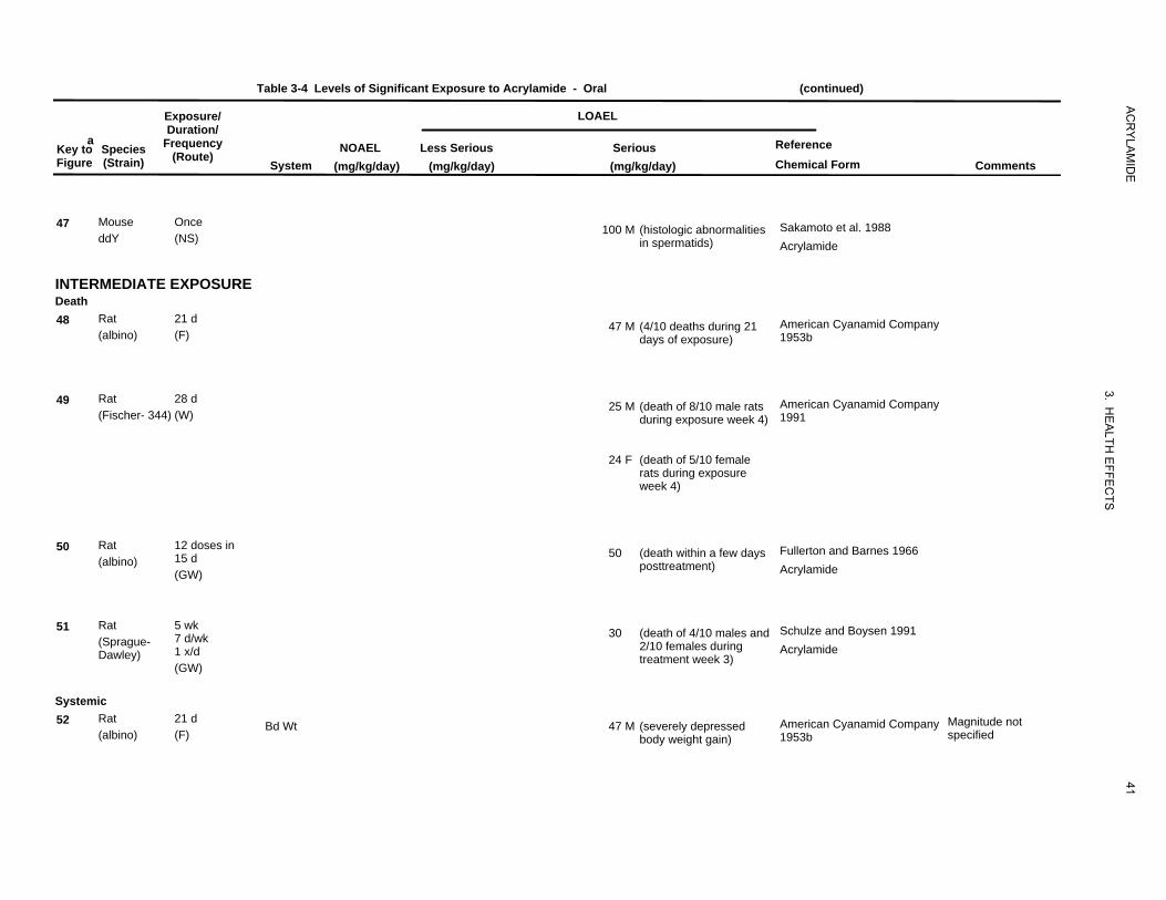


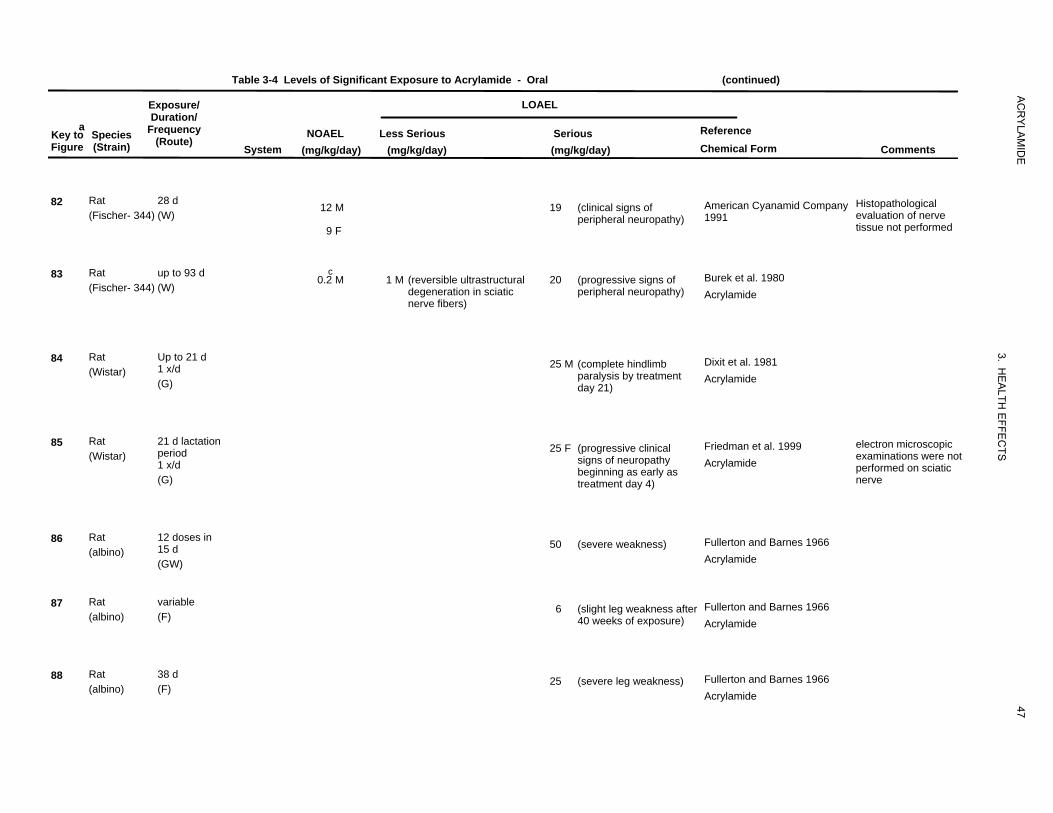


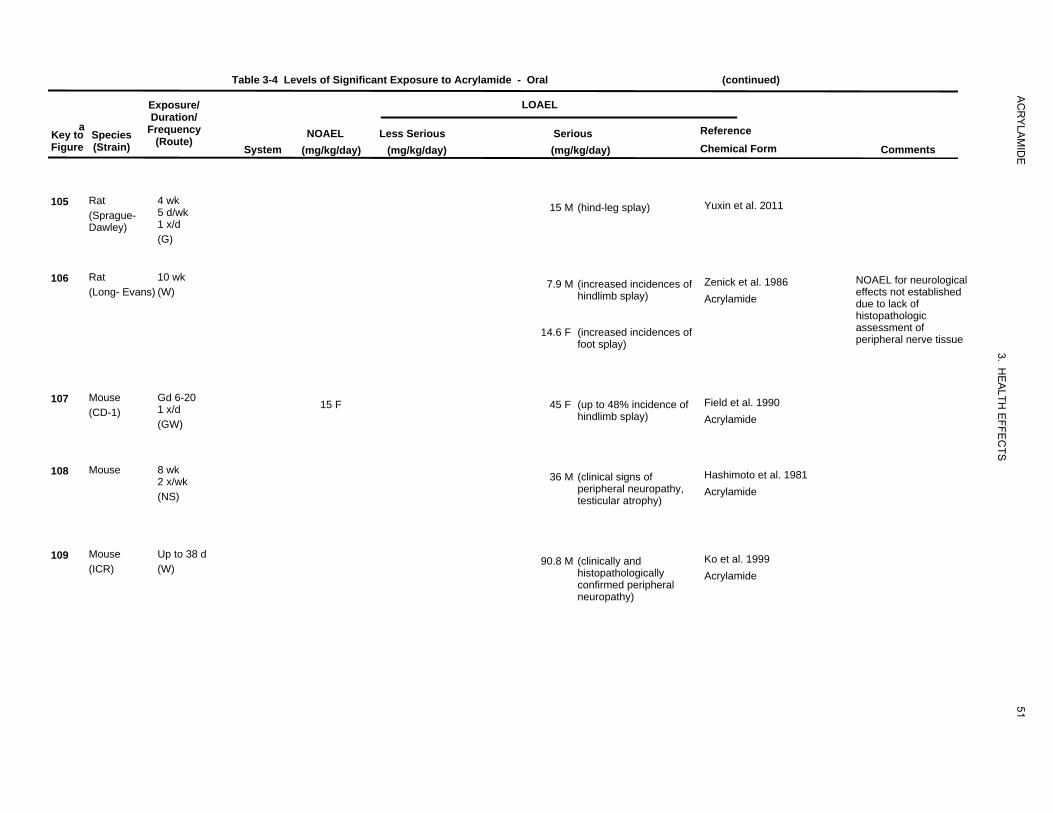


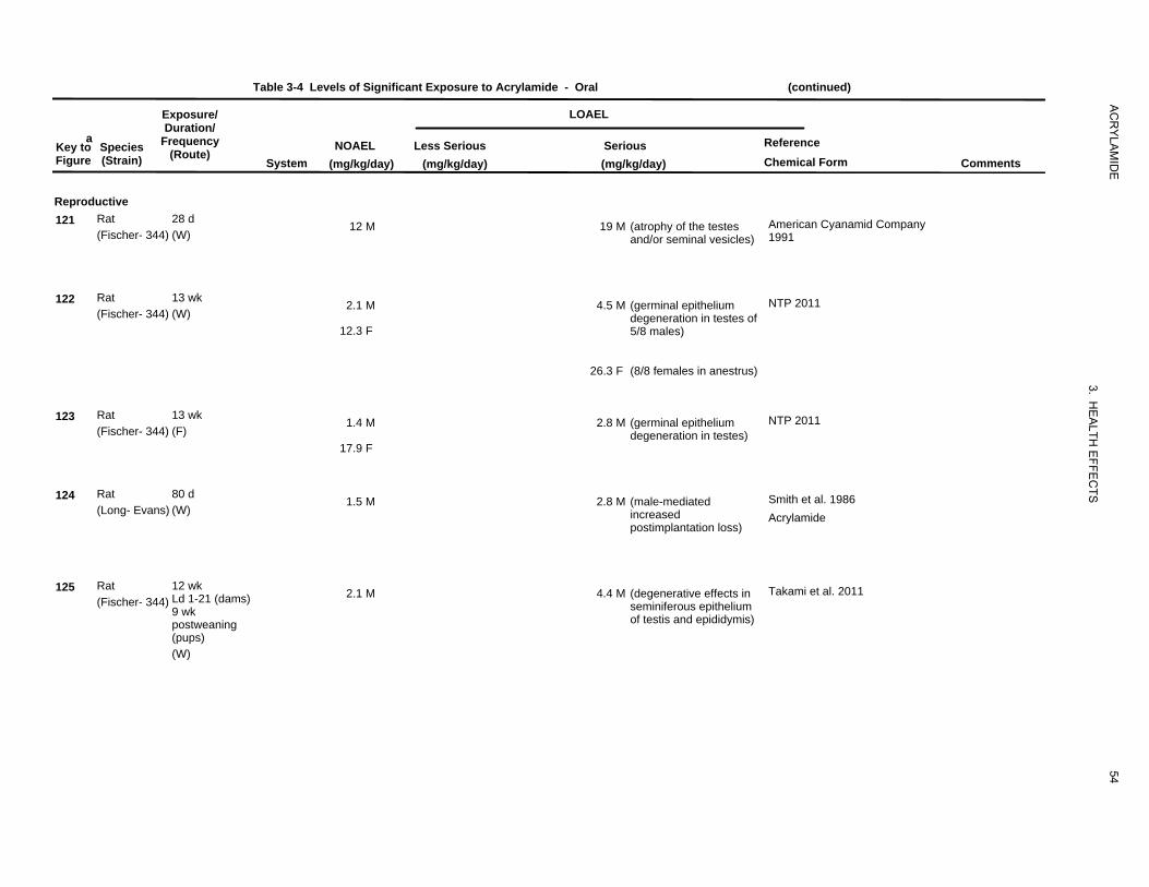


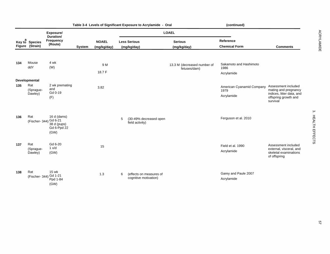

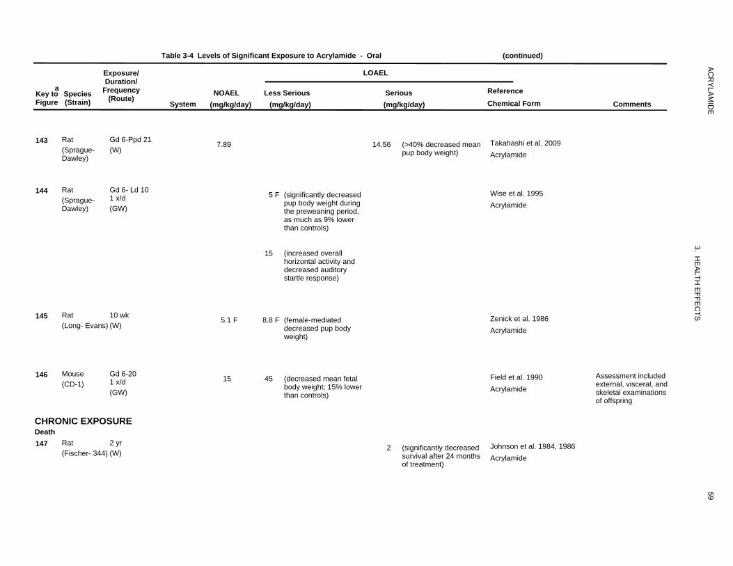
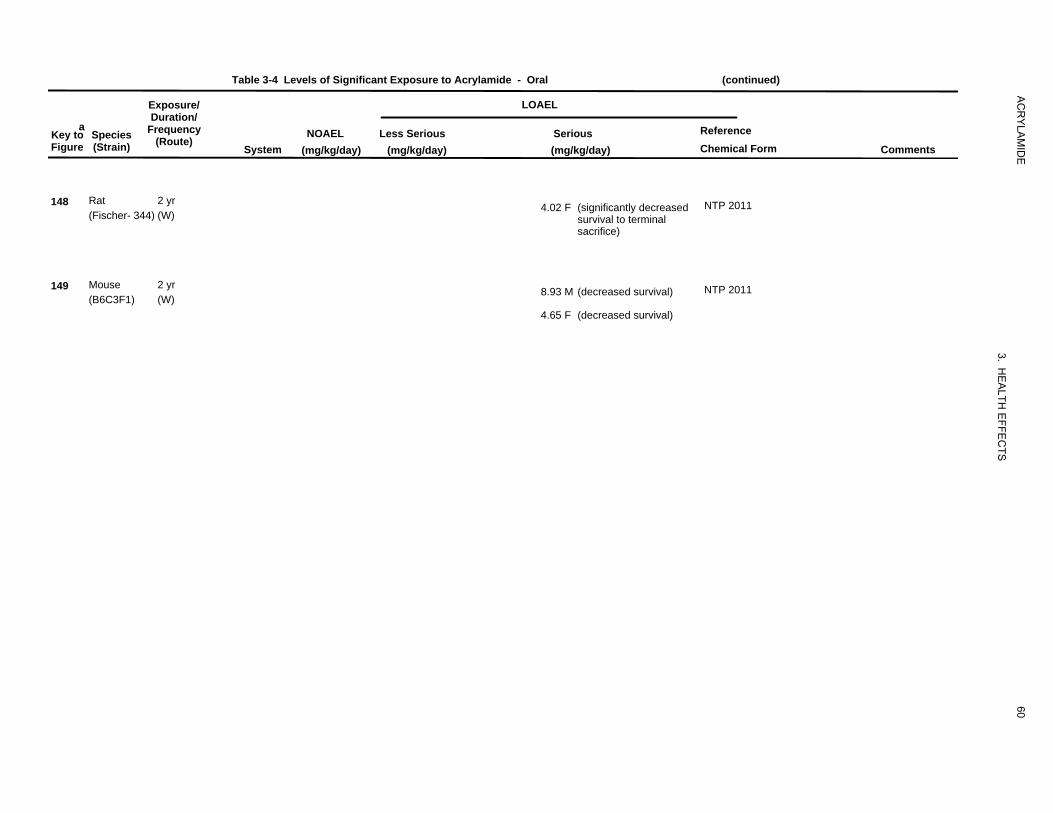


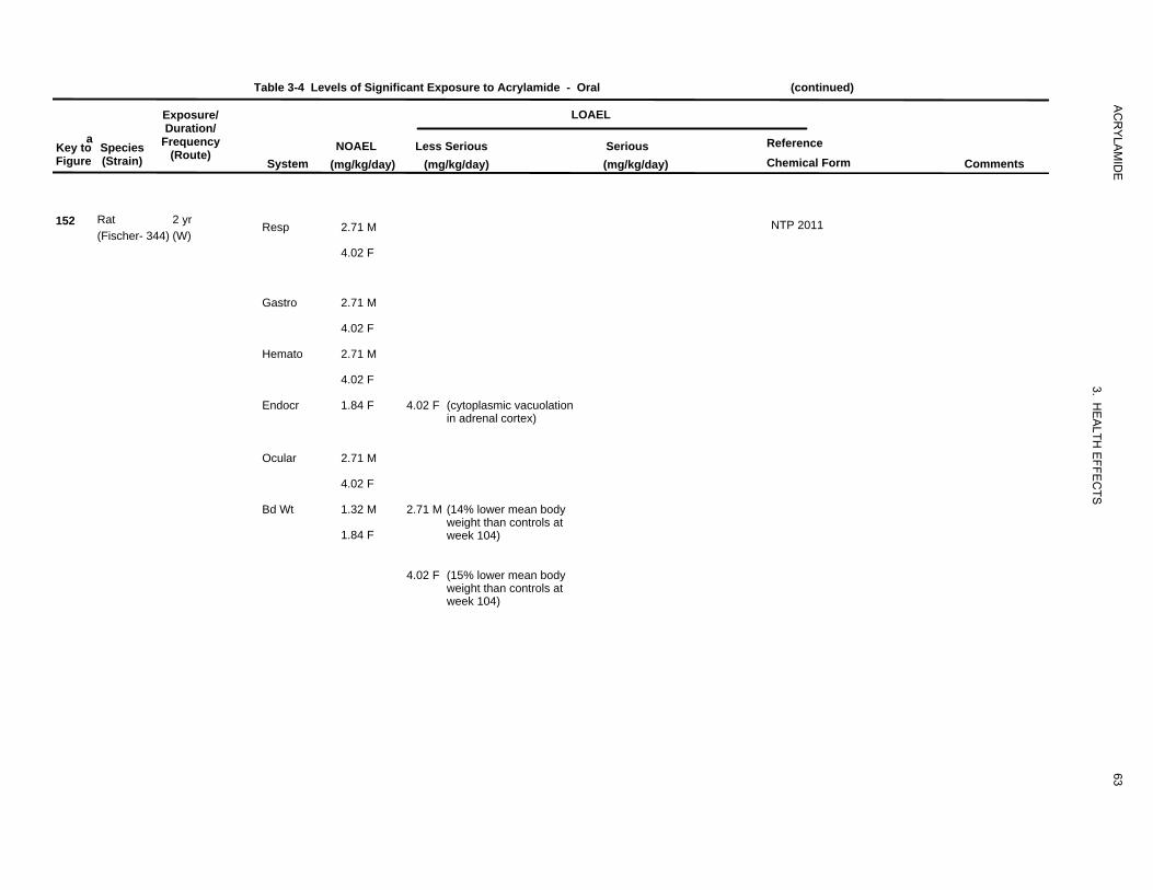


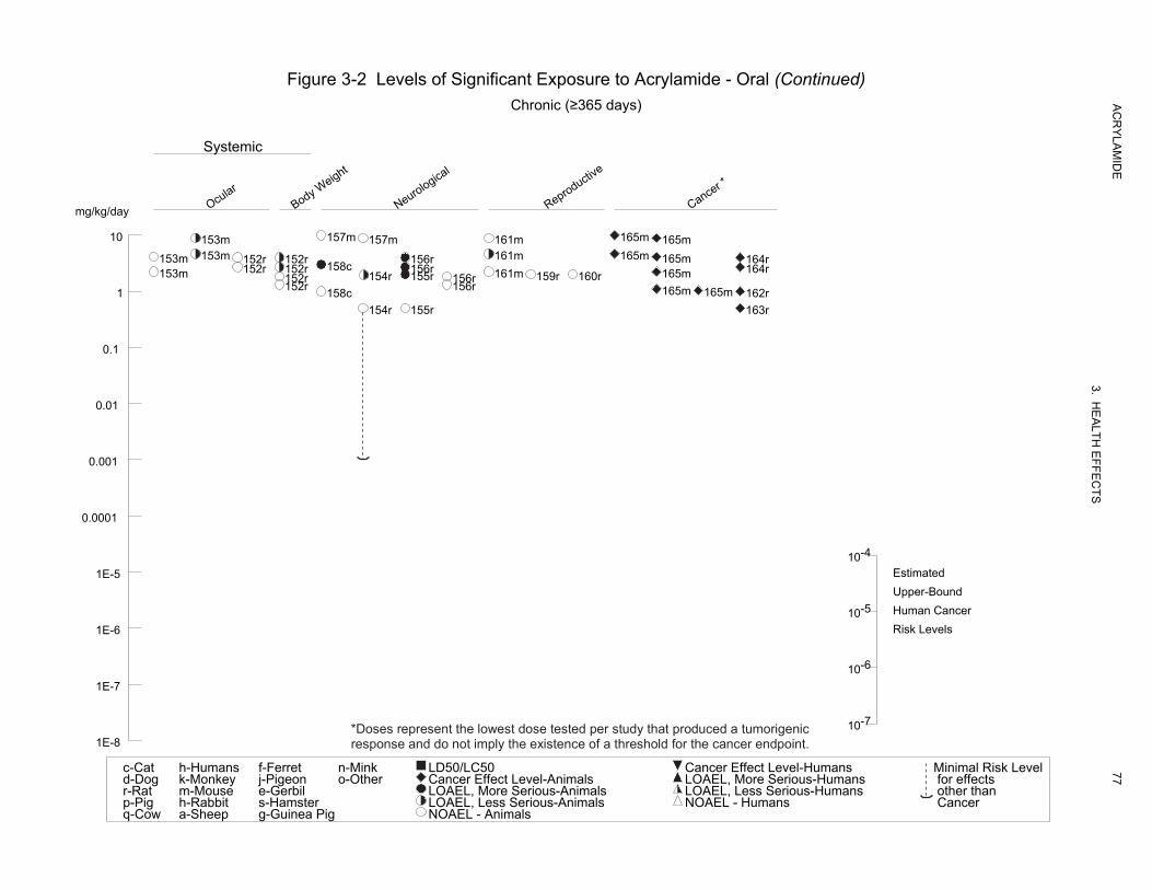


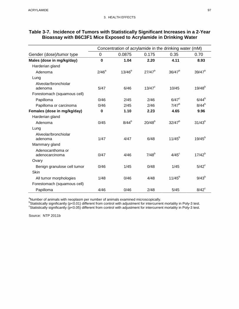


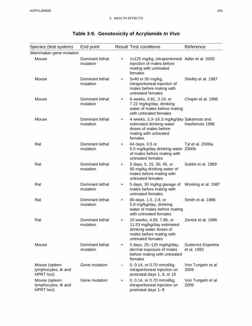


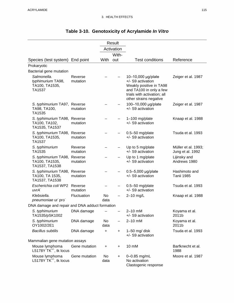


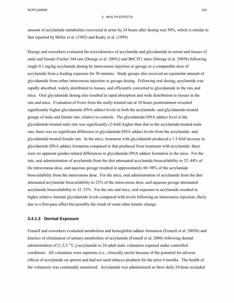


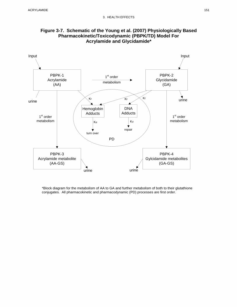


Recommended