Dipartimento di Scienze Anatomiche, Istologiche,
Medico-Legali e dell’Apparato Locomotore Sezione di Istologia ed Embriologia Medica
Facoltà di Medicina e Chirurgia
Dottorato di Ricerca in Scienze e Tecnologie Cellulari
XXIV CICLO
GLIAL CELL–DERIVED NEUROTROPHIC FACTOR (GDNF) PROMO TES
INVASIVE BEHAVIOR IN TESTICULAR SEMINOMA CELLS.
Tesi Dottorato di Ricerca
Dott.ssa Francesca Ferranti
Relatore Coordinatore del corso di Dottorato
Prof.ssa Angela Catizone Prof. Elio Ziparo
Prof.ssa Elena Vicini
a.a. 2010-2011

2
CONTENTS
ABSTRACT ............................................................................................................................... 4
INTRODUCTION ..................................................................................................................... 6
1. Testicular germ cell tumors (TGCTs) ............................................................................. 6
1.1 Origin and development .......................................................................................... 6
1.2 Epidemiology and risk factors ................................................................................. 8
1.3 TGCT gene expression ............................................................................................ 9
1.4 Genomic aberrations .............................................................................................. 10
1.5 TCam-2 cell line .................................................................................................... 11
2. Tumor tissue invasion and metastasis ........................................................................... 14
2.1 Mesenchymal and amoeboid cell invasion ............................................................ 14
2.2 Contributions of stromal cells to invasion and metastasis .................................... 15
3. TGCTs and seminoma invasion..................................................................................... 17
4. Glial derived neurotrophic factor (GDNF) .................................................................. 19
4.1 GDNF and co-receptors ......................................................................................... 19
4.2 GDNF transduction pathways ............................................................................... 21
4.2.1 Ret-indipendent transduction pathways ......................................................... 22
4.3 GDNF and spermatogenesis .................................................................................. 23
4.4 GDNF and cancer .................................................................................................. 26
AIMS OF THE PRESENT STUDY ....................................................................................... 28
MATERIALS AND METHODS ............................................................................................ 29
5.1 Tumor samples ...................................................................................................... 29
5.2 Immunohistochemistry (IHC) ............................................................................... 29
5.3 TCam-2 cell cultures ............................................................................................. 30
5.4 Reverse transcription-PCR (RT-PCR) Analysis ................................................... 30
5.5 Boyden chamber assays ......................................................................................... 31
5.6 Gelatin zymography for metalloproteinases (MMPs) detection ........................... 32
5.7 Statistical Analysis ................................................................................................ 32
RESULTS ................................................................................................................................. 33
6.1 GFRA1 is extensively expressed in CIS and invasive seminoma cells ................ 33
6.2 The TCam-2 seminoma cell line expresses the co-receptors for GDNF ............... 33
6.3 GDNF induces TCam-2 cell migration. ................................................................ 34
6.4 GDNF promotes TCam-2 cell invasion in a protease-dependent fashion. ............ 36

3
DISCUSSION........................................................................................................................... 38 BIBLIOGRAPHY .................................................................................................................... 42
PUBLICATIONS ..................................................................................................................... 54
4
ABSTRACT
The neurotrophic factor GDNF has multiple functions that promote cell survival, proliferation
and migration in different cell types. The experimental over-expression of GDNF in mouse
testis leads to infertility and promotes seminomatous germ cell tumors in older animals, which
suggests that although the underlying mechanisms are unknown, deregulation of the GDNF
pathway may be implicated in germ cell carcinogenesis. To explore the involvement of the
GDNF pathway in the onset and progression of testicular germ cell tumors, we firstly
analyzed the expression pattern of GFRA1 and Ret, the two major co-receptors for GDNF, in
seminoma samples. We report that GFRA1 was expressed more extensively in carcinoma in
situ (CIS) cells and intratubular invasive seminoma compared with normal testis.
Functional analysis of the GDNF biological activity was performed on TCam-2 human
seminoma cell line. RT-PCR and immunohistochemical analyses demonstrate that TCam-2
cells express both GFRA1 and Ret mRNA, but only GFRA1 was detected at the protein level.
It is well known that GDNF plays a central role in spermatogonial stem cell self-renewal and
proliferation (Hoffman MC et al., 2008). Therefore, in order to evaluate if GDNF could act as
a mitogenic factor in TCam-2 cells, we performed proliferation assays and cell cycle analyses.
Interestingly we observed that GDNF doesn’t induce increase in total cell number or S-phase
entry in TCam-2 cell line.
Several evidences suggested that GDNF is able to induce cell migration and invasion in
several normal and tumor cell types (Okada Y et al., 1999; Veit C et al., 2004; Su CM et al.,
2009; Song H et al., 2006; Paratcha G et al., 2006; Tang MJ et al., 1998; Young HM et al.,
2001). Therefore we have hypothesized that GDNF can act as a chemoattractant also in
seminoma cells. On this purpose we stimulated directional TCam-2 cell migration and
invasion in the presence of GDNF gradients and we investigated the downstream pathways
responsible for the GDNF-induced invasive behaviour. We demonstrated that GDNF is able

5
to induce migration, possibly through the Src and MEK pathways. It is documented that
tumor cell invasion can occur through two modalities: a proteolytic mesenchymal-like or non-
proteolytic amoeboid-like modality (Sahai E and Marshall CJ, 2003; Wolf K et al., 2003).
GDNF is able to induce TCam-2 seminoma cell invasion in a mesenchymal-like
metalloprotease-dependent manner.
In conclusion, GFRA1 over-expression in CIS and seminoma cells, along with the functional
analyses in TCam-2 cells, suggests an involvement of the GDNF pathway in the progression
of testicular germ cell cancer.

6
INTRODUCTION
1. Testicular germ cell tumors (TGCTs)
1.1 Origin and development
Testicular germ cell tumors (TGCTs) are the most frequent solid malignant tumors among
young men. Based on epidemiology, clinical presentation, phenotypic characterization,
chromosomal constitution and genomic imprinting, TGCTs are classified into three groups
(Oosterhuis JW et al., 2005). Type I comprises the teratomas and yolk-sac tumors of
newborns and infants (Figure 1); type II comprises the seminomatous and non-seminomatous
tumors of adolescents and young adults (Figure 1); and type III comprises the spermatocytic
seminomas of elderly people (Figure 1). During the carcinogenic process, the primordial germ
cells or gonocytes are converted into a pre-invasive lesion, known as in situ carcinoma (CIS)
(Kristensen DM et al., 2008; Skakkebaek NE et al., 1972). CIS cells further develop to give
rise to invasive TGCTs (Figure 2). However, not all TGCTs develop from a CIS, as is the
case of spermatocytic seminoma of elderly men and infantile germ cell tumors (Kristensen
DM et al., 2008).
The fetal origin of CIS cells is not only supported by the morphological similarities among
CIS cells, primordial germ cells and gonocytes but also by immunohistochemical studies of
proteins present in all these cell types such as the octamer binding transcription factor 3/4
(OCT3/4), the placental-like alkaline phosphatase (PLAP), the activating enhancer-binding
protein 2 gamma (AP-2γ), and the stem cell factor receptor c-KIT (van de Geijn GJM et al.,
2009). Moreover CIS lesions have been identified in prepuberal patients, who later developed
TGCTs, indicating that these cells originated prior to puberty (Muller J et al., 1984). Thus CIS
cells are most likely the result of a delayed or blocked differentiation of embryonic germ
cells.

i ii
iii iv
viv
Fig. 1 – Testicular germ cell tumors (TGCTs).Hematoxylin & Eosin stained section of: (i) seminoma, (ii) embryonic carcinoma, (iii) yolk-sac tumor, (iv) chorioncarcinoma, (v) teratoma and (vi) spermatocytic seminoma. (Modified from Wang P et al., 2010 Am J ClinPathol 134:604-612).

Fig. 2 - Germ cells transformation and tumor progression. During the carcinogenic process, primordial germ cells or gonocytes undergo arrest of differentiation and are converted into a pre-invasive lesion, known as carcinoma in situ (CIS) (Skakkebaek et al., 1972, Kristensen et al., 2008). CIS cells further develop to give rise to the invasive testicular germ-cell tumors (TGCTs) of the adolescent and young adults, that are seminomatous and non-seminomatous tumors. All these lesions are characterized by an up-regulation of germ cell pluripotency genes with an expression pattern more similar to embryonic germ cells. However not all TGCTs develop with a CIS, as in the case of spermatocytic seminoma of elderly men and infantile germ cell tumors. (From Kristensen DM et al., 2008 Mol.Cell Endocrinol. 288: 111-118).

7
CIS lesion appears in the form of enlarged cells, usually placed in the basal portion of
seminiferous tubules, with clear cytoplasm and a round, hyperchromatic nuclei (significantly
larger than those of spermatogonia) and one or more prominent nucleoli. Supporting its
neoplastic nature, CIS cells are aneuploid. CIS seminiferous tubules show decreased tubular
diameter, thickened peritubular basement membrane and absence of normal spermatogenesis.
Another observation that supports the precursor role of CIS in TGCTs development is that
CIS cells are found with high frequency in the seminiferous tubules adjacent to invasive germ
cell tumors. In the absence of an invasive tumor, CIS is asymptomatic and is usually detected
in testicular biopsies performed for other purposes. About the 50% of the patients with CIS
lesions develop an invasive TGCT within 5 years from diagnosis and the 70% within 7 years
(Giwercman A et al., 1993).
A morphological analysis of parenchyma adjacent to invasive TGCTs suggests that the
intermediate lesions between CIS and invasive TGCTs are either the intratubular seminoma or
the intratubular non-seminoma (Oosterhuis JW et al., 2003). During development of
seminomas, CIS cells become independent from the micro-environment (i.e. the Sertoli cells
produced factors) and fill up the lumen of the seminiferous tubules. Cell spreading out of the
seminiferous tubule constitutes the last step for the formation of the overt tumor. Conversely,
even though non-seminomas are thought to develop from CIS, intratubular non-seminoma is
invariably composed of pure embryonal carcinoma (EC) and when invasion starts the
differentiation into its derivatives occurs (teratoma, yolk-sac tumour and chorioncarcinoma)
(Oosterhuis JW et al., 2005). In this case EC has the ability to differentiate into a wide range
of cell types and represents the presumed stem cell compartment. EC may undergo down-
regulation of the pluripotency genes and activation of the somatic lineage (Kristensen DM et
al., 2008). Hence, while seminoma is a uniform tumor resembling early fetal germ cells and
CIS cells, the more malignant non-seminomas appear heterogeneous and can also contain
elements of seminoma (Ulbright TM et al., 1999).

8
1.2 Epidemiology and risk factors
TGCTs account for up to 60% of all malignancies affecting men between 20-40 years of age.
The rates of TGCTs display geographic and ethnic differences. The incidence of TGCTs in
Caucasian populations is 6–11/100,000 males but in the last decades an annual increase of
about 3-6% was registered (Oosterhuis JW and Looijenga LH, 2005). Black populations show
a lower risk for TGCTs than Caucasians (Moul JW et al., 1994). Moreover high oestrogen
levels relate with an increased risk of developing a TGCT while high testosterone levels
reduce the risk. These observations could possibly explain the different incidence rates among
Caucasians and Blacks (Henderson BE et al.,1988). However, even within the white
populations, marked differences in the rates of TGCTs development were observed, with
Denmark detaining the higher incidence. The relatively rapid rise in the incidence of TGCTs
and the differences among countries and ethinc groups suggest a possible involvement of
environmental or lifestyle factors in the etiology of these tumors (Rajpert-De ME and Hoei-
Hansen CE, 2007). The importance of this factors is supported by epidemiological studies on
populations migrating from countries with high incidence (Denmark) and with significantly
lower incidence (Finland) to Sweden. The first generation immigrants showed the same risk
of their country of origin, while the second generation, that was born in Sweden, had the same
incidence of native Swedes (Hemminki K and Li X, 2002).
In addition, one of the best known risk factor for TGCTs is cryptorchidism, suggesting that
also the microenvironment plays a crucial role in the regulation of the development of the
precursor lesion.
Other risk factors for TGCTs include hypotrophic (<12 ml) or atrophic testicle, Klinefelter’s
syndrome, familial history of testicular tumours among first-grade relatives, the presence of
contralateral tumour and infertility (Albers P et al., 2005) but also unspecific factors, such as
birth order, birth weight and factors related to maternal lifestyle (Rajpert-De ME and Hoei-
Hansen CE, 2007).

9
TGCTs can occur in a sporadic or familial manner. Twin studies showed a greater
concordance for disease in monozygotic than in dizygotic twins. The segregation analysis
suggests a recessive mode of inheritance. (Krausz C and Looijenga LH, 2008).
1.3 TGCT gene expression
In the last decades a significant number of markers has been identified to discriminate CIS,
seminoma and EC. As previously described, CIS cells share the expression of
immunohistochemical markers with primordial gem cells and early gonocytes.
Among the first proteins reported to be expressed both in gonocytes and their malignant
counterpart there is the stem cell factor receptor KIT (Rajpert-De ME and Skakkebaek NE,
1994). Together with PLAP is now commonly used as a clinical marker to detect CIS in
surgical biopsies.
More recently OCT3/4, a well-characterized marker for primordial germ cells, was discovered
to be positive in all cases of CIS, seminoma and derived invasive cancers (Looijenga LH et
al., 2003; Rajpert Rajpert-De ME et al., 2004). OCT3/4 is a transcription factor of the family
of the octamer-binding proteins and is a well known key regulator of pluripotency, (de Jong J
and Looijenga LH, 2006). Another important gene known for its association with
pluripotency in embryonal stem (ES) cells is the homeobox proteing NANOG. Similarly to
OCT3/4, NANOG is expressed not only in CIS cells but also in the overt germ cell tumor
(Hoei-Hansen CE et al., 2005).
Among the novel markers for TGCT there is AP-2γ, another transcription factor associated
with the undifferentiated state and suggested as a possible marker for CIS in semen specimen
of patient with early stages of TGCT (Hoei-Hansen CE et al., 2007).
All these stem cell-related proteins are physiologically abundantly expressed in fetal pre-
meiotic germ cells of both sexes. At the onset of meiotic prophase their expression is
normally rapidly down-regulated (Rajpert-De ME and Hoei-Hansen CE, 2007).

10
However, among the histological types of CIS-derived invasive tumors, marked differences in
the expression of embryonic genes were observed (Kristensen DM et al., 2008). While
seminoma phenotype is closely similar to CIS, the EC (the undifferentiated stem cell
compartment of non-seminomas) is strikingly related to that of ES cells, suggesting a re-
programming or a differentiation event. For example, the SRY (sex determining region Y)-
box 2 (SOX-2) is highly expressed in non-seminomas, whereas seminomas are negative for
this marker (Sperger JM et al., 2003; Santagata S et al., 2007). Accordingly SOX2 belong to
the SOX protein family of transcription factors that regulate the development from the early
embryonal stage to specific lineages (Avilion AA et al., 2003, Tay Y et al., 2008).
1.4 Genomic aberrations
CIS, seminomas, and all variants of nonseminomas are aneuploid, as demonstrated by flow
cytometry as well as cytogenetic approaches. (Oosterhuis JW et al., 1989; Murty VV et al.,
1990; De Graaff WE et al., 1992; Van Echten-Arends J et al., 1995). Even if altered copy
number of regions on chromosome 7, 8, 14, 17 and X have also been consistently reported
(Looijenga LHJ et al., 2000; Summersgill B et al.,2001; Ottesen AM et al., 2003; Ottesen AM
et al., 2004), the main recurrent structural imbalance in these tumors is the gain of the short
arm of chromosome 12, mostly in the form of an isochromosome (Looijenga LHJ et al.,
2003). At which stage during tumour development genomic instability is introduced is
uncertain (Skotheim RI and Lothe RA, 2003). However the observation that gain of 12p
material is detected in CIS cells near the overt tumours (Ottesen AM et al., 2003) suggests
that this event is related to the invasive growth of the type II TGCTs (Rosenberg C et al.,
2000; Summersgill B et al., 2001). Interestingly, gain of 12p region is also found in in vitro
cultured human ES cells, suggesting a relation with an improved capability to survive in a
non-physiological environment (Draper JS et al., 2003; Li SS et al., 2006).
11
One of the genes located on 12p region that could play a relevant role in favoring TGCTs
progressions is the cyclin D2 (CNND2). This gene is indeed overexpressed in type II TGCTs
and CIS (Houldsworth J et al., 1997; Kukoski R et al., 2003). However, this region contains
also other genes associated with pluripotency and proliferation, including the small GTPase
K-RAS and NANOG, that has been suggested to be relevant in invasive TGCTs progression,
although the actual proof is lacking so far (Krausz C and Looijenga LH, 2008).
1.5 TCam-2 cell line
Currently, various non-seminomatous cell lines have been established, but only one (i.e.
TCam-2) (de Jong J et al., 2008; Eckert D et al., 2008; Goddard NC et al., 2007; Mizuno Y et
al., 1993) has been established from seminoma, although they constitute more than 50% of all
TGCTs. This is probably due to the capability of seminoma cells to undergo to rapid
apoptosis after disruption of their microenvironment (Olie RA et al., 1996). TCam-2 cell line
originated from a primary testicular seminoma of a 35-year-old patient (Mizuno Y et al.,
1993). The initial in vitro culture was subcloned after 15 passages, and after 18 passages the
cells were also successfully transplanted into the back of CB-17 SCID mice. Electron
microscopy demonstrated the presence of significant amounts of glycogen and absence of
intercellular connection structures. Karyotyping after 10 passages in vitro showed 90–96
chromosomes, including the lack of the Y chromosome (Mizuno Y et al., 1993).
TCam-2 cells display the characteristic gain of chromosome 12p, a chromosomal aberration
common to all the invasive germ cell tumors but not spermatocytic seminoma (De Jong J et
al., 2008). Original cytogenetic analysis using Giemsa banding identified five copies of
chromosome 12 (Mizuno Y et al., 1993). Successively Goddard and coworkers identified by
metaphase and interphase fluorescence in situ hybridization analysis further four copies of the
12 centromere and material from the 12p11.2-12.1 region but no isochromosome of 12p
(Goddard NC et al., 2007). No expression of alpha fetal protein (αFP) or beta-human
12
choriogonadotrophin (βhCG), respectively markers for yolk-sac tumors and
chorioncarcinoma, was described, while strong staining for the seminoma marker PLAP has
been reported (Mizuno Y et al., 1993). As in the case of seminoma, the pluripotency genes
Oct3/4 and Nanog are highly expressed in TCam-2 cells as detected by
immunohistochemistry and western blot analysis (Goddard NC et al., 2007; De Jong J et al.,
2008). Instead the embryonic stem cell marker SOX2, typically highly expressed in EC but
absent in seminomas, is not expressed in TCam-2 cells both on protein and mRNA level (De
Jong J et al., 2008). The transcription factor AP-2γ, a known specific marker used for
diagnosis of seminomas, was expressed as revealed by RT-PCR, western blot analysis and
immuhistochemistry (Eckert D et al., 2008). In addition, the tumor necrosis factor receptor
superfamily member 8 (CD30) usually expressed in EC and EC-derived cell lines and related
to inhibition of apoptosis, is not expressed in TCam-2 cells on protein level nor on mRNA
level (de Jong J et al., 2008).
Moreover, the stem cell factor (SCF) receptor, KIT, and its ligand SCF were both expressed
in the TCam-2 cells as determined by immunoprecipitation for KIT (de Jong J et al., 2008,
Goddard NC et al., 2007), western blot analysis and immuhistochemistry (Eckert D et al.,
2008). KIT mutations have been described in bilateral TGCTs (Looijenga LH et al., 2003) but
sequencing analysis of exons 9, 11, 13 and 17 did not detect any activating mutations of KIT
in TCam-2 cells (Goddard NC et al., 2007). Finally, the expression of the typical germ cell
markers VASA (ATP-dependent RNA helicase), DAZL (DAZ (deleted in azoospermia)
ligand) and BOULE (another member of the human DAZ gene family) were observed (Eckert
D et al., 2008).
Unlike other solid tumors, type II TGCTs have a low mutation rate in protooncogenes,
including BRAF and KRAS2 (Sommerer F et al., 2005), as well as the tumor suppressor gene
TP53 (Kersemaekers AM et al., 2002). Mutation analysis of these genes in TCam-2 cells
showed a mutation in the protooncogene BRAF, due a thymine to adenine transversion at

13
nucleotide 1796 (T1796A), leading to a substitution of glutamic acid for valine at amino acid
600 (V600E) (de Jong J et al., 2008). It has been suggested that this mutation could be the
explanation for the successful propagation of these cells in vitro. Because of the lack of the
primary tumor, it is not possible to determine whether this mutation was present in the
original tumor, or was generated due to in vitro propagation (de Jong J et al., 2008). On the
other hand, recently Goddard and coworkers, described that the TCam-2 cell line does not
show evidence for a V600E BRAF mutation (Goddard NC et al., 2010). These authors
performed direct sequencing of both the forward and reverse strands of the BRAF gene in
TCam-2 cells and normal female DNA revealing that the sequence is wildtype at nucleotide
1796. They hypothesized that the mutation detected by de Jong J and coworkers (de Jong J et
al., 2008) could be present only in a small subpopulation of TCam-2 or alternatively, a BRAF
mutation may have arisen independently or the mutant allele may have been lost during
prolonged culture in different laboratories. In conclusion they affirmed that the BRAF
mutation can not account for the establishing of TCam-2 cell line from a seminoma sample. In
their opinion rather other factors, including mutations in other genes or the overexpression of
receptors or growth factors, may account for their in vitro growth (de Jong J et al., 2008).

14
2. Tumor tissue invasion and metastasis
2.1 Mesenchymal and amoeboid cell invasion
Cell migration plays a key role in metastatic dissemination of tumour cells from the primary
tumour to local and distant sites (McSherry EA et al., 2007; Farrow B et al., 2008; Condeelis
J and Segall JE, 2003).
Even if tumour cells in vivo can move both randomly and directionally, invasion is more
efficient when the cell is involved in directed migration toward a chemokine gradient
(chemotaxis) (Roussos ET et al., 2011). Thus, chemotaxis is on the base of several major
events during tumour progression: metastatic invasion and dissemination, angiogenesis and
immune cell extravasation (Roussos ET et al., 2011).
Different modes of invasive migration have been described for cancer cells: amoeboid
migration or mesenchymal migration for single cells and collective migration of groups of
cells (Roussos ET et al., 2011). Amoeboid cell motility of single tumour cells is independent
from the activity of matrix metalloproteinases (MMPs) because cells can infiltrate through
gaps in the extracellular matrix (ECM) because their cytoskeleton contractile capability (Wolf
K. et al., 2003; Wyckoff JB et al., 2006). Intravital multiphoton imaging had permitted to
observe migrating cells in vivo revealing that some carcinoma cells with an amoeboid
morphology can move at high speeds inside the tumours (~4 µm min–1) (Condeelis J and
Segall JE, 2003). On the other end, mesenchymal migration that typically involve single cells
but sometimes also collective migration, is characterized by polarized cells with an elongated
cell morphology and relatively low speeds of cell migration (0.1–1 µm min–1) (Sahai E,
2005). However, even if amoeboid and mesenchymal modes of migration can be observed as
separated mechanisms by in vitro studies, evidence suggests that in vivo they can convert into
each other in response to changes in the microenvironment (Sahai E and Marshall C, 2003;
Pankova K et al., 2009). For example, a transition from mesenchymal to amoeboid tumor cell
migration can be induced by inhibition of proteolysis (Wolf K et al., 2003).

15
Collective migration has been defined as the coordinate movement through the ECM of
clusters or sheets of tumour cells with functionally intact cell–cell adhesions (Friedl P and
Gilmour D, 2009). The leader cells positioned at the front of the migrating group actively
migrate toward a chemotactic gradient creating a track by matrix degradation (Rorth P, 2007;
Ilina O and Friedl P, 2009; Valentin G et al., 2007; Schmidt M et al., 2007; Aman A and
Piotrowski T, 2008). The other cells follow the leader cells along the remodeled matrix tracks
due to the physical coupling that generate drag forces (Rorth P, 2007). The leader cell can be
either a tumour cell with proteolytic activity or a stromal cell from the tumour
microenvironment (Gaggioli C et al., 2007; Rorth P, 2007; Wolf K, 2007).
The occurrence and frequency of these modes of migration in cancer is dependent on tumor
cell types and tumour microenvironment.
Despite the various patterns of directed migration during tumour cell dissemination, the
intracellular processes involved in cell motility are probably similar and are comprised of
three main steps: chemo-sensing, polarization and locomotion (Iglesias PA, Devreotes PN,
2008). First, polarized intracellular signals lead to asymmetric actin polymerization resulting
in extension of the cell membrane in the direction of movement, thus creating the leading-
edge protrusion. This is followed by integrin-mediated adhesion to the substrate in the
direction of movement, and by subsequent detachment from the substrate on the opposite side
after contraction of the trailing edge of the cell (Figure 3). Nodal point for regulation of these
migratory processes are the small GTPases such as Rho, cdc42, and Rac (Gupta GP and
Massague J, 2006)
2.2 Contributions of stromal cells to invasion and metastasis
Cancer cells typically produce mediators that induce chemotactic recruitment and activation
of a variety of cell types into the surrounding stroma (such as fibroblasts, myofibroblasts,
granulocytes, macrophages, mesenchymal stem cells, and lymphocytes) which contribute to

Fig. 3 - Schematic representation illustrating the different phases of cell migration. Polarized intracellular signals lead to asymmetric actin polymerization resulting in extension of the cell membrane in the direction of movement, creating the leading-edge protrusion. This event is followed by integrin-mediated cell-substrate adhesion (focal contacts) in the direction of movement, and by subsequent detachment on the opposite side after contraction of the trailing edge of the cell. (From Molecular Biology of the Cell; Alberts).

16
the formation of an inflammatory microenvironment (Gupta GP and Massagué J 2006).
Therefore, invasion occurs in a restricted zone of cross-talk and cooperation between this
“reactive” stroma and the premalignant epithelium (Figure 4).
The proteolytic machinery of the activated stromal cells, combined with that of tumor cells,
degrades extracellular matrix (ECM) proteins, uncovering cryptic sites that may display pro-
migratory properties and releasing sequestered growth and survival factors (Liotta LA and
Kohn EC, 2001).
In this scenario, invasive carcinoma may be viewed as “a pathology of multiple cell societies
inhabiting the epithelial/mesenchymal stromal unit” (Liotta LA and Kohn EC, 2001). Thus,
tumor cells could convert reactive stromal infiltrates from preservers of tissue homeostasis
into accomplices in malignancy. Accordingly, leukocytic infiltration, angiogenesis, and
lymphangiogenesis, all markers for stromal cells and tumor coexistence, are frequently
correlated with an increased likelihood of metastatic relapse (Gupta GP and Massagué J,
2006). However the stromal reaction to invading tumor cells is variable, depending in part
upon tumor cell properties and in part upon the local stromal composition (Bacac M and
Stamenkovic I, 2008).
During the biological cascade of metastasis, a cancer cell from a primary tumor executes
several discrete steps: i) emigration from the primary tumor and invasion of the surrounding
tissue and degradation of ECM; ii) intravasation into the microvasculature of blood or
lymphatic system iii) survival and translocation largely through the bloodstream and lymph to
microvessels of distant tissues and finally iv) extravasation and v) metastasis formation at
target tissues (Figure 5). Since cancer cells exit from the bloodstream, they have to adapt to
the foreign microenvironment to survive and give rise to a macroscopic secondary tumor
(colonization) (Fidler IJ et al., 2003).

Fig. 4 – Stromal reaction. Advanced malignancies are frequently coinhabited by different stromal cell types. The tumor-suppressive activities of lymphocytes (T, B, and NK cells) is in part antagonized through the release of immunosuppressive cytokines. Carcinoma-associated fibroblasts (CAFs) can secrete factors that promote both tumor cell growth and invasion and neoangiogenesis (through recruitment of bone marrow dendritic cells (BMDC) from circulation). Activated macrophages were recruited to tumors and can release many pro-tumorigenic growth factors. Additionally, macrophages may co-migrate with cancer cells within tumors through a paracrine growth-factor loop. Once cancer cells have invaded the blood stream, many of them will die from stresses associated with circulatory passage. The remaining survivors may attach to capillaries within a secondary organ through adhesion receptors. Subsequently, cancer cells can extravasate from capillaries and may or may not generate a viable niche at the secondary site.(From Gupta GP and Massagué J, 2006 Cell 127: 679-695).

Fig. 5 - The metastatic cascade. Metastasic process can be subdivided in two major phases: (i) physical translocation of cancer cells from the primary tumor to a distant organ and (ii) colonization of the translocated cells within that organ. (A) To begin the metastatic cascade, cancer cells within the primary tumor acquire an invasive phenotype. (B) Cancer cells can then invade into the surrounding matrix and toward blood vessels, where they intravasate to enter the circulation, which serves as their primary means of passage to distant organs. (C) Cancer cells traveling through the circulation are referred as circulating tumor cells (CTCs). (D) At the distant organ, CTCs exit the circulation and invade into the microenvironment of the foreign tissue. (E) At that foreign site, cancer cells must be able to evade the innate immune response and also survive as a single cell (or as a small cluster of cells). (F) To develop into an active macrometastatic deposit, the cancer cell must be able to adapt to the microenvironment and initiate proliferation. (From Chaffer CL and Weinberg RA, 2011 Science 331, 1559).
17
3. TGCTs and seminoma invasion
Although the cure rate for TGCT is high, presence of metastases reduces survival rates.
Interestingly, metastasis is a prevalent feature of human TGCTs showing a very high number
of metastases compared with other tumours, averaging 5.8 metastases per primary tumour (De
Giorgi U et al., 2008). Approximately 30% of patients affected by TGCTs presents metastases
at the time of diagnosis (Powles TB et al., 2005), and 15–20% of patients have subclinical
metastases in stage I seminoma (Benne R et al., 1986), the most common presentation among
TGCTs. While tumors remain confined to the testis in about 75% of patients, a retroperitoneal
involvement is observed in the 20%, and the remaining 5% shows supradiaphragmatic or
organ metastases (Ulbright TM et al., 2009).
Therefore metastases represent important factors directly affecting treatment modality,
tumour surveillance, and survival (Zechel JL et al., 2011). Treatment for TGCTs involves
surgery and chemotherapy with bleomycin, etoposide and cisplatin (BEP), obtaining a relative
low mortality risk of 2.3–4.5% (Fossa SD et al., 1998; Williams SD et al., 1987). Although
many TGCTs and their metastases respond readily, 10–25% of patients with metastases are
resistant to chemotherapy (Piulats JM et al., 2009), further complicating treatment and
worsening prognosis. Moreover, patients with a metastatic disease suffer frequently of a
higher incidence of relapse (5–10%) after treatment (Lutke Holzik MF et al., 2008) and a
lower survival rate (Loehrer PJ et al., 1998).
Even though the clinical and epidemiological aspects of TGCTs are well described, very scant
informations are available about the molecular mechanisms for tumor spreading and
metastasis. Testicular cancer metastases exhibit a conserved pattern targeting various tissues,
such as lymph nodes, lung, liver, bone and spleen (De Giorgi U et al., 2008).
It has been observed that this TGCT pattern of metastases abundantly overlap with that of
CXCL12 expression. In fact, metastases target organs that express high levels of the
chemokine CXCL12 (SDF-1), resembling patterns observed in other CXCR4 over-expressing

18
cancers (breast, colorectal and prostate cancers) (Gilbert DC et al., 2009). In normal human
testes, CXCL12 is expressed by Sertoli cells and its receptor CXCR4 by the germ cell
population. CXCL12 is able to stimulate invasive cell migration of TCam-2 seminoma cell
line in a CXCR4-dependent fashion via activation of ERK. More interestingly high-level
CXCL12 expression in tumors correlates with reduced risk of TGCT occult metastases and
relapse after surgical treatment (Gilbert DC et al., 2009).
Even if seminoma is generally pain-less, about 2,5% of patients presenting a metastatic
involvement suffers most commonly of lumbar pain due to retroperitoneal metastasis, even if
gastroinstestinal bleeding, bone pain, dyspnea and cough, a supraclavicular mass,
neurological symptoms, and lower extremity edema may also be presenting symptoms due to
spread to other sites. Such metastases, however, can be asymptomatic (Ulbright TM et al.,
1999).

19
4. Glial derived neurotrophic factor (GDNF)
4.1 GDNF and co-receptors
GDNF is a distant member of the transforming growth factor beta (TGF-β) superfamily (Lin
LF et al., 1993). GDNF was first identified through its activity on midbrain dopaminergic
neurons but subsequently, it has been shown to have pleiotropic functions, promoting
neuroprotection, cell proliferation, and migration (Sariola H and Saarma M, 2003).
Mice lacking GDNF die within the first day of birth due to severe defects in renal
differentiation and the absence of an enteric nervous system (Moore MW et al., 1996; Pichel
JG et al., 1996; Sanchez MP et al., 1996). The animals lacking GDNF receptors show the
same phenotypes, indicating that GDNF pathway is essential for postnatal survival in the
mouse (Schuchardt A et al., 1994; Enomoto H et al., 1998; Cacalano G et al., 1998).
GNDF, along with the other members of the GDNF family of neurotrophic factors (neurturin,
artemin and persephin), is a basic, dimeric, secretory protein. GDNF is first synthesized as an
inactive 211 amino acids long preproGDNF. Subsequently the secreted proGDNF is
proteolytically cleaved to form the mature GDNF protein of 134 amino acids (Saarma M and
Sariola H, 1999). The specific proteases that cleave and activate GDNF precursors have not
yet been identified. The GDNF mature protein is N-glycosylated at two amino acid residues,
even if the role of these carbohydrates in the biological activity of GDNF has not yet been
established (Lin LF et al., 1993). Moreover GDNF contains seven cysteine residues in the
same relative spacing as in the members of the TGF-b superfamily. Although the amino acid
sequence homology between the GDNF and the TGF-b superfamily is less than 20%, the
pattern of cysteine residues makes GDNF a distant member of this family (Lin LF et al.,
1993). According to the crystal structure, the GDNF monomer forms two finger-like (Finger 1
and Finger 2) structures by pairs of antiparallel β-strands with a α-helix at the opposite end
(Eigenbrot C and Geber N, 1997). The GDNF homodimer is formed by head-to-tail
dimerization, which is supported by one interchain disulphide bond among the unpaired

20
cysteines in monomers that are not part of the cysteine knot. Only the homodimer of GDNF is
biologically active (Saarma M and Sariola H, 1999).
GDNF exerts its effect on target cells by binding to a glycosyl phosphatidylinositol–linked
GDNF family receptor-a 1 (GFRA1); this binding recruits the tyrosine kinase receptor Ret to
form a multi-subunit signaling complex of GDNF/GFRA1/Ret.
While the other members of TGF-b superfamily signal through transmembrane serine-
threonine kinase receptors, the GDNF receptor Ret is a typical tyrosine kinase receptor. Ret is
a single-pass transmembrane protein that contains four cadherin-like repeats in the
extracellular domain (607 amino acids), a cysteine rich domain, a hydrophobic
transmembrane region and a cytoplasmic intracellular part with a kinase domain (Anders J et
al., 2001; Iwamoto T et al., 1993; Kuma K et al., 1993). Moreover a Ca2+-binding site is
localized between cadherin-like domain 2 and 3 (Anders 2001).
The GPI-anchored receptor GFRA1 contains a N-terminal hydrophobic domain codifying for
the secretory signal sequence and a C-terminal sequence with 23 hydrophobic amino acids. A
group of three small amino acids proceeding the stretch of hydrophobic amino acids indicates
a possible GPI-binding/cleavage site (Undenfriend S and Kodukula K, 1995).
GFRA1, as most GPI-linked proteins, can be localized to specialized detergent-resistant
membrane fractions, named lipid rafts. These structures are described as floating membrane
platforms in the exoplasmic leaflet of the membrane bilayer enriched in cholesterol and
sphingolipids (Simons K and Ikonen E, 1997). Several protein families have high affinity for
lipid rafts, such as Src-family kinases and small GTP-ases, making them signaling platforms
for various transmembrane and GPI-linked proteins (Saarma M, 2001; Simons K and Toomre
D, 2000).
The original model described the GDNF/GFRA1 binding and activation of Ret receptor as an
in cis interaction between two receptors expressed on the same cell (Jing S et al., 1996). This
model is supported by the overlapping expression of Ret receptor and GFRA1 in the same cell

21
population (Golden JP et al., 1998; Tansey MG et al., 2000). However also another
mechanism has been reported where the GFRA1 receptor can be released from the cell
surface by cleavage of the GPI-anchor and acts as a soluble receptor. According to this in
trans model, the soluble GFRA1 molecule presents the ligand to Ret located in the membrane
of another cell (Figure 6). It has been suggested that the in trans signaling can sustain and
increase the response achieved through the canonical in cis pathway (Paratcha G et al., 2001).
In this last case, GDNF binds to the GPI-linked GFRA1 receptor into lipids rafts, recruiting
Ret in this compartment which binds and activates the lipid-anchored adaptor protein FRS2
(fibroblast growth factor receptor substrate 2). During the in trans activation, instead, GDNF
binds to soluble GFRA1 and this complex binds and activates Ret on neighboring cells
outside the lipid rafts. The activated receptor associates with and phosporylates SHC (Src-
homologous and collagen-like protein) (Paratcha G et al., 2001; Airaksinen MS and Saarma
M, 2002).
Once GDNF/GFRA1 complex binds to Ret, two transmembrane Ret molecules dimerize and
transphosporylate cytoplasmic tyrosines, triggering several cell signaling cascades
(Airaksinen MS and Saarma M, 2002).
4.2 GDNF transduction pathways
The GDNF-GFRA1 binding to Ret extracellular domain leads to Ret autophosphorylation at
many tyrosine residues on its intracellular domain (Airaksinen MS and Saarma M, 2002). The
phosphorylated tyrosine residues of activated Ret act as docking sites for various adaptor
proteins that in turn may activate different pathways in target cells (Airaksinen MS and
Saarma M, 2002).
For instance, the Src signaling pathway, activated by Ret Tyrosine 981 phosphorilation, elicits
neurite outgrowth, neuronal survival and ureteric branching (Airaksinen MS and Saarma M,
2002) as well as spermatogonial stem cells (SSCs) self-renewal and proliferation in mouse

Fig. 6 - GDNF receptor complex signaling. In cis pathway (left): GDNF binds to GPI-anchored GFRa1 receptors in lipid rafts (1), resulting in the recruitment and activation of c-Ret in this compartment (2). c-Ret associates with and activates FRS2 inside and SHC outside lipid rafts. Activated c-Ret is in equilibrium between raft and non-raft compartments (3). During activation in trans (right), a complex of GDNF and soluble GFRa1 (sGFRa1) released from neighbouring cells binds to and activates c-Ret outside rafts (1), where the activated receptor associates with and phosphorylates SHC. c-Ret is then recruited to rafts by a mechanism dependent on its tyrosine kinase activity and phosphorylation of Tyr-1062 (2). Inside lipid rafts, c-Ret associates with and activates FRS2. Both c-Ret and GFRa1 are believed to function as homodimers; however, for simplicity, only monomers are represented in this cartoon. (From Paratcha G et al., 2001 Neuron 29: 171–184).

22
testes (Hoffman MC, 2008). Four Src family kinases have been so far implicated in the Ret
mediated SSCs proliferation: Src, Yes, Lyn and Fyn. Further Src activates the PI3K/Akt
signaling pathway leading to N-myc expression and promoting SSCs self-renewal (Hoffman
MC, 2008).
Another important transduction pathway activated by GDNF/GFRA1/Ret complex is the Ras
signaling pathway. In the developing enteric nervous system, the developing kidney, and in
neuroblastoma it is triggered by Ret Tyr 1062 phosphorylation (Worby CA et al., 1996; Jijiwa
M et al., 2004; Hayashi H et al., 2000).
Concerning SSCs, instead, the rapid and transient activation of the Ras/ERK1/2 pathway was
induced by GDNF binding and activation of the protein adaptors Shc and Grb2. At the end of
this signaling cascade, some transcription factors such as Creb-1, Atf-1, and Crem-1 were
phosphorilated and therefore activated. Finally, the Gdnf/Ret/Ras axis up-regulates the
transcription levels of the immediate-early gene c-fos, the cell cycle activator cyclin A, as
well as Cdk2 (Hoffman MC, 2008). Cyclin A is a key regulatory protein of the cell cycle S-
phase entry and associates with Cdk2 in mammalian cells (Cardoso MC et al., 1993).
Therefore, like in other cell types, Creb and c-Fos enhance the expression of cyclin A and
favor the G1/S cell cycle transition in GFRA-positive spermatogonia (Desdouets C et al.,
1995; Sunters A et al., 2004).
4.2.1 Ret-indipendent transduction pathways
Recently, some observations suggest that the GDNF/GFRA1 complex can transduce also in a
Ret-independent fashion (Sariola H and Saarma M, 2003). In support of this hypothesis, the
GFRA1 expression is widespread in many areas of the nervous system, and especially in the
forebrain, cortex and inner ear where no expression of Ret is detected (Trupp M et al., 1997;
Kokaia Z et al., 1999).

23
In RET deficient cell lines and primary neurons, GDNF is able to trigger Src family kinase
activation and phosphorylation of the ERK/MAP kinase, the phospholipase C-gamma (PLC-
γ) and the nuclear transcription factor cAMP Responsive Element Binding Protein (CREB),
and induction of the protoncogene Fos (Poteryaev D et al., 1999; Trupp M et al., 1999). In
RET-deficient mice exhibiting severe renal hypodysplasia, GDNF partially restores ureteric
branching morphogenesis (Popsueva A et al., 2003). In MDCK cells expressing GFRA1 but
not RET, GDNF stimulates branching but not chemotactic migration (Sariola H and Saarma
M, 2003). In several RET-deficient but GFRA1-positive cells GDNF induces Met
phosphorylation suggesting a contribute of Met to RET-independent GDNF signaling (Sariola
H and Saarma M, 2003). However, since GDNF does not immunoprecipitate Met, a direct
interaction between GDNF and Met seems improbable. Instead the GDNF-triggered RET-
independent Src and Met activation might be mediated by neural cell adhesion molecule
(NCAM). Ibañéz C. and co-workers investigated the Ret-indipendent GDNF signaling
mechanisms in the immortalized neuronal precursors RN33B and in primary Schwann cells,
which are reported to express relatively high levels of GFRA1, but no Ret receptor. They
observed that, in the absence of GFRA1, GDNF interacts with NCAM with low affinity
(Paratcha G et al., 2003). When GFRA1 is associated with NCAM, instead, GDNF binds with
high affinity to p140NCAM and activates Fyn and focal adhesion kinase (FAK) in the
cytoplasm (Paratcha G et al., 2003). According to these observations, GDNF effects on
midbrain dopaminergic neurons in vitro and in vivo are inhibited by an NCAM blocking
antibody, giving support to the physiological relevance of GDNF signaling through NCAM
(Chao CC et al., 2003).
4.3 GDNF and spermatogenesis
In rodent testis, GDNF is produced by the Sertoli cells, the somatic cells supporting germ cell
development (Tadokoro Y et al., 2002). However, in human testis, GDNF is produced by

24
Sertoli cells and by cells of the peritubular wall of the seminiferous tubules (Spinnler K et al.,
2010). GDNF is important for male fertility because it plays a central role in spermatogonial
stem cell self-renewal and proliferation (Hofmann MC et al., 2008). Reduced GDNF dosage
in heterozygous GDNF+/- animals leads to an excessive differentiation of spermatogonia and
finally to an exclusive Sertoli-cell-only phenotype, whereas over-expression of GDNF leads
to a block of stem cell differentiation (Meng X et al., 2000) (Figure 7).
Although mice lacking GDNF or its receptors GFRA1 and Ret die within the first day of
birth, due to severe defects in renal differentiation and the absence of an enteric nervous
system (Moore MW et al., 1996; Pichel JG et al., 1996; Sanchez MP et al., 1996), the
testicular morphology of these mice is normal before birth.
Transplantation of GDNF, GFRΑ-1 and Ret deficient neonatal testes under the back skin of
immunodeficient mice to monitor the development of the grafted testes revealed that any
disruption of GDNF-mediated Ret signaling results in a lack of SSCs self-renewal and
induces the progressive loss of spermatogenesis by germ cell depletion (Naughton CK et al.,
2006). On the contrary, normal spermatogenesis was observed in the grafted wild-type testes.
In the last decade, GDNF has been described as an essential factor in the maintenance and
expansion of in vitro SSCs in different mammals (Kanatsu-Shinohara M et al., 2003;
Kanatsu-Shinohara M et al., 2008; Kubota H et al., 2004; Wu Z et al., 2009). Microarray
analysis on in vitro cultured SSCs showed that GDNF administration induces the upregulation
of some transcription factor-encoding genes, including Bcl6b, Etv5 and Lhx1 and interestingly
of Gfra1 (Oatley JM et al., 2006). In order to determine whether these GDNF-induced
transcription factors are relevant to SSCs functions, their expression was transiently reduced
by RNA interference in cultured mouse SSCs. After cell transplantation in recipient mouse
testes, it has been observed that SSCs expansion in vitro was impaired, suggesting that
BCL6B, ETV5 and LHX1 are important factors for SSCs self-renewal (Oatley et JM al.,
2006; Oatley JM et al., 2007).

GDNF dosage
Fig. 7- GDNF dosage rule the correct balance between stem cell self-renewal and differentiation. When GDNF level is low, the sprematogonial stem cells undergo differentiation while high GDNF dosage induces stem cell self-renewal. Transgenic mice with loss or gain of function of Gdnf show disturbed spermatogenesis. (Modified from Sariola and Saarma, 2003 J. Cell. Sci. 116: 38-59).
LOW NORMAL HIGH
sc
sc
sc
sc sc
sc sc sc sc
sc
DC DC
DC DC DC DC
sc
DC
DC
DC
DC
DC
DC DC
DC
DC
DC
DC
DC DC
DC
sc
DC Differentiated Cells
Spermatogonial Progenitor/Stem Cells

25
The GDNF receptor GFRA1 has been found to be expressed by SSCs isolated from immature
mouse testes (Buageaw A et al., 2005) but not by SSCs from cryptorchid adult testes (Ebata
KT et al., 2005; Orwig KE et al., 2008). According to dependence of SSCs self-renewal in in
vitro culture upon GDNF administration, GFRA1 expression is maintained by cultured SSCs.
However both rodent and human SSCs do not uniformly express GFRA1 (Grisanti L et al.,
2009). In particular, in humans, where SSCs are among Adark (Ad) and Apale (Ap)
spermatogonia (De Rooij DG and Russell LD, 2000), only a fraction of the Ad and Ap
spermatogonia expresses GFRA1 (Grisanti L et al., 2009). In the same year, the Schlatt’s
group published a related paper about GFRA1 expression in human SSCs. Four distinct
subpopulations of GFRa1-positive cells were observed which could be distinguished
according to differences in the cell sizes and additional morphological criteria (Gassei K et
al., 2010).
In the testis, the regulation of GDNF expression is still poorly understood. However the
GDNF production by Sertoli cells is known to be dependent on the follicle-stimulating
hormone (FSH) (Tadokoro Y et al., 2002). In order to study the GDNF/FSH pathway, male
Sl/Sld mutant mice, whose testes produce only undifferentiated type A spermatogonia, were
used. FSH stimulation mediated by Sertoli cells was abolished by injecting a gonadotropin
releasing hormone antagonist (Nal-Glu) resulting in a decrease of GDNF production and
undifferentiated spermatogonia proliferation. Moreover primary cultures of Sertoli cells
respond to FSH increasing the GDNF production. This observation was also confirmed by
Simon and colleagues, who demonstrated that FSH increases the GDNF level expression in
the Sertoli cell line TM4 (Simon L et al., 2007). In addition, they observed that the GDNF
production by primary Sertoli cells in vitro is dependent on fibroblast growth factor 2 (FGF2),
tumor necrosis factor alpha (TNF-α) and interleukin-1beta (Il-1β) (Simon L et al., 2007).

26
4.4 GDNF and cancer
Even though physiological functions of GDNF in normal cells and tissues are well
documented, less is known concerning the role of GDNF in cancer. GDNF promotes cell
migration/chemotaxis and invasion of human pancreatic (Okada Y et al., 1999; Veit C et al.,
2004), chondrosarcoma (Su CM et al., 2009) and glioma cell lines (Song H et al., 2006).
Interestingly, older transgenic mice that over-express GDNF frequently develop testicular
tumors with the morphology and immunohistochemical profile (placental-like alkaline
phosphatase positive) of classical human seminomas (Meng X et al., 2001). The GDNF
transgene has been expressed in testes under the translation elongation factor 1α promoter. All
male GDNF overexpressing mice are infertile and after two weeks of age start to accumulate
undifferentiated spermatogonia which form large cell clusters occluding seminiferous tubules.
Around ten weeks of age, these clusters undergo apoptosis with subsequent testis atrophy.
However a rim of spermatogonia still remains in the periphery of the tubules. Invasion of
undifferentiating spermatogonia into the interstitium occurs after a year of age (Meng X et al.,
2000) and 89 percent of mice develops testicular tumors. No distant metastases were found by
autopsy or histological analysis (Meng X et al., 2001).
Even if the murine seminomatous tumors resemble human seminomas for many aspects, some
differences are also been observed. Firstly, in most transgenic mice tumors developed
bilaterally (56%), while in humans they were mainly unilateral. Secondly, whereas distorted
spermatogenesis was detected in mouse, a normal spermatogenetic process was maintained in
man. Moreover, tumors appeared at old age in mouse and at young age in man. Finally, the
large lymphocyte infiltrate present in most of human seminomas is absent in mouse tumors.
However, in spite of these differences, at the present time this is the only available animal
model for seminoma (Meng X et al., 2001).
More recently, a link between the DMRT1 (doublesex and mab-3 related transcription factor
1) tumor-suppressor protein and the deregulation of the GDNF signaling pathway has been

27
found in TGCTs. DMRT1 is a transcription factor implicated in testicular development. The
loss of Dmrt1 in fetal testis induces the reduction of Ret expression and increased teratoma
formation. However, increased DMRT1 expression is associated with increased GDNF
signaling and spermatocytic seminoma (Krentz AD et al., 2009). Thus, it is possible to
speculate that a reduced GDNF signaling, together with high expression of pluripotency
genes, leads to somatic differentiation programs and teratoma insurgence. On the contrary, the
postnatal overactivation of GDNF pathway may block differentiation of germ cell and
spermatocytic tumor formation (Krentz et al., 2009).
These observations suggest that a deregulation of the GDNF pathway might be implicated in
germ cell carcinogenesis even if the molecular alterations involved are not known (Meng X et
al., 2001).
Apart from a single study (Viglietto G et al., 2000), there are scant informations on the
expression of GDNF and its co-receptors in human TGCTs and their possible role in the
pathogenesis remains to be completely established.

28
AIM OF THE PRESENT STUDY
The neurotrophic factor GDNF has multiple functions involved in cell survival, proliferation
and migration of different cell types. The experimental over-expression of GDNF in mouse
testis leads to infertility and promotes seminomatous germ cell tumors in older animals. This
observation suggests that although the underlying mechanisms are unknown, deregulation of
the GDNF pathway may be implicated in germ cell carcinogenesis. However, apart from a
single study (Viglietto G et al., 2000), little is known about the expression of GDNF and its
co-receptors in human TGCTs.
The aim of the present study is to determine the potential role of the GDNF pathway in the
onset and progression of human seminoma germ cell tumors. For this purpose, we first
analyzed the expression pattern of GFRA1 and Ret, the two major co-receptors for GDNF, in
CIS and seminoma samples. We found that GFRA1 was expressed more extensively in CIS
cells and invasive seminoma compared with normal testis.
Therefore, to gain insights into the potential function of the activation of the GDNF pathway
in seminoma cells we used the TCam-2 human seminoma cell line as an experimental model
system.

29
MATERIAL AND METHODS
5.1 Tumor samples
Surgical samples were obtained from ten patients with testicular seminoma (mean age: 35.5
years, range: 23-50 years). The specimens were fixed in 10% formalin-buffered solution and
paraffin-embedded. Morphological analysis of hematoxylin-eosin stained sections indicated
that tumor samples contained variable amounts of seminiferous tubules with qualitatively
normal spermatogenesis (8/10), atrophic seminiferous tubules (8/10), seminiferous tubules
with pre-invasive CIS cells (8/10) and invasive seminoma cells (10/10).
5.2 Immunohistochemistry (IHC)
Immunohistochemistry was performed as described (Muciaccia B et al., 2010). Briefly, 3 µm
thick paraffin tissue sections were serially collected, treated for antigen retrieval and
incubated with primary antibodies (1:20 anti-human monoclonal RET; 1:100 anti-human
CD34, anti-human inhibin and anti-PLAP from Novocastra; 1:200 anti-human GFRα1 from
R&D Systems; 1:400 anti-human CD117 from Dako). The sections were then processed using
the avidin-biotin peroxidase complex (ABC) procedure, according to the manufacturer’s
instructions (UltraTek HRP Anti-Polyvalent Kit, ScyTek Laboratories). Negative controls
were performed by omitting the primary antibodies (data not shown). Peroxidase activity was
revealed using 3,3-diaminobenzidine tetrahydrochloride (Roche), and the nuclei were
counterstained using hematoxylin solution. For immunohistochemistry of the TCam-2 cells,
the cells were harvested, collected by centrifugation, fixed in 4% paraformaldehyde at 4°C for
10 min and paraffin-embedded. Paraffin sections, 5 µm thick, were processed as described
above for tissue sections. Negative controls were performed by omitting the primary
antibodies.

30
5.3 TCam-2 cell cultures
The TCam-2 human cell line was derived in 1993 from a primary testicular tumor sample of
pure classical seminoma (Mizuno Y et al., 1993). TCam-2 cell line is the only validated
seminoma cell line (de Jong, J et al., 2008; Eckert D et al., 2007; Goddard NC et al., 2007;
Mizuno Y et al., 1993).TCam-2 cells were cultured as described in RPMI 1640 (Lonza)
supplemented with 10% fetal bovine serum (Lonza) and penicillin/streptomycin (Invitrogen)
at 37°C in a humidified atmosphere with 5% carbon dioxide(Goddard NC et al., 2007). To
test the effect of GDNF on cell proliferation, TCam-2 cells were maintained for 16 hours
under serum-free conditions and then treated in the presence or absence of 100 ng/ml GDNF
(R&D Systems) or 10% (v/v) fetal bovine serum. After 24, 48 and 72 hours, the cells were
trypsinized, harvested and counted. Dead cells were evaluated using trypan blue exclusion
staining. To test the effect of GDNF on cell cycle entry, the cells were maintained for 16
hours under serum-free conditions and then cultured in the absence or in the presence of 100
ng/ml GDNF or 10% fetal bovine serum as positive controls for 12, 24 and 48 hours. The
cells were recovered and stained with a propidium iodide/RNase solution. The cell
suspensions were analyzed with a Beckman Coulter Epics XL Flow Cytometer. To test for
gelatinolytic activity, TCam-2 cell conditioned media were collected after 48 hours, and the
cells were lysed in 0.5% Triton X-100 in 0.1 M Tris-HCl, pH 8.1 (Catizone A et al., 2010).
5.4 Reverse transcription-PCR (RT-PCR) Analysis
The cells were harvested and total RNA was extracted with a phenol-chloroform extraction
(TRIreagent, Sigma-Aldrich). One microgram of total RNA per sample was used for cDNA
synthesis with random hexamers and Transcriptor reverse transcriptase (Roche). In control
samples, reverse transcriptase was omitted to monitor genomic DNA contaminations. PCR
was performed in a volume of 25 µl containing 2 µl cDNA, 50 µM KCl, 10 mM Tris–HCl
(pH 8.3), 1.5 mM MgCl, 50 pmol of each amplification primer, 200 mM dNTPs, and 0.5 U
31
Taq DNA polymerase (Roche). The primers and PCR details are described in Table 1. The
PCR products were fractionated by 1.8 % agarose gel electrophoresis and visualized by
ethidium bromide staining. Experiments were performed three times, using different RNA
preparations.
5.5 Boyden chamber assays
The cells were assayed for their ability to migrate through a polycarbonate filter (pore size, 8
µm; Whatman International) using Boyden chambers (NeuroProbe). The cells (5 X 10⁴/well)
were added to the upper chamber, and GDNF, anti-GDNF antibody (R&D Systems),
CXCL12 (SDF-1 from Peproteck), isotype control antibody or FBS (control wells) were
added alone or in combination in the lower chamber. To analyze the GDNF-triggered
pathways, 10 µm U0126 (MEK inhibitor), 10 µm or 15 µm LY294002 (PI3K inhibitor), 10
µm PP-2 (Src inhibitor) were added to both the upper and bottom chambers. The various
concentration of inhibitors used did not affect cell viability as assessed by FACS analysis of
propidium iodide stained cells (data not shown). The chambers were incubated for 5 hours at
37°C. The cells were assayed for their ability to invade through filters (8 µm) coated with
MatrigelTM Basement Membrane Matrix, Growth Factor Reduced (BD Biosciences) using
Boyden chambers. The cells (5 X 10⁴/well) were added to the upper chamber, and GDNF or
CXCL12 were added to the lower chamber. To obtain pericellular protease inhibition, a
cocktail of 250 µM E64, 2 µM leupeptin, 100 µM pepstatin A, 2.2 µM aprotinin and 50 µM
GM6001, termed the protease inhibitor mix (prot. inh. mix) was prepared as previously
described and added to both the upper and bottom chambers (Carragher NO et al., 2006).
Invasion was allowed to proceed for 24 or 48 hours at 37°C in a humidified atmosphere with
5% carbon dioxide, at which time filters were fixed and stained. The cells from the upper side
of the filter were carefully removed using a cotton swab. The cells that had migrated or
invaded to the lower side of the filters were quantified by bright-field microscopy using a 40x

32
objective, and the average number of cells per field was calculated. Data are expressed as a
migration index and calculated as the fold increase over the control. The control cells were
either in serum-free RPMI 1640 containing 0.1% BSA or in serum-free RPMI 1640
containing 0.1% BSA and DMSO. The results are reported as the means ± standard error
(SE).
5.6 Gelatin zymography for metalloproteinases (MMPs) detection
Gelatinolytic activity of the TCam-2 conditioned media and cell extracts were assayed as
previously described (Catizone A et al., 2010). Briefly, 20 µl aliquots of conditioned media
and cell extracts were fractionated by 10% SDS-polyacrylamide gel electrophoresis in the
presence of 0.1% gelatin under non-reducing conditions. Following electrophoresis, the gels
were washed twice in 2.5% Triton X-100 for 30 min at room temperature to remove SDS. The
gels were incubated at 37°C overnight in substrate buffer, stained with 0.5% Coomassie
Brilliant Blue R250 and destained in 30% methanol and 10% glacial acetic acid (vol/vol).
5.7 Statistical Analysis
All quantitative data are presented as the mean ± standard error of the mean (SEM). Data
were analyzed for significant differences between different subsets of migrated or invading
cells using a one-way analysis of variance (ANOVA) followed by a post hoc Dunnet test. The
significance level was fixed at α=0.05.

33
RESULTS
6.1 GFRA1 is extensively expressed in CIS and invasive seminoma cells
Investigation of the expression patterns of GFRA1 and Ret was performed by IHC on ten
different testicular seminoma samples (Figure 8, 9). Adjacent serial sections were stained with
antibodies against c-Kit (CD117) and PLAP, two diagnostic markers routinely used to
identify CIS and seminoma cells, and with anti-CD34 to detect endothelial cells (Figure 10).
In seminiferous tubules displaying qualitative normal spermatogenesis near a CIS lesion,
GFRA1 expression was detected, as previously described, in subsets of dark and pale type A
spermatogonia (Grisanti L et al., 2009) and in interstitial Leydig cells (Davidoff MS et al.,
2001) (Figure 8). Compared with preserved testicular parenchyma, GFRA1 expression was
expressed more extensively in both CIS and intratubular seminoma cells, while expression
levels varied from moderate to strong in invasive seminoma cells (Figure 8). In preserved
testicular parenchyma, Ret expression was not detected in seminiferous tubules, while it was
clearly detected in interstitial tissue, possibly in the Leydig cell lineage (Figure 9). In some
instances, a strong Ret expression was detected in Sertoli cells of atrophic seminiferous
tubules, with a thickened peritubular wall and lack of germ cells, as well as in Sertoli cell
cytoplasm of seminiferous tubules containing pre-invasive CIS cells. Finally, in invasive
seminoma lesions, no Ret expression was detected in seminoma cells, while Ret-positive
staining was detected in both endothelial cells (identified morphologically and by CD34
staining) and in cell clusters, possibly endothelial/pericyte precursors, located in perivascular
areas (Figure 9).
6.2 The TCam-2 seminoma cell line expresses the co-receptors for GDNF
Because CIS and seminoma cells highly express the GDNF co-receptor GFRA1, we tested the
hypothesis that GDNF is involved in tumor germ cell proliferation. To this end, we took

Fig. 8 - Immunohistochemical detection of GFRA1 receptor in TGCTs. Images are from six representative seminoma tissue samples. GFRA1 cellular localization: (i) seminiferous tubules with preserved spermatogenesis adjacent to CIS lesion. GFRA1 expression was detected in Leydig cells (asterisks) and in the basal compartment of seminiferous tubules where subsets of dark and pale type A spermatogonia were positive (arrows); (ii) seminiferous tubules with CIS cells showing high GFRA1 staining (arrows). CIS cells were recognized morphologically or with anti-c-Kit immunodetection on serial sections (not shown); (iii) GFRA1-positive intratubular seminoma cells (asterisk). GFRA1 expression was up-regulated in seminoma cells compared with spermatogonia (arrows) in adjacent seminiferous tubules with preserved spermatogenesis (star); (iv) c-Kit (CD117) and (v) GFRA1 immunostaining in adjacent sections of the same seminoma tissue sample. Note the fibrous septa rich in infiltrating inflammatory cells (arrows); (vi) high magnification of GFRA1-positive invasive seminoma cells (arrows), infiltrating lymphocytes are negative (asterisks).

Fig. 9 - Immunohistochemical detection of Ret receptor in TGCTs. Images are from six representative seminoma tissue samples. Ret cellular localization: (i) lack of Ret staining in preserved testicular parenchyma of seminiferous tubules both in germ and somatic cells, while Ret-positive cells (possibly Leydig cells) are detected in the interstitial compartment (asterisk). Dark type A spermatogonia are indicated by arrows, and a Sertoli cell nucleus is indicated by an arrowhead; (ii) high Ret cytoplasmic staining was detected in Sertoli cells (stars) present in seminiferous tubules containing Ret-negative CIS cells (arrows); (iii) in some seminiferous tubules, clusters of Ret-positive Sertoli cells were found sloughing into the lumen (asterisk). Arrowhead points to a Ret-positive Sertoli cell properly arranged in the epithelium. Arrows indicate the same CIS cell identified on adjacent sections, which is Ret-negative and GFRA1-positive (inset). An asterisk indicates the same cluster of Sertoli cells shown in (iii); (iv) in invasive seminoma cells, no Ret expression was detected. Arrows point to fibrous septa containing Ret-positive cells; (v) Ret expression was detected in endothelial cells (arrows) lining the lumen of vessels. Endothelial cells show a morphological activated phenotype with large heterochromatic nuclei; and (vi) Ret staining was detected in clusters of possibly endothelial/pericyte precursors (arrows). Seminoma cells are Ret-negative (asterisks).
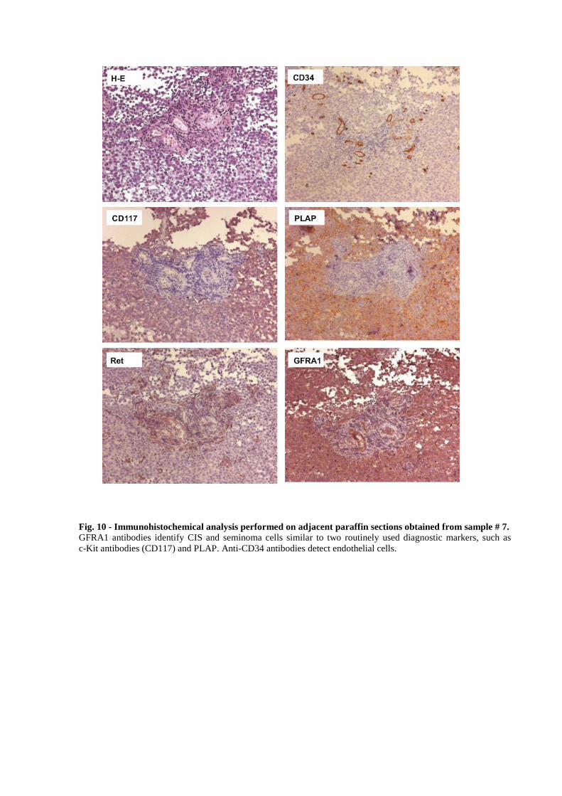
Fig. 10 - Immunohistochemical analysis performed on adjacent paraffin sections obtained from sample # 7. GFRA1 antibodies identify CIS and seminoma cells similar to two routinely used diagnostic markers, such as c-Kit antibodies (CD117) and PLAP. Anti-CD34 antibodies detect endothelial cells.

34
advantage of TCam-2 cells, currently, the only available validated seminoma cell line. First,
expression of GDNF co-receptors was analyzed by RT-PCR analysis (Figure 11A) and by
IHC (Figure 11B). GFRA1 is alternatively spliced into two highly homologous isoforms,
GFRA1a and GFRA1b, which differ at five amino acids (Shefelbine SE et al., 1998).
Recently GFRA1b, but not GFRA1a, has been demonstrated to be responsible of cell
migration and invasion in C6 glioma cells (Wan G et al., 2010). Both GFRA1 isoforms are
expressed in the TCam-2 cells (Figure 11A). Ret undergoes alternative splicing, which
generates the Ret9 and Ret51 isoforms, differing at the carboxyl-termini (Arighi E et al.,
2005). As observed by RT-PCR analysis, both Ret9 and Ret51 transcripts are present in the
TCam-2 cells (Figure 11A). In order to verify whether also GFRA1 and Ret receptor proteins
were expressed, we performed IHC analysis on TCam-2 cells. GFRA1, but not RET, was
detected by IHC (Figure 11B). We next addressed whether GDNF modulates TCam-2
proliferation or survival. In fact, GDNF is a known key factor regulating spermatogonial stem
cell self-renewal and proliferation (Hofmann MC et al., 2008). Serum-starved cells were
treated in the absence and in the presence of 100 ng/ml GDNF with or without 10% FBS for
24, 48 and 72 hours. The total number of cells, as well as the number of dead cells, did not
vary in GDNF-treated cells compared with the control at each time-point analyzed (Figure
12). In order to determine the percentage of TCam-2 cells in S phase FACS analysis, using
the same treatment conditions for 12, 24 and 48 hours, was performed (Figure 13). While FBS
significantly increased the percentage of cells in S phase compared with the control, GDNF
did not induce S-phase entry of TCam-2 cells at any time point analyzed (Figure 13).
6.3 GDNF induces TCam-2 cell migration
Intriguingly, in other cellular systems, GDNF has been implicated in the directed migration
and invasion of normal and tumor cells (Okada Y et al., 1999; Paratcha G et al., 2006; Song H
and Moon A, 2006; Su CM et al., 2009; Tang MJ et al., 1998; Veit C et al., 2004; Young HM

Fig. 11 - Identification of GDNF co-receptor variants expressed in TCam-2 cells. (A) RT-PCR analysis. Total RNA was prepared from TCam-2 cells and used as a template for RT-PCR analysis. A sample containing RNA from human brain and a sample containing no RNA (not shown) were used as controls. A β-actin primer set was used as a loading control. (B) Immunohistochemical detection of GDNF co-receptors in TCam-2 cells: (i) GFRA1 and (ii) Ret. No staining is present by omitting the primary antibodies (inset).
A B

0,0
2,0
4,0
6,0
8,0
10,0
12,0
14,0
16,0Control+FBSGDNF+FBSControlGDNF
5,5
6,5
7,5
8,5
9,5
10,5
11,5
12,5Control+FBS
GDNF+FBS
Control
GDNF
24 48 72hours
24 48 72hours
0
% D
ead
cel
ls/W
ell
Cel
ls(x
105 )
/Wel
l
Fig. 12 - GDNF is not a mitogenic factor for TCam-2 cells.(A) TCam-2 cells were serum-starved for 16 hours and then cultured in the presence or absence of 100 ng/ml GDNF with or without 10% FBS for 24, 48 and 72 hours. Percentage of dead cells at each time point is shown in (B). The results are from a representative of three experiments performed. Data are shown as the mean±SEM of triplicate samples.
A
B

a)
b) c)
Fig. 13 - GDNF does not induce S-phase entry in TCam-2 cells. (A) Flow cytometry analysis reveals that TCam-2 cells have a tetraploid DNA content. LYMPHO: human lymphocytes. (B) Cell cycle phase distribution of TCam-2 cells treated in the absence or presence of 100 ng/ml GDNF or with 10 % FBS (as a positive control) for 12, 24 and 48 hours of culture. Percentage of cells in G1, G2 and S phase for each experimental condition is presented in (C). The results are from a representative of three experiments performed. *p<0.05 (one-way Anova, Dunnet post hoc)
Control GDNF 100ng/ml FBS 1%
12h
ControlControl
GDNF
FBS
0
5
10
15
20
25
30
1 2 3
C o n tro l
G D N F 1 0 0 n g /m l
F BS 1 0 %
0
1
2
3
4
5
6
7
8
9
1 2 3
C o n tro l
G D N F 1 0 0 n g /m l
F BS 1 0 %
24h 48h
0
10
20
30
40
50
60
70
80
1 2 3
C o n tr o l
G D N F 1 0 0 n g /m l
F BS 1 0 %
12h
12h
12h 24h
24h
24h
48h
48h
48h
% C
ells
% C
ells
% C
ells
S Phase
G2 Phase
G1 Phase24h 48h
**
A
B C

35
et al., 2001). More interestingly, we recently found that in vitro GDNF is a chemoattractant
for murine undifferentiated spermatogonia (Dovere L, manuscript in preparation). Based on
these observations, we hypothesized that TCam-2 cells may migrate in response to a GDNF
gradient. Thus, in order to test whether the GDNF pathway could be involved in TCam-2
seminoma cell migration we performed Boyden chamber migration assays with increasing
concentrations of GDNF (20 to 150 ng/ml). Therefore to create a positive gradient for the
putative chemoattractant, the upper compartment was filled with DMEM+0,1% BSA alone,
while the lower chamber contained DMEM+0,1% BSA plus GDNF. The results showed that,
consistent with our hypothesis, after 5h of incubation, a significant effect on cell migration
was achieved with 100 ng/ml and 150 ng/ml GDNF (Figure 14A). As a control, we tested if
the anti-GDNF antibody was able to block this effect. Addition of the blocking antibody,
along with GDNF, completely neutralized the GDNF activity on TCam-2 cells, reducing cell
migration at a level comparable to the control (Figure 14B) No effect was observed after
administration of the antibody isotype control (Figure 14B). When GDNF was added to both
the upper and lower chambers, and therefore no chemokine gradient was present in the
Boyden chamber, cell migration was decreased to the control level (Figure 14B).
Chemoattractant activity of 1% FBS was comparable with 100 ng/ml GDNF, and no synergic
effect was found when the TCam-2 cells were stimulated with both 1% FBS and 100 ng/ml
GDNF (Figure 14B).
Recently the small cytokine CXCL12, a known chemoattractant for different cell types, has
been reported to be able to induce invasive migration on TCam-2 seminoma cells by
activation of the ERK pathway (Gilbert Dc et al., 2009). By comparing the GDNF
chemoattractant activity with that of CXCL12, we found that GDNF is more effective
(approximately 50%) than CXCL12 (Figure 14C). In conclusion, these data show for the first
time, that GDNF can stimulate directional migration of TCam-2 seminoma cell line.
Intriguingly, along with the observation that GDNF is an in vitro chemoattractant for

Fig. 14 - GDNF-dependent migration of TCam-2 cells. Cell migration was evaluated using the Boyden chamber assay as detailed in the Materials and Methods section. (A) TCam-2 cells were treated with increasing concentrations of GDNF. (B) TCam-2 cells were treated as indicated at final concentrations of 100 ng/ml GDNF, 15 µg/ml anti-GDNF antibody, 15 µg/ml control-IgG or fetal bovine serum (FBS) (1% v/v). To abrogate the GDNF gradient in the Boyden chamber, 100 ng/ml GDNF was added both in the upper and lower chamber. (C) TCam-2 cells were treated with 100 ng/ml GDNF or with increasing concentrations of CXCL12 (SDF-1). Data are expressed as the mean ± SEM (n=3, measured in triplicate). *P<0.001 vs. control (one-way Anova, Dunnet post hoc).
A
B
C
0
0,5
1
1,5
2
2,5
3
3,5
4
0
0,5
1
1,5
2
2,5
3
0,0
0,5
1,0
1,5
2,0
2,5
3,0
*
***
20 50 100 150
Control Control-IgG no GDNF gradient
FBSFBS
Mig
rate
d ce
lls (
Fol
d in
duct
ion)
* *
Mig
rate
d ce
lls (
Fold
indu
ctio
n) *
*
Control 100 20 50 100
GDNF
CXCL12GDNF
ng/ml
GDNF
ng/mlControl
Anti-GDNF
Mig
rate
d ce
lls (
Fol
d in
duct
ion)

36
undifferentiated mouse spermatogonia (Dovere L, manuscript in preparation), these data
suggest that the chemoattractant activity of GDNF is conserved in both normal and
transformed germ cells. We next investigated the GDNF-triggered pathway involved in
TCam-2 migration. In different tumor cell types, GDNF activates the MEK and PI3K
pathways (Su CM et al., 2009; Song H and Moon A, 2006; Veit C et al., 2004). Three
different inhibitors were tested as follows: PP-2 (a potent Src-inhibitor), LY294002 (a PI3K-
inhibitor), and U0126 (a MEK-inhibitor). PP-2 (Figure 15A) and UO126 (Figure 15B), but
not LY294002 (Figure 15C), completely abolished GDNF-induced cell migration. These data
suggest that GDNF-induced migration is mediated by the Src and MEK pathways but not by
the PI3K pathway.
6.4 GDNF promotes TCam-2 cell invasion in a protease-dependent fashion
We next addressed whether invasion could be triggered by GDNF in TCam-2 cells. For this
purpose we performed modified Boyden chamber assays with Matrigel-coated filters.
Matrigel is a solubilized basement membrane preparation whose major component is laminin,
followed by collagen IV, heparan sulfate proteoglycan, and entactin. This substrate is useful
to mimic the in vivo mechanisms on the base of tissue invasion where cells have to move
trough and degrade the ECM components to invade adjacent tissues. Invasion was analyzed at
two time points: 24 and 48 hours. At both time points, GDNF administration significantly
enhanced cell invasion compared with the control, as well as with CXCL12 (Figure 16A).
Tumor cell invasion can occur through two modalities: a proteolytic mesenchymal-like or
non-proteolytic amoeboid-like modality (Sahai E and Marshall CJ, 2003; Wolf K et al.,
2003). The mesenchymal-like strategy of invasion requires extracellular matrix degradation
by proteases, including matrix metalloproteinases (MMPs), and thus, it is sensitive to protease
inhibitors. In contrast, the amoeboid-type modality is protease-independent, with cells that
adopt a rounded morphology. To identify which of the two modalities is employed by the

Fig. 15 - The effect of different inhibitors on GDNF-induced TCam-2 cell migration. Cell migration was evaluated using the Boyden chamber assay as detailed in the Materials and Methods section. TCam-2 cells were pre-treated for 30 min with 10 µm PP-2 (A), 10 µm U0126 (B) or an indicated concentration of LY294002 (C) followed by stimulation with 100 ng/ml GDNF. In the control samples, DMSO was added to the highest concentration present in the treated samples. The results are expressed as the mean ± SEM, (n=3, measured in triplicate). *P<0.001 vs. control (one-way Anova, Dunnet post-hoc).
0,0
0,5
1,0
1,5
2,0
2,5
3,0
0,0
0,5
1,0
1,5
2,0
2,5
0,0
0,5
1,0
1,5
2,0
2,5
DMSO
Mig
rate
d ce
lls (
Fol
d in
duct
ion)
PP-2
Mig
rate
d ce
lls (
Fol
d in
duct
ion)
DMSO U0126
Mig
rate
d ce
lls (
Fol
d in
duct
ion)
DMSO ly29400210uM
ly294002+15uM
DMSO ly29400210uM
ly294002+15uM
GDNF
DMSO U0126
GDNF
DMSO PP-2
GDNF
* *
**
***
A
B
C
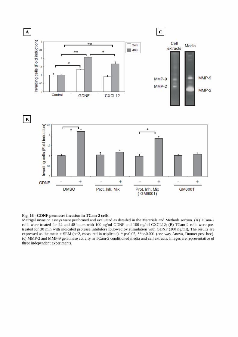
Fig. 16 - GDNF promotes invasion in TCam-2 cells. Matrigel invasion assays were performed and evaluated as detailed in the Materials and Methods section. (A) TCam-2 cells were treated for 24 and 48 hours with 100 ng/ml GDNF and 100 ng/ml CXCL12; (B) TCam-2 cells were pre-treated for 30 min with indicated protease inhibitors followed by stimulation with GDNF (100 ng/ml). The results are expressed as the mean ± SEM (n=2, measured in triplicate). * p<0.05, **p<0.001 (one-way Anova, Dunnet post-hoc). (c) MMP-2 and MMP-9 gelatinase activity in TCam-2 conditioned media and cell extracts. Images are representative of three independent experiments.
A
B
C

37
TCam-2 cells in GDNF-induced cell invasion, we tested a cocktail of protease inhibitors
(named prot. inh. mix), which included GM6001 (a broad matrix metalloproteinases
inhibitor), E64 (targets cysteine proteases such as cathepsins B, L, H and K), pepstatin A
(inhibits aspartic proteases including cathepsin D), leupeptin (a broad inhibitor of cysteine
proteases and cathepsin D) and aprotinin (an inhibitor of serine proteases, such as urokinase
and tissue plasminogen activator) (Carragher et al. 2006). Prot. inh. mix administration
completely abolished the GDNF-induced TCam-2 cell invasion (Figure 16B), suggesting a
protease-dependent invasion strategy. To evaluate the contribution of MMPs to TCam-2 cell
invasion, the MMPs-specific inhibitor GM6001 was tested alone, and its activity was
compared with prot. inh. mix without GM6001. GM6001 alone, but not the other inhibitors,
reduced the GDNF-induced TCam-2 cells invasion, suggesting the involvement of MMPs
(Figure 16B). MMP-2 and MMP-9 activity (Figure 16C) was detected in TCam-2 cell extracts
and conditioned medium by gelatin zymography.
38
DISCUSSION
In recent years, a large body of evidence pointed to the importance of GDNF for proper
spermatogenesis. In mice, GDNF is secreted by Sertoli cells and promotes spermatogonial
stem cell self-renewal (Hofmann MC, 2008). Over-expression of GDNF in adult transgenic
mice induces proliferation of clusters of undifferentiated spermatogonia within seminiferous
tubules (Meng X et al., 2001). Additionally, older animals develop testicular tumors that
mimic human seminoma, suggesting that over-activation of this pathway may lead to the
insurgence of TGCTs (Meng X et al., 2001). Supporting this hypothesis, Ret, a co-receptor
for GDNF, is a well-known oncogene that upon mutation and/or rearrangement undergoes
constitutive GDNF- and GFRA1-independent activation. Activating mutations of Ret were
previously reported in several types of cancer, including thyroid, pituitary, adrenal and
melanoma cancer. However, human seminoma does not appear to be linked to mutations or
relevant polymorphisms of Ret (Chevalier N et al., 2010). An alternative mechanism for
driving germ cell tumor progression could be the up-regulation of wild-type GFRA1 and/or
Ret proteins. Overexpression of GFRA1 and/or Ret has been found in different cancers
(Dawson DM et al., 1998; Nakashima M et al., 2007; Takaya K et al., 1996), and in some
instances, Ret over-expression correlates with poor survival (Ito Y et al., 2005) as well as the
development of a subgroup of breast tumors (Esseghir S et al., 2007). We demonstrate, for the
first time, that GFRA1 is extensively expressed in CIS, and its expression is maintained in
invasive seminoma. However, Ret was not detected by immunohistochemistry in normal
germ cells, in CIS cells, or in the invasive seminoma. In TCam-2 cells, Ret was detected at
mRNA but not protein levels, suggesting that in CIS and seminoma cells, Ret protein may be
expressed at very low levels. Alternatively, GFRA1 may transduce GDNF signal in a Ret-
independent fashion. To date, a number of CIS and seminoma cells markers are known to be
expressed in fetal gonocytes but not in adult spermatogonia. However, CIS cells also express

39
genes found in the germ cells of normal adult testis (i.e., VASA, TSPY, DAZ and DAZL)
(Rajpert-De ME, 2006). Because GFRA1 is detected in dark and pale type A spermatogonia,
GFRA1 lengthens the list of tumor germ cell markers that are expressed in normal germ cells.
To elucidate the biological activity of GDNF in TGCTs, we employed the TCam-2 cell line as
an experimental model system. Even though TCam-2 cells express the GDNF co-receptors,
GDNF did not affect cell survival and proliferation. Because GDNF acts as a chemoattractant
in several normal and tumor cell types (Okada Y et al. 1999; Paratcha G et al. 2006; Song H
and Moon A, 2006; Su CM et al., 2009; Tang MJ et al., 1998; Veit C et al., 2004; Young HM
et al., 2001), we tested the hypothesis that GDNF is a chemoattractant for the TCam-2
seminoma cell line. GDNF induced TCam-2 cell migration and was mediated by the Src and
MEK pathways. In contrast, PI3K does not seem to be involved in GDNF-induced migration.
Our findings are consistent with the observation that in some pancreatic carcinoma cells,
GDNF is not mitogenic but acts as a chemoattractant (Veit C et al., 2004). We found that
GDNF induces TCam-2 cell invasion through GFR-Matrigel, a matrix consisting of laminin,
collagen, entactin and growth factors. Invasive tumor dissemination involves proteolytic
degradation and remodeling of the extracellular matrix (ECM) barriers, a process dependent
upon extracellular proteases, including MMPs (Coussens LM et al., 2002). However, protease
inhibition does not completely abolish tumor cell migration and dissemination, suggesting
alternative compensating protease-independent mechanisms (Coussens LM et al., 2002;
Zucker S et al., 2000). Of note, when proteolysis is blocked, some tumor cell types switch
from a protease-dependent mesenchymal type of motility to an ameboid-like rounded mode of
motility that requires Rho/ROCK signaling (Sahai E and Marshall CJ, 2003; Wolf K et al.,
2003). By using inhibitors of a wide range of pericellular proteases, we found that the
invasive strategy used by TCam-2 cells is protease-dependent, which suggests a
mesenchymal-like invasion. Among candidate proteases implicated in GDNF-induced TCam-
2 cell invasion are the MMPs, the most prominent family of proteases involved in

40
tumorigenesis (Kessenbrock K et al., 2010). The lack of invasion upon inhibition of
pericellular proteases suggests that TCam-2 cells are not able to undergo the mesenchymal-
ameboid switch, thus suggesting a lack of plasticity compared with more aggressive tumor
cell types (Carragher NO et al., 2006).
We demonstrated that GFRA1 expression is upregulated in CIS and seminoma cells. This
raises the question as to the source of GDNF in seminoma. In normal human testis, GDNF is
produced by the Sertoli and peritubular cells, which form the wall of the seminiferous tubules
(Spinnler K et al,. 2010). The expression level of GDNF was found to be similar in
peritubular cells isolated from patients suffering from azoospermia and fibrotic remodeling of
the peritubular wall (Spinnler K et al., 2010). At present, data are not available regarding the
expression level and regulation of GDNF in normal human testis or those affected by
pathological conditions, including TGCTs. In infertile mouse models, GDNF levels are
inversely correlated with the germ cell content of the testis, possibly through the regulation of
the hypothalamus–pituitary axis (Tadokoro Y et al., 2002). Moreover, GDNF expression in
Sertoli cells is induced by inflammatory cytokines (Simon L et al., 2007). Therefore, it could
be speculated that, as described in some breast cancers (Esseghir S et al., 2007), local levels
of GDNF in the tumor microenvironment may be increased as a consequence of leukocyte
infiltration and/or spermatogenic arrest, which are commonly found in TGCTs. Although
currently it is not possible to directly address whether CIS and intratubular seminoma cells
respond to local GDNF by increasing their ability to migrate and invade the interstitial
compartment, our combined in vivo and in vitro observations suggests that GDNF may induce
tumor cell migration and dissemination within the testis. Recently, it has been suggested that
the expression level of the chemokine CXCL12 (SDF-1) in TGCT samples is a predictive
marker of relapse for patients affected by a subset of type II TGCTs, i.e., stage I non-
seminoma (NS). Patients with moderate to strong expression of CXCL12 have a reduced risk
of relapse, suggesting that a high level of chemokines in the tumor microenvironment may

41
impact tumor germ cell spreading and metastatic potential (Gilbert DC et al., 2009).
Following this line of reasoning, it would be interesting to assess whether the expression level
of GDNF signaling pathway molecules have a similar prognostic value in patients affected by
seminoma tumors.

42
BIBLIOGRAPHY
Airaksinen MS, Saarma M (2002) The GDNF family: signalling, biological functions and therapeutic value. Nat Rev Neurosci 3:383-394.
Albers P, Albrecht W, Algaba F, Bokemeyer C, Cohn-Cedermark G, Horwich A, Klepp O, Laguna MP, Pizzocaro G (2005) Guidelines on testicular cancer. Eur Urol 48:885-894.
Aman A, Piotrowski T (2008) Wnt/beta-catenin and Fgf signaling control collective cell migration by restricting chemokine receptor expression. Dev Cell 15:749-761.
Anders J, Kjar S, Ibanez CF (2001) Molecular modeling of the extracellular domain of the RET receptor tyrosine kinase reveals multiple cadherin-like domains and a calcium-binding site. J Biol Chem 276:35808-35817.
Arighi E, Borrello MG, Sariola H (2005) RET tyrosine kinase signaling in development and cancer. Cytokine Growth Factor Rev 16:441-467.
Avilion AA, Nicolis SK, Pevny LH, Perez L, Vivian N, Lovell-Badge R (2003) Multipotent cell lineages in early mouse development depend on SOX2 function. Genes Dev 17:126-140.
Bacac M, Stamenkovic I (2008) Metastatic cancer cell. Annu Rev Pathol 3:221-247.
bdel-Ghany M, Cheng HC, Elble RC, Pauli BU (2001) The breast cancer beta 4 integrin and endothelial human CLCA2 mediate lung metastasis. J Biol Chem 276:25438-25446.
bdel-Ghany M, Cheng HC, Elble RC, Pauli BU (2001) The breast cancer beta 4 integrin and endothelial human CLCA2 mediate lung metastasis. J Biol Chem 276:25438-25446.
Benne R, Van den BJ, Brakenhoff JP, Sloof P, Van Boom JH, Tromp MC (1986) Major transcript of the frameshifted coxII gene from trypanosome mitochondria contains four nucleotides that are not encoded in the DNA. Cell 46:819-826.
Buageaw A, Sukhwani M, Ben-Yehudah A, Ehmcke J, Rawe VY, Pholpramool C, Orwig KE, Schlatt S (2005) GDNF family receptor alpha1 phenotype of spermatogonial stem cells in immature mouse testes. Biol Reprod 73:1011-1016.
Cacalano G, Farinas I, Wang LC, Hagler K, Forgie A, Moore M, Armanini M, Phillips H, Ryan AM, Reichardt LF, Hynes M, Davies A, Rosenthal A (1998) GFRalpha1 is an essential receptor component for GDNF in the developing nervous system and kidney. Neuron 21:53-62.
Cardoso MC, Leonhardt H, Nadal-Ginard B (1993) Reversal of terminal differentiation and control of DNA replication: cyclin A and Cdk2 specifically localize at subnuclear sites of DNA replication. Cell 74:979-992.
Carragher NO, Walker SM, Scott Carragher LA, Harris F, Sawyer TK, Brunton VG, Ozanne BW, Frame MC (2006) Calpain 2 and Src dependence distinguishes mesenchymal and amoeboid modes of tumour cell invasion: a link to integrin function. Oncogene 25:5726-5740.

43
Catizone A, Ricci G, Tufano MA, Perfetto B, Canipari R, Galdieri M (2010) Hepatocyte growth factor (HGF) modulates Leydig cell extracellular matrix components. J Androl 31:306-313.
Chao CC, Ma YL, Chu KY, Lee EH (2003) Integrin alphav and NCAM mediate the effects of GDNF on DA neuron survival, outgrowth, DA turnover and motor activity in rats. Neurobiol Aging 24:105-116.
Chevalier N, Barlier A, Roche C, Mograbi B, Camparo P, vouassoux-Shisheboran M, Michiels JF, Nebout M, Chevallier D, Benahmed M, Enjalbert A, Fenichel P (2010) RET gene mutations are not involved in the origin of human testicular seminoma. Int J Androl 33:848-852.
Condeelis J, Segall JE (2003) Intravital imaging of cell movement in tumours. Nat Rev Cancer 3:921-930.
Coussens LM, Fingleton B, Matrisian LM (2002) Matrix metalloproteinase inhibitors and cancer: trials and tribulations. Science 295:2387-2392.
Davidoff MS, Middendorff R, Koeva Y, Pusch W, Jezek D, Muller D (2001) Glial cell line-derived neurotrophic factor (GDNF) and its receptors GFRalpha-1 and GFRalpha-2 in the human testis. Ital J Anat Embryol 106:173-180.
Dawson DM, Lawrence EG, MacLennan GT, Amini SB, Kung HJ, Robinson D, Resnick MI, Kursh ED, Pretlow TP, Pretlow TG (1998) Altered expression of RET proto-oncogene product in prostatic intraepithelial neoplasia and prostate cancer. J Natl Cancer Inst 90:519-523.
de Graaff WE, Oosterhuis JW, de JB, Dam A, van Putten WL, Castedo SM, Sleijfer DT, Schraffordt KH (1992) Ploidy of testicular carcinoma in situ. Lab Invest 66:166-168.
de Rooij DG, Russell LD (2000) All you wanted to know about spermatogonia but were afraid to ask. J Androl 21:776-798.
De Giorgi U, Nicolai N, Tana S, Tavolini IM, Palazzi S, Bracarda S, Tedeschi L, Palmieri G, Frassineti L, Da PL, Pastorino U, Emiliani E, Marangolo M, Pizzocaro G, Rosti G, Salvioni R (2008) IGG practice guidelines on germ cell tumor in adult male patients. Tumori 94:96-109.
de Jong J, Looijenga LH (2006) Stem cell marker OCT3/4 in tumor biology and germ cell tumor diagnostics: history and future. Crit Rev Oncog 12:171-203.
de Jong J, Stoop H, Gillis AJ, Hersmus R, van Gurp RJ, van de Geijn GJ, van DE, Beverloo HB, Schneider DT, Sherlock JK, Baeten J, Kitazawa S, van Zoelen EJ, van RK, Oosterhuis JW, Looijenga LH (2008) Further characterization of the first seminoma cell line TCam-2. Genes Chromosomes Cancer 47:185-196.
Desdouets C, Matesic G, Molina CA, Foulkes NS, Sassone-Corsi P, Brechot C, Sobczak-Thepot J (1995) Cell cycle regulation of cyclin A gene expression by the cyclic AMP-responsive transcription factors CREB and CREM. Mol Cell Biol 15:3301-3309.
Draper JS, Smith K, Gokhale P, Moore HD, Maltby E, Johnson J, Meisner L, Zwaka TP, Thomson JA, Andrews PW (2004) Recurrent gain of chromosomes 17q and 12 in cultured human embryonic stem cells. Nat Biotechnol 22:53-54.

44
Ebata KT, Zhang X, Nagano MC (2005) Expression patterns of cell-surface molecules on male germ line stem cells during postnatal mouse development. Mol Reprod Dev 72:171-181.
Eckert D, Nettersheim D, Heukamp LC, Kitazawa S, Biermann K, Schorle H (2008) TCam-2 but not JKT-1 cells resemble seminoma in cell culture. Cell Tissue Res 331:529-538.
Eigenbrot C, Gerber N (1997) X-ray structure of glial cell-derived neurotrophic factor at 1.9 A resolution and implications for receptor binding. Nat Struct Biol 4:435-438.
Enomoto H, Araki T, Jackman A, Heuckeroth RO, Snider WD, Johnson EM, Jr., Milbrandt J (1998) GFR alpha1-deficient mice have deficits in the enteric nervous system and kidneys. Neuron 21:317-324.
Esseghir S, Todd SK, Hunt T, Poulsom R, Plaza-Menacho I, Reis-Filho JS, Isacke CM (2007) A role for glial cell derived neurotrophic factor induced expression by inflammatory cytokines and RET/GFR alpha 1 receptor up-regulation in breast cancer. Cancer Res 67:11732-11741.
Farrow B, Albo D, Berger DH (2008) The role of the tumor microenvironment in the progression of pancreatic cancer. J Surg Res 149:319-328.
Fidler IJ (2003) The pathogenesis of cancer metastasis: the 'seed and soil' hypothesis revisited. Nat Rev Cancer 3:453-458.
Fossa SD, Kaye SB, Mead GM, Cullen M, de WR, Bodrogi I, van Groeningen CJ, de Mulder PH, Stenning S, Lallemand E, de PL, Collette L (1998) Filgrastim during combination chemotherapy of patients with poor-prognosis metastatic germ cell malignancy. European Organization for Research and Treatment of Cancer, Genito-Urinary Group, and the Medical Research Council Testicular Cancer Working Party, Cambridge, United Kingdom. J Clin Oncol 16:716-724.
Friedl P, Gilmour D (2009) Collective cell migration in morphogenesis, regeneration and cancer. Nat Rev Mol Cell Biol 10:445-457.
Gaggioli C, Hooper S, Hidalgo-Carcedo C, Grosse R, Marshall JF, Harrington K, Sahai E (2007) Fibroblast-led collective invasion of carcinoma cells with differing roles for RhoGTPases in leading and following cells. Nat Cell Biol 9:1392-1400.
Gassei K, Ehmcke J, Dhir R, Schlatt S (2010) Magnetic activated cell sorting allows isolation of spermatogonia from adult primate testes and reveals distinct GFRa1-positive subpopulations in men. J Med Primatol 39:83-91.
Gilbert DC, Chandler I, McIntyre A, Goddard NC, Gabe R, Huddart RA, Shipley J (2009) Clinical and biological significance of CXCL12 and CXCR4 expression in adult testes and germ cell tumours of adults and adolescents. J Pathol 217:94-102.
Giwercman A, Skakkebaek NE (1993) Carcinoma in situ of the testis: biology, screening and management. Eur Urol 23 Suppl 2:19-21.
Goddard NC, McIntyre A, Summersgill B, Gilbert D, Kitazawa S, Shipley J (2007) KIT and RAS signalling pathways in testicular germ cell tumours: new data and a review of the literature. Int J Androl 30:337-348.

45
Goddard NC, McIntyre A, Gilbert D, Kitazawa S, Shipley J (2010) No evidence for V600E BRAF mutation in the seminoma cell line TCam-2. Genes Chromosomes Cancer 49:963-966.
Golden JP, DeMaro JA, Osborne PA, Milbrandt J, Johnson EM, Jr. (1999) Expression of neurturin, GDNF, and GDNF family-receptor mRNA in the developing and mature mouse. Exp Neurol 158:504-528.
Grisanti L, Falciatori I, Grasso M, Dovere L, Fera S, Muciaccia B, Fuso A, Berno V, Boitani C, Stefanini M, Vicini E (2009) Identification of spermatogonial stem cell subsets by morphological analysis and prospective isolation. Stem Cells 27:3043-3052.
Grisanti L, Falciatori I, Grasso M, Dovere L, Fera S, Muciaccia B, Fuso A, Berno V, Boitani C, Stefanini M, Vicini E (2009) Identification of spermatogonial stem cell subsets by morphological analysis and prospective isolation. Stem Cells 27:3043-3052.
Hayashi H, Ichihara M, Iwashita T, Murakami H, Shimono Y, Kawai K, Kurokawa K, Murakumo Y, Imai T, Funahashi H, Nakao A, Takahashi M (2000) Characterization of intracellular signals via tyrosine 1062 in RET activated by glial cell line-derived neurotrophic factor. Oncogene 19:4469-4475.
Hemminki K, Li X (2002) Cancer risks in Nordic immigrants and their offspring in Sweden. Eur J Cancer 38:2428-2434.
Henderson BE, Bernstein L, Ross RK, Depue RH, Judd HL (1988) The early in utero oestrogen and testosterone environment of blacks and whites: potential effects on male offspring. Br J Cancer 57:216-218.
Hoei-Hansen CE, Almstrup K, Nielsen JE, Brask SS, Graem N, Skakkebaek NE, Leffers H, Rajpert-De ME (2005) Stem cell pluripotency factor NANOG is expressed in human fetal gonocytes, testicular carcinoma in situ and germ cell tumours. Histopathology 47:48-56.
Hoei-Hansen CE, Carlsen E, Jorgensen N, Leffers H, Skakkebaek NE, Rajpert-De ME (2007) Towards a non-invasive method for early detection of testicular neoplasia in semen samples by identification of fetal germ cell-specific markers. Hum Reprod 22:167-173.
Hofmann MC (2008) Gdnf signaling pathways within the mammalian spermatogonial stem cell niche. Mol Cell Endocrinol 288:95-103.
Houldsworth J, Reuter V, Bosl GJ, Chaganti RS (1997) Aberrant expression of cyclin D2 is an early event in human male germ cell tumorigenesis. Cell Growth Differ 8:293-299.
Iglesias PA, Devreotes PN (2008) Navigating through models of chemotaxis. Curr Opin Cell Biol 20:35-40.
Ilina O, Friedl P (2009) Mechanisms of collective cell migration at a glance. J Cell Sci 122:3203-3208.
Ito Y, Okada Y, Sato M, Sawai H, Funahashi H, Murase T, Hayakawa T, Manabe T (2005) Expression of glial cell line-derived neurotrophic factor family members and their receptors in pancreatic cancers. Surgery 138:788-794.

46
Iwamoto T, Taniguchi M, Asai N, Ohkusu K, Nakashima I, Takahashi M (1993) cDNA cloning of mouse ret proto-oncogene and its sequence similarity to the cadherin superfamily. Oncogene 8:1087-1091.
ix-Panabieres C, Riethdorf S, Pantel K (2008) Circulating tumor cells and bone marrow micrometastasis. Clin Cancer Res 14:5013-5021.
ix-Panabieres C, Riethdorf S, Pantel K (2008) Circulating tumor cells and bone marrow micrometastasis. Clin Cancer Res 14:5013-5021.
Jijiwa M, Fukuda T, Kawai K, Nakamura A, Kurokawa K, Murakumo Y, Ichihara M, Takahashi M (2004) A targeting mutation of tyrosine 1062 in Ret causes a marked decrease of enteric neurons and renal hypoplasia. Mol Cell Biol 24:8026-8036.
Jing S, Wen D, Yu Y, Holst PL, Luo Y, Fang M, Tamir R, Antonio L, Hu Z, Cupples R, Louis JC, Hu S, Altrock BW, Fox GM (1996) GDNF-induced activation of the ret protein tyrosine kinase is mediated by GDNFR-alpha, a novel receptor for GDNF. Cell 85:1113-1124.
Kanatsu-Shinohara M, Ogonuki N, Inoue K, Miki H, Ogura A, Toyokuni S, Shinohara T (2003) Long-term proliferation in culture and germline transmission of mouse male germline stem cells. Biol Reprod 69:612-616.
Kanatsu-Shinohara M, Muneto T, Lee J, Takenaka M, Chuma S, Nakatsuji N, Horiuchi T, Shinohara T (2008) Long-term culture of male germline stem cells from hamster testes. Biol Reprod 78:611-617.
Kersemaekers AM, Mayer F, Molier M, Van Weeren PC, Oosterhuis JW, Bokemeyer C, Looijenga LH (2002) Role of P53 and MDM2 in treatment response of human germ cell tumors. J Clin Oncol 20:1551-1561.
Kessenbrock K, Plaks V, Werb Z (2010) Matrix metalloproteinases: regulators of the tumor microenvironment. Cell 141:52-67.
Kokaia Z, Airaksinen MS, Nanobashvili A, Larsson E, Kujamaki E, Lindvall O, Saarma M (1999) GDNF family ligands and receptors are differentially regulated after brain insults in the rat. Eur J Neurosci 11:1202-1216.
Krausz C, Looijenga LH (2008) Genetic aspects of testicular germ cell tumors. Cell Cycle 7:3519-3524.
Krentz AD, Murphy MW, Kim S, Cook MS, Capel B, Zhu R, Matin A, Sarver AL, Parker KL, Griswold MD, Looijenga LH, Bardwell VJ, Zarkower D (2009) The DM domain protein DMRT1 is a dose-sensitive regulator of fetal germ cell proliferation and pluripotency. Proc Natl Acad Sci U S A 106:22323-22328.
Kristensen DM, Sonne SB, Ottesen AM, Perrett RM, Nielsen JE, Almstrup K, Skakkebaek NE, Leffers H, Rajpert-De ME (2008) Origin of pluripotent germ cell tumours: the role of microenvironment during embryonic development. Mol Cell Endocrinol 288:111-118.
Kubota H, Avarbock MR, Brinster RL (2004) Growth factors essential for self-renewal and expansion of mouse spermatogonial stem cells. Proc Natl Acad Sci U S A 101:16489-16494.

47
Kukoski R, Blonigen B, Macri E, Renshaw AA, Hoffman M, Loda M, Datta MW (2003) p27 and cyclin E/D2 associations in testicular germ cell tumors: implications for tumorigenesis. Appl Immunohistochem Mol Morphol 11:138-143.
Kuma K, Iwabe N, Miyata T (1993) Motifs of cadherin- and fibronectin type III-related sequences and evolution of the receptor-type-protein tyrosine kinases: sequence similarity between proto-oncogene ret and cadherin family. Mol Biol Evol 10:539-551.
Li SS, Liu YH, Tseng CN, Chung TL, Lee TY, Singh S (2006) Characterization and gene expression profiling of five new human embryonic stem cell lines derived in Taiwan. Stem Cells Dev 15:532-555.
Lin LF, Doherty DH, Lile JD, Bektesh S, Collins F (1993) GDNF: a glial cell line-derived neurotrophic factor for midbrain dopaminergic neurons. Science 260:1130-1132.
Loehrer PJ, Sr., Gonin R, Nichols CR, Weathers T, Einhorn LH (1998) Vinblastine plus ifosfamide plus cisplatin as initial salvage therapy in recurrent germ cell tumor. J Clin Oncol 16:2500-2504.
Looijenga LH, Rosenberg C, van Gurp RJ, Geelen E, van Echten-Arends J, de JB, Mostert M, Wolter OJ (2000) Comparative genomic hybridization of microdissected samples from different stages in the development of a seminoma and a non-seminoma. J Pathol 191:187-192.
Looijenga LH, Stoop H, de Leeuw HP, de Gouveia Brazao CA, Gillis AJ, van Roozendaal KE, van Zoelen EJ, Weber RF, Wolffenbuttel KP, van DH, Honecker F, Bokemeyer C, Perlman EJ, Schneider DT, Kononen J, Sauter G, Oosterhuis JW (2003) POU5F1 (OCT3/4) identifies cells with pluripotent potential in human germ cell tumors. Cancer Res 63:2244-2250.
Looijenga LH, Zafarana G, Grygalewicz B, Summersgill B, biec-Rychter M, Veltman J, Schoenmakers EF, Rodriguez S, Jafer O, Clark J, van Kessel AG, Shipley J, van Gurp RJ, Gillis AJ, Oosterhuis JW (2003) Role of gain of 12p in germ cell tumour development. APMIS 111:161-171.
Lutke Holzik MF, Sijmons RH, Hoekstra-Weebers JE, Sleijfer DT, Hoekstra HJ (2008) Clinical and genetic aspects of testicular germ cell tumours. Hered Cancer Clin Pract 6:3-14.
McSherry EA, Donatello S, Hopkins AM, McDonnell S (2007) Molecular basis of invasion in breast cancer. Cell Mol Life Sci 64:3201-3218.
Meng X, Lindahl M, Hyvonen ME, Parvinen M, de Rooij DG, Hess MW, Raatikainen-Ahokas A, Sainio K, Rauvala H, Lakso M, Pichel JG, Westphal H, Saarma M, Sariola H (2000) Regulation of cell fate decision of undifferentiated spermatogonia by GDNF. Science 287:1489-1493.
Meng X, de Rooij DG, Westerdahl K, Saarma M, Sariola H (2001) Promotion of seminomatous tumors by targeted overexpression of glial cell line-derived neurotrophic factor in mouse testis. Cancer Res 61:3267-3271.
Meng X, de Rooij DG, Westerdahl K, Saarma M, Sariola H (2001) Promotion of seminomatous tumors by targeted overexpression of glial cell line-derived neurotrophic factor in mouse testis. Cancer Res 61:3267-3271.

48
Mizuno Y, Gotoh A, Kamidono S, Kitazawa S (1993) [Establishment and characterization of a new human testicular germ cell tumor cell line (TCam-2)]. Nihon Hinyokika Gakkai Zasshi 84:1211-1218.
Moore MW, Klein RD, Farinas I, Sauer H, Armanini M, Phillips H, Reichardt LF, Ryan AM, Carver-Moore K, Rosenthal A (1996) Renal and neuronal abnormalities in mice lacking GDNF. Nature 382:76-79.
Moul JW, Schanne FJ, Thompson IM, Frazier HA, Peretsman SA, Wettlaufer JN, Rozanski TA, Stack RS, Kreder KJ, Hoffman KJ (1994) Testicular cancer in blacks. A multicenter experience. Cancer 73:388-393.
Muciaccia B, Sette C, Paronetto MP, Barchi M, Pensini S, D'Agostino A, Gandini L, Geremia R, Stefanini M, Rossi P (2010) Expression of a truncated form of KIT tyrosine kinase in human spermatozoa correlates with sperm DNA integrity. Hum Reprod 25:2188-2202.
Muller J, Skakkebaek NE, Nielsen OH, Graem N (1984) Cryptorchidism and testis cancer. Atypical infantile germ cells followed by carcinoma in situ and invasive carcinoma in adulthood. Cancer 54:629-634.
Murty VV, Dmitrovsky E, Bosl GJ, Chaganti RS (1990) Nonrandom chromosome abnormalities in testicular and ovarian germ cell tumor cell lines. Cancer Genet Cytogenet 50:67-73.
Nakashima M, Takamura N, Namba H, Saenko V, Meirmanov S, Matsumoto N, Hayashi T, Maeda S, Sekine I (2007) RET oncogene amplification in thyroid cancer: correlations with radiation-associated and high-grade malignancy. Hum Pathol 38:621-628.
Naughton CK, Jain S, Strickland AM, Gupta A, Milbrandt J (2006) Glial cell-line derived neurotrophic factor-mediated RET signaling regulates spermatogonial stem cell fate. Biol Reprod 74:314-321.
Oatley JM, Avarbock MR, Telaranta AI, Fearon DT, Brinster RL (2006) Identifying genes important for spermatogonial stem cell self-renewal and survival. Proc Natl Acad Sci U S A 103:9524-9529.
Oatley JM, Avarbock MR, Brinster RL (2007) Glial cell line-derived neurotrophic factor regulation of genes essential for self-renewal of mouse spermatogonial stem cells is dependent on Src family kinase signaling. J Biol Chem 282:25842-25851.
Okada Y, Takeyama H, Sato M, Morikawa M, Sobue K, Asai K, Tada T, Kato T, Manabe T (1999) Experimental implication of celiac ganglionotropic invasion of pancreatic-cancer cells bearing c-ret proto-oncogene with reference to glial-cell-line-derived neurotrophic factor (GDNF). Int J Cancer 81:67-73.
Olie RA, Boersma AW, Dekker MC, Nooter K, Looijenga LH, Oosterhuis JW (1996) Apoptosis of human seminoma cells upon disruption of their microenvironment. Br J Cancer 73:1031-1036.
Oosterhuis JW, Castedo SM, de JB, Cornelisse CJ, Dam A, Sleijfer DT, Schraffordt KH (1989) Ploidy of primary germ cell tumors of the testis. Pathogenetic and clinical relevance. Lab Invest 60:14-21.

49
Oosterhuis JW, Kersemaekers AM, Jacobsen GK, Timmer A, Steyerberg EW, Molier M, Van Weeren PC, Stoop H, Looijenga LH (2003) Morphology of testicular parenchyma adjacent to germ cell tumours. An interim report. APMIS 111:32-40.
Oosterhuis JW, Looijenga LH (2005) Testicular germ-cell tumours in a broader perspective. Nat Rev Cancer 5:210-222.
Orwig KE, Ryu BY, Master SR, Phillips BT, Mack M, Avarbock MR, Chodosh L, Brinster RL (2008) Genes involved in post-transcriptional regulation are overrepresented in stem/progenitor spermatogonia of cryptorchid mouse testes. Stem Cells 26:927-938.
Ottesen AM, Skakkebaek NE, Lundsteen C, Leffers H, Larsen J, Rajpert-De ME (2003) High-resolution comparative genomic hybridization detects extra chromosome arm 12p material in most cases of carcinoma in situ adjacent to overt germ cell tumors, but not before the invasive tumor development. Genes Chromosomes Cancer 38:117-125.
Ottesen AM, Larsen J, Gerdes T, Larsen JK, Lundsteen C, Skakkebaek NE, Rajpert-De ME (2004) Cytogenetic investigation of testicular carcinoma in situ and early seminoma by high-resolution comparative genomic hybridization analysis of subpopulations flow sorted according to DNA content. Cancer Genet Cytogenet 149:89-97.
Pankova K, Rosel D, Novotny M, Brabek J (2010) The molecular mechanisms of transition between mesenchymal and amoeboid invasiveness in tumor cells. Cell Mol Life Sci 67:63-71.
Paratcha G, Ledda F, Baars L, Coulpier M, Besset V, Anders J, Scott R, Ibanez CF (2001) Released GFRalpha1 potentiates downstream signaling, neuronal survival, and differentiation via a novel mechanism of recruitment of c-Ret to lipid rafts. Neuron 29:171-184.
Paratcha G, Ledda F, Ibanez CF (2003) The neural cell adhesion molecule NCAM is an alternative signaling receptor for GDNF family ligands. Cell 113:867-879.
Paratcha G, Ibanez CF, Ledda F (2006) GDNF is a chemoattractant factor for neuronal precursor cells in the rostral migratory stream. Mol Cell Neurosci 31:505-514.
Pichel JG, Shen L, Sheng HZ, Granholm AC, Drago J, Grinberg A, Lee EJ, Huang SP, Saarma M, Hoffer BJ, Sariola H, Westphal H (1996) Defects in enteric innervation and kidney development in mice lacking GDNF. Nature 382:73-76.
Piulats JM, Jimenez L, Garcia dM, X, Villanueva A, Vinals F, Germa-Lluch JR (2009) Molecular mechanisms behind the resistance of cisplatin in germ cell tumours. Clin Transl Oncol 11:780-786.
Popsueva A, Poteryaev D, Arighi E, Meng X, ngers-Loustau A, Kaplan D, Saarma M, Sariola H (2003) GDNF promotes tubulogenesis of GFRalpha1-expressing MDCK cells by Src-mediated phosphorylation of Met receptor tyrosine kinase. J Cell Biol 161:119-129.
Poteryaev D, Titievsky A, Sun YF, Thomas-Crusells J, Lindahl M, Billaud M, Arumae U, Saarma M (1999) GDNF triggers a novel ret-independent Src kinase family-coupled signaling via a GPI-linked GDNF receptor alpha1. FEBS Lett 463:63-66.
Powles TB, Bhardwa J, Shamash J, Mandalia S, Oliver T (2005) The changing presentation of germ cell tumours of the testis between 1983 and 2002. BJU Int 95:1197-1200.

50
Rajpert-De ME, Skakkebaek NE (1994) Expression of the c-kit protein product in carcinoma-in-situ and invasive testicular germ cell tumours. Int J Androl 17:85-92.
Rajpert-De ME, Hanstein R, Jorgensen N, Graem N, Vogt PH, Skakkebaek NE (2004) Developmental expression of POU5F1 (OCT-3/4) in normal and dysgenetic human gonads. Hum Reprod 19:1338-1344.
Rajpert-De ME (2006) Developmental model for the pathogenesis of testicular carcinoma in situ: genetic and environmental aspects. Hum Reprod Update 12:303-323.
Rajpert-De ME, Hoei-Hansen CE (2007) From gonocytes to testicular cancer: the role of impaired gonadal development. Ann N Y Acad Sci 1120:168-180.
Rorth P (2007) Collective guidance of collective cell migration. Trends Cell Biol 17:575-579.
Rosenberg C, van Gurp RJ, Geelen E, Oosterhuis JW, Looijenga LH (2000) Overrepresentation of the short arm of chromosome 12 is related to invasive growth of human testicular seminomas and nonseminomas. Oncogene 19:5858-5862.
Roussos ET, Condeelis JS, Patsialou A (2011) Chemotaxis in cancer. Nat Rev Cancer 11:573-587.
Saarma M, Sariola H (1999) Other neurotrophic factors: glial cell line-derived neurotrophic factor (GDNF). Microsc Res Tech 45:292-302.
Saarma M (2001) GDNF recruits the signaling crew into lipid rafts. Trends Neurosci 24:427-429.
Sahai E, Marshall CJ (2003) Differing modes of tumour cell invasion have distinct requirements for Rho/ROCK signalling and extracellular proteolysis. Nat Cell Biol 5:711-719.
Sahai E (2005) Mechanisms of cancer cell invasion. Curr Opin Genet Dev 15:87-96.
Sanchez MP, Silos-Santiago I, Frisen J, He B, Lira SA, Barbacid M (1996) Renal agenesis and the absence of enteric neurons in mice lacking GDNF. Nature 382:70-73.
Santagata S, Ligon KL, Hornick JL (2007) Embryonic stem cell transcription factor signatures in the diagnosis of primary and metastatic germ cell tumors. Am J Surg Pathol 31:836-845.
Sariola H, Saarma M (2003) Novel functions and signalling pathways for GDNF. J Cell Sci 116:3855-3862.
Schmidt M, Paes K, De MA, Smyczek T, Yang S, Gray A, French D, Kasman I, Klumperman J, Rice DS, Ye W (2007) EGFL7 regulates the collective migration of endothelial cells by restricting their spatial distribution. Development 134:2913-2923.
Schuchardt A, D'Agati V, Larsson-Blomberg L, Costantini F, Pachnis V (1994) Defects in the kidney and enteric nervous system of mice lacking the tyrosine kinase receptor Ret. Nature 367:380-383.

51
Shefelbine SE, Khorana S, Schultz PN, Huang E, Thobe N, Hu ZJ, Fox GM, Jing S, Cote GJ, Gagel RF (1998) Mutational analysis of the GDNF/RET-GDNFR alpha signaling complex in a kindred with vesicoureteral reflux. Hum Genet 102:474-478.
Simon L, Ekman GC, Tyagi G, Hess RA, Murphy KM, Cooke PS (2007) Common and distinct factors regulate expression of mRNA for ETV5 and GDNF, Sertoli cell proteins essential for spermatogonial stem cell maintenance. Exp Cell Res 313:3090-3099.
Simons K, Ikonen E (1997) Functional rafts in cell membranes. Nature 387:569-572.
Simons K, Toomre D (2000) Lipid rafts and signal transduction. Nat Rev Mol Cell Biol 1:31-39.
Skakkebaek NE (1972) Possible carcinoma-in-situ of the testis. Lancet 2:516-517.
Skotheim RI, Lothe RA (2003) The testicular germ cell tumour genome. APMIS 111:136-150.
Sommerer F, Hengge UR, Markwarth A, Vomschloss S, Stolzenburg JU, Wittekind C, Tannapfel A (2005) Mutations of BRAF and RAS are rare events in germ cell tumours. Int J Cancer 113:329-335.
Song H, Moon A (2006) Glial cell-derived neurotrophic factor (GDNF) promotes low-grade Hs683 glioma cell migration through JNK, ERK-1/2 and p38 MAPK signaling pathways. Neurosci Res 56:29-38.
Sperger JM, Chen X, Draper JS, Antosiewicz JE, Chon CH, Jones SB, Brooks JD, Andrews PW, Brown PO, Thomson JA (2003) Gene expression patterns in human embryonic stem cells and human pluripotent germ cell tumors. Proc Natl Acad Sci U S A 100:13350-13355.
Spinnler K, Kohn FM, Schwarzer U, Mayerhofer A (2010) Glial cell line-derived neurotrophic factor is constitutively produced by human testicular peritubular cells and may contribute to the spermatogonial stem cell niche in man. Hum Reprod 25:2181-2187.
Su CM, Lu DY, Hsu CJ, Chen HT, Huang CY, Yang WH, Su YC, Yang SN, Fong YC, Tseng WP, Tang CH (2009) Glial cell-derived neurotrophic factor increases migration of human chondrosarcoma cells via ERK and NF-kappaB pathways. J Cell Physiol 220:499-507.
Summersgill B, Osin P, Lu YJ, Huddart R, Shipley J (2001) Chromosomal imbalances associated with carcinoma in situ and associated testicular germ cell tumours of adolescents and adults. Br J Cancer 85:213-220.
Sunters A, Thomas DP, Yeudall WA, Grigoriadis AE (2004) Accelerated cell cycle progression in osteoblasts overexpressing the c-fos proto-oncogene: induction of cyclin A and enhanced CDK2 activity. J Biol Chem 279:9882-9891.
Tadokoro Y, Yomogida K, Ohta H, Tohda A, Nishimune Y (2002) Homeostatic regulation of germinal stem cell proliferation by the GDNF/FSH pathway. Mech Dev 113:29-39.
Takaya K, Yoshimasa T, Arai H, Tamura N, Miyamoto Y, Itoh H, Nakao K (1996) Expression of the RET proto-oncogene in normal human tissues, pheochromocytomas, and other tumors of neural crest origin. J Mol Med (Berl) 74:617-621.

52
Tang MJ, Worley D, Sanicola M, Dressler GR (1998) The RET-glial cell-derived neurotrophic factor (GDNF) pathway stimulates migration and chemoattraction of epithelial cells. J Cell Biol 142:1337-1345.
Tansey MG, Baloh RH, Milbrandt J, Johnson EM, Jr. (2000) GFRalpha-mediated localization of RET to lipid rafts is required for effective downstream signaling, differentiation, and neuronal survival. Neuron 25:611-623.
Tay Y, Zhang J, Thomson AM, Lim B, Rigoutsos I (2008) MicroRNAs to Nanog, Oct4 and Sox2 coding regions modulate embryonic stem cell differentiation. Nature 455:1124-1128.
Trupp M, Belluardo N, Funakoshi H, Ibanez CF (1997) Complementary and overlapping expression of glial cell line-derived neurotrophic factor (GDNF), c-ret proto-oncogene, and GDNF receptor-alpha indicates multiple mechanisms of trophic actions in the adult rat CNS. J Neurosci 17:3554-3567.
Trupp M, Scott R, Whittemore SR, Ibanez CF (1999) Ret-dependent and -independent mechanisms of glial cell line-derived neurotrophic factor signaling in neuronal cells. J Biol Chem 274:20885-20894.
Udenfriend S, Kodukula K (1995) How glycosylphosphatidylinositol-anchored membrane proteins are made. Annu Rev Biochem 64:563-591.
Ulbright TM, Amin MB, Young RH (1999) Tumors of the Testis, Adnexa, Spermatic Cord, and Scrotum, 1st edition. Washington, DC: Armed Forces Institute of Pathology.
Valentin G, Haas P, Gilmour D (2007) The chemokine SDF1a coordinates tissue migration through the spatially restricted activation of Cxcr7 and Cxcr4b. Curr Biol 17:1026-1031.
van de Geijn GJM, Hersmus R, Looijenga LHJ (2009) Recent Developments in Testicular Germ Cell Tumor Research. Birth Defects Research Part C-Embryo Today-Reviews 87:96-113.
van EJ, Oosterhuis JW, Looijenga LH, van de PM, Wiersema J, te Meerman GJ, Schaffordt KH, Sleijfer DT, de JB (1995) No recurrent structural abnormalities apart from i(12p) in primary germ cell tumors of the adult testis. Genes Chromosomes Cancer 14:133-144.
Veit C, Genze F, Menke A, Hoeffert S, Gress TM, Gierschik P, Giehl K (2004) Activation of phosphatidylinositol 3-kinase and extracellular signal-regulated kinase is required for glial cell line-derived neurotrophic factor-induced migration and invasion of pancreatic carcinoma cells. Cancer Res 64:5291-5300.
Viglietto G, Dolci S, Bruni P, Baldassarre G, Chiariotti L, Melillo RM, Salvatore G, Chiappetta G, Sferratore F, Fusco A, Santoro M (2000) Glial cell line-derived neutrotrophic factor and neurturin can act as paracrine growth factors stimulating DNA synthesis of Ret-expressing spermatogonia. Int J Oncol 16:689-694.
Wan G, Too HP (2010) A specific isoform of glial cell line-derived neurotrophic factor family receptor alpha 1 regulates RhoA expression and glioma cell migration. J Neurochem 115:759-770.

53
Wang W, Goswami S, Lapidus K, Wells AL, Wyckoff JB, Sahai E, Singer RH, Segall JE, Condeelis JS (2004) Identification and testing of a gene expression signature of invasive carcinoma cells within primary mammary tumors. Cancer Res 64:8585-8594.
Williams SD, Birch R, Einhorn LH, Irwin L, Greco FA, Loehrer PJ (1987) Treatment of disseminated germ-cell tumors with cisplatin, bleomycin, and either vinblastine or etoposide. N Engl J Med 316:1435-1440.
Wolf K, Mazo I, Leung H, Engelke K, von Andrian UH, Deryugina EI, Strongin AY, Brocker EB, Friedl P (2003) Compensation mechanism in tumor cell migration: mesenchymal-amoeboid transition after blocking of pericellular proteolysis. J Cell Biol 160:267-277.
Wolf K, Wu YI, Liu Y, Geiger J, Tam E, Overall C, Stack MS, Friedl P (2007) Multi-step pericellular proteolysis controls the transition from individual to collective cancer cell invasion. Nat Cell Biol 9:893-904.
Worby CA, Vega QC, Zhao Y, Chao HH, Seasholtz AF, Dixon JE (1996) Glial cell line-derived neurotrophic factor signals through the RET receptor and activates mitogen-activated protein kinase. J Biol Chem 271:23619-23622.
Wu Z, Falciatori I, Molyneux LA, Richardson TE, Chapman KM, Hamra FK (2009) Spermatogonial culture medium: an effective and efficient nutrient mixture for culturing rat spermatogonial stem cells. Biol Reprod 81:77-86.
Wyckoff JB, Pinner SE, Gschmeissner S, Condeelis JS, Sahai E (2006) ROCK- and myosin-dependent matrix deformation enables protease-independent tumor-cell invasion in vivo. Current Biology 16:1515-1523.
Ylikoski J, Pirvola U, Virkkala J, Suvanto P, Liang XQ, Magal E, Altschuler R, Miller JM, Saarma M (1998) Guinea pig auditory neurons are protected by glial cell line-derived growth factor from degeneration after noise trauma. Hear Res 124:17-26.
Young HM, Hearn CJ, Farlie PG, Canty AJ, Thomas PQ, Newgreen DF (2001) GDNF is a chemoattractant for enteric neural cells. Dev Biol 229:503-516.
Zechel JL, MacLennan GT, Heaney JD, Nadeau JH (2011) Spontaneous metastasis in mouse models of testicular germ-cell tumours. Int J Androl 34:e278-e287.
Zucker S, Cao J, Chen WT (2000) Critical appraisal of the use of matrix metalloproteinase inhibitors in cancer treatment. Oncogene 19:6642-6650.

54
PUBLICATIONS
Ferranti F, Muciaccia B, Catizone A, Ricci G, Dovere L, Canipari R, Magliocca F, Stefanini M and Vicini E. Glial cell–derived neurotrophic factor (GDNF) promotes invasive behavior in testicular seminoma cells. Int J of Androl. Revised with minor revisions needed.
Catizone A, Ricci G, Caruso M, Ferranti F, Canipari R and Galdieri M. Hepatocyte growth factor (HGF) regulates blood-testis barrier (BTB) in adult rats. Mol Cell Endocrinol. 2012 Jan 2; 348(1):135-46. Epub 2011 Aug 5.

55

56
Recommended