Diagnosis and treatment of cancer
with very low levels EMFs modulated
at tumor-specific frequencies
Boris Pasche, MD, PhD
Director, Comprehensive Cancer Center
Chair, Department of Cancer Biology
COI: Boris Pasche has filed applications for patent protection and holds
patents related to electromagnetic fields amplitude-modulated at tumor-
specific frequencies as they relate to the diagnosis and treatment of cancer. He
hold stocks in TheraBionic LLC and TheraBionic Gmbh.

Patient-based discovery of tumor-
specific frequencies
Clinical results
In vitro results
In vivo results
Conclusions

DISCOVERY OF CANCER-SPECIFIC
MODULATION FREQUENCIES
Biofeedback methods identified changes in pulse pressure upon exposure to specific frequencies
Pulse pressure changes occur at identical frequencies
for patients with the same type of cancer Frequencies eliciting the best biofeedback responses,
defined by the magnitude of increased amplitude and/or the number of beats with increased amplitude, were selected as tumor-specific frequencies
Control individuals without a diagnosis of cancer do
not exhibit changes in pulse pressure upon exposure to frequencies identified in patients with a diagnosis of cancer
J Exp Clin Cancer Res 2009, 28:51

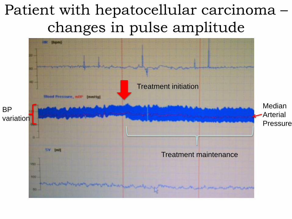
Patient with hepatocellular carcinoma –
changes in pulse amplitude
BP
variation
Treatment initiation
Treatment maintenance
Median
Arterial
Pressure

J Exp Clin Cancer Res 2009, 28:51
DISCOVERY OF TUMOR-SPECIFIC
FREQUENCIES
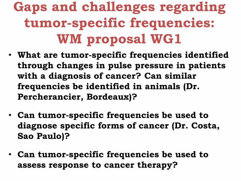
Gaps and challenges regarding
tumor-specific frequencies:
WM proposal WG1 • What are tumor-specific frequencies identified
through changes in pulse pressure in patients
with a diagnosis of cancer? Can similar
frequencies be identified in animals (Dr.
Percherancier, Bordeaux)?
• Can tumor-specific frequencies be used to
diagnose specific forms of cancer (Dr. Costa,
Sao Paulo)?
• Can tumor-specific frequencies be used to
assess response to cancer therapy?

Patient-based discovery of tumor-
specific frequencies
Clinical results
In vitro results
In vivo results
Conclusions

• Output power: 100 mW
• Carrier frequency: 27.12 MHz
• Amplitude modulation (sine wave) with at least one or several of the following frequencies: 1873.477 Hz, 2221.323 Hz, 3669.513 Hz, 4486.384 Hz, 5882.292 Hz, 6350.333 Hz, 8452.119 Hz, 10456.383 Hz
• Frequencies are emitted sequentially for 3s and the sequence is repeated for 60 min
• Emitting power and absorbed doses of electromagnetic fields are identical to those of the device previously presented to the FDA and used in several IRB-approved U.S. and European clinical trials
J Exp Clin Cancer Res 2009, 28:51
RF AMPLITUDE-MODULATED
ELECTROMAGNETIC FIELDS FOR THE
TREATMENT OF CANCER

A
B 27 11
Tumor-specific modulation frequencies range between 410 Hz and 20 kHz
D
C
Br J Cancer 2011, 105:640-648
DEVICE, FREQUENCY PROGRAM AND TREATMENT ADMINISTRATION

Distribution of RF EMF in
Humans: the body antenna
concept
The device delivers
whole body mean SAR
in the range of only 0.2
to 1mW/kg, with a 1g
peak spatial SAR
between 150 and
350mW/kg

Compassionate treatment of patients
with cancer: a feasibility study
Eligibility: Patients with advanced cancer and no curative
treatment options. Patients with measurable disease either
by imaging studies or tumor markers. Patients may not
receive other anti-cancer therapies
Pre-treatment examination: In order to identify tumor-
specific frequencies, each patient was examined using
biofeedback methods
Baseline exams: Imaging studies and/or tumor markers were
obtained within four weeks prior treatment initiation
Treatment: tumor-specific frequencies administered
sequentially for 60 min, three times a day
Study registration: clinicaltrials.gov identifier NCT00805337
J Exp Clin Cancer Res 2009, 28:51

59 yo postmenopausal
female with ER/PR
positive, ERBB2
negative breast cancer
with biopsy confirmed
metastasis to the left
ischium and right
adrenal gland.
A) Baseline PET MIP
image
B) PET MIP image four
months after baseline
C) Baseline PET/CT (left
panel)
D) Baseline PET/CT (right
panel)
E) Follow-up PET/CT: The
fused PET/CT four
months after baseline
J Exp Clin Cancer Res
2009 , 28:51

64 year old man with recurrent thyroid cancer
metastatic to the lungs: stable disease at + 8 years and 2
months (Br J Cancer 2011, 105:640-648; Chinese J Cancer, 2013, 32:573-581)
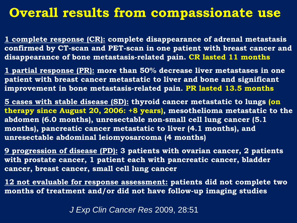
Overall results from compassionate use
1 complete response (CR): complete disappearance of adrenal metastasis
confirmed by CT-scan and PET-scan in one patient with breast cancer and
disappearance of bone metastasis-related pain. CR lasted 11 months
1 partial response (PR): more than 50% decrease liver metastases in one
patient with breast cancer metastatic to liver and bone and significant
improvement in bone metastasis-related pain. PR lasted 13.5 months
5 cases with stable disease (SD): thyroid cancer metastatic to lungs (on
therapy since August 20, 2006: +8 years), mesothelioma metastatic to the
abdomen (6.0 months), unresectable non-small cell lung cancer (5.1
months), pancreatic cancer metastatic to liver (4.1 months), and
unresectable abdominal leiomyosarcoma (4 months)
9 progression of disease (PD): 3 patients with ovarian cancer, 2 patients
with prostate cancer, 1 patient each with pancreatic cancer, bladder
cancer, breast cancer, small cell lung cancer
12 not evaluable for response assessment: patients did not complete two
months of treatment and/or did not have follow-up imaging studies
J Exp Clin Cancer Res 2009, 28:51

• HCC diagnosis
– Confirmed histology
– Clinical presentation
• BLCL C (advanced stage)
– Portal vein thrombosis
– Extra-hepatic metastasis
– Symptoms
• Progression post conventional therapy
• Advanced cirrhosis (B8-9)
• Progressed after sorafenib or systemic chemotherapy
• Study registration: clinicaltrials.gov identifier
NCT00534664
Treatment of advanced hepatocellular
carcinoma (HCC)
Br J Cancer 2011, 105:640-648

76 year old woman with
hepatitis C and Child-Pugh
A5, BCLC C, biopsy-proven
hepatocellular carcinoma
with lung metastases who
had evidence of disease
progression (+36% by
RECIST criteria) between
May 3, 2006 (first column)
and July 26, 2006 (second
column). Treatment with
amplitude-modulated
electromagnetic fields was
initiated on August 9, 2006.
The patient had a partial
(near complete) response of
intrahepatic tumor (white
arrows) and left lower lobe
lung metastasis (black
arrows fourth row), with
stable size of right lower
lobe lesion (black arrows
third row) that persists as
she continued receiving
treatment for more than five
years (fourth column).
Long term near complete response in a patient
with biopsy-proven hepatocellular carcinoma
British Journal of Cancer 2011, 105:640; Chinese Journal of Cancer 2013, 32:573

Objective responses
Type of response
#
eligible for
response
(28 patients)
intent to
treat
analysis
(41 patients)
Partial response 4 14% 10%
Stable disease 16 57% 39%
Progression 8 29% N/A
Br J Cancer 2011, 105:640-648

Comparison of TheraBionic AM EMF with sorafenib (NexavarR) in Child-Pugh A and B patients
None of the 137 patients treated with Nexavar had stable disease lasting longer than 15 months. Four of 41 (9.8%) patients treated with AM EMF had stable disease for more than 15 months, three (7.3%) had stable disease for more than 27 months, although several of them had received prior systemic treatment.
J Clin Onc 2006, 24:4293
Br J Cancer 2011, 105:640
TheraBionic response
rate (9.8%) is fourfold
higher than that of
sorafenib (2.2%)

Clinical results in patients with
advanced HCC as of October 2014
41 patients treated in phase I/II study (Costa et al,
Br J Cancer 2011, 105:640)
1 patient treated in feasibility study (Barbault et al.,
J Exp Clin Cancer Res 2009, 28:51)
6 patients treated off protocol (Brazil and France)
1 patient treated with FDA compassionate use
approval (USA)
Partial responses and/or long-term survival in excess
of 24 months observed in 11 (22.4%) of 49 patients

78 year old man with hormone-refractory disease since January 2014
Treatment with amplitude-modulated electromagnetic fields: 259 frequencies
0,83 1,24 1,52
2,84
4,79 5,46
4,6
3,33 3,62 4,02
5,01 5,6
6,12
6,95
6,05 6,61
7,84
9,01
7,36
9,62
12,9
11,7
10,8
0
2
4
6
8
10
12
14
PSA
TheraBionic treatment initiation on May 8, 2013
9 patients with prostate cancer treated thus far, four with
PSA response

0 500
1000 1500 2000 2500 3000 3500 4000 4500
CA 19-9 in 61 yo patient stage IV (lungs, bones) pancreatic cancer after FOLFIRINOX and progression on gemcitabine/abraxene
Treatment with pancreatic cancer specific frequencies as of May 5, 2014
7 patients with stage IV pancreatic cancer treated to date, 4 patients with
CA 19-9 and/or PET/CT responses

Gaps and challenges regarding
clinical efficacy: WM proposal
WG1 • What genetic features predict
response and duration of response to
tumor-specific frequencies?
• Phase III study in advanced
hepatocellular carcinoma
• Phase II studies in breast cancer,
pancreatic cancer, prostate cancer,
ovarian cancer
• Other tumor types?

Patient-based discovery of tumor-
specific frequencies
Clinical results
In vitro results
In vivo results
Conclusions

In Vitro Exposure Equipment and
Conditions
Br J Cancer 2012, 106:307-313
Cells: HepG2, Huh7,
THLE2, MCF7, MCF10A
Exposure: 3 hours per day
for 7 days (21 total hours)
Initial Seeding: 40,000-
50,000 cells
Exposure Dose (SAR): 0.03
W/kg to 0.4 W/kg
Medium Exchange: every
48 hours
Comparison: Treated cells
to cells not receiving
exposure
Subculturing (long-term):
every 7 days (1:15)

HCC, BREAST CANCER AND RANDOMLY-CHOSEN MODULATION
FREQUENCIES
Br J Cancer 2012, 106:307-313

Inhibition of cancer cell proliferation by
specific modulation frequencies
Br J Cancer 2012, 106:307-313
HCC-specific
modulation
frequencies
Breast
cancer-
specific
modula
tion
frequen
cies
HCC-
specific
modula
tion
frequen
cies p=0.001
p=0.01
p=0.02
N=6

Inhibition of cancer cell proliferation by
specific modulation frequencies
Br J Cancer 2012, 106:307-313
HepG2
p=0.03
p=0.01
N=6

Inhibition of cancer cell proliferation by
specific modulation frequencies
Br J Cancer 2012, 106:307-313
Breast
cancer-
specific
modulation
frequencies
HCC-specific
modulation
frequencies
N=6
p=0.02

qPCR validation of differentially expressed genes as assessed by RNA Seq
HepG2
Br J Cancer 2012, 106:307-313
N=4
p=0.009
p=0.02

A B
Fluorescence microscopy assessing the effects of HCC-specific frequencies on
microtubule dynamics and cell division in HepG2 cells
A HepG2 cells are able to efficiently assemble a bipolar mitotic spindle,
allowing cells to pass through the mitotic assembly checkpoint and
successfully progress from metaphase to anaphase
B HepG2 cells exposed to HCC-specific frequencies display microtubule-
associated errors such as tripolar spindle formation, potentially resulting in
aneuploid cellular states. Successive rounds of unsuccessful mitosis could
be a major determinant in cell survival. (Cyan=DAPI; Gray=Microtubules;
Arrows=mitotic spindle)
HCC-specific frequencies disrupt the mitotic spindle
Br J Cancer 2012, 106:307-313

NFAT5 (nuclear factor of activated T-cells
5)
MBOAT1 (membrane bound O-
acyltransferase domain containing 1)
ARHGDIB (Rho GDP dissociation
inhibitor (GDI) beta)
S100B (S100 calcium binding protein B)
ANXA1 (Annexin I)
Source: NCBI Gene
Galaxy Evaluation of RNA-Seq Data
http://www.ncbi.nlm.nih.gov/books/NBK21705/bin/ch20f39.jpg
Goal: Use more comprehensive
software technology to build a
pathway based on changes in
gene expression

miRNA sequences are
short beyond the scope of
RNA-Seq sensitivity
Hsa-mir-1246
NFAT5
PLCB1
Hsa-mir-148a
PLCXD3
PLCB4
Hsa-let-7g
PLCB2
PLCD4
Common link: signal
transduction through
IP3/DAG http://www.ncbi.nlm.nih.gov/books/NBK21705/bin/ch20f39.jpg
MicroRNA (miRNA) Screen

L
A
T TCR-CD3
Complex
p300 CREB CBP
Gene Expression NFAT
Ca2+
Ga Gb Gg
Gb Gg
Ga-Q GTP
GDP
BTK
PLCg
PIP3 PIP2
PLC
IP3
PTEN
PLCb2
ITK4
PI3K
Ca2+
Ca2+
GADS
Fyb
SLP76
Fyn ZAP70
PLCg PLCe
Ca2+
Calm
CaMKK CaMKII CaMKIV
Caln
2009
ProteinLounge.com
C
Hsa-mir-1246
Hsa-mir-148a
Hsa-let-7g
Hsa-mir-1246
ARHGDIB
NFAT5
S100B
ANXA1 MBOAT1
IP3/DAG Signaling

A
B
Calcium is a necessary mediator of RF EMF-
mediated inhibition of cell proliferation
27872,78
30494,55
13513,43
25119,52
0,00
5000,00
10000,00
15000,00
20000,00
25000,00
30000,00
35000,00
40000,00
H3
(C
ou
nts
pe
r M
inu
te)
Huh7 cell proliferation assay following exposure to HCC-specific or random-chosen frequencies for 3h/day for seven days
P < 0.01
NS
N=6
Random
Frequencies
Random
Frequencies
+BAPTA 100 µM
HCC-specific
Frequencies
HCC-specific
Frequencies
+BAPTA 100 µM

Patient-based discovery of tumor-
specific frequencies
Clinical results
In vitro results
In vivo results
Conclusions

AM EMF exposure system: uses transverse electromagnetic (TEM) wave in a
stripline integrated into a ring resonator. Dosimetry analysis confirmed that
exposure remained homogenous regardless of mouse posture. Both computer
simulations and actual exposures using “phantom” anatomical models were used
for verification. The TEM equipment is capable of exposing 16 mice simultaneously
to each treatment and control conditions.
Euthanize upon reaching excessive tumor burden (>2cm)
Harvest tumor tissue and normal organ tissue
Evaluate tumor doubling time as well as treatment time to euthanasia
In Vivo Experimental Methods

In Vivo Experimental Methods
NOD scid mice 1x10⁷ cells: HCC, breast and ovarian
cancer cell lines derived from patient
tumors
Palpable Tumor
Formation Injections 1x10⁷ cells
AM RF EMF
Exposure
0.4 W/kg, three
one hour
treatments daily

Volu
me (
mm
3)
Weeks 1 2 3 4 5 6 7 8 9 10 11 12 13 14 15 16 17 18
Treated Observation
Only
Subcutaneous HepG2 Cellular Xenografts Treated
with AM RF EMF – Complete response
Each volume is an
average of 3 weekly
measurements

HepG2-1
Magnification: 200x
Subcutaneous HepG2 Cellular Xenografts Treated
with AM RF EMF – Residual Encapsulated Tumor

Week 1 2 3 4 5 6
Subcutaneous Huh7 Cellular Xenografts Treated
with AM RF EMF – Change in Total Volume Model: Subcutaneous Cellular
Xenograft in NOD SCID Mice
Cells: Huh7 hepatocellular carcinoma
To
tal C
han
ge i
n T
um
or
Vo
lum
e (Δ
mm
3)
Control
n = 4
Treated
n = 5
p = 0.0106

RF EMF Exposed Control
Proliferative Impact on Normal Tissue Following
RF EMF Exposure
Mag
nific
atio
n: 20
0x
Mag
nific
atio
n: 20
0x
Ki-67 stain identifies proliferation in the crypts of the small
intestine
There are no differences in proliferation in small intestine tissue
following exposure to HCC-specific RF EMF
Ki-67 Mouse Small Intestine Ki-67 Mouse Small Intestine

Gaps and challenges regarding
mechanism of action: WM
proposal WG1 • Replication of growth inhibition of
Huh7 cells in vitro and in vivo by 194
hepatocellular carcinoma-specific
frequencies (Prof. Schär, Basel)
• Dissection of the molecular
mechanism underlying cancer cell
growth inhibition
• Identification of novel pathways
implicated in cancer growth

FUTURE DIRECTIONS • The TheraBionic device will become commercially available for
the treatment of advanced hepatocellular carcinoma and breast
cancer as a class IIa device in Europe in 2015 (DIN ISO 9001
certified, ISO 13485 certification in progress). Five European
centers will perform a phase IV study starting in 2015
• A randomized study assessing pulse pressure changes in 60
individuals with hepatocellular carcinoma, breast cancer and
healthy individuals exposed to cancer specific frequencies and
randomly chosen frequencies (PI Dr. Frederico Costa, Hospital
Sirio-Libanes, Sao Paulo, Brazil; clinicaltrials.gov # CT01688412)
• The FDA has reviewed and approved a phase III protocol in
which patients with advanced hepatocellular carcinoma who
have failed sorafenib will receive treatment with hepatocellular
carcinoma-specific frequencies or randomly chosen frequencies.
• A phase II study in advanced breast cancer will start in early
2015

CONCLUSIONS • Intrabuccally-administered 27 MHz electromagnetic
fields, amplitude-modulated at tumor-specific
frequencies elicit objective responses in patients with
advanced breast cancer and hepatocellular carcinoma
• Several patients experience long-lasting responses or
disease-stabilization without significant side effects
• Tumor-specific frequencies block proliferation of
cancer cells both in vitro as well as in vivo at levels of
exposure similar to those yielding therapeutic
responses in humans (< 0.4 W/kg)
• Tumor-specific frequencies do not affect the
proliferation of normal hepatocytes or other tumor
types, which suggest a cell-specific resonance
mechanism for specific frequencies

CONCLUSIONS
• Hepatocellular carcinoma-specific frequencies downregulate
the expression of the PLP2 and XCL2 genes in hepatocellular
carcinoma cells. Expression of the same genes is
downregulated in ovarian cancer cell lines, which suggests a
common mechanism of action for certain tumor types
• Changes in siRNA suggest that the IP3/DAG signaling pathway
is modulated by AM EMF, affecting calcium efflux as originally
reported by Ross Adey and Carl F. Blackman.
• Calcium chelation with Bapta confirms the central role of
Calcium in AM EMF targeted antiproliferative effects
• Hepatocellular carcinoma-specific frequencies disrupt the
mitotic spindle in HepG2 cells
• The experimental findings suggest the existence of a novel
receptor mechanism through which cancer growth can be
effectively targeted, thus offering a new therapeutic approach

ACKNOWLEDGMENTS
• Alexandre Barbault
• Pasche Lab, Wake Forest
Carl Blackman
Jackie Zimmerman
Michael Pennison
Hugo Jimenez
• Hospital Sirio-Libanes, Sao Paulo
Frederico P. Costa
Brenda Gumz
• UAB Oncology
James Posey
Desiree Morgan
• Swiss Federal Institute
• of Technology
Niels Kuster
Recommended