Damage to Cobalt-Chromium Surfaces During Arthroscopy of Total Knee Replacements
by Gregory E. Raab, Christopher M. Jobe, Paul A. Williams, and Qiang G. Dai
J Bone Joint Surg AmVolume 83(1):46-46
January 1, 2001
©2001 by The Journal of Bone and Joint Surgery, Inc.
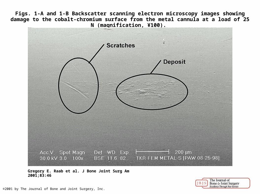
Figs. 1-A and 1-B Backscatter scanning electron microscopy images showing damage to the cobalt-chromium surface from the metal cannula at a load of 25 N (magnification, ¥100).
Gregory E. Raab et al. J Bone Joint Surg Am 2001;83:46
©2001 by The Journal of Bone and Joint Surgery, Inc.

Atomic number contrast showing two different materials.
Gregory E. Raab et al. J Bone Joint Surg Am 2001;83:46
©2001 by The Journal of Bone and Joint Surgery, Inc.

Backscatter scanning electron microscopy images with topographical contrast, showing the damage to the cobalt-chromium surface from the metal cannula at different normal loads.
Gregory E. Raab et al. J Bone Joint Surg Am 2001;83:46
©2001 by The Journal of Bone and Joint Surgery, Inc.

Regression curve of normal load versus Ra value.
Gregory E. Raab et al. J Bone Joint Surg Am 2001;83:46
©2001 by The Journal of Bone and Joint Surgery, Inc.
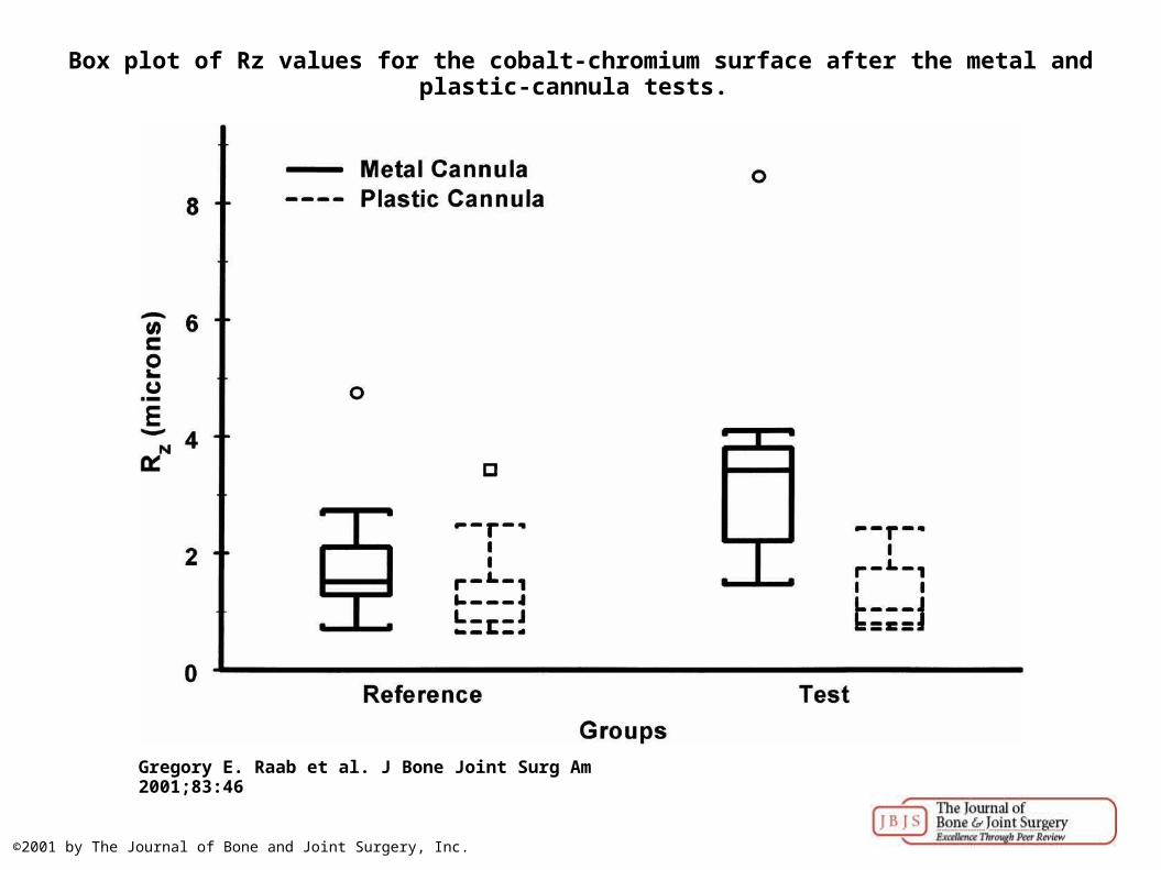
Box plot of Rz values for the cobalt-chromium surface after the metal and plastic-cannula tests.
Gregory E. Raab et al. J Bone Joint Surg Am 2001;83:46
©2001 by The Journal of Bone and Joint Surgery, Inc.
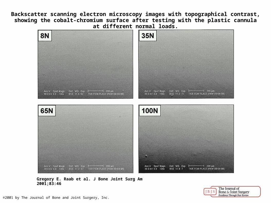
Backscatter scanning electron microscopy images with topographical contrast, showing the cobalt-chromium surface after testing with the plastic cannula at different normal loads.
Gregory E. Raab et al. J Bone Joint Surg Am 2001;83:46
©2001 by The Journal of Bone and Joint Surgery, Inc.
Recommended